Review

Effects of NAD+ in Caenorhabditis elegans Models of Neuronal Damage

1
Department of Life Science, Hallym University, Chuncheon 24252, Korea
2
Convergence Program of Material Science for Medicine and Pharmaceutics, Hallym University, Chuncheon 24252, Korea
3
Multidisciplinary Genome Institute, Hallym University, Chuncheon 24252, Korea
*
Author to whom correspondence should be addressed.
Biomolecules 2020, 10(7), 993; https://doi.org/10.3390/biom10070993
Submission received: 1 April 2020 / Revised: 20 June 2020 / Accepted: 30 June 2020 / Published: 2 July 2020
(This article belongs to the Special Issue Nicotinamide in Health and Diseases)

Abstract

:
Nicotinamide adenine dinucleotide (NAD+) is an essential cofactor that mediates numerous biological processes in all living cells. Multiple NAD+ biosynthetic enzymes and NAD+-consuming enzymes are involved in neuroprotection and axon regeneration. The nematode Caenorhabditis elegans has served as a model to study the neuronal role of NAD+ because many molecular components regulating NAD+ are highly conserved. This review focuses on recent findings using C. elegans models of neuronal damage pertaining to the neuronal functions of NAD+ and its precursors, including a neuroprotective role against excitotoxicity and axon degeneration as well as an inhibitory role in axon regeneration. The regulation of NAD+ levels could be a promising therapeutic strategy to counter many neurodegenerative diseases, as well as neurotoxin-induced and traumatic neuronal damage.

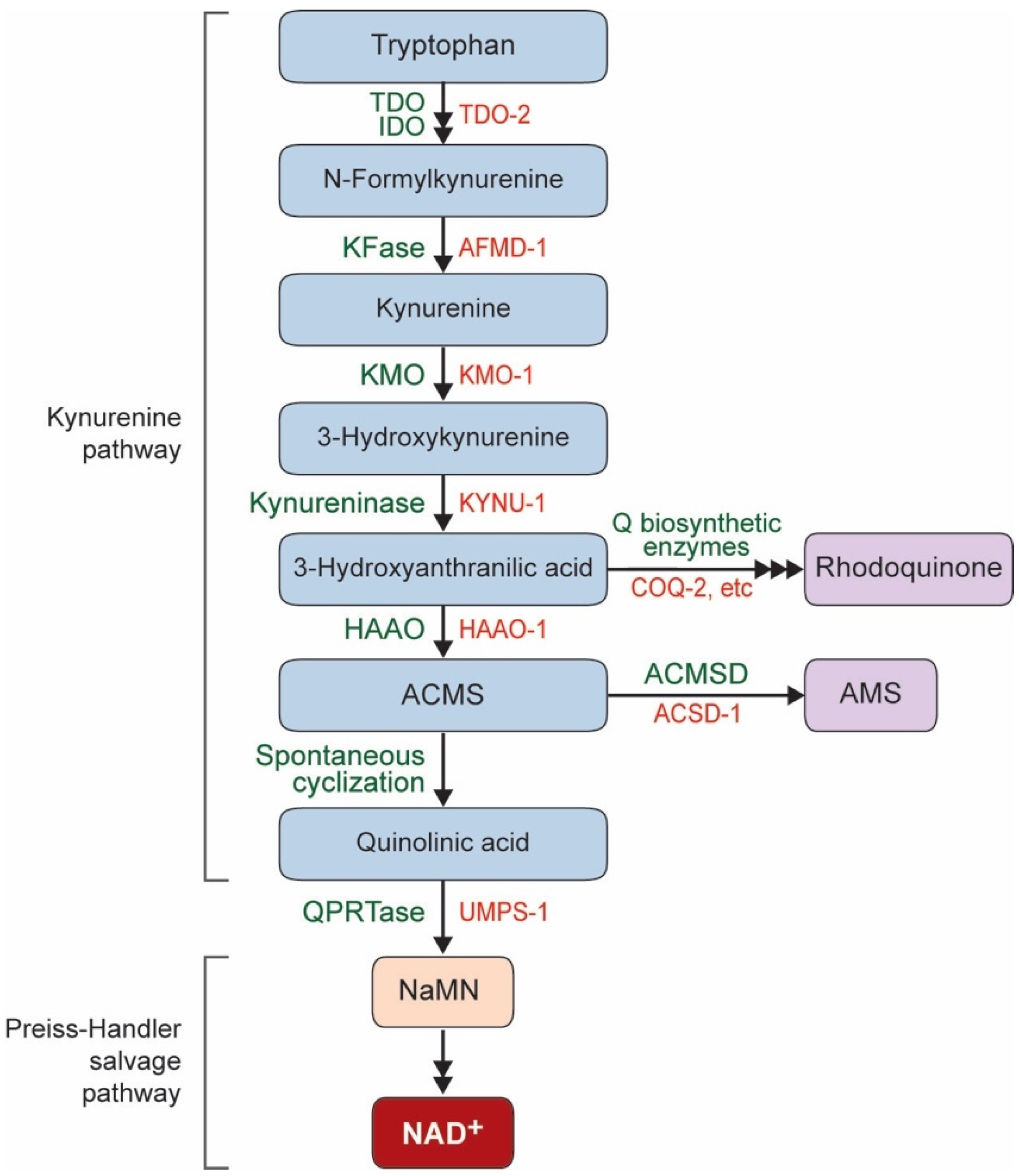
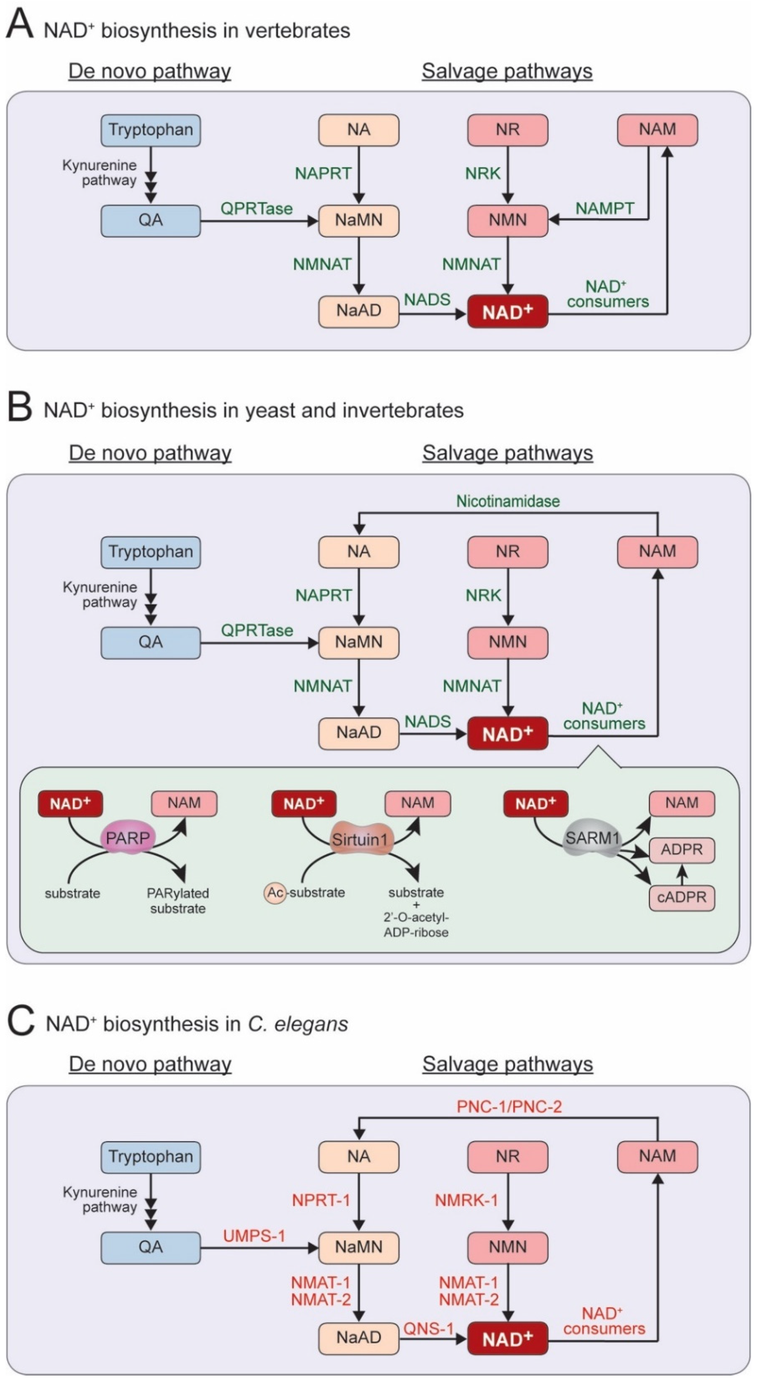
1. NAD+ Biosynthesis Pathway in C. elegans

Nicotinamide adenine dinucleotide (NAD+) is found in all living cells and plays an essential role in many fundamental biological processes, such as metabolism, cell signaling, gene expression, and DNA repair [1]. NAD+ is synthesized through two metabolic pathways, either a de novo pathway or salvage pathways. In a de novo pathway, NAD+ can be synthesized from the degradation of the essential amino acid L-tryptophan via the kynurenine pathway (Figure 1) [2,3]. The derivatives of the kynurenine pathway have been linked to both the progression and protection of neurological disorders and neurodegenerative diseases [4]. The kynurenine pathway produces quinolinic acid (QA), which is converted to nicotinic acid mononucleotide (NaMN) by QA phosphoribosyltransferase (QPRTase) and merges with the Preiss–Handler salvage pathway [5]. QA is a known neurotoxin [6,7]; thus, a proper clearing mechanism should be fulfilled by QPRTase. Although the de novo pathway is conserved in C. elegans, no QPRTase homolog has been found [8]. A recent study showed that the enzyme uridine monophosphate phosphoribosyltransferase (UMPS), encoded by the umps-1 gene in C. elegans, possesses QPRTase activity [9]. A lack of umps-1 in C. elegans results in decreased levels of global NAD+ and increased steady-state levels of QA, indicating that UMPS-1 is required for NAD+ de novo synthesis in place of the missing QPRTase in C. elegans [9]. Thus, the de novo NAD+ biosynthesis pathway is functionally conserved in C. elegans. However, the neuronal roles of the kynurenine pathway in C. elegans are yet to be determined.
Alternatively, NAD+ can be produced from NAD+ precursors, which include nicotinic acid (NA), nicotinamide riboside (NR), and nicotinamide (NAM) (Figure 2A–C) [10,11]; the salvage synthesis from NA is termed the Preiss–Handler pathway [12,13]. These three compounds are termed vitamin B3 or niacin and can be taken up from the diet. These compounds are also produced within cells as intermediates of NAD+ biosynthesis. NAD+ is consumed and converted to NAM by various enzymes, such as poly(ADP-ribose) polymerases (PARPs), sirtuins, and Sarm1 [8,14]. At least two types of NAM salvage pathways are known: vertebrates use a one-step pathway, whereas yeast and invertebrates use a two-step pathway. Vertebrates convert NAM into nicotinamide mononucleotide (NMN) by Nicotinamide phosphoribosyltransferase (NAMPT), which is then converted to NAD+ by Nicotinamide mononucleotide adenylyltransferase (NMNAT) (Figure 2A). In primitive eukaryotes, including yeast, C. elegans, and Drosophila, NAMPT activity has not been found [8]. In these species, NAM is converted to NA using a nicotinamidase, which then enters the Preiss–Handler pathway (Figure 2B,C).
Despite the presence of the de novo pathway, the salvage pathways are essential in animals. Interestingly, in C. elegans, mutants lacking components of the de novo pathway show normal viability, while some mutants lacking components of the salvage pathways show lethality. So far, two genes are found to be essential for viability, nmat-2 (Nicotinamide Mononucleotide AdenylylTransferase-2) and qns-1 (Glutamine-dependent NAD+ Synthase-1) [15]. nmat-2 encodes a C. elegans NMNAT homolog, and qns-1 encodes a NAD+ synthase (NADS) homolog. The homozygous nmat-2 or qns-1 offspring of heterozygous mutants are viable but sterile, whereas homozygous mutants produce no offspring [15], suggesting that maternally or paternally derived NAD+ is likely critical for embryogenesis. In contrast, the other enzymes in either de novo or salvage pathways (the protein encoded by tdo-2, afmd-1, kmo-1, haao-1, umps-1, pnc-1, pnc-2, nprt-1, nmat-1, or nmrk-1) are found to be non-essential for animal survival, although some mutants show a low fecundity. Thus, in C. elegans, NMAT-2 and QNS-1 are likely rate-limiting or nonredundant among the components in the NAD+ biosynthesis pathways.

2. Neuroprotective Effect of NAD+ in C. elegans

The NAD+ biosynthesis pathway appears to play a role in protecting neurons from damage and stresses. Depending on the duration of the harmful stimulus, the types of damage can be classified into acute (short-term) and chronic (long-term) injuries (Figure 3A,B) [16]. Acute axon injuries physically sever axons into two parts, which can be manifested by transecting neurons or laser-assisted microsurgical cutting of the individual axons. Chronic injuries result from persistent neuronal stresses such as the expression of harmful proteins or hazardous environmental conditions. In response to these acute or chronic injuries, neurons may undergo axon degeneration, cell death, or axon regeneration. These neuronal responses can vary depending on the types of damage [16].

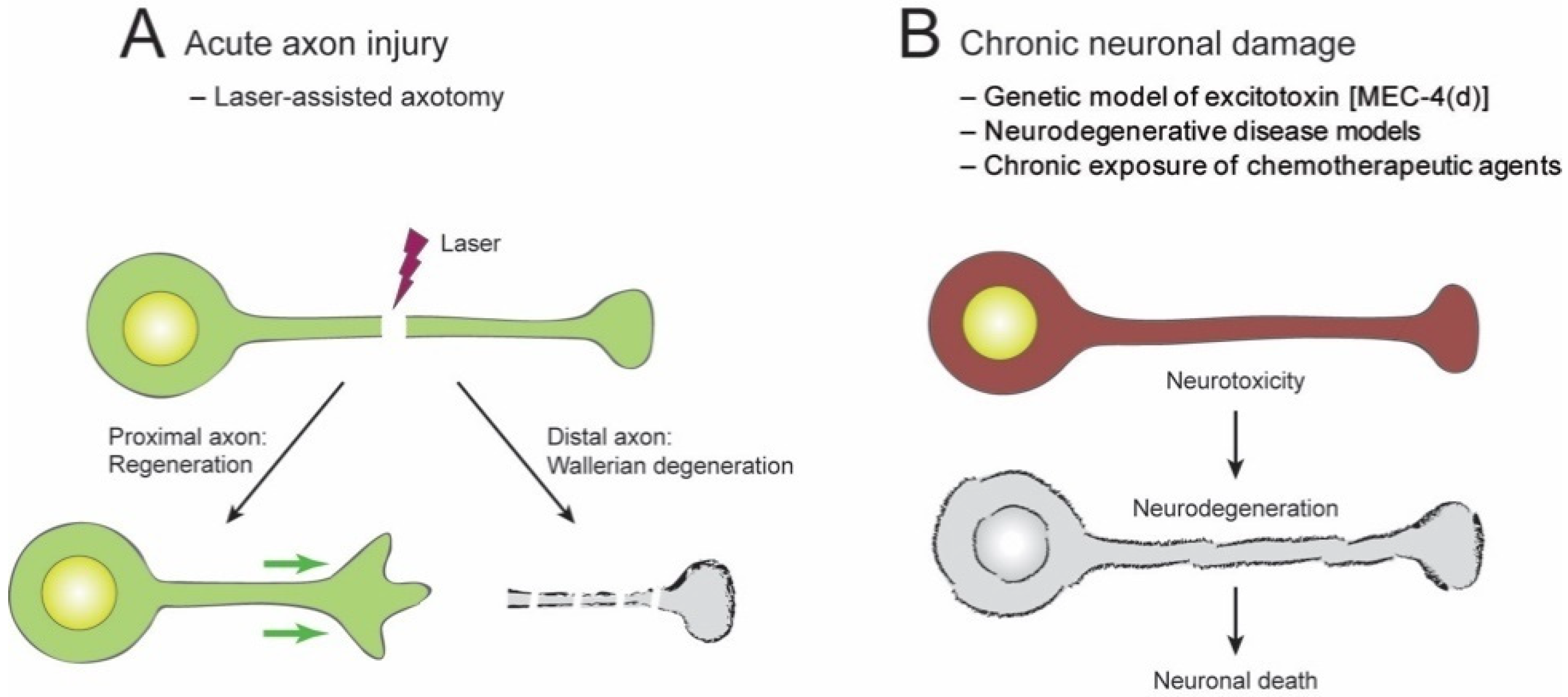
2.1. Acute Axon Injuries

In an acute axon injury, the axon is severed (axotomy) into two fragments in relation to the cell body: a proximal fragment and a distal fragment. The proximal axon fragment may undergo axonal regeneration, whereas the distal axon fragment often undergoes a rapid axonal degeneration (Figure 3A). Axon degeneration is an early feature of most neuronal injuries and is a primary cause of functional impairment. The degeneration of axons distal to a lesion site is highly stereotyped and is known as Wallerian degeneration [17,18]. In this process, the distal part of an axon first experiences a latent phase. It then undergoes a catastrophic fragmentation into numerous pieces, after which the pieces are completely removed via a genetically encoded “self-destruction” program [19].
Wallerian degeneration is delayed dramatically in (1) the Wallerian degeneration slow (Wlds) mutant mice, (2) the Sarm1 null mutant mice, and (3) Nmnat overexpressed mice [20,21,22,23], suggesting that the Wallerian degeneration is an active molecular mechanism. First, Wlds is a spontaneous mutant gene that encodes a fusion protein, WLDs, which likely has a sustained NMNAT activity in the cytosol [24]. Second, SARM1 exhibits a NADase activity [25], and SARM1 activation thus reduces NAD+ levels and vice versa [26,27]. Third, NMNAT is an NAD+-synthesizing enzyme, and loss-of-function studies suggest that NMNAT in healthy neurons plays a protective role in inhibiting axon degeneration [28,29]. These observations support the idea that sustaining high levels of NAD+ inhibits or delays Wallerian degeneration.
The Wallerian degeneration paradigm has also been studied in C. elegans. Following a laser axotomy, C. elegans sensory and motor neurons exhibit an axon degeneration with a morphological similarity to the Wallerian degeneration, such as axonal swelling, thinning, breaks, and clearance. However, this axon degeneration occurs independently of the WLDs, NMNAT, and SARM1 pathways because the overexpression of Wlds or the endogenous Nmnat gene, or the lack of Sarm1 (C. elegans homolog, tir-1) showed no protective role against axon degeneration [30]. These observations suggest that high levels of NAD+ may not inhibit axon degeneration in all neuron types or acute injuries. Further experiments are needed to clarify this point.

2.2. Chronic Injuries

Chronic neuronal stressors can cause axon degeneration and excitotoxicity (Figure 3B). Chronic stressors include several neurodegenerative diseases and exposure to chronic neurotoxins such as heavy metals and chemotherapeutic drugs [31]. Exposure to low levels of chronic excitotoxin may result in a slow pathological cascade, axonal degeneration, and pathology. In many neuropathies, the axon’s most terminal connections are lost and are termed “dying back degeneration” [32,33]. Severe excitotoxicity can result in the initiation of cell death pathways.
C. elegans is a valuable model for studying the underlying mechanisms of chronic axon injuries. The processes of synaptic release, trafficking, and production of neurotransmitters are conserved in C. elegans, and the neuronal morphology, changes in gene expression and neurotransmitters, and behaviors can be examined. We summarize the function of the C. elegans NAD+ pathway in chronic neuronal injuries by stressors, including a genetic model of excitotoxin, several neurodegenerative diseases, and exposure to chemotherapeutic agents.

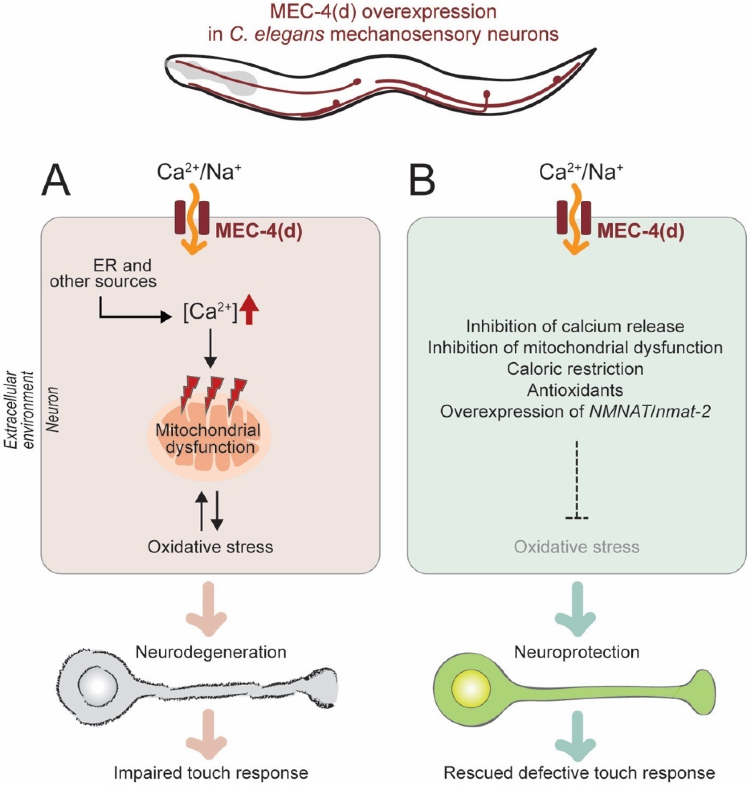
2.2.1. Genetic Model of Neuronal Excitotoxicity

Neuronal excitotoxicity is often induced by an excessive release of the neurotransmitter glutamate and triggers neurite degeneration and neuronal death [34,35,36]. Hyperactivation of the degenerin channel MEC-4 (MEChanosensory abnormality-4) (MEC-4(d)) can trigger axon degeneration and the cell death of C. elegans neurons (Figure 4A) [37]. Stereotypically, axons become swollen and later truncated; cell bodies become vacuolated and then disappear. In the mec-4(d) excitotoxic model of C. elegans mechanosensory (touch-sensing) neurons, touch response is impaired [38]. These defects are rescued by the inhibition of calcium increase and mitochondrial dysfunction [38]. In addition, overexpression of NMNAT/NMAT-2 protects the cell body and axons against mec-4(d)-triggered degeneration, and enables a neuronal function such as touch response (Figure 4B) [38]. Here, reactive oxygen species (ROS) are identified as a key intermediate of neuronal degeneration triggered by MEC-4(d) stimuli. Interestingly, caloric restriction and systemic antioxidant treatment (ROS scavengers such as trolox and ascorbic acid) help decrease oxidative damage and protect both cell bodies and axons from mec-4(d)-triggered degeneration. However, it remains to be determined how an increase of NMNAT or NAD+ levels is responsible for relieving oxidative damage and playing a neuroprotective role. Interestingly, the neuroprotective effects of NMNAT seem to be variable according to the type of nerve injuries. In C. elegans, NMNAT is required to inhibit or delay a genetic insult-induced chronic axon degeneration, but not an injury-induced acute axon degeneration [30,38]. Additionally, the relationships between NAD+ levels and the regulation of calcium signaling also remain to be further studied. Advances in live-cell and in vivo imaging technology will allow us to track the changes of NAD+ and NAD+ intermediates in different subcellular compartments. They will enable us to reveal linkages among NAD+ levels, oxidative stress, calcium surge, and neuron degeneration.
Another C. elegans study using the mec-4(d) neurotoxic model identifies that a lack of mitochondrial sirtuin sir-2.3, homologous to mammalian Sirt4, is protective in neuronal death [39]. Sirtuins use NAD+ as a co-substrate in their enzymatic reaction. Interestingly, blocking the NAD+ salvage pathway by the knockout of nicotinamidase/pnc-1 protects neurons from another neurotoxic model such as hypoxic ischemia with azide at low pH [39]. The neuroprotective effects seen in sir-2.3 or pnc-1 loss-of-function mutants are not additive, suggesting that sir-2.3 and pnc-1 are in a common genetic pathway. pnc-1 mutants display a dramatic increase in NAM levels and a mild decrease in NAD+ levels [40]. When considering NAM as a pan-sirtuin inhibitor, although the effects of NAM are highly complicated [41], high levels of NAM in pnc-1 mutants may inhibit SIR-2.3. Furthermore, ROS is eliminated more efficiently in animals that lack sir-2.3 than wild types, under caloric restriction conditions. Thus, the protective effects of sir-2.3 mutants seem to be mediated by the reduction of oxidative damage, which is similar to that seen under nmat-2-overexpressed conditions, suggesting that sir-2.3 and nmat-2 have opposite roles in neuronal protection. Therefore, it would be interesting to explore whether or how NAD+ augmentation in mitochondria is critical to efficiently eliminating ROS.

2.2.2. C. elegans Models of Neurodegenerative Diseases

Neuronal excitotoxicity has been described as a contributing factor in several pathologies, including Huntington’s disease, Alzheimer’s disease, and Parkinson’s disease. C. elegans serves as an efficient model system for these age-dependent neurodegenerative disease studies [42].

Huntington’s Disease

Huntington’s disease (HD) is an inherited, autosomal, dominant neurological disorder that leads to the progressive degeneration of neurons. In HD, protein denaturation occurs because of a toxic mutation of the huntingtin protein containing an abnormally long polyglutamine (polyQ) tract. Parker and colleagues established the HD model in C. elegans and studied the expression of a fragment of the huntingtin gene in the mechanosensory neurons. These worms show a mutant huntingtin-dependent cytotoxicity as well as a touch response defect [43]. The upregulation of C. elegans sir-2.1/Sirt1 recovers the axonal morphology, which in turn allows the touch response to function [43]. In addition, treatment with a sirtuin activator, resveratrol [44], shows a similar effect. Such effects are lost in sir-2.1 mutants, indicating that the mode of action for the protection from mutant huntingtin toxicity is through SIR-2.1. Considering that NAD+ is the rate-limiting co-substrate for SIRT1, increased levels of NAD+ presumably activate SIRT1 like resveratrol; however, this remains to be determined. Conversely, as mentioned above, the knockout of a mitochondrial sirtuin sir-2.3/Sirt4 protects neurons from mec-4(d)-triggered degeneration [39]. Thus, the neuroprotective effects of sirtuins are variable.
Mammalian sirtuins are NAD+-dependent deacylases, and there are seven sirtuin homologs (SIRT1–SIRT7) with varied subcellular localizations [45]. SIRT1 in the brain exerts neuroprotection against ischemic injury and neurodegenerative diseases, such as HD [46]. In the mouse model of HD, SIRT1 activation by resveratrol reduces the peripheral nerve deficits. However, it leads to no significant improvement in central nervous system deficits such as motor impairment and striatal atrophy [47]. SIRT1 mediates neuroprotection in mice HD models through activation of the brain-derived neurotrophic factor expression [48]. Additionally, the brain-specific knockout of Sirt1 exacerbates the brain pathology, and the overexpression of Sirt1 improves neuronal survival [49]. In contrast to SIRT1, the closest homolog of SIRT1, SIRT2, is detrimental to HD, because inhibition of SIRT2 protects neurons in animal models of HD [50,51]. These results again show the variable effects of sirtuins in neuroprotection.
In fly HD models, the genetic and pharmacological reduction of the Drosophila SIRT1 homolog results in the clearance of mutant huntingtin and neuroprotection [52]. Based on this finding, clinical trials using Selisistat (an inhibitor of SIRT1) [53] to counter the toxicity induced by huntingtin were conducted, but no clinically relevant effects have been published [54,55,56]. Currently, no therapeutics are available to treat HD.

Alzheimer’s Disease

A study using another C. elegans model of neurodegenerative disease, the Alzheimer’s disease (AD) model [57,58,59], showed a link between NAD+ levels and mitophagy induction [60]. Mitophagy, the removal of damaged mitochondria through autophagy, is impaired in AD patients and the C. elegans AD model [60]. The typical pathology of AD is the accumulation of damaged mitochondria [61]. C. elegans models of AD have been established by overexpressing amyloid-β or tau proteins. One of the NAD+ precursors, NR, reduces amyloid-β proteotoxicity in both C. elegans and mouse models of AD [62]. Mitophagy stimulation via supplementation with another NAD+ precursor, NMN, reverses memory impairment in the C. elegans model of AD [60]. Mitophagy enhancement also abolishes AD-related tau hyperphosphorylation in human neurons and reverses memory impairment in transgenic tau mice [60]. These results suggest that the supplementation of NAD+ precursors may help remove defective mitochondria and reduce proteotoxicity in AD patients, although the underlying mechanism remains unknown.

Parkinson’s Disease

Parkinson’s disease (PD) is an adult-onset neurological movement disorder with a progressive degeneration of the dopaminergic neurons. At the cellular level, PD is characterized by the accumulation of α-synuclein in large masses called Lewy bodies [63]. Both genetic and toxicant C. elegans models of PD have been derived [64]. One of the genetic models of PD is generated by expressing the human SNCA/PARK gene, which encodes α-synuclein [65]. In this PD model, the expression of α-synuclein causes the loss of the dopaminergic neurons, deficits in dopamine-dependent behavior, and decreased dopamine levels. One study shows that blueberry extracts attenuate α-synuclein protein expression, and lower the expression of sir-2.1/Sirt1 [66]. These observations suggest that a reduced SIR-2.1/SIRT1 activity may be beneficial to the attenuation of α-synuclein.
Exposure to heavy metals, such as methylmercury or excess manganese, also makes a toxicant model of PD because it is linked to a dopaminergic dysfunction in C. elegans and mammals [67,68,69,70,71]. Exposure to methylmercury or chronic exposure to manganese in low doses causes the depletion of the cellular NAD+ levels, mitochondrial dysfunction, and oxidative stress [72]. It is possible that high levels of ROS induce DNA damage and, in turn, trigger the activity of the DNA damage response protein, PARP, which likely causes the depletion of the NAD+ levels (Figure 5). Interestingly, NAD+ pretreatment decreases methylmercury-induced oxidative stress, dopaminergic toxicity, and behavioral deficits [72]. It would be interesting to investigate how the global changes of mitochondrial dysfunction and oxidative stress cause dopaminergic neuron-specific defects. A cell-specific visualization study of NAD+ levels would help explore the precise mechanisms.

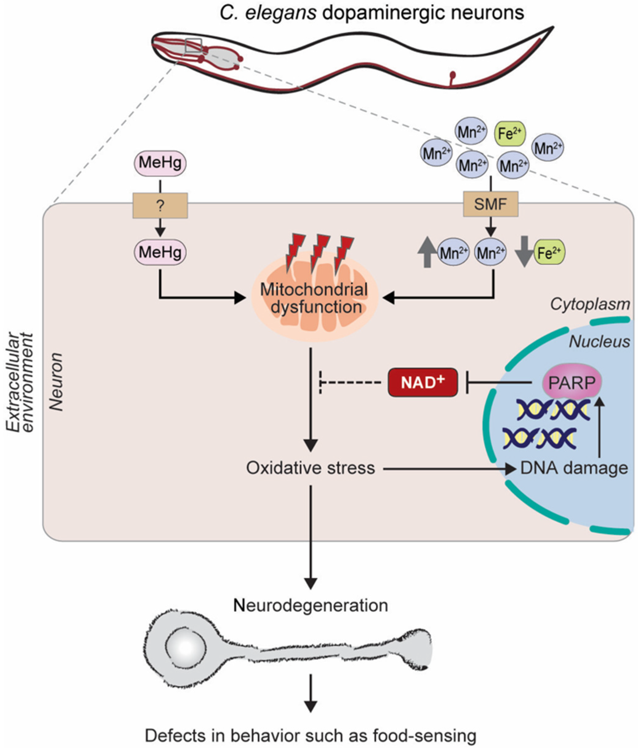
2.2.3. Chemotherapeutic Agents

Chemotherapy-induced peripheral neuropathy (CIPN) is one of the most common disorders caused by cancer treatments, affecting up to 80% of cancer patients [73]. CIPN pathology is a “dying back” axon degeneration of the distal nerve endings [74]. Taxol is used widely as a chemotherapeutic agent, even though it has been shown to induce axonal degeneration in cancer patients and in experimental models [75]. Axon degeneration in the form of axonal swelling, thinning, and gaps is seen in elderly worms and is significantly greater in taxol-treated animals [76].
C. elegans treated with taxol shows a significant growth retardation. This defect is reversed by the expression of the non-nuclear-localized mutant form of the mouse Nmnat1 gene. In addition, taxol dramatically reduces the basal expression of a mitoUPR (mitochondrial Unfolded Protein Response) reporter, serving as a marker of mitochondrial proteostasis, and it was restored by the expression of non-nuclear mouse NMNAT1 [76]. Thus, NMNAT is suggested to provide protection from a taxol-induced axonal pathology. These observations suggest that NMNAT may ameliorate mitochondrial proteostasis stress and protect neurons from an axonal damage.

3. Regulation of Axon Regeneration in C. elegans

The laser-assisted axotomy technique has established C. elegans as a valuable model to study the molecular mechanisms of axon regeneration [77]. Transgenic animals with fluorescently labeled single neurons are subjected to laser axotomy, and then the regenerating axons are analyzed. Laser severing of the C. elegans axon does not cause neuronal death but does cause severe axonal damage. Although a calcium surge is often neurotoxic, a proper calcium influx following axonal damage is considered to be beneficial because it triggers injury responses, including robust axon regeneration [78,79,80,81]. This neuronal injury response contributes to the recovery of the functional impairments caused by the injury. For example, the axonal injury of motor neurons causes locomotory defects, and regenerating this injured axon is likely required to recover the animal’s locomotion [77].

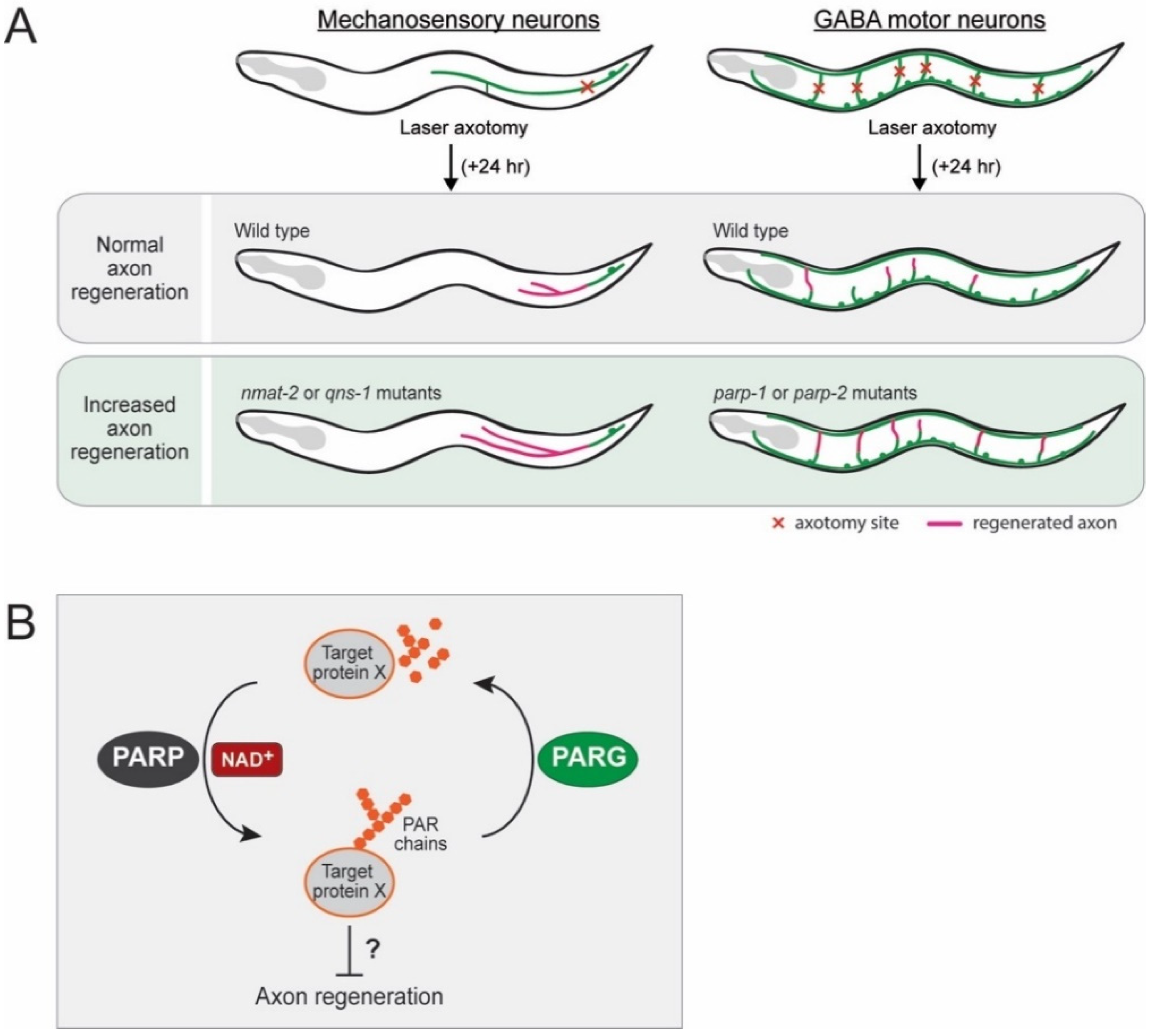
3.1. Inhibitory Role of NMNAT and NADS in Axon Regeneration

This laser axotomy paradigm enables the screening of many genetic regulators controlling axon regeneration. Yishi Jin and Andrew D. Chisholm laboratories have pioneered this technique in C. elegans and screened numerous genes involving axon regeneration of the mechanosensory neurons [15,82]. One of their findings shows that NAD+ biosynthesis likely inhibits axon regeneration [15]. They have examined loss-of-function mutants of each gene encoding enzymes in the NAD+ salvage pathways, such as the C. elegans orthologs of NMNAT (nmat-1 and nmat-2), NADS (qns-1), nicotinamide riboside kinase (NRK) (nmrk-1), nicotinate phosphoribosyltransferase (NAPRT) (nprt-1), and nicotinamidase (pnc-1 and pnc-2) (Figure 2C). Among them, three independent nmat-2 mutants and a qns-1 mutant show increased axon regeneration following the injury of mechanosensory neurons, indicating that nmat-2 and qns-1, catalyzing the terminal steps of the NAD+ salvage pathways, inhibit axon regeneration in C. elegans (Figure 6A).
Importantly, the enzymatic properties of NMAT-2 are essential for the inhibition of axon regeneration because the active site motif [83] mutation of nmat-2 exhibits a phenotype similar to that of the null mutants of nmat-2 [15]. Thus, the role of NMAT-2 in axon regeneration likely requires its enzymatic activity so that it may activate through a different downstream pathway. Consistent with the inhibitory role of nmat-2/NMNAT in axon regeneration in C. elegans, the overexpression of NMNAT leads to impaired sensory axon regeneration in Drosophila [84]. Together, these data suggest conserved roles of NMNAT in hindering axon regeneration, at least in invertebrate animal models.
Another distinct feature of NMAT-2 in axon regeneration, when compared to its neuroprotection effect, is cell autonomy. In Drosophila and mice, the neuroprotective effect of NMNAT is cell-autonomous [29,85]. In contrast, NMAT-2 inhibits axon regeneration via several tissues in C. elegans. The phenotype of the nmat-2 null mutant is restored by an nmat-2 transgene under the endogenous promoter. However, the transgenic expression of NMAT-2 in individual tissues of the intestine, epidermis, or neurons fails to restore the phenotype. It is restored to normal only by the combined expression of NMAT-2 in all three tissues. These results suggest that NAD+ may have activated inhibitory factors in neuronal and non-neuronal tissues. An intriguing possibility that has not yet been explored is that Nmnat or NAD+ biosynthesis plays a role in the gut–brain–skin axis regulating axon regeneration. It would be interesting to know how the interplay among NMNAT proteins expressed in different tissues affects neuronal events.

3.2. Inhibitory Role of PARPs in Axon Regeneration

In C. elegans, PARPs and poly(ADP-ribose) glycohydrolases (PARGs) show opposing effects on axon regeneration [86]. PARPs catalyze the transfer of ADP-ribose from NAD+ onto protein substrates (Figure 2B), whereas PARGs remove poly(ADP-ribose) from proteins [87]. The NAD+-consuming PARP encoding genes in C. elegans, parp-1 and parp-2, inhibit the axon regeneration of the GABA motor neurons (Figure 6A), whereas the PARG encoding genes, parg-1 and parg-2, enhance it [86,88]. Importantly, a pharmacological PARP inhibitor (A966492) significantly enhances axon regeneration when administered after injury in vivo in C. elegans motor neurons and in vitro in mammalian cortical neurons [86]. Treatment using a PARP inhibitor post-injury also improves the behavioral recovery in C. elegans, indicating that PARP inhibition after injury is sufficient to improve axon regeneration. Currently, chemical PARP inhibitors are in preclinical and clinical trials for indications, including various cancer and stroke therapies [89,90]. These results suggest that the PARG–PARP balance may determine axon regeneration by regulating the poly(ADP-ribose) levels on target proteins [91]. Thus, identifying protein substrates of PARPs and PARGs will require further investigation in the future. When considering the enhanced axon regeneration seen in both nmat-2 and parp-1/-2 mutant worms, it is speculated that decreased NAD+ levels in nmat-2 mutants are presumably not sufficient to activate regeneration inhibitory PARP proteins (Figure 6B). Thus, it is possible that the NAD+ may act through the PARP pathway, which remains to be determined.
The effects of PARPs on mammalian axon regeneration have been tested in multiple studies with variable results [92,93]. Brochier and colleagues identified PARP1 as a critical mediator of multiple growth-inhibitory signals [93]. In vitro models of axonal injury reveal that the exposure to growth-inhibitory signals promotes PARP1 activation and the accumulation of PAR in neurons, which leads to an axon regeneration failure. The pharmacological inhibition or genetic depletion of PARP1 is sufficient to promote regeneration. However, Wang and colleagues found that the inhibition of PARylation fails to increase axon regeneration or improve functional recovery after an adult mammalian CNS injury [92]. These conflicts might be due to the different types of neurons being tested. The axonal regenerative effects of PARPs remain unclear.

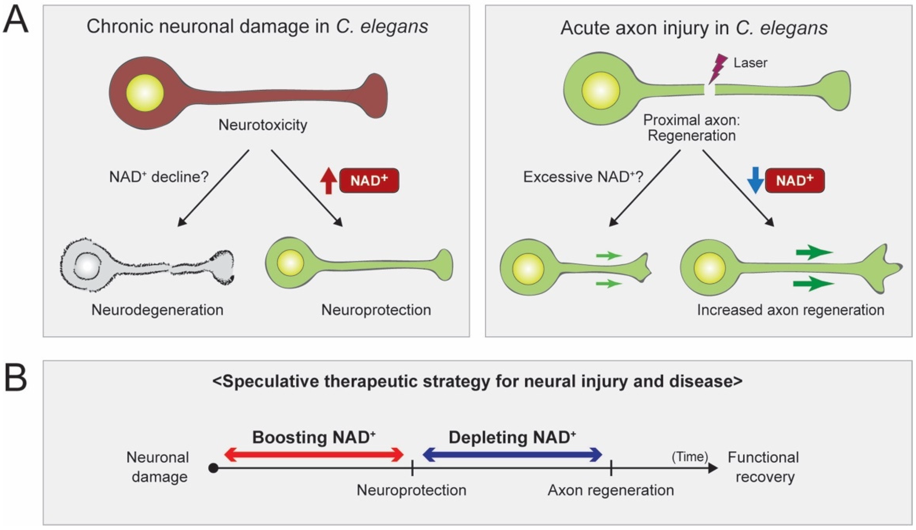
4. Conclusions and Future Perspectives

NAD+ biosynthesis pathways are functionally conserved in C. elegans, and emerging evidence is revealing the critical role that NAD+ plays in neuronal protection and axon regeneration. The effects of NAD+ have been studied in various neuronal damage models of C. elegans. Unlike the mammalian Wallerian degeneration paradigm, the sustained high levels of NAD+ show no effects in inhibiting or delaying axon degeneration in an acute injury model of C. elegans. In chronic injury models, however, high levels of NAD+ influence neuronal protection, which presumably helps reduce the oxidative stress produced by the cytosolic calcium surge and mitochondrial dysfunction (Figure 7A). The C. elegans laser-assisted axotomy paradigm enables the screening of many genes in the NAD+ pathways. All known components in NAD+ salvage pathways and several NAD+ consuming enzymes have been tested. In sensory neurons, NMNAT/NMAT-2 and NADS/QNS-1 are found to inhibit axon regeneration, suggesting that decreased levels of NAD+ promote axon regeneration (Figure 7A). In motor neurons, PARPs inhibit axon regeneration, whereas PARGs promote it. It is speculated that PARP proteins are activated by high levels of NAD+ and that they activate regeneration inhibitory target proteins by PARylation (Figure 6B). Nonetheless, given the inhibitory role of NMNAT/NMAT-2 and NADS/QNS-1 in sensory axon regeneration, it is necessary to issue a caution before manipulating NAD+ levels as a therapeutic strategy. As we come to understand NAD+ biology in greater detail, we may need to regulate the NAD+ levels in proper time frames and in specific tissues. For example, after neuronal damage, the treatment of high-dose NAD+ may help protect damaged neurons, but later, depleting NAD+ may help regenerate axons (Figure 7B). In the near future, a precise understanding of NAD+ biology may allow the development of NAD+-based therapeutic strategies for various neuronal damage conditions.

Author Contributions

Conceptualization, K.W.K.; Writing—Original Draft Preparation, Review & Editing, Y.L., H.J., K.H.P., K.W.K.; Supervision, K.W.K. All authors have read and agreed to the published version of the manuscript.

Funding

This research was supported by the Original Technology Research Program for Brain Science through the National Research Foundation of Korea (NRF) funded by the Ministry of Education, Science and Technology (NRF-2019M3C7A1031836) and the Hallym University research funds (HRF-202001-009).

Conflicts of Interest

The authors declare no conflict of interest.

References

  1. Canto, C.; Menzies, K.J.; Auwerx, J. NAD(+) Metabolism and the Control of Energy Homeostasis: A Balancing Act between Mitochondria and the Nucleus. Cell Metab. 2015, 22, 31–53. [Google Scholar] [CrossRef] [PubMed] [Green Version]
  2. Gazzaniga, F.; Stebbins, R.; Chang, S.Z.; McPeek, M.A.; Brenner, C. Microbial NAD metabolism: Lessons from comparative genomics. Microbiol. Mol. Biol. Rev. 2009, 73, 529–541. [Google Scholar] [CrossRef] [Green Version]
  3. Kurnasov, O.; Goral, V.; Colabroy, K.; Gerdes, S.; Anantha, S.; Osterman, A.; Begley, T.P. NAD biosynthesis: Identification of the tryptophan to quinolinate pathway in bacteria. Chem. Biol. 2003, 10, 1195–1204. [Google Scholar] [CrossRef] [PubMed] [Green Version]
  4. Maddison, D.C.; Giorgini, F. The kynurenine pathway and neurodegenerative disease. Semin. Cell Dev. Biol. 2015, 40, 134–141. [Google Scholar] [CrossRef] [PubMed] [Green Version]
  5. Magni, G.; Amici, A.; Emanuelli, M.; Orsomando, G.; Raffaelli, N.; Ruggieri, S. Enzymology of NAD+ homeostasis in man. Cell. Mol. Life Sci. 2004, 61, 19–34. [Google Scholar] [CrossRef]
  6. Schwarcz, R.; Bruno, J.P.; Muchowski, P.J.; Wu, H.Q. Kynurenines in the mammalian brain: When physiology meets pathology. Nat. Rev. Neurosci. 2012, 13, 465–477. [Google Scholar] [CrossRef]
  7. Guillemin, G.J. Quinolinic acid, the inescapable neurotoxin. FEBS J. 2012, 279, 1356–1365. [Google Scholar] [CrossRef]
  8. Rongvaux, A.; Andris, F.; Van Gool, F.; Leo, O. Reconstructing eukaryotic NAD metabolism. Bioessays 2003, 25, 683–690. [Google Scholar] [CrossRef] [PubMed]
  9. McReynolds, M.R.; Wang, W.; Holleran, L.M.; Hanna-Rose, W. Uridine monophosphate synthetase enables eukaryotic de novo NAD(+) biosynthesis from quinolinic acid. J. Biol. Chem. 2017, 292, 11147–11153. [Google Scholar] [CrossRef] [PubMed] [Green Version]
  10. Belenky, P.; Bogan, K.L.; Brenner, C. NAD+ metabolism in health and disease. Trends Biochem. Sci. 2007, 32, 12–19. [Google Scholar] [CrossRef] [PubMed]
  11. Bieganowski, P.; Brenner, C. Discoveries of nicotinamide riboside as a nutrient and conserved NRK genes establish a Preiss-Handler independent route to NAD+ in fungi and humans. Cell 2004, 117, 495–502. [Google Scholar] [CrossRef] [Green Version]
  12. Preiss, J.; Handler, P. Biosynthesis of diphosphopyridine nucleotide. I. Identification of intermediates. J. Biol. Chem. 1958, 233, 488–492. [Google Scholar] [PubMed]
  13. Preiss, J.; Handler, P. Biosynthesis of diphosphopyridine nucleotide. II. Enzymatic aspects. J. Biol. Chem. 1958, 233, 493–500. [Google Scholar] [PubMed]
  14. Song, S.B.; Park, J.S.; Chung, G.J.; Lee, I.H.; Hwang, E.S. Diverse therapeutic efficacies and more diverse mechanisms of nicotinamide. Metabolomics 2019, 15, 137. [Google Scholar] [CrossRef]
  15. Kim, K.W.; Tang, N.H.; Piggott, C.A.; Andrusiak, M.G.; Park, S.; Zhu, M.; Kurup, N.; Cherra, S.J., III; Wu, Z.; Chisholm, A.D.; et al. Expanded genetic screening in Caenorhabditis elegans identifies new regulators and an inhibitory role for NAD(+) in axon regeneration. eLife 2018, 7. [Google Scholar] [CrossRef]
  16. Li, J.; Collins, C.A. Mechanisms of Axonal Degeneration and Regeneration. In The Oxford Handbook of Invertebrate Neurobiology; Byrne, J.H., Ed.; Oxford University Press: Oxford, UK, 2017; pp. 574–592. [Google Scholar] [CrossRef]
  17. Llobet Rosell, A.; Neukomm, L.J. Axon death signalling in Wallerian degeneration among species and in disease. Open Biol. 2019, 9, 190118. [Google Scholar] [CrossRef] [Green Version]
  18. Waller, A. Experiments on the Section of the Glossopharyngeal and Hypoglossal Nerves of the Frog, and Observations of the Alterations Produced Thereby in the Structure of Their Primitive Fibres. Philos. Trans. R. Soc. Lond. 1850, 140, 423–429. [Google Scholar]
  19. Raff, M.C.; Whitmore, A.V.; Finn, J.T. Axonal self-destruction and neurodegeneration. Science 2002, 296, 868–871. [Google Scholar] [CrossRef]
  20. Araki, T.; Sasaki, Y.; Milbrandt, J. Increased nuclear NAD biosynthesis and SIRT1 activation prevent axonal degeneration. Science 2004, 305, 1010–1013. [Google Scholar] [CrossRef] [Green Version]
  21. Osterloh, J.M.; Yang, J.; Rooney, T.M.; Fox, A.N.; Adalbert, R.; Powell, E.H.; Sheehan, A.E.; Avery, M.A.; Hackett, R.; Logan, M.A.; et al. dSarm/Sarm1 is required for activation of an injury-induced axon death pathway. Science 2012, 337, 481–484. [Google Scholar] [CrossRef] [Green Version]
  22. Perry, V.H.; Lunn, E.R.; Brown, M.C.; Cahusac, S.; Gordon, S. Evidence that the Rate of Wallerian Degeneration is Controlled by a Single Autosomal Dominant Gene. Eur. J. Neurosci. 1990, 2, 408–413. [Google Scholar] [CrossRef]
  23. Lunn, E.R.; Perry, V.H.; Brown, M.C.; Rosen, H.; Gordon, S. Absence of Wallerian Degeneration does not Hinder Regeneration in Peripheral Nerve. Eur. J. Neurosci. 1989, 1, 27–33. [Google Scholar] [CrossRef]
  24. Conforti, L.; Gilley, J.; Coleman, M.P. Wallerian degeneration: An emerging axon death pathway linking injury and disease. Nat. Rev. Neurosci. 2014, 15, 394–409. [Google Scholar] [CrossRef]
  25. Essuman, K.; Summers, D.W.; Sasaki, Y.; Mao, X.; Yim, A.K.Y.; DiAntonio, A.; Milbrandt, J. TIR Domain Proteins Are an Ancient Family of NAD(+)-Consuming Enzymes. Curr. Biol. 2018, 28, 421–430. [Google Scholar] [CrossRef] [PubMed] [Green Version]
  26. Summers, D.W.; Gibson, D.A.; DiAntonio, A.; Milbrandt, J. SARM1-specific motifs in the TIR domain enable NAD+ loss and regulate injury-induced SARM1 activation. Proc. Natl. Acad. Sci. USA 2016, 113, E6271–E6280. [Google Scholar] [CrossRef] [Green Version]
  27. Gerdts, J.; Brace, E.J.; Sasaki, Y.; DiAntonio, A.; Milbrandt, J. SARM1 activation triggers axon degeneration locally via NAD(+) destruction. Science 2015, 348, 453–457. [Google Scholar] [CrossRef] [PubMed] [Green Version]
  28. Gilley, J.; Coleman, M.P. Endogenous Nmnat2 is an essential survival factor for maintenance of healthy axons. PLoS Biol. 2010, 8, e1000300. [Google Scholar] [CrossRef] [PubMed] [Green Version]
  29. Wen, Y.; Parrish, J.Z.; He, R.; Zhai, R.G.; Kim, M.D. Nmnat exerts neuroprotective effects in dendrites and axons. Mol. Cell. Neurosci. 2011, 48, 1–8. [Google Scholar] [CrossRef] [Green Version]
  30. Nichols, A.L.A.; Meelkop, E.; Linton, C.; Giordano-Santini, R.; Sullivan, R.K.; Donato, A.; Nolan, C.; Hall, D.H.; Xue, D.; Neumann, B.; et al. The Apoptotic Engulfment Machinery Regulates Axonal Degeneration in C. elegans Neurons. Cell Rep. 2016, 14, 1673–1683. [Google Scholar] [CrossRef] [Green Version]
  31. Salvadores, N.; Sanhueza, M.; Manque, P.; Court, F.A. Axonal Degeneration during Aging and Its Functional Role in Neurodegenerative Disorders. Front. Neurosci. 2017, 11, 451. [Google Scholar] [CrossRef] [Green Version]
  32. Yaron, A.; Schuldiner, O. Common and Divergent Mechanisms in Developmental Neuronal Remodeling and Dying Back Neurodegeneration. Curr. Biol. 2016, 26, R628–R639. [Google Scholar] [CrossRef]
  33. Cavanagh, J.B. The ‘dying back’ process. A common denominator in many naturally occurring and toxic neuropathies. Arch. Pathol. Lab. Med. 1979, 103, 659–664. [Google Scholar] [PubMed]
  34. Ankarcrona, M.; Dypbukt, J.M.; Bonfoco, E.; Zhivotovsky, B.; Orrenius, S.; Lipton, S.A.; Nicotera, P. Glutamate-induced neuronal death: A succession of necrosis or apoptosis depending on mitochondrial function. Neuron 1995, 15, 961–973. [Google Scholar] [CrossRef] [Green Version]
  35. Lewerenz, J.; Maher, P. Chronic Glutamate Toxicity in Neurodegenerative Diseases-What is the Evidence? Front. Neurosci. 2015, 9, 469. [Google Scholar] [CrossRef] [PubMed]
  36. Hosie, K.A.; King, A.E.; Blizzard, C.A.; Vickers, J.C.; Dickson, T.C. Chronic excitotoxin-induced axon degeneration in a compartmented neuronal culture model. ASN Neuro. 2012, 4. [Google Scholar] [CrossRef] [PubMed]
  37. Hall, D.H.; Gu, G.; Garcia-Anoveros, J.; Gong, L.; Chalfie, M.; Driscoll, M. Neuropathology of degenerative cell death in Caenorhabditis elegans. J. Neurosci. 1997, 17, 1033–1045. [Google Scholar] [CrossRef]
  38. Calixto, A.; Jara, J.S.; Court, F.A. Diapause formation and downregulation of insulin-like signaling via DAF-16/FOXO delays axonal degeneration and neuronal loss. PLoS Genet. 2012, 8, e1003141. [Google Scholar] [CrossRef] [Green Version]
  39. Sangaletti, R.; D’Amico, M.; Grant, J.; Della-Morte, D.; Bianchi, L. Knock-out of a mitochondrial sirtuin protects neurons from degeneration in Caenorhabditis elegans. PLoS Genet. 2017, 13, e1006965. [Google Scholar] [CrossRef] [Green Version]
  40. Wang, W.; McReynolds, M.R.; Goncalves, J.F.; Shu, M.; Dhondt, I.; Braeckman, B.P.; Lange, S.E.; Kho, K.; Detwiler, A.C.; Pacella, M.J.; et al. Comparative Metabolomic Profiling Reveals That Dysregulated Glycolysis Stemming from Lack of Salvage NAD+ Biosynthesis Impairs Reproductive Development in Caenorhabditis elegans. J. Biol. Chem. 2015, 290, 26163–26179. [Google Scholar] [CrossRef] [Green Version]
  41. Hwang, E.S.; Song, S.B. Nicotinamide is an inhibitor of SIRT1 in vitro, but can be a stimulator in cells. Cell. Mol. Life Sci. 2017, 74, 3347–3362. [Google Scholar] [CrossRef]
  42. Dimitriadi, M.; Hart, A.C. Neurodegenerative disorders: Insights from the nematode Caenorhabditis elegans. Neurobiol. Dis. 2010, 40, 4–11. [Google Scholar] [CrossRef] [PubMed] [Green Version]
  43. Parker, J.A.; Arango, M.; Abderrahmane, S.; Lambert, E.; Tourette, C.; Catoire, H.; Neri, C. Resveratrol rescues mutant polyglutamine cytotoxicity in nematode and mammalian neurons. Nat. Genet. 2005, 37, 349–350. [Google Scholar] [CrossRef] [PubMed]
  44. Alcain, F.J.; Villalba, J.M. Sirtuin activators. Expert Opin. Ther. Pat. 2009, 19, 403–414. [Google Scholar] [CrossRef] [PubMed]
  45. Michan, S.; Sinclair, D. Sirtuins in mammals: Insights into their biological function. Biochem. J. 2007, 404, 1–13. [Google Scholar] [CrossRef] [Green Version]
  46. Herskovits, A.Z.; Guarente, L. SIRT1 in neurodevelopment and brain senescence. Neuron 2014, 81, 471–483. [Google Scholar] [CrossRef] [Green Version]
  47. Ho, D.J.; Calingasan, N.Y.; Wille, E.; Dumont, M.; Beal, M.F. Resveratrol protects against peripheral deficits in a mouse model of Huntington’s disease. Exp. Neurol. 2010, 225, 74–84. [Google Scholar] [CrossRef]
  48. Jiang, M.; Wang, J.; Fu, J.; Du, L.; Jeong, H.; West, T.; Xiang, L.; Peng, Q.; Hou, Z.; Cai, H.; et al. Neuroprotective role of Sirt1 in mammalian models of Huntington’s disease through activation of multiple Sirt1 targets. Nat. Med. 2011, 18, 153–158. [Google Scholar] [CrossRef]
  49. Jeong, H.; Cohen, D.E.; Cui, L.; Supinski, A.; Savas, J.N.; Mazzulli, J.R.; Yates, J.R., 3rd; Bordone, L.; Guarente, L.; Krainc, D. Sirt1 mediates neuroprotection from mutant huntingtin by activation of the TORC1 and CREB transcriptional pathway. Nat. Med. 2011, 18, 159–165. [Google Scholar] [CrossRef]
  50. Luthi-Carter, R.; Taylor, D.M.; Pallos, J.; Lambert, E.; Amore, A.; Parker, A.; Moffitt, H.; Smith, D.L.; Runne, H.; Gokce, O.; et al. SIRT2 inhibition achieves neuroprotection by decreasing sterol biosynthesis. Proc. Natl. Acad. Sci. USA 2010, 107, 7927–7932. [Google Scholar] [CrossRef] [Green Version]
  51. Chopra, V.; Quinti, L.; Kim, J.; Vollor, L.; Narayanan, K.L.; Edgerly, C.; Cipicchio, P.M.; Lauver, M.A.; Choi, S.H.; Silverman, R.B.; et al. The sirtuin 2 inhibitor AK-7 is neuroprotective in Huntington’s disease mouse models. Cell Rep. 2012, 2, 1492–1497. [Google Scholar] [CrossRef] [Green Version]
  52. Pallos, J.; Bodai, L.; Lukacsovich, T.; Purcell, J.M.; Steffan, J.S.; Thompson, L.M.; Marsh, J.L. Inhibition of specific HDACs and sirtuins suppresses pathogenesis in a Drosophila model of Huntington’s disease. Hum. Mol. Genet. 2008, 17, 3767–3775. [Google Scholar] [CrossRef] [PubMed] [Green Version]
  53. Neugebauer, R.C.; Uchiechowska, U.; Meier, R.; Hruby, H.; Valkov, V.; Verdin, E.; Sippl, W.; Jung, M. Structure-activity studies on splitomicin derivatives as sirtuin inhibitors and computational prediction of binding mode. J. Med. Chem. 2008, 51, 1203–1213. [Google Scholar] [CrossRef] [PubMed]
  54. Kumar, A.; Kumar, V.; Singh, K.; Kumar, S.; Kim, Y.S.; Lee, Y.M.; Kim, J.J. Therapeutic Advances for Huntington’s Disease. Brain Sci. 2020, 10, 43. [Google Scholar] [CrossRef] [PubMed] [Green Version]
  55. Dai, H.; Sinclair, D.A.; Ellis, J.L.; Steegborn, C. Sirtuin activators and inhibitors: Promises, achievements, and challenges. Pharmacol. Ther. 2018, 188, 140–154. [Google Scholar] [CrossRef] [PubMed]
  56. Sussmuth, S.D.; Haider, S.; Landwehrmeyer, G.B.; Farmer, R.; Frost, C.; Tripepi, G.; Andersen, C.A.; Di Bacco, M.; Lamanna, C.; Diodato, E.; et al. An exploratory double-blind, randomized clinical trial with selisistat, a SirT1 inhibitor, in patients with Huntington’s disease. Br. J. Clin. Pharmacol. 2015, 79, 465–476. [Google Scholar] [CrossRef] [Green Version]
  57. Brandt, R.; Gergou, A.; Wacker, I.; Fath, T.; Hutter, H. A Caenorhabditis elegans model of tau hyperphosphorylation: Induction of developmental defects by transgenic overexpression of Alzheimer’s disease-like modified tau. Neurobiol. Aging 2009, 30, 22–33. [Google Scholar] [CrossRef]
  58. Miyasaka, T.; Ding, Z.; Gengyo-Ando, K.; Oue, M.; Yamaguchi, H.; Mitani, S.; Ihara, Y. Progressive neurodegeneration in C. elegans model of tauopathy. Neurobiol. Dis. 2005, 20, 372–383. [Google Scholar] [CrossRef]
  59. Alexander, A.G.; Marfil, V.; Li, C. Use of Caenorhabditis elegans as a model to study Alzheimer’s disease and other neurodegenerative diseases. Front. Genet. 2014, 5, 279. [Google Scholar] [CrossRef] [Green Version]
  60. Fang, E.F.; Hou, Y.; Palikaras, K.; Adriaanse, B.A.; Kerr, J.S.; Yang, B.; Lautrup, S.; Hasan-Olive, M.M.; Caponio, D.; Dan, X.; et al. Mitophagy inhibits amyloid-beta and tau pathology and reverses cognitive deficits in models of Alzheimer’s disease. Nat. Neurosci. 2019, 22, 401–412. [Google Scholar] [CrossRef]
  61. Cai, Q.; Jeong, Y.Y. Mitophagy in Alzheimer’s Disease and Other Age-Related Neurodegenerative Diseases. Cells 2020, 9, 150. [Google Scholar] [CrossRef] [Green Version]
  62. Sorrentino, V.; Romani, M.; Mouchiroud, L.; Beck, J.S.; Zhang, H.; D’Amico, D.; Moullan, N.; Potenza, F.; Schmid, A.W.; Rietsch, S.; et al. Enhancing mitochondrial proteostasis reduces amyloid-beta proteotoxicity. Nature 2017, 552, 187–193. [Google Scholar] [CrossRef] [PubMed]
  63. Spillantini, M.G.; Crowther, R.A.; Jakes, R.; Hasegawa, M.; Goedert, M. Alpha-Synuclein in filamentous inclusions of Lewy bodies from Parkinson’s disease and dementia with lewy bodies. Proc. Natl. Acad. Sci. USA 1998, 95, 6469–6473. [Google Scholar] [CrossRef] [PubMed] [Green Version]
  64. Cooper, J.F.; Van Raamsdonk, J.M. Modeling Parkinson’s Disease in C. elegans. J. Parkinsons Dis. 2018, 8, 17–32. [Google Scholar] [CrossRef] [PubMed] [Green Version]
  65. Lakso, M.; Vartiainen, S.; Moilanen, A.M.; Sirvio, J.; Thomas, J.H.; Nass, R.; Blakely, R.D.; Wong, G. Dopaminergic neuronal loss and motor deficits in Caenorhabditis elegans overexpressing human alpha-synuclein. J. Neurochem. 2003, 86, 165–172. [Google Scholar] [CrossRef] [PubMed] [Green Version]
  66. Maulik, M.; Mitra, S.; Hunter, S.; Hunstiger, M.; Oliver, S.R.; Bult-Ito, A.; Taylor, B.E. Sir-2.1 mediated attenuation of alpha-synuclein expression by Alaskan bog blueberry polyphenols in a transgenic model of Caenorhabditis elegans. Sci. Rep. 2018, 8, 10216. [Google Scholar] [CrossRef] [PubMed]
  67. Farina, M.; Avila, D.S.; da Rocha, J.B.; Aschner, M. Metals, oxidative stress and neurodegeneration: A focus on iron, manganese and mercury. Neurochem. Int. 2013, 62, 575–594. [Google Scholar] [CrossRef] [Green Version]
  68. Garza-Lombo, C.; Posadas, Y.; Quintanar, L.; Gonsebatt, M.E.; Franco, R. Neurotoxicity Linked to Dysfunctional Metal Ion Homeostasis and Xenobiotic Metal Exposure: Redox Signaling and Oxidative Stress. Antioxid. Redox Signal 2018, 28, 1669–1703. [Google Scholar] [CrossRef] [Green Version]
  69. Chen, P.; Martinez-Finley, E.J.; Bornhorst, J.; Chakraborty, S.; Aschner, M. Metal-induced neurodegeneration in C. elegans. Front. Aging Neurosci. 2013, 5, 18. [Google Scholar] [CrossRef] [Green Version]
  70. Benedetto, A.; Au, C.; Avila, D.S.; Milatovic, D.; Aschner, M. Extracellular dopamine potentiates mn-induced oxidative stress, lifespan reduction, and dopaminergic neurodegeneration in a BLI-3-dependent manner in Caenorhabditis elegans. PLoS Genet. 2010, 6. [Google Scholar] [CrossRef] [Green Version]
  71. Gubert, P.; Puntel, B.; Lehmen, T.; Fessel, J.P.; Cheng, P.; Bornhorst, J.; Trindade, L.S.; Avila, D.S.; Aschner, M.; Soares, F.A.A. Metabolic effects of manganese in the nematode Caenorhabditis elegans through DAergic pathway and transcription factors activation. Neurotoxicology 2018, 67, 65–72. [Google Scholar] [CrossRef]
  72. Caito, S.W.; Aschner, M. NAD+ Supplementation Attenuates Methylmercury Dopaminergic and Mitochondrial Toxicity in Caenorhabditis elegans. Toxicol. Sci. 2016, 151, 139–149. [Google Scholar] [CrossRef] [PubMed] [Green Version]
  73. Zajaczkowska, R.; Kocot-Kepska, M.; Leppert, W.; Wrzosek, A.; Mika, J.; Wordliczek, J. Mechanisms of Chemotherapy-Induced Peripheral Neuropathy. Int. J. Mol. Sci. 2019, 20, 1451. [Google Scholar] [CrossRef] [Green Version]
  74. Fukuda, Y.; Li, Y.; Segal, R.A. A Mechanistic Understanding of Axon Degeneration in Chemotherapy-Induced Peripheral Neuropathy. Front. Neurosci. 2017, 11, 481. [Google Scholar] [CrossRef]
  75. Scripture, C.D.; Figg, W.D.; Sparreboom, A. Peripheral neuropathy induced by paclitaxel: Recent insights and future perspectives. Curr. Neuropharmacol. 2006, 4, 165–172. [Google Scholar] [CrossRef] [PubMed] [Green Version]
  76. Mao, X.R.; Kaufman, D.M.; Crowder, C.M. Nicotinamide mononucleotide adenylyltransferase promotes hypoxic survival by activating the mitochondrial unfolded protein response. Cell Death Dis. 2016, 7, e2113. [Google Scholar] [CrossRef] [PubMed] [Green Version]
  77. Yanik, M.F.; Cinar, H.; Cinar, H.N.; Chisholm, A.D.; Jin, Y.; Ben-Yakar, A. Neurosurgery: Functional regeneration after laser axotomy. Nature 2004, 432, 822. [Google Scholar] [CrossRef] [PubMed]
  78. Ghosh-Roy, A.; Wu, Z.; Goncharov, A.; Jin, Y.; Chisholm, A.D. Calcium and cyclic AMP promote axonal regeneration in Caenorhabditis elegans and require DLK-1 kinase. J. Neurosci. 2010, 30, 3175–3183. [Google Scholar] [CrossRef] [Green Version]
  79. He, Z.; Jin, Y. Intrinsic Control of Axon Regeneration. Neuron 2016, 90, 437–451. [Google Scholar] [CrossRef] [Green Version]
  80. Kim, K.W.; Jin, Y. Neuronal responses to stress and injury in C. elegans. FEBS Lett. 2015, 589, 1644–1652. [Google Scholar] [CrossRef] [Green Version]
  81. Sun, L.; Shay, J.; McLoed, M.; Roodhouse, K.; Chung, S.H.; Clark, C.M.; Pirri, J.K.; Alkema, M.J.; Gabel, C.V. Neuronal regeneration in C. elegans requires subcellular calcium release by ryanodine receptor channels and can be enhanced by optogenetic stimulation. J. Neurosci. 2014, 34, 15947–15956. [Google Scholar] [CrossRef] [Green Version]
  82. Chen, L.; Wang, Z.; Ghosh-Roy, A.; Hubert, T.; Yan, D.; O’Rourke, S.; Bowerman, B.; Wu, Z.; Jin, Y.; Chisholm, A.D. Axon regeneration pathways identified by systematic genetic screening in C. elegans. Neuron 2011, 71, 1043–1057. [Google Scholar] [CrossRef] [PubMed] [Green Version]
  83. Zhang, H.; Zhou, T.; Kurnasov, O.; Cheek, S.; Grishin, N.V.; Osterman, A. Crystal structures of E. coli nicotinate mononucleotide adenylyltransferase and its complex with deamido-NAD. Structure 2002, 10, 69–79. [Google Scholar] [CrossRef] [Green Version]
  84. Chen, L.; Nye, D.M.; Stone, M.C.; Weiner, A.T.; Gheres, K.W.; Xiong, X.; Collins, C.A.; Rolls, M.M. Mitochondria and Caspases Tune Nmnat-Mediated Stabilization to Promote Axon Regeneration. PLoS Genet. 2016, 12, e1006503. [Google Scholar] [CrossRef]
  85. Gilley, J.; Adalbert, R.; Yu, G.; Coleman, M.P. Rescue of peripheral and CNS axon defects in mice lacking NMNAT2. J. Neurosci. 2013, 33, 13410–13424. [Google Scholar] [CrossRef] [PubMed] [Green Version]
  86. Byrne, A.B.; McWhirter, R.D.; Sekine, Y.; Strittmatter, S.M.; Miller, D.M.; Hammarlund, M. Inhibiting poly(ADP-ribosylation) improves axon regeneration. eLife 2016, 5. [Google Scholar] [CrossRef] [PubMed]
  87. St-Laurent, J.F.; Desnoyers, S. Poly(ADP-ribose) metabolism analysis in the nematode Caenorhabditis elegans. Methods Mol. Biol. 2011, 780, 413–425. [Google Scholar] [CrossRef]
  88. Gagnon, S.N.; Hengartner, M.O.; Desnoyers, S. The genes pme-1 and pme-2 encode two poly(ADP-ribose) polymerases in Caenorhabditis elegans. Biochem. J. 2002, 368, 263–271. [Google Scholar] [CrossRef] [Green Version]
  89. Ford, A.L.; Lee, J.M. Climbing STAIRs towards clinical trials with a novel PARP-1 inhibitor for the treatment of ischemic stroke. Brain Res. 2011, 1410, 120–121. [Google Scholar] [CrossRef] [PubMed] [Green Version]
  90. Anwar, M.; Aslam, H.M.; Anwar, S. PARP inhibitors. Hered. Cancer Clin. Pract. 2015, 13, 4. [Google Scholar] [CrossRef] [Green Version]
  91. Bai, P. Biology of Poly(ADP-Ribose) Polymerases: The Factotums of Cell Maintenance. Mol. Cell 2015, 58, 947–958. [Google Scholar] [CrossRef] [Green Version]
  92. Wang, X.; Sekine, Y.; Byrne, A.B.; Cafferty, W.B.; Hammarlund, M.; Strittmatter, S.M. Inhibition of Poly-ADP-Ribosylation Fails to Increase Axonal Regeneration or Improve Functional Recovery after Adult Mammalian CNS Injury. eNeuro 2016, 3. [Google Scholar] [CrossRef] [PubMed] [Green Version]
  93. Brochier, C.; Jones, J.I.; Willis, D.E.; Langley, B. Poly(ADP-ribose) polymerase 1 is a novel target to promote axonal regeneration. Proc. Natl. Acad. Sci. USA 2015, 112, 15220–15225. [Google Scholar] [CrossRef] [PubMed] [Green Version]
Figure 1. The de novo Nicotinamide adenine dinucleotide (NAD+) biosynthesis pathway starts with the degradation of the essential amino acid tryptophan via the kynurenine pathway and produces quinolinic acid, which merges with the Preiss–Handler salvage pathway and produces NAD+. TDO, Tryptophan-2,3-dioxygenase; IDO, Indoleamine-2,3-dioxygenase; KFase, Kynurenine formamidase; KMO, Kynurenine-3-monooxygenase; HAAO, 3-hydroxyanthranilate 3,4-dioxygenase; ACMS, α-amino-β-carboxymuconate-ε-semialdehyde; AMS, α-aminomuconate semialdehyde; ACMSD, ACMS decarboxylase; QPRTase, QA phosphoribosyltransferase. Green text: general enzyme names or events; red text: C. elegans genes encoding corresponding enzymes.
Biomolecules 10 00993 g001
Figure 2. NAD+ biosynthesis pathway in (A) vertebrates, (B) yeast and invertebrates, and (C) C. elegans. PARylated, Poly Adenosine diphosphate (ADP)-Ribosylated; ADPR, ADP-ribose; cADPR, cyclic ADPR.
Biomolecules 10 00993 g002
Figure 3. C. elegans models of neuronal damage. (A) Acute axon injury by laser-assisted axotomy. (B) Chronic neuronal damage models.
Biomolecules 10 00993 g003
Figure 4. C. elegans models of chronic neuronal damage. (A) In the mec-4(d) excitotoxic model of C. elegans mechanosensory neurons, intracellular calcium increase and mitochondrial dysfunction produce oxidative stress, which triggers neurodegeneration and eventually an impaired touch response. (B) Relieving oxidative stress with various strategies protects neurons against degeneration and rescues touch response defects.
Biomolecules 10 00993 g004
Figure 5. C. elegans models of Parkinson’s disease with chronic heavy metals treatments. Methylmercury (MeHg) and excess manganese (Mn2+) lead to a dopaminergic neuronal loss resembling Parkinson’s disease. (Left) Methylmercury is transported across the membrane into the dopaminergic neuron and induces mitochondrial dysfunction, oxidative stress, and, finally, neurodegeneration. (Right) Excess manganese hinders iron transport, and iron deficiency causes mitochondrial dysfunction, oxidative stress, and DNA damage. PARPs are activated to prevent DNA damage, but an excessive PARP activity leads to NAD+ depletion, which presumably fails to reduce oxidative stress, and eventually induces dopaminergic neurodegeneration. SMF, divalent cation transporter.
Biomolecules 10 00993 g005
Figure 6. NAD+ regulating axon regeneration. (A) Axon regeneration in mechanosensory neurons is increased in nmat-2 and qns-1 mutants, and axon regeneration in GABA motor neurons is increased in parp-1 and parp-2 mutants. Thus, these genes play an inhibitory role in axon regeneration. (B) Speculation of the relationship between NAD+ levels and the PARP–PARG pathway in inhibiting axon regeneration. By utilizing NAD+, PARP proteins synthesize long chains of poly(ADP-ribose) (PAR) on target protein X that may inhibit axon regeneration.
Biomolecules 10 00993 g006
Figure 7. The relationship between NAD+ levels and the responses of damaged neurons in C. elegans. (A) High levels of NAD+ help protect damaged neurons, while low levels of NAD+ help promote axon regeneration (B) A proposed therapeutic strategy to improve recovery from neuronal damage by controlling NAD+ supplementation.
Biomolecules 10 00993 g007

