Next Article in Journal
Regulation of Nrf2 by Mitochondrial Reactive Oxygen Species in Physiology and Pathology
Previous Article in Journal
Kernel Differential Subgraph Analysis to Reveal the Key Period Affecting Glioblastoma
 
 
Retraction published on 27 November 2023, see Biomolecules 2023, 13(12), 1710.
Article

RETRACTED: Role of Serum and Urine Biomarkers (PLA2R and THSD7A) in Diagnosis, Monitoring and Prognostication of Primary Membranous Glomerulonephritis

by
Sadiq Mu’azu Maifata
1,2,3,
Rafidah Hod
2,
Fadhlina Zakaria
4 and
Fauzah Abd Ghani
1,*
1
Histopathology Unit, Department of Pathology, Faculty of Medicine and Health Science, Universiti Putra Malaysia, Serdang, Selangor 43400, Malaysia
2
Physiology Unit, Department of Anatomy, Faculty of Medicine and Health Science, Universiti Putra Malaysia, Serdang, Selangor 43400, Malaysia
3
Department of Physiology, Faculty of Basic Medical Science, College of Medicine, Federal University Lafia, Lafia, Nasarawa 950102, Nigeria
4
Nephrology Unit, Department of Medicine, Faculty of Medicine and Health Science, Universiti Putra Malaysia, Serdang, Selangor 43400, Malaysia
*
Author to whom correspondence should be addressed.
Biomolecules 2020, 10(2), 319; https://doi.org/10.3390/biom10020319
Submission received: 20 January 2020 / Revised: 10 February 2020 / Accepted: 11 February 2020 / Published: 17 February 2020 / Retracted: 27 November 2023

Abstract

:
Differentiating primary and secondary membranous glomerulonephritis (MGN) using biomarkers for MGN is essential in patients’ diagnosis, treatment and follow-up. Although biopsy has been the primary tool in making the diagnosis, not all patients can withstand it due to its invasive nature, and it cannot be used to monitor treatment. Hence, there is the need for less invasive or even non-invasive biomarkers for effective diagnosis, treatment monitoring and prognostication. This study aimed at providing an alternative way of differentiating primary and secondary MGN using enzyme-linked immunosorbent assay (ELISA) technique for serum and urine biomarkers (M-type phospholipase A2 receptor (PLA2R) and thrombospondin type-1 domain-containing 7A (THSD7A)) for prompt diagnosis, treatment and prognosis. A total of 125 subjects, including 81 primary and 44 secondary MGN subjects, were diagnosed from January 2012 to October 2019 at Hospital Serdang and Hospital Kuala Lumpur from which 69 subjects consisting of 47 primary and 22 secondary MGN subjects participated in the study. Of these, 13 primary MGN subjects were positive for both serum and urine anti-PLA2R antibodies (Ab) whereas only one secondary MGN subject associated with hepatitis B virus was positive for both serum and urine anti-PLA2R Ab. At the same time, anti-THSD7A Ab was found positive in four primary MGN subjects and two secondary MGN subjects with malignancy.

1. Introduction

Primary membranous glomerulonephritis (MGN) is one of the most common types of primary glomerulonephritis among adults [1,2], associated with a frequent increase in prevalence in Southeast Asia [3] and Malaysia [4] in particular. Although biopsy remains the confirmatory test for MGN, the clinical outcome is variable and often unpredictable. Some patients with secondary MGN may present with clinical features of MGN months or years before the manifestation of the underlying illness. Likewise, treatment with costly and potentially toxic drugs is challenging. Patients may undergo a series of renal biopsy to diagnose and monitor the disease condition. The discovery of anti-PLA2R Ab [5] in the serum of primary MGN patients and anti-THSD7A Ab [6] among primary MGN variants who are seronegative for anti-PLA2R Ab changes the dimension of diagnosis monitoring of patients using different techniques like western blot, recombinant immunofluorescence, enzyme-linked immunosorbent assay (ELISA) and many others [7]. Several studies suggested that anti-PLA2R Ab could be used as a prognostic biomarker for primary MGN and thus, used in monitoring primary MGN patients [8,9]. In addition, most studies supported the use of ELISA as a technique of choice for the detection of biomarkers (anti-PLA2R and anti-THSD7A) based on the following reasons: high sensitivity and specificity, ability to measure both qualitative and quantitative assays and affordability [7,10]. Despite all this progress mentioned on serum anti-PLA2R Ab and anti-THSD7A Ab above, urine anti-PLA2R and anti-THSD7A are lacking [7,11]. This study contributes to highlighting the role of these serum and urine biomarkers in early diagnosis, treatment decision, monitoring and prognostication of primary MGN patients.

2. Materials and Methods

2.1. Study Population and Study Design

A retrospective study (from January 2012 to October 2019) design involving biopsy-proven fully consented MGN in Hospital Serdang and Hospital Kuala Lumpur, Malaysia. Ethical approval was obtained from the National Medical Research Register (NMRR-18–3245–44092). Each subject was coded and blinded from its clinical data using a proforma to ensure confidentiality.

2.2. Methodology

2.2.1. Data Collection

Primary and secondary MGN were defined based on renal biopsy and clinical parameters. Those with associated chronic conditions like hepatitis B, hepatitis C viral infection, diabetes nephropathy, malignancy, lupus nephritis type V and many other secondary causes of MGN were deemed to be secondary MGN. In contrast, those with no known associated clinical conditions were considered as primary MGN.
A total of 125 patients were diagnosed with MGN from January 2012 to October 2019 at Hospitals Serdang and Kuala Lumpur (presented in Figure 1). General information such as age, sex and contacts of subjects were obtained from the hospitals’ databases where 69 subjects agreed to participate in the study following detailed explanation, oral and written consents.
ELISA technique was used to detect the presence of biomarkers (PLA2R, THSD7A) in serum and urine of all subjects. Urine and serum samples were measured by human anti-PLA2R ELISA kit (Cat. No: MBS2600483) by double antibody sandwich technique while human anti-THSD7A ELISA kits (Cat. No: MBS109100) by quantitative sandwich technique. Both human PLA2R1 and human THSD7A biomarkers were obtained from mybiosource.com. The standard curve for each of the biomarker was plotted as shown in Appendix A. In contrast, some samples were sent for laboratory tests including serum albumin, creatinine, urea, urinary protein creatinine index (UPCr Index) and estimated glomerular filtration rate (eGFR) was calculated using CKD-EPI Creatinine 2009 Equation.

2.2.2. Cut-Off Points Value for Laboratory Parameters

Complete remission and no remission were defined as <0.03 g/mmol and >0.03 g/mmol respectively according to Kidney Disease Improving Global Outcome (KDIGO) (12). Serum albumin was considered low when <35 g/L, serum urea and creatinine level were considered normal at value range 2.76–8.07 mmol/L and 44–80 μmol/L respectively. Estimated glomerular filtration rate (eGFR) was defined as follows: ≥90, 60–89, 45–59, 30–44, 15–29 and <15 mL/min/1.73 m2 for normal, chronic kidney disease stage I (CKD 1), CKD 2, CKD 3, CKD 4 and CKD 5 respectively, calculated using CKD-EPI Creatinine 2009 Equation. The cut-off point values for PLA2R and THSD7A were determined by taking the mean ± standard deviation of 24 normal samples and validated using receiver operating characteristic (ROC) curve: anti-PLA2R Ab (cut-off point= 0.411 ng/mL, sensitivity = 84.6%, specificity = 100%, AOC = 1.00) and anti-THSD7A Ab (cut-off point = 0.67ng/mL, sensitivity = 90%, specificity = 100%, AUC = 1.00). The primary outcome was defined as subjects at high risk of end-stage renal disease (ESRD) (eGFR < 60) and those that could not achieve remission at the end of follow-up (UPCr Index > 0.03 g/mmol) [12].

2.3. Data Analysis

A standard statistical software package, IBM SPSS statistics for MacBook, SPSS version 25.0 was used to analyze all the results. Normally distributed continuous variables were expressed as the mean ± standard deviation (SD) while the median (interquartile) was used for variables that were not normally distributed. Simple and multiple regression analysis was conducted to determine the prognosis, validated by the ROC curve. Differences were considered statistically significant at p < 0.05.

2.4. Flow Chart

3. Results

3.1. General Characteristics of the Subjects at the Time of Renal Biopsy

A total of 69 subjects, consisting of 47 primary MGN and 22 secondary MGN subjects, participated in this study. The subjects consist of 29 males and 40 females with an average age (year) of 45.5 (34.0–58.0) years, with the majority of them ≤ 30 years, most of whom were Malay and Chinese. Nephrotic syndrome was the most common presentation among primary MGN (38 of 47) subjects while hypertension was seen among significant subjects with primary (28 of 47) and secondary MGN (10 of 22) as shown in Table 1.

3.2. General Characteristics of Subjects at the End of Follow-Up

Table 2 shows that after a median follow-up period of 39.0 (17.5–59.5) and 27.5 (13.0–49.8) months for both primary and secondary MGN, 14 (29.8%) and 7 (31.8%) of both primary and secondary MGN respectively were at risk of ESRD. Likewise, out of the 47 primary MGN subjects, 27 (57.4%) are yet achieve remission while only 5 (22.7%) secondary MGN subjects achieved remission.
The biomarker results from the Table 2 as follows; serum and urine anti-PLA2R antibodies (Ab) were detected in 13 (27.7%) of primary MGN subjects and 1 (4.5%) for serum and urine secondary MGN (due to hepatitis B virus infection) respectively. Serum anti-THSD7A Ab was positive among 4 (8.5%) primary MGN and 2 (9.1%) secondary MGN.

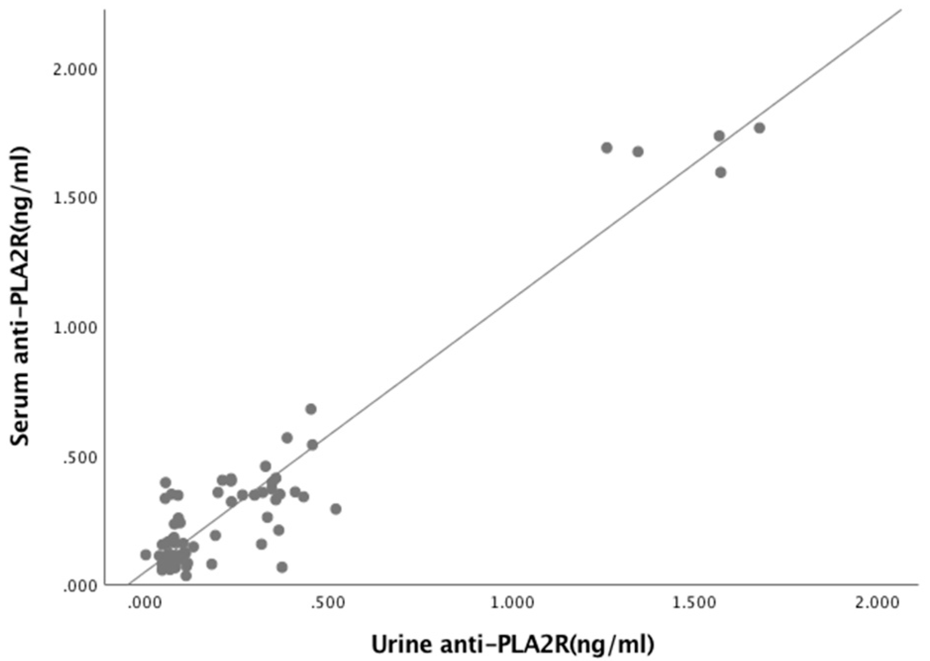
3.3. Relationships Between Biomarkers and Laboratory Parameters

Table 3 described the relationship between biomarkers (PLA2R and THSD7A) and respective laboratory parameters. There was a strong positive significant relationship between serum anti-PLA2R Ab and urine protein creatinine index (UPCrI) (R = 0.522, p < 0.05) with no significant relationship between serum anti-PLA2R Ab and urea, creatinine and estimated glomerular filtration rate (eGFR). Laboratory parameters such as urea (R = 0.251, p < 0.038) and UPCr Index (R = 0.437, p < 0.05) were, respectively, poorly and fairly correlated with urine anti-PLA2R Ab. Urine anti-PLA2R Ab and serum anti-PLA2R Ab were highly correlated (R = 0.902, p < 0.05). There was no correlation between serum anti-THSD7A Ab and urine anti-THSD7A Ab and other laboratory parameters. The graphical presentations of some of the variables with significant correlation are presented in Appendix B.

3.4. The Prognostic Outcome of Primary MGN Subjects

3.4.1. The Prognostic Outcome of Primary MGN using eGFR (CKD ≥ 3)

Most of the variables except for sex, albumin and serum anti-THSD7A were retained following simple logistic regression described in Table 4 below. However, variables like urine anti-PLA2R Ab and serum creatinine were excluded due to multicollinearity. Following multiple logistic regression, only urea (B = −1.174, S.E. = 0.441, 95% C.I. = 0.130–0.734, AOR = 0.309, p = 0.008) and serum anti-PLA2R Ab (B = −1.447, S.E. = 1.467, C. I. = 0.013–4.175, AOR = 0.235) as shown in Table 5. Urea increases the risk of the primary outcome (CKD ≥ 3) by the odd of 0.309 (69.1%) for any unit increase in urea (95 % C.I.) while high serum anti-PLA2R Ab titre increased the risk of the primary outcome (CKD ≥ 3) by 4.3 odds compared with those subjects with normal serum anti-PLA2R Ab titre. The result was validated by receiver operating characteristics (ROC) curve (AUC = 0.960, sensitivity = 87.0%, specificity = 93.3%) as shown in Figure 2.

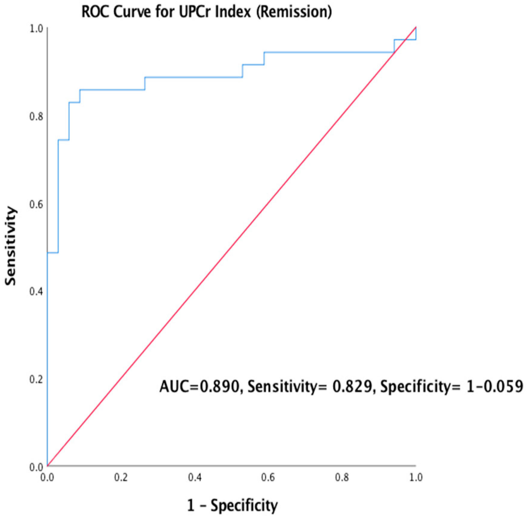
3.4.2. The Prognostic Outcome of Primary MGN using UPCr Index (Remission)

Factors associated with remission were assessed as important prognostic factors. Simple logistic regression was used where creatinine (SE = 0.015, COR = 1.047, 95% CI = 1.016–1.079, p = 0.002), eGFR(SE = 0.013, COR = 0.954, CI = 0.931–0.978, p < 0.05) and urine anti-PLA2R Ab(SE = 1.249, COR = 11.845, 95% CI = 1.025–136.924, p = 0.048) were found to be significant as shown in Table 6, while Table 7 shows multiple regression validated by ROC curve in Figure 3 (AUC=0.890, sensitivity= 82.9%, specificity= 94.1%); only urine anti-PLA2R Ab was retained as a primary factor associated with remission. Therefore, any rise in one unit of urine anti-PLA2R Ab titre increases the risk of not achieving remission by 18.486% (95% C.I. = 0.153–2234.834, p < 0.05).

4. Discussion

Discovery of serum biomarkers for MGN has been part of exceptional achievement in the management of primary MGN patients. These biomarkers can help in making the diagnosis, making a decision based on whether to give supportive or immunosuppressive therapy or both. The importance of serum and urine biomarkers (anti-PLA2R Ab and anti-THSD7A Ab) in the management of primary MGN patient was emphasized in this study.
A total of 69 subjects consisting of 47 primary and 22 secondary MGN fully consented and enrolled in this study with an average follow-up of 36.0 (15.0–57.0) in months.

4.1. Role of Biomarkers (PLA2R and THSD7A) in the Diagnosis of MGN

A high index of suspicion is needed from history and clinical presentation to rule out secondary causes before making a diagnosis of primary MGN and confirm by renal biopsy. However, this might be time-consuming, and patients on an anticoagulant may have delayed biopsy. For these reasons, biomarkers like serum and urine anti-PLA2R Ab complimented by serum anti-THSD7A Ab were employed to make the diagnosis of MGN without waiting long for the manifestation of other features [6,13]. From this study, 13 of the 47 biopsy-proven primary MGN patients were found to be positive for both serum and urine anti-PLA2R Ab at the end of follow-up. In contrast, a serum and urine sample of the same subject associated with hepatitis B virus was positive for secondary MGN at the end of follow-up. Although anti-PLA2R Ab was considered a reliable biomarker for primary MGN [5], its appearance in secondary MGN was not surprising since the biomarker was reported to be associated with hepatitis B virus infection [14,15]. A biomarker, anti-THSD7A Ab can be used to make a diagnosis of primary MGN variants not detected by anti-PLA2R Ab [16]. In this study, serum anti-THSD7A Ab was positive in four biopsy-proven primary MGN subjects and two secondary MGN subjects (one of which was associated with malignancy and the other was due to lupus nephritis type V). This result may be possible for the fact that anti-THSD7A Ab was associated with malignancy [17,18,19] and we can’t also rule out the presence of malignancy or lupus nephritis coexisting with primary MGN. Therefore, malignancy should be suspected among anti-THSD7A Ab positive subjects.

4.2. Role of Biomarkers in Monitoring MGN Subjects

It was widely reported that biomarker like PLA2R Ab could be used to monitor subjects with primary MGN [20,21]. Therefore, positive detection of anti-PLA2R Ab in 13 subjects and anti-THSD7A Ab in four subjects with biopsy-proven primary MGN after a prolonged period of follow-up justified the importance of these biomarkers in monitoring MGN. Studies have shown that changes in biomarker titre are immunological, and the depletion of anti-PLA2R Ab and an increase in serum albumin level preceded the decrease in proteinuria. Therefore, anti-PLA2R Ab can predict remission better than proteinuria [9,22]. The above finding is another reason why anti-PLA2R Ab is needed to monitor patients with primary MGN.

4.3. Prognosis of MGN

From Table 3 above, it was demonstrated that there was a significant positive relationship between anti-PLA2R Ab and UPCr Index, thereby confirming the fact that those biomarkers can be used to determine prognosis since they are significantly associated with prognostic laboratory parameters [23].

Anti-PLA2R Ab as a Prognostic Biomarker for Primary MGN

An elevated anti-PLA2R Ab titre is associated with poor prognosis and increased risk of progression to ESRD [9] or post kidney transplantation failure [24,25,26] whereas low anti-PLA2R Ab titre is associated with spontaneous remission [21,27,28,29]. Therefore, the low prevalence of anti-PLA2R Ab at the end of follow-up in this study demonstrated that anti-PLA2R Ab is a prognostic biomarker for primary MGN as against UPCr Index level, which showed fewer subjects achieving remission at the end of follow-up. Thus, anti-PLA2R Ab could help in deciding when to commence or stop immunosuppressive therapy and also those patients that may likely have ESRD.

5. Conclusions

Serum (anti-PLA2R Ab and anti-THSD7A Ab) and urine anti-PLA2R Ab are essential biomarkers for diagnosis, treatment decision and prognosis of patients with primary MGN. In addition, biomarkers should be clinically recommended to make a diagnosis of primary MGN in patients who were unable to undergo renal biopsy and to monitor the response to treatment.

Author Contributions

The conceptualization of this study was done by S.M.M. and F.A.G.; methodology, S.M.M. and F.A.G.; validation, N.F.Z., F.A.G. and R.H.; formal analysis, S.M.M.; investigation, F.Z. and S.M.M.; resources, F.A.G.; data curation, S.M.M.; writing—original draft preparation, S.M.M.; writing—review and editing, F.Z., R.H. and F.A.G.; visualization, S.M.M.; supervision, F.A.G., R.H. and F.Z.; project administration, F.A.G.; funding acquisition, F.A.G. and F.Z. All authors have read and agreed to the published version of the manuscript.

Funding

Grants from University Putra Malaysia funded this research (Grant ID 9594200).

Acknowledgments

The staff of Hospital Serdang and Hospital Kuala Lumpur.

Conflicts of Interest

No conflict of interest. The funders had no role in the design of the study; in the collection, analyses or interpretation of data, in the writing of the manuscript or in the decision to publish the results.

Appendix A

Figure A1. Anti-PLA2R ELISA standard curve.
Biomolecules 10 00319 g0a1

a0.2209
b−2.203
c1.188
d1.738
MSE0.003579
0.9886
SS0.04294
SYX0.07327
Figure A2. Anti-THSD7A standard curve.
Biomolecules 10 00319 g0a2

a0.05272
b−1.051
c0.2162
d5.961
MSE0.0003364
0.9995
SS0.002018
SYX0.03177

Appendix B

Figure A3. Relationship between Serum anti-PLA2R and urine anti-PLA2R (R = 902, p < 0.001).
Biomolecules 10 00319 g0a3
Figure A4. Relationship between Serum anti-PLA2R and UPCr Index (R = 0.502, p < 0.001).
Biomolecules 10 00319 g0a4
Figure A5. Relationship between Urine anti-PLA2R and UPCr Index (R = 437, p < 0.001).
Biomolecules 10 00319 g0a5
Figure A6. Relationship between Urine anti-PLA2R and serum Urea (R = 0.201, p < 0.038).
Biomolecules 10 00319 g0a6

References

  1. Hanko, J.B.; O’Rourke, D.M.; McNamee, P.T.; MAxwell, A.P.; Courtney, E.A. The changing pattern of adult primary glomerular disease. Nephrol Dial. Transpl. 2009, 24, 3050–3054. [Google Scholar] [CrossRef]
  2. Pan, X.; Xu, J.; Ren, H.; Zhang, W.; Xu, Y.; Shen, P.; Li, X.; Wang, W.; Chen, X.; Wu, P.; et al. Changing spectrum of biopsy-proven primary glomerular diseases over the past 15 years: A single-centre study in China. Contrib Nephrol. 2013, 181, 22–30. [Google Scholar] [PubMed]
  3. Xiaofan, H.; Jing, X.; Chenni, G.; Yifan, W.; Xialian, Y.; Li, L.; Hong, R.; Wen, Z.; Weiming, W.; Xiaoxia, P.; et al. New risk score for predicting progression of membranous nephropathy. J. Transl. Med. 2019, 17, 1–7. [Google Scholar] [CrossRef] [PubMed]
  4. Yahya, R.; Jazilah, W.; Ismail, W. Fifth report of the Malaysian Registry of Renal Biopsy 2012. Malaysian Registry of Renal Biopsy, 2014. Available online: http://www.msn.org.my or https://www.macr.org.my/emrrb (accessed on 30 June 2014).
  5. Beck, L.H., Jr.; Bonegio, R.G.; Lambeau, G.; Beck, D.M.; Powell, D.W.; Cummins, T.D.; Klein, J.B.S.D. M-type phospholipase A2 receptor as target antigen in idiopathic membranous nephropathy. N. Engl. J. Med. 2009, 361, 11–21. [Google Scholar] [CrossRef] [PubMed]
  6. Tomas, N.M.; Beck, L.H., Jr.; Meyer-Schwesinger, C.; Seitz-Polski, B.; Ma, H.; Zahner, G.; Dolla, G.; Hoxha, E.; Dabert-Gay, A.; Debayle, D.; et al. Thrombospondin type-1 domain-containing 7A in idiopathic membranous nephropathy. N. Engl. J. Med. 2014, 371, 2277–2287. [Google Scholar] [CrossRef]
  7. Maifata, S.M.A.; Hod, R.; Zakaria, N.F.; Ghani, F.A. Membranous Glomerulonephritis: Overview of the Role of Serum and Urine Biomarkers in the Management. Biomedicine 2019, 7, 86. [Google Scholar] [CrossRef]
  8. Cravedi, P.; Jarque, M.; Angeletti, A.; Favà, À.; Cantarelli, C.; Bastard, O. Immune-Monitoring Disease Activity in Primary Membranous Nephropathy. Front. Med. 2019, 6, 1–11. [Google Scholar] [CrossRef]
  9. Pourcine, F.; Dahan, K.; Mihout, F.; Cachanado, M.; Brocheriou, I.; Debiec, H.; Pierre, R. Prognostic value of PLA2R autoimmunity detected by measurement of anti-PLA2R antibodies combined with detection of PLA2R antigen in membranous nephropathy: A single-centre study over 14 years. PLoS ONE 2017, 12, e0173201. [Google Scholar] [CrossRef]
  10. Timmermans, S.A.M.E.G.; Damoiseaux, J.G.M.C.; Heerings-Rewinkel, P.T.J.; Ayalon, R.; Beck, L.H.; Schlumberger, W.; Salant, D.J.; van Paassen, P.; Tervaert, J.W.C. Evaluation of anti-PLA2R1 as measured by a novel ELISA in patients with idiopathic membranous nephropathy: A cohort study. Am. J. Clin. Pathol. 2014, 142, 29–34. [Google Scholar] [CrossRef]
  11. Varghese, S.A.; Powell, T.B.; Budisavljevic, M.N.; Oates, J.C.; Raymond, J.R.; Almeida, J.S.; Arthur, J.M. Urine Biomarkers Predict the Cause of Glomerular Disease. Oncotarget 2007, 7, 200–205. [Google Scholar] [CrossRef]
  12. KDIGO. KDIGO clinical practice guideline for glomerulonephritis. Kidney Int. Suppl. 2012, 2, 142. [Google Scholar]
  13. Hofstra, J.M.; Beck, L.H.; Beck, D.M.; Wetzels, J.F.; Salant, D.J. Anti-phospholipase a2 receptor antibodies correlate with clinical status in idiopathic membranous nephropathy. Clin. J. Am. Soc. Nephrol. 2011, 6, 1286–1291. [Google Scholar] [CrossRef] [PubMed]
  14. Sasaki, Y.; Nagai, Y.; Mikami, T.; Akasaka, Y.; Shibuya, K.; Urita, Y. Anti-phospholipase A2 receptor antibody-positive hepatitis B virus-associated membranous nephropathy remitted with entecavir after relapse with lamivudine. J. Nephropathol. 2018, 7, 93–97. [Google Scholar] [CrossRef]
  15. Dong, H.R.; Wang, Y.Y.; Cheng, X.H.; Wang, G.Q.; Sun, L.J.; Cheng, H.; Chen, Y.P. A retrospective study of phospholipase A2 receptor and IgG subclasses in glomerular deposits in Chinese patients with membranous nephropathy. PLoS ONE 2016, 11. [Google Scholar] [CrossRef] [PubMed]
  16. Zaghrini, C.; Seitz-Polski, B.; Justino, J.; Dolla, G.; Payré, C.; Jourde-Chiche, N.; Van de Logt, A.E.; Booth, C.; Rigby, E.; Lonnbro-Widgren, J.; et al. Novel ELISA for thrombospondin type 1 domain-containing 7A autoantibodies in membranous nephropathy. Int. Soc. Nephrol. 2019, 95, 666–679. [Google Scholar]
  17. Pozdzik., A.; Brochériou, I.; David, C.; Touzani, F.; Goujon, J.M.; Wissing, K.M. Membranous Nephropathy and Anti-Podocytes Antibodies: Implications for the Diagnostic Workup and Disease Management. Biomed. Res. Int. 2018, 1–19. [Google Scholar] [CrossRef]
  18. Hoxha, E.; Beck, L.H., Jr.; Wiech, T.; Nicola, M.T.; Probst, C.; Mindorf, S.; Meyer-Schwesinger, C.; Zahner, G.; Stahl, P.R.; Schopper, R.; et al. An indirect immunofluorescence method facilitates detection of thrombospondin type 1 domain-containing 7A-specific antibodies in membranous nephropathy. J. Am. Soc. Nephrol. 2016, 28, 520–531. [Google Scholar] [CrossRef]
  19. Xian, L.; Dong, D.; Luo, J.; Zhuo, L.; Li, K.; Zhang, P.; Xu, Y.; Xu, G.; Wang, L.; Li, G. Expression of THSD7A in neoplasm tissues and its relationship with proteinuria. BMC Nephrol. 2019, 20, 1–6. [Google Scholar] [CrossRef]
  20. Wu, X.; Liu, L.; Guo, Y.; Yang, L. Clinical value of a serum anti-PLA2R antibody in the diagnosis and monitoring of primary membranous nephropathy in adults. Int. J. Nephrol. Renovasc. Dis. 2018, 11, 241–247. [Google Scholar] [CrossRef]
  21. Jullien, P.; Polski, B.S.; Maillard, N.; Thibaudin, D.; Laurent, B.; Ollier, E.; Alamartine, E.L.; Mariat, C. Anti-phospholipase A2 receptor antibody levels at diagnosis predict spontaneous remission of idiopathic membranous nephropathy. Clin. Kidney J. 2017, 10, 209–214. [Google Scholar] [CrossRef]
  22. Mastroianni-Kirsztajn, G.; Hornig, N.; Schlumberger, W. Autoantibodies in renal diseases—Clinical significance and recent developments in serological detection. Front. Immunol. 2015, 6, 1–6. [Google Scholar] [CrossRef] [PubMed]
  23. Wang, Y.; He, Y.X.; Diao, T.T.; Wei, S.Y.; Qi, W.R.; Wang, C.C.; Song, S.M.; Bi, M.; Li, C.M.; Zhnag, C.X.; et al. Urine anti-PLA2R antibody is a novel biomarker of idiopathic membranous nephropathy. Oncotarget 2018, 9, 67–74. [Google Scholar] [CrossRef] [PubMed]
  24. Quintana, L.F.; Miguel Seras, N.S.; Perez, M.; Lopez-Hoyos, P.; Villarroel, E.R.; Odette, V.; Guadalupe, E.; Fritz, D.; Jose, J.G.R.; Gema, F.F.; et al. Antiphospholipase A2 antibody predicts the risk of post-transplantation recurrence of membranous nephropathy. Transplantation 2015, 99, 1709–1714. [Google Scholar] [CrossRef]
  25. Gupta, G.; Fattah, H.; Ayalon, R.; Kidd, J.; Gehr, T.; Quintana, L.F.; Sadruddin, S.; Massey, D.H.; Kumar., D.; King, A.L.; et al. Pretransplant phospholipase A2 receptor autoantibody concentration is associated with clinically significant recurrence of membranous nephropathy post-kidney transplantation. Clin. Transpl. 2016, 30, 461–469. [Google Scholar] [CrossRef]
  26. Leon, J.; Pérez-Sáez, M.J.; Batal, I.; Beck, L.H.; Rennke, H.; Canaud, G.; Legendre, C.; Pascual, J.; Reilla, L. Membranous Nephropathy Post-Transplantation. Transplantation 2019, 103, 1. [Google Scholar]
  27. Diaz, M.; Agraz, I.; Soler, M.J. Anti-phospholipase A2 receptor antibody and spontaneous remission in membranous nephropathy. Clin. Kidney J. 2019, 12, 33–35. [Google Scholar] [CrossRef]
  28. Rodas, L.M.; Matas-García, A.; Barros, X.; Blasco, M.; Vinas, O.; Llobell, A.; Martin, N.; Quintana, L.F. Antiphospholipase 2 receptor antibody levels to predict complete spontaneous remission in primary membranous nephropathy. Clin. Kidney J. 2019, 12, 36–41. [Google Scholar] [CrossRef]
  29. Ayalon, R.L.H.B., Jr.; Schlumberger, W. Evaluation of Anti-PLA2R1 as Measured by a Novel ELISA in Patients With Idiopathic Membranous Nephropathy: A Cohort Study. Am. J. Clin. Pathol. 2014, 29–34. [Google Scholar]
Figure 1. Flow chart of events from ethical approval to data analysis.
Biomolecules 10 00319 g001
Figure 2. Receiver operating characteristics (ROC) curve was used to validate predictors of primary outcome using eGFR (CKD ≥ 3).
Biomolecules 10 00319 g002
Figure 3. ROC Curve was used to validate the predictors of outcome using UPCr Index (Remission).
Biomolecules 10 00319 g003
Table 1. Characteristics of subjects at the time of renal biopsy.
VariablePrimary MGN (n = 47)
Frequency Percentage (%)
Secondary MGN (n = 22)
Frequency Percentage (%)
Age at Diagnosis:
≤30
31–40
41–50
51–60
>60

16
8
8
8
7

34.1
17.0
17.0
17.0
14.9

7
4
1
4
6

31.8
18.2
4.5
18.2
27.3
Sex:
Male
Female

21
26

44.7
55.3

8
14

36.4
63.6
Ethnicity:
Malay
Chinese
Indian
Others

24
12
9
2

51.1
25.5
19.1
4.3

16
5
0
1

72.8
22.7
0
4.5
Nephrotic Syndrome:
Present
absent

38
9

80.8
19.1

13
9

59.1
40.9
Hypertension:
Present
Absent

28
19

59.6
40.4

10
12

45.5
54.5
Haematuria:
Present
Absent

9
38

19.1
80.9

2
20

9.1
90.9
Albumin:
Normal
Low

14
33

29.8
70.2

8
14

36.4
63.6
Serum Urea(mmol/L)
Normal
High

26
21

55.3
44.7

13
9

59.1
40.9
Serum Creatinine(µmol/L)
Normal
High

6
41

12.8
87.2

8
14

36.4
63.6
eGFR (mL/min/1.73m2)
Low risk
High risk

33
14

70.2
29.8

15
7

68.2
31.8
UPCr Index(g/mmol)
Normal
High

20
27

42.6
57.4

5
17

22.7
77.3
Estimated Glomerular Filtration Rate (eGFR) (high risk ≤ 60 mL/min/1.73m2, low risk ≥ 60 mL/min/1.73m2), Urine creatinine Index (UPCr Index) (normal ≤ 0.03 g/mmol, high ≥ 0.03 g/mmol), Categorical variables were expressed as frequency and percentage.
Table 2. General characteristics of subjects at the end of the follow-up period.
VariablePrimary MGN (n = 47)
Frequency Percentage (%)
Secondary MGN (n = 22)
Frequency Percentage (%)
Median (Interquartile)
Follow-Up (months)39.0(17.5–59.5)27.5(13.0–49.8)
eGFR(mL/min/1.73m2)
Low risk
High risk

33
14

70.2
29.8

15
7

68.2
31.8
UPCr Index (g/mmol)
Remission
No remission

20
27

42.6
57.4

5
17

22.7
77.3
Biomarkers
Serum anti-PLA2R Ab (ng/mL)
Negative
Positive

34
13

72.3
27.7

21
1

95.5
4.5
Urine anti-PLA2R Ab (ng/mL)
Negative
Positive

34
13

71.1
28.9

21
1

95.5
4.5
Serum Anti-THSD7A Ab (ng/mL)
Negative
Positive

43
4

91.5
8.5

20
2

90.9
9.1
Urine Anti-THSD7A Ab (ng/mL)
Negative
Positive

47
0

100.0
0.0

22
0

100.0
0.0
Categorical variables were expressed as frequency and percentage, continuous variables as interquartile.
Table 3. Relationship between biomarkers and laboratory biomarkers.
CovariateRp-Value
Serum anti-PLA2R Ab (ng/mL)
Urine anti-PLA2R Ab (ng/mL)

0.902

<0.05 *
Albumin (g/L)
Urea (mmol/L)
Creatinine (μmol/L)
eGFR (mL/min/1.73m2)
UPCr Index (g/mmol)
0.056
0.226
0.034
0.216
0.502
0.647
0.062
0.782
0.075
<0.05 *
Urine anti-PLA2R Ab (ng/mL)
Albumin (g/L)
Urea (mmol/L)
Creatinine (μmol/L)
eGFR (mL/min/1.73m2)
UPCr Index (g/mmol)

0.040
0.251
0.550
−0.08
0.437

0.746
0.038 *
0.652
0.514
<0.05 *
Serum anti-THSD7A Ab (ng/mL)
Urine anti-THSD7A Ab (ng/mL)

0.063

0.604
Albumin (g/L)
Urea (mmol/L)
Creatinine (μmol/L)
eGFR (mL/min/1.73m2)
UPCr Index (g/mmol)
−0.069
−0.137
−0.110
0.136
−0.069
0.574
0.999
0.367
0.266
0.395
eGFR = estimated glomerular filtration rate, UPCr index = urine protein creatinine index. Correlation (R) given as <0.25 as poor, 0.26–0.5 as fair, 0.51–0.75 as good, and >0.75 as excellent. Level of significance <0.05 *.
Table 4. Simple logistic regression for the prognostic outcome of primary MGN using eGFR (CKD ≥ 3).
VariablesBSECORCI (95%)p-Value
Age0.0620.0230.9400.898-0.9930.008 *
Gender
Male
female


0.241


0.587

1.000
1.273


0.403–4.021


0.681
Albumin (g/L)
Urea (mmol/L)
0.111
0.883
0.077
0.259
1.117
2.417
0.961–1.299
1.454–4.019
0.149
0.002 *
Creatinine (μmol/L)0.1780.0681.1951.046–1.3160.009 *
UPCr Index (g/mmol)
Remission
No remission


−2.565


0.813

1.000
0.077


0.016–0.378


0.002 *
Serum anti-PLA2R Ab(ng/mL)
Negative
Positive


−1.978


0.971

1.000
0.138


0.021–0.928


0.042 *
Urine anti-PLA2R Ab(ng/mL)
Negative
Positive


−1.833


0.755

1.000
0.160


0.036–0.703


0.015 *
Serum anti-THSD7A Ab(ng/mL)
Negative
Positive


1.872


0.967

1.000
6.500


0.976–43.289


0.053
SE = Standard error, COR = Crude Odd Ratio, CI = Confidence Interval, level of significant p < 0.05 *.
Table 5. Multiple logistic regression for the prognostic outcome of primary MGN using eGFR (CKD ≥ 3).
VariablesBSEAORC.I. 95%p-Value
Age at diagnosis (years)−0.0940.0520.9100.822–1.0090.534
Serum anti-PLA2R Ab(ng/mL)
−1.447

1.467
1.000
0.235

0.013–4.175

0.048 *
Urea(mmol/L)
−1.174

0.441
1.000
0.309

0.130–0.734

0.008 *
SE = Standard Error, AOR= Adjusted Odd Ratio, C.I. = Confidence Interval, level of significance, p < 0.05 *.
Table 6. Simple logistic regression for the prognostic outcome of primary MGN using UPCr Index (Remission).
VariableBSECOR95% C.I.p-Value
Age at diagnosis0.0250.0181.0250.989–1.0630.181
Sex0.3080.4901.3600.521–3.5510.530
Albumin(g/L)−0.0750.0600.9280.825–1.0430.209
Urea(mmol/L)0.2570.1361.2930.991–1.6860.058
Creatinine (μmol/L)0.0460.0151.0471.016–1.0790.002 *
eGFR(mL/min/1.73m2)−0.0470.0130.9540.931–0.978<0.05 *
Serum anti-PLA2R Ab
(ng/mL)
1.3770.8103.9640.810–19.3990.015 *
Urine anti-PLA2R Ab(ng/mL)2.4721.24911.8451.025–136.9240.05 *
Serum anti-THSD7A Ab(ng/mL)−0.9371.0220.3920.530–2.9020.392
SE = Standard Error, COR = Crude Odd Ratio, C.I. = Confidence Interval, level of significance <0.05 *.
Table 7. Multivariate logistic regression for the prognostic outcome of primary MGN using UPCr Index (Remission).
VariableBSEAOR95% C.I.p-Value
Creatinine
eGFR (mL/min/1.73m2)
Urine anti-PLA2R Ab (ng/mL)
0.018
0.002
2.917
0.044
0.039
2.446
1.018
1.002
18.486
0.934–1.110
0.928–1.882
0.153–2234.834
0.660
0.958
0.038
SE = Standard Error, AOR = adjusted odd ratio, C.I. = Confidence Interval, level of significance <0.05 *.

