Review

Biomarkers to Personalize the Treatment of Rheumatoid Arthritis: Focus on Autoantibodies and Pharmacogenetics

by
Valeria Conti
1,*,†,
Graziamaria Corbi
2,†,
Maria Costantino
1,3,
Emanuela De Bellis
1,
Valentina Manzo
1,
Carmine Sellitto
1,
Berenice Stefanelli
1,
Francesca Colucci
1 and
Amelia Filippelli
1
1
Department of Medicine, Surgery and Dentistry “Scuola Medica Salernitana”, University of Salerno, 84081 Baronissi, Italy
2
Department of Medicine and Health Sciences, University of Molise, 86100 Campobasso, Italy
3
Association Non-Profit F.I.R.S.Thermae (Interdisciplinary Training, Researches and Spa Sciences) in Italian National Register of Research of MIUR, Naples, 80070 Bacoli, Italy
*
Author to whom correspondence should be addressed.
These authors contributed equally to this work.
Biomolecules 2020, 10(12), 1672; https://doi.org/10.3390/biom10121672
Submission received: 15 November 2020 / Revised: 8 December 2020 / Accepted: 12 December 2020 / Published: 14 December 2020
(This article belongs to the Special Issue Pathogenesis of Arthritis)

Abstract

:
Rheumatoid arthritis (RA) is a chronic inflammatory disease that is very complex and heterogeneous. If not adequately treated, RA patients are likely to manifest excess of morbidity and disability with an important impact on the quality of life. Pharmacological treatment is based on the administration of the disease-modifying antirheumatic drugs (DMARDs), subdivided into conventional synthetic (csDMARDs), targeted synthetic (tsDMARDs), and biological (bDMARDs). bDMARDs are now frequently administered in patients, both as alternative treatment and together with csDMARDs. Unfortunately, there is a therapeutic response variability both to old and new drugs. Therefore, to identify pre-therapeutic and on-treatment predictors of response is a priority. This review aims to summarize recent advances in understanding the causes of the variability in treatment response in RA, with particular attention to predictive potential of autoantibodies and DMARD pharmacogenetics. In recent years, several biomarkers have been proposed to personalize the therapy. Unfortunately, a magic bullet does not exist, as many factors concur to disease susceptibility and treatment outcomes, acting around the patient’s congenital background. Models integrating demographic, clinical, biochemical, and genetic data are needed to enhance the predictive capacity of specific factors singularly considered to optimize RA treatment in light of multidisciplinary patient management.

1. Introduction

Rheumatoid arthritis (RA) is a chronic inflammatory, autoimmune disorder affecting nearly 1% of the general population [1]. Women suffer from RA three times more often than men. Indeed, the prevalence of immune-mediated inflammatory diseases is lower in male subjects, and male RA patients appear to react better to specific treatments, such as anti-cytokines and antiproliferative drugs [2,3]. Possible reasons could be mainly related to a greater immune response in females than in males. Moreover, lymphocytes/monocytes from female subjects show higher immune/inflammatory reactivity, suggesting involvement of sex hormones in the regulation of the immune/inflammatory response in immune-mediated diseases [4,5]. In fact, sex hormones play an important role in regulating the immune/inflammatory response in RA [6].
RA initially affects small joints, progresses to larger joints and internal organs, ultimately causing an excess of morbidity and disability with an important impact on the quality of life (QoL).
The therapy is based on a treat-to-target approach, which requires tight monitoring of the disease activity in order to implement a prompt correction of the therapy regimen. The ideal therapeutic target coincides with the clinical remission or, at the very least, the low activity disease (LDA) [1].
According to the updated recommendations for the management of RA of the European League Against Rheumatism (EULAR) [7], pharmacological therapy, based on the administration of disease-modifying antirheumatic drugs (DMARDs), should be started as soon as the diagnosis of RA is made. DMARDs may slow down or even arrest the progression of joint destruction, seeking to get the treatment target, mainly assessed by the Disease Activity Score based on 28 joint counts (DAS28) [7].
DMARDs are classified in conventional synthetic (csDMARDs), targeted synthetic (tsDMARDs), and biological (bDMARDs). All the DMARDs are indicated for both non-rheumatic and rheumatic diseases thanks to their various activities, mainly immunomodulatory and anti-inflammatory ones.
Among csDMARDs, methotrexate (MTX) represents the mainstay of RA treatment.
MTX is an antimetabolite that interferes with the folate pathway binding to dihydrofolate reductase (DHFR) and thus inhibiting the transformation of dihydrofolic acid (FH2) into tetrahydrofolic acid (FH4), which is an important cofactor for the synthesis of nucleic acids and amino acids [8,9].
Under the last recommendation of the European Medicine Agency (EMA), MTX must be administered only once a week, starting with a low dose and then performing a titration as needed [10]. Serious side effects, including liver problems, bone marrow depression, and even death, occur when MTX is taken more often [10] The maximum weekly dose varies depending on the patient’s ethnic group, considering variables such as body weight and genetic factors [1].
Besides MTX, other csDMARDs suitable for RA are leflunomide and sulfasalazine. These are used as part of the first therapeutic strategy in patients with a contraindication or who manifest an early intolerance to MTX [1].
Today, besides the conventional options, clinicians have other therapeutic options, represented by ts- and bDMARDs. Tofacitinib, baricitinib, filgotinib, and upadacitinib are tsDMARDs that work by inhibiting the activity of one or more enzymes belonging to the Janus kinase family. The EULAR has included tsDMARDs among the drugs to administer in second- and later-line treatment with or without MTX. They are used in patients with moderate to severe RA who are non-responders or intolerant to csDMARDs [1].
Biological DMARDs include infliximab, adalimumab, etanercept, golimumab, and certolizumab pegol, which are all anti-tumor necrosis factor α (TNFα), and abatacept, rituximab, tocilizumab, and sarilumab. According to the EULAR recommendations [1], these drugs must be started in second- and third-line therapy, with or without MTX.
It is useful to combine pharmacological and non-pharmacological treatments with the promotion of a healthy and active lifestyle to reach the treatment target. However, despite huge advances in clinical management, diagnostic tool, and pharmacological therapy, not all patients reach an adequate response.
A variable response occurs, in fact, both to drugs and nonpharmacological therapy; therefore, there is a need to deeply investigate the factors involved in such variability observed in daily clinical practice [1].
This review aims to summarize recent advances in understanding the causes of the variability in treatment response observed in daily clinical practice in patients with RA, with particular attention to more recent data on autoantibodies and DMARD pharmacogenetics.

2. Role of Autoantibodies in Predicting the Response to RA Treatment

In RA, many antibodies, such as anti-citrullinated protein antibodies (ACPAs), rheumatoid factor (RF), and anti-carbamylated protein (anti-CarP), with diagnostic and prognostic role have been identified. All of these are principally present in RA patients’ serum and synovial fluid (SF), but ACPAs and RF are also found in lung, sputum, periodontium, intestine, and cervical–vaginal mucosa [11]. For this reason, RF and ACPAs are routinely used, mainly as biomarkers for diagnostic purpose [12]. Indeed, the European League Against Rheumatism/American College of Rheumatology (EULAR/ACR) has also introduced RF and ACPAs in the diagnostic criteria of RA, and it suggests their use in guiding the choice of treatments to prevent the development of symptomatic RA [13]. The ACPAs are the most relevant diagnostic and prognostic biomarkers for RA [14] because their determination permits to classify RA patients into seropositive versus seronegative, as well as RF measurement. The ACPAs include a group of autoantibodies directed against peptides and proteins that are present in 70–90% of RA patients and have high disease-specificity (90–95%) [15]. In fact, unlike the RF, they are rarely found in other diseases or healthy subjects. In a meta-analysis by Nishimura et al., ACPAs showed a sensitivity similar to RF (≈67% vs. ≈69%), but a higher specificity (≈95% vs. ≈85%) [16]. These autoantibodies are predictive for RA development in patients without a previous diagnosis, and for more severe disease [17]. The autoimmune response in RA is presumably initiated by citrullination of self-peptides, leading to alterations of their properties. ACPAs are locally produced in RA joints, where proteins are citrullinated during the inflammatory process [18]. Citrullination [19] leads to the activation of complex immune responses and specific ACPA generation, found in approximately 75% of RA patients [20]. The risk of developing RA also depends on hereditary factors [21], and over 100 genetic loci are involved in an increased risk of RA [22]. Interestingly, twin studies have found no difference in heritability in subsets of ACPA-positive and ACPA-negative RA [23]. Instead, an analysis of large Swedish population registers showed that inheritability accounted for around 50% of ACPA-positive RA, but only 20% of ACPA-negative RA [24]. In several studies, HLA-DRB1 alleles [25], including DRB1*0401, *0404/*0408, *0405, *0101, *1001, and *1402, presenting a similar sequence at amino acid positions 67–74 in the third hypervariable region of the DR molecule (called the “shared epitope”) were associated with RA. Similarly, susceptibility to RA is associated with certain DQB1/DQA1 combinations (DQB1*03 and *04 combined with DQA1*03 (referred to as DQ3), and DQB1*0501 combined with DQA1*0101), while RA protection is mediated by other DRB1 alleles (*0103, *0402, *1102, *1103, *1301, *1302, and *1304) [26]. In North American white people, the DRB1*0401 and *0404 alleles are the most prevalent, and these genes are associated with the highest risk of severe disease. In a recent study, Wysocki et al. underlined that HLA-DRB1 genotyping is neither used in clinical practice, nor included in the current RA classification criteria [20]. Nonetheless, some HLA-DRB1 variants seem to predict the unfavorable course of the disease, including a higher risk of radiographic damage progression, higher incidence of interstitial lung disease, or lymphoproliferative diseases. The authors also suggested the possible identification of high-risk patients bearing the HLA-DRB1 risk allele, with a consequent important role of this marker in the personalization of the therapy [20]. Analyzing data from patients with early RA treated according to different strategies, by considering DQB1 typing and DRB1 typing and subtyping, Lard et al. showed that the association between the HLA class II antigens and joint destruction is affected by early and aggressive therapy with DMARDs. This interaction was independent of other prognostic factors, such as RF and baseline disease activity. Then, the authors suggested that an early and aggressive treatment with DMARDs, such as MTX, sulfasalazine, and cloroquine, can regulate the immune process, maybe by preventing a secondary release of autoantigens, by modulating the autoantigen presentation by APCs to the CD4+ T cells, and/or by inhibiting the response of T cells [27]. RFs, first identified autoantibodies in RA, are also present in infectious diseases, wherein they facilitate the removal of pathogens, and in cancer [28,29]. RFs and ACPAs show a broad spectrum of isotypes (immunoglobulin (Ig)M, IgG, IgA) [30]. The RF is an autoantibody that recognizes domains CH2 and CH3 of the Fc portion of human immunoglobulin G (IgG). According to different studies, RF testing in RA patients has a sensitivity that varies from 60 to 90% and specificity between 48 and 92% [31]. The limited specificity of RF is due to its presence also in healthy controls and patients with other autoimmune and non-autoimmune diseases [32]; consequently, RF alone is insufficient for diagnosis. High RF levels are related to an increased risk of developing RA, which may increase 26 times if they are >100 IU/mL [33], while the presence of the IgA isotype is associated with extra-articular manifestations [34]. Due to RF variable specificity, ACPA testing has been added in the latest RA classification criteria in order to increase the specificity. In RA, 50–70% of patients are positive for ACPAs, which recognize several citrullinated antigens, such as α-enolase, fibrinogen, filaggrin, vimentin, and type II collagen [35,36]. In various diseases, IgG and IgA avidity is associated with different clinical response and, in the case of ACPAs, low avidity IgG is linked to more severe bone damage [37]. About 50% of RA patients have an abnormal serologic profile several years before the development of symptoms. Moreover, elevated serum level of IgM-RF or ACPAs in healthy subjects indicates a high risk for RA onset [38]. Anti-CarP recognize other post-translational modified (PTM)-antigens and often occur together with other antibodies (e.g., ACPAs) with which they can cross-react. Anti-CarP are prognostic of the new RA onset, being detectable years before, and increasing shortly before the RA onset [39], and are predictive of joint destruction, especially in ACPA-negative RA patients [40], and they represent an important serological marker in ACPA-negative RA patients because predictive of more severe disease, as shown by radiological progression [40].
In antibody-positive patients, an increase of IgA plasmablast in peripheral blood is identified, suggesting that mucosal immune responses are involved in early RA pathogenesis [41], as well as the presence of ACPAs and RFs in the subclinical stage [42]. Therefore, Anti-CarPs, ACPAs, and RFs have a predictive role, appearing 1–3 years before RA symptoms onset, and their presence is associated with joint destruction, contributing into RA pathogenesis and development [43]. ACPAs and RFs could enhance the secretion of neutrophil extracellular traps (NETs), which show a pro-inflammatory effect, exposing autoantigens [44]. In RA patients, ACPAs are associated with parenchymal lung abnormalities [45] and may have a role in bone erosion affecting osteoclasts and increasing osteoclast precursor differentiation in vitro and in vivo [46]. This is very important because RA is characterized by chronic inflammation and bone erosion, also caused by immune complexes (ICs) composed of autoantibodies that worsen pro-inflammatory status, leading to complement activation and increased levels of chemokines and cytokines [47,48,49,50]. In a cohort of 200 RA patients, it has been demonstrated that RF testing, followed by ACPA and anti-RA33 analysis, can be a useful workflow in diagnosing the early inflammatory joint damage [51].
De Moel et al. investigated the association between the breadth of the serum autoantibody profile with early and late treatment outcomes in 399 seropositive RA patients enrolled in the IMPROVED (the induction therapy with methotrexate and prednisone in rheumatoid or very early arthritic disease) trial. The seropositive patients with a broader autoantibody profile at baseline exhibited an early treatment response but they did not maintain such favorable outcome over time. The authors suggested that the presence at baseline of multiple autoantibody types, including ACPAs, anti-CARP, RF, and the anti-modified protein antibodies (AMPAs), could improve an early response to DMARDs by stimulating a more active humoral immunity [52].
A study by Martin-Mola et al. suggested that ACPAs, especially when present in high titers, may predict a better response to rituximab and abatacept, but not to anti-TNF agents and tocilizumab [53]. Therapy with these drugs can be complicated by the development of anti-drug antibodies (ADA), since they are proteins and, therefore, inherently immunogenic [54]. The presence of ADA may compromise therapeutic efficacy, leading to a reduction of serum drug levels or even to a neutralization of the drug, causing a loss of clinical response [55,56,57]. ADA development is multifactorial and depends on many factors, even if they are not still clarified. In a study on RA patients receiving infliximab as a second agent, ADA development, low serum infliximab, and RF positivity were more common in females and associated with treatment failure [58]. This finding is interesting, since, as already mentioned, women are more frequently affected by autoantibody-positive autoimmune diseases [59]. In a multicohort analysis evaluating different variables, factors associated with ADA development were longer disease duration, moderate disease activity, and lifetime smoking [60]. Previous studies have shown that 28% of patients treated with adalimumab reported having ADA within 3 years of treatment [56]. ADA have also been observed in up to 44% of patients treated with infliximab in the first 6 months of treatment [61]. On the other hand, in different studies, etanercept has been shown to have mild immunogenicity, reporting an incidence of ADA only in 0–7% of patients [54,62]. A meta-analysis of 17 studies on the immunogenicity of TNF inhibitors reported that ADA were not detected in patients treated with etanercept [63]. Similar results were observed in a recent study in which it was found that all the etanercept-treated patients were negative for ADA, while 31.2% and 17.4% of those treated with adalimumab and infliximab, respectively, were positive for ADA [64]. A meta-analysis of 34 studies enrolling 4273 patients evaluated the clinical response in patients with ADA treated with anti-TNF alpha drugs, showing a significant reduction of response in patients with ADA compared to patients without, especially when treated with infliximab or adalimumab. In this view, early detection of serum ADA levels may improve patients’ management [65]. The current guidelines do not recommend routine monitoring of ADA in cases of primary or secondary failure of anti-TNF therapy. Consequently, when patients show a loss or lack of response to a first anti-TNF alpha, the clinical practice is usually based on switching to a biologic drug with a different mechanism of action, or switching to another anti-TNF alpha, or increasing the dose without changing drug [66]. The decision is often empirical and depends on physician discretion. Several algorithms, which foresee the ADA and therapeutic drug monitoring, have been proposed—they are all based on the hypothesis that immunogenicity assessment may help to guide therapeutic decisions, going beyond the “empirical switching” and obtaining better control of the disease [67]. In a recently proposed algorithm, the measurement of drug levels plays a crucial role. In the first branching of the algorithm, testing ADAs is not recommended because they are not always clinically relevant. In case of treatment failure, they recommend the supplementary ADA test, which may be helpful in personalizing the treatment choice [68].
Table 1 reports the studies, discussed in the text, highlighting the relationship between serum autoantibodies and treatment outcomes in patients with RA.

3. Pharmacogenetic Biomarkers

Up until now, there are no data on the role of genetic polymorphism concerning tsDMARDs. On the contrary, several studies highlighted the impact of the pharmacogenetics (PGx) to personalize the therapy with cs- and bDMARDs in patients suffering from RA.

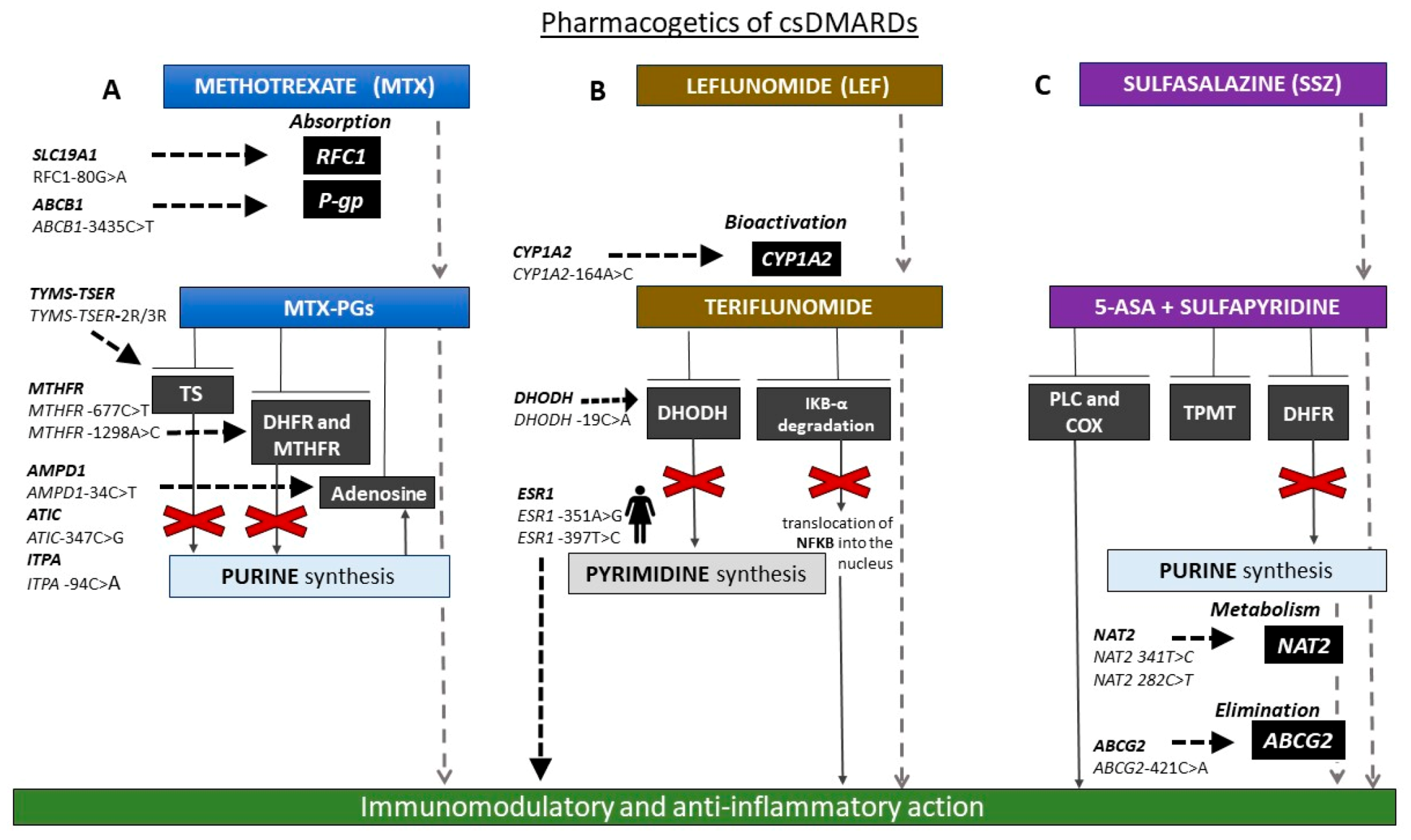
3.1. Pharmacogenetics of csDMARDs

Owing to its efficacy, long-term safety, dose-titratable range, and cost-effectiveness, methotrexate (MTX) is still one of the most important drugs used in RA therapy.
MTX is recommend as first-line treatment in patients with RA, both as monotherapy and as an “anchor drug” in association with other DMARDs [7,69]. Following oral administration, it is absorbed from the small intestine via folate transporters and then converted through hepatic and intracellular metabolism into polyglutamated forms (MTX-PGs). Within the cell, MTX-PGs bind to and inhibit dihydrofolate reductase (DHFR) and other folate pathway enzymes required for purine and pyrimidine synthesis [70].
Despite its widespread use, the mechanism by which MTX exerts its therapeutic effects in RA is not fully understood. The reason is that MTX has anti-inflammatory effects, mediated through a variety of molecular pathways that do not necessarily involve the folate antagonism. Multiple mechanisms appear to be involved, including increase and release of adenosine; inhibition of methyl-donor production; generation of radical oxygen species (ROS); downregulation of adhesion molecules levels, eicosanoids, and matrix metalloproteinases; and interference with T cell activity and cytokine secretion [70].
Unfortunately a considerable number of patients with RA (nearly 30–40%) do not respond to MTX adequately, and many of them discontinue the therapy often because of MTX-related adverse drug reactions (ADRs). The most frequently reported ADRs include ulcerative stomatitis, leukopenia, nausea, and abdominal distress but also malaise, undue fatigue, chills and fever, dizziness, and infections [71].
However, at present, there are no biomarkers or models to predict MTX responsiveness so robust as to effectively tailor the MTX treatment in patients with RA.
Many investigations, using various technical approaches, have been proposed with the main aim to stratify the patients on their risk to not respond to MTX. For example, mRNA expression profiling [72,73,74] are considered biomarkers valuable to predict patients’ clinical outcomes before the beginning of the treatment.
In this context, the MTX PGx may be very helpful. Actually, it is well known that, besides disease-specific characteristics, numerous factors, including drug–drug interaction and patient’s genetic background may influence both the MTX efficacy and tolerability [75,76].
Recently, several genetic variants possibly influencing the MTX pharmacokinetics and pharmacodynamics have been explored. The interindividual variability in MTX pharmacokinetics can be partly explained by the presence of polymorphisms (mainly single nucleotide polymorphisms, SNPs) in the genes encoding membrane transporter proteins. MTX is absorbed through active transport by the reduced folate carrier protein 1 (RFC1) and the proton-coupled folate transporter (PCFT), encoded by the human solute carrier family 19 and 46 member 1 (SLC19A1 and SLC46A1), respectively [75,77].
One of these SNPs is RFC1-80G>A (rs1051266), which results in variable intracellular levels of MTX-PGs, and consequently variable MTX efficacy. It has been shown that patients carrying RFC1 80AA genotype responded better to the treatment than RFC1-80AG and RFC1-80GG individuals, and the probability of remission of RA symptoms was 3.32-fold higher in carriers of RFC1-80AA when compared with those with RFC1-80GG genotype [78].
As expected, there is a relevant variability depending upon the ethnic population. As matter of a fact, Li et al. found a significant association between RFC1-80G>A and MTX efficacy in Asians (p = 0.002 for A allele; p = 0.003 for AA genotype) but not in Caucasians (p = 0.15 for A allele; p = 0.05 for AA genotype), and no association with toxicity was found [79]. In contrast, Qiu et al. demonstrated an association between MTX-related toxicity and RFC1-80G>A SNP in Caucasian patients (odds ratio (OR) = 1.36, 95% CI = 1.01–1.83, Z = 2.05, p = 0.041) [76].
However, the studies included in these metanalyses reported different results also considering study populations belonging to the same ethnicity. Drodzik et al. [78] conducted a prospective study on 174 Caucasian patients affected by RA and treated with a regimen of MTX (7.5–15.0 mg weekly) plus low doses of methylprednisone. The efficacy of treatment was evaluated according to the American College of Rheumatology response criteria (ACR20%) in 12 months, and the safety as number of adverse events. The probability of remission of RA symptoms was 3.32-fold higher in patients with RFC1-80AA genotype than in those with -80GG genotype. Adverse events, such as increase of transaminase, were mostly detected in patients with -80AA genotype than in -80GA and -80GG genotypes, but without reaching statistical significance [78]. Conversely, in a retrospective study enrolling 150 Caucasian RA patients, Bohanec-Grabar et al. found an association between the risk of MTX-related toxicity and the presence of RFC1-G80A SNP. In fact, patients carrying RFC1-80GG genotype had a higher risk for MTX overall toxicity. Gastrointestinal complaints and hepatotoxicity were the most frequent drug adverse events, and such a finding was found also by Lima et al., who demonstrated an increased gastrointestinal MTX-related toxicity associated with the presence of RFC1-80G allele in 233 Caucasian RA patients. Differently from Bohanec, these authors found no association between this SNP and overall toxicity [80,81].
Another prospective study performed in Caucasian patients stressed the importance to take into account different variables to evaluate variability response to RA treatment. The research analysed the possible association between several polymorphisms in folate pathway and efficacy of MTX combined with sulfasalazine and hydroxychloroquine in 98 patients with early RA. In line with the results reported by Drodzik et al. [78], the SNP was significantly associated with the status of responder to the therapy. Moreover, patients carrying such a polymorphism combined with methionine synthase (MTR)-2756A>G or TYMS3R-del6 polymorphisms were more likely to have remission of symptoms up to 3 years [82].
Hayashi et al. analyzed 170 Japanese patients with RA. Of the patients, 89 were treated with MTX alone (MTX group), and 81 with MTX plus bDMARDs. The disease activity score (DAS)28 was the same (2.3–2.4) in the two groups. RFC1-80A allele frequency was higher in MTX group while more patients carrying the G allele were observed in the MTX plus bDMARDS arm. Not coincidentally, the presence of the G allele may be associated to a poor response to MTX monotherapy that requires the co-administration with a bDMARD [83]. Conversely, another study performed in Japanese RA patients failed to find a significant association between RFC1-80A>G, neither with MTX efficacy nor MTX safety [84].
Several others carriers involved in MTX influx have also been examined [77]. One of the most promising pharmacogenes is the ABC transporter family members (ATP-binding cassette transporter), coding for the P-glycoprotein (P-gp-1) efflux pump [85,86]. This P-gp, expressed on the membrane of a variety of cells, serves to decrease the intracellular accumulation of various drugs [87]. The SNP ABCB1 3435C>T is one of the most investigated polymorphisms because it may influence, especially together with other genetic variants, the response to several drugs, including the antiplatelet clopidogrel [88], statins [89], the antineoplastic agent irinotecan [90], and others.
The SNP ABCB1 3435C>T has been related to the clinical course of RA, as well as to the drug response [84,91,92]. However, the available literature data are often contradictory.
Two meta-analyses have investigated the potential of ABCB1 C3435T in predicting the response to MTX in RA patients. They shared four articles and reached opposite conclusions.
The study by Lee et al, including nine articles involving 1275 RA patients, reported that patients carrying the ABCB1 C3435T may have an increased risk of MTX-related toxicity (TC vs. TT + CC; OR = 0.483, 95 % CI = 0.259–0.900, p = 0.022), but such a SNP does not predict RA susceptibility nor responsiveness to DMARDs [93]. These authors highlighted that a limitation of their study is the small statistic power; therefore, they stressed the need to analyze larger scale studies to validate the association between the ABCB1 C3435T SNP with efficacy and toxicity in RA. Conversely, the study by He et al., including 12 articles involving more RA patients with respect to the study by Lee et al. (a total of 2014 patients), suggested that the ABCB1 C3435T might be helpful to predict the response to MTX in Asian patients (additive: OR = 1.64, 95% CI = 1.09–2.48, p = 0.019; homozygous model: OR = 2.54, 95% CI = 1.51–4.8, p < 0.001; and recessive model: OR = 2.09, 95% CI = 1.37–3.19, p = 0.001) but not the MTX-related ADRs [92].
Regarding MTX target genes, some polymorphisms in the gene “TYMS” encoding TS, which is a key enzyme for DNA synthesis and repair inhibited by MTX-PGs, seem to be related to the response to MTX. The most common genetic variant, located in the enhancer region (TSER), consists of two or three 28-bp tandem repeats (TYMS-TSER-2R/3R). It has been reported that individuals carrying -3R allele have higher TYMS mRNA expression than those with -2R and require a higher MTX dosage in order to achieve an adequate therapeutic response [77,94,95].
Different levels of TYMS mRNA has been also considered to understand a possible role of the TSER-2R/3R polymorphism in predicting fluoropyrimidine-related toxicity in oncological patients [96,97].
Several meta-analyses focused mostly on SNPs in the gene encoding the 5,10-methylenetetrahydrofolate reductase (MTHFR) [76,98,99,100,101], a rate-limiting enzyme in the methyl cycle catalyzing the conversion of 5,10-methylenetetrahydrofolate to 5-methyltetrahydrofolate, which in turn serves as a methyl donor for the methylation of homocysteine to methionine. Variants in the MTHFR may interfere with these physiological events, thus leading to unexpected MTX side-effects.
The MTHFR C677T (rs1801133) and A1298C (rs1801131) are two SNPs that are well known to affect enzyme activity and drug metabolism. The first results in a thermolabile form of MTHFR with a reduced enzyme activity (30% of wild-type activity for TT carriers) and hyperhomocysteinemia, and increased toxicity of MTX has been associated with such a polymorphism [95].
Moreover, the MTHFR A1298C leads to lower enzyme activity and hyperhomocysteinemia but without resulting in a thermolabile protein and, in contrast to the MTHFR 677C>T, it does not seem to increase the frequency and severity of MTX-associated toxicity [95].
Additionally in this case, the available data are controversial. For example Bohanec-Grabar et al. observed that carriers of MTHFR A1298C SNP showed a decreased overall MTX toxicity but no effects on MTX efficacy [87].
Moreover, the already mentioned HLA-DRB1-shared epitope is linked to the response to MTX. In particular, the presence of HLA-DRB1*04 allele in combination with anti–cyclic citrullinated peptide (anti-CCP) antibodies has been associated with development of MTX resistance and it may justify the use of an anti-TNFα agent in patients with early RA [25,102]
Other genetic variants in genes involved in MTX transport and metabolism (e.g., FOLR1, FPGS and GGH), as well as in purine synthesis (e.g., ATIC, AMPD1, ADA, and ADORA2A) or adenosine signalling (AMPD1, ATIC, ITPA, MTR, and MTRR), have been explored, often with contrasting results [86,94].
For example, Wessels et al. found that patients carrying AMPD1 34T allele, or ATIC 347CC or ITPA 94CC genotypes were more likely to have a good clinical response [103], while Weisman et al. reported that ATIC 347GG genotype was associated with an increased risk of MTX-related gastrointestinal ADRs [104]. In a recent meta-analysis, Lee et al. investigated the potential of the ATIC 347C/G polymorphism in predicting efficacy and safety of MTX. Examining nine comparative studies including 1056 RA patients, the authors found that ATIC 347 GG + GC genotype was associated with the lack of response to MTX (OR = 1.884, 95% CI = 1.236–2.873, p = 0.003) and with MTX-related toxicity in Caucasian RA patients (OR = 1.741, 95% CI = 1.080–2.806, p = 0.023) [105].
To date, only two genome-wide association studies (GWASs) have been carried out to investigate genetic predictors of response to MTX IN RA patients [106,107]. Taylor et al combined data from two consortia to carry out a GWAS on the response to MTX in 1424 early RA patients. The authors found that no SNP reached significance for any outcome measure [106]. Senapati et al. performed a GWAS in 457 RA patients on MTX monotherapy, classified as good responders (GRs, n = 297) and poor-responders (PRs, n = 160) according to DAS28 values. None of the association signals reached genome-wide significance (p < 5 × 10⁻⁸). However, seven novel risk variants with a suggestive association (p-value between 5 × 10⁻⁸ and 5 × 10⁻⁵) were identified. These included three genes, namely, ADP-ribosylation factor-like 14; protein phosphatase, Mg2+/Mn2+ dependent, 1L (ARL14|PPM1L); protein tyrosine phosphatase, receptor type M (PTPRM); and bone morphogenetic protein 2 (BMP2) and four intergenic regions. All the markers are common among RA patients and confer risk for poor response to MTX. Only eight SNPs from five candidate genes (MTRR, DHFR, GGH, FPGS, and TYMS) were found to be associated with at p ≤ 0.05, of which only five SNPs from DHFR, FPGS, and TYMS withstood gene-wide testing. The most significant allelic and genotypic association was present in the intron-2 of TYMS (rs2244500-G>A; p allelic = 0.005, OR = 1.48; p genotypic = 0.004, OR = 1.48). Moreover, of the 334 SNPs included by using a support vector machine (SVM) approach to identify potential interactions, three novel significant genes, namely, adenosine deaminase RNA-specific B2 (ADARB2), WW domain-containing oxidoreductase (WWOX), and neuroligin 1 (NLGN1) for MTX (poor) response were found. An additional four suggestive risk genes, namely, ROBO2, GALR1, DNAH8, and BMP2 were also identified. The authors observed that at all the SVM- and GWAS-identified genes showed genetic interactions and were co-expressed with the three well-known candidate genes DHFR, FPGS, and TYMS and also notably involved in a common network. At the TYMS locus in addition to the top index SNP(rs2244500) observed in the candidate GWAS, three stronger association signals at rs1824993, rs71363250, and rs11662367 that localized in intron-2 of COLEC12, intron-9 of ENOSF1, and 10 kb downstream of ADCYAP1, respectively, were found. Notably, all the markers at this locus that are in high linkage disequilibrium (LD) with the index SNP were present only within a B48-kb (659.2–707.5 Mb) region, falling within the TYMS-ENOSF1 gene region defined by a strong LD block. Therefore, the authors suggested that the significant association of variants in only three genes (i.e., DHFR, FPGS, and TYMS) and the marginal association of GGH and MTRR may be interpreted to reiterate the contribution of these genes from the pharmacokinetic pathway toward MTX (poor) response in RA [107].
Despite considerably large analyzed cohorts, these studies failed to identify genetic markers usable in clinical practice. However, the lack of univocal consensus from pharmacogenetic studies is unsurprising, given the pleiotropic nature of the RA and the heterogeneity of the enrolled study populations. It is necessary to perform large-scale prospective studies, which should be more homogenous, in order to clarify the impact of pharmacogenetics in determining the MTX response in RA.

3.2. Pharmacogenetics of Other csDMARDS

Leflunomide (LEF) is used in patients with a contraindication to MTX. It is a prodrug that is converted into the active form A77 126 (teriflunomide). This form inhibits the dihydroorotate dehydrogenase (DHODH), an enzyme involved in the biosynthesis of pyrimidines. More than 50% of patients fail to respond to this treatment because of genetic polymorphisms.
It has been reported that the efficacy of the therapy with LEF is superior in men in comparison with women. In fact, estrogens may contrast the anti-inflammatory action of leflunomide by enhancing cytokines production.
Dziedziejko et al. [108] investigated the association between polymorphisms in the genes coding for two estrogen receptors (i.e., ESR1 and ESR2) and leflunomide treatment outcome in female RA patients. The authors found that the patients who were homozygous AA for ESR1-351A>G (rs9340799) and homozygous TT for ESR1-397T>C (rs2234693) responded to the therapy. The subjects bearing ESR1 rs9340799-rs2234693 A-T haplotype showed a better response, while those with ESR1 rs9340799-rs2234693 G-C haplotype demonstrated a worse response. There was no statistically significant association with response to treatment considering ESR2 rs4986938 and rs1256049 polymorphisms [108]. Similarly, Cutolo et al. [109] demonstrated that sex hormones interfere with expression/production of pro-inflammatory cytokines by activated macrophages (including RA synovial macro-phages) and modulate the downregulatory activity exerted by drugs such as leflunomide on the immune/inflammatory reaction. The authors found that 17b-estradiol and testosterone respectively seem to contrast and to synergize the LEF downregulatory activity [110]. In another study, Dziedziejko et al. [111] examined the association between CAG repeat polymorphism in the androgen receptor (AR) gene and response to therapy in women with RA treated with LEF. The AR gene, present on the X chromosome, contains a highly polymorphic CAG (glutamine) repeat in the region encoding NH2-terminal transactivation domain, which normally varies between 9 and 37 repeats (approximately 10–30 in Caucasians) [112,113]. CAG length ((CAG)n) is inversely associated with the AR transcriptional activity, such that, having longer (CAG)n, it reduces AR transcriptional activity and subsequent intracellular androgenic activity [110,113].
The authors studied 114 women diagnosed with RA and treated with LEF 20 mg daily, with a 12-month follow-up. No statistically significant associations between CAG repeat polymorphism in the AR gene and improvement of disease activity parameters was found, suggesting no correlation between CAG repeat polymorphism in the AR gene and response to treatment with LEF in women with RA [111].
Pawlik et al. reported the relationship between DHODH 19C>A SNP and the efficacy of LEF treatment in 147 RA patients. Clinical improvements, evaluated according to the American College of Rheumatology 20% and 50% response criteria (ACR20 and ACR50), were found in C allele carriers as opposed to in those carrying the allele A [114].
Some genetic variants possibly related to response to LEF in RA patients have been found in the genes coding for CYP1A2, CYP2C19, and CYP2C9 isoenzymes that are all involved in the bioactivation of the drug. It seems that the CC genotype of CYP1A2-164A>C SNP is significantly associated to an increased risk of developing LEF toxicity, mainly gastrointestinal and neutropenia [115,116].
Besides LEF, sulfasalazine represents another therapeutic option in patients who are non-responders to MTX. It is also a prodrug converted in its active form by coliform bacterial enzyme, azoreductase, to sulfapyridine and 5-aminosalicylic acid (5-ASA). The mechanism of action is not clear, but it seems that sulfasalazine and its metabolites interfere with folate and purine metabolism by inhibiting DHFR and thiopurine methyltransferase. Polymorphisms in genes coding for N-acetyl transferase 2 (NAT2) and ABCG2, involved in drug metabolism and elimination, respectively, could influence both response to sulfasalazine and its related toxicity. Wiese at al. analyzed these polymorphisms in RA patients treated with a DMARD regimen including sulfasalazine. A total of 26 out of 140 patients carrying loss of function allele A of the ABCG2 421C>A SNP showed a beneficial effect after 12 months of treatment (OR = 3.34, 95% CI = 1.18–9.50, p = 0.024). Instead, the toxicity profile appeared to be linked to the NAT2 genotype (hazard ratio = 1.74, 95% CI = 1.01–3.21, p = 0.044). In fact, carriers of loss of function (LoF) allele of NAT2 341T>C and NAT2 282C>T, thus classified as intermediate or slow acetylators, developed more toxicities than rapid acetylators within 12 months of treatment [117].
Figure 1 reports the main pharmacogenetic biomarkers associated with csDMARD treatment.

3.3. Pharmacogenetics of bDMARDs

Biological DMARDs are now frequently administered in patients with RA both as alternative treatment and together with csDMARDs. However, accumulating evidence demonstrates therapeutic response variability also for this class of drugs. Therefore, given the variety of bDMARDs now available and their high costs, the identification of pre-therapeutic predictors of response is becoming a priority.
It has been reported that 25–30% of patients with RA do not respond adequately to TNFα inhibitors [118].
The anti-TNFα PGx is not yet used in daily clinical practice. Indeed, the results are promising but yet inconclusive.
Several studies have shown that the presence of polymorphisms in the genes coding for TNFα and TNF receptor (TNFR) could influence the response to treatment. Among them, three studies evaluated the association between TNFα 308A>G (rs1800629) SNP and clinical response to infliximab [119], etanercept [120], and adalimumab [121], independently. All of these studies showed that RA patients with a TNFα-308 G/G genotype responded to biological drugs better than patients with -308 A/G or -A/A genotypes, suggesting that TNFα-308A>G genotyping may be a useful tool for predicting the response to anti-TNF agents. Similar results were obtained in the meta-analysis [122] by Zeng et al. that, including 15 studies with a total of 2127 RA patients, suggests that TNFα-308G allele plays a major role in guiding the response to anti-TNFα treatment (OR = 1.87, 95 % CI = 1.26–2.79) [122].
Maxwell et al. [123], in a cohort of RA patients treated with anti-TNF drugs (455 with etanercept and 450 with infliximab), showed that TNFα-308AA genotype was associated with a significantly poorer response compared with TNFα-308GG among the patients treated with etanercept but not in those with infliximab [123]. In addition, Kang et al. proposed another polymorphism in the TNFα gene, (i.e., TNFα-857C/T SNP, rs1799724) as a biomarker of response to etanercept. These authors analyzed 70 RA patients and found that those carrying TNFα-857T allele responded better than the homozygous for TNFα- 857CC [124].
Miceli-Richard et al. [125] stressed the importance to concomitantly consider more than one genetic variation. These authors performed a large pharmacogenetic study including 388 Caucasian RA patients, providing evidence that patients who were homozygous for a single TNF locus haplotype (-238G/-308G/-857C) showed a lower response to adalimumab, mainly when such a drug was combined with MTX [125].
Unlike all previously mentioned studies, Ongaro et al. [126] did not find a significant association between TNFα-308G>A polymorphism and clinical response. On the contrary, these authors report that another SNP, the TNF receptor II (TNFRII) 676TG, could be associated to anti-TNF outcomes.
In particular, they analyzed a total of 105 RA patients (55 treated with etanercept, 40 with infliximab, and 10 with adalimumab), finding that the TNFRII-676TG genotype was significantly associated with lower ACR response compared with -676TT genotype, 3 and 12 months after the beginning of the therapy [126].
Apart from the already mentioned study by Ongaro et al. [126], other researchers investigated possible association between polymorphisms within the TNF receptors (TNFR) genes and responsiveness to TNFα blockers. Among them, Swierkot et al. [127] analyzed five SNPs in the TNFα and TNFR genes (TNFα-G308A, -G238A, and -C857T; TNFR1A-G36A and TNFR1B-T676G), finding that, after a 6-month therapy, RA patients with TNFR1A-36AA genotype showed a better response than those who were TNFR1A-36GG homozygotes. In addition, according to the results of Kang et al. [124], patients bearing TNFα-857TT genotype were better responders to anti-TNF treatment than the TNFα-857CC homozygotes [127]. In a multicenter study, the association between SNPs in the gene encoding TNFR superfamily member 1B (TNFRSF1B) and treatment response was investigated in 596 anti-TNF-naïve RA patients [128]. The results showed that subjects who were carriers of the TNFRSF1B-676GG genotype had an increased risk of having a worse response to TNF blockers. However, this association only reached a marginal statistical significance and was not confirmed through a meta-analysis [128].
A meta-analysis investigating the impact of the TNFRSF1B polymorphisms in influencing the therapy with anti-TNF was conducted by Chen et al. [129]. These authors investigated the association between TNFRS1B-T676G and TNFRSF1A-A36G SNPs with responsiveness to anti-TNF therapy in autoimmune diseases (RA, psoriasis, and Crohn’s disease). An association between the TNFRSF1B (rs1061622) allele and non-responders to TNF inhibitors in RA was found (T/G OR = 0.69, 95% CI = 0.48–0.99, p < 0.05). The patients who were carriers of TNFRSF1B-T allele were the better responders than the others. Notably, these results are consistent with those obtained by Ongaro et al. [126], while they are in contrast with those reported by Swierkot et al. [127].
Besides polymorphisms in TNF and TNFR genes, the response to anti-TNF agents can be modulated by other genetic variants.
Cui et al. analyzed a GWAS meta-analysis of nearly 2 million variants in 2706 patients suffering from RA and treated with etanercept, infliximab, or adalimumab. Primary endpoints were the change in disease activity score based on DAS28 evaluated up to 3–12 months from the beginning of the therapy [130]. When the authors tried to analyze all patients without making distinctions about the drug used, no significant association with therapy response was provided. Then, when they separated the data on the specific anti-TNF used, considering their different pharmacodynamics and biochemical characteristics, a locus on chromosome 1q23 achieved a very high statistical significance for the patients treated with etanercept but not for those with infliximab or adalimumab. The top SNP was CD84 rs6427528 (p = 8 × 10⁻⁸) in etanercept, but not in the infliximab or adalimumab subsets (p > 0.05), able to influence the immune related gene CD84 by disrupting a transcription factor site motif in its 3′-untranslated region (UTR). Interestingly the expression protein levels of CD84 correlated positively with disease activity (assessed by DAS28) in patients (only those of European ancestry) treated with etanercept. The results of this study highlight the importance to investigate the response to specific drug treatment and are consistent with the fact that RA patients who did not adequately respond to one TNF inhibitor may still benefit from another [130]. Plant et al. analyzed the GWAS data conducted from Wellcome Trust Case Control Consortium in 566 anti-TNF-treated RA patients [131]. They found that the SNP (rs173011249) was significantly associated with improved response (coefficient −0.27, p = 5.67 × 10⁻⁵). This involved an intronic variant located in the gene EYA4 (eyes absent homolog 4) that may induce the expression of interferon β (IFN-β) whose high levels have been correlated with a poor clinical response to infliximab [132]. Another gene candidate to predict the response to anti-TNF is FCGR2A, which codes for immunoglobulin gamma Fc region receptor II-a, principally expressed in dendritic cells and macrophages. TNFα inhibitors possess an IgG1 Fc portion with binding affinity to FCGR; therefore, it is unsurprising that variation in Fc regions could influence the response to the therapy [133]. FCGR2A rs1801274 SNP has been associated with variable response to anti-TNF therapy in RA patients [134]. Recent studies suggest that FCGR2A genetic variations could predict the response to the therapy with TNF inhibitors, but it depends on the type of the agents administered. In fact, more convincing results involved patients treated with infliximab [134,135], while no association with the response to etanercept was found [135,136]. Avila-Pedretti et al., in a cohort of 348 RA patients, observed a statistically significant association between FCGR2A and the clinical response to adalimumab. Moreover, FCGR2A was significantly associated with the response to infliximab but only in patients positive for anti-CCP, and the lack of association between FCGR2A and etanercept was confirmed [137]. Bek et al. performed a systematic review with meta-analysis to evaluate the status of the anti-TNF PGx. considering 47 studies, they evaluated 19 polymorphisms from candidate gene and GWA studies, and other 6 genetic variants from their meta-analysis. In total, 25 single nucleotide polymorphisms were found to be associated with clinical response to anti-TNF treatment. Such genetic variants are located in genes involved in T cell function, nuclear factor kappa-light-chain-enhancer of activated B cells (NFκB), and TNF signaling pathways, including CTCN5, TEC, PTPRC, FCGR2A, NFKBIB, IRAK3, and FCGR2A [138].
FCGR2A is considered a pharmacogene also in the context of treatment with other bDMARDs such as tocilizumab and rituximab. Morales et al. investigated the role of polymorphisms in genes encoding the surface receptors FCGR2A and FCGR3A in influencing the response to tocilizumab and rituximab in 142 patients with RA previously treated with csDMARDs. These genetic variants altered the affinity of FCRs to constant region (Fc) of the bDMARDS. Patients bearing the lower-affinity genotype (i.e., FCGR3A rs396991-TT), probably lowering plasmatic clearance of tocilizumab, demonstrated a higher response to this drug. Conversely the best response to rituximab was found in patients carrying FCGR3A rs396991-G allele, associated with a high affinity of FCR for the Fc region, maybe by increasing cellular immunological response [139]. Moreover, besides FCGR3A rs396991 polymorphism, the response to both bDMARDs tested in this study depended also on higher baseline DAS28, a smaller number of previous treatments with biologic drugs, and the presence of serum anti-CCP and RF value, according to previous literature data [139,140].
Figure 2 reports the main pharmacogenetic biomarkers associated with bDMARDs treatment.

4. Conclusions

RA is a very complex disease characterized by numerous interactions between genetic and environmental factors.
In recent years, several biomarkers have been proposed to personalize the therapy. Unfortunately, a magic bullet does not exist, as several patients’ characteristics (such as gender, drug pharmacokinetics, previous pharmacological treatments, comorbidity, and polypharmacy) are deeply interconnected. For instance, RF, ACPAs, and ADA autoantibodies could also have a role in guiding the choice of treatment. Although the current guidelines do not recommend their routine monitoring, accumulating evidence confirms their predictive potential.
Pharmacogenetic testing may be of great help to identify patients at high risk of treatment failure and to choose the treatment predicted to have the highest probability of success. However, DMARD pharmacogenetics has not yet been implemented in daily clinical practice because no single polymorphism has reached a satisfactory level of evidence. The genetic variants explored until now, although promising predictive factors, do not fully explain susceptibility to the disease nor whether and how a patient will respond to a specific treatment. Again, the individual’s susceptibility to treatment depends on many factors. Therefore, the influence of a single gene is usually very limited. That makes it necessary to concomitantly evaluate the presence of various polymorphisms and haplotypes and, more generally, to set up complementary strategies taking into account all the measurable variables that act around the congenital background.
In this context, models integrating demographic, clinical, biochemical, and genetic data are needed to enhance the predictive capacity of specific factors singularly considered.
Such predictive models will allow a personalized approach to both pharmacological and nonpharmacological therapy currently used in patients with RA and other chronic diseases in light of multidisciplinary patient management.

Author Contributions

Conceptualization, V.C., G.C. and A.F.; Supervision, V.C. and G.C.; Writing—original draft, V.C., G.C., M.C., E.D.B., V.M., C.S., B.S. and F.C.; Writing—review and editing, V.C., G.C., and A.F.; V.C. and G.C. contributed equally to this work. All authors have read and agreed to the published version of the manuscript.

Funding

This research received no external funding.

Conflicts of Interest

The authors declare no conflict of interest.

References

  1. Smolen, J.S.; Aletaha, D.; McInnes, I.B. Rheumatoid arthritis. Lancet 2016, 388, 2023–2038. [Google Scholar] [CrossRef]
  2. Sokka, T.; Toloza, S.; Cutolo, M.; Kautiainen, H.; Makinen, H.; Gogus, F.; Skakic, V.; Badsha, H.; Peets, T.; Baranauskaite, A.; et al. QUEST-RA Group. Women, men, and rheumatoid arthritis: Analyses of disease activity, disease characteristics, and treatments in the QUEST-RA study. Arthritis Res. Ther. 2009, 11, R7. [Google Scholar] [CrossRef] [PubMed] [Green Version]
  3. Kvien, T.K.; Uhlig, T.; Ødegård, S.; Heiberg, M.S. Epidemiological aspects of rheumatoid arthritis: The sex ratio. Ann. N. Y. Acad. Sci. 2006, 69, 212–222. [Google Scholar] [CrossRef] [PubMed]
  4. Kanda, N.; Tsuchida, T.; Tamaki, K. Estrogen enhancement of anti-double-stranded DNA antibody and immunoglobulin G production in peripheral blood mononuclear cells from patients with systemic lupus erythematosus. Arthritis Rheum. 1999, 42, 328–337. [Google Scholar] [CrossRef]
  5. Kramer, P.R.; Kramer, S.F.; Guan, G. 17 beta-estradiol regulates cytokine release through modulation of CD16 expression in monocytes and monocyte-derived macrophages. Arthritis Rheum. 2004, 50, 1967–1975. [Google Scholar] [CrossRef] [PubMed]
  6. Cutolo, M.; Capellino, S.; Montagna, P.; Villaggio, B.; Sulli, A.; Seriolo, B.; Straub, R.H. New roles for estrogens in rheumatoid arthritis. Clin. Exp. Rheumatol. 2003, 21, 687–690. [Google Scholar]
  7. Smolen, J.S.; Landewé, R.; Bijlsma, J.W.; Burmester, G.R.; Dougados, M.; Kerschbaumer, A.; McInnes, I.B.; Sepriano, A.; Van Vollenhoven, R.F.; De Wit, M.; et al. EULAR recommendations for the management of rheumatoid arthritis with synthetic and biological disease-modifying antirheumatic drugs: 2019 update. Ann. Rheum. Dis. 2020, 76, 685–699. [Google Scholar] [CrossRef] [Green Version]
  8. Cronstein, B.N. Low-dose methotrexate: A mainstay in the treatment of rheumatoid arthritis. Pharmacol. Rev. 2005, 57, 163–172. [Google Scholar] [CrossRef] [Green Version]
  9. Bedoui, Y.; Guillot, X.; Sélambarom, J.; Guiraud, P.; Giry, C.; Jaffar-Bandjee, M.C.; Ralandison, S.; Gasque, P. Methotrexate an Old Drug with New Tricks. Int. J. Mol. Sci. 2019, 20, 5023. [Google Scholar] [CrossRef] [Green Version]
  10. Italian Medicines Agency Home Page. Available online: https://www.aifa.gov.it/documents/20142/847374/Methotrexate_public_health_communication_IT.pdf/a8fb75db-4b62-2783-528b-6701ce5cfc66 (accessed on 15 November 2020).
  11. Holers, V.M.; Demoruelle, M.K.; Kuhn, K.A.; Buckner, J.H.; Robinson, W.H.; Okamoto, Y.; Norris, J.M.; Deane, K.D. Rheumatoid Arthritis and the Mucosal Origins Hypothesis: Protection Turns to Destruction. Nat. Rev. Rheumatol. 2018, 14, 542–557. [Google Scholar] [CrossRef]
  12. Trouw, L.A.; Rispens, T.; Toes, R.E.M. Beyond Citrullination: Other Post-Translational Protein Modifications in Rheumatoid Arthritis. Nat. Rev. Rheumatol. 2017, 13, 331–339. [Google Scholar] [CrossRef] [PubMed]
  13. Aletaha, D.; Neogi, T.; Silman, A.J.; Funovits, J.; Felson, D.T.; Bingham, C.O., 3rd; Birnbaum, N.S.; Burmester, G.R.; Bykerk, V.P.; Cohen, M.D.; et al. 2010 Rheumatoid Arthritis Classification Criteria: An American College of Rheumatology/European League Against Rheumatism Collaborative Initiative. Ann. Rheum. Dis. 2010, 69, 1580–1588. [Google Scholar] [CrossRef] [PubMed]
  14. van Boekel, M.A.M.; Vossenaar, E.R.; van den Hoogen, F.H.J.; van Venrooij, W.J. Autoantibody Systems in Rheumatoid Arthritis: Specificity, Sensitivity and Diagnostic Value. Arthritis Res. 2002, 4, 87–93. [Google Scholar] [CrossRef] [PubMed] [Green Version]
  15. Schellekens, G.A.; Visser, H.; de Jong, B.A.; van den Hoogen, F.H.; Hazes, J.M.; Breedveld, F.C.; van Venrooij, W.J. The Diagnostic Properties of Rheumatoid Arthritis Antibodies Recognizing a Cyclic Citrullinated Peptide. Arthritis Rheum. 2000, 43, 155–163. [Google Scholar] [CrossRef]
  16. Nishimura, K.; Sugiyama, D.; Kogata, Y.; Tsuji, G.; Nakazawa, T.; Kawano, S.; Saigo, K.; Morinobu, A.; Koshiba, M.; Kuntz, K.M.; et al. Meta-Analysis: Diagnostic Accuracy of Anti-Cyclic Citrullinated Peptide Antibody and Rheumatoid Factor for Rheumatoid Arthritis. Ann. Intern. Med. 2007, 146, 797–808. [Google Scholar] [CrossRef]
  17. Verpoort, K.N.; Jol-van der Zijde, C.M.; Papendrecht-van der Voort, E.A.M.; Ioan-Facsinay, A.; Drijfhout, J.W.; van Tol, M.J.D.; Breedveld, F.C.; Huizinga, T.W.J.; Toes, R.E.M. Isotype Distribution of Anti-Cyclic Citrullinated Peptide Antibodies in Undifferentiated Arthritis and Rheumatoid Arthritis Reflects an Ongoing Immune Response. Arthritis Rheum. 2006, 54, 3799–3808. [Google Scholar] [CrossRef]
  18. Reparon-Schuijt, C.C.; van Esch, W.J.; van Kooten, C.; Schellekens, G.A.; de Jong, B.A.; van Venrooij, W.J.; Breedveld, F.C.; Verweij, C.L. Secretion of Anti-Citrulline-Containing Peptide Antibody by B Lymphocytes in Rheumatoid Arthritis. Arthritis Rheum. 2001, 44, 41–47. [Google Scholar] [CrossRef]
  19. Wang, S.; Wang, Y. Peptidylarginine Deiminases in Citrullination, Gene Regulation, Health and Pathogenesis. Biochim. Biophys. Acta 2013, 1829, 1126–1135. [Google Scholar] [CrossRef] [Green Version]
  20. Wysocki, T.; Olesińska, M.; Paradowska-Gorycka, A. Current Understanding of an Emerging Role of HLA-DRB1 Gene in Rheumatoid Arthritis-From Research to Clinical Practice. Cells 2020, 9, 1127. [Google Scholar] [CrossRef]
  21. Gregersen, P.K. Genetics of Rheumatoid Arthritis: Confronting Complexity. Arthritis Res. 1999, 1, 37–44. [Google Scholar] [CrossRef] [Green Version]
  22. Okada, Y.; Wu, D.; Trynka, G.; Raj, T.; Terao, C.; Ikari, K.; Kochi, Y.; Ohmura, K.; Suzuki, A.; Yoshida, S.; et al. Genetics of Rheumatoid Arthritis Contributes to Biology and Drug Discovery. Nature 2014, 506, 376–381. [Google Scholar] [CrossRef] [PubMed]
  23. van der Woude, D.; Houwing-Duistermaat, J.J.; Toes, R.E.M.; Huizinga, T.W.J.; Thomson, W.; Worthington, J.; van der Helm-van Mil, A.H.M.; de Vries, R.R.P. Quantitative Heritability of Anti-Citrullinated Protein Antibody-Positive and Anti-Citrullinated Protein Antibody-Negative Rheumatoid Arthritis. Arthritis Rheum. 2009, 60, 916–923. [Google Scholar] [CrossRef] [PubMed]
  24. Frisell, T.; Holmqvist, M.; Källberg, H.; Klareskog, L.; Alfredsson, L.; Askling, J. Familial Risks and Heritability of Rheumatoid Arthritis: Role of Rheumatoid Factor/Anti-Citrullinated Protein Antibody Status, Number and Type of Affected Relatives, Sex, and Age. Arthritis Rheum. 2013, 65, 2773–2782. [Google Scholar] [CrossRef] [PubMed]
  25. O’Dell, J.R.; Nepom, B.S.; Haire, C.; Gersuk, V.H.; Gaur, L.; Moore, G.F.; Drymalski, W.; Palmer, W.; Eckhoff, P.J.; Klassen, L.W.; et al. HLA-DRB1 Typing in Rheumatoid Arthritis: Predicting Response to Specific Treatments. Ann. Rheum. Dis. 1998, 57, 209–213. [Google Scholar] [CrossRef] [Green Version]
  26. Zanelli, E.; Breedveld, F.C.; de Vries, R.R. HLA Class II Association with Rheumatoid Arthritis: Facts and Interpretations. Hum. Immunol. 2000, 61, 1254–1261. [Google Scholar] [CrossRef]
  27. Lard, L.R.; Boers, M.; Verhoeven, A.; Vos, K.; Visser, H.; Hazes, J.M.W.; Zwinderman, A.H.; Schreuder, G.M.T.; Breedveld, F.C.; De Vries, R.R.P.; et al. Early and Aggressive Treatment of Rheumatoid Arthritis Patients Affects the Association of HLA Class II Antigens with Progression of Joint Damage. Arthritis Rheum. 2002, 46, 899–905. [Google Scholar] [CrossRef]
  28. Van Snick, J.L.; Van Roost, E.; Markowetz, B.; Cambiaso, C.L.; Masson, P.L. Enhancement by IgM Rheumatoid Factor of in Vitro Ingestion by Macrophages and in Vivo Clearance of Aggregated IgG or Antigen-Antibody Complexes. Eur. J. Immunol. 1978, 8, 279–285. [Google Scholar] [CrossRef]
  29. Hogben, D.N.; Devey, M.E. Studies on Rheumatoid Factor: I. The Effect of Rheumatoid Factor on the Clearance of Preformed Immune Complexes in Mice. Clin. Exp. Immunol. 1986, 66, 648–653. [Google Scholar]
  30. Falkenburg, W.J.J.; van Schaardenburg, D.; Ooijevaar-de Heer, P.; Wolbink, G.; Rispens, T. IgG Subclass Specificity Discriminates Restricted IgM Rheumatoid Factor Responses From More Mature Anti-Citrullinated Protein Antibody-Associated or Isotype-Switched IgA Responses. Arthritis Rheumatol. 2015, 67, 3124–3134. [Google Scholar] [CrossRef] [Green Version]
  31. Ingegnoli, F.; Castelli, R.; Gualtierotti, R. Rheumatoid Factors: Clinical Applications. Dis. Mark. 2013, 35, 727–734. [Google Scholar] [CrossRef] [Green Version]
  32. Verheul, M.K.; Fearon, U.; Trouw, L.A.; Veale, D.J. Biomarkers for Rheumatoid and Psoriatic Arthritis. Clin. Immunol. 2015, 161, 2–10. [Google Scholar] [CrossRef] [PubMed]
  33. Nielsen, S.F.; Bojesen, S.E.; Schnohr, P.; Nordestgaard, B.G. Elevated Rheumatoid Factor and Long Term Risk of Rheumatoid Arthritis: A Prospective Cohort Study. BMJ 2012, 345, e5244. [Google Scholar] [CrossRef] [PubMed] [Green Version]
  34. Jónsson, T.; Arinbjarnarson, S.; Thorsteinsson, J.; Steinsson, K.; Geirsson, A.J.; Jónsson, H.; Valdimarsson, H. Raised IgA Rheumatoid Factor (RF) but Not IgM RF or IgG RF Is Associated with Extra-Articular Manifestations in Rheumatoid Arthritis. Scand. J. Rheumatol. 1995, 24, 372–375. [Google Scholar] [CrossRef] [PubMed]
  35. Takizawa, Y.; Suzuki, A.; Sawada, T.; Ohsaka, M.; Inoue, T.; Yamada, R.; Yamamoto, K. Citrullinated Fibrinogen Detected as a Soluble Citrullinated Autoantigen in Rheumatoid Arthritis Synovial Fluids. Ann. Rheum. Dis. 2006, 65, 1013–1020. [Google Scholar] [CrossRef] [PubMed]
  36. Burkhardt, H.; Koller, T.; Engström, A.; Nandakumar, K.S.; Turnay, J.; Kraetsch, H.G.; Kalden, J.R.; Holmdahl, R. Epitope-Specific Recognition of Type II Collagen by Rheumatoid Arthritis Antibodies Is Shared with Recognition by Antibodies That Are Arthritogenic in Collagen-Induced Arthritis in the Mouse. Arthritis Rheum. 2002, 46, 2339–2348. [Google Scholar] [CrossRef] [PubMed]
  37. Fialová, L. Avidity of Selected Autoantibodies—Usefulness of Their Determination for Clinical Purposes. Epidemiol. Mikrobiol. Imunol. Cas. Spol. Pro Epidemiol. Mikrobiol. Ces. Lek. Spol. J.E. Purkyne 2016, 65, 155–163. [Google Scholar]
  38. Nielen, M.M.J.; van Schaardenburg, D.; Reesink, H.W.; van de Stadt, R.J.; van der Horst-Bruinsma, I.E.; de Koning, M.H.M.T.; Habibuw, M.R.; Vandenbroucke, J.P.; Dijkmans, B.A.C. Specific Autoantibodies Precede the Symptoms of Rheumatoid Arthritis: A Study of Serial Measurements in Blood Donors. Arthritis Rheum. 2004, 50, 380–386. [Google Scholar] [CrossRef]
  39. Gan, R.W.; Trouw, L.A.; Shi, J.; Toes, R.E.M.; Huizinga, T.W.J.; Demoruelle, M.K.; Kolfenbach, J.R.; Zerbe, G.O.; Deane, K.D.; Edison, J.D.; et al. Anti-Carbamylated Protein Antibodies Are Present Prior to Rheumatoid Arthritis and Are Associated with Its Future Diagnosis. J. Rheumatol. 2015, 42, 572–579. [Google Scholar] [CrossRef] [Green Version]
  40. Shi, J.; Knevel, R.; Suwannalai, P.; van der Linden, M.P.; Janssen, G.M.C.; van Veelen, P.A.; Levarht, N.E.W.; van der Helm-van Mil, A.H.M.; Cerami, A.; Huizinga, T.W.J.; et al. Autoantibodies Recognizing Carbamylated Proteins Are Present in Sera of Patients with Rheumatoid Arthritis and Predict Joint Damage. Proc. Natl. Acad. Sci. USA 2011, 108, 17372–17377. [Google Scholar] [CrossRef] [Green Version]
  41. Kinslow, J.D.; Blum, L.K.; Deane, K.D.; Demoruelle, M.K.; Okamoto, Y.; Parish, M.C.; Kongpachith, S.; Lahey, L.J.; Norris, J.M.; Robinson, W.H.; et al. Elevated IgA Plasmablast Levels in Subjects at Risk of Developing Rheumatoid Arthritis. Arthritis Rheumatol. 2016, 68, 2372–2383. [Google Scholar] [CrossRef] [Green Version]
  42. van de Stadt, L.A.; Witte, B.I.; Bos, W.H.; van Schaardenburg, D.A. Prediction Rule for the Development of Arthritis in Seropositive Arthralgia Patients. Ann. Rheum. Dis. 2013, 72, 1920–1926. [Google Scholar] [CrossRef] [PubMed]
  43. Verheul, M.K.; Böhringer, S.; van Delft, M.A.M.; Jones, J.D.; Rigby, W.F.C.; Gan, R.W.; Holers, V.M.; Edison, J.D.; Deane, K.D.; Janssen, K.M.J.; et al. Triple Positivity for Anti-Citrullinated Protein Autoantibodies, Rheumatoid Factor, and Anti-Carbamylated Protein Antibodies Conferring High Specificity for Rheumatoid Arthritis: Implications for Very Early Identification of At-Risk Individuals. Arthritis Rheumatol. 2018, 70, 1721–1731. [Google Scholar] [CrossRef] [PubMed] [Green Version]
  44. Husby, G.; Gran, J.T.; Johannessen, A. Epidemiological and Genetic Aspects of IgM Rheumatoid Factors. Scand. J. Rheumatol. Suppl. 1988, 75, 213–218. [Google Scholar] [CrossRef] [PubMed]
  45. Reynisdottir, G.; Karimi, R.; Joshua, V.; Olsen, H.; Hensvold, A.H.; Harju, A.; Engström, M.; Grunewald, J.; Nyren, S.; Eklund, A.; et al. Structural Changes and Antibody Enrichment in the Lungs Are Early Features of Anti-Citrullinated Protein Antibody-Positive Rheumatoid Arthritis. Arthritis Rheumatol. 2014, 66, 31–39. [Google Scholar] [CrossRef]
  46. Roosnek, E.; Lanzavecchia, A. Efficient and Selective Presentation of Antigen-Antibody Complexes by Rheumatoid Factor B Cells. J. Exp. Med. 1991, 173, 487–489. [Google Scholar] [CrossRef] [Green Version]
  47. Daha, N.A.; Banda, N.K.; Roos, A.; Beurskens, F.J.; Bakker, J.M.; Daha, M.R.; Trouw, L.A. Complement Activation by (Auto-) Antibodies. Mol. Immunol. 2011, 48, 1656–1665. [Google Scholar] [CrossRef]
  48. Trouw, L.A.; Haisma, E.M.; Levarht, E.W.N.; van der Woude, D.; Ioan-Facsinay, A.; Daha, M.R.; Huizinga, T.W.J.; Toes, R.E. Anti-Cyclic Citrullinated Peptide Antibodies from Rheumatoid Arthritis Patients Activate Complement via Both the Classical and Alternative Pathways. Arthritis Rheum. 2009, 60, 1923–1931. [Google Scholar] [CrossRef]
  49. Aleyd, E.; Al, M.; Tuk, C.W.; van der Laken, C.J.; van Egmond, M. IgA Complexes in Plasma and Synovial Fluid of Patients with Rheumatoid Arthritis Induce Neutrophil Extracellular Traps via FcαRI. J. Immunol. 2016, 197, 4552–4559. [Google Scholar] [CrossRef]
  50. Paoliello-Paschoalato, A.B.; Marchi, L.F.; de Andrade, M.F.; Kabeya, L.M.; Donadi, E.A.; Lucisano-Valim, Y.M. Fcγ and Complement Receptors and Complement Proteins in Neutrophil Activation in Rheumatoid Arthritis: Contribution to Pathogenesis and Progression and Modulation by Natural Products. Evid. Based Complement. Altern. Med. 2015, 2015, 429878. [Google Scholar] [CrossRef]
  51. Nell, V.P.K.; Machold, K.P.; Stamm, T.A.; Eberl, G.; Heinzl, H.; Uffmann, M.; Smolen, J.S.; Steiner, G. Autoantibody Profiling as Early Diagnostic and Prognostic Tool for Rheumatoid Arthritis. Ann. Rheum. Dis. 2005, 64, 1731–1736. [Google Scholar] [CrossRef]
  52. de Moel, E.C.; Derksen, V.F.A.M.; Stoeken, G.; Trouw, L.A.; Bang, H.; Goekoop, R.J.; Speyer, I.; Huizinga, T.W.J.; Allaart, C.F.; Toes, R.E.M.; et al. Baseline Autoantibody Profile in Rheumatoid Arthritis Is Associated with Early Treatment Response but Not Long-Term Outcomes. Arthritis Res. Ther. 2018, 20, 33. [Google Scholar] [CrossRef] [PubMed] [Green Version]
  53. Martin-Mola, E.; Balsa, A.; García-Vicuna, R.; Gómez-Reino, J.; González-Gay, M.A.; Sanmartí, R.; Loza, E. Anti-Citrullinated Peptide Antibodies and Their Value for Predicting Responses to Biologic Agents: A Review. Rheumatol. Int. 2016, 36, 1043–1063. [Google Scholar] [CrossRef] [PubMed]
  54. Anderson, P.J. Tumor Necrosis Factor Inhibitors: Clinical Implications of Their Different Immunogenicity Profiles. Semin. Arthritis Rheum. 2005, 34, 19–22. [Google Scholar] [CrossRef] [PubMed]
  55. van Schouwenburg, P.A.; Rispens, T.; Wolbink, G.J. Immunogenicity of Anti-TNF Biologic Therapies for Rheumatoid Arthritis. Nat. Rev. Rheumatol. 2013, 9, 164–172. [Google Scholar] [CrossRef]
  56. Bartelds, G.M.; Krieckaert, C.L.M.; Nurmohamed, M.T.; van Schouwenburg, P.A.; Lems, W.F.; Twisk, J.W.R.; Dijkmans, B.A.C.; Aarden, L.; Wolbink, G.J. Development of Antidrug Antibodies against Adalimumab and Association with Disease Activity and Treatment Failure during Long-Term Follow-Up. JAMA 2011, 305, 1460–1468. [Google Scholar] [CrossRef] [Green Version]
  57. Pascual-Salcedo, D.; Plasencia, C.; Ramiro, S.; Nuño, L.; Bonilla, G.; Nagore, D.; Ruiz Del Agua, A.; Martínez, A.; Aarden, L.; Martín-Mola, E.; et al. Influence of Immunogenicity on the Efficacy of Long-Term Treatment with Infliximab in Rheumatoid Arthritis. Rheumatology 2011, 50, 1445–1452. [Google Scholar] [CrossRef] [Green Version]
  58. Hambardzumyan, K.; Hermanrud, C.; Marits, P.; Vivar, N.; Ernestam, S.; Wallman, J.K.; van Vollenhoven, R.F.; Fogdell-Hahn, A.; Saevarsdottir, S. Association of Female Sex and Positive Rheumatoid Factor with Low Serum Infliximab and Anti-Drug Antibodies, Related to Treatment Failure in Early Rheumatoid Arthritis: Results from the SWEFOT Trial Population. Scand. J. Rheumatol. 2019, 48, 362–366. [Google Scholar] [CrossRef] [Green Version]
  59. Ngo, S.T.; Steyn, F.J.; McCombe, P.A. Gender Differences in Autoimmune Disease. Front. Neuroendocrinol. 2014, 35, 347–369. [Google Scholar] [CrossRef] [Green Version]
  60. Quistrebert, J.; Hässler, S.; Bachelet, D.; Mbogning, C.; Musters, A.; Tak, P.P.; Wijbrandts, C.A.; Herenius, M.; Bergstra, S.A.; Akdemir, G.; et al. Incidence and Risk Factors for Adalimumab and Infliximab Anti-Drug Antibodies in Rheumatoid Arthritis: A European Retrospective Multicohort Analysis. Semin. Arthritis Rheum. 2019, 48, 967–975. [Google Scholar] [CrossRef]
  61. Bendtzen, K.; Geborek, P.; Svenson, M.; Larsson, L.; Kapetanovic, M.C.; Saxne, T. Individualized Monitoring of Drug Bioavailability and Immunogenicity in Rheumatoid Arthritis Patients Treated with the Tumor Necrosis Factor Alpha Inhibitor Infliximab. Arthritis Rheum. 2006, 54, 3782–3789. [Google Scholar] [CrossRef]
  62. Dore, R.K.; Mathews, S.; Schechtman, J.; Surbeck, W.; Mandel, D.; Patel, A.; Zhou, L.; Peloso, P. The Immunogenicity, Safety, and Efficacy of Etanercept Liquid Administered Once Weekly in Patients with Rheumatoid Arthritis. Clin. Exp. Rheumatol. 2007, 25, 40–46. [Google Scholar] [PubMed]
  63. Garcês, S.; Demengeot, J.; Benito-Garcia, E. The Immunogenicity of Anti-TNF Therapy in Immune-Mediated Inflammatory Diseases: A Systematic Review of the Literature with a Meta-Analysis. Ann. Rheum. Dis. 2013, 72, 1947–1955. [Google Scholar] [CrossRef] [PubMed]
  64. Moots, R.J.; Xavier, R.M.; Mok, C.C.; Rahman, M.U.; Tsai, W.-C.; Al-Maini, M.H.; Pavelka, K.; Mahgoub, E.; Kotak, S.; Korth-Bradley, J.; et al. The Impact of Anti-Drug Antibodies on Drug Concentrations and Clinical Outcomes in Rheumatoid Arthritis Patients Treated with Adalimumab, Etanercept, or Infliximab: Results from a Multinational, Real-World Clinical Practice, Non-Interventional Study. PLoS ONE 2017, 12, e0175207. [Google Scholar] [CrossRef] [Green Version]
  65. Pecoraro, V.; De Santis, E.; Melegari, A.; Trenti, T. The Impact of Immunogenicity of TNFα Inhibitors in Autoimmune Inflammatory Disease. A Systematic Review and Meta-Analysis. Autoimmun. Rev. 2017, 16, 564–575. [Google Scholar] [CrossRef] [PubMed]
  66. Perry, M.; Abdullah, A.; Frleta, M.; MacDonald, J.; McGucken, A. The Potential Value of Blood Monitoring of Biologic Drugs Used in the Treatment of Rheumatoid Arthritis. Ther. Adv. Musculoskelet. Dis. 2020, 12, 1759720X20904850. [Google Scholar] [CrossRef] [PubMed]
  67. Garcês, S.; Antunes, M.; Benito-Garcia, E.; da Silva, J.C.; Aarden, L.; Demengeot, J. A Preliminary Algorithm Introducing Immunogenicity Assessment in the Management of Patients with RA Receiving Tumour Necrosis Factor Inhibitor Therapies. Ann. Rheum. Dis. 2014, 73, 1138–1143. [Google Scholar] [CrossRef]
  68. Mehta, P.; Manson, J.J. What Is the Clinical Relevance of TNF Inhibitor Immunogenicity in the Management of Patients With Rheumatoid Arthritis? Front. Immunol. 2020, 11, 589. [Google Scholar] [CrossRef]
  69. Singh, J.A.; Saag, K.G.; Bridges, S.L.J.; Akl, E.A.; Bannuru, R.R.; Sullivan, M.C.; Vaysbrot, E.; McNaughton, C.; Osani, M.; Shmerling, R.H.; et al. 2015 American College of Rheumatology Guideline for the Treatment of Rheumatoid Arthritis. Arthritis Care Res. 2016, 68, 1–25. [Google Scholar] [CrossRef] [Green Version]
  70. Friedman, B.; Cronstein, B. Methotrexate Mechanism in Treatment of Rheumatoid Arthritis. Jt. Bone Spine 2019, 86, 301–307. [Google Scholar] [CrossRef]
  71. Food and Drug Administration Home Page. Available online: https://www.accessdata.fda.gov/drugsatfda_docs/label/2018/008085s068lbl.pdf (accessed on 15 November 2020).
  72. Plant, D.; Maciejewski, M.; Smith, S.; Nair, N.; Hyrich, K.; Ziemek, D.; Barton, A.; Verstappen, S. Profiling of Gene Expression Biomarkers as a Classifier of Methotrexate Nonresponse in Patients With Rheumatoid Arthritis. Arthritis Rheumatol. 2019, 71, 678–684. [Google Scholar] [CrossRef]
  73. Singh, A.; Patro, P.S.; Aggarwal, A. MicroRNA-132, MiR-146a, and MiR-155 as Potential Biomarkers of Methotrexate Response in Patients with Rheumatoid Arthritis. Clin. Rheumatol. 2019, 38, 877–884. [Google Scholar] [CrossRef] [PubMed]
  74. Sode, J.; Krintel, S.B.; Carlsen, A.L.; Hetland, M.L.; Johansen, J.S.; Hørslev-Petersen, K.; Stengaard-Pedersen, K.; Ellingsen, T.; Burton, M.; Junker, P.; et al. Plasma MicroRNA Profiles in Patients with Early Rheumatoid Arthritis Responding to Adalimumab plus Methotrexate vs Methotrexate Alone: A Placebo-Controlled Clinical Trial. J. Rheumatol. 2018, 45, 53–61. [Google Scholar] [CrossRef] [PubMed]
  75. Mikkelsen, T.S.; Thorn, C.F.; Yang, J.J.; Ulrich, C.M.; French, D.; Zaza, G.; Dunnenberger, H.M.; Marsh, S.; McLeod, H.L.; Giacomini, K.; et al. PharmGKB Summary: Methotrexate Pathway. Pharmacogenet. Genom. 2011, 21, 679–686. [Google Scholar] [CrossRef] [PubMed]
  76. Qiu, Q.; Huang, J.; Lin, Y.; Shu, X.; Fan, H.; Tu, Z.; Zhou, Y.; Xiao, C. Polymorphisms and Pharmacogenomics for the Toxicity of Methotrexate Monotherapy in Patients with Rheumatoid Arthritis: A Systematic Review and Meta-Analysis. Medicine 2017, 96, e6337. [Google Scholar] [CrossRef] [PubMed]
  77. Giletti, A.; Esperon, P. Genetic Markers in Methotrexate Treatments. Pharmacogenom. J. 2018, 18, 689–703. [Google Scholar] [CrossRef] [PubMed]
  78. Drozdzik, M.; Rudas, T.; Pawlik, A.; Gornik, W.; Kurzawski, M.; Herczynska, M. Reduced Folate Carrier-1 80G>A Polymorphism Affects Methotrexate Treatment Outcome in Rheumatoid Arthritis. Pharmacogenom. J. 2007, 7, 404–407. [Google Scholar] [CrossRef] [Green Version]
  79. Li, H.; Hu, M.; Li, W. The association between reduced folate carrier-1 gene 80G/A polymorphism and methotrexate efficacy or methotrexate related-toxicity in rheumatoid arthritis: A meta-analysis. Int. Immunopharmacol. 2016, 38, 8–15. [Google Scholar] [CrossRef]
  80. Bohanec Grabar, P.; Logar, D.; Lestan, B.; Dolzan, V. Genetic Determinants of Methotrexate Toxicity in Rheumatoid Arthritis Patients: A Study of Polymorphisms Affecting Methotrexate Transport and Folate Metabolism. Eur. J. Clin. Pharmacol. 2008, 64, 1057–1068. [Google Scholar] [CrossRef]
  81. Lima, A.; Bernardes, M.; Azevedo, R.; Monteiro, J.; Sousa, H.; Medeiros, R.; Seabra, V. SLC19A1, SLC46A1 and SLCO1B1 Polymorphisms as Predictors of Methotrexate-Related Toxicity in Portuguese Rheumatoid Arthritis Patients. Toxicol. Sci. 2014, 142, 196–209. [Google Scholar] [CrossRef] [Green Version]
  82. James, H.M.; Gillis, D.; Hissaria, P.; Lester, S.; Somogyi, A.A.; Cleland, L.G.; Proudman, S.M. Common Polymorphisms in the Folate Pathway Predict Efficacy of Combination Regimens Containing Methotrexate and Sulfasalazine in Early Rheumatoid Arthritis. J. Rheumatol. 2008, 35, 562–571. [Google Scholar]
  83. Hayashi, H.; Tazoe, Y.; Tsuboi, S.; Horino, M.; Morishita, M.; Arai, T.; Ohshima, M.; Matsuyama, T.; Kosuge, K.; Yamada, H.; et al. A Single Nucleotide Polymorphism of Reduced Folate Carrier 1 Predicts Methotrexate Efficacy in Japanese Patients with Rheumatoid Arthritis. Drug Metab. Pharmacokinet. 2013, 28, 164–168. [Google Scholar] [CrossRef] [PubMed]
  84. Takatori, R.; Takahashi, K.A.; Tokunaga, D.; Hojo, T.; Fujioka, M.; Asano, T.; Hirata, T.; Kawahito, Y.; Satomi, Y.; Nishino, H.; et al. ABCB1 C3435T Polymorphism Influences Methotrexate Sensitivity in Rheumatoid Arthritis Patients. Clin. Exp. Rheumatol. 2006, 24, 546–554. [Google Scholar] [PubMed]
  85. Jekic, B.; Maksimovic, N.; Damnjanovic, T. Methotrexate Pharmacogenetics in the Treatment of Rheumatoid Arthritis. Pharmacogenomics 2019, 20, 1235–1245. [Google Scholar] [CrossRef] [PubMed]
  86. Brown, P.M.; Pratt, A.G.; Isaacs, J.D. Mechanism of Action of Methotrexate in Rheumatoid Arthritis, and the Search for Biomarkers. Nat. Rev. Rheumatol. 2016, 12, 731–742. [Google Scholar] [CrossRef] [PubMed]
  87. Dean, M.; Hamon, Y.; Chimini, G. The Human ATP-Binding Cassette (ABC) Transporter Superfamily. J. Lipid Res. 2001, 42, 1007–1017. [Google Scholar] [CrossRef]
  88. Valeria, C.; Carmine, S.; Valentina, M.; Teresa, I.; Maria, C.; Martina, T.; Giancarlo, A.; Giovanna, N.; Graziamaria, C.; Amelia, F. The Need of a Multicomponent Guiding Approach to Personalize Clopidogrel Treatment. Pharmacogenom. J. 2020. [Google Scholar] [CrossRef]
  89. Guan, Z.-W.; Wu, K.-R.; Li, R.; Yin, Y.; Li, X.-L.; Zhang, S.-F.; Li, Y. Pharmacogenetics of Statins Treatment: Efficacy and Safety. J. Clin. Pharm. Ther. 2019, 44, 858–867. [Google Scholar] [CrossRef]
  90. Li, M.; Seiser, E.L.; Baldwin, R.M.; Ramirez, J.; Ratain, M.J.; Innocenti, F.; Kroetz, D.L. ABC Transporter Polymorphisms Are Associated with Irinotecan Pharmacokinetics and Neutropenia. Pharmacogenom. J. 2018, 18, 35–42. [Google Scholar] [CrossRef]
  91. Pawlik, A.; Wrzesniewska, J.; Fiedorowicz-Fabrycy, I.; Gawronska-Szklarz, B. The MDR1 3435 Polymorphism in Patients with Rheumatoid Arthritis. Int. J. Clin. Pharmacol. Ther. 2004, 42, 496–503. [Google Scholar] [CrossRef]
  92. He, X.; Sun, M.; Liang, S.; Li, M.; Li, L.; Yang, Y. Association between ABCB1 C3435T Polymorphism and Methotrexate Treatment Outcomes in Rheumatoid Arthritis Patients: A Meta-Analysis. Pharmacogenomics 2019, 20, 381–392. [Google Scholar] [CrossRef]
  93. Lee, Y.H.; Bae, S.-C.; Song, G.G. Association of the ABCB1 C3435T Polymorphism with Responsiveness to and Toxicity of DMARDs in Rheumatoid Arthritis: A Meta-Analysis. Z. Rheumatol. 2016, 75, 707–715. [Google Scholar] [CrossRef] [PubMed]
  94. Tavakolpour, S.; Darvishi, M.; Ghasemiadl, M. Pharmacogenetics: A Strategy for Personalized Medicine for Autoimmune Diseases. Clin. Genet. 2018, 93, 481–497. [Google Scholar] [CrossRef] [PubMed] [Green Version]
  95. Cao, M.; Guo, M.; Wu, D.-Q.; Meng, L. Pharmacogenomics of Methotrexate: Current Status and Future Outlook. Curr. Drug Metab. 2018, 19, 1182–1187. [Google Scholar] [CrossRef] [PubMed]
  96. Conti, V.; De Bellis, E.; Manzo, V.; Sabbatino, F.; Iannello, F.; Dal Piaz, F.; Izzo, V.; Charlier, B.; Stefanelli, B.; Torsiello, M.; et al. A Genotyping/Phenotyping Approach with Careful Clinical Monitoring to Manage the Fluoropyrimidines-Based Therapy: Clinical Cases and Systematic Review of the Literature. J. Personal. Med. 2020, 10, 113. [Google Scholar] [CrossRef] [PubMed]
  97. Lecomte, T.; Ferraz, J.-M.; Zinzindohoué, F.; Loriot, M.-A.; Tregouet, D.-A.; Landi, B.; Berger, A.; Cugnenc, P.-H.; Jian, R.; Beaune, P.; et al. Thymidylate Synthase Gene Polymorphism Predicts Toxicity in Colorectal Cancer Patients Receiving 5-Fluorouracil-Based Chemotherapy. Clin. Cancer Res. Off. J. Am. Assoc. Cancer Res. 2004, 10, 5880–5888. [Google Scholar] [CrossRef] [PubMed] [Green Version]
  98. Huang, J.; Fan, H.; Qiu, Q.; Liu, K.; Lv, S.; Li, J.; Yang, H.; Shu, X.; Xu, Y.; Lu, X.; et al. Are Gene Polymorphisms Related to Adverse Events of Methotrexate in Patients with Rheumatoid Arthritis? A Retrospective Cohort Study Based on an Updated Meta-Analysis. Ther. Adv. Chronic Dis. 2020, 11, 2040622320916026. [Google Scholar] [CrossRef]
  99. Fisher, M.C.; Cronstein, B.N. Metaanalysis of Methylenetetrahydrofolate Reductase (MTHFR) Polymorphisms Affecting Methotrexate Toxicity. J. Rheumatol. 2009, 36, 539–545. [Google Scholar] [CrossRef] [Green Version]
  100. Lee, Y.H.; Song, G.G. Associations between the C677T and A1298C Polymorphisms of MTHFR and the Efficacy and Toxicity of Methotrexate in Rheumatoid Arthritis: A Meta-Analysis. Clin. Drug Investig. 2010, 30, 101–108. [Google Scholar] [CrossRef]
  101. Song, G.G.; Bae, S.-C.; Ati, Y.H. Association of the MTHFR C677T and A1298C Polymorphisms with Methotrexate Toxicity in Rheumatoid Arthritis: A Meta-Analysis. Clin. Rheumatol. 2014, 33, 1715–1724. [Google Scholar] [CrossRef]
  102. Mori, S.; Hirose, J.; Yonemura, K. Contribution of HLA-DRB1*04 Alleles and Anti-Cyclic Citrullinated Antibodies toDevelopment of Resistance to Disease-Modifying Antirheumatic Drugs in Early Rheumatoid Arthritis. Clin. Rheumatol. 2010, 29, 1357–1366. [Google Scholar] [CrossRef]
  103. Wessels, J.A.M.; Kooloos, W.M.; De Jonge, R.; De Vries-Bouwstra, J.K.; Allaart, C.F.; Linssen, A.; Collee, G.; De Sonnaville, P.; Lindemans, J.; Huizinga, T.W.J.; et al. Relationship between Genetic Variants in the Adenosine Pathway and Outcome of Methotrexate Treatment in Patients with Recent-Onset Rheumatoid Arthritis. Arthritis Rheum. 2006, 54, 2830–2839. [Google Scholar] [CrossRef] [PubMed]
  104. Weisman, M.H.; Furst, D.E.; Park, G.S.; Kremer, J.M.; Smith, K.M.; Wallace, D.J.; Caldwell, J.R.; Dervieux, T. Risk Genotypes in Folate-Dependent Enzymes and Their Association with Methotrexate-Related Side Effects in Rheumatoid Arthritis. Arthritis Rheum. 2006, 54, 607–612. [Google Scholar] [CrossRef] [PubMed] [Green Version]
  105. Lee, Y.H.; Bae, S.-C. Association of the ATIC 347 C/G Polymorphism with Responsiveness to and Toxicity of Methotrexate in Rheumatoid Arthritis: A Meta-Analysis. Rheumatol. Int. 2016, 36, 1591–1599. [Google Scholar] [CrossRef] [PubMed]
  106. Taylor, J.C.; Bongartz, T.; Massey, J.; Mifsud, B.; Spiliopoulou, A.; Scott, I.C.; Wang, J.; Morgan, M.; Plant, D.; Colombo, M.; et al. Genome-Wide Association Study of Response to Methotrexate in Early Rheumatoid Arthritis Patients. Pharmacogenom. J. 2018, 18, 528–538. [Google Scholar] [CrossRef] [PubMed]
  107. Senapati, S.; Singh, S.; Das, M.; Kumar, A.; Gupta, R.; Kumar, U.; Jain, S.; Juyal, R.C.; Thelma, B.K. Genome-Wide Analysis of Methotrexate Pharmacogenomics in Rheumatoid Arthritis Shows Multiple Novel Risk Variants and Leads for TYMS Regulation. Pharmacogenet. Genom. 2014, 24, 211–219. [Google Scholar] [CrossRef] [PubMed]
  108. Dziedziejko, V.; Kurzawski, M.; Safranow, K.; Chlubek, D.; Pawlik, A. The Effect of ESR1 and ESR2 Gene Polymorphisms on the Outcome of Rheumatoid Arthritis Treatment with Leflunomide. Pharmacogenomics 2011, 12, 41–47. [Google Scholar] [CrossRef] [PubMed]
  109. Cutolo, M.; Montagna, P.; Brizzolara, R.; Sulli, A.; Seriolo, B.; Villaggio, B.; Triolo, P.; Clerico, P.; Soldano, S. Sex Hormones Modulate the Effects of Leflunomide on Cytokine Production by Cultures of Differentiated Monocyte/Macrophages and Synovial Macrophages from Rheumatoid Arthritis Patients. J. Autoimmun. 2009, 32, 254–260. [Google Scholar] [CrossRef]
  110. Chamberlain, N.L.; Driver, E.D.; Miesfeld, R.L. The Length and Location of CAG Trinucleotide Repeats in the Androgen Receptor N-Terminal Domain Affect Transactivation Function. Nucleic Acids Res. 1994, 22, 3181–3186. [Google Scholar] [CrossRef] [Green Version]
  111. Dziedziejko, V.; Kurzawski, M.; Safranow, K.; Ossowski, A.; Piatek, J.; Parafiniuk, M.; Chlubek, D.; Pawlik, A. Lack of Association between CAG Repeat Polymorphism in the Androgen Receptor Gene and the Outcome of Rheumatoid Arthritis Treatment with Leflunomide. Eur. J. Clin. Pharmacol. 2012, 68, 371–377. [Google Scholar] [CrossRef] [Green Version]
  112. Giwercman, Y.L.; Xu, C.; Arver, S.; Pousette, A.; Reneland, R. No Association between the Androgen Receptor Gene CAG Repeat and Impaired Sperm Production in Swedish Men. Clin. Genet. 1998, 54, 435–436. [Google Scholar]
  113. Zitzmann, M.; Nieschlag, E. The CAG Repeat Polymorphism within the Androgen Receptor Gene and Maleness. Int. J. Androl. 2003, 26, 76–83. [Google Scholar] [CrossRef]
  114. Pawlik, A.; Herczynska, M.; Kurzawski, M.; Safranow, K.; Dziedziejko, V.; Drozdzik, M. The Effect of Exon (19C>A) Dihydroorotate Dehydrogenase Gene Polymorphism on Rheumatoid Arthritis Treatment with Leflunomide. Pharmacogenomics 2009, 10, 303–309. [Google Scholar] [CrossRef]
  115. Hopkins, A.M.; Wiese, M.D.; Proudman, S.M.; O’Doherty, C.E.; Upton, R.N.; Foster, D.J.R. Genetic Polymorphism of CYP1A2 but Not Total or Free Teriflunomide Concentrations Is Associated with Leflunomide Cessation in Rheumatoid Arthritis. Br. J. Clin. Pharmacol. 2016, 81, 113–123. [Google Scholar] [CrossRef] [PubMed] [Green Version]
  116. Bohanec Grabar, P.; Rozman, B.; Tomsic, M.; Suput, D.; Logar, D.; Dolzan, V. Genetic Polymorphism of CYP1A2 and the Toxicity of Leflunomide Treatment in Rheumatoid Arthritis Patients. Eur. J. Clin. Pharmacol. 2008, 64, 871–876. [Google Scholar] [CrossRef] [PubMed]
  117. Wiese, M.D.; Alotaibi, N.; O’Doherty, C.; Sorich, M.J.; Suppiah, V.; Cleland, L.G.; Proudman, S.M. Pharmacogenomics of NAT2 and ABCG2 Influence the Toxicity and Efficacy of Sulphasalazine Containing DMARD Regimens in Early Rheumatoid Arthritis. Pharmacogenom. J. 2014, 14, 350–355. [Google Scholar] [CrossRef] [PubMed]
  118. Salliot, C.; Finckh, A.; Katchamart, W.; Lu, Y.; Sun, Y.; Bombardier, C.; Keystone, E. Indirect Comparisons of the Efficacy of Biological Antirheumatic Agents in Rheumatoid Arthritis in Patients with an Inadequate Response to Conventional Disease-Modifying Antirheumatic Drugs or to an Anti-Tumour Necrosis Factor Agent: A Meta-Analysis. Ann. Rheum. Dis. 2011, 70, 266–271. [Google Scholar] [CrossRef] [PubMed]
  119. Mugnier, B.; Balandraud, N.; Darque, A.; Roudier, C.; Roudier, J.; Reviron, D. Polymorphism at position -308 of the tumor necrosis factor alpha gene influences outcome of infliximab therapy in rheumatoid arthritis. Arthritis Rheum. 2003, 48, 1849–1852. [Google Scholar] [CrossRef] [PubMed]
  120. Guis, S.; Balandraud, N.; Bouvenot, J.; Auger, I.; Toussirot, E.; Wendling, D.; Mattei, J.P.; Nogueira, L.; Mugnier, B.; Legeron, P. Influence of -308 A/G polymorphism in the tumor necrosis factor alpha gene on etanercept treatment in rheumatoid arthritis. Arthritis Rheum. 2007, 57, 1426–1430. [Google Scholar] [CrossRef]
  121. Cuchacovich, M.; Soto, L.; Edwardes, M.; Gutierrez, M.; Llanos, C.; Pacheco, D.; Sabugo, F.; Alamo, M.; Fuentealba, C.; Villanueva, L.; et al. Tumour necrosis factor (TNF)alpha -308 G/G promoter polymorphism and TNFalpha levels correlate with a better response to adalimumab in patients with rheumatoid arthritis. Scand. J. Rheumatol. 2006, 35, 435–440. [Google Scholar] [CrossRef]
  122. Zeng, Z.; Duan, Z.; Zhang, T.; Wang, S.; Li, G.; Gao, J.; Ye, D.; Xu, S.; Xu, J.; Zhang, L. Association between tumor necrosis factor-α (TNF-α) promoter -308 G/A and response to TNF-α blockers in rheumatoid arthritis: A meta-analysis. Mod. Rheumatol. 2013, 23, 489–495. [Google Scholar] [CrossRef]
  123. Maxwell, J.R.; Potter, C.; Hyrich, K.L.; Biologics in Rheumatoid Arthritis Genetics and Genomics Study Syndicate; Barton, A.; Worthington, J.; Isaacs, J.D.; Morgan, A.W.; Wilson, A.G. Association of the tumour necrosis factor-308 variant with differential response to anti-TNF agents in the treatment of rheumatoid arthritis. Hum. Mol Genet. 2008, 17, 3532–3538. [Google Scholar] [CrossRef] [PubMed] [Green Version]
  124. Kang, C.P.; Lee, K.W.; Yoo, D.H.; Kang, C.; Bae, S.C. The influence of a polymorphism at position -857 of the tumour necrosis factor alpha gene on clinical response to etanercept therapy in rheumatoid arthritis. Rheumatology 2005, 44, 547–552. [Google Scholar] [CrossRef] [PubMed] [Green Version]
  125. Miceli-Richard, C.; Comets, E.; Verstuyft, C.; Tamouza, R.; Loiseau, P.; Ravaud, P.; Kupper, H.; Becquemont, L.; Charron, D.; Mariette, X. A single tumour necrosis factor haplotype influences the response to adalimumab in rheumatoid arthritis. Ann. Rheum Dis. 2008, 67, 478–484. [Google Scholar] [CrossRef] [PubMed]
  126. Ongaro, A.; De Mattei, M.; Pellati, A.; Caruso, A.; Ferretti, S.; Masieri, F.F.; Fotinidi, M.; Farina, I.; Trotta, F.; Padovan, M. Can tumor necrosis factor receptor II gene 676T>G polymorphism predict the response grading to anti-TNFalpha therapy in rheumatoid arthritis? Rheumatol Int. 2008, 28, 901–908. [Google Scholar] [CrossRef]
  127. Swierkot, J.; Bogunia-Kubik, K.; Nowak, B.; Bialowas, K.; Korman, L.; Gebura, K.; Kolossa, K.; Jeka, S.; Wiland, P. Analysis of associations between polymorphisms within genes coding for tumour necrosis factor (TNF)-alpha and TNF receptors and responsiveness to TNF-alpha blockers in patients with rheumatoid arthritis. Jt. Bone Spine 2015, 82, 94–99. [Google Scholar] [CrossRef]
  128. Canet, L.M.; Filipescu, I.; Cáliz, R.; Lupiañez, C.B.; Canhão, H.; Escudero, A.; Segura-Catena, J.; Soto-Pino, M.J.; Ferrer, M.A.; García, A.; et al. Genetic variants within the TNFRSF1B gene and susceptibility to rheumatoid arthritis and response to anti-TNF drugs: A multicenter study. Pharmacogenet. Genom. 2015, 25, 323–333. [Google Scholar] [CrossRef]
  129. Chen, W.; Xu, H.; Wang, X.; Gu, J.; Xiong, H.; Shi, Y. The tumor necrosis factor receptor superfamily member 1B polymorphisms predict response to anti-TNF therapy in patients with autoimmune disease: A meta-analysis. Int. Immunopharmacol. 2015, 28, 146–153. [Google Scholar] [CrossRef]
  130. Cui, J.; Stahl, E.A.; Saevarsdottir, S.; Miceli, C.; Diogo, D.; Trynka, G.; Raj, T.; Mirkov, M.U.; Canhao, H.; Ikari, K.; et al. Genome-Wide Association Study and Gene Expression Analysis Identifies CD84 as a Predictor of Response to Etanercept Therapy in Rheumatoid Arthritis. PLoS Genet. 2013, 9, e1003394. [Google Scholar] [CrossRef] [Green Version]
  131. Plant, D.; Bowes, J.; Potter, C.; Hyrich, K.L.; Morgan, A.W.; Wilson, A.G.; Isaacs, J.D.; Barton, A. Genome-Wide Association Study of Genetic Predictors of Anti-Tumor Necrosis Factor Treatment Efficacy in Rheumatoid Arthritis Identifies Associations with Polymorphisms at Seven Loci. Arthritis Rheum. 2011, 63, 645–653. [Google Scholar] [CrossRef] [Green Version]
  132. van Baarsen, L.G.; Wijbrandts, C.A.; Rustenburg, F.; Cantaert, T.; van der Pouw Kraan, T.C.; Baeten, D.L.; Dijkmans, B.A.; Tak, P.P.; Verweij, C.L. Regulation of IFN Response Gene Activity during Infliximab Treatment in Rheumatoid Arthritis Is Associated with Clinical Response to Treatment. Arthritis Res. Ther. 2010, 12, R11. [Google Scholar] [CrossRef] [Green Version]
  133. Guilliams, M.; Bruhns, P.; Saeys, Y.; Hammad, H.; Lambrecht, B.N. The Function of Fcγ Receptors in Dendritic Cells and Macrophages. Nat. Rev. Immunol. 2014, 14, 94–108. [Google Scholar] [CrossRef] [PubMed]
  134. Cañete, J.D.; Suárez, B.; Hernández, M.V.; Sanmartí, R.; Rego, I.; Celis, R.; Moll, C.; Pinto, J.A.; Blanco, F.J.; Lozano, F. Influence of Variants of Fc Gamma Receptors IIA and IIIA on the American College of Rheumatology and European League Against Rheumatism Responses to Anti-Tumour Necrosis Factor Alpha Therapy in Rheumatoid Arthritis. Ann. Rheum. Dis. 2009, 68, 1547–1552. [Google Scholar] [CrossRef] [PubMed]
  135. Montes, A.; Perez-Pampin, E.; Narváez, J.; Cañete, J.D.; Navarro-Sarabia, F.; Moreira, V.; Fernández-Nebro, A.; Del Carmen Ordóñez, M.; de la Serna, A.R.; Magallares, B.; et al. Association of FCGR2A with the Response to Infliximab Treatment of Patients with Rheumatoid Arthritis. Pharmacogenet. Genom. 2014, 24, 238–245. [Google Scholar] [CrossRef] [PubMed]
  136. Criswell, L.A.; Lum, R.F.; Turner, K.N.; Woehl, B.; Zhu, Y.; Wang, J.; Tiwari, H.K.; Edberg, J.C.; Kimberly, R.P.; Moreland, L.W.; et al. The Influence of Genetic Variation in the HLA-DRB1 and LTA-TNF Regions on the Response to Treatment of Early Rheumatoid Arthritis with Methotrexate or Etanercept. Arthritis Rheum. 2004, 50, 2750–2756. [Google Scholar] [CrossRef] [PubMed]
  137. Avila-Pedretti, G.; Tornero, J.; Fernández-Nebro, A.; Blanco, F.; González-Alvaro, I.; Cañete, J.D.; Maymó, J.; Alperiz, M.; Fernández-Gutiérrez, B.; Olivé, A.; et al. Variation at FCGR2A and Functionally Related Genes Is Associated with the Response to Anti-TNF Therapy in Rheumatoid Arthritis. PLoS ONE 2015, 10, e0122088. [Google Scholar] [CrossRef] [Green Version]
  138. Bek, S.; Bojesen, A.B.; Nielsen, J.V.; Sode, J.; Bank, S.; Vogel, U.; Andersen, V. Systematic Review and Meta-Analysis: Pharmacogenetics of Anti-TNF Treatment Response in Rheumatoid Arthritis. Pharmacogenom. J. 2017, 17, 403–411. [Google Scholar] [CrossRef]
  139. Jiménez Morales, A.; Maldonado-Montoro, M.; Martínez de la Plata, J.E.; Pérez Ramírez, C.; Daddaoua, A.; Alarcón Payer, C.; Expósito Ruiz, M.; García Collado, C. FCGR2A/FCGR3A Gene Polymorphisms and Clinical Variables as Predictors of Response to Tocilizumab and Rituximab in Patients With Rheumatoid Arthritis. J. Clin. Pharmacol. 2019, 59, 517–531. [Google Scholar] [CrossRef]
  140. Mirouse, A.; Seror, R.; Vicaut, E.; Mariette, X.; Dougados, M.; Fauchais, A.-L.; Deroux, A.; Dellal, A.; Costedoat-Chalumeau, N.; Denis, G.; et al. Arthritis in Primary Sjögren’s Syndrome: Characteristics, Outcome and Treatment from French Multicenter Retrospective Study. Autoimmun. Rev. 2019, 18, 9–14. [Google Scholar] [CrossRef]
Figure 1. Schematic representation of the main pharmacogenetic biomarkers associated with conventional synthetic disease-modifying antirheumatic drug (csDMARD) treatment. (A) Methetrexate (MTX) is absorbed by reduced folate carrier 1 (RFC1) transporter, encoded by human solute carrier family 19 member 1 (SLC19A1). Patients with RFC1-80AA genotype responded better to MTX than -80AG and -80GG individuals. ATP-binding cassette subfamily B member 1 (ABCB1)-3435CT single nucleotide polymorphism (SNP) may influence the MTX absorption and therapy response. MTX is converted into MTX-polyglutamates (MTX-PGs), which in turn inhibit folate pathway enzymes thymidilate synthase (TS), dihydrofolate reductase (DHFR), and methylene tetrahydrofolate reductase (MTHFR) and increase adenosine release. Individuals carrying -3R allele of the genetic variant in the enhancer region of thymidylate synthase (TYMS; thymidylate synthase enhancer region (TSER)), encoding TS (TYMS-TSER-2R/3R) have higher TYMS mRNA expression than those with -2R and require a higher MTX dosage. MTHFR -677CT and -A1298AC SNPs lead to lower MTHFR enzyme activity. Moreover, MTX response and toxicity may be affected by SNPs in genes involved in adenosine signaling (adenosine monophosphate deaminase 1 (AMPD1)-34C>T, 5-aminoimidazole-4-carboxamide ribonucleotide formyltransferase/inosine monophosphate cyclohydrolase (ATIC)-347C>G, inosine triphosphate pyrophosphatase (ITPA)-94C>A). (B) Leflunomide (LEF) is converted into the active form, teriflunomide, mainly by CYP1A2. CC genotype of CYP1A2-164AC SNP is associated with increased LEF toxicity. Teriflunomide inhibits dihydroorotate dehydrogenase (DHODH; enzyme involved in the biosynthesis of pyrimidines) and nuclear factor of kappa light polypeptide gene enhancer in B cells inhibitor alpha (IκBα) degradation. DHODH-19CC patients respond better than -19AA. The efficacy of LEF is superior in patients with ESR1 rs9340799 AA and ESR1 rs2234693 TT genotypes. (C) Polymorphisms in genes coding for N-acetyltransferase 2 (NAT2) and ATP-binding cassette subfamily G member 2 (ABCG2), involved, respectively, in drug metabolism and elimination, could influence response and safety of sulfasalazine. Patients carrying loss of function (LoF) allele A of the ABCG2 -421CA SNP respond better than carriers of LoF allele of NAT2-341TC, and -282CT is associated with high-risk toxicity. csDMARDs, conventional synthetic disease-modifying antirheumatic drugs; MTX, methetrexate; SLC19A1, human solute carrier family 19 member 1; RFC1, reduced folate carrier 1; P-gp, P-glycoprotein; ABCB1, ATP-binding cassette subfamily B member 1; MTX-PGs, MTX-polyglutamate: TYMS-TSER, thymidylate synthase-thymidylate synthase enhancer region; TS, thymidilate synthase; DHFR, dihydrofolate reductase; MTHFR, methylene tetrahydrofolate reductase; AMPD1, adenosine monophosphate deaminase 1; ATIC, 5-aminoimidazole-4-carboxamide ribonucleotide formyltransferase/inosine monophosphate cyclohydrolase; ITPA, inosine triphosphate pyrophosphatase; DHODH, dihydroorotate dehydrogenase; IκBα, nuclear factor of kappa light polypeptide gene enhancer in B cells inhibitor, alpha; NFκB, nuclear factor kappa-light-chain-enhancer of activated B cells; ESR1, estrogen receptor 1; TPMT, thiopurine methyltransferase; 5-ASA, 5-aminosalicylic acid; TPMT, thiopurine S-methyltransferase; NAT2, N-acetyltransferase 2; ABCG2, ATP-binding cassette subfamily G member 2.
Biomolecules 10 01672 g001
Figure 2. Schematic representation of the main pharmacogenetic biomarkers associated with bDMARDs treatment. Rheumatoid arthritis (RA) patients with tumor necrosis factor α (TNFα)-308 G/G genotype may respond to anti-TNFα drugs better than patients with -308 A/G or -A/A genotypes. TNFα -857C>T and CD84 rs6427528 SNPs also affect the treatment with etanercept. TNFα-308AA genotype is associated with a poorer response compared with TNFα-308GG, and CD84 rs6427528 SNP is positively correlated with disease activity in patients treated with etanercept. In apoptosis, DNA from dead cells is digested by DNase II in the macrophages after they are engulfed. Interferon β (IFN-β) and TNFα are produced from the macrophages carrying undigested DNA. Eyes absent homolog 4 (EYA4), originally identified as a co-transcription factor, stimulates the expression of IFN-β in response to undigested DNA of apoptotic cells, worsening the response to infliximab. The SNP rs173011249 in EYA4 gene is associated with improved therapy response. Treatment with infliximab and adalimumab may be affected by fragment crystallizable region of immunoglobulin (Ig)G receptor 2A (FCGR2A) rs173011249 in gene coding for immunoglobulin gamma Fc region receptor II-a. Patients bearing FCGR3A rs396991-TT genotype, probably lowering plasmatic clearance, respond better to tocilizumab. Rituximab is more efficacious in patients carrying FCGR3A rs396991-G allele, associated with higher affinity of FCR for the Fc region. bDMARDs, biologics disease-modifying antirheumatic drugs; TNFα, tumor necrosis factor α; TNFRSF-1B/1A, TNF receptor superfamily member 1B/1A; IFN-β, interferon β; EYA4, eyes absent homolog 4; FCGR-2A/3A, fragment crystallizable region of IgG receptor 2A/3A; IL-6, interleukin-6.
Biomolecules 10 01672 g002
Table 1. Studies on association between autoantibodies and treatment outcomes in patients with rheumatoid arthritis.
ReferencesPts (n)BiomarkersConclusions of the Study
Lard et al. (2002) [27]361HLA class II antigensAn early and aggressive treatment with DMARDs, such as MTX, sulfasalazine, and chloroquine can regulate the immune process, maybe by preventing a secondary release of autoantigens, by modulating the autoantigen presentation by APCs to the CD4+ T cells, and/or by inhibiting the response of T cells. However, this approach is mainly effective in a subgroup of patients with shared epitope positivity.
de Moel et al. (2018) [52]399IgG, IgM, and IgA isotypes for ACPA and anti-CarP, IgM, and IgA RFs; AMPAsThe presence at baseline of multiple autoantibody types could improve the early response to drug therapy, reflecting a more active humoral immunity. The impact of the baseline autoantibody profile on treatment outcomes diminishes over time.
Pascual-Salcedo et al. (2011) [57]85ADAThe presence of ADA, like anti-infliximab Abs, may compromise efficacy and safety of the therapy. The formation of anti-infliximab Abs is associated with appearance of infusion reactions, discontinuation of therapy, and poor clinical response.
Hambardzumyan et al. (2019) [58]128ADA, RFADA development, low serum infliximab, and RF positivity were more common in females and were associated with treatment failure.
Quistrebert et al. (2019) [60]366ADAIn RA patients treated with adalimumab or infliximab and co-treated with MTX, longer disease duration, moderate disease activity, and lifetime smoking were all factors associated with ADA development.
Bartelds et al. (2011) [56]272ADAThe production of ADA is associated with a negative outcome. Patients with ADA against adalimumab discontinue therapy earlier and more frequently and less often achieved remission. The presence of ADA influenced serum adalimumab concentrations.
Bendtzen et al. (2006) [61]106ADAUp to 44% of patients treated with infliximab developed ADA in the first 6 months of treatment. ADA development is associated with increased risk of infusion reaction and treatment failure.
Dore et al. (2007) [62]222ADAADA, all non-neutralizing Abs, were detected in few patients and their presence did not affect etanercept safety or efficacy, except for few cases of serious adverse events (6.3%) and serious infections (2.3%).
Garcês et al. (2012) [63]1801ADAIn patients treated with anti-TNFα, the development of ADA reduces the drug efficacy. This event can be avoided by administration of other immunosuppressive agents.
Moots et al. (2017) [64]595ADAAll patients treated with etanercept did not develop ADA, while ADA were present in patients treated with adalimumab and infliximab. Patients negative for ADA generally showed better clinical outcomes than those who were ADA-positive.
Pecoraro et al. (2017) [65]4273ADAADA reduced drug response in patients treated with anti-TNFα, especially infliximab or adalimumab.
DMARDs: disease-modifying antirheumatic drugs; ACPAs: anti-citrullinated protein antibodies; anti-CarP: anti-carbamylated protein antibodies; RF: rheumatoid factor; AMPAs: acetylated peptides (anti-modified protein antibodies); ADA: anti-drug antibodies; RA: rheumatoid arthritis; MTX: methotrexate.
Publisher’s Note: MDPI stays neutral with regard to jurisdictional claims in published maps and institutional affiliations.

