Article

Identification of Feldin, an Antifungal Polyyne from the Beefsteak Fungus Fistulina hepatica

1
Institute for Microbiology, Cluster of Excellence on Plant Sciences, Bioeconomy Science Centre, Heinrich Heine University Düsseldorf, 40204 Düsseldorf, Germany
2
Molecular Biotechnology, Department of Biosciences, Goethe University Frankfurt, 60438 Frankfurt am Main, Germany
3
Soil Science of Temperate Ecosystems, Georg-August University Göttingen, 37077 Göttingen, Germany
4
Buchmann Institute for Life Sciences (BMLS), Goethe University Frankfurt, 60438 Frankfurt am Main, Germany
5
Senckenberg Gesellschaft für Naturforschung, 60325 Frankfurt, Germany
6
Project Group Genetics and Genomics of Fungi, Chair Evolution of Plants and Fungi, Ruhr-University Bochum (RUB), Universitätsstr. 150, 44780 Bochum, Germany
*
Author to whom correspondence should be addressed.
Biomolecules 2020, 10(11), 1502; https://doi.org/10.3390/biom10111502
Submission received: 28 August 2020 / Revised: 16 October 2020 / Accepted: 27 October 2020 / Published: 31 October 2020
(This article belongs to the Section Chemical Biology)

Abstract

:
Fruiting body-forming members of the Basidiomycota maintain their ecological fitness against various antagonists like ascomycetous mycoparasites. To achieve that, they produce myriads of bioactive compounds, some of which are now being used as agrochemicals or pharmaceutical lead structures. Here, we screened ethyl acetate crude extracts from cultures of thirty-five mushroom species for antifungal bioactivity, for their effect on the ascomycete Saccharomyces cerevisiae and the basidiomycete Ustilago maydis. One extract that inhibited the growth of S. cerevisiae much stronger than that of U. maydis was further analyzed. For bioactive compound identification, we performed bioactivity-guided HPLC/MS fractionation. Fractions showing inhibition against S. cerevisiae but reduced activity against U. maydis were further analyzed. NMR-based structure elucidation from one such fraction revealed the polyyne we named feldin, which displays prominent antifungal bioactivity. Future studies with additional mushroom-derived eukaryotic toxic compounds or antifungals will show whether U. maydis could be used as a suitable host to shortcut an otherwise laborious production of such mushroom compounds, as could recently be shown for heterologous sesquiterpene production in U. maydis.

Graphical Abstract

1. Introduction

Many species of Basidiomycota, the second-largest division of the kingdom Fungi, with currently more than 30,000 described species [1], form conspicuous fruiting bodies (basidiomes, basidiocarps, mushrooms) for reproduction. As a result, these fungi are commonly referred to as ‘(basidiomycete) mushrooms’. In particular, fruiting bodies are produced by species of the class Agaricomycetes, which is assigned to the Basidiomycota subphylum Agaricomycotina [2,3]. Especially this subphylum and, in particular, basidiomes of respective species, have been tapped for secondary metabolites. Accordingly, 80–85% of medicinal mushroom products are derived from basidiomes, while only 15% are obtained from mycelia [2]. Like other fungi, mushrooms maintain their ecological fitness mainly by evolving efficient chemical defense mechanisms to protect their vegetative mycelia and their fruiting bodies against antagonists [4]. Hence, they produce a cornucopia of diverse unique bioactive substances. A few such ‘biologicals’ have made it into development as agrochemical pesticides or pharmaceutical lead structures, e.g., the cytotoxic illudins, the antifungal strobilurins, and the antibacterial pleuromutilins [2,5,6]. To date, the biosynthesis of at least two Agaricomycotina molecules has been accomplished by means of heterologous production in an ascomycetous host, including the tricyclic diterpenoid pleuromutilin and the polyketide strobilurin A [7,8]. However, customary hosts, such as the Gram-negative bacterium Escherichia coli or ascomycete model fungi like Saccharomyces cerevisiae, do not always work well in the expression of many of the numerous relevant metabolite families. Amongst other problems, e.g., on the level of transcription or translation, intermediates or final products biosynthesized via heterologous gene expression can be toxic, and enzymatic activities of heterologously expressed cytochrome P450 monooxygenases may be very low [9,10,11,12]. As an example for final product toxicity, certain antifungals may be more toxic to ascomycetes than to basidiomycetes, such as described by Schlingmann et al. [13]. In that study, the human-pathogenic ascomycetous yeast Candida albicans [14] proved more susceptible to the compound of interest than the tested basidiomycetes [13].
The beefsteak fungus Fistulina hepatica, formerly a Schizophyllaceae [15], is an edible mushroom incertae sedis from the order of Agaricales [1] that is a producer of a number of different metabolites with interesting chemical properties or bioactivities. This wood-decaying species has been genome-sequenced to understand the brown-rot (heart rot) it causes in Quercus spp. and Castanea spp. [16,17,18]. Among several other metabolites, some bioactive polyynes (‘polyacetylenes’) from F. hepatica have been described [19,20,21,22,23,24]. In addition to the above-mentioned fungal biologicals, polyynes comprise another important class of fungal compounds. They are even better known in plants, exemplified by the antifungals falcarinol and falcarindiol from carrot roots [25,26,27].
In the present study, we screened ethyl acetate crude extracts from mycelial cultures of 41 strains representing 35 species of basidiomycete mushrooms against the ascomycete S. cerevisiae and the basidiomycete Ustilago maydis, identifying one extract from a wild strain of F. hepatica that showed selective antifungal activity. Bioactivity-guided substance purification, and, eventually, structure elucidation from one bioactive fraction revealed an antifungal polyyne that we named feldin.

2. Materials and Methods

2.1. Fungal Material, Fungal Cultivation, and Media

Fruiting bodies of 35 different saprotrophic basidiomycete mushroom species were collected to initiate axenic cultures (Table 1). Their generation, maintenance, and storage is exemplarily described with the fungus that served for production of basidiomycete ethyl acetate crude extract BE05, F. hepatica. Basidiomes of F. hepatica were collected on August 18th 2013 from an oak tree stump in a deciduous forest on chalky soil dominated by Fagus sylvatica and Quercus spp. This forest is situated to the East of the German city of Jena, about 300 m to the northeast from the Fürstenbrunnen, the spring of the creek Pennickenbach, and about 1 km to the southwest from the stony monument ‘Steinkreuz Ziegenhain’ (50°54′51.71″ N, 11°38′23.36″ E). Dikaryotic mycelium of F. hepatica was isolated by sterile explanting of hyphal tufts onto potato dextrose agar (PDA, 413758.1210, AppliChem, Darmstadt, Germany), complemented with 100 µg/mL ampicillin and 50 µg/mL chloramphenicol from freshly harvested F. hepatica fruiting bodies, which were torn open aseptically. Cultures were generally grown at 25 °C in the dark. When hyphal outgrowth from the fruiting body explants occurred after about a week, subcultures were made by transferring hyphal tufts that were taken from the edge of uncontaminated areas. Subcultures were then grown for one week. After one additional round of contamination-free subcultivation, culture characteristics of the pure culture of F. hepatica were assessed under the microscope. Cultures were maintained on PDA, and stored as fully grown refrigerated mineral oil stocks [28] at the Department of Mycology (Goethe University Frankfurt, Frankfurt, Germany), using light paraffin oil (J217-500ML, VWR, Radnor, PA, USA) and corn meal agar slants (42347-500G-F, Sigma-Aldrich Chemie GmbH, Munich, Germany).

2.2. Microscopy

To confirm the morphological identification, microscopy of the mycelium culture on PDA at 25 °C for 4 weeks was carried out as previously described [29,30], using a wide-field microscope set-up from Visitron Systems (Puchheim, Germany), Axio Imager M1 equipped with a Spot Pursuit CCD camera (Diagnostic Instruments, Sterling Heights, MI, USA), and the objective lens Plan Neofluar (40 ×, NA 1.3; 63 ×, NA 1.25; Carl Zeiss, Jena, Germany). The microscopic system was controlled by MetaMorph software (Molecular Devices, version 7, Sunnyvale, CA, USA). The program was also used for image processing, including the adjustment of brightness and contrast.

2.3. Extraction

For each of the 41 basidiomycete strains (see Table 1), two PDA plates (equals about 50 mL volume) grown for four weeks were extracted in their entirety with the twofold volume of pure ethyl acetate (100 mL). For bioactive compound isolation, this step was repeated on a large scale for F. hepatica. The plates were sliced and soaked in the twofold volume of pure ethyl acetate shaking overnight at 150 rpm. The extraction procedure was performed twice and the ethyl acetate extract was filtrated through filter paper (Munktell & Filtrak GmbH, Bärenstein, Germany). Ethyl acetate was then evaporated at 40 °C and 180 mbar using a rotary evaporator equipped with a cooling system working at 4 °C. Further removal of residual organic solvent in vacuum yielded basidiomycete ethyl acetate crude extract of each strain from Table 1. In the case of F. hepatica, about 3 g of brown ethyl acetate crude extract was obtained from 4000 fully colonized PDA plates.

2.4. Liquid Chromatography/Mass Spectrometry

Fractionation and subfractionation of the crude extract and isolation of pure substance were carried out on preparative and semipreparative Agilent LC-MS 1260 Infinity II coupled to a DAD and a single quadrupole detector. The crude extract was resuspended in methanol and then subjected to the preparative HPLC with a C18 column (30 × 250 mm, 10 µm) using an acetonitrile/water gradient (0.1% formic acid) 0–18 min, 5–100%, 40 mL/min to afford eight fractions. Fraction 8.2 (29.5 mg), containing the bioactive compound, was subjected to semipreparative HPLC with a phenyl column (9.8 × 250 mm, 5 µm) using an acetonitrile/water gradient (0.1% formic acid) 0–10 min, 45–60%, 3 mL/min to afford six subfractions. Subfraction 8.2.6 (5.8 mg), mainly containing the target compound, was further purified by the semipreparative HPLC with a C18 column (9.8 × 250 mm, 5 µm) using 45% acetonitrile/water isocratic elution (0.1% formic acid), 3 mL/min to afford feldin (2.0 mg), from which, beforehand, a small subsample (0.3 mg) had been saved and dissolved in DMSO for an immediate bioactivity test against S. cerevisiae before NMR. The rest of the 2.0 mg feldin retrieved after NMR were used for another round of bioactivity testing against S. cerevisiae.

2.5. NMR Spectroscopy

The 1D and 2D NMR spectra were recorded on a 500 MHz NMR spectrometer for 1H, and 125 MHz for 13C. Chemical shifts (δ) were given on parts per million (ppm) scale and referenced to the solvent signals. Coupling constants were expressed in hertz (Hz).

2.6. Antifungal and Antibacterial Assays

The antifungal and antibacterial assays were performed using the agar diffusion (Kirby-Bauer) method applying a protocol described previously [31]. Ustilago maydis AB33 [32] and S. cerevisiae ESM356-1 strains [33] were precultured in CM medium supplemented with 10 g/L glucose [34,35] and YPD, respectively. Cultivation of fungi was performed at 28 °C, shaking in baffled flasks at 200 rpm. The Gram-positive bacterium Corynebacterium glutamicum ATCC13032 [36] and the Gram-negative bacterium Escherichia coli K-12 derivate Top10 (Life Technologies, Carlsbad, CA, USA) were precultured in LB medium by shaking in baffled flasks at 200 rpm at 28 °C and 37 °C, respectively. Then, 500 μL of diluted overnight cultures to an OD600 of 0.5 was inoculated on LB agar plates. Sterile 5 mm diameter Whatman filter paper disks (GE Healthcare Life Sciences, Munich, Germany) were placed on the agar plates. For the antifungal assay, each dried fungal extract, a subfraction of the dried fungal extract or pure feldin compound (100 μg) was dissolved in DMSO to impregnate the disks. In the case of the antibacterial assay, different amounts (100 μg, 200 μg, and 500 μg) of dried basidiomycete ethyl acetate crude extract BE05 were dissolved in DMSO to impregnate the disks. For this, 200 µg nourseothricin (clonNAT, AB-102L, Jena Bioscience, Jena, Germany) was used as the positive control, while DMSO was used as negative control. To observe antifungal activity, inoculated agar plates were incubated at 28 °C for 48 h. In the case of the antibacterial activity assay, the inoculated agar plates were incubated at 28 °C (C. glutamicum) or 37 °C (E. coli) for 48 h. Afterwards, the growth inhibition zone surrounding the disk was recorded photographically. Three independent biological experiments (n = 3) were carried out.

3. Results

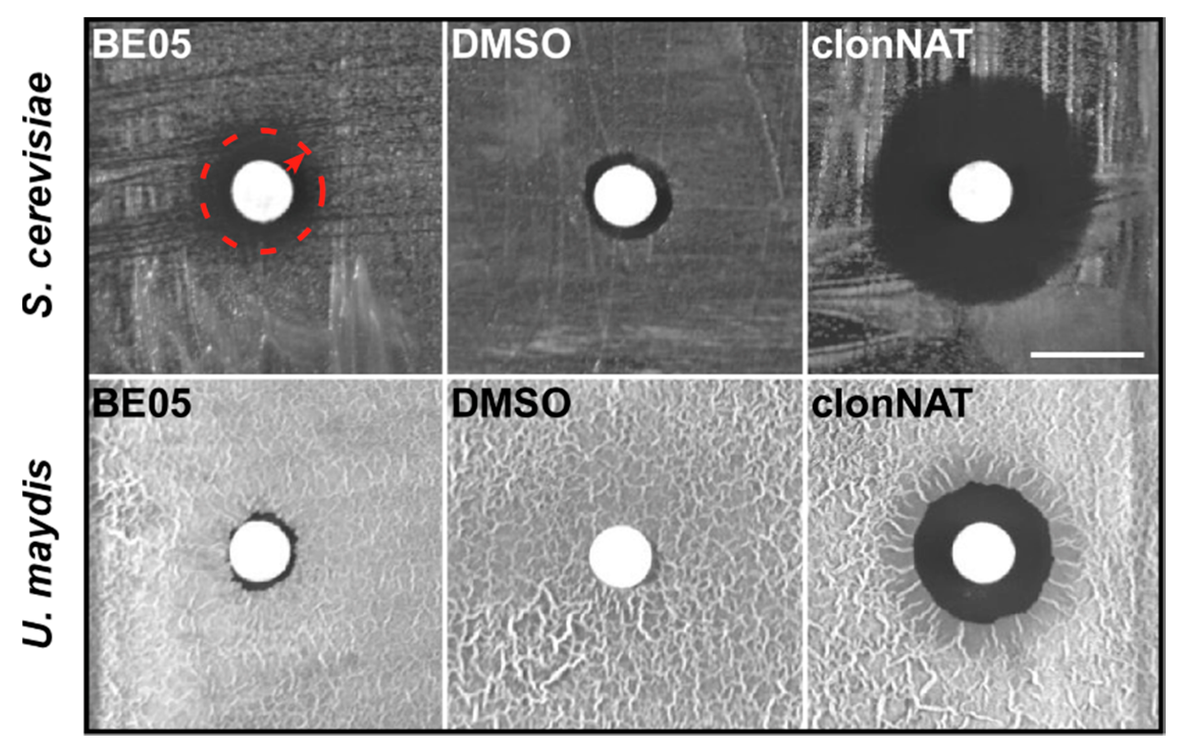
3.1. Bioactivity Tests with Ethyl Acetate Extracts from 41 Basidiomycete Mushroom Strains

In order to identify useful bioactive secondary metabolites, ethyl acetate extracts from mycelial cultures of wild strains of 41 basidiomycete mushroom strains representing 35 species were tested (Table 1). Their antifungal activity was analyzed in an antimicrobial assay against the yeast S. cerevisiae and the yeast form of U. maydis. Forty extracts showed no activity under these conditions (Figure S1). However, we detected bioactivity of the crude extract BE05 from F. hepatica against S. cerevisiae. The same extract was hardly active against U. maydis (Figure 1).
The antifungal crude extract BE05 was further tested against Gram-positive C. glutamicum and Gram-negative E. coli bacteria. With both bacteria, application of 500 µg of BE05 extract resulted in the formation of a two-zone halo, the inner zone of which completely lacked bacterial growth. In the adjacent outer halo zone that appeared optically brighter than the inner halo, bacterial lawn was still formed but at a lower density compared to the surrounding biofilm (Figure S2). Compared to S. cerevisiae (see Figure 1), where halo formation was already striking with 100 µg of BE05 extract, C. glutamicum as well as E. coli exhibited only a slight growth inhibition when exposed to BE05 extract.

3.2. Characteristics of Fistulina hepatica

The fungal strain from which the basidiomycete extract BE05 originates has been isolated from basidiomes of F. hepatica, exhibiting unambiguous ecological and morphological features of this species as described by Knudsen and Vesterholt [37], as well as by Krieglsteiner [38]: tongue-shaped basidiome; upper surface reddish-brown; hymenophore light yellowish, consisting of individual tubules; context very soft, reddish-brown, with lighter streaks, exuding a dull red liquid when cut; grown on Quercus sp. (Figure 2a).
After isolation by aseptical explanting of hyphal tufts from freshly harvested fruiting bodies and subcultivation, F. hepatica mycelium was cultivated for four weeks at 25 °C on potato dextrose agar (PDA). Mycelial characteristics as recorded for F. hepatica strains by Stalpers and Vlug [39] on two different media, reappeared with our strain of F. hepatica on PDA: the mycelium displayed a whitish wooly to cottony texture of the mycelium that peripherally collapsed into a velutinous yellowishly colored mat (Figure 2b). The less intense yellow pigmentation in contrast to the cultures by Stalpers and Vlug [39] is in agreement with the work of Griffith et al. [40], even though the latter work only included ascomycetes. In further agreement with its identity, microscopy of F. hepatica mycelium of our strain on PDA yielded typical features of F. hepatica cultures described by Stalpers and Vlug [39]. Clamp connections were noticed on a regular basis on dikaryotic hyphae (Figure 2c). Some clamps grew out to form new hyphae (Figure 2d). Also, the typical branching of hyphae was noticed, which normally takes place either at acute angles (Figure 2d) or the branching-off of very narrow hyphae growing from wide hyphae could be observed (Figure 2c).

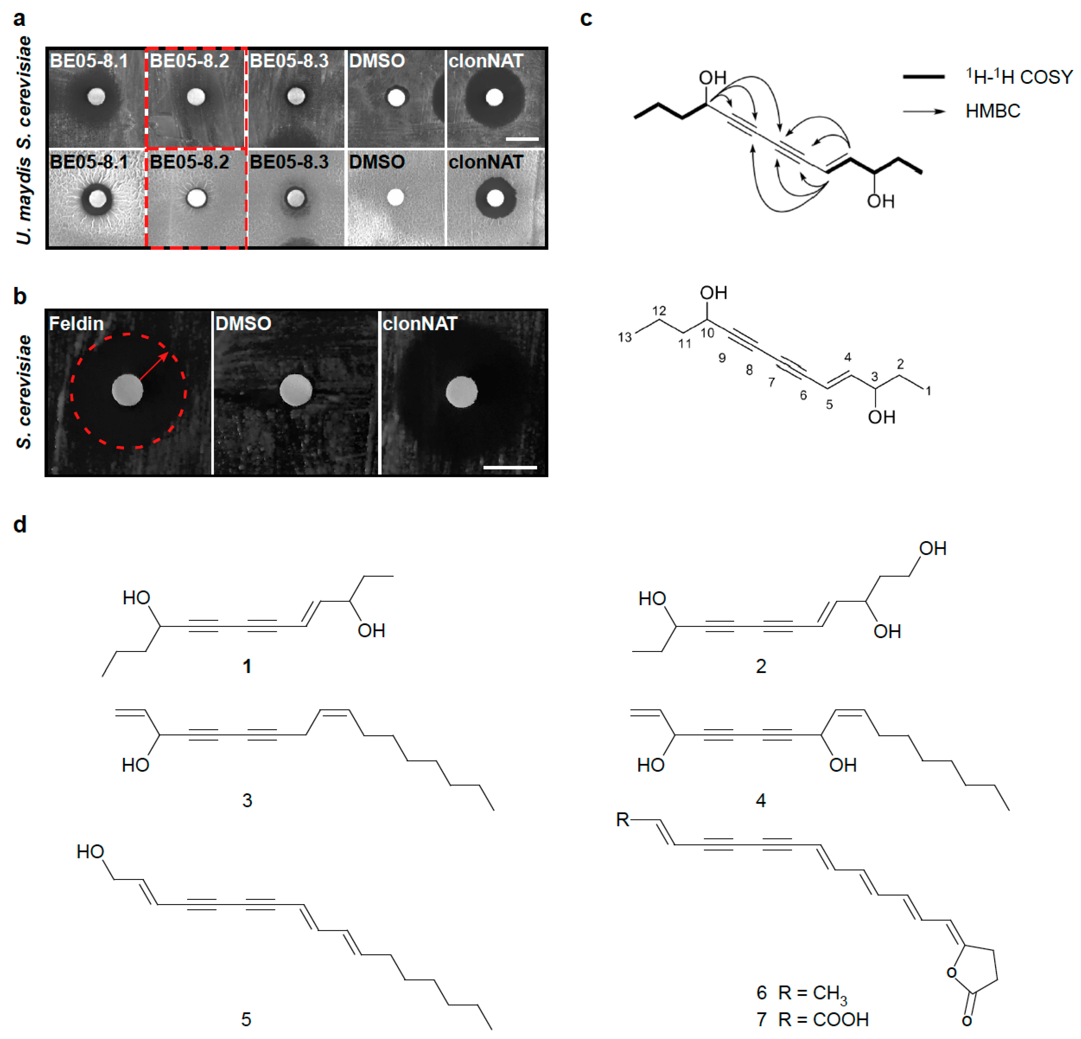
3.3. Identification of the Antifungal Polyyne Feldin from Basidiomycete Extract BE05

According to the bioactivity results (see Figure 1), we largely upscaled the cultivation of F. hepatica on PDA, followed by ethyl acetate extraction and LC-MS-based fractionation and subfractionation of a large amount of BE05 (see Materials and Methods). All fractions were retested for their antifungal activity against S. cerevisiae and U. maydis. Fractions showing growth inhibition of S. cerevisiae but hardly of U. maydis, such as fraction 8.2, were further processed (Figure 3a). Applying LC-MS-mediated clean-up, we were able to purify one bioactive fraction (fraction 8.2.6) containing only one promising mass signal, which was structure-elucidated via NMR. To obviate potential substance instability that could result in substance decomposition after NMR—a known phenomenon with some natural products (see discussion)—we immediately saved a subsample of freshly obtained fraction 8.2.6. This subsample was immediately applied to biotesting against S. cerevisiae (Figure 3b), leaving just enough of the fraction 8.2.6 (2.0 mg) to execute NMR analysis to resolve the structure.
The molecular formula of the corresponding compound (Figure 3c), designated as feldin, was established from the quasimolecular [M + Na]+ ion peak at m/z 229.1178 (calcd for C13H18O2Na, 229.1199, ∆ppm 9.2), with a degree of unsaturation of five. All proton signals were associated with their respective carbons via analysis of the HSQC spectrum. The resemblance of the NMR data of feldin (Table 2) and 4-dodecene-6,8-diyne-1,3,10-triol [41], another polyyne, suggested that they were structurally similar, except that C-1 hydroxymethyl in 4-dodecene-6,8-diyne-1,3,10-triol is replaced by a methyl group (δC = 8.6) in feldin. These structural differences were determined by 2D NMR data (Figure S3–S7). In addition, feldin has one more methylene at C-12 (δC = 18.1) than 4-dodecene-6,8-diyne-1,3,10-triol. Furthermore, feldin also exhibits some structural resemblance to falcarinol and falcarindiol from carrot roots [25,26,27]; oenanthetol from leaves, as well as seeds, of Trachyspermum ammi (L.) Spr. [42]; and also with xerulin as well as xerulinic acid [43] from the fir-wood decomposing agaric Xerula melanotricha (Figure 3d). In parallel, employing the subsample of feldin saved before NMR, we ascertained bioactivity of this compound against S. cerevisiae (see Figure 3b). In essence, we, thus, not only succeeded in solving the structure of the compound feldin from the basidiomycete F. hepatica. We recorded that growth of the ascomycete S. cerevisiae is inhibited when it is exposed to feldin.

4. Discussion

In the present study, we have bioactivity-screened PDA culture-derived ethyl acteate extracts of 41 strains, representing 35 basidiomycete mushroom species. One out of the 41 strains produced an extract that displayed antifungal bioactivity. From this strain of F. hepatica, we characterized a bioactive compound via a bioactivity-guided isolation, employing the model ascomycete S. cerevisiae and the model basidiomycete U. maydis as test microorganisms. Pure bioactive compound isolation and structure analysis revealed the bioactive polyyne (‘polyacetylene’) named feldin.
Comparing the present study to the work by Suay et al. [44], who extensively screened Southern European mushroom strain methanolic extracts for potential antimicrobial bioactivities without isolating and analyzing any pure bioactive compounds, we observed a lower “hit rate” of extracts showing antimicrobial bioactivity. Our hit rate was only 2.4%, while the hit rate of those authors was 45.1%. However, their general hit rate for antifungal extracts was comparable to ours, i.e., 6% to our 2.4%. The detected discrepancy of the general hit rates may at least partially relate to the cultivation technique employed by Suay et al. [44], who cultivated in more nutritious aerated liquid medium (8% glucose, 5% corn meal), whereas the here-employed fungi were cultivated on PDA medium (2% glucose, 5.75% potatoes [40]). Accordingly, the average amount of crude extract solids and of different secondary metabolites per strain produced by at least some of their fungi may have, thus, generally been higher compared to the one obtained per strain in the present study. On the one hand, this implies that a more extensive screening of the strains in the present study (more diverse cultivation regimes including different liquid media and solid-state media) would certainly lead to a higher diversity of secondary metabolites and a higher bioactivity hit rate due to the OSMAC (“one strain many compounds”) effect [31,45,46]. Still, media that lead to increased fungal growth and large amounts of crude extract solids, or high metabolic diversity for one fungal strain, may induce poor metabolic diversity for a different fungal strain, or not serve well for production of certain compounds of interest within the same strain [47].
Although known from fungi, polyynes from plants are even better studied, such as falcarinol and falcarindiol from oil-filled channels within the periderm/pericyclic parenchyma tissue running parallel to the length of the root of carrot plants. Falcarindiol exhibits antifungal bioactivity protecting the young roots, supposedly via alteration or damage of the plasma membrane or other membrane functions [25,26,27], which is also a proposed mode of action of certain bacterial polyynes [48]. In addition, certain falcarindiol isoforms display antibacterial bioactivities [49,50]. Being potent antibacterials, some such polyynes have been the source of a number of unique pharmacophores, like phomallenic acid C identified from a Phoma sp. (Ascomycota). This one exhibits a 20-fold higher potency than thiolactomycin or cerulenin against the Gram-positive human-pathogenic bacterium Staphylococcus aureus [51].
So far, some polyynes from F. hepatica [19,21] have been described, as well as from F. pallida, including a polyyne glycoside [52]. The two structurally similar polyynes, classified as triacetylene derivatives, the Cinnatriacetins A and B, were isolated from F. hepatica basidiomes and reported to exhibit bioactivity against Gram-positive bacteria, but none against Gram-negative bacteria and S. cerevisiae [21]. This, and the fact that the polyyne feldin we describe originates from F. hepatica mycelium showing activity against S. cerevisiae, supports the notion that F. hepatica may employ a versatile arsenal of polyynes to equip its chemical defense system against antagonists, including fungal ones. Well-known with cultivated mushroom species like the button mushroom (Agaricus bisporus), mushroom-parasitic bacteria and microfungi impose a severe threat to mushrooms during substrate colonizing or basidiome formation. Blotch-disease-causing pseudomonads [53] or the ascomycetous button mushroom pathogen Lecanicillium fungicola [54] are certainly among the economically most relevant representatives of such mushroom parasites. Thus, in nature, polyynes accumulating in F. hepatica mycelium and fruiting bodies may potentially help the fungus to keep such antagonists at bay. In contrast to induced mushroom defense systems, such as the antifungal strobilurin A of Oudemansiella mucida [55], the polyyne feldin from F. hepatica we describe here belongs to the autonomous defense molecules [4], at least under the tested conditions, as it is constitutively produced by its mycelium on PDA. This is well in line with the fact that it is relatively common to observe the accumulation of such compounds in fungi, in contrast to the situation in plants [27].
Fungal polyynes, such as 10-hydroxy-undeca-2,4,6,8-tetraynamide, may display broad bioactivity spectra. Besides activity against Gram-negative and Gram-positive bacteria, it also affects a broad spectrum of fungi (several ascomycetes, including S. cerevisiae, as well as the closely related human-pathogenic yeast C. albicans, and the basidiomycetous yeast Rhodotorula glutinis), the oomycete Phytophtora infestans, and Ehrlich ascites tumor cells [56]. Other fungal polyynes, like the allenic fungal polyyne of Schlingmann et al. [14], show a narrower bioactivity spectrum. While basidiomycetes like U. maydis and Rhodotorula rubra are hardly affected, it is strongly active against Gram-positive bacteria and C. albicans.
Fungal polyynes have been studied as potential drugs of high potency, like the tuberculosis cure mycomycin [57], the cholesterol biosynthesis inhibitors from the silver fir wood-decomposer X. melanotricha [43], or 10-hydroxy-undeca-2,4,6,8-tetraynamide from Mycena viridimarginata [56]. In this context, stability and cytotoxicity are major challenges to the applicability of the individual substance under study [43,56,57]. Polyynes tend to be unstable succumbing, for example, to oxidative, photolytic, and/or pH-dependent decomposition [27]. Trying to obviate that the bioactive compound in subfraction 8.2.6 (which was revealed as a polyyne after NMR) might get damaged after NMR solvent evaporation, as is known with unstable natural products like polyynes [48,58,59], bioactivity of feldin against S. cerevisiae was ascertained with a subsample of fresh feldin saved before NMR (see Figure 3b). Similarly, the antibacterial polyyne isolated from the basidiomycete Baeospora myosura proved to be very unstable, i.e., it polymerized when the solvent was removed [58]. Likewise, the 3,4,5,6-tetrahydro-6-hydroxy-derivative of the tuberculosis cure mycomycin (reported as potent but unstable by Celmer and Solomons [57]) was reported to be too unstable to isolate in a pure form [14]. Moreover, bacterial polyynes can also be unstable for the same or similar reasons. This makes very careful handling indispensable for bioactivity assessment, e.g., by avoiding light, oxygen, and solvent evaporation as much as possible [48,59]. Feldin apparently got damaged only after NMR solvent evaporation. Potentially, as in the case of the antibacterial polyyne from the basidiomycete B. myosura [58], polymerization has happened causing a loss of bioactivity. Accordingly, we cautiously assume that a customized minimalistic NMR solvent evaporation would be required to avoid feldin disintegration after NMR.
A possible solution for stabilizing bioactive polyynes like feldin may come from a (temporary) derivatization, e.g., creating a polyyne glycoside. Such derivatives may be found in fungi and plants and can exhibit antibacterial and anti-inflammatory activities [60,61,62]. Even in a very close relative of F. hepatica a natural polyyne glycoside is known [52]. Depicting a conjugation of a sugar moiety and a polyyne, such glycosides are judged as more stable than pure polyynes. Interestingly, no bioactivity was reported in the one described by Ahmed et al. [52]. This may relate to one suggested mode of action of polyynes. Bäuerle et al. [56] discussed the activity of 10-hydroxy-undeca-2,4,6,8-tetraynamide as correlated to its high chemical reactivity due to carbon–carbon triple bonds. Accordingly, such compounds, as stated by Walsh et al. [63], cause inactivation of enzymes by alkylation. Pan et al. [60] suggested that the carbohydrate part of polyyne glycosides might play a ‘protecting’ role in either stabilizing the polyyne moiety or in masking its biological activity until cleavage of the carbohydrate moiety by a glycosidase. Otherwise, it may increase solubility and facilitate the delivery of these molecules to certain cell types, e.g., via binding to certain sugar transporters.

5. Conclusions

In this exemplary study, we characterized the unstable bioactive mushroom polyyne feldin which at least qualitatively appears to be more toxic to the model ascomycete S. cerevisiae than to the basidiomycete model fungus U. maydis. This can be used as a starting point for future studies examining whether basidiomycetes like U. maydis may generally be more tolerant to prominent basidiomycete-derived eukaryotic toxic compounds, such as stobilurins, illudins, or even basidiome-derived antifungals like ageritin [2,5,6,64]. Such may be achieved by exposing U. maydis to a selection of these compounds. Tests may also include yet-to-discover differentially bioactive compounds from basidiomycetes by applying a more extensive screening. The latter should include additional strains also of the same species [28,65], a few more test microorganisms, and additional cultivation setups that are known to elicit the OSMAC effect [31,45,46], such as liquid media or bulk solid-state media. If this revealed a higher tolerance of U. maydis against certain such compounds, consequently U. maydis might be considered a candidate host for heterologous expression of mushroom-derived biologicals.
Moreover, if an understanding of polyyne biosynthesis were desirable, heterologous expression, including a derivatization approach, could be meaningful. Following identification of potential polyyne biosynthesis genes in the F. hepatica genome sequence of Floudas et al. [16], overexpression of respective candidate genes should be attempted, similar to the proof-of-principle approach recently accomplished by Lee et al. [66] on sesquiterpene production. Simultaneous expression of plant enzymes for a coupling approach might, eventually, allow the engineering of polyyne-derivatives, which are relatively stable and secreted into the culture medium. In a final step, the conjugation could be cleaved by a glycosidase to release the engineered compound’s polyyne moiety.

Supplementary Materials

The following are available online at https://www.mdpi.com/2218-273X/10/11/1502/s1, Figure S1: Bioactivity test of basidiomycete crude extracts against S. cerevisiae (a) and U. maydis (b), Figure S2: Bioactivity test with crude extract BE05 against C. glutamicum and E. coli, Figure S3: 1H NMR spectrum of feldin in MeOH-d4, Figure S4: 13C NMR spectrum of feldin in MeOH-d4, Figure S5: HSQC spectrum of feldin in MeOH-d4, Figure S6: HMBC spectrum of feldin in MeOH-d4, Figure S7: 1H-1H COSY spectrum of feldin in MeOH-d4.

Author Contributions

Conceptualization, F.H. and M.F.; methodology, J.L., Y.-M.S., P.G. and F.H.; validation, J.L. and F.H.; formal analysis, J.L., M.F. and F.H.; investigation, J.L., Y.-M.S., P.G. and F.H.; resources, M.G., M.F., H.B. and F.H.—fungal strains were acquired by M.G. and F.H.; writing—original draft preparation, J.L. and F.H., with contributions from co-authors Y.-M.S., P.G., M.G. and M.F.; writing—review and editing, J.L., M.F. and F.H., with minor contributions from Y.-M.S. and H.B.; visualization, J.L., Y.-M.S. and F.H.; supervision, M.F. and F.H.; project administration, M.F. and F.H.; funding acquisition, M.F, H.B. and F.H. All authors have read and agreed to the published version of the manuscript.

Funding

The work was funded by the Deutsche Forschungsgemeinschaft under Germany’s Excellence Strategy EXC-2048/1—Project ID 39068111 to MF). The scientific activities of the Bioeconomy Science Center were financially supported by the Ministry of Culture and Science within the framework of the NRW Strategieprojekt BioSC (No. 313/323-400-002 13 to MF). FH acknowledges financial support from the Deutsche Forschungsgemeinschaft under grant HE 7849/3-1. Work in the Bode lab was funded by the LOEWE Center for Translational Biodiversity Genomics (TBG).

Acknowledgments

We acknowledge support by the DFG Open Access Publication Funds of the Ruhr-University Bochum. We express our sincere gratitude to Meike Piepenbring (Goethe University Frankfurt, Germany) for giving us access to the fungal strain collection at the Department of Mycology of the Goethe University, where the culture of F. hepatica P052 is maintained. We also thank Erika Kothe (Institute of Microbiology, Friedrich Schiller University Jena) for her support of M.G.’s sampling activities of basidiomycete mushrooms, not least at the former uranium mining site in Eastern Thuringia (near Ronneburg), and the permission of including the thereof isolated strains in this study. Furthermore, we cordially thank Heinrich Dörfelt (Institute of Microbiology, Friedrich Schiller University Jena) for providing us with a strain of Pleurotus dryinus, and we express our sincere gratitude to Lilli Bismar (Institute for Microbiology, Heinrich Heine University Düsseldorf) and Frank Surup (HZI Braunschweig) for critical reading of the manuscript. We also kindly acknowledge English language editing by Elisabeth Stratmann. Last but not least, we thank the three anonymous reviewers whose comments helped to improve the manuscript.

Conflicts of Interest

The authors declare no conflict of interest.

References

  1. He, M.; Zhao, R.; Hyde, K.D.; Begerow, D.; Kemler, M.; Yurkov, A.; McKenzie, E.H.C.; Raspé, O.; Kakishima, M.; Sánchez-Ramírez, S.; et al. Notes, outline and divergence times of Basidiomycota. Fungal Divers. 2019, 99, 105–367. [Google Scholar] [CrossRef] [Green Version]
  2. Sandargo, B.; Chepkirui, C.; Cheng, T.; Chaverra-Munoz, L.; Thongbai, B.; Stadler, M.; Hüttel, S. Biological and chemical diversity go hand in hand: Basidiomycota as source of new pharmaceuticals and agrochemicals. Biotechnol. Adv. 2019, 37, 107344. [Google Scholar] [CrossRef] [PubMed]
  3. Zhao, R.-L.; Li, G.-J.; Sánchez-Ramírez, S.; Stata, M.; Yang, Z.L.; Wu, G.; Dai, Y.C.; He, S.H.; Cui, B.K.; Zhou, J.L.; et al. A six-gene phylogenetic overview of Basidiomycota and allied phyla with estimated divergence times of higher taxa and a phyloproteomics perspective. Fungal Divers. 2017, 84, 43–74. [Google Scholar] [CrossRef]
  4. Künzler, M. How fungi defend themselves against microbial competitors and animal predators. PLoS Pathog. 2018, 14, e1007184. [Google Scholar] [CrossRef] [PubMed]
  5. Hyde, K.D.; Xu, J.; Rapior, S.; Jeewon, R.; Lumyong, S.; Niego, A.G.T.; Abeywickrama, P.D.; Aluthmuhandiram, J.V.S.; Brahamanage, R.S.; Brooks, S.; et al. The amazing potential of fungi: 50 ways we can exploit fungi industrially. Fungal Divers. 2019, 97, 1–136. [Google Scholar] [CrossRef] [Green Version]
  6. Tayyrov, A.; Azevedo, S.; Herzog, R.; Vogt, E.; Arzt, S.; Lüthy, P.; Müller, P.; Rühl, M.; Hennicke, F.; Künzler, M. Heterologous production and functional characterization of ageritin, a novel type of ribotoxin highly expressed during fruiting of the edible mushroom Agrocybe aegerita. Appl. Environ. Microbiol. 2019, 85, e01549-19. [Google Scholar] [CrossRef]
  7. Alberti, F.; Khairudin, K.; Venegas, E.; Davies, J.A.; Hayes, P.M.; Willis, C.L.; Bailey, A.M.; Foster, G.D. Heterologous expression reveals the biosynthesis of the antibiotic pleuromutilin and generates bioactive semi-synthetic derivatives. Nat. Commun. 2017, 8, 1831. [Google Scholar] [CrossRef] [PubMed] [Green Version]
  8. Nofiani, R.; de Mattos-Shipley, K.; Lebe, K.E.; Han, L.-C.; Iqbal, Z.; Bailey, A.M.; Willis, C.L.; Simpson, T.J.; Cox, R.J. Strobilurin biosynthesis in basidiomycete fungi. Nat. Commnun. 2018, 9, 3940. [Google Scholar] [CrossRef] [PubMed] [Green Version]
  9. Engels, B.; Heinig, U.; Grothe, T.; Stadler, M.; Jennewein, S. Cloning and characterization of an Armillaria gallica cDNA encoding protoilludene synthase, which catalyzes the first committed step in the synthesis of antimicrobial melleolides. J. Biol. 2011, 286, 6871–6878. [Google Scholar] [CrossRef] [Green Version]
  10. Croteau, R.; Ketchum, R.E.; Long, R.M.; Kaspera, R.; Wildung, M.R. Taxol biosynthesis and molecular genetics. Phytochem. Rev. 2006, 5, 75–97. [Google Scholar] [CrossRef] [Green Version]
  11. Dejong, J.M.; Liu, Y.; Bollon, A.P.; Long, R.M.; Jennewein, S.; Williams, D.; Croteau, R.B. Genetic engineering of taxol biosynthetic genes in Saccharomyces cerevisiae. Biotechnol. Bioeng. 2006, 93, 212–224. [Google Scholar] [CrossRef] [PubMed]
  12. Paddon, C.J.; Westfall, P.J.; Pitera, D.J.; Benjamin, K.; Fisher, K.; McPhee, D.; Leavell, M.D.; Tai, A.; Main, A.; Eng, D.; et al. High-level semi-synthetic production of the potent antimalarial artemisinin. Nature 2013, 496, 528–532. [Google Scholar] [CrossRef] [PubMed] [Green Version]
  13. Schlingmann, G.; Milne, L.; Pearce, C.J.; Borders, D.B.; Greenstein, M.; Maiese, W.M.; Carter, G.T. Isolation, characterization and structure of a new allenic polyine antibiotic produced by fungus LL-07F275. J. Antibiot. Res. 1995, 48, 375–379. [Google Scholar] [CrossRef] [PubMed] [Green Version]
  14. Costa-de-Oliveira, S.; Rodrigues, A.G. Candida albicans antifungal resistance and tolerance in bloodstream infections: The triad yeast-host-antifungal. Microorganisms 2020, 8, 154. [Google Scholar] [CrossRef] [PubMed] [Green Version]
  15. Matheny, P.B.; Curtis, J.M.; Hofstetter, V.; Aime, M.C.; Moncalvo, J.M.; Ge, Z.W.; Slot, J.C.; Ammirati, J.F.; Baroni, T.J.; Bougher, N.L.; et al. Major clades of Agaricales: A multilocus phylogenetic overview. Mycologia 2006, 98, 982–995. [Google Scholar] [CrossRef]
  16. Floudas, D.; Held, B.W.; Riley, R.; Nagy, L.G.; Koehler, G.; Ransdell, A.S.; Younus, H.; Chow, J.; Chiniquy, J.; Lipzen, A.; et al. Evolution of novel wood decay mechanisms in Agaricales revealed by the genome sequences of Fistulina hepatica and Cylindrobasidium torrendii. Fungal Genet. Biol. 2015, 76, 78–92. [Google Scholar] [CrossRef] [Green Version]
  17. Kaffenberger, J.T.; Schilling, J.S. Comparing lignocellulose physiochemistry after decomposition by brown rot fungi with distinct evolutionary origins. Environ. Microbiol. 2015, 17, 4885–4897. [Google Scholar] [CrossRef]
  18. Regué, A.; Bassié, L.; de-Miguel, S.; Colinas, C. Environmental and stand conditions related to Fistulina hepatica heart rot attack on Castanea sativa. For. Pathol. 2019, 49, e12517. [Google Scholar] [CrossRef]
  19. Jones, E.R.H.; Lowe, G.; Shannon, P.V.R. Natural acetylenes. Part XX. Tetra-acetylenic and other metabolites from Fistulina hepatica (Huds) Fr. J. Chem. Soc. C 1966, 139–144. [Google Scholar] [CrossRef]
  20. Ivanova, V.; Kolarova, M.; Aleksieva, K.; Schlegel, R.; Schumann, P.; Graefe, U. Octadeca-8,11-dienoic acid methylester, a new fatty acid metabolite from Fistulina hepatica. J. Mod. Med. 2013, 1, 43–48. [Google Scholar] [CrossRef] [Green Version]
  21. Tsuge, N.; Mori, T.; Hamano, T.; Tanaka, H.; Shin-ya, K.; Seto, H. Cinnatriacetins A and B, new antibacterial triacetylene derivatives from the fruiting bodies of Fistulina hepatica. J. Antibiot. Res. 1999, 52, 578–581. [Google Scholar] [CrossRef] [PubMed] [Green Version]
  22. Vaz, J.A.; Barros, L.; Martins, A.; Morais, J.S.; Vasconcelos, M.H.; Ferreira, I.C.F.R. Phenolic profile of seventeen Portuguese wild mushrooms. LWT Food Sci. Technol. 2011, 44, 343–346. [Google Scholar] [CrossRef] [Green Version]
  23. Wu, S.; Krings, U.; Zorn, H.; Berger, R.G. Volatile compounds from the fruiting bodies of beefsteak fungus Fistulina hepatica (Schaeffer: Fr.) Fr. Food Chem. 2005, 92, 221–226. [Google Scholar] [CrossRef]
  24. Wu, S.; Zorn, H.; Krings, U.; Berger, R.G. Volatiles from submerged and surface-cultured beefsteak fungus, Fistulina hepatica. Flavour Frag. J. 2007, 22, 53–60. [Google Scholar] [CrossRef]
  25. Garrod, B.; Lewis, B.G. Location of the antifungal compound falcarindiol in carrot root tissue. Trans. Brit. 1979, 72, 515–517. [Google Scholar] [CrossRef]
  26. Garrod, B.; Lea, E.J.A.; Lewis, B.G. Studies on the mechanism of action of the antifungal compound falcarindiol. New Phytol. 1979, 83, 463–471. [Google Scholar] [CrossRef] [Green Version]
  27. Minto, R.E.; Blacklock, B.J. Biosynthesis and function of polyacetylenes and allied natural products. Prog. Lipid Res. 2008, 47, 233–306. [Google Scholar] [CrossRef] [Green Version]
  28. Stevens, R. Mycology Guidebook; University of Washington Press: Seattle, WA, USA; London, UK, 1974; p. 719. [Google Scholar]
  29. Baumann, S.; Zander, S.; Weidtkamp-Peters, S.; Feldbrügge, M. Live cell imaging of septin dynamics in Ustilago maydis. Methods Cell Biol. 2016, 136, 143–159. [Google Scholar] [CrossRef]
  30. Jankowski, S.; Pohlmann, T.; Baumann, S.; Muntjes, K.; Devan, S.K.; Zander, S.; Feldbrügge, M. The multi PAM2 protein Upa2 functions as novel core component of endosomal mRNA transport. EMBO Rep. 2019, 20, e47381. [Google Scholar] [CrossRef]
  31. Harwoko, H.; Daletos, G.; Stuhldreier, F.; Lee, J.; Wesselborg, S.; Feldbrügge, M.; Müller, W.E.G.; Kalscheuer, R.; Ancheeva, E.; Proksch, P. Dithiodiketopiperazine derivatives from endophytic fungi Trichoderma harzianum and Epicoccum nigrum. Nat. Prod. Res. 2019, 18, 1–9. [Google Scholar] [CrossRef]
  32. Brachmann, A.; Weinzierl, G.; Kämper, J.; Kahmann, R. Identification of genes in the bW/bE regulatory cascade in Ustilago maydis. Mol. Microbiol. 2001, 42, 1047–1063. [Google Scholar] [CrossRef] [PubMed]
  33. Pereira, G.; Tanaka, T.U.; Nasmyth, K.; Schiebel, E. Modes of spindle pole body inheritance and segregation of the Bfa1p-Bub2p checkpoint protein complex. EMBO J. 2001, 20, 6359–6370. [Google Scholar] [CrossRef] [Green Version]
  34. Holliday, R. Molecular aspects of genetic exchange and gene conversion. Genetics 1974, 78, 273–287. [Google Scholar] [PubMed]
  35. Banuett, F.; Herskowitz, I. Different a alleles of Ustilago maydis are necessary for maintenance of filamentous growth but not for meiosis. Proc. Natl. Acad. Sci. USA 1989, 86, 5878–5882. [Google Scholar] [CrossRef] [PubMed] [Green Version]
  36. Abe, S.; Takayarna, K.; Kinoshita, S. Taxonomical studies on glutamic acid producing bacteria. J. Gen. Appl. Microbiol. 1967, 13, 279–301. [Google Scholar] [CrossRef]
  37. Knudsen, H.; Vesterholt, J. Funga Nordica; Nordsvamp: Copenhagen, Denmark, 2008; 966p. [Google Scholar]
  38. Krieglsteiner, G.J. Die Grosspilze Baden-Württembergs. Bd. 1: Allgemeiner Teil, Ständerpilze: Gallert-Rinden-, Stachel- und Porenpilze; Ulmer: Stuttgart, Germany, 2000; p. 629. (In German) [Google Scholar]
  39. Stalpers, J.A.; Vlug, I. Confistulina, the anamorph of Fistulina hepatica. Can. J. Bot. 1983, 61, 1660–1666. [Google Scholar] [CrossRef]
  40. Griffith, G.W.; Easton, G.L.; Detheridge, A.; Roderick, K.; Edwards, A.; Worgan, H.J.; Nicholson, J.; Perkins, W.T. Copper deficiency in potato dextrose agar causes reduced pigmentation in cultures of various fungi. FEMS Microbiol. Lett. 2007, 276, 165–171. [Google Scholar] [CrossRef]
  41. Bohlmann, F.; Knoll, K.-H. New acetylenic compounds from Emilia species. Phytochemistry 1978, 17, 557–558. [Google Scholar] [CrossRef]
  42. Christensen, L.P.; Brandt, K. Bioactive polyacetylenes in food plants of the Apiaceae family: Occurrence, bioactivity and analysis. J. Pharm. Biomed. Anal. 2006, 41, 683–693. [Google Scholar] [CrossRef]
  43. Kuhnt, D.; Anke, T.; Besl, H.; Bross, M.; Herrmann, R.; Mocek, U.; Steffan, B.; Steglich, W. Antibiotics from basidiomycetes. XXXVII. New inhibitors of cholesterol biosynthesis from cultures of Xerula melanotricha Dörfelt. J. Antibiot. Res. 1990, 43, 1413–1420. [Google Scholar] [CrossRef] [Green Version]
  44. Suay, I.; Arenal, F.; Asensio, F.J.; Basilio, A.; Cabello, M.A.; Díez, M.T.; García, J.B.; del Val, A.G.; Gorrochategui, J.; Hernández, P.; et al. Screening of basidiomycetes for antimicrobial activities. Antonie Van Leeuwenhoek 2000, 78, 129–139. [Google Scholar] [CrossRef] [PubMed]
  45. Kjer, J.; Debbab, A.; Aly, A.H.; Proksch, P. Methods for isolation of marine-derived endophytic fungi and their bioactive secondary products. Nat. Protoc. 2010, 3, 479–490. [Google Scholar] [CrossRef]
  46. Pan, F.; El-Kashef, D.H.; Kalscheuer, R.; Müller, W.E.G.; Lee, J.; Feldbrügge, M.; Mándi, A.; Kurtán, T.; Liu, Z.; Wu, W.; et al. New hybrid polyketides from the endophytic fungus Cladosporium sphaerospermum WBS017. Eur. J. Med. Chem. 2020, 191, 112159. [Google Scholar] [CrossRef] [PubMed]
  47. Vandermolen, K.M.; Raja, H.A.; El-Elimat, T.; Oberlies, N.H. Evaluation of culture media for the production of secondary metabolites in a natural products screening program. AMB Express 2013, 3, 71. [Google Scholar] [CrossRef] [PubMed] [Green Version]
  48. Fritsche, K.; Berg, M.V.D.; Boer, W.D.; Beek, T.A.V.; Raaijmakers, J.M.; Veen, J.A.V.; Leveau, J.H.J. Biosynthetic genes and activity spectrum of antifungal polyynes from Collimonas fungivorans Ter331. Environ. Microbiol. 2014, 16, 1334–1345. [Google Scholar] [CrossRef] [Green Version]
  49. Kobaisy, M.; Abramowski, Z.; Lermer, L.; Saxena, G.; Hancock, R.E.W.; Towers, G.H.N.; Doxsee, D.; Stokes, R.W. Antimycobacterial polyynes of Devil’s Club (Oplopanax horridus), a North American native medicinal plant. J. Nat. Prod. 1997, 60, 1210–1213. [Google Scholar] [CrossRef]
  50. Lechner, D.; Stavri, M.; Oluwatuyi, M.; Pereda-Miranda, R.; Gibbons, S. The anti-staphylococcal activity of Angelica dahurica (Bai Zhi). Phytochemistry 2004, 65, 331–335. [Google Scholar] [CrossRef]
  51. Ondeyka, J.G.; Zink, D.L.; Young, K.; Painter, R.; Kodali, S.; Galgoci, A.; Collado, J.; Tormo, J.R.; Basilio, A.; Vicente, F.; et al. Discovery of bacterial fatty acid synthase inhibitors from a Phoma species as antimicrobial agents using a new antisense-based strategy. J. Nat. Prod. 2006, 69, 377–380. [Google Scholar] [CrossRef]
  52. Ahmed, M.; Barley, G.C.; Hearn, M.T.W.; Jones, E.R.H.; Thaller, V.; Yates, J.A. Natural acetylenes. Part XLIII. Polyacetylenes from cultures of the fungus Fistulina pallida (berk. and rev.). J. Chem. Soc. Perkin Trans. 1974, 1974, 1981–1987. [Google Scholar] [CrossRef]
  53. Frey-Klett, P.; Burlinson, P.; Deveau, A.; Barret, M.; Tarkka, M.; Sarniguet, A. Bacterial-fungal interactions: Hyphens between agricultural, clinical, environmental, and food microbiologists. Microbiol. Mol. Biol. Rev. 2011, 75, 583–609. [Google Scholar] [CrossRef] [Green Version]
  54. Berendsen, R.L.; Baars, J.J.; Kalkhove, S.I.; Lugones, L.G.; Wösten, H.A.; Bakker, P.A. Lecanicillium fungicola: Causal agent of dry bubble disease in white-button mushroom. Mol. Plant Pathol. 2010, 11, 585–595. [Google Scholar] [CrossRef] [PubMed]
  55. Kettering, M.; Sterner, O.; Anke, T. Antibiotics in the chemical communication of fungi. Z. Naturforsch. 2004, 59, 816–823. [Google Scholar] [CrossRef] [PubMed] [Green Version]
  56. Bäuerle, J.; Anke, T.; Jente, R.; Bosold, F. Antibiotics from Basidiomycetes. XVI. Antimicrobial and cytotoxic polyines from Mycena viridimarginata Karst. Arch. Microbiol. 1982, 132, 194–196. [Google Scholar] [CrossRef]
  57. Celmer, W.D.; Solomons, I.A. The structure of the antibiotic mycomycin. J. Am. Chem. Soc. 1952, 74, 1870–1871. [Google Scholar] [CrossRef]
  58. Parish, C.A.; Huber, J.; Baxter, J.; González, A.; Collado, J.; Platas, G.; Diez, M.T.; Vicente, F.; Dorso, K.; Abruzzo, G.; et al. A new ene-triyne antibiotic from the fungus Baeospora myosura. J. Nat. Prod. 2004, 67, 1900–1902. [Google Scholar] [CrossRef] [PubMed]
  59. Kai, K.; Sogame, M.; Sakurai, F.; Nasu, N.; Fujita, M. Collimonins A-D, unstable polyynes with antifungal or pigmentation activities from the fungus-feeding bacterium Collimonas fungivorans Ter331. Org. Lett. 2018, 20, 3536–3540. [Google Scholar] [CrossRef]
  60. Pan, Y.; Lowary, T.D.; Tykwinski, R.R. Naturally occurring and synthetic polyyne glycosides. Can. J. Chem. 2009, 87, 1565–1582. [Google Scholar] [CrossRef]
  61. He, J.; Shen, Y.; Jiang, J.S.; Yang, Y.-N.; Feng, Z.-M.; Zhang, P.-C.; Yuan, S.-P.; Hou, Q. New polyacetylene glucosides from the florets of Carthamus tinctorius and their weak anti-inflammatory activities. Carbohydr. Res. 2011, 346, 1903–1908. [Google Scholar] [CrossRef]
  62. Konovalov, D.A. Polyacetylene compounds of plants of the Asteraceae family (review). Pharm. Chem. J. 2014, 48, 615–633. [Google Scholar] [CrossRef]
  63. Walsh, C.T.; Schonbrunn, A.; Lockridge, O.; Massey, V.; Abeles, R.H. Inactivation of a flavoprotein, lactate oxidase, by an acetylenic substance. J. Biol. 1972, 247, 6004–6006. [Google Scholar]
  64. Citores, L.; Ragucci, S.; Ferreras, J.M.; Di Maro, A.; Iglesias, R. Ageritin, a ribotoxin from poplar mushroom (Agrocybe aegerita) with defensive and antiproliferative activities. ACS Chem. Biol. 2019, 14, 1319–1327. [Google Scholar] [CrossRef] [PubMed]
  65. Dresch, P.; D’Aguanno, M.N.; Rosam, K.; Grienke, U.; Rollinger, J.M.; Peintner, U. Fungal strain matters: Colony growth and bioactivity of the European medicinal polypores Fomes fomentarius, Fomitopsis pinicola and Piptoporus betulinus. AMB Express 2015, 5, 4. [Google Scholar] [CrossRef] [PubMed] [Green Version]
  66. Lee, J.; Hilgers, F.; Loeschke, A.; Jaeger, K.-E.; Feldbrügge, M. Ustilago maydis serves as a novel production host for the synthesis of plant and fungal sesquiterpenoids. Front. Microbiol. 2020, 11, 1655. [Google Scholar] [CrossRef]
Figure 1. Bioactivity test of the basidiomycete ethyl acetate crude extract BE05 against S. cerevisiae and U. maydis. In total, 100 µg of crude extract was dissolved in DMSO to impregnate a filter paper disk centrally placed on each agar plate. DMSO was used instead of the crude extract as negative control and 200 µg of clonNAT dissolved in ddH2O was used as positive control. The scale bar represents 1 cm. Three independent biological experiments (n = 3) were carried out. The biotesting with extract BE05 (left part of the panel) yielded a zone of growth inhibition (halo, dashed red circle with arrow) with S. cerevisiae, while resulting in minimal growth inhibition with U. maydis.
Biomolecules 10 01502 g001
Figure 2. Basidiome and mycelial morphology of the F. hepatica wild strain employed in the present study. The shown mycelial culture served to generate basidiomycete ethyl acetate crude extract BE05. (a) Pileal surface (left specimen) as well as stipe and pore surface (right specimen) of two fruiting bodies growing from a stump of an oak tree in a mixed Fagus sylvatica forest. The scale bar represents 5 cm. (b) Mycelial colony morphology of F. hepatica growing on potato dextrose agar (PDA) at 25 °C. The scale bar represents 1 cm. (c,d) Typical hyphal morphology of F. hepatica, indicating a clamp connection (red-framed yellow arrowhead) at a septum between two dikaryotic hyphal segments of aerial mycelium at the colony margin. Such clamp connections may grow out to form new hyphae (d, white-framed black arrowhead). Side-branch formation on F. hepatica hyphae normally occurs via outgrowth of side branches at acute angles (c-d, black-framed white arrowheads). Such a side branch is either more or less equally sized to the hypha from which it grows out (d, black-framed white arrowhead) or it branches off as a very narrow hypha (c, black-framed white arrowhead) from a wide hypha. The scale bar represents 10 µm.
Biomolecules 10 01502 g002
Figure 3. Bioactivity-guided fractionation from the basidiomycete ethyl acetate crude extract BE05; bioactivity of the freshly isolated bioactive compound against S. cerevisiae, and structure analysis of this compound (feldin). (a) Bioactivity test of fraction 8.2 (highlighted by a dashed frame) and two adjacent fractions from basidiomycete ethyl acetate crude extract BE05 against S. cerevisiae and U. maydis. The biotesting with fraction 8.2 yielded a zone of growth inhibition with S. cerevisiae (halo), while resulting in minimal growth inhibition with U. maydis. In total, 100 µg of this fraction were dissolved in DMSO to impregnate the filter paper disk. DMSO was used instead of the extract as negative control and 200 µg of clonNAT dissolved in ddH2O was used as positive control, respectively. The scale bar represents 1 cm. Three independent biological experiments (n = 3) were carried out. (b) Bioactivity test against S. cerevisiae of a subsample of the F. hepatica polyyne feldin, which was directly saved from fraction 8.2.6 before running NMR. The biotesting yielded a zone of growth inhibition (halo, dashed red circle with arrow) with S. cerevisiae. Here, 100 µg of feldin was dissolved in DMSO to impregnate the filter paper disk. DMSO was used instead of the extract as negative control and 200 µg of clonNAT dissolved in ddH2O was used as positive control, respectively. The scale bar represents 1 cm. Three independent biological experiments (n = 3) were carried out. (c) Structure of the F. hepatica polyyne feldin. (d) Similarity of chemical structure of feldin with known polyynes (1 in bold face, feldin; 2, 4-dodecene-6,8-diyne-1,3,10-triol; 3, falcarinol; 4, falcarindiol; 5, oenanthetol; 6, xerulin; 7, xerulinic acid).
Biomolecules 10 01502 g003
Table 1. Forty-one mushroom strain extracts (BE01-41) tested for potential antifungal bioactivity.
ExtractSpeciesStrainGeographic 1 and Ecological Data
BE01Armillaria ostoyaeP089Göttingen (Gö), Kerstlingeröder Feld, under Pinus nigra, tc 2, leg., det. M.G. (2009-10-07)
BE02Armillaria cf. ostoyaeP1451.2 km S of Grobsdorf (Gd), Nordhalde (Nh), lc 3, Tilia sp. stump, leg., det. M.G. (2010-09-27)
BE03Coprinopsis atramentariaP125Jena (J), Beutenberg, half buried softwood, lc, leg., det. M.G. (2013-11-18)
BE04Coprinopsis picaceaP1061 km W of Gö-Herberhausen (GöHe), Fagus sylvatica forest (Fsf), tc, leg., det. M.G. (2013-11-25)
BE05Fistulina hepaticaP052J, 1 km SW of “Steinkreuz Ziegenhain”, Fsf, lc, Quercus sp. stump, leg., det. F.H. (2013-08-18)
BE06Flammulina velutipesP111J, Cospedaer Grund, lc, Fraxinus excelsior stump, leg., det. M.G. (2009-12-01)
BE07Fomes fomentariusP116Jonsdorf, Weißer Stein, lc, Betula pendula wood, leg., det. M.G. (2012-03-05)
BE08Pleurotus dryinusP090Dederstedt, lc, living stem of F. excelsior, leg., det. Dr. habil. H. Dörfelt (2009-11-18)
BE09Pleurotus cf. pulmonariusP046Oberursel-Hohemark (OHm), mixed Fsf, lc, F. sylvatica wood, leg., det. F.H. (2013-09-13)
BE10Pleurotus ostreatusP118Hannoversche Klippen 1.5 km NW of Bad Karlshafen, lc, F. sylvatica wood, leg., det. M.G. (2012-07-30)
BE11Agaricus arvensisP151Kauern, Nh, 1 km S of Gd, mixed forest, tc 2, leg., det. M. G. (2009-09-23)
BE12Agaricus augustusP0962.5 km NE of Maria Laach (ML), mixed Fsf, tc, leg., det. M. G. (2010-08-12)
BE13Agaricus augustusP148Gö., Brüder-Grimm-Allee, tc, under Tilia sp., leg., det. M. G. (2013-10-25)
BE14Auricularia auricula-judaeP093J, Mühltal, lc 3, Sambucus nigra wood, leg., det. M. G. (2009-12-07)
BE15Bovista nigrescensP123Rothesütte, about 500 m SE of Rothesütte, tc, pasture, leg., det. M. G. (2013-09-23)
BE16Clitocybe geotropaP149Gö, close to GöHe, Fsf, tc, leg., det. M. G. (2013-12-02)
BE17Clitocybe odoraP053Morgenröthe, mixed Picea abies forest, tc, needle litter, leg., det. F. H. (2013-09-07)
BE18Galerina marginataP057OHm, mixed Fsf, lc, P. abies wood, leg., det. F. H. (2013-09-13)
BE19Ganoderma lucidumP095Königsfeld/SW, 500 m N of Königsfeld, lc, Quercus sp. stump, leg., det. M. G. (2010-07-17)
BE20Gloeophyllum odoratumP124J, Mühltal, lc, P. abies stump, leg., det. M. G. (2013-11-04)
BE21Hypholoma fasciculareP099750 m SE of Gd, mixed forest, lc, Quercus sp. stump, leg., det. M. G. (2010-09-07)
BE22Hypholoma sublateritiumP169200 m S of Closewitz, Quercus spp. forest, lc, Quercus sp. stump, leg., det. M. G. (2014-02-17)
BE23Inonotus obliquusP150Oybin, Töpfer/Brandhöhe, lc, B. pendula wood, leg., det. M. G. (2014-01-06)
BE24Kuehneromyces mutabilisP050OHm, mixed Fsf, lc, on F. sylvatica wood, leg., det. F. H. (2013-09-13)
BE25Langermannia giganteaP051Bad Berka, Trebestrasse, tc, garden lawn, leg., det. F. H. (2013-08-17)
BE26Lycoperdon excipuliformeP122800 m S of Gd, tc, under scattered B. pendula trees, leg., det. M. G. (2013-09-12)
BE27Lycoperdon molleP154ML, Laacher-See-Haus, 1.3 km SE of ML, on a path in a Fsf, tc, leg., det. M. G. (2010-08-12)
BE28Macrolepiota proceraP097ML, 2.5 km NE of ML, mixed Fsf, tc, leg., det. M. G. (2010-08-16)
BE29Macrolepiota proceraP130Kauern, Nh, 1.2 km S of Gd, mixed forest, tc, leg., det. M. G. (2009-09-25)
BE30Marasmius oreadesP1441 km S of Gd, tc, grassy forest margin, leg., det. M. G. (2010-09-08)
BE31Marasmius scorodoniusP055OHm, mixed Fsf, tc, F. sylvatica leaf litter, leg., det. F. H. (2013-09-13)
BE32Megacollybia platyphyllaP0262.5 km N of Rambach, 2 km NW of Naurod, mixed Fsf, tc, leg., det. F. H. (2013-05-27)
BE33Pleurotus ostreatusP114ML, Laacher-See-Haus, 1.3 km SE of ML, lc, on F. sylvatica wood, leg., det. M. G. (2011-08-02)
BE34Pleurotus ostreatusP119Rosdorf, Kiessee, lc, on wood of Salix sp., leg., det. M. G. (2013-08-23)
BE35Pleurotus ostreatusP128Hainewalde, near graveyard, lc, Populus x canadensis stump, leg., det. M. G. (2014-01-06)
BE36Polyporus brumalisP1171.5 km SW of GöHe, mixed Fsf, lc, Tilia sp. wood, leg., det. M. G. (2012-03-05)
BE37Polyporus tuberasterP036J, 300 m S of “Steinkreuz Ziegenhain”, Fsf, lc, on decayed wood, leg., det. F. H. (2013-08-18)
BE38Sarcomyxa serotinaP126800 m W of GöHe, mixed Fsf, lc, F. sylvatica stump, leg., det. M. G. (2013-11-25)
BE39Stropharia aeruginosaP102Jenaprießnitz, Tännicht, Fsf, tc, leg., det. M. G. (2010-10-06)
BE40Trametes gibbosaP129J, Mühltal, 1.3 km SW of Cospeda, F, sylvatica wood, leg., det. M. G. (2013-03-17)
BE41Tricholomopsis rutilansP056OHm, mixed Fsf, lc, F. sylvatica wood, leg., det. F. H. (2013-09-13)
1 Fungal material was collected in Germany; 2 terricolous; 3 lignicolous.
Table 2. NMR data assignment of feldin.
No.1H (mult., J)13C, mult.
10.95 (t, 6.8)8.6, CH3
21.53 (m)29.3, CH2
34.06 (td, 7.1, 1.5)72.4, CH
46.32 (15.9, 5.6)149.8, CH
55.78 (ddd, 15.9, 1.7, 0.7)107.3, CH
6-76.0, C
7-72.8, C
8-68.0, C
9-83.2, C
104.41 (t, 6.7)61.5, CH
111.68 (m)39.5, CH2
121.48 (m)18.1, CH2
130.98 (t, 6.7)12.6, CH3
Publisher’s Note: MDPI stays neutral with regard to jurisdictional claims in published maps and institutional affiliations.

