An Overview of the Algae-Mediated Biosynthesis of Nanoparticles and Their Biomedical Applications


 ,
,  and
and
Abstract
1. Introduction
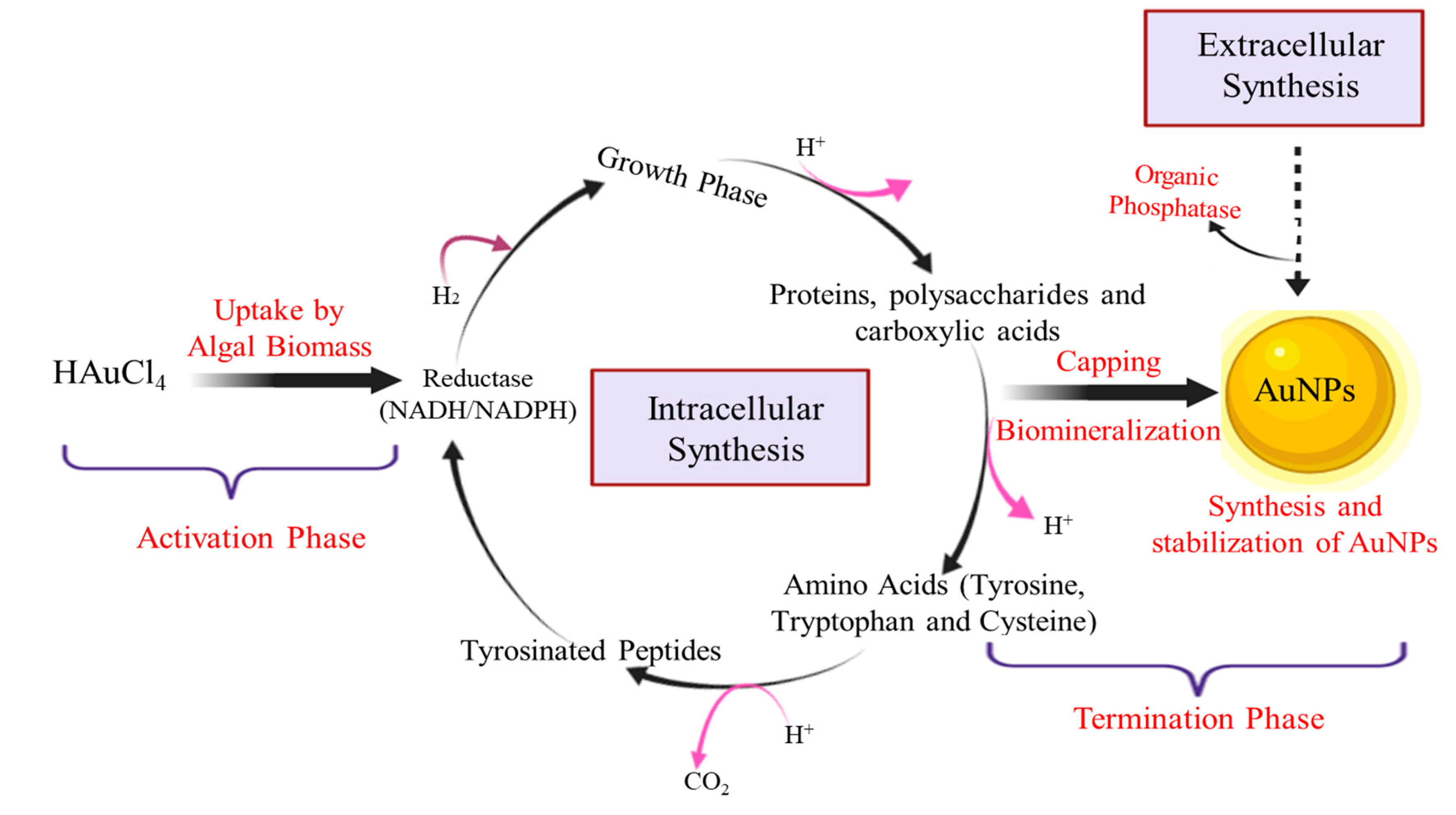
2. Mechanism Involved in the Algae-Mediated Biosynthesis of NPs
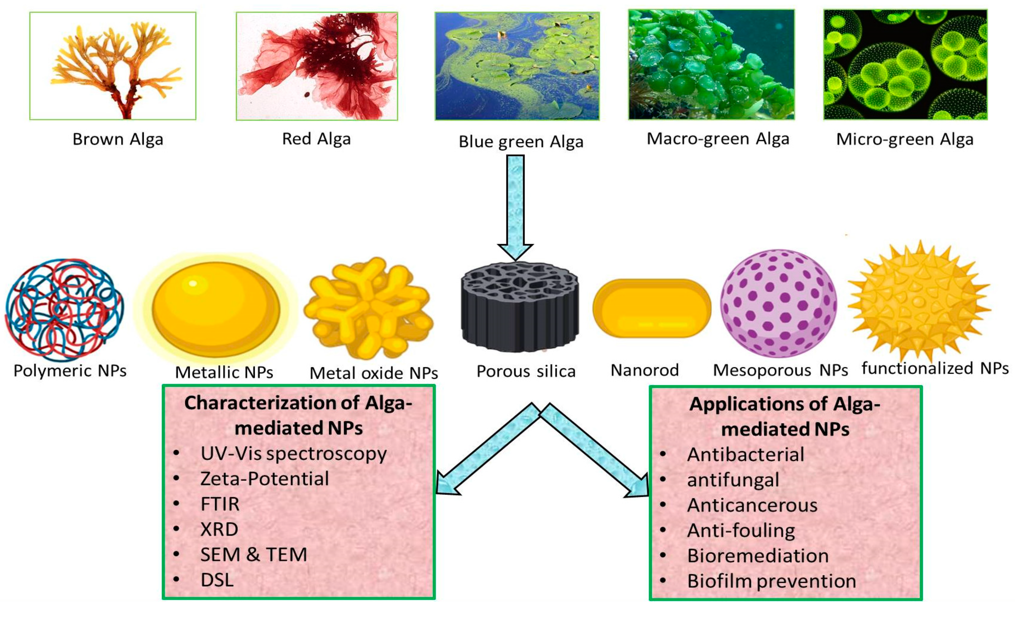
3. Biosynthesis of NPs from Algae
3.1. Brown Algae-Mediated Biosynthesis of NPs
3.2. Red Algae-Mediated Biosynthesis of NPs
3.3. Blue-Green Algae-Mediated Biosynthesis of NPs
3.4. Green Algae-Mediated Biosynthesis of NPs
3.4.1. Green Micro Algae-Mediated Biosynthesis of NPs
3.4.2. Green Macro Algae-Mediated Biosynthesis of NPs
4. Biomedical Applications of Algae-Mediated NPs
4.1. Antibacterial Activity
4.2. Antifungal Activity
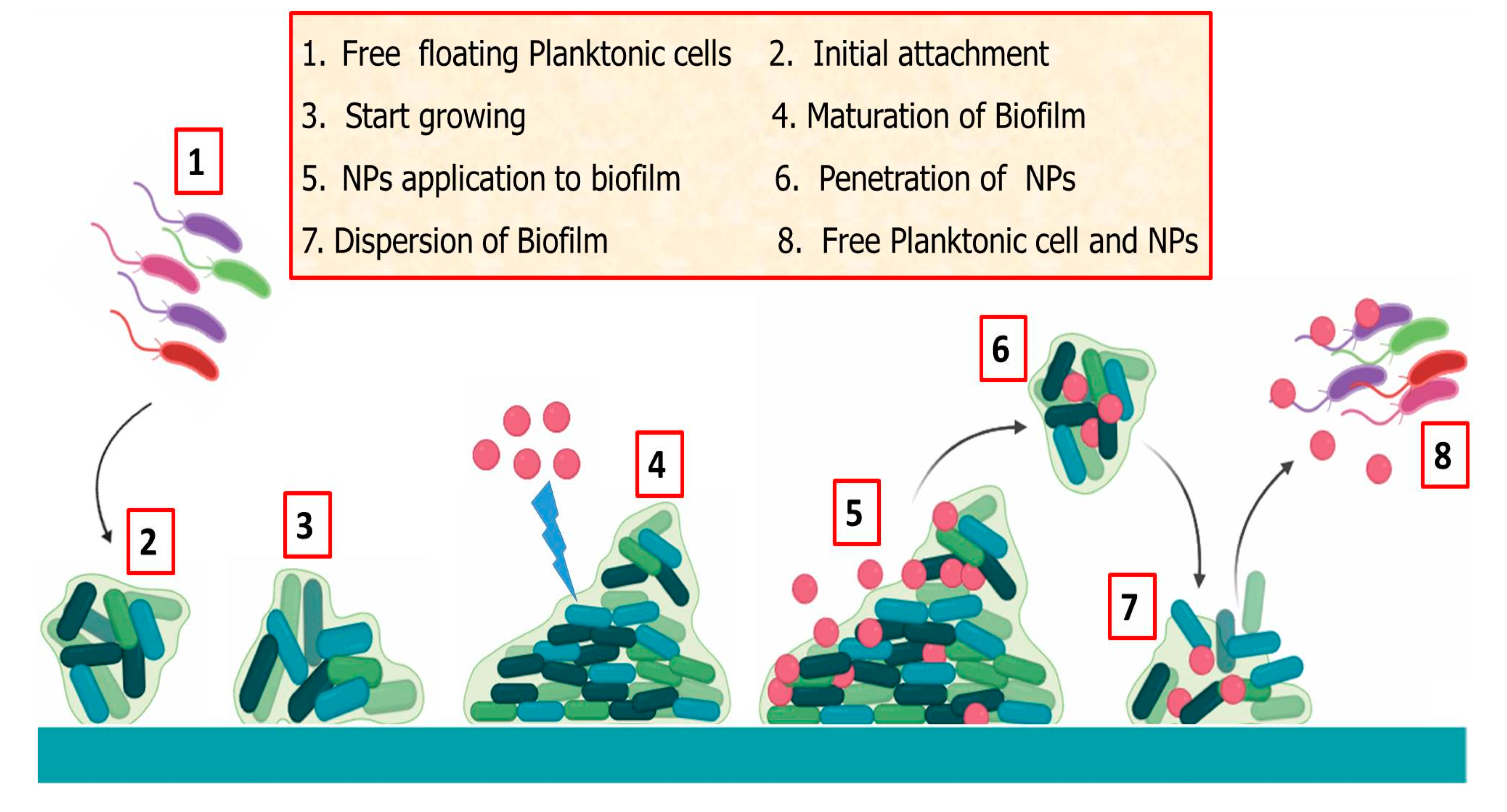
4.3. Antifouling Agent and Biofilm Prevention
4.4. Anti-Cancerous Activity
4.5. Nano-Bioremediation
4.6. Biosensing
5. Limitations and Future Prospects
Author Contributions
Funding
Acknowledgments
Conflicts of Interest
References
- Tran, P.H.-L.; Tran, T.T.-D.; Van Vo, T.; Lee, B.-J. Promising iron oxide-based magnetic nanoparticles in biomedical engineering. Arch. Pharmacal. Res. 2012, 35, 2045–2061. [Google Scholar] [CrossRef] [PubMed]
- Cho, E.J.; Holback, H.; Liu, K.C.; Abouelmagd, S.A.; Park, J.; Yeo, Y. Nanoparticle characterization: State of the art, challenges, and emerging technologies. Mol. Pharm. 2013, 10, 2093–2110. [Google Scholar] [CrossRef] [PubMed]
- Baker, S.; Harini, B.; Rakshith, D.; Satish, S. Marine microbes: Invisible nanofactories. J. Pharm. Res. 2013, 6, 383–388. [Google Scholar] [CrossRef]
- D’Amato, R.; Falconieri, M.; Gagliardi, S.; Popovici, E.; Serra, E.; Terranova, G.; Borsella, E. Synthesis of ceramic nanoparticles by laser pyrolysis: From research to applications. J. Anal. Appl. Pyrolysis 2013, 104, 461–469. [Google Scholar] [CrossRef]
- Zharov, V.P.; Kim, J.-W.; Curiel, D.T.; Everts, M. Self-assembling nanoclusters in living systems: Application for integrated photothermal nanodiagnostics and nanotherapy. Nanomed. Nanotechnol. Biol. Med. 2005, 1, 326–345. [Google Scholar] [CrossRef]
- Guo, S.; Wang, E. Noble metal nanomaterials: Controllable synthesis and application in fuel cells and analytical sensors. Nano Today 2011, 6, 240–264. [Google Scholar] [CrossRef]
- Kumar, V.G.; Gokavarapu, S.D.; Rajeswari, A.; Dhas, T.S.; Karthick, V.; Kapadia, Z.; Shrestha, T.; Barathy, I.; Roy, A.; Sinha, S. Facile green synthesis of gold nanoparticles using leaf extract of antidiabetic potent Cassia auriculata. Colloids Surf. B. Biointerfaces 2011, 87, 159–163. [Google Scholar] [CrossRef]
- Cai, W.; Gao, T.; Hong, H.; Sun, J. Applications of gold nanoparticles in cancer nanotechnology. Nanotechnol. Sci. Appl. 2008, 1, 17. [Google Scholar] [CrossRef]
- Dhillon, G.S.; Brar, S.K.; Kaur, S.; Verma, M. Green approach for nanoparticle biosynthesis by fungi: Current trends and applications. Crit. Rev. Biotechnol. 2012, 32, 49–73. [Google Scholar] [CrossRef]
- Cheng, W.; Zhang, W.; Hu, L.; Ding, W.; Wu, F.; Li, J. Etching synthesis of iron oxide nanoparticles for adsorption of arsenic from water. RSC Adv. 2016, 6, 15900–15910. [Google Scholar] [CrossRef]
- Azizi, S.; Ahmad, M.B.; Namvar, F.; Mohamad, R. Green biosynthesis and characterization of zinc oxide nanoparticles using brown marine macroalga Sargassum muticum aqueous extract. Mater. Lett. 2014, 116, 275–277. [Google Scholar] [CrossRef]
- Kalaiarasi, R.; Jayallakshmi, N.; Venkatachalam, P. Phytosynthesis of nanoparticles and its applications. Plant Cell Biotechnol. Mol. Biol. 2010, 11, 1–16. [Google Scholar]
- Sinha, S.N.; Paul, D.; Halder, N.; Sengupta, D.; Patra, S.K. Green synthesis of silver nanoparticles using fresh water green alga Pithophora oedogonia (Mont.) Wittrock and evaluation of their antibacterial activity. Appl. Nanosci. 2015, 5, 703–709. [Google Scholar] [CrossRef]
- Iravani, S. Green synthesis of metal nanoparticles using plants. Green Chem. 2011, 13, 2638–2650. [Google Scholar] [CrossRef]
- Pillai, Z.S.; Kamat, P.V. What factors control the size and shape of silver nanoparticles in the citrate ion reduction method? J. Phys. Chem. B 2004, 108, 945–951. [Google Scholar] [CrossRef]
- Dahoumane, S.A.; Djediat, C.; Yéprémian, C.; Couté, A.; Fiévet, F.; Coradin, T.; Brayner, R. Species selection for the design of gold nanobioreactor by photosynthetic organisms. J. Nanopart. Res. 2012, 14, 883. [Google Scholar] [CrossRef]
- Dahoumane, S.A.; Wijesekera, K.; Filipe, C.D.; Brennan, J.D. Stoichiometrically controlled production of bimetallic gold-silver alloy colloids using micro-alga cultures. J. Colloid Interface Sci. 2014, 416, 67–72. [Google Scholar] [CrossRef]
- Parial, D.; Pal, R. Biosynthesis of monodisperse gold nanoparticles by green alga Rhizoclonium and associated biochemical changes. J. Appl. Phycol. 2015, 27, 975–984. [Google Scholar] [CrossRef]
- Aboelfetoh, E.F.; El-Shenody, R.A.; Ghobara, M.M. Eco-friendly synthesis of silver nanoparticles using green algae (Caulerpa serrulata): Reaction optimization, catalytic and antibacterial activities. Environ. Monit. Assess. 2017, 189, 349. [Google Scholar] [CrossRef]
- Hulkoti, N.I.; Taranath, T. Biosynthesis of nanoparticles using microbes—A review. Colloids Surf. B Biointerfaces 2014, 121, 474–483. [Google Scholar] [CrossRef]
- Rahdar, A. Study of different capping agents effect on the structural and optical properties of Mn doped ZnS nanostructures. World Appl. Program 2013, 3, 56–60. [Google Scholar]
- Menon, S.; Rajeshkumar, S.; Kumar, V. A review on biogenic synthesis of gold nanoparticles, characterization, and its applications. Resour. Effic. Technol. 2017, 3, 516–527. [Google Scholar] [CrossRef]
- Shah, M.; Fawcett, D.; Sharma, S.; Tripathy, S.K.; Poinern, G.E.J. Green synthesis of metallic nanoparticles via biological entities. Materials 2015, 8, 7278–7308. [Google Scholar] [CrossRef]
- Shakibaie, M.; Forootanfar, H.; Mollazadeh-Moghaddam, K.; Bagherzadeh, Z.; Nafissi-Varcheh, N.; Shahverdi, A.R.; Faramarzi, M.A. Green synthesis of gold nanoparticles by the marine microalga Tetraselmis suecica. Biotechnol. Appl. Biochem. 2010, 57, 71–75. [Google Scholar] [CrossRef]
- Patel, V.; Berthold, D.; Puranik, P.; Gantar, M. Screening of cyanobacteria and microalgae for their ability to synthesize silver nanoparticles with antibacterial activity. Biotechnol. Rep. 2015, 5, 112–119. [Google Scholar] [CrossRef]
- Chandran, P.R.; Naseer, M.; Udupa, N.; Sandhyarani, N. Size controlled synthesis of biocompatible gold nanoparticles and their activity in the oxidation of NADH. Nanotechnology 2011, 23, 015602. [Google Scholar] [CrossRef] [PubMed]
- Fawcett, D.; Verduin, J.J.; Shah, M.; Sharma, S.B.; Poinern, G.E.J. A review of current research into the biogenic synthesis of metal and metal oxide nanoparticles via marine algae and seagrasses. J. Nanosci. 2017, 2017, 1–15. [Google Scholar] [CrossRef]
- Sahoo, P.C.; Kausar, F.; Lee, J.H.; Han, J.I. Facile fabrication of silver nanoparticle embedded CaCO3 microspheres via microalgae-templated CO2 biomineralization: Application in antimicrobial paint development. RSC Adv. 2014, 4, 32562–32569. [Google Scholar] [CrossRef]
- Agnihotri, S.; Mukherji, S.; Mukherji, S. Size-controlled silver nanoparticles synthesized over the range 5–100 nm using the same protocol and their antibacterial efficacy. RSC Adv. 2014, 4, 3974–3983. [Google Scholar] [CrossRef]
- Prasad, R.; Pandey, R.; Barman, I. Engineering tailored nanoparticles with microbes: Quo vadis? Wiley Interdiscip. Rev. Nanomed. Nanobiotechnol. 2016, 8, 316–330. [Google Scholar] [CrossRef]
- Sharma, A.; Sharma, S.; Sharma, K.; Chetri, S.P.; Vashishtha, A.; Singh, P.; Kumar, R.; Rathi, B.; Agrawal, V. Algae as crucial organisms in advancing nanotechnology: A systematic review. J. Appl. Phycol. 2016, 28, 1759–1774. [Google Scholar] [CrossRef]
- Dahoumane, S.A.; Yéprémian, C.; Djédiat, C.; Couté, A.; Fiévet, F.; Coradin, T.; Brayner, R. A global approach of the mechanism involved in the biosynthesis of gold colloids using micro-algae. J. Nanopart. Res. 2014, 16, 2607. [Google Scholar] [CrossRef]
- Sicard, C.; Brayner, R.; Margueritat, J.; Hémadi, M.; Couté, A.; Yéprémian, C.; Djediat, C.; Aubard, J.; Fiévet, F.; Livage, J. Nano-gold biosynthesis by silica-encapsulated micro-algae: A “living” bio-hybrid material. J. Mater. Chem. 2010, 20, 9342–9347. [Google Scholar] [CrossRef]
- Senapati, S.; Syed, A.; Moeez, S.; Kumar, A.; Ahmad, A. Intracellular synthesis of gold nanoparticles using alga Tetraselmis kochinensis. Mater. Lett. 2012, 79, 116–118. [Google Scholar] [CrossRef]
- Vijayan, S.R.; Santhiyagu, P.; Singamuthu, M.; Kumari Ahila, N.; Jayaraman, R.; Ethiraj, K. Synthesis and characterization of silver and gold nanoparticles using aqueous extract of seaweed, Turbinaria conoides, and their antimicrofouling activity. Sci. World J. 2014, 2014, 938272. [Google Scholar] [CrossRef] [PubMed]
- Dahoumane, S.A.; Wujcik, E.K.; Jeffryes, C. Noble metal, oxide and chalcogenide-based nanomaterials from scalable phototrophic culture systems. Enzym. Microb. Technol. 2016, 95, 13–27. [Google Scholar] [CrossRef]
- Oza, G.; Pandey, S.; Mewada, A.; Kalita, G.; Sharon, M.; Phata, J.; Ambernath, W.; Sharon, M. Facile biosynthesis of gold nanoparticles exploiting optimum pH and temperature of fresh water algae Chlorella pyrenoidusa. Adv. Appl. Sci. Res. 2012, 3, 1405–1412. [Google Scholar]
- Parial, D.; Patra, H.K.; Roychoudhury, P.; Dasgupta, A.K.; Pal, R. Gold nanorod production by cyanobacteria—A green chemistry approach. J. Appl. Phycol. 2012, 24, 55–60. [Google Scholar] [CrossRef]
- Namvar, F.; Azizi, S.; Ahmad, M.B.; Shameli, K.; Mohamad, R.; Mahdavi, M.; Tahir, P.M. Green synthesis and characterization of gold nanoparticles using the marine macroalgae Sargassum muticum. Res. Chem. Intermed. 2015, 41, 5723–5730. [Google Scholar] [CrossRef]
- Kalabegishvili, T.L.; Kirkesali, E.I.; Rcheulishvili, A.N.; Ginturi, E.N.; Murusidze, I.G.; Pataraya, D.T.; Gurielidze, M.A.; Tsertsvadze, G.I.; Gabunia, V.N.; Lomidze, L.G. Synthesis of gold nanoparticles by some strains of Arthrobacter genera. Mater. Sci. Eng. A 2012, 2, 164–173. [Google Scholar]
- Sahayaraj, K.; Rajesh, S.; Rathi, J. Silver nanoparticles biosynthesis using marine alga Padina pavonica (linn.) and its microbicidal activity. Dig. J. Nanomater. Bios. 2012, 7, 1557–1567. [Google Scholar]
- Prasad, T.N.; Kambala, V.S.R.; Naidu, R. Phyconanotechnology: Synthesis of silver nanoparticles using brown marine algae Cystophora moniliformis and their characterisation. J. Appl. Phycol. 2013, 25, 177–182. [Google Scholar] [CrossRef]
- Kumar, S.S.; Kumar, Y.; Khan, M.; Anbu, J.; De Clercq, E. Antihistaminic and antiviral activities of steroids of Turbinaria conoides. Nat. Prod. Res. 2011, 25, 723–729. [Google Scholar] [CrossRef]
- Rajeshkumar, S.; Kannan, C.; Annadurai, G. Green synthesis of silver nanoparticles using marine brown algae Turbinaria conoides and its antibacterial activity. Int. J. Pharma. Bio Sci. 2012, 3, 502–510. [Google Scholar]
- Liu, B.; Xie, J.; Lee, J.; Ting, Y.; Chen, J.P. Optimization of high-yield biological synthesis of single-crystalline gold nanoplates. J. Phys. Chem. B 2005, 109, 15256–15263. [Google Scholar] [CrossRef] [PubMed]
- Mata, Y.; Torres, E.; Blazquez, M.; Ballester, A.; González, F.; Munoz, J. Gold (III) biosorption and bioreduction with the brown alga Fucus vesiculosus. J. Hazard. Mater. 2009, 166, 612–618. [Google Scholar] [CrossRef]
- Khanna, P.; Kaur, A.; Goyal, D. Algae-based metallic nanoparticles: Synthesis, characterization and applications. J. Microbiol. Methods 2019, 163, 105656. [Google Scholar] [CrossRef]
- Khalil, M.M.; Ismail, E.H.; El-Baghdady, K.Z.; Mohamed, D. Green synthesis of silver nanoparticles using olive leaf extract and its antibacterial activity. Arab. J. Chem. 2014, 7, 1131–1139. [Google Scholar] [CrossRef]
- Madhiyazhagan, P.; Murugan, K.; Kumar, A.N.; Nataraj, T.; Dinesh, D.; Panneerselvam, C.; Subramaniam, J.; Kumar, P.M.; Suresh, U.; Roni, M. Sargassum muticum-synthesized silver nanoparticles: An effective control tool against mosquito vectors and bacterial pathogens. Parasitol. Res. 2015, 114, 4305–4317. [Google Scholar] [CrossRef]
- Ghodake, G.; Lee, D.S. Biological synthesis of gold nanoparticles using the aqueous extract of the brown algae Laminaria japonica. J. Nanoelectron. Optoelectron. 2011, 6, 268–271. [Google Scholar] [CrossRef]
- Rajeshkumar, S.; Malarkodi, C.; Gnanajobitha, G.; Paulkumar, K.; Vanaja, M.; Kannan, C.; Annadurai, G. Seaweed-mediated synthesis of gold nanoparticles using Turbinaria conoides and its characterization. J. Nanostructure Chem. 2013, 3, 44. [Google Scholar] [CrossRef]
- Venkatesan, J.; Manivasagan, P.; Kim, S.-K.; Kirthi, A.V.; Marimuthu, S.; Rahuman, A.A. Marine algae-mediated synthesis of gold nanoparticles using a novel Ecklonia cava. Bioprocess Biosyst. Eng. 2014, 37, 1591–1597. [Google Scholar] [CrossRef]
- Dahoumane, S.A.; Mechouet, M.; Wijesekera, K.; Filipe, C.D.; Sicard, C.; Bazylinski, D.A.; Jeffryes, C. Algae-mediated biosynthesis of inorganic nanomaterials as a promising route in nanobiotechnology—A review. Green Chem. 2017, 19, 552–587. [Google Scholar] [CrossRef]
- Singaravelu, G.; Arockiamary, J.; Kumar, V.G.; Govindaraju, K. A novel extracellular synthesis of monodisperse gold nanoparticles using marine alga, Sargassum wightii Greville. Colloids Surf. B. Biointerfaces 2007, 57, 97–101. [Google Scholar] [CrossRef] [PubMed]
- González-Ballesteros, N.; Rodríguez-Argüelles, M.; Lastra-Valdor, M.; González-Mediero, G.; Rey-Cao, S.; Grimaldi, M.; Cavazza, A.; Bigi, F. Synthesis of silver and gold nanoparticles by Sargassum muticum biomolecules and evaluation of their antioxidant activity and antibacterial properties. J. Nanostructure Chem. 2020, 1–14. [Google Scholar] [CrossRef]
- Dhas, T.S.; Kumar, V.G.; Abraham, L.S.; Karthick, V.; Govindaraju, K. Sargassum myriocystum mediated biosynthesis of gold nanoparticles. Spectrochim. Acta. A Mol. Biomol. Spectrosc. 2012, 99, 97–101. [Google Scholar] [CrossRef]
- Rajathi, F.A.A.; Parthiban, C.; Kumar, V.G.; Anantharaman, P. Biosynthesis of antibacterial gold nanoparticles using brown alga, Stoechospermum marginatum (kützing). Spectrochim. Acta. A Mol. Biomol. Spectrosc. 2012, 99, 166–173. [Google Scholar] [CrossRef]
- Singh, M.; Kalaivani, R.; Manikandan, S.; Sangeetha, N.; Kumaraguru, A. Facile green synthesis of variable metallic gold nanoparticle using Padina gymnospora, a brown marine macroalga. Appl. Nanosci. 2013, 3, 145–151. [Google Scholar] [CrossRef]
- Varun, S.; Sudha, S. P Senthil Kumar. Biosynthesis of gold nanoparticles from aqueous extract of Dictyota bartayresiana and their anti-fungal activity. Indian J. Adv. Chem. Sci. 2014, 2, 190–193. [Google Scholar]
- Ramakrishna, M.; Babu, D.R.; Gengan, R.M.; Chandra, S.; Rao, G.N. Green synthesis of gold nanoparticles using marine algae and evaluation of their catalytic activity. J. Nanostructure Chem. 2016, 6, 1–13. [Google Scholar] [CrossRef]
- González-Ballesteros, N.; Prado-López, S.; Rodríguez-González, J.; Lastra, M.; Rodríguez-Argüelles, M. Green synthesis of gold nanoparticles using brown algae Cystoseira baccata: Its activity in colon cancer cells. Colloids Surf. B Biointerfaces 2017, 153, 190–198. [Google Scholar] [CrossRef] [PubMed]
- Govindaraju, K.; Basha, S.K.; Kumar, V.G.; Singaravelu, G. Silver, gold and bimetallic nanoparticles production using single-cell protein (Spirulina platensis) Geitler. J. Mater. Sci. 2008, 43, 5115–5122. [Google Scholar] [CrossRef]
- Li, X. Green Energy for Sustainability and Energy Security; Springer: New York, NY, USA, 2011; pp. 1–16. [Google Scholar]
- Thangaraju, N.; Venkatalakshmi, R.; Chinnasamy, A.; Kannaiyan, P. Synthesis of silver nanoparticles and the antibacterial and anticancer activities of the crude extract of Sargassum polycystum C. Agardh. Nano Biomed. Eng. 2012, 4, 89–94. [Google Scholar] [CrossRef]
- Dhanalakshmi, P.; Azeez, R.; Rekha, R.; Poonkodi, S.; Nallamuthu, T. Synthesis of silver nanoparticles using green and brown seaweeds. Phykos 2012, 42, 39–45. [Google Scholar]
- Bhuyar, P.; Rahim, M.H.A.; Sundararaju, S.; Ramaraj, R.; Maniam, G.P.; Govindan, N. Synthesis of silver nanoparticles using marine macroalgae Padina sp. and its antibacterial activity towards pathogenic bacteria. Beni-Suef Univ. J. Basic Appl. Sci. 2020, 9, 3. [Google Scholar] [CrossRef]
- Shiny, P.; Mukherjee, A.; Chandrasekaran, N. Marine algae mediated synthesis of the silver nanoparticles and its antibacterial efficiency. Int. J. Pharm. Pharm. Sci. 2013, 5, 239–241. [Google Scholar]
- El-Rafie, H.; El-Rafie, M.; Zahran, M. Green synthesis of silver nanoparticles using polysaccharides extracted from marine macro algae. Carbohydr. Polym. 2013, 96, 403–410. [Google Scholar] [CrossRef]
- Mohandass, C.; Vijayaraj, A.; Rajasabapathy, R.; Satheeshbabu, S.; Rao, S.; Shiva, C.; De-Mello, I. Biosynthesis of silver nanoparticles from marine seaweed Sargassum cinereum and their antibacterial activity. Indian J. Pharm. Sci. 2013, 75, 606. [Google Scholar]
- Devi, J.S.; Bhimba, B.V.; Peter, D.M. Production of biogenic silver nanoparticles using Sargassum longifolium and its applications. Indian J. Geomarine Sci. 2013, 42, 125–130. [Google Scholar]
- Prasad, T.; Elumalai, E. Marine algae mediated synthesis of silver nanopaticles using Scaberia agardhü Greville. J. Biol. Sci. 2013, 13, 566. [Google Scholar] [CrossRef]
- Asha, K.; Johnson, M.; Chandra, P.; Shibila, T.; Revathy, I. Extracellular synthesis of silver nanoparticles from a marine Alga, Sargassum polycystum C. agardh and their biopotentials. WJPPS 2015, 4, 1388–1400. [Google Scholar]
- Govindaraju, K.; Krishnamoorthy, K.; Alsagaby, S.A.; Singaravelu, G.; Premanathan, M. Green synthesis of silver nanoparticles for selective toxicity towards cancer cells. IET Nanobiotechnol. 2015, 9, 325–330. [Google Scholar] [CrossRef]
- Krishnan, M.; Sivanandham, V.; Hans-Uwe, D.; Murugaiah, S.G.; Seeni, P.; Gopalan, S.; Rathinam, A.J. Antifouling assessments on biogenic nanoparticles: A field study from polluted offshore platform. Mar. Pollut. Bull. 2015, 101, 816–825. [Google Scholar] [CrossRef]
- Mahdavi, M.; Namvar, F.; Ahmad, M.B.; Mohamad, R. Green biosynthesis and characterization of magnetic iron oxide (Fe3O4) nanoparticles using seaweed (Sargassum muticum) aqueous extract. Molecules 2013, 18, 5954–5964. [Google Scholar] [CrossRef]
- Mohanpuria, P.; Rana, N.K.; Yadav, S.K. Biosynthesis of nanoparticles: Technological concepts and future applications. J. Nanopart. Res. 2008, 10, 507–517. [Google Scholar] [CrossRef]
- Vaidyanathan, R.; Kalishwaralal, K.; Gopalram, S.; Gurunathan, S. Nanosilver—The burgeoning therapeutic molecule and its green synthesis (Retraction of vol 27, pg 924, 2009). Biotechnol. Adv. 2010, 28, 940. [Google Scholar] [CrossRef]
- Maneerung, T.; Tokura, S.; Rujiravanit, R. Impregnation of silver nanoparticles into bacterial cellulose for antimicrobial wound dressing. Carbohydr. Polym. 2008, 72, 43–51. [Google Scholar] [CrossRef]
- Kumar-Krishnan, S.; Prokhorov, E.; Hernández-Iturriaga, M.; Mota-Morales, J.D.; Vázquez-Lepe, M.; Kovalenko, Y.; Sanchez, I.C.; Luna-Bárcenas, G. Chitosan/silver nanocomposites: Synergistic antibacterial action of silver nanoparticles and silver ions. Eur. Polym. J. 2015, 67, 242–251. [Google Scholar] [CrossRef]
- Kushnerova, N.; Fomenko, S.; Sprygin, V.; Kushnerova, T.; Khotimchenko, Y.S.; Kondrat’eva, E.; Drugova, L. An extract from the brown alga Laminaria japonica: A promising stress-protective preparation. Russ. J. Mar. Biol. 2010, 36, 209–214. [Google Scholar] [CrossRef]
- Khodashenas, B.; Ghorbani, H.R. Synthesis of silver nanoparticles with different shapes. Arab. J. Chem. 2019, 12, 1823–1838. [Google Scholar] [CrossRef]
- Sirelkhatim, A.; Mahmud, S.; Seeni, A.; Kaus, N.H.M.; Ann, L.C.; Bakhori, S.K.M.; Hasan, H.; Mohamad, D. Review on zinc oxide nanoparticles: Antibacterial activity and toxicity mechanism. Nanomicro Lett. 2015, 7, 219–242. [Google Scholar] [CrossRef]
- Azizi, S.; Mahdavi Shahri, M.; Mohamad, R. Green synthesis of zinc oxide nanoparticles for enhanced adsorption of lead ions from aqueous solutions: Equilibrium, kinetic and thermodynamic studies. Molecules 2017, 22, 831. [Google Scholar] [CrossRef] [PubMed]
- Yoon, H.S.; Müller, K.M.; Sheath, R.G.; Ott, F.D.; Bhattacharya, D. Defining the major lineages of red algae (Rhodophyta) 1. J. Phycol. 2006, 42, 482–492. [Google Scholar] [CrossRef]
- Ramakritinan, C.; Shankar, S.; Anand, M.; Kumaraguru, A. Biosynthesis of silver, gold and bimetallic alloy (Ag: Au) Nanoparticles from green alga, Lyngpya sp. In Proceedings of the 3rd National Conference on Nanaomaterials and Nanotechnology, Lucknow, India, 21–23 December 2020; pp. 174–187. [Google Scholar]
- Rao, P.S.; Mantri, V.A.; Ganesan, K. Mineral composition of edible seaweed Porphyra vietnamensis. Food Chem. 2007, 102, 215–218. [Google Scholar] [CrossRef]
- Venkatpurwar, V.; Pokharkar, V. Green synthesis of silver nanoparticles using marine polysaccharide: Study of in-vitro antibacterial activity. Mater. Lett. 2011, 65, 999–1002. [Google Scholar] [CrossRef]
- Pugazhendhi, A.; Prabakar, D.; Jacob, J.M.; Karuppusamy, I.; Saratale, R.G. Synthesis and characterization of silver nanoparticles using Gelidium amansii and its antimicrobial property against various pathogenic bacteria. Microb. Pathog. 2018, 114, 41–45. [Google Scholar] [CrossRef]
- Kumar, P.; Govindaraju, M.; Senthamilselvi, S.; Premkumar, K. Photocatalytic degradation of methyl orange dye using silver (Ag) nanoparticles synthesized from Ulva lactuca. Colloids Surf. B Biointerfaces 2013, 103, 658–661. [Google Scholar] [CrossRef]
- Rajasulochana, P.; Krishnamoorthy, P.; Dhamotharan, R. Potential application of Kappaphycus alvarezii in agricultural and pharmaceutical industry. J. Chem. Pharm. Res. 2012, 4, 33–37. [Google Scholar]
- Abdel-Raouf, N.; Al-Enazi, N.M.; Ibraheem, I.B. Green biosynthesis of gold nanoparticles using Galaxaura elongata and characterization of their antibacterial activity. Arab. J. Chem. 2017, 10, S3029–S3039. [Google Scholar] [CrossRef]
- Castro, L.; Blázquez, M.L.; Muñoz, J.A.; González, F.; Ballester, A. Biological synthesis of metallic nanoparticles using algae. IET Nanobiotechnol. 2013, 7, 109–116. [Google Scholar] [CrossRef]
- Sharma, B.; Purkayastha, D.D.; Hazra, S.; Thajamanbi, M.; Bhattacharjee, C.R.; Ghosh, N.N.; Rout, J. Biosynthesis of fluorescent gold nanoparticles using an edible freshwater red alga, Lemanea fluviatilis (L.) C. Ag. and antioxidant activity of biomatrix loaded nanoparticles. Bioprocess Biosyst. Eng. 2014, 37, 2559–2565. [Google Scholar] [CrossRef] [PubMed]
- Naveena, B.E.; Prakash, S. Biological synthesis of gold nanoparticles using marine algae Gracilaria corticata and its application as a potent antimicrobial and antioxidant agent. Asian J. Pharm. Clin. Res. 2013, 6, 179–182. [Google Scholar]
- El-Kassas, H.Y.; El-Sheekh, M.M. Cytotoxic activity of biosynthesized gold nanoparticles with an extract of the red seaweed Corallina officinalis on the MCF-7 human breast cancer cell line. Asian Pac. J. Cancer Prev. 2014, 15, 4311–4317. [Google Scholar] [CrossRef] [PubMed]
- Vivek, M.; Kumar, P.S.; Steffi, S.; Sudha, S. Biogenic silver nanoparticles by Gelidiella acerosa extract and their antifungal effects. Avicenna J. Med. Biotechnol. 2011, 3, 143. [Google Scholar] [PubMed]
- Kumar, P.; Senthamilselvi, S.; Lakshmipraba, A.; Premkumar, K.; Muthukumaran, R.; Visvanathan, P.; Ganeshkumar, R.; Govindaraju, M. Efficacy of bio-synthesized silver nanoparticles using Acanthophora spicifera to encumber biofilm formation. Dig. J. Nanomater. Biostructures 2012, 7, 511–522. [Google Scholar]
- Shukla, M.K.; Singh, R.P.; Reddy, C.; Jha, B. Synthesis and characterization of agar-based silver nanoparticles and nanocomposite film with antibacterial applications. Bioresour. Technol. 2012, 107, 295–300. [Google Scholar] [CrossRef] [PubMed]
- Devi, J.S.; Bhimba, B.V.; Ratnam, K. In vitro anticancer activity of silver nanoparticles synthesized using the extract of Gelidiella sp. Int. J. Pharm. Pharm. Sci. 2012, 4, 710–715. [Google Scholar]
- Baskar, B.B. Biosynthesis of silver nanoparticles using Kappa phycus species. Int. J. Pharm. Sci. Rev. Res. 2013, 3. [Google Scholar]
- Kumar, P.; Selvi, S.S.; Govindaraju, M. Seaweed-mediated biosynthesis of silver nanoparticles using Gracilaria corticata for its antifungal activity against Candida spp. Appl. Nanosci. 2013, 3, 495–500. [Google Scholar] [CrossRef]
- Ganesan, V.; Aruna Devi, J.; Astalakshmi, A.; Nima, P.; Thangaraja, A. Eco-friendly synthesis of silver nanoparticles using a sea weed, Kappaphycus alvarezii (Doty) Doty ex PC Silva. Int. J. Eng. Adv. Technol. 2013, 2, 559–563. [Google Scholar]
- de Aragao, A.P.; de Oliveira, T.M.; Quelemes, P.V.; Perfeito, M.L.G.; Araujo, M.C.; Santiago, J.d.A.S.; Cardoso, V.S.; Quaresma, P.; de Almeida, J.R.d.S.; da Silva, D.A. Green synthesis of silver nanoparticles using the seaweed Gracilaria birdiae and their antibacterial activity. Arab. J. Chem. 2019, 12, 4182–4188. [Google Scholar] [CrossRef]
- Ibraheem, I.; Abd-Elaziz, B.; Saad, W.; Fathy, W. Green biosynthesis of silver nanoparticles using marine Red Algae Acanthophora specifera and its antimicrobial activity. J. Nanomed Nanotechnol. 2016, 7, 6. [Google Scholar]
- Sajidha Parveen, K.; Lakshmi, D. Biosynthesis of silver nanoparticles using red algae, Amphiroa fragilissima and its antibacterial potential against gram positive and gram negative bacteria. Int. J. Curr. Sci. 2016, 19, 93–100. [Google Scholar]
- González-Ballesteros, N.; González-Rodríguez, J.; Rodríguez-Argüelles, M.; Lastra, M. New application of two Antarctic macroalgae Palmaria decipiens and Desmarestia menziesii in the synthesis of gold and silver nanoparticles. Polar Sci. 2018, 15, 49–54. [Google Scholar] [CrossRef]
- Al-Naamani, L.; Dobretsov, S.; Dutta, J.; Burgess, J.G. Chitosan-zinc oxide nanocomposite coatings for the prevention of marine biofouling. Chemosphere 2017, 168, 408–417. [Google Scholar] [CrossRef]
- Kumar, P.; Senthamil Selvi, S.; Lakshmi Prabha, A.; Prem Kumar, K.; Ganeshkumar, R.; Govindaraju, M. Synthesis of silver nanoparticles from Sargassum tenerrimum and screening phytochemicals for its antibacterial activity. Nano Biomed. Eng. 2012, 4, 12–16. [Google Scholar] [CrossRef]
- Vadlapudi, V.; Amanchy, R. Synthesis, characterization and antibacterial activity of Silver Nanoparticles from Red Algae, Hypnea musciformis. Adv. Biol. Res. 2017, 11, 242–249. [Google Scholar]
- Nabikhan, A.; Kandasamy, K.; Raj, A.; Alikunhi, N.M. Synthesis of antimicrobial silver nanoparticles by callus and leaf extracts from saltmarsh plant, Sesuvium portulacastrum L. Colloids Surf. B Biointerfaces 2010, 79, 488–493. [Google Scholar] [CrossRef]
- Selvam, G.G.; Sivakumar, K. Phycosynthesis of silver nanoparticles and photocatalytic degradation of methyl orange dye using silver (Ag) nanoparticles synthesized from Hypnea musciformis (Wulfen) JV Lamouroux. Appl. Nanosci. 2015, 5, 617–622. [Google Scholar] [CrossRef]
- Singh, C.R.; Kathiresan, K.; Anandhan, S. A review on marine based nanoparticles and their potential applications. Afr. J. Biotechnol. 2015, 14, 1525–1532. [Google Scholar]
- Murugesan, S.; Bhuvaneswari, S.; Sivamurugan, V. Green synthesis, characterization of silver nanoparticles of a marine red alga Spyridia fusiformis and their antibacterial activity. Int. J. Pharm. Pharm. Sci. 2017, 9, 192–197. [Google Scholar] [CrossRef]
- Mosulishvili, L.; Belokobylsky, A.; Kirkesali, E.; Frontasyeva, M.; Pavlov, S.; Aksenova, N. Neutron activation analysis for studying Cr uptake in the blue-green microalga Spirulina platensis. J. Neutron Res. 2007, 15, 49–54. [Google Scholar] [CrossRef]
- Khan, A.U.; Khan, M.; Malik, N.; Cho, M.H.; Khan, M.M. Recent progress of algae and blue–green algae-assisted synthesis of gold nanoparticles for various applications. Bioprocess Biosyst. Eng. 2019, 42, 1–15. [Google Scholar] [CrossRef] [PubMed]
- Parial, D.; Gopal, P.K.; Paul, S.; Pal, R. Gold (III) bioreduction by cyanobacteria with special reference to in vitro biosafety assay of gold nanoparticles. J. Appl. Phycol. 2016, 28, 3395–3406. [Google Scholar] [CrossRef]
- El-Sheekh, M.M.; Shabaan, M.T.; Hassan, L.; Morsi, H.H. Antiviral activity of algae biosynthesized silver and gold nanoparticles against Herps Simplex (HSV-1) virus in vitro using cell-line culture technique. Int. J. Environ. Health Res. 2020, 1–12. [Google Scholar] [CrossRef]
- Suganya, K.U.; Govindaraju, K.; Kumar, V.G.; Dhas, T.S.; Karthick, V.; Singaravelu, G.; Elanchezhiyan, M. Blue green alga mediated synthesis of gold nanoparticles and its antibacterial efficacy against Gram positive organisms. Mater. Sci. Eng. C 2015, 47, 351–356. [Google Scholar] [CrossRef] [PubMed]
- Lengke, M.F.; Fleet, M.E.; Southam, G. Morphology of gold nanoparticles synthesized by filamentous cyanobacteria from gold (I)− thiosulfate and gold (III)− chloride complexes. Langmuir 2006, 22, 2780–2787. [Google Scholar] [CrossRef] [PubMed]
- Focsan, M.; Ardelean, I.I.; Craciun, C.; Astilean, S. Interplay between gold nanoparticle biosynthesis and metabolic activity of cyanobacterium Synechocystis sp. Nanotechnology 2011, 22, 485101. [Google Scholar] [CrossRef]
- Chakraborty, N.; Banerjee, A.; Lahiri, S.; Panda, A.; Ghosh, A.N.; Pal, R. Biorecovery of gold using cyanobacteria and an eukaryotic alga with special reference to nanogold formation—A novel phenomenon. J. Appl. Phycol. 2009, 21, 145. [Google Scholar] [CrossRef]
- Parial, D.; Patra, H.K.; Dasgupta, A.K.; Pal, R. Screening of different algae for green synthesis of gold nanoparticles. Eur. J. Phycol. 2012, 47, 22–29. [Google Scholar] [CrossRef]
- MubarakAli, D.; Gopinath, V.; Rameshbabu, N.; Thajuddin, N. Synthesis and characterization of CdS nanoparticles using C-phycoerythrin from the marine cyanobacteria. Mater. Lett. 2012, 74, 8–11. [Google Scholar] [CrossRef]
- Mosulishvili, L.; Kirkesali, E.; Belokobylsky, A.; Khizanishvili, A.; Frontasyeva, M.; Pavlov, S.; Gundorina, S. Experimental substantiation of the possibility of developing selenium-and iodine-containing pharmaceuticals based on blue–green algae Spirulina platensis. J. Pharm. Biomed 2002, 30, 87–97. [Google Scholar] [CrossRef]
- Parial, D.; Pal, R. Green synthesis of gold nanoparticles using cyanobacteria and their characterization. Indian J. Appl. Res 2014, 4, 69–72. [Google Scholar] [CrossRef]
- Rösken, L.M.; Cappel, F.; Körsten, S.; Fischer, C.B.; Schönleber, A.; van Smaalen, S.; Geimer, S.; Beresko, C.; Ankerhold, G.; Wehner, S. Time-dependent growth of crystalline Au0-nanoparticles in cyanobacteria as self-reproducing bioreactors: 2. Anabaena cylindrica. Beilstein J. Nanotechnol. 2016, 7, 312–327. [Google Scholar] [CrossRef]
- Ali, A.; Ali, M.A.; Ali, M.U.; Mohammad, S. Hospital outcomes of obstetrical-related acute renal failure in a tertiary care teaching hospital. Ren. Fail. 2011, 33, 285–290. [Google Scholar] [CrossRef] [PubMed]
- Doshi, H.; Ray, A.; Kothari, I. Bioremediation potential of live and dead Spirulina: Spectroscopic, kinetics and SEM studies. Biotechnol. Bioeng. 2007, 96, 1051–1063. [Google Scholar] [CrossRef] [PubMed]
- Lengke, M.F.; Fleet, M.E.; Southam, G. Biosynthesis of silver nanoparticles by filamentous cyanobacteria from a silver (I) nitrate complex. Langmuir 2007, 23, 2694–2699. [Google Scholar] [CrossRef] [PubMed]
- Sudha, S.; Rajamanickam, K.; Rengaramanujam, J. Microalgae mediated synthesis of silver nanoparticles and their antibacterial activity against pathogenic bacteria. Int. J. Nanomed. 2013, 51, 393–399. [Google Scholar]
- Husain, S.; Afreen, S.; Yasin, D.; Afzal, B.; Fatma, T. Cyanobacteria as a bioreactor for synthesis of silver nanoparticles-an effect of different reaction conditions on the size of nanoparticles and their dye decolorization ability. J. Microbiol. Methods 2019, 162, 77–82. [Google Scholar] [CrossRef]
- Husain, S.; Sardar, M.; Fatma, T. Screening of cyanobacterial extracts for synthesis of silver nanoparticles. World J. Microbiol. Biotechnol. 2015, 31, 1279–1283. [Google Scholar] [CrossRef]
- Mahdieh, M.; Zolanvari, A.; Azimee, A. Green biosynthesis of silver nanoparticles by Spirulina platensis. Sci. Iran. 2012, 19, 926–929. [Google Scholar] [CrossRef]
- Rao, M.D.; Pennathur, G. Green synthesis and characterization of cadmium sulphide nanoparticles from Chlamydomonas reinhardtii and their application as photocatalysts. Mater. Res. Bull. 2017, 85, 64–73. [Google Scholar] [CrossRef]
- Mukherjee, P.; Senapati, S.; Mandal, D.; Ahmad, A.; Khan, M.I.; Kumar, R.; Sastry, M. Extracellular synthesis of gold nanoparticles by the fungus Fusarium oxysporum. Chembiochem 2002, 3, 461–463. [Google Scholar] [CrossRef]
- Iravani, S.; Thota, S.; Crans, D. Metal Nanoparticles: Synthesis and Applications in Pharmaceutical Sciences; Wiley: New York, NY, USA, 2018; pp. 15–32. Available online: https://onlinelibrary.wiley.com/doi/book/10.1002/9783527807093 (accessed on 30 October 2020).
- Sosa, I.O.; Noguez, C.; Barrera, R.G. Optical properties of metal nanoparticles with arbitrary shapes. J. Phys. Chem. B 2003, 107, 6269–6275. [Google Scholar] [CrossRef]
- Kalabegishvili, T.; Kirkesali, E.; Rcheulishvili, A. Synthesis of Gold Nanoparticles by Blue-Green Algae Spirulina Platensis; Frank Lab. of Neutron Physics: Dubna, Russia, 2012. [Google Scholar]
- Kim, S.-K.; Thomas, N.V.; Li, X. Anticancer compounds from marine macroalgae and their application as medicinal foods. Adv. Food Nutr. Res. 2011, 64, 213–224. [Google Scholar]
- Deglint, J.L.; Jin, C.; Wong, A. Investigating the automatic classification of algae using the spectral and morphological characteristics via deep residual learning. In Proceedings of International Conference on Image Analysis and Recognition; Karray, F., Campilho, A., Yu, A., Eds.; Springer: Cham, Switzerland, 2019. [Google Scholar]
- Yousefzadi, M.; Rahimi, Z.; Ghafori, V. The green synthesis, characterization and antimicrobial activities of silver nanoparticles synthesized from green alga Enteromorpha flexuosa (wulfen) J. Agardh. Mater. Lett. 2014, 137, 1–4. [Google Scholar] [CrossRef]
- Wei, D.; Qian, W. Facile synthesis of Ag and Au nanoparticles utilizing chitosan as a mediator agent. Colloids Surf. B Biointerfaces 2008, 62, 136–142. [Google Scholar] [CrossRef]
- Vigneshwaran, N.; Ashtaputre, N.; Varadarajan, P.; Nachane, R.; Paralikar, K.; Balasubramanya, R. Biological synthesis of silver nanoparticles using the fungus Aspergillus flavus. Mater. Lett. 2007, 61, 1413–1418. [Google Scholar] [CrossRef]
- Lengke, M.F.; Ravel, B.; Fleet, M.E.; Wanger, G.; Gordon, R.A.; Southam, G. Mechanisms of gold bioaccumulation by filamentous cyanobacteria from gold (III)− chloride complex. Environ. Sci. Technol. 2006, 40, 6304–6309. [Google Scholar] [CrossRef] [PubMed]
- Jena, J.; Pradhan, N.; Nayak, R.R.; Dash, B.P.; Sukla, L.B.; Panda, P.K.; Mishra, B.K. Microalga Scenedesmus sp.: A potential low-cost green machine for silver nanoparticle synthesis. J. Microbiol. Biotechnol. 2014, 24, 522–533. [Google Scholar] [CrossRef]
- Xie, J.; Lee, J.Y.; Wang, D.I.; Ting, Y.P. Identification of active biomolecules in the high-yield synthesis of single-crystalline gold nanoplates in algal solutions. Small 2007, 3, 672–682. [Google Scholar] [CrossRef]
- MubarakAli, D.; Arunkumar, J.; Nag, K.H.; SheikSyedIshack, K.; Baldev, E.; Pandiaraj, D.; Thajuddin, N. Gold nanoparticles from Pro and eukaryotic photosynthetic microorganisms—Comparative studies on synthesis and its application on biolabelling. Colloids Surf. B Biointerfaces 2013, 103, 166–173. [Google Scholar] [CrossRef]
- Roychoudhury, P.; Pal, R. Spirogyra submaxima—A green alga for nanogold production. J. Algal Biomass Util. 2014, 5, 15–19. [Google Scholar]
- Xie, J.; Lee, J.Y.; Wang, D.I.; Ting, Y.P. Silver nanoplates: From biological to biomimetic synthesis. ACS Nano 2007, 1, 429–439. [Google Scholar] [CrossRef]
- Mohseniazar, M.; Barin, M.; Zarredar, H.; Alizadeh, S.; Shanehbandi, D. Potential of microalgae and lactobacilli in biosynthesis of silver nanoparticles. BioImpacts 2011, 1, 149. [Google Scholar]
- Barwal, I.; Ranjan, P.; Kateriya, S.; Yadav, S.C. Cellular oxido-reductive proteins of Chlamydomonas reinhardtii control the biosynthesis of silver nanoparticles. J. Nanobiotechnol. 2011, 9, 56. [Google Scholar] [CrossRef]
- Jena, J.; Pradhan, N.; Dash, B.P.; Sukla, L.B.; Panda, P.K. Biosynthesis and characterization of silver nanoparticles using microalga Chlorococcum humicola and its antibacterial activity. Int. J. Nanomater. Biostructures 2013, 3, 1–8. [Google Scholar]
- Li, X.; Schirmer, K.; Bernard, L.; Sigg, L.; Pillai, S.; Behra, R. Silver nanoparticle toxicity and association with the alga Euglena gracilis. Environ. Sci. Nano 2015, 2, 594–602. [Google Scholar] [CrossRef]
- Aziz, N.; Faraz, M.; Pandey, R.; Shakir, M.; Fatma, T.; Varma, A.; Barman, I.; Prasad, R. Facile algae-derived route to biogenic silver nanoparticles: Synthesis, antibacterial, and photocatalytic properties. Langmuir 2015, 31, 11605–11612. [Google Scholar] [CrossRef]
- Bhimba, B.; Kumari, P. Phytosynthesis of silver nanoparticles from the extracts of seaweed Ulva lactuca and its antimicrobial activity. Int. J. Pharm. BioSci. 2014, 5, 666–677. [Google Scholar]
- Kathiraven, T.; Sundaramanickam, A.; Shanmugam, N.; Balasubramanian, T. Green synthesis of silver nanoparticles using marine algae Caulerpa racemosa and their antibacterial activity against some human pathogens. Appl. Nanosci. 2015, 5, 499–504. [Google Scholar] [CrossRef]
- Kannan, R.R.R.; Arumugam, R.; Ramya, D.; Manivannan, K.; Anantharaman, P. Green synthesis of silver nanoparticles using marine macroalga Chaetomorpha linum. Appl. Nanosci. 2013, 3, 229–233. [Google Scholar]
- Kannan, R.; Stirk, W.; Van Staden, J. Synthesis of silver nanoparticles using the seaweed Codium capitatum PC Silva (Chlorophyceae). S. Afr. J. Bot. 2013, 86, 1–4. [Google Scholar]
- Chokshi, K.; Pancha, I.; Ghosh, T.; Paliwal, C.; Maurya, R.; Ghosh, A.; Mishra, S. Green synthesis, characterization and antioxidant potential of silver nanoparticles biosynthesized from de-oiled biomass of thermotolerant oleaginous microalgae Acutodesmus dimorphus. RSC Adv. 2016, 6, 72269–72274. [Google Scholar]
- Arya, A.; Mishra, V.; Chundawat, T.S. Green synthesis of silver nanoparticles from green algae (Botryococcus braunii) and its catalytic behavior for the synthesis of benzimidazoles. Chem. Data Collect. 2019, 20, 100190. [Google Scholar]
- Beganskienė, A.; Sirutkaitis, V.; Kurtinaitienė, M.; Juškėnas, R.; Kareiva, A. FTIR, TEM and NMR investigations of Stöber silica nanoparticles. Mater. Sci. (Medžiagotyra) 2004, 10, 287–290. [Google Scholar]
- Sharma, B.; Purkayastha, D.D.; Hazra, S.; Gogoi, L.; Bhattacharjee, C.R.; Ghosh, N.N.; Rout, J. Biosynthesis of gold nanoparticles using a freshwater green alga, Prasiola crispa. Mater. Lett. 2014, 116, 94–97. [Google Scholar]
- Priyadharshini, R.I.; Prasannaraj, G.; Geetha, N.; Venkatachalam, P. Microwave-mediated extracellular synthesis of metallic silver and zinc oxide nanoparticles using macro-algae (Gracilaria edulis) extracts and its anticancer activity against human PC3 cell lines. Appl. Biochem. Biotechnol. 2014, 174, 2777–2790. [Google Scholar]
- Salari, Z.; Danafar, F.; Dabaghi, S.; Ataei, S.A. Sustainable synthesis of silver nanoparticles using macroalgae Spirogyra varians and analysis of their antibacterial activity. J. Saudi Chem. Soc. 2016, 20, 459–464. [Google Scholar]
- González-Ballesteros, N.; Diego-González, L.; Lastra-Valdor, M.; Rodríguez-Argüelles, M.; Grimaldi, M.; Cavazza, A.; Bigi, F.; Simón-Vázquez, R. Immunostimulant and biocompatible gold and silver nanoparticles synthesized using the Ulva intestinalis L. aqueous extract. J. Mater. Chem. B 2019, 7, 4677–4691. [Google Scholar]
- Suriya, J.; Raja, S.B.; Sekar, V.; Rajasekaran, R. Biosynthesis of silver nanoparticles and its antibacterial activity using seaweed Urospora sp. Afr. J. Biotechnol. 2012, 11, 12192–12198. [Google Scholar]
- Rahimi, Z.; Yousefzadi, M.; Noori, A.; Akbarzadeh, A. Green synthesis of silver nanoparticles using Ulva flexousa from the Persian Gulf, Iran. J. Persian Gulf 2014, 5, 9–16. [Google Scholar]
- Madhiyazhagan, P.; Murugan, K.; Kumar, A.N.; Nataraj, T.; Subramaniam, J.; Chandramohan, B.; Panneerselvam, C.; Dinesh, D.; Suresh, U.; Nicoletti, M. One pot synthesis of silver nanocrystals using the seaweed Gracilaria edulis: Biophysical characterization and potential against the filariasis vector Culex quinquefasciatus and the midge Chironomus circumdatus. J. Appl. Phycol. 2017, 29, 649–659. [Google Scholar] [CrossRef]
- Bhattacharya, P.; Swarnakar, S.; Ghosh, S.; Majumdar, S.; Banerjee, S. Disinfection of drinking water via algae mediated green synthesized copper oxide nanoparticles and its toxicity evaluation. J. Environ. Chem. Eng. 2019, 7, 102867. [Google Scholar] [CrossRef]
- Wang, L.; Hu, C.; Shao, L. The antimicrobial activity of nanoparticles: Present situation and prospects for the future. Int. J. Nanomed. 2017, 12, 1227. [Google Scholar] [CrossRef]
- Rajeshkumar, S.; Kannan, C.; Annadurai, G. Synthesis and characterization of antimicrobial silver nanoparticles using marine brown seaweed Padina tetrastromatica. Drug Invent. Today 2012, 4, 511–513. [Google Scholar]
- Borase, H.P.; Patil, C.D.; Suryawanshi, R.K.; Koli, S.H.; Mohite, B.V.; Benelli, G.; Patil, S.V. Mechanistic approach for fabrication of gold nanoparticles by Nitzschia diatom and their antibacterial activity. Bioprocess Biosyst. Eng. 2017, 40, 1437–1446. [Google Scholar] [CrossRef]
- Omomowo, I.; Adenigba, V.; Ogunsona, S.; Adeyinka, G.; Oluyide, O.; Adedayo, A.; Fatukasi, B. Antimicrobial and antioxidant activities of algal-mediated silver and gold nanoparticles. In IOP Conference Series: Materials Science and Engineering; IOP Publishing: Bristol, UK, 2020; p. 012010. [Google Scholar]
- Mallmann, E.J.J.; Cunha, F.A.; Castro, B.N.; Maciel, A.M.; Menezes, E.A.; Fechine, P.B.A. Antifungal activity of silver nanoparticles obtained by green synthesis. Rev. Inst. Med. Trop. São Paulo 2015, 57, 165–167. [Google Scholar] [CrossRef]
- Rajeshkumar, S.; Malarkodi, C.; Paulkumar, K.; Vanaja, M.; Gnanajobitha, G.; Annadurai, G. Algae mediated green fabrication of silver nanoparticles and examination of its antifungal activity against clinical pathogens. Met. Mater. Int. 2014, 2014, 692643. [Google Scholar] [CrossRef]
- Dhavale, R.; Jadhav, S.; Sibi, G. Microalgae mediated silver nanoparticles (ag-nps) synthesis and their biological activities. J. Crit. Rev. 2019, 7, 2020. [Google Scholar]
- El-Sheekh, M.M.; El-Kassas, H.Y. Algal production of nano-silver and gold: Their antimicrobial and cytotoxic activities: A review. J. Genet. Eng. Biotechnol. 2016, 14, 299–310. [Google Scholar] [CrossRef]
- Romero, D.; Aguilar, C.; Losick, R.; Kolter, R. Amyloid fibers provide structural integrity to Bacillus subtilis biofilms. Proc. Natl. Acad. Sci. USA 2010, 107, 2230–2234. [Google Scholar] [CrossRef]
- Bixler, G.D.; Bhushan, B. Biofouling: Lessons from nature. Philos. Trans. R. Soc. A Math. Phys. Eng. Sci. 2012, 370, 2381–2417. [Google Scholar] [CrossRef] [PubMed]
- LewisOscar, F.; Vismaya, S.; Arunkumar, M.; Thajuddin, N.; Dhanasekaran, D.; Nithya, C. Algal nanoparticles: Synthesis and biotechnological potentials. Algae Org. Imminent Biotechnol. 2016, 7, 157–182. [Google Scholar]
- Kumar, P.; Selvi, S.S.; Praba, A.; Selvaraj, M.; Rani, L.M.; Suganthi, P.; Devi, B.S.; Govindaraju, M. Antibacterial activity and in-vitro cytotoxicity assay against brine shrimp using silver nanoparticles synthesized from Sargassum ilicifolium. Dig. J. Nanomater. Biostructures 2012, 7, 1447–1455. [Google Scholar]
- Balaraman, P.; Balasubramanian, B.; Kaliannan, D.; Durai, M.; Kamyab, H.; Park, S.; Chelliapan, S.; Lee, C.T.; Maluventhen, V.; Maruthupandian, A. Phyco-synthesis of Silver Nanoparticles Mediated from Marine Algae Sargassum myriocystum and Its Potential Biological and Environmental Applications. Waste Biomass Valorization 2020, 11, 5255–5271. [Google Scholar] [CrossRef]
- Amutha, V.; Deepak, P.; Kamaraj, C.; Balasubramani, G.; Aiswarya, D.; Arul, D.; Santhanam, P.; Ballamurugan, A.; Perumal, P. Mosquito-Larvicidal Potential of Metal and Oxide nanoparticles Synthesized from Aqueous Extract of the Seagrass, Cymodocea serrulata. J. Clust. Sci. 2019, 30, 797–812. [Google Scholar] [CrossRef]
- Cheung, R.C.F.; Ng, T.B.; Wong, J.H.; Chan, W.Y. Chitosan: An update on potential biomedical and pharmaceutical applications. Mar. Drugs 2015, 13, 5156–5186. [Google Scholar] [CrossRef]
- KJ, P. Multi-functional silver nanoparticles for drug delivery: A review. Int. J. Curr. Pharm. Rev. Res 2017, 9, 1–5. [Google Scholar]
- Supraja, N.; Prasad, T.; Soundariya, M.; Babujanarthanam, R. Synthesis, characterization and dose dependent antimicrobial and anti-cancerous activity of phycogenic silver nanoparticles against human hepatic carcinoma (HepG2) cell line. AIMS Bioeng. 2016, 3, 425–440. [Google Scholar] [CrossRef]
- Gopu, M.; Kumar, P.; Selvankumar, T.; Senthilkumar, B.; Sudhakar, C.; Govarthanan, M.; Kumar, R.S.; Selvam, K. Green biomimetic silver nanoparticles utilizing the red algae Amphiroa rigida and its potent antibacterial, cytotoxicity and larvicidal efficiency. Bioprocess Biosyst. Eng. 2020, 1–7. [Google Scholar] [CrossRef]
- Babu, B.; Palanisamy, S.; Vinosha, M.; Anjali, R.; Kumar, P.; Pandi, B.; Tabarsa, M.; You, S.; Prabhu, N.M. Bioengineered gold nanoparticles from marine seaweed Acanthophora spicifera for pharmaceutical uses: Antioxidant, antibacterial, and anticancer activities. Bioprocess Biosyst. Eng. 2020, 43, 2231–2242. [Google Scholar] [CrossRef] [PubMed]
- Acharya, D.; Satapathy, S.; Somu, P.; Parida, U.K.; Mishra, G. Apoptotic effect and anticancer activity of biosynthesized silver nanoparticles from marine algae Chaetomorpha linum extract against human colon cancer cell HCT-116. Biol. Trace Elem. Res. 2020, 1–11. [Google Scholar] [CrossRef] [PubMed]
- van Horssen, R.; Ten Hagen, T.L.; Eggermont, A.M. TNF-α in cancer treatment: Molecular insights, antitumor effects, and clinical utility. Oncologist 2006, 11, 397–408. [Google Scholar] [CrossRef]
- Borah, D.; Das, N.; Das, N.; Bhattacharjee, A.; Sarmah, P.; Ghosh, K.; Chandel, M.; Rout, J.; Pandey, P.; Ghosh, N.N. Alga-mediated facile green synthesis of silver nanoparticles: Photophysical, catalytic and antibacterial activity. Appl. Organomet. Chem. 2020, 34, e5597. [Google Scholar] [CrossRef]
- Jena, J.; Pradhan, N.; Aishvarya, V.; Nayak, R.R.; Dash, B.P.; Sukla, L.B.; Panda, P.K.; Mishra, B.K. Biological sequestration and retention of cadmium as CdS nanoparticles by the microalga Scenedesmus-24. J. Appl. Phycol. 2015, 27, 2251–2260. [Google Scholar] [CrossRef]
- Zhang, Y.; Gao, G.; Qian, Q.; Cui, D. Chloroplasts-mediated biosynthesis of nanoscale Au-Ag alloy for 2-butanone assay based on electrochemical sensor. Nanoscale Res. Lett. 2012, 7, 475. [Google Scholar] [CrossRef]
- Elgamouz, A.; Idriss, H.; Nassab, C.; Bihi, A.; Bajou, K.; Hasan, K.; Abu Haija, M.; Patole, S.P. Green Synthesis, Characterization, Antimicrobial, Anti-Cancer, and Optimization of Colorimetric Sensing of Hydrogen Peroxide of Algae Extract Capped Silver Nanoparticles. Nanomaterials 2020, 10, 1861. [Google Scholar] [CrossRef] [PubMed]
- Kuppusamy, P.; Mashitah, M.Y.; Maniam, G.P.; Govindan, N. Biosynthesized gold nanoparticle developed as a tool for detection of HCG hormone in pregnant women urine sample. Asian Pac. J. Trop. Dis. 2014, 4, 237. [Google Scholar] [CrossRef]
- Sharma, D.; Kanchi, S.; Bisetty, K. Biogenic synthesis of nanoparticles: A review. Arab. J. Chem. 2019, 12, 3576–3600. [Google Scholar] [CrossRef]


| Algae | NPs | Location | Synthesis Conditions | Shape and Size | Characterization | Reducing Agent | References |
|---|---|---|---|---|---|---|---|
| Turbinaria conoides | Ag | Extracellular | 1 h incubation in dark | Spherical, 96 nm | SEM, TEM, FTIR, and XRD | Carbonyl groups and polyamines | [44] |
| Sargassum spp. | Au | Extracellular | Neutral pH, 5 h incubation at room temperature | Longest edges, hexagonal and triangular, 300–400 nm | AFM, XRD, FTIR, and UV-Vis | Hydroxyl, amine functional groups | [45] |
| Fucus vesiculosus | Au | Extracellular | pH range (2–9), incubation time (1–8 h) | Spherical, 20–50 nm | XRD, TEM, SEM, EDS, and FTIR | Hydroxyl groups present in polysaccharides. | [46] |
| Sargassum polycystum | Ag | Extracellular | - | Spherical, 7 nm | UV-Vis, XRD, FTIR, SEM, and TEM | Extracellular terpenoids | [47] |
| Turbinaria ornata | Ag | Extracellular | 24 h incubation | Spherical, polydispersed, 22 nm | UV-Vis, EDS, FE-SEM, XRD, and FTIR | Organic moieties | [48] |
| Sargassum muticum | Ag | Extracellular | 2 h incubation at room temperature | Spherical, crystalline, 43–79 nm | UV–Vis, SEM, FTIR, EDX, and XRD | Sulfate and hydroxyl groups | [49] |
| Sargassum muticum | ZnO | Extracellular | Heating at 450 °C for 4 h | Hexagonal, 30–57 nm | UV–Vis, SEM, FESEM, FTIR, and XRD | Sulfate and hydroxyl groups | [11] |
| Laminaria Japonica | Au | Extracellular | - | Spherical, 15–20 nm | UV-Vis, XRD, FTIR, and TEM | Polysaccharides | [50] |
| Turbinaria conoides | Au | Extracellular | 24 h incubation | Polydispersed, rectangular, triangular, 6–10 nm | UV-Vis, FTIR, and TEM | Carboxylic, fucoidan and polyphenolic groups | [51] |
| Ecklonia cava | Au | Extracellular | 1 min incubation at 80 °C on magnetic stirrer | Spherical, 20–50 nm | UV-Vis, FESEM, TEM, XRD, and FTIR | Hydroxyl and phenolic groups | [52] |
| Gilidiella acerosa | Ag | Extracellular | - | Spherical 18–46 nm | UV-Vis, XRD, HR-TEM, and SEM | Metabolites | [53] |
| Sargassum wightii | Au | Extracellular | 12–15 h continuous stirring | Thin planner, 8–12 nm | UV-Vis, XRD and HR-TEM | - | [54] |
| Sargassum muticum | Ag Au | Extracellular | - | Spherical, 41.0 ± 5.7 nm 10.4 ± 1.2 nm | UV-Vis, XRD, and HR-TEM | Polysaccharides | [55] |
| Sargassum myriocystum | Au | Extracellular | 15 min incubation in water bath (72 °C) | Polydispersed, spherical and triangular, 15 nm | GC-MS, EDAX, XRD, UV-Vis, and FTIR | 1-cyclopentyl group | [56] |
| Turbinaria conoides | Au | Extracellular | 1 h of continuous stirring at room temperature | Rectangular, square, and triangular, 15 nm | EDS and SEM | Biochemical groups | [44] |
| Stoechospermum marginatum | Au | Extracellular | 2 h incubation | Hexagonal, triangular and spherical, 18.7–93.7 nm | TEM, XRD, WDXRF, SEM, and FTIR | Hydroxyl groups | [57] |
| Padina gymnospora | Au | Extracellular | Temperature range of 24–95 °C, incubation of (0–20 h) and pH (4–12) | Spherical, 8–21 nm | AFM, UV-Vis, FTIR, TEM, and XRD | Fucoxanthin and flavonoids | [58] |
| Dictyota bartayresiana | Au | Extracellular | - | Polydispersed and spherical nanoparticles | FTIR, SEM, and UV-Vis | Phenolics, carboxylic and amine groups | [59] |
| Ecklonia cava | Au | Extracellular | 1 min at 80 °C | Spherical, FCC, and triangular, 20–50 nm | FTIR, FESEM, EDX, and UV-Vis | Amine and hydroxyl groups | [52] |
| Padina gymnospora | Au | Extracellular | Temperature range of 24–95 °C, incubation of (0–20 h) and pH (4–12) | Spherical, 8–21 nm | AFM, UV-Vis, XRD, and HR-TEM | Fucoxanthin and flavonoids | [58] |
| Sargassum muticum | Au | Extracellular | 1 h stirring at 45 °C | FCC and spherical, 5.42 nm | Zeta potential, UV-Vis, and TEM | Proteins and bio-organic compounds | [39] |
| Sargassum tenerrimum | Au | Extracellular | 90 min incubation | Anisotropic and poly-dispersed, 4–45 nm | HR-TEM, UV-Vis, FTIR, DLS, and zeta potential | Secondary metabolites | [60] |
| Cystoseira baccata | Au | Extracellular | 24 h stirring at room temperature | Poly-crystalline and spherical, 8.4 ± 2.2 nm | Zeta potential, UV-Vis, EELS, EDS, FTIR, and TEM | Poly-phenols and polysaccharides | [61] |
| Sargassum wightiigrevilli | Ag | Extracellular | 120 h incubation at 37 °C, 5.6 pH | Spherical, 8–27 nm | UV-Vis, HR-TEM, XRD, and FTIR | Oxidation of alcoholic group | [62] |
| Sargassum ilicifolium | Ag | Extracellular | - | Spherical, 33–40 nm | UV-Vis, SEM, and TEM | Bio-active compounds | [63] |
| Sargassum polycystum | Ag | Extracellular | 24 h incubation at room temperature | FCC and spherical, 5–7 nm | UV-Vis, FTIR, GC-MS, and HR-TEM | Hexadecane and octadecanol | [64] |
| Sargassum plagiophyllum | Ag | Intracellular | - | Spherical, 20–50 nm | SEM, FTIR, and UV-Vis | Carboxylic and benzene rings | [65] |
| Padina pavonica | Ag | Extracellular | 24 h incubation at room temperature | Polydispersed and spherical, 10–72 nm | UV-Vis, XRD, SEM, and TEM | Terpenoids | [41] |
| Padina tetrastromatica1 | Ag | Extracellular | 48 h incubation on magnetic stirrer at 60 °C | Spherical, 4 nm | FTIR, TEM, and UV-Vis | Alkanes | [66] |
| Padina gymnospora | Ag | Extracellular | - | Spherical, 25–40 nm | UV-Vis and TEM | Aqueous extract | [67] |
| Colpmenia sinusa | Ag | Extracellular | Continuous stirring for 20 min at 70 °C, pH 10 | Spherical, 20 nm | TEM, FTIR, GLC-C-O, and UV-Vis | Polysaccharides | [68] |
| Cystophora moniliformis | Ag | Extracellular | 30 min incubation at 65 °C | FCC, 75 nm | SEM, DLS, EDX, and zeta-potential | Metabolites, phenolic compounds | [42] |
| Sargassum cinereum | Ag | Extracellular | 3 h incubation and centrifuged at 13,000 rpm | Triangular and spherical 45–76 nm | UV-Vis, and SEM | - | [69] |
| Sargassum longifolium | Ag | Extracellular | - | Cubical, 30 nm | UV-Vis, SEM, FTIR, and EDS | Ketone and carboxylic groups | [70] |
| Scaberia agardhii | Ag | Extracellular | 24 h incubation | Polydispersed, 40–50 nm | UV-Vis, SEM, EDAX, and FTIR | Proteins | [71] |
| Sargassum polycystum C. Agardh | Ag | Extracellular | - | Spherical | UV-Vis, XRD, and FTIR | Metabolites and proteins | [72] |
| Sargassum vulgare | Ag | Extracellular | - | Spherical, 10 nm | TEM, XRD, HR-TEM, EDX, and FACS | Secondary OH groups | [73] |
| Turbinaria ornate | Ag | Extracellular | Incubation at 70 °C water bath and centrifugation at 10,000 rpm | Polydispersed and spherical, 22 nm | UV-Vis, SEM, FESEM, and XRD | Organic moieties | [74] |
| Sargassum muticum | Ag | Extracellular | 90 min incubation | Crystalline and spherical, 43–79 nm | UV-Vis, FTIR, SEM, EDX, and XRD | Sulfate and hydroxyl groups | [75] |
| Algae | NPs | Location | Synthesis Conditions | Shape and Size | Characterization | Reducing Agent | References |
|---|---|---|---|---|---|---|---|
| Gracilaria edulis | Ag | Extracellular | Incubation on orbital shaker at 150 rpm under 40 °C | Spherical, 12.5–100 nm, | UV-Vis, SEM, TEM, XRD, FTIR | Proteins and terpenoids | [88] |
| Gelidium amansii | Ag | Extracellular | 48 h incubation at room temperature and centrifugation at 13,000 rpm | Spherical | UV-Vis | - | [89] |
| Kappaphycus alvarezii | Au | Extracellular | - | Polydispersed, 10–40 nm | XRD, TEM, FTIR FAAS, and UV-Vis | Polyphenolic compounds | [90] |
| Galaxaura elongate | Au | Extracellular | Incubation (10 min–12 h) at continuous stirring at 120 rpm | Rod, truncated and triangular shaped, 3.85–77.13 nm | Zeta-potential, TEM, HPLC, and GC-MS | Palmitic acid | [91] |
| Chondrus crispus | Au | Extracellular | Room temperature stirring, pH 2, 4, and 10 | Spherical and polyhedral, 30–50 nm | UV-Vis, SEM, EDS, TEM, FAAS, and FTIR | Proteins | [92] |
| Lemanea fluviatilis | Au | Extracellular | 12 h room temperature stirring | Polydispersed, FCC and spherical, 5–15 nm | UV-Vis, XRD, FTIR, DLS, and TEM | Protein and organic molecules | [93] |
| Gracilaria corticata | Au | Extracellular | Room temperature stirring | Spherical, 45–57 nm | UV-Vis and SEM | [94] | |
| Corallina officinalis | Au | Extracellular | - | Functional groups | [95] | ||
| Gelidiella acerosa | Ag | Extracellular | 48 h incubation on 120 rpm continuous stirring | FCC and spherical, 22 nm | UV-Vis, SEM, XRD, FTIR, and TEM | Aromatic compounds | [96] |
| Acanthophora spicifera | Ag | Extracellular | 20 min of incubation at 60 °C | Spherical, 48 nm | TEM, FTIR, and UV-Vis | Alcoholic, carboxylic acid and phenolic compounds | [97] |
| Gracilaria dura | Ag | Extracellular | Incubation period (1, 4, and 48 h), temperature (25, 60, and 100 °C) and pH 6 | Spherical, 6 nm | EDX, XRD, SAED, TGA, TEM, and TGA | Polymers | [98] |
| Gelidiella sp. | Ag | Extracellular | 10 min incubation at 121 °C and centrifugation at 10,000 rpm | Spherical, 50 nm | UV-Vis, XRD, EDS, SEM, and FTIR | Proteins | [99] |
| Kappa phycus sp. | Ag | Extracellular | - | 52–104 nm | UV-Vis, AFM, and FTIR | [100] | |
| Gracilaria corticata | Ag | Extracellular | 20 min incubation at 60 °C heating mantle | 46 nm | UV-Vis, TEM, FTIR, DLS, and zeta-potential | Phenolic and amides | [101] |
| Kappaphycus alverazii | Ag | Extracellular | 96 h of incubation at 27 °C at 250 rpm in orbital shaker | FCC, 73 nm | UV-Vis, SEM, FTIR, and EDX | Polysaccharides and functional groups | [102] |
| Pterocladia capillacae | Ag | Extracellular | 20 min magnetic stirring at 70 °C | Spherical, 7 nm | UV-Vis, TEM, GLC, and FTIR | Carboxylic and sulfate groups | [68] |
| Gracilaria birdiae | Ag | Extracellular | 30 min incubation at 0 °C at pH 10 | Spherical, 20.3 nm | DLS, FTIR, TEM, UV-Vis, and zeta-potential | Hydroxyl and carbonyl groups | [103] |
| Jania rubins | Ag | Extracellular | 20 min magnetic stirring at 70 °C | Spherical, 12 nm | TEM, FTIR, GLC, and UV-Vis | Carbonyl groups | [68] |
| Acanthophora specifera | Ag | Extracellular | Room temperature incubation | Cubic, 81 nm | FTIR and XRD | Polysaccharides and uronic acids | [104] |
| Amphiroa fragilissima | Ag | Extracellular | 20 min incubation, centrifugation at 12,000 rpm | Crystalline | UV-Vis, XRD, and FTIR | Peptide | [105] |
| Porphyra vietnamensis | Ag | Extracellular | 15 min incubation at 70 °C, pH 11 | 13 ± 3 nm | FTIR | Polysaccharides | [87] |
| Desmarestia menziesii | Ag Au | Extracellular | - | 7.0 ± 1.2 nm 17.8 ± 2.6 nm | FTIR, UV-Vis, SEM, and TEM | Carbonyl groups | [106] |
| Gracilaria sp. | Ag-Au | Extracellular | Overnight incubation at 60 °C, pH 6 | Crystalline | FTIR, UV-Vis, SEM, and TEM | Functional groups | [107] |
| Algae | NPs | Location | Shape and Size (nm) | Synthesis Conditions | Characterization | Reducing Agent | References |
|---|---|---|---|---|---|---|---|
| Nostoc ellipsosporum | Au | Extracellular | Decahedral and icosahedron, 20–40 nm | 3 h incubation, pH 5 | UV-Vis, SEM, and FT-IR | Proteins and carboxylate groups | [116] |
| Spirulina platenesis | Au | Extracellular | Monodispersed and spherical, 2–8 nm | - | FT-IR, UV-Vis, HR-TEM, and EDAX | Proteins | [117] |
| Spirulina platensis | Au | Extracellular | Spherical, 20–30 nm | 40 h incubation | UV-Vis, FTIR, TEM, SEM-EDAX, XRD, NAA, AAS | Biomolecules (amino, carboxylic, phosphate, thiol) | [40] |
| Spirulina platensis | Au | Extracellular | Octahedral and cubic | 48 h incubation at room temperature, centrifugation at 10,000 rpm | UV-Vis and SEM | Proteins and peptides | [118] |
| Plectonema boryanum | Au | Extracellular | 20–25 nm | 24 h incubation at 200 °C | TEM, XPS, TOF-SIMS, and SEM | - | [119] |
| Synechocystis sp. | Au | Extracellular | Spherical, 3–13 nm | 16 h incubation at 20 °C in light (50 μmol photons m−2 s−1), dark and 4 °C in dark respectively | TEM, SERS, and zeta-potential | Carboxylic groups and polysaccharides | [120] |
| Lyngbya majuscula and Spirulina subsalsa | Au | Intracellular | Spherical, 20 nm | 72 h incubation, pH 6, 7 and 8 | TEM | - | [121] |
| Phormidium tenue | Au | Extracellular | Spherical and irregular, 14.84 nm | 72 h exposure, pH 7 and 0 | TEM, UV-Vis, and XRD | Enzymes and metabolites | [122] |
| Phormidium sp. | Au | Intracellular | Triangular, 25 nm | Incubation at room temperature | UV-Vis, HR-SEM, TEM, FT-IR, and EDX | Cytoplasmic proteins | [123] |
| Phormidium sp. | Au | Intracellular | Monodispersed, Triangular | - | UV-Vis, SEM, and TEM | Proteins | [124] |
| Phormidium valderianum | Au | Extracellular | Spherical, hexagonal, and FCC, 24 nm | 72 h incubation at 20 °C | TEM, UV-Vis, and XRD | Metabolites | [122] |
| Lyngbya majuscule | Au | Extracellular | Hexagonal and spherical, 2–25 nm | Incubation at room temperature and centrifugation 10,000 rpm | UV-Vis, MLDLS, FT-IR and TEM | Proteins | [125] |
| Anabaena sp. | Au | Extracellular | Spherical, 10 nm | 4–40 h incubation | TEM and XRD | Protein moieties | [126] |
| Spirulina subsalsa | Au | Extracellular | Spherical, 30 nm | - | UV-Vis, XRD, FT-IR, and MLDLS | Extracellular proteins | [125] |
| Oscillato riawillei | Ag | Extracellular | Spherical, 10–25 nm | 24 h incubation | UV-Vis, EDS, FT-IR, and SEM | Tryptophan | [127] |
| Spirulina platensis | Ag | Extracellular | Spherical, 2–8 nm | 48 h incubation | SEM and UV-Vis | Phytochemicals | [128] |
| Plectonemaboryanum | Ag | Intracellular | Octahedral, 200 nm | 25–100 °C incubation upto 28 days | XPS, EDS, and TEM | - | [129] |
| Aphanothece sp. and Oscillitoria sp. | Ag | Extracellular | Spherical, 44–79 nm | - | UV-Vis, EDX, and SEM | Bio-active compounds | [130] |
| Microchaete | Ag | Extracellular | Polydispersed and spherical, 80 nm | 60 min incubation at 60 °C, pH 5.6 | TEM | Cellular metabolites | [131] |
| Cylindrospermum stagnale | Ag | Extracellular | Pentagonal, 38–88 nm | 45 h incubation at 40 °C | UV-Vis and SEM | Extracellular proteins | [132] |
| Spirulina platensis | Si | Extracellular | Crystalline | 24 h of incubation at 25 °C, pH 7 | UV-Vis | - | [133] |
| Spirulina platensis | Au-Ag | Extracellular | Core-shell | 120 h of incubation 37 °C, pH 5.6 | UV-Vis and SEM | Bio-active compounds | [62] |
| Chlamydomonas reinhardtii | CdSNPs | Extracellular | - | Incubation at 65 °C, centrifugation | UV-Vis and SEM | Extracellular proteins | [134] |
| Algae | NPs | Location | Shape and Size | Synthesis Conditions | Characterization | Reducing Agent | References |
|---|---|---|---|---|---|---|---|
| Chlorella vulgaris | Au | Extracellular | 9–20 nm | 3 h incubation for 45–90 °C | XRD, FTIR, SEM, UV-Vis | Biomolecules (amino, carboxylic, phosphate, thiol) | [142] |
| Scencedesmus sp. | Ag | Extracellular | 15–20 nm | Incubation at 37 °C on 200 rpm shaking | XRD, FTIR, SEM, UV-Vis | - | [143] |
| Pithophora oedogonia | Ag | Extracellular | Cubical and hexagonal, 24–55 nm | Incubation at room temperature, centrifugation at 15,000 rpm for 15 min | UV-Vis, SEM, EDS, and DLS | Proteins and functional groups | [13] |
| Phormidiumtenue | Au | Extracellular | Spherical- and irregular-shaped, 14.84 nm, | - | UV-Vis, TEM, XRD | Cellular metabolites | [144] |
| Plectonema boryanum | Ag | Intracellular | Less than 10 nm | Incubation at 25 °C | TEM, XPS, TEM-ED | Nitrate ions | [47] |
| Chlorococcum humicola | Ag | Intracellular | Spherical, 16 nm | 72 h incubation at 28 °C, centrifugation at 12,000 rpm | UV-Vis, XRD, SEM, EDX, TEM, and FTIR | Intracellular protein molecules | [145] |
| Enteromorpha flexuosa | Ag | Extracellular | Spherical, 2–32 nm | 1 h incubation | UV-Vis, XRD, SEM, EDX, EDS and FTIR | Functional groups | [141] |
| Chlorella vulgaris | Au | Extracellular | Triangular and truncated | 48 h of incubation | FESEM, FTIR, UV-Vis, SAED, and AFM | Proteins | [146] |
| Klebsormidium a flaccidum | Au | Intracellular | 10–20 nm | Total 100 h incubation | UV-Vis, PAM, SERS, XPS, and TEM | In thylakoids | [33] |
| Chlorella pyrenoidusa | Au | Extracellular | Icosahedral and spherical, 25–30 nm | Incubation at 100 °C at 100 rpm, pH 8 | XRD, HR-TEM, and UV-Vis | Enzymes | [37] |
| Tetraselmis suecica | Au | Extracellular | Spherical, 120 nm | 5 min incubation at 90 °C | XRD, FTIR, and UV-Vis | Hydroxyl, carbonyl, and nitrate function groups | [24] |
| Tetraselmis kochinensis | Au | Intracellular | Triangular, FCC, and spherical, 5–35 nm | 48 h incubation at 28–29 °C on 200 rpm shaker | UV-Vis and TEM | Cell wall and cytoplasm’s enzymes | [34] |
| Coelastrella sp. | Au | Intracellular | Spherical, 30 nm | 1 h incubation in dark, centrifugation at 6,000 rpm for 7 min | UV-Vis, EDAX, SEM, TEM, and FTIR | Cytoplasm’s micro-proteins | [147] |
| Cosmarium impressulum | Au | Extracellular | Spherical, 5.7 nm | Incubation of 16 h light and 8 h dark at 20 °C | TEM and UV-Vis | Enzymes | [16] |
| Spirogyra submaxima | Au | Intracellular | Triangular and spherical | - | UV-Vis, TEM, XRD and zeta potential | At cytoplasm and chloroplast | [148] |
| Chlorella vulgaris | Ag | Extracellular | Triangular, 28 nm | 12 h incubation at room temperature | UV-Vis, HR- TEM and FESEM | Carbonyl groups | [149] |
| Nannochloropsis oculata and Chlorella vulgaris | Ag | Extracellular | FCC, 15 nm | 24–48 h of incubation | XRD and TEM | Proteins | [150] |
| Chlamydomonas reinhardtii | Ag | Extracellular | Rectangular and rounded, 1–15 nm | 5–10 h of incubation | SEM, EDAX, MALDI-TOF-MS and UV-Vis | Cellular proteins like histones | [151] |
| Chlorococcum humicola | Ag | Intracellular | Spherical, 16 nm | 48 h of incubation at 24 °C | XRD, EDX, SEM, FTIR, and UV-Vis | Intracellular proteins | [152] |
| Euglena gracilis | Ag | Extracellular | Polydispersed and spherical, 15–60 nm | 1–4 h incubation, pH 7.5 | UV-Vis, ICP-AES, EDAX, SEM, FTIR, and TEM | Primary proteins | [153] |
| Chlorella pyrenoidosa | Ag | Extracellular | FCC, 5–20 nm | 24 h incubation at 28 °C, centrifugation at 10,000 rpm for 15 min | XRD, SEM, TEM, EDX, UV-Vis, and FTIR | Cyclic peptides | [154] |
| Euglena intermedia | Ag | Extracellular | Polydispersed and spherical, 24 nm | 1–4 h incubation, pH 7.5 | UV-Vis, ICP-AES, EDAX, SEM, FTIR, and TEM | Proteins | [153] |
| Enteromorpha flexuosa | Ag | Extracellular | Circular, 15 nm | 1 h incubation | UV-Vis, TEM, EDS, XRD, and FTIR | Amines and peptides | [141] |
| Ulva lactuca | Ag | Extracellular | Spherical, 20–50 nm | - | UV-Vis, TEM, XRD, TGA, EDAX, and SEM | Protein molecules | [155] |
| Caulerpa racemosa | Ag | Extracellular | FCC, 5–25 nm | 24 h incubation at room temperature | XRD, TEM, FTIR, and SEM | Cyclic peptides | [156] |
| Prasiola crispa | Au | Extracellular | Spherical, 5–25 nm | - | UV-Vis, XRD, FTIR, DLS, HR-TEM | Proteins | [125] |
| Pithophora oedogonia | Ag | Extracellular | Cubical and hexagonal, 25–44 nm | Incubation at room temperature, centrifugation at 15,000 rpm for 15 min | EDS, UV-Vis, DLS, and FTIR | Phytochemicals and proteins | [13] |
| Caulerpa serrulata | Ag | Extracellular | Spherical and FCC, 10 nm | Different time and pH ranges were studied | HR-TEM, FTIR, XRD, and UV-Vis | - | [19] |
| Chaetomorpha linum | Ag | Extracellular | Coalescence, 3–44 nm | 30 min of incubation | UV-Vis, FTIR, SEM, EDX, and TEM | - | [157] |
| Codium captitatum | Ag | Extracellular | Coalescence, 3–44 nm | 48 h room temperature incubation | UV-Vis, DLS, FTIR, and TEM | Carboxylic group | [158] |
| Acutodesmus dimorphus | Ag | Extracellular | Spherical, 2–20 nm | 24 h incubation, centrifugation at 16,000 rpm for 20 min at 4 °C | UV-Vis, FTIR and TEM | Proteins | [159] |
| Botryococcus braunii | Ag | Extracellular | Spherical, 16 nm | 3 h continuous stirring at room temperature, dried AgNPs at 55 °C for 5 h | - | Peptides and amines | [160] |
| Cholera vulgaris | Si | Extracellular | Spherical | 13 days slow incubation | UV-Vis, TEM, and SEM | Peptides and proteins | [161] |
| Prasiola crispa | Au | Extracellular | FCC, facile, and spherical, 25 nm | 12 h incubation, centrifugation at 10,000 rpm | UV-Vis, XRD, FTIR, and DLS | Proteins | [162] |
| Algae | NPs | Location | Shape and Size | Synthesis Conditions | Characterization | Reducing Agent | References |
|---|---|---|---|---|---|---|---|
| Rhizoclonium fontinale | Au | Intracellular | Spherical (5–20 nm), nano-triangles (15–88 nm) | 72 h incubation, pH 9 | UV-Vis, HR-TEM, DLS, EDAX | Intracellular synthesis | [65] |
| Rhizoclonium fontinale | Au | Intracellular | Spherical (5–20 nm) and hexagonal (34 nm) | Incubation intervals (1, 3, 24, 48 and 72 h) pH 9 | UV-Vis, HR-TEM, EDAX, DLS, and SEM | Intracellular proteins | [18] |
| Ulva reticulata | Ag | Extracellular | Spherical, 40–50 nm | Incubation at room temperature | UV-Vis, FT-IR, SEM, XRD | Carboxylic acids, benzene rings, and fluoro alkane | [158] |
| Ulva intestinalis | Ag Au | Extracellular | - | Incubation at room temperature | UV-Vis, HR-TEM, EDAX, and SEM | Polysaccharides | [165] |
| Enteromorpha compressa4 | Ag | Extracellular | Spherical, 0–50 nm | - | UV-Vis, FT-IR, SEM, XRD | Benzene rings and hydrogen bonded alcohols | [158] |
| Codium capitatum | Ag | Extracellular | 3–44 nm | Incubation in dark at room temperature | UV-Vis, EDX, FT-IR | Amine, peptide, and sulfate groups | [163] |
| Gracilaria edulis | Ag and ZnO | Extracellular | 55–99 nm and 66–95 nm | Centrifugation at 10,000 rpm | UV-Vis, XRD, EDX, FT-IR, and FESEM | Alcohol, amides, and nitro groups | [163] |
| Urospora sp. | Ag | Extracellular | FCC and spherical, 20–30 nm | Incubation in dark at 70 °C on magnetic stirrer | FT-IR, HR-TEM, XRD, and UV-Vis | Hydroxyl and carbonyl groups | [166] |
| Chaetomorpha linum | Ag | Extracellular | 3–44 nm | Incubation at 37 °C at static condition | SEM, FT-IR, and UV-Vis | Peptides, flavonoids and terpenoids | [157] |
| Ulva faciata | Ag | Extracellular | Spherical, 7–20 nm | 20 mi of continuous stirring at 70 °C | TEM, UV-Vis, FT-IR, and GLC. | Polyphenolic groups | [68] |
| Rhizoclonium fontinale | Au | Intracellular | Spherical, 5–20 nm and hexagonal, 34 nm | 72 h of incubation, pH 9 | UV-Vis, HR-TEM, EDAX, DLS, and SEM | Intracellular proteins | [18] |
| Ulva reticula | Ag | Extracellular | Spherical, 40–50 nm | - | HR-TEM, FT-IR, XRD, and UV-Vis | - | [65] |
| Ulva faciata | Ag | Extracellular | - | - | UV-Vis | Proteins | [68] |
| Ulva flexousa | Ag | Extracellular | Circular and FCC, 2–32 nm | 24 h incubation in illuminated conditions, centrifugation at 5000 rpm | XRD, TEM, FT-IR, and UV-Vis | Peptides | [167] |
| Spirogyra varians | Ag | Extracellular | FCC, 17.6 nm | 20 min incubation in dark | UV-Vis, SEM, and FT-IR | Amino, carboxylic, and hydroxyl groups | [164] |
| Spirogyra insignis | Ag | Extracellular | Spherical, 28–41 nm | pH 2, 4 and 10 | UV-Vis, FT-IR, and TEM | Proteins | [92] |
Publisher’s Note: MDPI stays neutral with regard to jurisdictional claims in published maps and institutional affiliations. |
© 2020 by the authors. Licensee MDPI, Basel, Switzerland. This article is an open access article distributed under the terms and conditions of the Creative Commons Attribution (CC BY) license (http://creativecommons.org/licenses/by/4.0/).
Share and Cite
Chaudhary, R.; Nawaz, K.; Khan, A.K.; Hano, C.; Abbasi, B.H.; Anjum, S. An Overview of the Algae-Mediated Biosynthesis of Nanoparticles and Their Biomedical Applications. Biomolecules 2020, 10, 1498. https://doi.org/10.3390/biom10111498
Chaudhary R, Nawaz K, Khan AK, Hano C, Abbasi BH, Anjum S. An Overview of the Algae-Mediated Biosynthesis of Nanoparticles and Their Biomedical Applications. Biomolecules. 2020; 10(11):1498. https://doi.org/10.3390/biom10111498
Chicago/Turabian StyleChaudhary, Rimsha, Khadija Nawaz, Amna Komal Khan, Christophe Hano, Bilal Haider Abbasi, and Sumaira Anjum. 2020. "An Overview of the Algae-Mediated Biosynthesis of Nanoparticles and Their Biomedical Applications" Biomolecules 10, no. 11: 1498. https://doi.org/10.3390/biom10111498
APA StyleChaudhary, R., Nawaz, K., Khan, A. K., Hano, C., Abbasi, B. H., & Anjum, S. (2020). An Overview of the Algae-Mediated Biosynthesis of Nanoparticles and Their Biomedical Applications. Biomolecules, 10(11), 1498. https://doi.org/10.3390/biom10111498