Sulfur-Containing Amino Acid Homeostasis in the Central Nervous System: From Physiology Regulation to Metal-Induced Neurotoxicity
 , , , and
Abstract
1. Introduction
2. Sulfur-Containing Amino Acid Homeostasis and Physiological Roles
2.1. Sulfur-Containing Amino Acids (SCAA)
2.2. SCAA Homeostasis
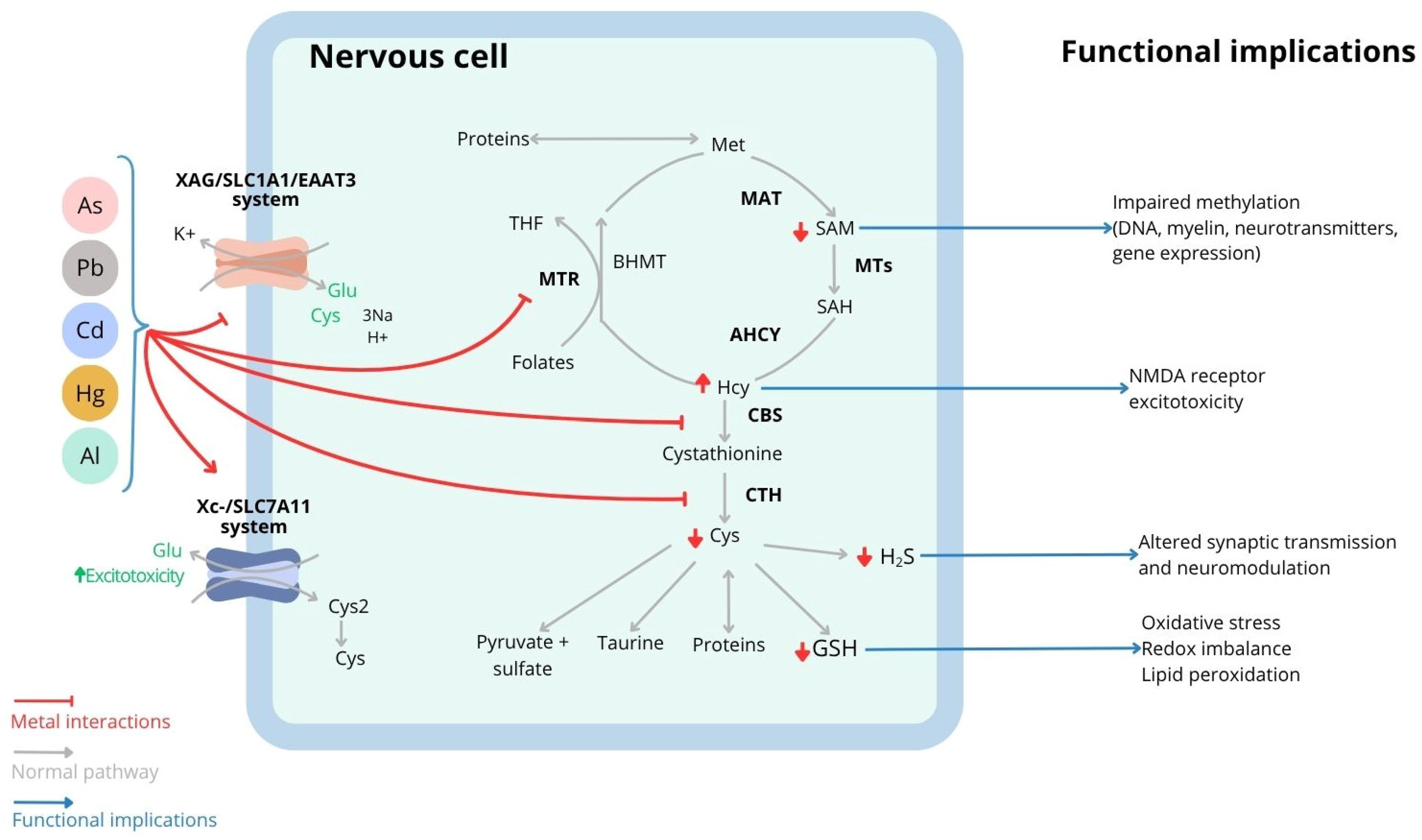
2.2.1. SCAA Metabolism
2.2.2. Genes Involved in SCAA Homeostasis Are Differentially Expressed Across the Brain
2.3. Role of SCAA in Brain Physiology
3. Metal-Induced Neurotoxicity and Potential Contribution of SCAA Dysregulation
3.1. Convergent Mechanisms of Metal-Induced Neurotoxicity
3.2. Toxic Metals Disrupt Sulfur Amino Acid Homeostasis
3.2.1. Cadmium (Cd)

| Population | Exposure Levels | Main Findings | References |
|---|---|---|---|
| Cadmium | |||
| China | |||
| Smoking (n = 61) Non-smoking adult (n = 98) | Blood median (IQR) Male: 1.11 (0.55–2.75) µg/dL, and female: 0.72 (0.29–1.33) µg/dL | Blood Cd levels are positively associated with blood Hcy. Higher Hcy levels in the smoking vs. non-smoking population | [90] |
| Arsenic (As) | |||
| Argentina | |||
| Lung cancer patient (n = 109) Control (n = 141) | Water < 200 µg/L | Lung cancer risk increased with the increase in MMA levels (%) in urine. The risk is associated with CBS SNP. | [94] |
| Brazil | |||
| 35–50 years old Taxi drivers (n = 42) Non-smoking men controls (n = 27) | Blood levels Drivers: 14.87 ± 1.01 µg/L Control: 9.26 ± 0.96 µg/L | As levels are positively associated with Hcy and proinflammatory cytokines (IL-1β, IL-6, TNF-α) and negatively associated with GPx activity, in taxi drivers | [95] |
| Mexico | |||
| Pregnant women (n = 197) | Water: 24.7 (0.33–235.6) µg/L | Vitamin B12 deficiency in the population. Maternal vitamin B9/B12 levels positively correlate with urine MMAs, and DMAs in cord serum. | [96] * |
| India | |||
| 18–86 years old Populations with access to drinking water with low As vs. high As levels Control (n = 193) Exposed (n = 226) | Water: Control < 10 µg/L As exposure > 50 µg/L Urinay Control male: 5 ± 5.15 µg/L, Control female: 5.2 ± 5.9 µg/L As-exposed male: 65.4 ± 82.3 µg/L As-exposed female: 51.7 ± 51.1 µg/L | Vitamin B9/B12 deficiency increased with As consumption. As decreased Cys and increased Hcy plasmatic levels | [97] * |
| Bangladesh | |||
| 20–65 years old (n = 1650) 6-year-old children (n = 165) 30–65 years old (n = 353) | Water: <650 µg/L Urine-As Female: 97.6 ± 119.7 µg/L Male: 121.4 ± 140.4 µg/L Median (Range) Water: 114 (0–700) µg/L Urine: 124 (3–1990) µg/L Blood: 10.8 (1.2–57.0) µg/L | Higher prevalence of hyperhomocysteinemia. As levels in water are negatively associated with plasma folate. Urine DMA is positively associated with folate levels and negatively associated with Hcy. Marginal folate levels in 20% of children. Urine iAs (%) inversely correlated with blood folate and Cys levels. Blood SAM levels are negatively associated with urinary As. Vitamin B9/B12 status modified this association, no association with SAH | [98] * [99] * [100] * [101] * |
| Taiwan | |||
| >40 years old Patient with carotid atherosclerosis (n = 163) Control (n = 163) | Water < 3590 µg/L | Cumulative As exposure positively correlated with blood Hcy levels. High Hcy and MMA% levels increased the risk of developing atherosclerosis 5.4-fold. | [102] * |
| Lead (Pb) | |||
| USA | |||
| >20 years old (n = 4089) >20 years old (n = 4482) 20–59 years old (n = 2492) men > 55 years old (n = 1218) New York, women 18–44 years old (n = 259) Maryland older adults 50–70 years old (n = 1037) | Blood Mean ± SD (Range): 2.1 ± 1.8 (0.2–33.0) µg/dL Blood Mean ± SEM: 1.75 ± 0.04 μg/dL Blood Mean ± SEM: 2.88 ± 0.13 µg/dL Blood Mean ± SD: 4.9 ± 2.7 μg/dL Blood Mean (95% CI): 0.91 (0.86–0.96) μg/dL Blood Mean ± SD: 3.5 ± 2.4 µg/dL | Blood Pb levels positively correlated with blood Hcy in vitamin B6, B9, B12-deficient population. Increased hyperhomocysteinemia prevalence. Pb levels were strongly correlated with blood Hcy, C reactive protein, cholesterol, and BMI in patients with cardiovascular disease. | [103,104,105,106,107] * [108] |
| Pakistan | |||
| Low-income population 18–60 years old (n = 872) | Blood median (IQR): 10.82 µg/dL (8.29–13.60) | Subjects in the higher Pb levels quartile presented higher levels of Hcy. Model was adjusted for age, gender, folate and vitamin B12. | [109] * |
| Poland | |||
| Occupationally exposed men (20–60 years old) (n = 231) Metal workers (25–55 years old) (n = 183) | Blood Median (95% CI): 2.56 (0.9–4.72) μg/dL Blood Range Low Exposure: 20–45 μg/dL High exposure: 45–60 μg/dL | High Pb levels positively correlated with Hcy levels, C reactive protein, fibrinogen. Workers with higher Pb levels had higher blood Hcy, high carbonylated proteins, and lower blood GSH levels. | [110,111] |
| South Korea | |||
| 41–71 years old (n = 386) | Blood Mean ± SD: 4.4 ±1.9 µg/dL | Blood Pb levels positively correlated with plasma Hcy. Polymorphism in CBS, MTR and MTHFR did not show interaction with blood levels. | [112] |
| Ukraine | |||
| Occupationally exposed men (38–47 years old) (n = 146) Control (n = 57) | Blood Mean ± SEM: High exposure: 2.12 ± 0.56 μmol/L Low exposure: 1.72 ± 0.03 μmol/L | Higher Pb and Hcy levels in patients with cardiovascular manifestations. Positive correlation between Hcy and Pb levels. | [113] |
| Singapore and Vietnam | |||
| Battery factory workers (19–66 years old) Singapore (n = 183) Vietnam (n = 323) | Blood Mean (Range): 22.7 (2.0–66.9) µg/dL | Positive correlation between blood Pb and plasma Hcy. | [114] |
| Brazil | |||
| 35–50 years old Taxi drivers (n = 42), Non-smoking men controls (n = 27) Male adult (n = 45) | Blood Mean ± SEM: Drivers: 2.60 ± 0.21 µg/dL, Control: 1.95 ± 0.24 µg/dL Blood Mean ± SEM: 11.38 ± 1.92 µg/dL | Pb levels are positively associated with Hcy and proinflammatory cytokine levels (IL-1β, IL-6, RNF-α). Subjects with blood Pb > 5 µg/dL had lower plasmatic H2S levels. | [66,95] |
| China | |||
| Smoking (n = 61), Non-smoking adult (n = 98) | Blood Median (IQR) male: 35.91 (23.45–48.77) μg/dL female: 27.10 (21.46–32.76) μg/dL | Hcy levels correlated with Pb levels, especially in smoking men. | [90] |
| India | |||
| Pb-exposed workers male > 18 years old (n = 338) | Blood Range <10 µg/dL, 10–30 µg/dL, 30–50 µg/dL, >50 µg/dL | Blood Pb levels > 10 µg/dL decreased SAM levels and methylation index, while SAH levels increased. Lifestyle, working experience > 5 years and age, synergically influence in SAM levels. | [115] |
| Aluminum | |||
| Italy | |||
| 18–75 years old | Urine >35 μg/g creatinine | Hyperhomocysteinemia. Chelating therapy reduced serum Hcy and ROS levels and increased serum vitamins B9 and B12, increased GSH, antioxidant capacity, and Al elimination. | [116] * |
| Mercury | |||
| Brazil | |||
| 35–50 years old Taxi drivers (n = 42), Non-smoking men controls (n = 27). | Blood Mean ± SEM Drivers: 33.65 ± 2.95 µg/L, Control: 11.8 ± 1.57 µg/L | Increased Hg blood levels in drivers. Hg levels are positively associated with Hcy levels and negatively with GPx activity. Increased IL-1β, IL-6, TNF-α in taxi drivers. | [95] |
| USA | |||
| NHANES study Children 3–5 years (n = 1005) | Blood Q1: ≤0.70 μmol/L, Q2: 0.70–1.50 μmol/L, Q3: 1.50–3.49 μmol/L, Q4: >3.49 μmol/L | Inverse association between Hg and Hcy in boys, but not in girls with lower vitamin B9/B12. | [117] * |
| Oman | |||
| Autistic children (n = 27) Control (n = 27) | Hair (mean ± SD) Autistic: 6.93 ± 0.36 mg/g, Control: 0.611 ± 0.033 mg/g | Vit B9/B12 deficiency in children. Lower serum GSH levels and higher Hcy and SAH levels in autistic children. Hair Hg levels were markedly elevated in autistic subjects and inversely associated with Cys levels. | [70] * |
3.2.2. Arsenic (As)
| Model | Exposure Dose | Main Finding | References |
|---|---|---|---|
| Cadmium (Cd) | |||
| Chang cell line | CdCl2 1 µM for 1 h + fresh medium for up to 24 h | ↑ CTH protein and H2S production in a time-dependent manner, inducing an adaptative response to radiation. | [136] |
| C2C12 cell line (Murine Myoblast) | CdCl2: 1–100 µM (30 µM) for 24 h | ↑ CTH expression and H2S production, inhibition of CTH aggravate Cd-induced apoptosis. | [137] |
| C57BL/6J mice and CTH KO (C57BL/6J background) | CdCl2 5 mg/kg (i.p) for 24 h | RSS produced by CTH neutralized Cd through the formation of CdS. CTH deletion enhanced Cd-induced hepatotoxicity. | [138] |
| Male Sprague-Dawley rats | CdCl2 0.6 mg/kg/day (s.c), 5 day/week for 6 weeks | ↓ SAM, cystathionine, MAT2B and CBS proteins in the kidney, associated with nephrotoxicity and oxidative stress (↑ MDA, 4HNE and ↓ GSH). | [92] |
| Arsenic (As) | |||
| RWPE1 human non-tumorigenic prostate cell line (do not readily methylate As) | NaAsO2 5 µM up to 16 weeks | ↑ MAT2B, SAHH, CBS, GSH-synthesis enzymes, and MRP1 transcript in a time-dependent manner. ↓ MAT2A transcript but returned to normal levels by 12 weeks. ↓ BHMT transcript. | [131] |
| SH-S5Y5 cells | NaAsO2 Up to 10 µM | ↓ GSH and Cys levels and inhibited MTR activity. | [130] |
| N2a cell line (neuron) + N9 cell line (microglia) | NaAsO2 10 and 20 µM | As induces extracellular Cys depletion by N9 cells, dependent on SLC7A11 overexpression in N9 cell, leading to neuron oxidative cell death. | [71] |
| HepG2 hepatocellular carcinoma cell line Macaca fascicularis | NaAsO2 3.75, 7.5, 15 µM for 24 h NaAsO2 1 mg/kg/day for 28 days | ↓ CBS and GPx1 protein in cell line and ↓ monkey liver. | [139] |
| CH3 mice | NaAsO2 85 mg/L from embryonic day 8–18 (model of hepatocellular carcinoma) | In fetal liver: ↓ MAT1A and BHMT expression. In newborn liver: ↑ GSH-synthesis genes and metallothionein expression and ↓ BHMT expression | [132] [133] |
| Male ICR mice | Realgar 0.15, 0.45, 1.35 g/kg/day (i.g) for 8 weeks | ↓ GSH, RSS, cysteine, and cystine transporters in the hippocampus. Alteration associated with ultrastructural changes and cognitive deficiencies (NOR test). | [72] |
| C57BL/6 mice | NaAsO2 5–10 mg/L for 6 months | ↓ SAM levels, MTR expression and activity in liver, and vitamin B9 and B12 (especially at 5 mg/L) in serum, associated with lipid accumulation and inflammation in liver, and microbiota dysbiosis (↑ vitamin B9/12 producing bacteria). Serum: ↓ Met, SAM levels, ↑ SAH Liver: ↓ SAM associated with ↓ m6A RNA modification, inducing ↑ fatty acid synthesis and NAFLD phenotype. | [140,141] |
| CD1 mice | NaAsO2 20 mg/L from gestation until 3 months after birth NaAsO2: 2 mg/L + NaF: 25 mg/L (oral) from gestation until 3 months after birth | ↑ GSH levels in cortex and hippocampus, associated with ↑ SLC1A1, SLC7A5 and SLC7A11 protein, and alteration in glutamate receptor and cognitive impairment (LOR test) As and As+F ↓ the activity of the transsulfuration pathway in the cortex and hippocampus at 1 month and 3 months after birth, associated with cognitive impairment (NOR/LOR test). | [73,86] |
| Female Wistar rat | NaAsO2 3 mg/L from gestation up to 4 months old | ↓ SAM and phosphatidylcholine levels and ↑ choline levels in the liver, associated with histological alteration in liver. No changes in the brain, although important myelin and nerve alteration in the striatum were observed. | [142] |
| Male Sprague Dawley rat | NaAsO2 5, 10, 50 mg/L for 6 months | As (10 and 50 ppm) ↑ Hcy levels in the brain and serum and induced histopathological changes in CA1, endoplasmic reticulum stress and memory impairment (MWM test). | [143] |
| Lead (Pb) | |||
| SH-S5Y5 cell line | Pb(NO3)2 100 nM for 60 min | ↓ GSH levels and Cys uptake and inhibited MTR activity. | [130] |
| C57BL/6J mice | PbAc 30 mg/L 100 mg/kg (oral) from P21–P60 Followed by alcohol seeking paradigm. | Pb ↑ propensity to alcohol seeking relapses, associated with ↑ SLC7A11 expression in nucleus accumbens and dorso-lateral striatum, and aberrant glutamatergic transmission | [74] |
| Rat | PbAc 120 mg/kg (oral) for 60 days | ↓ Cys and Met levels in the liver. | [144] |
| Fisher 344 rats | PbAc 2000 mg/L for 5 weeks | ↓ Cys and GSH, and ↑ MDA levels in rat lenses | [145] |
| Male Wistar rat | PbAc 100 mg/kg (oral) for 1 and 3 months | ↓ H2S level, CTH expression, and GSH levels in the kidney. ↑ oxidative stress, inflammation, and ↓ H2S and CTH protein levels in the liver. NaHS treatment recovered GSH levels, ↑ CTH expression, ↓ liver damage and inflammation, and reduced renal toxicity. | [146,147] |
| Aluminum (Al) | |||
| SH-SY5Y cell line | Up to 10 µM | ↓ GSH levels and Cys uptake and inhibited MTR activity. | [130] |
| Male Wistar rat | AlCl3 50 mg/kg/day (oral) for 3 months | ↑ blood Hcy levels, AchE, BACE1, and IL1β levels in the brain. Induced cognitive impairment (MWM test). | [148] |
| Male Wistar rat (Al-induced AD model) | AlCl3 17 mg/kg/day (oral) for 4 weeks | ↓ antioxidant capacity, acetylcholine, and dopamine levels, ↑ Hcy levels, induced histological degeneration in the cortex and cerebellum, and behavioral impairment (OFT). | [149] |
| Mercury | |||
| HeLa cell line | HgCl2 1–100 µM for 24 h | At 10 µM: ↑ GSH levels, Cys and Hcy utilization, and GSSG release. At 100 µM: ↓ GSH levels and delayed cell growth. | [150,151] |
| Cerebellar granular neuron, Cell line: PC12, NPC1231, SH-S5Y5 | MeHg 2 µM for 24 h | ↓ RSS levels and activated redox signaling. RSS pretreatment attenuated cell damage and oxidative stress | |
| SH-S5Y5 cell line | TH: 10 µM HgCl2: 10 µM NaHS (500 µM) for 12 h +/− MeHg (0.5–2 µM) for 24 h | ↓ MTR activity by a redox-induced GSCbI/MeCbl cofactor imbalance and indirectly through IGF1 and dopamine-mediated signaling. Pretreatment with NaHS or CBS overexpression suppressed MeHg toxicity. Endogenous RSS interacts in vitro with MeHg to form an inert metabolite (MeHg)2S | [129,130] |
| ICR mice | HgCl2 1.5, 3, and 4.5 mg/kg/day from embryonic day 5.5–9.5 | Hg alters SCAA flux between placenta and embryos. In placenta: ↑ oxidative stress, SLC7A5 and SLC7A11 expression. In embryos: ↑ Cys and Met, but ↓ SAM and Hcy SCAA metabolism imbalance in embryo is associated with neural tube defect. | [152] |
| C57BL/6J mice and CTH KO (C57BL/6J background) | MeHg 5 mg/kg/day for 12 days (oral) | CTH KO mice: ↑ MeHg levels in the cerebellum, and higher susceptibility to MeHg-induced motor impairment (rotarod test) and cerebellum damage | [75] |
| Rat | HgCl2 1 mg/kg/day for 45 days (s.c) | ↑ Cys levels in a time-dependent manner. ↑ Met levels increased after 15 days but returned to control levels on the 45th day. | [144] |
| Small Indian mongoose (Herpestes auropunctatus) | Natural exposure Liver MeHg levels Mean (range): 12.7 (1.75–55.5) µg/g | Mongoose expresses CBS/CTH in different tissues and produces significant RSS levels, including CySSH, H2S, and (MeHg)2S metabolites. | [153] |
3.2.3. Lead (Pb)
3.2.4. Aluminum (Al)
3.2.5. Mercury (Hg)
4. Dysregulation of SCAA Metabolism in Neurological Disorders
4.1. Alzheimer’s Disease (AD)
4.2. Huntington’s Disease (HD)
4.3. Parkinson Disease (PD)
4.4. Multiple Sclerosis (MS)
4.5. Autism Spectrum Disorder (ASD)
5. Therapeutic Implications and Future Perspectives
6. Concluding Remarks
Supplementary Materials
Author Contributions
Funding
Institutional Review Board Statement
Informed Consent Statement
Data Availability Statement
Conflicts of Interest
Abbreviations
| AD | Alzheimer Disease |
| AHCY | S-adenosylhomocysteine hydrolase, also SAHH |
| Al | Aluminum |
| APP/PS1 mice | Doble transgenic strain with mutations in amyloid precursor protein and presenilin 1 |
| As | Arsenic |
| ASC | Alanine serine cysteine transporter |
| ASD | Autism Spectrum Disorder |
| As3MT | Arsenite (As3+) methyltransferase |
| Aβ | Amyloid Beta |
| BACE1 | Beat secretase 1 (beta-site amyloid precursor protein cleaving enzyme 1) |
| BHMT | Betaine homocysteine methyl transferase |
| CBS | Cystathionine β-synthase |
| Cd | Cadmium |
| CI | Confidence interval |
| CNS | Central Nervous System |
| CTH | Cystathionine γ-lyase, also CSE (cystathionase) |
| Cys | Cysteine |
| CySSH | Thiocysteine |
| DATS | Diallyl trisulfide |
| DNA | Deoxyribonucleic acid |
| DMA | Dimethyl arsenic |
| DOPA | 3,4-dihydroxyphenylalanine |
| GCLc | Gamma-glutamyl-cysteinyl-ligase |
| GPx | Glutathione peroxidase |
| GSH | Glutathione |
| GSSG | Glutathione oxidized |
| H2S | Hydrogen sulfide |
| Hcy | Homocysteine |
| HD | Huntington Disease |
| Hg | Mercury |
| IFNy | Interferon gamma |
| IL1β | Interleukin 1 beta |
| IQR | Interquartile range |
| KO | Knock out |
| MeHg | Methylmercury |
| Met | Methionine |
| MMA | Monomethyl arsenic |
| MAT | Methionine adenosyl transferase |
| MAT1 | Methionine adenosyl transferase isoform type 1 |
| MAT2 | Methionine adenosyl transferase isoform type 2 |
| MAT2A | Methionine adenosyl transferase subunit 2A (catalytic) |
| MAT2b | Methionine adenosyl transferase subunit 2Bb (regulatory) |
| MPR1 | Multidrug resistance-associated protein 1 |
| MS | Multiple Sclerosis |
| MTR | Methionine synthase |
| MTHFR | Methyl tetrahydrofolate reductase |
| NaHS | Sodium hydrosulfide |
| NMDA | N-methyl-D Aspartate |
| NR2A | NMDA receptor subunit 2A |
| NR2B | NMDA receptor subunit 2B |
| Pb | Lead |
| PD | Parkinson Disease |
| ROS | Reactive Oxygen Species |
| RNA | Ribonucleic acid |
| RNS | Reactive Nitrogen Species |
| RSS | Reactive Sulfur Species |
| SAH | S-adenosyl-homocysteine |
| SAM | S Adenosyl methionine |
| SD | Standard deviation |
| SCAA | Sulfur-containing amino acid |
| SE | Standard error |
| SEM | Standard error of the mean |
| -SH | Sulfhydryl group/thiol group |
| SLC1 | Solute carrier family 1 |
| SLC1A1 | Solute carrier family 1 member 1, also EAAC1 or EAAT3 (excitatory amino acid carrier or transporter) |
| SLC1A4 | Solute carrier family 1 member 1, also ASCT1 (alanine serine cysteine transporter 1) |
| SLC6 | Solute carrier family 6 |
| SLC6A6 | Solute carrier family 6 member 6, also TauT (taurine transporter) |
| SLC7 | Solute carrier family 7 |
| SLC7A5 | Solute carrier family 7 member 5, also LAT1 (large amino acid transporter 1) |
| SLC7A6 | Solute carrier family 7 member 6 |
| SLC7A8 | Solute carrier family 7 member 6, also LAT2 (large amino acid transporter 2) |
| SLC7A11 | Solute carrier family 7 member 6, also xCT (glutamate cystine transporter) |
| SNP | Single nucleotide polymorphism |
| THF | Tetrahydrofolate |
| TNFα | Tumor necrosis factor alpha |
| TSP | Transsulfuration pathway |
| XAG | Aspartic acid-glutamate transport system |
| Xc | Cystine system |
References
- Brosnan, J.T.; Brosnan, M.E. The Sulfur-Containing Amino Acids: An Overview. J. Nutr. 2006, 136, 1636S–1640S. [Google Scholar] [CrossRef] [PubMed]
- Corona-Trejo, A.; Gonsebatt, M.E.; Trejo-Solis, C.; Campos-Peña, V.; Quintas-Granados, L.I.; Villegas-Vázquez, E.Y.; Daniel Reyes-Hernández, O.; Hernández-Abad, V.J.; Figueroa-González, G.; Silva-Adaya, D. Transsulfuration pathway: A targeting neuromodulator in Parkinson’s disease. Rev. Neurosci. 2023, 34, 915–932. [Google Scholar] [CrossRef] [PubMed]
- Paul, B.D.; Sbodio, J.I.; Snyder, S.H. Cysteine Metabolism in Neuronal Redox Homeostasis. Trends Pharmacol. Sci. 2018, 39, 513–524. [Google Scholar] [CrossRef] [PubMed]
- Paul, B.D.; Sbodio, J.I.; Xu, R.; Vandiver, M.S.; Cha, J.Y.; Snowman, A.M.; Snyder, S.H. Cystathionine γ-lyase deficiency mediates neurodegeneration in Huntington’s disease. Nature 2014, 509, 96–100. [Google Scholar] [CrossRef] [PubMed]
- Netto, L.E.S.; de Oliveira, M.A.; Monteiro, G.; Demasi, A.P.D.; Cussiol, J.R.R.; Discola, K.F.; Demasi, M.; Silva, G.M.; Alves, S.V.; Faria, V.G.; et al. Reactive cysteine in proteins: Protein folding, antioxidant defense, redox signaling and more. Comp. Biochem. Physiol. Part C Toxicol. Pharmacol. 2007, 146, 180–193. [Google Scholar] [CrossRef] [PubMed]
- Poole, L.B. The basics of thiols and cysteines in redox biology and chemistry. Free Radic. Biol. Med. 2015, 80, 148–157. [Google Scholar] [CrossRef] [PubMed]
- Fujii, J.; Osaki, T.; Soma, Y.; Matsuda, Y. Critical Roles of the Cysteine-Glutathione Axis in the Production of γ-Glutamyl Peptides in the Nervous System. Int. J. Mol. Sci. 2023, 24, 8044. [Google Scholar] [CrossRef] [PubMed]
- Stipanuk, M.H. Sulfur amino acid metabolism: Pathways for production and removal of homocysteine and cysteine. Annu. Rev. Nutr. 2004, 24, 539–577. [Google Scholar] [CrossRef] [PubMed]
- Vitvitsky, V.; Thomas, M.; Ghorpade, A.; Gendelman, H.; Banerjee, R. A Functional Transsulfuration Pathway in the Brain Links to Glutathione Homeostasis. J. Biol. Chem. 2006, 281, 35785–35793. [Google Scholar] [CrossRef] [PubMed]
- McBean, G.J. Cysteine, Glutathione, and Thiol Redox Balance in Astrocytes. Antioxidants 2017, 6, 62. [Google Scholar] [CrossRef] [PubMed]
- Garza-Lombó, C.; Posadas, Y.; Quintanar, L.; Gonsebatt, M.E.; Franco, R. Neurotoxicity Linked to Dysfunctional Metal Ion Homeostasis and Xenobiotic Metal Exposure: Redox Signaling and Oxidative Stress. Antioxid. Redox Signal. 2018, 28, 1669–1703. [Google Scholar] [CrossRef] [PubMed]
- Ahmed, G.; Rahaman, M.S.; Perez, E.; Khan, K.M. Associations of Environmental Exposure to Arsenic, Manganese, Lead, and Cadmium with Alzheimer’s Disease: A Review of Recent Evidence from Mechanistic Studies. J. Xenobiotics 2025, 15, 47. [Google Scholar] [CrossRef] [PubMed]
- Chin-Chan, M.; Navarro-Yepes, J.; Quintanilla-Vega, B. Environmental pollutants as risk factors for neurodegenerative disorders: Alzheimer and Parkinson diseases. Front. Cell. Neurosci. 2015, 9, 124. [Google Scholar] [CrossRef] [PubMed]
- Rehman, Q.; Rehman, K.; Akash, M.S.H. Heavy Metals and Neurological Disorders: From Exposure to Preventive Interventions. In Environmental Contaminants and Neurological Disorders; Akash, M.S.H., Rehman, K., Eds.; Springer International Publishing: Cham, Switzerland, 2021; pp. 69–87. [Google Scholar]
- Rana, A.; Katiyar, A.; Arun, A.; Berrios, J.N.; Kumar, G. Natural sulfur compounds in mental health and neurological disorders: Insights from observational and intervention studies. Front. Nutr. 2025, 12, 1534000. [Google Scholar] [CrossRef] [PubMed]
- Gao, J.; Cahill, C.M.; Huang, X.; Roffman, J.L.; Lamon-Fava, S.; Fava, M.; Mischoulon, D.; Rogers, J.T. S-Adenosyl Methionine and Transmethylation Pathways in Neuropsychiatric Diseases Throughout Life. Neurotherapeutics 2018, 15, 156–175. [Google Scholar] [CrossRef] [PubMed]
- Giuliani, D.; Ottani, A.; Zaffe, D.; Galantucci, M.; Strinati, F.; Lodi, R.; Guarini, S. Hydrogen sulfide slows down progression of experimental Alzheimer’s disease by targeting multiple pathophysiological mechanisms. Neurobiol. Learn. Mem. 2013, 104, 82–91. [Google Scholar] [CrossRef] [PubMed]
- Koehler, D.; Shah, Z.A.; Hensley, K.; Williams, F.E. Lanthionine ketimine-5-ethyl ester provides neuroprotection in a zebrafish model of okadaic acid-induced Alzheimer’s disease. Neurochem. Int. 2018, 115, 61–68. [Google Scholar] [CrossRef] [PubMed]
- Paul, B.D.; Snyder, S.H. Gasotransmitter hydrogen sulfide signaling in neuronal health and disease. Biochem. Pharmacol. 2018, 149, 101–109. [Google Scholar] [CrossRef] [PubMed]
- Mulica, P.; Grünewald, A.; Pereira, S.L. Astrocyte-Neuron Metabolic Crosstalk in Neurodegeneration: A Mitochondrial Perspective. Front. Endocrinol. 2021, 12, 668517. [Google Scholar] [CrossRef] [PubMed]
- Valdovinos-Flores, C.; Gonsebatt, M.E. The role of amino acid transporters in GSH synthesis in the blood-brain barrier and central nervous system. Neurochem. Int. 2012, 61, 405–414. [Google Scholar] [CrossRef] [PubMed]
- Kanai, Y.; Clémençon, B.; Simonin, A.; Leuenberger, M.; Lochner, M.; Weisstanner, M.; Hediger, M.A. The SLC1 high-affinity glutamate and neutral amino acid transporter family. Mol. Asp. Med. 2013, 34, 108–120. [Google Scholar] [CrossRef] [PubMed]
- Verrey, F.; Closs, E.I.; Wagner, C.A.; Palacin, M.; Endou, H.; Kanai, Y. CATs and HATs: The SLC7 family of amino acid transporters. Pflug. Arch. Eur. J. Physiol. 2004, 447, 532–542. [Google Scholar] [CrossRef] [PubMed]
- Sosnoski, H.M.; Sears, S.M.S.; He, Y.; Frare, C.; Hewett, S.J. Sexually dimorphic and brain region-specific transporter adaptations in system xc− null mice. Neurochem. Int. 2020, 141, 104888. [Google Scholar] [CrossRef] [PubMed]
- Williams, L.E.; Featherstone, D.E. Regulation of hippocampal synaptic strength by glial xCT. J. Neurosci. 2014, 34, 16093–16102. [Google Scholar] [CrossRef] [PubMed]
- Sears, S.M.S.; Roberts, S.H.; Hewett, S.J. Hyperexcitability and brain morphological differences in mice lacking the cystine/glutamate antiporter, system xc−. J. Neurosci. Res. 2021, 99, 3339–3353. [Google Scholar] [CrossRef] [PubMed]
- Ramírez-Guerrero, S.; Guardo-Maya, S.; Medina-Rincón, G.J.; Orrego-González, E.E.; Cabezas-Pérez, R.; González-Reyes, R.E. Taurine and Astrocytes: A Homeostatic and Neuroprotective Relationship. Front. Mol. Neurosci. 2022, 15, 937789. [Google Scholar] [CrossRef] [PubMed]
- Sbodio, J.I.; Snyder, S.H.; Paul, B.D. Regulators of the transsulfuration pathway. Br. J. Pharmacol. 2019, 176, 583–593. [Google Scholar] [CrossRef] [PubMed]
- Bolaños, J.P.; Magistretti, P.J. The neuron-astrocyte metabolic unit as a cornerstone of brain energy metabolism in health and disease. Nat. Metab. 2025, 7, 2414–2423. [Google Scholar] [CrossRef] [PubMed]
- Jessa, S.; Blanchet-Cohen, A.; Krug, B.; Vladoiu, M.; Coutelier, M.; Faury, D.; Poreau, B.; De Jay, N.; Hebert, S.; Monlong, J.; et al. Stalled developmental programs at the root of pediatric brain tumors. Nat. Genet. 2019, 51, 1702–1713. [Google Scholar] [CrossRef] [PubMed]
- Ianevski, A.; Giri, A.K.; Aittokallio, T. Fully-automated and ultra-fast cell-type identification using specific marker combinations from single-cell transcriptomic data. Nat. Commun. 2022, 13, 1246. [Google Scholar] [CrossRef] [PubMed]
- Knaus, L.S.; Basilico, B.; Malzl, D.; Gerykova Bujalkova, M.; Smogavec, M.; Schwarz, L.A.; Gorkiewicz, S.; Amberg, N.; Pauler, F.M.; Knittl-Frank, C.; et al. Large neutral amino acid levels tune perinatal neuronal excitability and survival. Cell 2023, 186, 1950–1967.E25. [Google Scholar] [CrossRef] [PubMed]
- Górny, M.; Wnuk, A.; Kamińska, A.; Kamińska, K.; Chwatko, G.; Bilska-Wilkosz, A.; Iciek, M.; Kajta, M.; Rogóż, Z.; Lorenc-Koci, E. Glutathione Deficiency and Alterations in the Sulfur Amino Acid Homeostasis during Early Postnatal Development as Potential Triggering Factors for Schizophrenia-Like Behavior in Adult Rats. Molecules 2019, 24, 4253. [Google Scholar] [CrossRef] [PubMed]
- Ramani, K.; Lu, S.C. Methionine adenosyltransferases in liver health and diseases. Liver Res. 2017, 1, 103–111. [Google Scholar] [CrossRef] [PubMed]
- Jena, A.; Montoya, C.A.; Mullaney, J.A.; Dilger, R.N.; Young, W.; McNabb, W.C.; Roy, N.C. Gut-Brain Axis in the Early Postnatal Years of Life: A Developmental Perspective. Front. Integr. Neurosci. 2020, 14, 44. [Google Scholar] [CrossRef] [PubMed]
- Lonsdale, J.; Thomas, J.; Salvatore, M.; Phillips, R.; Lo, E.; Shad, S.; Hasz, R.; Walters, G.; Garcia, F.; Young, N.; et al. The Genotype-Tissue Expression (GTEx) project. Nat. Genet. 2013, 45, 580–585. [Google Scholar] [CrossRef] [PubMed]
- R Team Core. R: A Language and Environment for Statistical Computing; R Foundation for Statistical Computing: Vienna, Austria, 2023. [Google Scholar]
- Warwick, A.N.; Zuckerman, B.; Ung, C.Y.; Luben, R.; Olvera-Barrios, A. gtexr: A convenient R interface to the Genotype-Tissue Expression (GTEx) Portal API. J. Open Source Softw. 2025, 10, 8249. [Google Scholar] [CrossRef]
- Huang, F.; Chen, X.; Jiang, X.; Niu, J.; Cui, C.; Chen, Z.; Sun, J. Betaine ameliorates prenatal valproic-acid-induced autism-like behavioral abnormalities in mice by promoting homocysteine metabolism. Psychiatry Clin. Neurosci. 2019, 73, 317–322. [Google Scholar] [CrossRef] [PubMed]
- Singhal, N.K.; Sternbach, S.; Fleming, S.; Alkhayer, K.; Shelestak, J.; Popescu, D.; Weaver, A.; Clements, R.; Wasek, B.; Bottiglieri, T.; et al. Betaine restores epigenetic control and supports neuronal mitochondria in the cuprizone mouse model of multiple sclerosis. Epigenetics 2020, 15, 871–886. [Google Scholar] [CrossRef] [PubMed]
- Enokido, Y.; Suzuki, E.; Iwasawa, K.; Namekata, K.; Okazawa, H.; Kimura, H. Cystathionine beta-synthase, a key enzyme for homocysteine metabolism, is preferentially expressed in the radial glia/astrocyte lineage of developing mouse CNS. FASEB J. Off. Publ. Fed. Am. Soc. Exp. Biol. 2005, 19, 1854–1856. [Google Scholar] [CrossRef] [PubMed]
- Robert, K.; Vialard, F.; Thiery, E.; Toyama, K.; Sinet, P.M.; Janel, N.; London, J. Expression of the cystathionine beta synthase (CBS) gene during mouse development and immunolocalization in adult brain. J. Histochem. Cytochem. Off. J. Histochem. Soc. 2003, 51, 363–371. [Google Scholar] [CrossRef] [PubMed]
- Castillo, P.E.; Jung, H.; Klann, E.; Riccio, A. Presynaptic Protein Synthesis in Brain Function and Disease. J. Neurosci. Off. J. Soc. Neurosci. 2023, 43, 7483–7488. [Google Scholar] [CrossRef] [PubMed]
- Desai, M.; Sun, B. Positions of cysteine residues reveal local clusters and hidden relationships to Sequons and Transmembrane domains in Human proteins. Sci. Rep. 2024, 14, 25886. [Google Scholar] [CrossRef] [PubMed]
- Trivedi, M.V.; Laurence, J.S.; Siahaan, T.J. The role of thiols and disulfides on protein stability. Curr. Protein Pept. Sci. 2009, 10, 614–625. [Google Scholar] [CrossRef] [PubMed]
- Ulrich, K.; Jakob, U. The role of thiols in antioxidant systems. Free Radic. Biol. Med. 2019, 140, 14–27. [Google Scholar] [CrossRef] [PubMed]
- Li, B.; Ming, H.; Qin, S.; Nice, E.C.; Dong, J.; Du, Z.; Huang, C. Redox regulation: Mechanisms, biology and therapeutic targets in diseases. Signal Transduct. Target. Ther. 2025, 10, 72. [Google Scholar] [CrossRef] [PubMed]
- Nagpure, B.V.; Bian, J.-S. Brain, Learning, and Memory: Role of H2S in Neurodegenerative Diseases. In Chemistry, Biochemistry and Pharmacology of Hydrogen Sulfide; Moore, P.K., Whiteman, M., Eds.; Springer International Publishing: Cham, Switzerland, 2015; pp. 193–215. [Google Scholar]
- Percio, A.; Cicchinelli, M.; Masci, D.; Summo, M.; Urbani, A.; Greco, V. Oxidative Cysteine Post Translational Modifications Drive the Redox Code Underlying Neurodegeneration and Amyotrophic Lateral Sclerosis. Antioxidants 2024, 13, 883. [Google Scholar] [CrossRef] [PubMed]
- Wang, P.; Wang, F.; Ni, L.; Wu, P.; Chen, J. Targeting redox-altered plasticity to reactivate synaptic function: A novel therapeutic strategy for cognitive disorder. Acta Pharm. Sin. B 2021, 11, 599–608. [Google Scholar] [CrossRef] [PubMed]
- Forrester, S.J.; Kikuchi, D.S.; Hernandes, M.S.; Xu, Q.; Griendling, K.K. Reactive Oxygen Species in Metabolic and Inflammatory Signaling. Circ. Res. 2018, 122, 877–902. [Google Scholar] [CrossRef] [PubMed]
- Liu, S.; Liu, J.; Wang, Y.; Deng, F.; Deng, Z. Oxidative Stress: Signaling Pathways, Biological Functions, and Disease. MedComm 2025, 6, e70268. [Google Scholar] [CrossRef] [PubMed]
- Nath, K.A.; Salahudeen, A.K. Autoxidation of cysteine generates hydrogen peroxide: Cytotoxicity and attenuation by pyruvate. Am. J. Physiol.-Ren. Physiol. 1993, 264, F306–F314. [Google Scholar] [CrossRef] [PubMed]
- D’Emilia, D.M.; Lipton, S.A. Ratio of S-nitrosohomocyst(e)ine to homocyst(e)ine or other thiols determines neurotoxicity in rat cerebrocortical cultures. Neurosci. Lett. 1999, 265, 103–106. [Google Scholar] [CrossRef] [PubMed]
- Jara-Prado, A.; Ortega-Vazquez, A.; Ruano, L.M.; Rios, C.; Santamaria, A. Homocysteine-induced brain lipid peroxidation: Effects of NMDA receptor blockade, antioxidant treatment, and nitric oxide synthase inhibition. Neurotox. Res. 2003, 5, 237–243. [Google Scholar] [CrossRef] [PubMed]
- Mukwevho, E.; Ferreira, Z.; Ayeleso, A. Potential Role of Sulfur-Containing Antioxidant Systems in Highly Oxidative Environments. Molecules 2014, 19, 19376–19389. [Google Scholar] [CrossRef] [PubMed]
- Park, S.Y.; Kim, K.Y.; Gwak, D.S.; Shin, S.Y.; Jun, D.Y.; Kim, Y.H. L-Cysteine mitigates ROS-induced apoptosis and neurocognitive deficits by protecting against endoplasmic reticulum stress and mitochondrial dysfunction in mouse neuronal cells. Biomed. Pharmacother. 2024, 180, 117538. [Google Scholar] [CrossRef] [PubMed]
- Vitvitsky, V.; Kabil, O.; Banerjee, R. High turnover rates for hydrogen sulfide allow for rapid regulation of its tissue concentrations. Antioxid. Redox Signal. 2012, 17, 22–31. [Google Scholar] [CrossRef] [PubMed]
- Hanna, D.A.; Chen, B.; Shah, Y.M.; Khalimonchuk, O.; Cunniff, B.; Banerjee, R. H(2)S remodels mitochondrial ultrastructure and destabilizes respiratory supercomplexes. J. Biol. Chem. 2025, 301, 108433. [Google Scholar] [CrossRef] [PubMed]
- Xiao, Q.; Ying, J.; Xiang, L.; Zhang, C. The biologic effect of hydrogen sulfide and its function in various diseases. Medicine 2018, 97, e13065. [Google Scholar] [CrossRef] [PubMed]
- Nada, S.E.; Tulsulkar, J.; Raghavan, A.; Hensley, K.; Shah, Z.A. A derivative of the CRMP2 binding compound lanthionine ketimine provides neuroprotection in a mouse model of cerebral ischemia. Neurochem. Int. 2012, 61, 1357–1363. [Google Scholar] [CrossRef] [PubMed]
- Dupuis, J.P.; Nicole, O.; Groc, L. NMDA receptor functions in health and disease: Old actor, new dimensions. Neuron 2023, 111, 2312–2328. [Google Scholar] [CrossRef] [PubMed]
- McCully, K.S. Chemical Pathology of Homocysteine. IV. Excitotoxicity, Oxidative Stress, Endothelial Dysfunction, and Inflammation. Ann. Clin. Lab. Sci. 2009, 39, 219–232. [Google Scholar] [PubMed]
- Lu, M.; Hu, L.F.; Hu, G.; Bian, J.S. Hydrogen sulfide protects astrocytes against H(2)O(2)-induced neural injury via enhancing glutamate uptake. Free Radic. Biol. Med. 2008, 45, 1705–1713. [Google Scholar] [CrossRef] [PubMed]
- Possomato-Vieira, J.S.; Gonçalves-Rizzi, V.H.; Nascimento, R.A.d.; Wandekin, R.R.; Caldeira-Dias, M.; Chimini, J.S.; da Silva, M.L.S.; Dias-Junior, C.A. Clinical and Experimental Evidences of Hydrogen Sulfide Involvement in Lead-Induced Hypertension. BioMed Res. Int. 2018, 2018, 4627391. [Google Scholar] [CrossRef] [PubMed]
- Rafaiee, R.; Khastar, H.; Garmabi, B.; Taleb, M.; Norouzi, P.; Khaksari, M. Hydrogen sulfide protects hippocampal CA1 neurons against lead mediated neuronal damage via reduction oxidative stress in male rats. J. Chem. Neuroanat. 2021, 112, 101917. [Google Scholar] [CrossRef] [PubMed]
- Massie, A.; Boillée, S.; Hewett, S.; Knackstedt, L.; Lewerenz, J. Main path and byways: Non-vesicular glutamate release by system xc(-) as an important modifier of glutamatergic neurotransmission. J. Neurochem. 2015, 135, 1062–1079. [Google Scholar] [CrossRef] [PubMed]
- Nasca, C.; Bigio, B.; Zelli, D.; de Angelis, P.; Lau, T.; Okamoto, M.; Soya, H.; Ni, J.; Brichta, L.; Greengard, P.; et al. Role of the Astroglial Glutamate Exchanger xCT in Ventral Hippocampus in Resilience to Stress. Neuron 2017, 96, 402–413.e5. [Google Scholar] [CrossRef] [PubMed]
- Hodgson, N.W.; Waly, M.I.; Al-Farsi, Y.M.; Al-Sharbati, M.M.; Al-Farsi, O.; Ali, A.; Ouhtit, A.; Zang, T.; Zhou, Z.S.; Deth, R.C. Decreased glutathione and elevated hair mercury levels are associated with nutritional deficiency-based autism in Oman. Exp. Biol. Med. 2014, 239, 697–706. [Google Scholar] [CrossRef] [PubMed]
- Singh, V.; Gera, R.; Kushwaha, R.; Sharma, A.K.; Patnaik, S.; Ghosh, D. Hijacking microglial glutathione by inorganic arsenic impels bystander death of immature neurons through extracellular cystine/glutamate imbalance. Sci. Rep. 2016, 6, 30601. [Google Scholar] [CrossRef] [PubMed]
- Wang, Y.; Chen, M.; Zhang, Y.; Huo, T.; Fang, Y.; Jiao, X.; Yuan, M.; Jiang, H. Effects of realgar on GSH synthesis in the mouse hippocampus: Involvement of system XAG−, system XC−, MRP-1 and Nrf2. Toxicol. Appl. Pharmacol. 2016, 308, 91–101. [Google Scholar] [CrossRef] [PubMed]
- Ramos-Chavez, L.A.; Rendon-Lopez, C.R.; Zepeda, A.; Silva-Adaya, D.; Del Razo, L.M.; Gonsebatt, M.E. Neurological effects of inorganic arsenic exposure: Altered cysteine/glutamate transport, NMDA expression and spatial memory impairment. Front. Cell. Neurosci. 2015, 9, 21. [Google Scholar] [CrossRef] [PubMed]
- Rangel-Barajas, C.; Coronel, I.; Zhang, Y.; Hernandez, M.; Boehm, S.L., II. Low-level developmental lead exposure does not predispose to adult alcohol self-administration, but does increase the risk of relapsing to alcohol seeking in mice: Contrasting role of GLT1 and xCT brain expression. Neuropharmacology 2020, 181, 108339. [Google Scholar] [CrossRef] [PubMed]
- Akiyama, M.; Unoki, T.; Yoshida, E.; Ding, Y.; Yamakawa, H.; Shinkai, Y.; Ishii, I.; Kumagai, Y. Repression of mercury accumulation and adverse effects of methylmercury exposure is mediated by cystathionine gamma-lyase to produce reactive sulfur species in mouse brain. Toxicol. Lett. 2020, 330, 128–133. [Google Scholar] [CrossRef] [PubMed]
- Chen, P.; Miah, M.R.; Aschner, M. Metals and Neurodegeneration. F1000Research 2016, 5, 366. [Google Scholar] [CrossRef] [PubMed]
- Pamphlett, R.; Bishop, D.P. The toxic metal hypothesis for neurological disorders. Front. Neurol. 2023, 14, 1173779. [Google Scholar] [CrossRef] [PubMed]
- Howard, G.; Bartram, J.; Pedley, S.; Schmoll, O.; Chorus, I.; Berger, P. Groundwater and public health. In Protecting Groundwater for Health: Managing the Quality of Drinking-Water Sources; Schmoll, O., Howard, G., Chilton, J., Chorus, I., World Health Organization, Water, Sanitation, Hygiene and Health (WSH), Eds.; World Health Organization: Geneva, Switzerland; IWA Publishing: London, UK, 2006; pp. 3–19. [Google Scholar]
- Wang, R.; Sang, P.; Guo, Y.; Jin, P.; Cheng, Y.; Yu, H.; Xie, Y.; Yao, W.; Qian, H. Cadmium in food: Source, distribution and removal. Food Chem. 2023, 405, 134666. [Google Scholar] [CrossRef] [PubMed]
- Goulet, S.; Doré, F.Y.; Mirault, M.E. Neurobehavioral changes in mice chronically exposed to methylmercury during fetal and early postnatal development. Neurotoxicol. Teratol. 2003, 25, 335–347. [Google Scholar] [CrossRef] [PubMed]
- Ramirez Ortega, D.; Gonzalez Esquivel, D.F.; Blanco Ayala, T.; Pineda, B.; Gomez Manzo, S.; Marcial Quino, J.; Carrillo Mora, P.; Perez de la Cruz, V. Cognitive Impairment Induced by Lead Exposure during Lifespan: Mechanisms of Lead Neurotoxicity. Toxics 2021, 9, 23. [Google Scholar] [CrossRef] [PubMed]
- Vázquez Cervantes, G.I.; González Esquivel, D.F.; Ramírez Ortega, D.; Blanco Ayala, T.; Ramos Chávez, L.A.; López-López, H.E.; Salazar, A.; Flores, I.; Pineda, B.; Gómez-Manzo, S.; et al. Mechanisms Associated with Cognitive and Behavioral Impairment Induced by Arsenic Exposure. Cells 2023, 12, 2537. [Google Scholar] [CrossRef] [PubMed]
- Braga, M.F.M.; Pereira, E.F.R.; Mike, A.; Albuquerque, E.X. Pb2+ via Protein Kinase C Inhibits Nicotinic Cholinergic Modulation of Synaptic Transmission in the Hippocampus. J. Pharmacol. Exp. Ther. 2004, 311, 700–710. [Google Scholar] [CrossRef] [PubMed]
- Nagaraja, T.N.; Desiraju, T. Regional alterations in the levels of brain biogenic amines, glutamate, GABA, and GAD activity due to chronic consumption of inorganic arsenic in developing and adult rats. Bull. Environ. Contam. Toxicol. 1993, 50, 100–107. [Google Scholar] [CrossRef] [PubMed]
- Pyatha, S.; Kim, H.; Lee, D.; Kim, K. Co-exposure to lead, mercury, and cadmium induces neurobehavioral impairments in mice by interfering with dopaminergic and serotonergic neurotransmission in the striatum. Front. Public Health 2023, 11, 1265864. [Google Scholar] [CrossRef] [PubMed]
- Gonzalez-Alfonso, W.L.; Pavel, P.; Karina, H.M.; Del Razo, L.M.; Sanchez-Pena, L.C.; Zepeda, A.; Gonsebatt, M.E. Chronic exposure to inorganic arsenic and fluoride induces redox imbalance, inhibits the transsulfuration pathway, and alters glutamate receptor expression in the brain, resulting in memory impairment in adult male mouse offspring. Arch. Toxicol. 2023, 97, 2371–2383. [Google Scholar] [CrossRef] [PubMed]
- Toscano, C.D.; Hashemzadeh-Gargari, H.; McGlothan, J.L.; Guilarte, T.R. Developmental Pb2+ exposure alters NMDAR subtypes and reduces CREB phosphorylation in the rat brain. Dev. Brain Res. 2002, 139, 217–226. [Google Scholar] [CrossRef] [PubMed]
- Awadh, S.M.; Yaseen, Z.M.; Al-Suwaiyan, M.S. The role of environmental trace element toxicants on autism: A medical biogeochemistry perspective. Ecotoxicol. Environ. Saf. 2023, 251, 114561. [Google Scholar] [CrossRef] [PubMed]
- Knyszyńska, A.; Skonieczna-Żydecka, K.; Koziarska, D.; Stachowska, L.; Kotwas, A.; Kulaszyńska, M.; Lubkowska, A.; Karakiewicz, B. Searching for the Relationship between the Concentration of Heavy Metals in the Blood and the Clinical Course of Multiple Sclerosis: A Cross-Sectional Study in Poland. Int. J. Environ. Res. Public Health 2022, 19, 6548. [Google Scholar] [CrossRef] [PubMed]
- Cai, R.; Zheng, Y.F.; Bu, J.G.; Zhang, Y.Y.; Fu, S.L.; Wang, X.G.; Guo, L.L.; Zhang, J.R. Effects of blood lead and cadmium levels on homocysteine level in plasma. Eur. Rev. Med. Pharmacol. Sci. 2017, 21, 162–166. [Google Scholar] [PubMed]
- Sugiura, Y.; Kashiba, M.; Maruyama, K.; Hoshikawa, K.; Sasaki, R.; Saito, K.; Kimura, H.; Goda, N.; Suematsu, M. Cadmium Exposure Alters Metabolomics of Sulfur-Containing Amino Acids in Rat Testes. Antioxid. Redox Signal. 2005, 7, 781–787. [Google Scholar] [CrossRef] [PubMed]
- Kuang, W.; Zhang, X.; Zhu, W.; Lan, Z. Ligustrazine modulates renal cysteine biosynthesis in rats exposed to cadmium. Environ. Toxicol. Pharmacol. 2017, 54, 125–132. [Google Scholar] [CrossRef] [PubMed]
- Zhang, G.B.; Wang, H.; Hu, J.; Guo, M.Y.; Wang, Y.; Zhou, Y.; Yu, Z.; Fu, L.; Chen, Y.H.; Xu, D.X. Cadmium-induced neural tube defects and fetal growth restriction: Association with disturbance of placental folate transport. Toxicol. Appl. Pharmacol. 2016, 306, 79–85. [Google Scholar] [CrossRef] [PubMed]
- Steinmaus, C.; Yuan, Y.; Kalman, D.; Rey, O.A.; Skibola, C.F.; Dauphine, D.; Basu, A.; Porter, K.E.; Hubbard, A.; Bates, M.N.; et al. Individual differences in arsenic metabolism and lung cancer in a case-control study in Cordoba, Argentina. Toxicol. Appl. Pharmacol. 2010, 247, 138–145. [Google Scholar] [CrossRef] [PubMed]
- Brucker, N.; Moro, A.; Charao, M.; Bubols, G.; Nascimento, S.; Goethel, G.; Barth, A.; Prohmann, A.C.; Rocha, R.; Moresco, R.; et al. Relationship between blood metals and inflammation in taxi drivers. Clin. Chim. Acta 2015, 444, 176–181. [Google Scholar] [CrossRef] [PubMed]
- Clark, J.; Bommarito, P.; Styblo, M.; Rubio-Andrade, M.; Garcia-Vargas, G.G.; Gamble, M.V.; Fry, R.C. Maternal serum concentrations of one-carbon metabolism factors modify the association between biomarkers of arsenic methylation efficiency and birth weight. Environ. Health 2022, 21, 68. [Google Scholar] [CrossRef] [PubMed]
- Mukherjee, A.K.; Manna, S.K.; Roy, S.K.; Chakraborty, M.; Das, S.; Naskar, J.P. Plasma-aminothiols status and inverse correlation of total homocysteine with B-vitamins in arsenic exposed population of West Bengal, India. J. Environ. Sci. Health Part A 2016, 51, 962–973. [Google Scholar] [CrossRef] [PubMed]
- Gamble, M.V.; Liu, X.; Ahsan, H.; Pilsner, J.R.; Ilievski, V.; Slavkovich, V.; Parvez, F.; Chen, Y.; Levy, D.; Factor-Litvak, P.; et al. Folate and arsenic metabolism: A double-blind, placebo-controlled folic acid-supplementation trial in Bangladesh. Am. J. Clin. Nutr. 2006, 84, 1093–1101. [Google Scholar] [CrossRef] [PubMed]
- Hall, M.N.; Liu, X.; Slavkovich, V.; Ilievski, V.; Pilsner, J.R.; Alam, S.; Factor-Litvak, P.; Graziano, J.H.; Gamble, M.V. Folate, Cobalamin, Cysteine, Homocysteine, and Arsenic Metabolism among Children in Bangladesh. Environ. Health Perspect. 2009, 117, 825–831. [Google Scholar] [CrossRef] [PubMed]
- Howe, C.G.; Niedzwiecki, M.M.; Hall, M.N.; Liu, X.; Ilievski, V.; Slavkovich, V.; Alam, S.; Siddique, A.B.; Graziano, J.H.; Gamble, M.V. Folate and cobalamin modify associations between S-adenosylmethionine and methylated arsenic metabolites in arsenic-exposed Bangladeshi adults. J. Nutr. 2014, 144, 690–697. [Google Scholar] [CrossRef] [PubMed]
- Niedzwiecki, M.M.; Hall, M.N.; Liu, X.; Oka, J.; Harper, K.N.; Slavkovich, V.; Ilievski, V.; Levy, D.; van Geen, A.; Mey, J.L.; et al. A dose-response study of arsenic exposure and global methylation of peripheral blood mononuclear cell DNA in Bangladeshi adults. Environ. Health Perspect. 2013, 121, 1306–1312. [Google Scholar] [CrossRef] [PubMed]
- Wu, M.M.; Chiou, H.Y.; Hsueh, Y.M.; Hong, C.T.; Su, C.L.; Chang, S.F.; Huang, W.L.; Wang, H.T.; Wang, Y.H.; Hsieh, Y.C.; et al. Effect of plasma homocysteine level and urinary monomethylarsonic acid on the risk of arsenic-associated carotid atherosclerosis. Toxicol. Appl. Pharmacol. 2006, 216, 168–175. [Google Scholar] [CrossRef] [PubMed]
- Krieg, E.F., Jr.; Butler, M.A. Blood lead, serum homocysteine, and neurobehavioral test performance in the third National Health and Nutrition Examination Survey. Neurotoxicology 2009, 30, 281–289. [Google Scholar] [CrossRef] [PubMed]
- Lee, Y.M.; Lee, M.K.; Bae, S.G.; Lee, S.H.; Kim, S.Y.; Lee, D.H. Association of homocysteine levels with blood lead levels and micronutrients in the US general population. J. Prev. Med. Public Health 2012, 45, 387–393. [Google Scholar] [CrossRef] [PubMed]
- Bakulski, K.M.; Park, S.K.; Weisskopf, M.G.; Tucker, K.L.; Sparrow, D.; Spiro, A., 3rd; Vokonas, P.S.; Nie, L.H.; Hu, H.; Weuve, J. Lead exposure, B vitamins, and plasma homocysteine in men 55 years of age and older: The VA normative aging study. Environ. Health Perspect. 2014, 122, 1066–1074. [Google Scholar] [CrossRef] [PubMed]
- Pollack, A.Z.; Mumford, S.L.; Sjaarda, L.; Perkins, N.J.; Malik, F.; Wactawski-Wende, J.; Schisterman, E.F. Blood lead, cadmium and mercury in relation to homocysteine and C-reactive protein in women of reproductive age: A panel study. Environ. Health 2017, 16, 84. [Google Scholar] [CrossRef] [PubMed]
- Wei, J.; Ji, J.S. Modification of vitamin B6 on the associations of blood lead levels and cardiovascular diseases in the US adults. BMJ Nutr. Prev. Health 2020, 3, 180–187. [Google Scholar] [CrossRef] [PubMed]
- Schafer, J.H.; Glass, T.A.; Bressler, J.; Todd, A.C.; Schwartz, B.S. Blood lead is a predictor of homocysteine levels in a population-based study of older adults. Environ. Health Perspect. 2005, 113, 31–35. [Google Scholar] [CrossRef] [PubMed]
- Yakub, M.; Iqbal, M.P. Association of blood lead (Pb) and plasma homocysteine: A cross sectional survey in Karachi, Pakistan. PLoS ONE 2010, 5, e11706. [Google Scholar] [CrossRef] [PubMed]
- Prokopowicz, A.; Sobczak, A.; Szula-Chraplewska, M.; Zaciera, M.; Kurek, J.; Szoltysek-Boldys, I. Effect of occupational exposure to lead on new risk factors for cardiovascular diseases. Occup. Environ. Med. 2017, 74, 366–373. [Google Scholar] [CrossRef] [PubMed]
- Kasperczyk, S.; Blaszczyk, I.; Dobrakowski, M.; Romuk, E.; Kapka-Skrzypczak, L.; Adamek, M.; Birkner, E. Exposure to lead affects male biothiols metabolism. Ann. Agric. Environ. Med. 2013, 20, 721–725. [Google Scholar] [PubMed]
- Kim, K.N.; Lee, M.R.; Lim, Y.H.; Hong, Y.C. Blood lead levels, iron metabolism gene polymorphisms and homocysteine: A gene-environment interaction study. Occup. Environ. Med. 2017, 74, 899–904. [Google Scholar] [CrossRef] [PubMed]
- Karlova, O.; Grinzovskyy, A.; Kuzminska, O.; Karvatsky, I. Hyperhomocysteinemia as a Predictor of Cardiovascular Diseases in Lead-Exposed Subjects. Georgian Med. News 2017, 271, 86–90. [Google Scholar]
- Chia, S.E.; Ali, S.M.; Lee, B.L.; Lim, G.H.; Jin, S.; Dong, N.V.; Tu, N.T.; Ong, C.N.; Chia, K.S. Association of blood lead and homocysteine levels among lead exposed subjects in Vietnam and Singapore. Occup. Environ. Med. 2007, 64, 688–693. [Google Scholar] [CrossRef] [PubMed]
- Adepu, V.K.; Kumar, H.S.S.; Ravibabu, K.; Nagaraju, R. Effect of Lead Exposure and Lifestyle Factors on Methylation Index Markers Among Pb-Exposed Workers. Biol. Trace Elem. Res. 2025, 203, 1342–1350. [Google Scholar] [CrossRef] [PubMed]
- Fulgenzi, A.; De Giuseppe, R.; Bamonti, F.; Vietti, D.; Ferrero, M.E. Efficacy of chelation therapy to remove aluminium intoxication. J. Inorg. Biochem. 2015, 152, 214–218. [Google Scholar] [CrossRef] [PubMed]
- Gallagher, C.M.; Meliker, J.R. Total blood mercury, plasma homocysteine, methylmalonic acid and folate in US children aged 3–5 years, NHANES 1999–2004. Sci. Total Environ. 2011, 409, 1399–1405. [Google Scholar] [CrossRef] [PubMed]
- Watanabe, T.; Hirano, S. Metabolism of arsenic and its toxicological relevance. Arch. Toxicol. 2013, 87, 969–979. [Google Scholar] [CrossRef] [PubMed]
- Chung, C.-J.; Pu, Y.-S.; Su, C.-T.; Chen, H.-W.; Huang, Y.-K.; Shiue, H.-S.; Hsueh, Y.-M. Polymorphisms in one-carbon metabolism pathway genes, urinary arsenic profile, and urothelial carcinoma. Cancer Causes Control 2010, 21, 1605–1613. [Google Scholar] [CrossRef] [PubMed]
- Hall, M.; Gamble, M.; Slavkovich, V.; Liu, X.; Levy, D.; Cheng, Z.; van Geen, A.; Yunus, M.; Rahman, M.; Pilsner, J.R.; et al. Determinants of Arsenic Metabolism: Blood Arsenic Metabolites, Plasma Folate, Cobalamin, and Homocysteine Concentrations in Maternal–Newborn Pairs. Environ. Health Perspect. 2007, 115, 1503–1509. [Google Scholar] [CrossRef] [PubMed]
- Heck, J.E.; Gamble, M.V.; Chen, Y.; Graziano, J.H.; Slavkovich, V.; Parvez, F.; Baron, J.A.; Howe, G.R.; Ahsan, H. Consumption of folate-related nutrients and metabolism of arsenic in Bangladesh. Am. J. Clin. Nutr. 2007, 85, 1367–1374. [Google Scholar] [CrossRef] [PubMed]
- Hsueh, Y.-M.; Lin, Y.-C.; Chung, C.-J.; Huang, Y.-L.; Hsieh, R.-L.; Huang, P.-T.; Wu, M.-Y.; Shiue, H.-S.; Chien, S.-N.; Lee, C.-Y.; et al. Combined effect of polymorphisms of MTHFR and MTR and arsenic methylation capacity on developmental delay in preschool children in Taiwan. Arch. Toxicol. 2020, 94, 2027–2038. [Google Scholar] [CrossRef] [PubMed]
- López-Carrillo, L.; Gamboa-Loira, B.; Becerra, W.; Hernández-Alcaraz, C.; Hernández-Ramírez, R.U.; Gandolfi, A.J.; Franco-Marina, F.; Cebrián, M.E. Dietary micronutrient intake and its relationship with arsenic metabolism in Mexican women. Environ. Res. 2016, 151, 445–450. [Google Scholar] [CrossRef] [PubMed]
- Porter, K.E.; Basu, A.; Hubbard, A.E.; Bates, M.N.; Kalman, D.; Rey, O.; Smith, A.; Smith, M.T.; Steinmaus, C.; Skibola, C.F. Association of genetic variation in cystathionine-beta-synthase and arsenic metabolism. Environ. Res. 2010, 110, 580–587. [Google Scholar] [CrossRef] [PubMed]
- Saxena, R.; Liu, X.; Navas-Acien, A.; Parvez, F.; LoIacono, N.J.; Islam, T.; Uddin, M.N.; Ilievski, V.; Slavkovich, V.; Balac, O.; et al. Nutrition, one-carbon metabolism and arsenic methylation in Bangladeshi adolescents. Environ. Res. 2021, 195, 110750. [Google Scholar] [CrossRef] [PubMed]
- Spratlen, M.J.; Gamble, M.V.; Grau-Perez, M.; Kuo, C.C.; Best, L.G.; Yracheta, J.; Francesconi, K.; Goessler, W.; Mossavar-Rahmani, Y.; Hall, M.; et al. Arsenic metabolism and one-carbon metabolism at low-moderate arsenic exposure: Evidence from the Strong Heart Study. Food Chem. Toxicol. 2017, 105, 387–397. [Google Scholar] [CrossRef] [PubMed]
- Gamble Mary, V.; Liu, X.; Ahsan, H.; Pilsner, J.R.; Ilievski, V.; Slavkovich, V.; Parvez, F.; Levy, D.; Factor-Litvak, P.; Graziano Joseph, H. Folate, Homocysteine, and Arsenic Metabolism in Arsenic-Exposed Individuals in Bangladesh. Environ. Health Perspect. 2005, 113, 1683–1688. [Google Scholar] [CrossRef] [PubMed]
- Laine, J.E.; Ilievski, V.; Richardson, D.B.; Herring, A.H.; Stýblo, M.; Rubio-Andrade, M.; Garcia-Vargas, G.; Gamble, M.V.; Fry, R.C. Maternal one carbon metabolism and arsenic methylation in a pregnancy cohort in Mexico. J. Expo. Sci. Environ. Epidemiol. 2018, 28, 505–514. [Google Scholar] [CrossRef] [PubMed]
- Waly, M.; Olteanu, H.; Banerjee, R.; Choi, S.W.; Mason, J.B.; Parker, B.S.; Sukumar, S.; Shim, S.; Sharma, A.; Benzecry, J.M.; et al. Activation of methionine synthase by insulin-like growth factor-1 and dopamine: A target for neurodevelopmental toxins and thimerosal. Mol. Psychiatry 2004, 9, 358–370. [Google Scholar] [CrossRef] [PubMed]
- Waly, M.; Power-Charnitsky, V.A.; Hodgson, N.; Sharma, A.; Audhya, T.; Zhang, Y.; Deth, R. Alternatively Spliced Methionine Synthase in SH-SY5Y Neuroblastoma Cells: Cobalamin and GSH Dependence and Inhibitory Effects of Neurotoxic Metals and Thimerosal. Oxid. Med. Cell Longev. 2016, 2016, 6143753. [Google Scholar] [CrossRef] [PubMed]
- Coppin, J.-F.; Qu, W.; Waalkes, M.P. Interplay between cellular methyl metabolism and adaptive efflux during oncogenic transformation from chronic arsenic exposure in human cells. J. Biol. Chem. 2008, 283, 19342–19350. [Google Scholar] [CrossRef] [PubMed]
- Liu, J.; Xie, Y.; Cooper, R.; Ducharme, D.M.; Tennant, R.; Diwan, B.A.; Waalkes, M.P. Transplacental exposure to inorganic arsenic at a hepatocarcinogenic dose induces fetal gene expression changes in mice indicative of aberrant estrogen signaling and disrupted steroid metabolism. Toxicol. Appl. Pharmacol. 2007, 220, 284–291. [Google Scholar] [CrossRef] [PubMed][Green Version]
- Xie, Y.; Liu, J.; Benbrahim-Tallaa, L.; Ward, J.M.; Logsdon, D.; Diwan, B.A.; Waalkes, M.P. Aberrant DNA methylation and gene expression in livers of newborn mice transplacentally exposed to a hepatocarcinogenic dose of inorganic arsenic. Toxicology 2007, 236, 7–15. [Google Scholar] [CrossRef] [PubMed]
- Jou, Y.C.; Wang, S.C.; Dai, Y.C.; Chen, S.Y.; Shen, C.H.; Lee, Y.R.; Chen, L.C.; Liu, Y.W. Gene expression and DNA methylation regulation of arsenic in mouse bladder tissues and in human urothelial cells. Oncol. Rep. 2019, 42, 1005–1016. [Google Scholar] [CrossRef] [PubMed]
- Silva-Adaya, D.; Ramos-Chavez, L.A.; Petrosyan, P.; Gonzalez-Alfonso, W.L.; Perez-Acosta, A.; Gonsebatt, M.E. Early Neurotoxic Effects of Inorganic Arsenic Modulate Cortical GSH Levels Associated with the Activation of the Nrf2 and NFkappaB Pathways, Expression of Amino Acid Transporters and NMDA Receptors and the Production of Hydrogen Sulfide. Front. Cell. Neurosci. 2020, 14, 17. [Google Scholar] [CrossRef] [PubMed]
- Pan, Y.; Yuan, D.; Zhang, J.; Shao, C. Cadmium induced radioadaptive response via an ATM-independent H(2)S/cystathionine gamma-lyase modulation. Mutat. Res. 2011, 707, 67–73. [Google Scholar] [CrossRef] [PubMed]
- Zhang, Y.; Ali, A.; Jin, Z.; Pei, Y.; Yang, G. Induction of cystathionine gamma-lyase expression and metallothionein-1 S-sulfhydration alleviate cadmium-induced cell death in myoblast cells. Ecotoxicol. Environ. Saf. 2019, 179, 222–231. [Google Scholar] [CrossRef] [PubMed]
- Akiyama, M.; Shinkai, Y.; Unoki, T.; Shim, I.; Ishii, I.; Kumagai, Y. The Capture of Cadmium by Reactive Polysulfides Attenuates Cadmium-Induced Adaptive Responses and Hepatotoxicity. Chem. Res. Toxicol. 2017, 30, 2209–2217. [Google Scholar] [CrossRef] [PubMed]
- Kim, S.; Lee, S.H.; Lee, S.; Park, J.D.; Ryu, D.Y. Arsenite-induced changes in hepatic protein abundance in cynomolgus monkeys (Macaca fascicularis). Proteomics 2014, 14, 1833–1843. [Google Scholar] [CrossRef] [PubMed]
- Li, H.; Ye, F.; Li, Z.; Peng, X.; Wu, L.; Liu, Q. The response of gut microbiota to arsenic metabolism is involved in arsenic-induced liver injury, which is influenced by the interaction between arsenic and methionine synthase. Environ. Int. 2024, 190, 108824. [Google Scholar] [CrossRef] [PubMed]
- Li, H.; Wu, L.; Ye, F.; Wang, D.; Wang, L.; Li, W.; Xu, Y.; Li, Z.; Zhang, J.; Wang, S.; et al. As3MT via consuming SAM is involved in arsenic-induced nonalcoholic fatty liver disease by blocking m(6)A-mediated miR-142-5p maturation. Sci. Total Environ. 2023, 892, 164746. [Google Scholar] [CrossRef] [PubMed]
- Rios, R.; Santoyo, M.E.; Cruz, D.; Delgado, J.M.; Zarazua, S.; Jimenez-Capdeville, M.E. Methyl group balance in brain and liver: Role of choline on increased S-adenosyl methionine (SAM) demand by chronic arsenic exposure. Toxicol. Lett. 2012, 215, 110–118. [Google Scholar] [CrossRef] [PubMed]
- Sun, H.; Yang, Y.; Shao, H.; Sun, W.; Gu, M.; Wang, H.; Jiang, L.; Qu, L.; Sun, D.; Gao, Y. Sodium Arsenite-Induced Learning and Memory Impairment Is Associated with Endoplasmic Reticulum Stress-Mediated Apoptosis in Rat Hippocampus. Front. Mol. Neurosci. 2017, 10, 286. [Google Scholar] [CrossRef] [PubMed]
- Halatcheva, L.; Nikolova, P. The effect of heavy metals (lead, manganese, mercury) on the concentration of free amino acids in the liver of rats. Arch. Toxicol. Suppl. 1980, 4, 355–357. [Google Scholar] [CrossRef] [PubMed]
- Neal, R.; Cooper, K.; Gurer, H.; Ercal, N. Effects of N-acetylcysteine and 2,3-dimercaptosuccinic acid on lead induced oxidative stress in rat lenses. Toxicology 1998, 130, 167–174. [Google Scholar] [CrossRef] [PubMed][Green Version]
- Abdel-Zaher, A.O.; Abd-Ellatief, R.B.; Aboulhagag, N.A.; Farghaly, H.S.M.; Al-Wasei, F.M.M. The interrelationship between gasotransmitters and lead-induced renal toxicity in rats. Toxicol. Lett. 2019, 310, 39–50. [Google Scholar] [CrossRef] [PubMed]
- Abdel-Zaher, A.O.; Abd-Ellatief, R.B.; Aboulhagag, N.A.; Farghaly, H.S.M.; Al-Wasei, F.M.M. The potential relationship between gasotransmitters and oxidative stress, inflammation and apoptosis in lead-induced hepatotoxicity in rats. Tissue Cell 2021, 71, 101511. [Google Scholar] [CrossRef] [PubMed]
- Ismail, M.F.; Elmeshad, A.N.; Salem, N.A. Potential therapeutic effect of nanobased formulation of rivastigmine on rat model of Alzheimer’s disease. Int. J. Nanomed. 2013, 8, 393–406. [Google Scholar] [CrossRef] [PubMed]
- Cheng, X.J.; Gu, J.X.; Pang, Y.P.; Liu, J.; Xu, T.; Li, X.R.; Hua, Y.Z.; Newell, K.A.; Huang, X.F.; Yu, Y.; et al. Tacrine-Hydrogen Sulfide Donor Hybrid Ameliorates Cognitive Impairment in the Aluminum Chloride Mouse Model of Alzheimer’s Disease. ACS Chem. Neurosci. 2019, 10, 3500–3509. [Google Scholar] [CrossRef] [PubMed]
- Hultberg, B.; Andersson, A.; Isaksson, A. Copper ions differ from other thiol reactive metal ions in their effects on the concentration and redox status of thiols in HeLa cell cultures. Toxicology 1997, 117, 89–97. [Google Scholar] [CrossRef] [PubMed]
- Hultberg, B.; Andersson, A.; Isaksson, A. Interaction of metals and thiols in cell damage and glutathione distribution: Potentiation of mercury toxicity by dithiothreitol. Toxicology 2001, 156, 93–100. [Google Scholar] [CrossRef] [PubMed]
- Fu, X.; Yao, B.; Pan, Z.; Chen, L.; Jin, L. Mercury chloride exposure disrupts placental function and embryonic methionine metabolism leading to neural tube defects in ICR mice. Reprod. Toxicol. 2026, 143, 109254. [Google Scholar] [CrossRef] [PubMed]
- Horai, S.; Abiko, Y.; Unoki, T.; Shinkai, Y.; Akiyama, M.; Nakata, K.; Kunisue, T.; Kumagai, Y. Concentrations of nucleophilic sulfur species in small Indian mongoose (Herpestes auropunctatus) in Okinawa, Japan. Chemosphere 2022, 295, 133833. [Google Scholar] [CrossRef] [PubMed]
- Ramirez Ortega, D.; Ovalle Rodríguez, P.; Pineda, B.; González Esquivel, D.F.; Ramos Chávez, L.A.; Vázquez Cervantes, G.I.; Roldán Roldán, G.; Pérez de la Cruz, G.; Díaz Ruiz, A.; Méndez Armenta, M.; et al. Kynurenine Pathway as a New Target of Cognitive Impairment Induced by Lead Toxicity During the Lactation. Sci. Rep. 2020, 10, 3184. [Google Scholar] [CrossRef] [PubMed]
- Borai, I.H.; Ezz, M.K.; Rizk, M.Z.; Aly, H.F.; El-Sherbiny, M.; Matloub, A.A.; Fouad, G.I. Therapeutic impact of grape leaves polyphenols on certain biochemical and neurological markers in AlCl3-induced Alzheimer’s disease. Biomed. Pharmacother. 2017, 93, 837–851. [Google Scholar] [CrossRef] [PubMed]
- Kim, H.; Kim, K.-N.; Hwang, J.-Y.; Ha, E.-H.; Park, H.; Ha, M.; Kim, Y.; Hong, Y.-C.; Chang, N. Relation between serum folate status and blood mercury concentrations in pregnant women. Nutrition 2013, 29, 514–518. [Google Scholar] [CrossRef] [PubMed]
- Kim, B.; Shah, S.; Park, H.-S.; Hong, Y.-C.; Ha, M.; Kim, Y.; Kim, B.-N.; Kim, Y.; Ha, E.-H. Adverse effects of prenatal mercury exposure on neurodevelopment during the first 3 years of life modified by early growth velocity and prenatal maternal folate level. Environ. Res. 2020, 191, 109909. [Google Scholar] [CrossRef] [PubMed]
- Benchoam, D.; Cuevasanta, E.; Julió Plana, L.; Capece, L.; Banerjee, R.; Alvarez, B. Heme-Thiolate Perturbation in Cystathionine β-Synthase by Mercury Compounds. ACS Omega 2021, 6, 2192–2205. [Google Scholar] [CrossRef] [PubMed]
- Bridges, C.C.; Krasnikov, B.F.; Joshee, L.; Pinto, J.T.; Hallen, A.; Li, J.; Zalups, R.K.; Cooper, A.J.L. New insights into the metabolism of organomercury compounds: Mercury-containing cysteine S-conjugates are substrates of human glutamine transaminase K and potent inactivators of cystathionine γ-lyase. Arch. Biochem. Biophys. 2012, 517, 20–29. [Google Scholar] [CrossRef] [PubMed]
- Taoka, S.; Green, E.L.; Loehr, T.M.; Banerjee, R. Mercuric chloride-induced spin or ligation state changes in ferric or ferrous human cystathionine β-synthase inhibit enzyme activity. J. Inorg. Biochem. 2001, 87, 253–259. [Google Scholar] [CrossRef] [PubMed]
- Sood, P.P.; Vijayalakshmi, K.; Bapu, C. Ameliorative capacities of vitamins and monothiols administered alone or in combinations in methylmercury mobilisation in nervous and non-nervous tissues of mice. Cell Mol. Biol. 1993, 39, 213–219. [Google Scholar] [PubMed]
- Abiko, Y.; Yoshida, E.; Ishii, I.; Fukuto, J.M.; Akaike, T.; Kumagai, Y. Involvement of Reactive Persulfides in Biological Bismethylmercury Sulfide Formation. Chem. Res. Toxicol. 2015, 28, 1301–1306. [Google Scholar] [CrossRef] [PubMed]
- Yoshida, E.; Toyama, T.; Shinkai, Y.; Sawa, T.; Akaike, T.; Kumagai, Y. Detoxification of Methylmercury by Hydrogen Sulfide-Producing Enzyme in Mammalian Cells. Chem. Res. Toxicol. 2011, 24, 1633–1635. [Google Scholar] [CrossRef] [PubMed]
- Paul, B.D.; Pieper, A.A. Neuroprotective signaling by hydrogen sulfide and its dysregulation in Alzheimer’s disease. Curr. Opin. Chem. Biol. 2024, 82, 102511. [Google Scholar] [CrossRef] [PubMed]
- Duerson, K.; Woltjer, R.L.; Mookherjee, P.; Leverenz, J.B.; Montine, T.J.; Bird, T.D.; Pow, D.V.; Rauen, T.; Cook, D.G. Detergent-insoluble EAAC1/EAAT3 aberrantly accumulates in hippocampal neurons of Alzheimer’s disease patients. Brain Pathol. 2009, 19, 267–278. [Google Scholar] [CrossRef] [PubMed]
- Lauriola, M.; D’Onofrio, G.; Ciccone, F.; Germano, C.; Cascavilla, L.; Paris, F.; Greco, A. Relationship of Homocysteine Plasma Levels with Mild Cognitive Impairment, Alzheimer’s Disease, Vascular Dementia, Psychobehavioral, and Functional Complications. J. Alzheimers Dis. 2021, 82, 235–248. [Google Scholar] [CrossRef] [PubMed]
- Linnebank, M.; Popp, J.; Smulders, Y.; Smith, D.; Semmler, A.; Farkas, M.; Kulic, L.; Cvetanovska, G.; Blom, H.; Stoffel-Wagner, B.; et al. S-adenosylmethionine is decreased in the cerebrospinal fluid of patients with Alzheimer’s disease. Neurodegener. Dis. 2010, 7, 373–378. [Google Scholar] [CrossRef] [PubMed]
- Román, G.C.; Mancera-Páez, O.; Bernal, C. Epigenetic Factors in Late-Onset Alzheimer’s Disease: MTHFR and CTH Gene Polymorphisms, Metabolic Transsulfuration and Methylation Pathways, and B Vitamins. Int. J. Mol. Sci. 2019, 20, 319. [Google Scholar] [CrossRef] [PubMed]
- Troesch, B.; Weber, P.; Mohajeri, M.H. Potential Links between Impaired One-Carbon Metabolism Due to Polymorphisms, Inadequate B-Vitamin Status, and the Development of Alzheimer’s Disease. Nutrients 2016, 8, 803. [Google Scholar] [CrossRef] [PubMed]
- Vandini, E.; Ottani, A.; Zaffe, D.; Calevro, A.; Canalini, F.; Cavallini, G.M.; Rossi, R.; Guarini, S.; Giuliani, D. Mechanisms of Hydrogen Sulfide against the Progression of Severe Alzheimer’s Disease in Transgenic Mice at Different Ages. Pharmacology 2019, 103, 50–60. [Google Scholar] [CrossRef] [PubMed]
- Frederick, N.M.; Bertho, J.; Patel, K.K.; Petr, G.T.; Bakradze, E.; Smith, S.B.; Rosenberg, P.A. Dysregulation of system xc− expression induced by mutant huntingtin in a striatal neuronal cell line and in R6/2 mice. Neurochem. Int. 2014, 76, 59–69. [Google Scholar] [CrossRef] [PubMed]
- Li, X.; Valencia, A.; Sapp, E.; Masso, N.; Alexander, J.; Reeves, P.; Kegel, K.B.; Aronin, N.; Difiglia, M. Aberrant Rab11-dependent trafficking of the neuronal glutamate transporter EAAC1 causes oxidative stress and cell death in Huntington’s disease. J. Neurosci. 2010, 30, 4552–4561. [Google Scholar] [CrossRef] [PubMed]
- Sbodio, J.I.; Snyder, S.H.; Paul, B.D. Transcriptional control of amino acid homeostasis is disrupted in Huntington’s disease. Proc. Natl. Acad. Sci. USA 2016, 113, 8843–8848. [Google Scholar] [CrossRef] [PubMed]
- Wright, D.J.; Gray, L.J.; Finkelstein, D.I.; Crouch, P.J.; Pow, D.; Pang, T.Y.; Li, S.; Smith, Z.M.; Francis, P.S.; Renoir, T.; et al. N-acetylcysteine modulates glutamatergic dysfunction and depressive behavior in Huntington’s disease. Hum. Mol. Genet. 2016, 25, 2923–2933. [Google Scholar] [CrossRef] [PubMed]
- Vandiver, M.S.; Paul, B.D.; Xu, R.; Karuppagounder, S.; Rao, F.; Snowman, A.M.; Ko, H.S.; Lee, Y.I.; Dawson, V.L.; Dawson, T.M.; et al. Sulfhydration mediates neuroprotective actions of parkin. Nat. Commun. 2013, 4, 1626. [Google Scholar] [CrossRef] [PubMed]
- Müller, T.; Kuhn, W. Homocysteine levels after acute levodopa intake in patients with Parkinson’s disease. Mov. Disord. 2009, 24, 1339–1343. [Google Scholar] [CrossRef] [PubMed]
- Obeid, R.; Schadt, A.; Dillmann, U.; Kostopoulos, P.; Fassbender, K.; Herrmann, W. Methylation status and neurodegenerative markers in Parkinson disease. Clin. Chem. 2009, 55, 1852–1860. [Google Scholar] [CrossRef] [PubMed]
- Kida, K.; Yamada, M.; Tokuda, K.; Marutani, E.; Kakinohana, M.; Kaneki, M.; Ichinose, F. Inhaled hydrogen sulfide prevents neurodegeneration and movement disorder in a mouse model of Parkinson’s disease. Antioxid. Redox Signal. 2011, 15, 343–352. [Google Scholar] [CrossRef] [PubMed]
- Xie, L.; Hu, L.F.; Teo, X.Q.; Tiong, C.X.; Tazzari, V.; Sparatore, A.; Del Soldato, P.; Dawe, G.S.; Bian, J.S. Therapeutic effect of hydrogen sulfide-releasing L-Dopa derivative ACS84 on 6-OHDA-induced Parkinson’s disease rat model. PLoS ONE 2013, 8, e60200. [Google Scholar] [CrossRef] [PubMed]
- Dubchenko, E.; Ivanov, A.; Spirina, N.; Smirnova, N.; Melnikov, M.; Boyko, A.; Gusev, E.; Kubatiev, A. Hyperhomocysteinemia and Endothelial Dysfunction in Multiple Sclerosis. Brain Sci. 2020, 10, 637. [Google Scholar] [CrossRef] [PubMed]
- Kirsty, C.W.; Mary, H.; Sumner, J. The Relationship of Cobalamin and/or Folate to the Patient-Centred Outcomes in Multiple Sclerosis: A Systematic Review and Meta-analysis. Nutr. Health 2022, 28, 527–542. [Google Scholar] [CrossRef] [PubMed]
- Ramsaransing, G.S.; Fokkema, M.R.; Teelken, A.; Arutjunyan, A.V.; Koch, M.; De Keyser, J. Plasma homocysteine levels in multiple sclerosis. J. Neurol. Neurosurg. Psychiatry 2006, 77, 189–192. [Google Scholar] [CrossRef] [PubMed]
- Vrethem, M.; Mattsson, E.; Hebelka, H.; Leerbeck, K.; Osterberg, A.; Landtblom, A.M.; Balla, B.; Nilsson, H.; Hultgren, M.; Brattström, L.; et al. Increased plasma homocysteine levels without signs of vitamin B12 deficiency in patients with multiple sclerosis assessed by blood and cerebrospinal fluid homocysteine and methylmalonic acid. Mult. Scler. 2003, 9, 239–245. [Google Scholar] [CrossRef] [PubMed]
- Singhal, N.K.; Li, S.; Arning, E.; Alkhayer, K.; Clements, R.; Sarcyk, Z.; Dassanayake, R.S.; Brasch, N.E.; Freeman, E.J.; Bottiglieri, T.; et al. Changes in Methionine Metabolism and Histone H3 Trimethylation Are Linked to Mitochondrial Defects in Multiple Sclerosis. J. Neurosci. Off. J. Soc. Neurosci. 2015, 35, 15170–15186. [Google Scholar] [CrossRef] [PubMed]
- Ineichen, B.V.; Keskitalo, S.; Farkas, M.; Bain, N.; Kallweit, U.; Weller, M.; Klotz, L.; Linnebank, M. Genetic variants of homocysteine metabolism and multiple sclerosis: A case-control study. Neurosci. Lett. 2014, 562, 75–78. [Google Scholar] [CrossRef] [PubMed]
- Veerareddy, P.; Dao, N.; Yun, J.W.; Stokes, K.Y.; Disbrow, E.; Kevil, C.G.; Cvek, U.; Trutschl, M.; Kilgore, P.; Ramanathan, M.; et al. Dysregulated Sulfide Metabolism in Multiple Sclerosis: Serum and Vascular Endothelial Inflammatory Responses. Pathophysiology 2022, 29, 570–582. [Google Scholar] [CrossRef] [PubMed]
- Sener, E.F.; Oztop, D.B.; Ozkul, Y. MTHFR Gene C677T Polymorphism in Autism Spectrum Disorders. Genet. Res. Int. 2014, 2014, 698574. [Google Scholar] [CrossRef] [PubMed]
- Shafae, M.M.E.; Sabry, J.H.; Behiry, E.G.; Elshahat, S.; Zaki, M.S.; Esmaiel, N.N. Association of cystathionine beta synthase gene polymorphism with cognitive disorders in autistic children. J. Pharm. Biol. Sci. 2017, 4, 20–24. [Google Scholar]
- Tisato, V.; Silva, J.A.; Longo, G.; Gallo, I.; Singh, A.V.; Milani, D.; Gemmati, D. Genetics and Epigenetics of One-Carbon Metabolism Pathway in Autism Spectrum Disorder: A Sex-Specific Brain Epigenome? Genes 2021, 12, 782. [Google Scholar] [CrossRef] [PubMed]
- Geier, D.A.; Geier, M.R. A clinical and laboratory evaluation of methionine cycle-transsulfuration and androgen pathway markers in children with autistic disorders. Horm. Res. 2006, 66, 182–188. [Google Scholar] [CrossRef] [PubMed]
- Geier, D.A.; Kern, J.K.; Garver, C.R.; Adams, J.B.; Audhya, T.; Geier, M.R. A prospective study of transsulfuration biomarkers in autistic disorders. Neurochem. Res. 2009, 34, 386–393. [Google Scholar] [CrossRef] [PubMed]
- Wang, Z.; Wei, X.; Yang, J.; Suo, J.; Chen, J.; Liu, X.; Zhao, X. Chronic exposure to aluminum and risk of Alzheimer’s disease: A meta-analysis. Neurosci. Lett. 2016, 610, 200–206. [Google Scholar] [CrossRef] [PubMed]
- Yang, Y.W.; Liou, S.H.; Hsueh, Y.M.; Lyu, W.S.; Liu, C.S.; Liu, H.J.; Chung, M.C.; Hung, P.H.; Chung, C.J. Risk of Alzheimer’s disease with metal concentrations in whole blood and urine: A case-control study using propensity score matching. Toxicol. Appl. Pharmacol. 2018, 356, 8–14. [Google Scholar] [CrossRef] [PubMed]
- O’Bryant, S.E.; Edwards, M.; Menon, C.V.; Gong, G.; Barber, R. Long-term low-level arsenic exposure is associated with poorer neuropsychological functioning: A Project FRONTIER study. Int. J. Environ. Res. Public Health 2011, 8, 861–874. [Google Scholar] [CrossRef] [PubMed]
- Coon, S.; Stark, A.; Peterson, E.; Gloi, A.; Kortsha, G.; Pounds, J.; Chettle, D.; Gorell, J. Whole-body lifetime occupational lead exposure and risk of Parkinson’s disease. Environ. Health Perspect. 2006, 114, 1872–1876. [Google Scholar] [CrossRef] [PubMed]
- Weisskopf, M.G.; Weuve, J.; Nie, H.; Saint-Hilaire, M.H.; Sudarsky, L.; Simon, D.K.; Hersh, B.; Schwartz, J.; Wright, R.O.; Hu, H. Association of cumulative lead exposure with Parkinson’s disease. Environ. Health Perspect. 2010, 118, 1609–1613. [Google Scholar] [CrossRef] [PubMed]
- Tyler, C.R.; Allan, A.M. The Effects of Arsenic Exposure on Neurological and Cognitive Dysfunction in Human and Rodent Studies: A Review. Curr. Environ. Health Rep. 2014, 1, 132–147. [Google Scholar] [CrossRef] [PubMed]
- Wasserman, G.A.; Liu, X.; Loiacono, N.J.; Kline, J.; Factor-Litvak, P.; van Geen, A.; Mey, J.L.; Levy, D.; Abramson, R.; Schwartz, A.; et al. A cross-sectional study of well water arsenic and child IQ in Maine schoolchildren. Environ. Health 2014, 13, 23. [Google Scholar] [CrossRef] [PubMed]
- Rossignol, D.A.; Genuis, S.J.; Frye, R.E. Environmental toxicants and autism spectrum disorders: A systematic review. Transl. Psychiatry 2014, 4, e360. [Google Scholar] [CrossRef] [PubMed]
- Dickerson, A.S.; Rahbar, M.H.; Bakian, A.V.; Bilder, D.A.; Harrington, R.A.; Pettygrove, S.; Kirby, R.S.; Durkin, M.S.; Han, I.; Moye, L.A., 3rd; et al. Autism spectrum disorder prevalence and associations with air concentrations of lead, mercury, and arsenic. Environ. Monit. Assess. 2016, 188, 407. [Google Scholar] [CrossRef] [PubMed]
- Baydar, T.; Nagymajtényi, L.; Isimer, A.; Sahin, G. Effect of folic acid supplementation on aluminum accumulation in rats. Nutrition 2005, 21, 406–410. [Google Scholar] [CrossRef] [PubMed]
- Bozack, A.K.; Howe, C.G.; Hall, M.N.; Liu, X.; Slavkovich, V.; Ilievski, V.; Lomax-Luu, A.M.; Parvez, F.; Siddique, A.B.; Shahriar, H.; et al. Betaine and choline status modify the effects of folic acid and creatine supplementation on arsenic methylation in a randomized controlled trial of Bangladeshi adults. Eur. J. Nutr. 2021, 60, 1921–1934. [Google Scholar] [CrossRef] [PubMed]
- Cao, X.-J.; Huang, S.-H.; Wang, M.; Chen, J.-T.; Ruan, D.-Y. S-adenosyl-l-methionine improves impaired hippocampal long-term potentiation and water maze performance induced by developmental lead exposure in rats. Eur. J. Pharmacol. 2008, 595, 30–34. [Google Scholar] [CrossRef] [PubMed]
- Chen, W.H.; Wang, M.; Yu, S.S.; Su, L.; Zhu, D.M.; She, J.Q.; Cao, X.J.; Ruan, D.Y. Clioquinol and vitamin B12 (cobalamin) synergistically rescue the lead-induced impairments of synaptic plasticity in hippocampal dentate gyrus area of the anesthetized rats in vivo. Neuroscience 2007, 147, 853–864. [Google Scholar] [CrossRef] [PubMed]
- Fan, G.; Feng, C.; Li, Y.; Wang, C.; Yan, J.; Li, W.; Feng, J.; Shi, X.; Bi, Y. Selection of nutrients for prevention or amelioration of lead-induced learning and memory impairment in rats. Ann. Occup. Hyg. 2009, 53, 341–351. [Google Scholar] [CrossRef] [PubMed][Green Version]
- Fan, G.; Feng, C.; Wu, F.; Ye, W.; Lin, F.; Wang, C.; Yan, J.; Zhu, G.; Xiao, Y.; Bi, Y. Methionine choline reverses lead-induced cognitive and N-methyl-d-aspartate receptor subunit 1 deficits. Toxicology 2010, 272, 23–31. [Google Scholar] [CrossRef] [PubMed]
- Mukherjee, S.; Das, D.; Mukherjee, M.; Das, A.S.; Mitra, C. Synergistic effect of folic acid and vitamin B12 in ameliorating arsenic-induced oxidative damage in pancreatic tissue of rat. J. Nutr. Biochem. 2006, 17, 319–327. [Google Scholar] [CrossRef] [PubMed]
- Nandi, D.; Patra, R.C.; Swarup, D. Effect of cysteine, methionine, ascorbic acid and thiamine on arsenic-induced oxidative stress and biochemical alterations in rats. Toxicology 2005, 211, 26–35. [Google Scholar] [CrossRef] [PubMed]
- Pal, S.; Chatterjee, A.K. Protective effect of methionine supplementation on arsenic-induced alteration of glucose homeostasis. Food Chem. Toxicol. 2004, 42, 737–742. [Google Scholar] [CrossRef] [PubMed]
- Quan, F.S.; Yu, X.F.; Gao, Y.; Ren, W.Z. Protective effects of folic acid against central nervous system neurotoxicity induced by lead exposure in rat pups. Genet. Mol. Res. 2015, 14, 12466–12471. [Google Scholar] [CrossRef] [PubMed]
- Sreekumaran, E.; Ramakrishna, T.; Madhav, T.R.; Anandh, D.; Prabhu, B.M.; Sulekha, S.; Bindu, P.N.; Raju, T.R. Loss of dendritic connectivity in CA1, CA2, and CA3 neurons in hippocampus in rat under aluminum toxicity: Antidotal effect of pyridoxine. Brain Res. Bull. 2003, 59, 421–427. [Google Scholar] [CrossRef] [PubMed]
- Wen, Y.-F.; Zhao, J.-Q.; Bhadauria, M.; Nirala, S.K. Pyridoxine mitigates cadmium induced hepatic cytotoxicity and oxidative stress. Environ. Toxicol. Pharmacol. 2010, 30, 169–174. [Google Scholar] [CrossRef] [PubMed]
- Xie, L.; Gao, Q.; Xu, H. Ameliorative effect of l-methionine on Pb-exposed mice. Biol. Trace Elem. Res. 2003, 93, 227–236. [Google Scholar] [CrossRef] [PubMed]
- Li, N.; Zhao, Y.; Wang, F.; Song, L.; Qiao, M.; Wang, T.; Huang, X. Folic acid alleviates lead acetate-mediated cardiotoxicity by down-regulating the expression levels of Nrf2, HO-1, GRP78, and CHOP proteins. Environ. Sci. Pollut. Res. Int. 2022, 29, 55916–55927. [Google Scholar] [CrossRef] [PubMed]
- Yan, R.; Tan, H.W.; Zhao, X.Y.; Wu, J.Y.; Zhong, Q.H.; Wang, X.Y.; Cai, N.L.; Xu, Y.M.; Lau, A.T.Y. A methionine/aspartate-rich synthetic peptide delineated from N-terminal region of nucleophosmin protein effectively protects against cadmium-induced toxicity. Environ. Int. 2025, 199, 109443. [Google Scholar] [CrossRef] [PubMed]
- Li, L.; Chen, Q.; Cui, S.; Ishfaq, M.; Zhou, L.; Zhou, X.; Liu, Y.; Peng, Y.; Yu, Y.; Wu, W. Exogenous Application of Amino Acids Alleviates Toxicity in Two Chinese Cabbage Cultivars by Modulating Cadmium Distribution and Reducing Its Translocation. Int. J. Mol. Sci. 2024, 25, 8478. [Google Scholar] [CrossRef] [PubMed]
- Sreevaram, H.H.; Kishore, A.; Sivanesan, S.; Prabhakaran, M.; Karunakaran, B.; Kasirajan, S.P.; Jyothi, A.K.; Maria Francis, Y. Comparative neuroprotective efficacy of N-acetylcysteine and naringin in lead-induced neurotoxicity: Restoration of BDNF, neurotransmitters, and cognitive function. Morphologie 2025, 109, 101075. [Google Scholar] [CrossRef] [PubMed]
- Shaker, H.O.; Rageh, O.E.S.; Alnajar, M.; Alshamaly, N.F.; Abdelmaged, W.A.; Abd-ElGawad, M. Efficacy of intravenous N acetylcysteine as an adjuvant therapy in the treatment of acute aluminum phosphide Poisoning: A systematic review and meta-analysis. BMC Pharmacol. Toxicol. 2023, 24, 59. [Google Scholar] [CrossRef] [PubMed]
- Nasir, A.; Ur Rahman, M.; Khan, M.; Zahid, M.; Shahab, M.; Jiao, H.; Zeb, A.; Shah, S.A.; Khan, H. Vitamin B6 Via p-JNK/Nrf-2/NF-kappaB Signaling Ameliorates Cadmium Chloride-Induced Oxidative Stress Mediated Memory Deficits in Mice Hippocampus. Curr. Neuropharmacol. 2024, 23, 116–127. [Google Scholar] [CrossRef] [PubMed]
- Majumdar, S.; Maiti, A.; Karmakar, S.; Das, A.S.; Mukherjee, S.; Das, D.; Mitra, C. Antiapoptotic efficacy of folic acid and vitamin B(1)(2) against arsenic-induced toxicity. Environ. Toxicol. 2012, 27, 351–363. [Google Scholar] [CrossRef] [PubMed]
- Hassan, E.; Kahilo, K.; Kamal, T.; El-Neweshy, M.; Hassan, M. Protective effect of diallyl sulfide against lead-mediated oxidative damage, apoptosis and down-regulation of CYP19 gene expression in rat testes. Life Sci. 2019, 226, 193–201. [Google Scholar] [CrossRef] [PubMed]
- Miltonprabu, S.; Sumedha, N.C. Arsenic-induced hepatic mitochondrial toxicity in rats and its amelioration by diallyl trisulfide. Toxicol. Mech. Methods 2014, 24, 124–135. [Google Scholar] [CrossRef] [PubMed]
- Sumedha, N.C.; Miltonprabu, S. Diallyl trisulfide ameliorates arsenic-induced hepatotoxicity by abrogation of oxidative stress, inflammation, and apoptosis in rats. Hum. Exp. Toxicol. 2014, 34, 506–525. [Google Scholar] [CrossRef] [PubMed]
- Bandyopadhyay, R.; Pattnaik, S. A Comparative Review of Phytochemical Composition, Bioactivity, and Health Implications of Single- and Multiple-Clove Garlic. ACS Omega 2026, 11, 22393–22406. [Google Scholar] [CrossRef] [PubMed]
- Kalayarasan, S.; Sriram, N.; Sureshkumar, A.; Sudhandiran, G. Chromium (VI)-induced oxidative stress and apoptosis is reduced by garlic and its derivative S-allylcysteine through the activation of Nrf2 in the hepatocytes of Wistar rats. J. Appl. Toxicol. 2008, 28, 908–919. [Google Scholar] [CrossRef] [PubMed]
- Liu, J.L.; Wang, M.; Zhang, R.; Zheng, J.F.; Jiang, X.X.; Hu, Q.N. Hydrogen Sulfide Alleviates Lipid Peroxidation-Mediated Carbonyl Stress in Uranium-Intoxicated Kidney Cells via Nrf2/ARE Signaling. Biomed. Environ. Sci. 2025, 38, 484–500. [Google Scholar] [CrossRef] [PubMed]
- Wang, Y.; Mandal, A.K.; Son, Y.O.; Pratheeshkumar, P.; Wise, J.T.F.; Wang, L.; Zhang, Z.; Shi, X.; Chen, Z. Roles of ROS, Nrf2, and autophagy in cadmium-carcinogenesis and its prevention by sulforaphane. Toxicol. Appl. Pharmacol. 2018, 353, 23–30. [Google Scholar] [CrossRef] [PubMed]
- Gisondi, P.; Bellinato, F. Scurvy as Presenting Sign of Anorexia Nervosa. J. Cutan. Med. Surg. 2022, 26, 645. [Google Scholar] [CrossRef] [PubMed]
- Flora, S.J.; Pachauri, V. Chelation in metal intoxication. Int. J. Environ. Res. Public Health 2010, 7, 2745–2788. [Google Scholar] [CrossRef] [PubMed]
- Stangle, D.E.; Smith, D.R.; Beaudin, S.A.; Strawderman, M.S.; Levitsky, D.A.; Strupp, B.J. Succimer chelation improves learning, attention, and arousal regulation in lead-exposed rats but produces lasting cognitive impairment in the absence of lead exposure. Environ. Health Perspect. 2007, 115, 201–209. [Google Scholar] [CrossRef] [PubMed]
- Sannadi, S.; Kadeyala, P.K.; Gottipolu, R.R. Reversal effect of monoisoamyl dimercaptosuccinic acid (MiADMSA) for arsenic and lead induced perturbations in apoptosis and antioxidant enzymes in developing rat brain. Int. J. Dev. Neurosci. 2013, 31, 586–597. [Google Scholar] [CrossRef] [PubMed]
- Ram Kumar, M.; Flora, S.J.; Reddy, G.R. Monoisoamyl 2,3-dimercaptosuccinic acid attenuates arsenic induced toxicity: Behavioral and neurochemical approach. Environ. Toxicol. Pharmacol. 2013, 36, 231–242. [Google Scholar] [CrossRef] [PubMed]
- S, A.K.B.; Gopalakrishnan, A.V. Lead-Induced Nephrotoxicity and Its Therapeutic Interventions: An Updated Review. Biol. Trace Elem. Res. 2026, 204, 495–513. [Google Scholar] [CrossRef] [PubMed]
- Pachauri, V.; Dubey, M.; Yadav, A.; Kushwaha, P.; Flora, S.J. Monensin potentiates lead chelation efficacy of MiADMSA in rat brain post chronic lead exposure. Food Chem. Toxicol. 2012, 50, 4449–4460. [Google Scholar] [CrossRef] [PubMed]
- Sharma, P.; Mishra, K.P. Aluminum-induced maternal and developmental toxicity and oxidative stress in rat brain: Response to combined administration of Tiron and glutathione. Reprod. Toxicol. 2006, 21, 313–321. [Google Scholar] [CrossRef] [PubMed]
- Flora, S.J. Arsenic-induced oxidative stress and its reversibility following combined administration of N-acetylcysteine and meso 2,3-dimercaptosuccinic acid in rats. Clin. Exp. Pharmacol. Physiol. 1999, 26, 865–869. [Google Scholar] [CrossRef] [PubMed]
- Sears, M.E. Chelation: Harnessing and enhancing heavy metal detoxification—A review. Sci. World J. 2013, 2013, 219840. [Google Scholar] [CrossRef] [PubMed]
- Waidande, S.S.; Kshirsagar, M.; Thorat, V.M.; Tiwari, D.D. Role of Antioxidant Supplementation in Enhancing Chelation Therapy for Lead-Induced Oxidative Stress in Rats. Cureus 2025, 17, e79699. [Google Scholar] [CrossRef] [PubMed]
- Kushwaha, P.; Yadav, A.; Samim, M.; Flora, S.J.S. Combinatorial drug delivery strategy employing nano-curcumin and nano-MiADMSA for the treatment of arsenic intoxication in mouse. Chem. Biol. Interact. 2018, 286, 78–87. [Google Scholar] [CrossRef] [PubMed]
- Pachauri, V.; Saxena, G.; Mehta, A.; Mishra, D.; Flora, S.J. Combinational chelation therapy abrogates lead-induced neurodegeneration in rats. Toxicol. Appl. Pharmacol. 2009, 240, 255–264. [Google Scholar] [CrossRef] [PubMed]


Disclaimer/Publisher’s Note: The statements, opinions and data contained in all publications are solely those of the individual author(s) and contributor(s) and not of MDPI and/or the editor(s). MDPI and/or the editor(s) disclaim responsibility for any injury to people or property resulting from any ideas, methods, instructions or products referred to in the content. |
© 2026 by the authors. Licensee MDPI, Basel, Switzerland. This article is an open access article distributed under the terms and conditions of the Creative Commons Attribution (CC BY) license.
Share and Cite
González-Alfonso, W.L.; Vázquez-Cervantes, G.I.; Flores, I.; Gonsebatt, M.E.; Pérez de la Cruz, G.; Gómez Manzo, S.; Salazar, A.; Pineda, B.; Pérez de la Cruz, V. Sulfur-Containing Amino Acid Homeostasis in the Central Nervous System: From Physiology Regulation to Metal-Induced Neurotoxicity. Metabolites 2026, 16, 461. https://doi.org/10.3390/metabo16070461
González-Alfonso WL, Vázquez-Cervantes GI, Flores I, Gonsebatt ME, Pérez de la Cruz G, Gómez Manzo S, Salazar A, Pineda B, Pérez de la Cruz V. Sulfur-Containing Amino Acid Homeostasis in the Central Nervous System: From Physiology Regulation to Metal-Induced Neurotoxicity. Metabolites. 2026; 16(7):461. https://doi.org/10.3390/metabo16070461
Chicago/Turabian StyleGonzález-Alfonso, Wendy Leslie, Gustavo Ignacio Vázquez-Cervantes, Itamar Flores, María E. Gonsebatt, Gonzalo Pérez de la Cruz, Saúl Gómez Manzo, Aleli Salazar, Benjamín Pineda, and Verónica Pérez de la Cruz. 2026. "Sulfur-Containing Amino Acid Homeostasis in the Central Nervous System: From Physiology Regulation to Metal-Induced Neurotoxicity" Metabolites 16, no. 7: 461. https://doi.org/10.3390/metabo16070461
APA StyleGonzález-Alfonso, W. L., Vázquez-Cervantes, G. I., Flores, I., Gonsebatt, M. E., Pérez de la Cruz, G., Gómez Manzo, S., Salazar, A., Pineda, B., & Pérez de la Cruz, V. (2026). Sulfur-Containing Amino Acid Homeostasis in the Central Nervous System: From Physiology Regulation to Metal-Induced Neurotoxicity. Metabolites, 16(7), 461. https://doi.org/10.3390/metabo16070461