Pentose Phosphate Pathway Is Critical for Providing Energy by Bypassing 6-Phosphofructo-1-Kinase (PFK1) During Increased Neuronal Activity



Abstract
1. Introduction
2. Glucose-Derived Brain Energy Metabolism
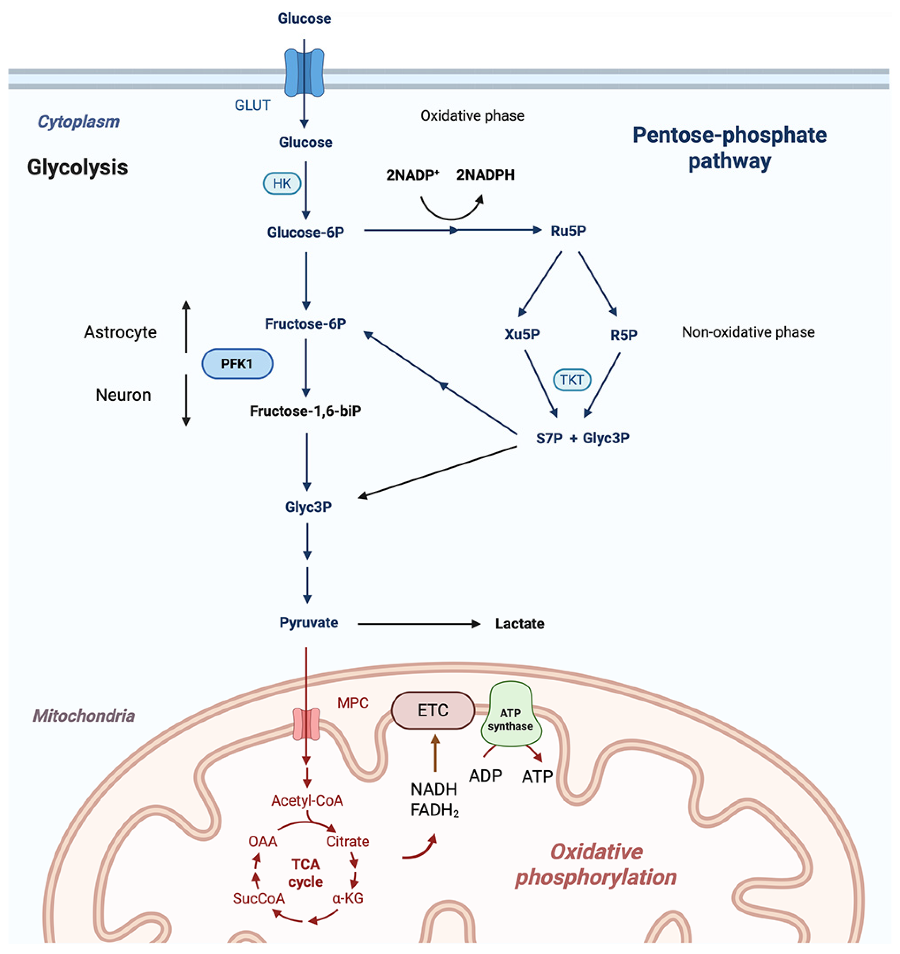
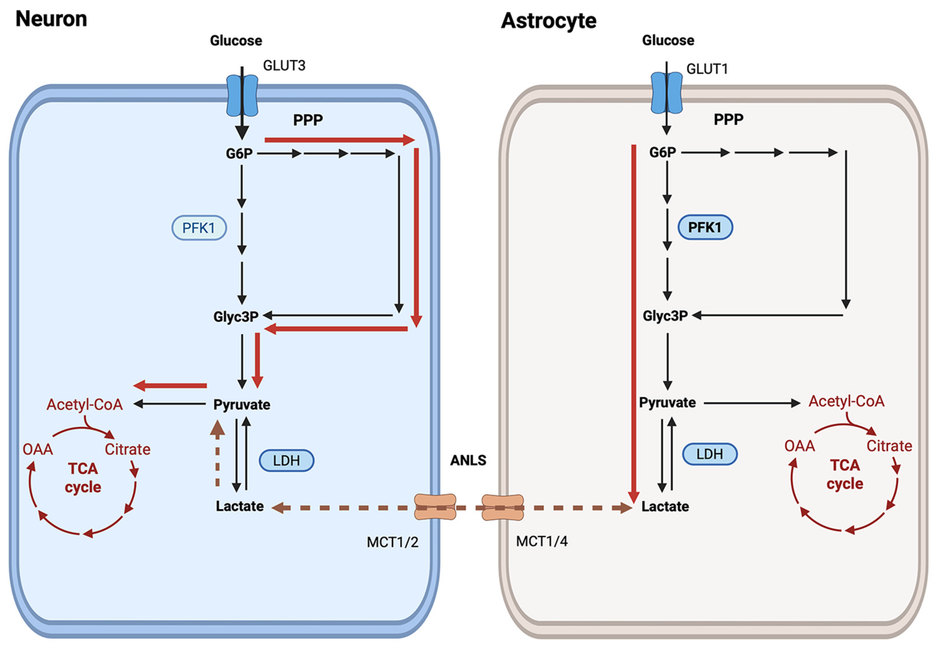
2.1. Cell-Type Specificity of Glycolytic Metabolism
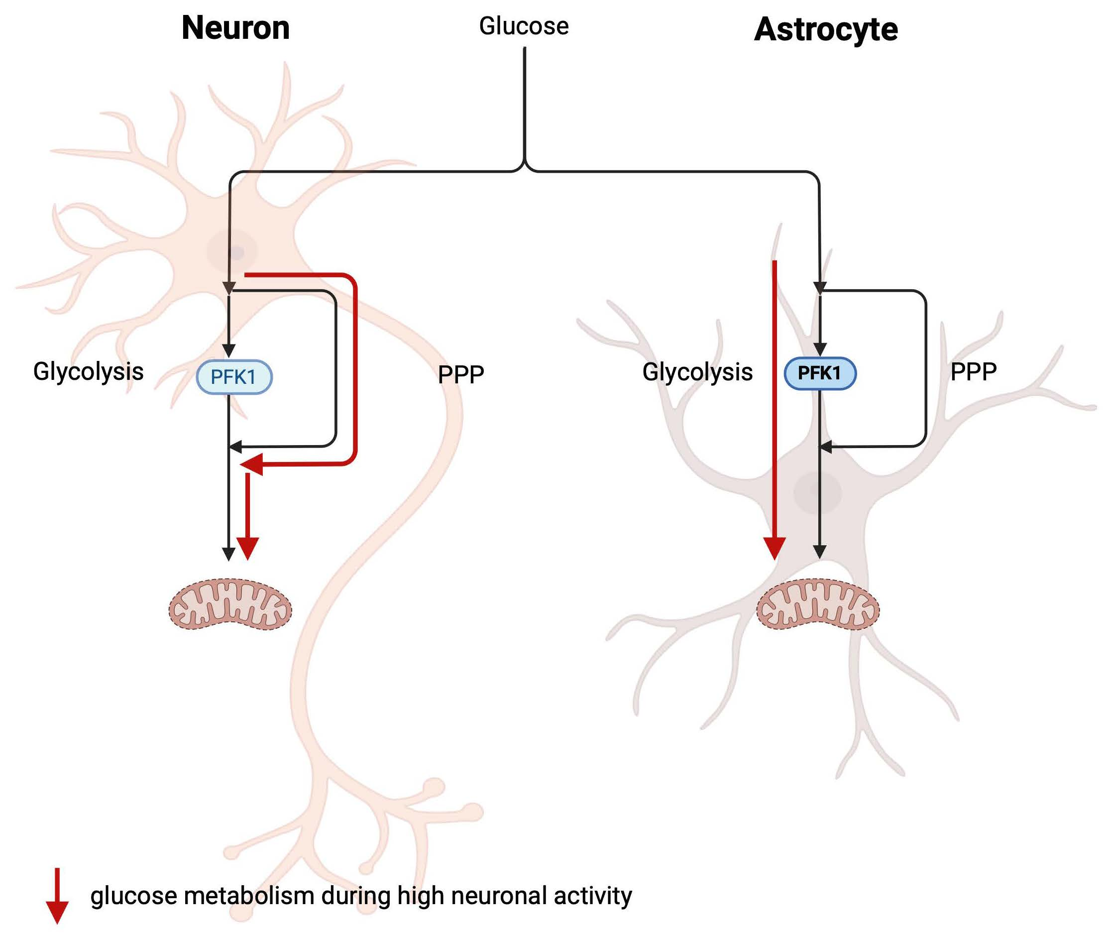
2.2. Glucose Metabolism Through the Pentose Phosphate Pathway Can Support Neuronal Activity
2.3. Role of Glucose Metabolism via PPP in Astrocytes
2.4. Regulation of PPP
2.5. Brain Region-Dependent PPP Activity
2.6. PPP Activity During Disease Conditions
3. Conclusions
Author Contributions
Funding
Institutional Review Board Statement
Informed Consent Statement
Data Availability Statement
Conflicts of Interest
Abbreviations
| ANLS | Astrocyte-to-neuron lactate shuttle |
| APC/C | Anaphase-promoting complex/cyclosome |
| ATP | Adenosine triphosphate |
| ETC | Electron transport chain |
| F6P | Fructose-6-phosphate |
| G6P | Glucose-6-phosphate |
| G6PD | Glucose-6-phosphate dehydrogenase |
| GAPDH | Glyceraldehyde 3-phosphate dehydrogenase |
| GLUT | Glucose transporter |
| Glyc3P | Glyceraldehyde-3-phosphate |
| GSH | Glutathione |
| HK | Hexokinase |
| Keap1 | Kelch-like enoyl-CoA hydratase-associated protein 1 |
| LDH | Lactate dehydrogenase |
| MCT | Monocarboxylate transporter |
| MPC | Mitochondrial pyruvate carrier |
| NADPH | Nicotinamide dinucleotide phosphate |
| Nrf2 | Nuclear respiratory factor 2 |
| OAA | Oxaloacetic acid |
| PDH | Pyruvate dehydrogenase complex |
| PFK1 | 6-phosphofructo-1-kinase |
| PFKFB3 | 6-phosphofructo-2-kinase-3 |
| PK | Pyruvate kinase |
| PPP | Pentose-phosphate pathway |
| ROS | Reactive oxygen species |
| Ru5P | Ribulose-5-phosphate |
| R5P | Ribose-5-phosphate |
| S7P | Sedoheptulose 7-phosphate |
| TCA | Tricarboxylic acid cycle |
| TKT | Transketolase |
| Xu5P | Xylulose-5-phosphate |
| α-KG | α-ketoglutarate |
References
- McKenna, M.C.; Tildon, J.T.; Stevenson, J.H.; Hopkins, I.B. Energy metabolism in cortical synaptic terminals from weanling and mature rat brain: Evidence for multiple compartments of tricarboxylic acid cycle activity. Dev. Neurosci. 1994, 16, 291–300. [Google Scholar] [CrossRef]
- McKenna, M.C.; Dienel, G.A.; Sonnewald, U.; Waagepetersen, H.S.; Schousboe, A. Energy metabolism of the brain. In Basic Neurochemistry; Brady, S.T., Siegel, G.J., Alberts, R.W., Price, D.L., Eds.; Academic Press: Burlington, MA, USA, 2012; pp. 200–231. [Google Scholar]
- Rae, C.D.; Baur, J.A.; Borges, K.; Dienel, G.; Diaz-Garcia, C.M.; Douglass, S.R.; Drew, K.; Duarte, J.M.N.; Duran, J.; Kann, O.; et al. Brain energy metabolism: A roadmap for future research. J. Neurochem. 2024, 168, 910–954. [Google Scholar] [CrossRef]
- Stincone, A.; Prigione, A.; Cramer, T.; Wamelink, M.M.; Campbell, K.; Cheung, E.; Olin-Sandoval, V.; Gruning, N.M.; Kruger, A.; Tauqeer Alam, M.; et al. The return of metabolism: Biochemistry and physiology of the pentose phosphate pathway. Biol. Rev. Camb. Philos. Soc. 2015, 90, 927–963. [Google Scholar] [CrossRef]
- Ramos-Martinez, J.I. The regulation of the pentose phosphate pathway: Remember Krebs. Arch. Biochem. Biophys. 2017, 614, 50–52. [Google Scholar] [CrossRef]
- Schousboe, A.; Bak, L.K.; Waagepetersen, H.S. Astrocytic Control of Biosynthesis and Turnover of the Neurotransmitters Glutamate and GABA. Front. Endocrinol. 2013, 4, 102. [Google Scholar] [CrossRef] [PubMed]
- Pellerin, L.; Magistretti, P.J. Glutamate uptake into astrocytes stimulates aerobic glycolysis: A mechanism coupling neuronal activity to glucose utilization. Proc. Natl. Acad. Sci. USA 1994, 91, 10625–10629. [Google Scholar] [CrossRef] [PubMed]
- Dienel, G.A.; Rothman, D.L.; Mangia, S. A Bird’s-Eye View of Glycolytic Upregulation in Activated Brain: The Major Fate of Lactate Is Release From Activated Tissue, Not Shuttling to Nearby Neurons. J. Neurochem. 2025, 169, e70111. [Google Scholar] [CrossRef] [PubMed]
- Benard, G.; Faustin, B.; Passerieux, E.; Galinier, A.; Rocher, C.; Bellance, N.; Delage, J.P.; Casteilla, L.; Letellier, T.; Rossignol, R. Physiological diversity of mitochondrial oxidative phosphorylation. Am. J. Physiol. Cell Physiol. 2006, 291, C1172–C1182. [Google Scholar] [CrossRef]
- Tuttle, J.P.; Wilson, J.E. Brain hexokinase: A kinetic comparison of soluble and particulate forms. Biochim. Biophys. Acta 1970, 212, 185–188. [Google Scholar] [CrossRef]
- Wilson, J.E. Brain hexokinase. A proposed relation between soluble-particulate distribution and activity in vivo. J. Biol. Chem. 1968, 243, 3640–3647. [Google Scholar] [CrossRef]
- Wilson, J.E. Isozymes of mammalian hexokinase: Structure, subcellular localization and metabolic function. J. Exp. Biol. 2003, 206, 2049–2057. [Google Scholar] [CrossRef]
- da-Silva, W.S.; Gomez-Puyou, A.; de Gomez-Puyou, M.T.; Moreno-Sanchez, R.; De Felice, F.G.; de Meis, L.; Oliveira, M.F.; Galina, A. Mitochondrial bound hexokinase activity as a preventive antioxidant defense: Steady-state ADP formation as a regulatory mechanism of membrane potential and reactive oxygen species generation in mitochondria. J. Biol. Chem. 2004, 279, 39846–39855. [Google Scholar] [CrossRef] [PubMed]
- de-Souza-Ferreira, E.; Rios-Neto, I.M.; Martins, E.L.; Galina, A. Mitochondria-coupled glucose phosphorylation develops after birth to modulate H2O2 release and calcium handling in rat brain. J. Neurochem. 2019, 149, 624–640. [Google Scholar] [CrossRef] [PubMed]
- Vyssokikh, M.Y.; Holtze, S.; Averina, O.A.; Lyamzaev, K.G.; Panteleeva, A.A.; Marey, M.V.; Zinovkin, R.A.; Severin, F.F.; Skulachev, M.V.; Fasel, N.; et al. Mild depolarization of the inner mitochondrial membrane is a crucial component of an anti-aging program. Proc. Natl. Acad. Sci. USA 2020, 117, 6491–6501. [Google Scholar] [CrossRef]
- Patel, M.S.; Nemeria, N.S.; Furey, W.; Jordan, F. The pyruvate dehydrogenase complexes: Structure-based function and regulation. J. Biol. Chem. 2014, 289, 16615–16623. [Google Scholar] [CrossRef] [PubMed]
- Attwell, D.; Laughlin, S.B. An energy budget for signaling in the grey matter of the brain. J. Cereb. Blood Flow. Metab. 2001, 21, 1133–1145. [Google Scholar] [CrossRef]
- Rolfe, D.F.; Brown, G.C. Cellular energy utilization and molecular origin of standard metabolic rate in mammals. Physiol. Rev. 1997, 77, 731–758. [Google Scholar] [CrossRef]
- Hertz, L.; Gibbs, M.E.; Dienel, G.A. Fluxes of lactate into, from, and among gap junction-coupled astrocytes and their interaction with noradrenaline. Front. Neurosci. 2014, 8, 261. [Google Scholar] [CrossRef]
- Dienel, G.A.; Hertz, L. Glucose and lactate metabolism during brain activation. J. Neurosci. Res. 2001, 66, 824–838. [Google Scholar] [CrossRef]
- McKenna, M.C. Substrate competition studies demonstrate oxidative metabolism of glucose, glutamate, glutamine, lactate and 3-hydroxybutyrate in cortical astrocytes from rat brain. Neurochem. Res. 2012, 37, 2613–2626. [Google Scholar] [CrossRef]
- Dobolyi, A.; Cservenak, M.; Bago, A.G.; Chen, C.; Stepanova, A.; Paal, K.; Lee, J.; Palkovits, M.; Hudson, G.; Chinopoulos, C. Cell-specific expression of key mitochondrial enzymes limits OXPHOS in astrocytes of the adult human neocortex and hippocampal formation. Commun. Biol. 2024, 7, 1045. [Google Scholar] [CrossRef]
- McKenna, M.C. Glutamate pays its own way in astrocytes. Front. Endocrinol. 2013, 4, 191. [Google Scholar] [CrossRef]
- Bolanos, J.P.; Almeida, A.; Moncada, S. Glycolysis: A bioenergetic or a survival pathway? Trends Biochem. Sci. 2010, 35, 145–149. [Google Scholar] [CrossRef]
- Herrero-Mendez, A.; Almeida, A.; Fernandez, E.; Maestre, C.; Moncada, S.; Bolanos, J.P. The bioenergetic and antioxidant status of neurons is controlled by continuous degradation of a key glycolytic enzyme by APC/C-Cdh1. Nat. Cell Biol. 2009, 11, 747–752. [Google Scholar] [CrossRef]
- Almeida, A.; Moncada, S.; Bolanos, J.P. Nitric oxide switches on glycolysis through the AMP protein kinase and 6-phosphofructo-2-kinase pathway. Nat. Cell Biol. 2004, 6, 45–51. [Google Scholar] [CrossRef] [PubMed]
- Rider, M.H.; Bertrand, L.; Vertommen, D.; Michels, P.A.; Rousseau, G.G.; Hue, L. 6-phosphofructo-2-kinase/fructose-2,6-bisphosphatase: Head-to-head with a bifunctional enzyme that controls glycolysis. Biochem. J. 2004, 381, 561–579. [Google Scholar] [CrossRef] [PubMed]
- Pesin, J.A.; Orr-Weaver, T.L. Regulation of APC/C activators in mitosis and meiosis. Annu. Rev. Cell Dev. Biol. 2008, 24, 475–499. [Google Scholar] [CrossRef] [PubMed]
- Jimenez-Blasco, D.; Agulla, J.; Lapresa, R.; Garcia-Macia, M.; Bobo-Jimenez, V.; Garcia-Rodriguez, D.; Manjarres-Raza, I.; Fernandez, E.; Jeanson, Y.; Khoury, S.; et al. Weak neuronal glycolysis sustains cognition and organismal fitness. Nat. Metab. 2024, 6, 1253–1267. [Google Scholar] [CrossRef]
- Salas, M.L.; Vinuela, E.; Salas, M.; Sols, A. Citrate Inhibition of Phosphofructokinase and the Pasteur Effect. Biochem. Biophys. Res. Commun. 1965, 19, 371–376. [Google Scholar] [CrossRef]
- Colombo, G.; Tate, P.W.; Girotti, A.W.; Kemp, R.G. Interaction of inhibitors with muscle phosphofructokinase. J. Biol. Chem. 1975, 250, 9404–9412. [Google Scholar] [CrossRef]
- Kondo, S.; Adachi, K. The nature of the inhibition and activation of epidermal phosphofructokinase: An allosteric enzyme. J. Investig. Dermatol. 1972, 59, 397–401. [Google Scholar] [CrossRef] [PubMed]
- Usenik, A.; Legisa, M. Evolution of allosteric citrate binding sites on 6-phosphofructo-1-kinase. PLoS ONE 2010, 5, e15447. [Google Scholar] [CrossRef] [PubMed]
- Rosen, P.C.; Fu, P.; Ferran, B.; Kim, E.; Brooks, D.J.; Lim, D.C.; Diaz-Garcia, C.M.; Yellen, G. Activity-dependent citrate dynamics in neurons. Proc. Natl. Acad. Sci. USA 2025, 122, e2519902122. [Google Scholar] [CrossRef] [PubMed]
- Magistretti, P.J.; Sorg, O.; Yu, N.; Martin, J.L.; Pellerin, L. Neurotransmitters regulate energy metabolism in astrocytes: Implications for the metabolic trafficking between neural cells. Dev. Neurosci. 1993, 15, 306–312. [Google Scholar] [CrossRef]
- Pellerin, L.; Magistretti, P.J. Sweet sixteen for ANLS. J. Cereb. Blood Flow. Metab. 2012, 32, 1152–1166. [Google Scholar] [CrossRef]
- Chih, C.P.; Lipton, P.; Roberts, E.L., Jr. Do active cerebral neurons really use lactate rather than glucose? Trends Neurosci. 2001, 24, 573–578. [Google Scholar] [CrossRef]
- Gjedde, A.; Marrett, S. Glycolysis in neurons, not astrocytes, delays oxidative metabolism of human visual cortex during sustained checkerboard stimulation in vivo. J. Cereb. Blood Flow. Metab. 2001, 21, 1384–1392. [Google Scholar] [CrossRef]
- Mangia, S.; DiNuzzo, M.; Giove, F.; Carruthers, A.; Simpson, I.A.; Vannucci, S.J. Response to ‘comment on recent modeling studies of astrocyte-neuron metabolic interactions’: Much ado about nothing. J. Cereb. Blood Flow. Metab. 2011, 31, 1346–1353. [Google Scholar] [CrossRef]
- Dienel, G.A. Brain lactate metabolism: The discoveries and the controversies. J. Cereb. Blood Flow. Metab. 2012, 32, 1107–1138. [Google Scholar] [CrossRef]
- Dienel, G.A. Lack of appropriate stoichiometry: Strong evidence against an energetically important astrocyte-neuron lactate shuttle in brain. J. Neurosci. Res. 2017, 95, 2103–2125. [Google Scholar] [CrossRef]
- Barros, L.F.; Weber, B. CrossTalk proposal: An important astrocyte-to-neuron lactate shuttle couples neuronal activity to glucose utilisation in the brain. J. Physiol. 2018, 596, 347–350. [Google Scholar] [CrossRef]
- Yellen, G. Fueling thought: Management of glycolysis and oxidative phosphorylation in neuronal metabolism. J. Cell Biol. 2018, 217, 2235–2246. [Google Scholar] [CrossRef] [PubMed]
- Brooks, G.A. The Science and Translation of Lactate Shuttle Theory. Cell Metab. 2018, 27, 757–785. [Google Scholar] [CrossRef] [PubMed]
- Brooks, G.A. Lactate as a fulcrum of metabolism. Redox Biol. 2020, 35, 101454. [Google Scholar] [CrossRef] [PubMed]
- Brooks, G.A.; Curl, C.C.; Leija, R.G.; Osmond, A.D.; Duong, J.J.; Arevalo, J.A. Tracing the lactate shuttle to the mitochondrial reticulum. Exp. Mol. Med. 2022, 54, 1332–1347. [Google Scholar] [CrossRef]
- Kann, O. Lactate as a supplemental fuel for synaptic transmission and neuronal network oscillations: Potentials and limitations. J. Neurochem. 2024, 168, 608–631. [Google Scholar] [CrossRef]
- Schurr, A. The Feud over Lactate and Its Role in Brain Energy Metabolism: An Unnecessary Burden on Research and the Scientists Who Practice It. Int. J. Mol. Sci. 2025, 26, 4429. [Google Scholar] [CrossRef]
- Andersen, J.V.; McKenna, M.C. Neurons in Need: Glucose, but Not Lactate, Is Required to Support Energy-Demanding Synaptic Transmission. J. Neurochem. 2025, 169, e70255. [Google Scholar] [CrossRef]
- Bak, L.K.; Walls, A.B. CrossTalk opposing view: Lack of evidence supporting an astrocyte-to-neuron lactate shuttle coupling neuronal activity to glucose utilisation in the brain. J. Physiol. 2018, 596, 351–353. [Google Scholar] [CrossRef]
- McKenna, M.C.; Tildon, J.T.; Stevenson, J.H.; Huang, X. New insights into the compartmentation of glutamate and glutamine in cultured rat brain astrocytes. Dev. Neurosci. 1996, 18, 380–390. [Google Scholar] [CrossRef]
- Love, S. Oxidative stress in brain ischemia. Brain Pathol. 1999, 9, 119–131. [Google Scholar] [CrossRef]
- Nakamura, T.; Lipton, S.A. S-nitrosylation of critical protein thiols mediates protein misfolding and mitochondrial dysfunction in neurodegenerative diseases. Antioxid. Redox Signal. 2011, 14, 1479–1492. [Google Scholar] [CrossRef]
- Mattson, M.P. Calcium and neuronal injury in Alzheimer’s disease. Contributions of beta-amyloid precursor protein mismetabolism, free radicals, and metabolic compromise. Ann. N. Y. Acad. Sci. 1994, 747, 50–76. [Google Scholar] [CrossRef]
- Klimova, N.; Fearnow, A.; Kristian, T. Role of NAD+-Modulated Mitochondrial Free Radical Generation in Mechanisms of Acute Brain Injury. Brain Sci. 2020, 10, 449. [Google Scholar] [CrossRef] [PubMed]
- Kowaltowski, A.J.; de Souza-Pinto, N.C.; Castilho, R.F.; Vercesi, A.E. Mitochondria and reactive oxygen species. Free Radic. Biol. Med. 2009, 47, 333–343. [Google Scholar] [CrossRef] [PubMed]
- Stowe, D.F.; Camara, A.K. Mitochondrial reactive oxygen species production in excitable cells: Modulators of mitochondrial and cell function. Antioxid. Redox Signal. 2009, 11, 1373–1414. [Google Scholar] [CrossRef] [PubMed]
- Horecker, B.L. The pentose phosphate pathway. J. Biol. Chem. 2002, 277, 47965–47971. [Google Scholar] [CrossRef]
- Kruger, N.J.; von Schaewen, A. The oxidative pentose phosphate pathway: Structure and organisation. Curr. Opin. Plant Biol. 2003, 6, 236–246. [Google Scholar] [CrossRef]
- Ben-Yoseph, O.; Boxer, P.A.; Ross, B.D. Assessment of the role of the glutathione and pentose phosphate pathways in the protection of primary cerebrocortical cultures from oxidative stress. J. Neurochem. 1996, 66, 2329–2337. [Google Scholar] [CrossRef]
- Garcia-Nogales, P.; Almeida, A.; Bolanos, J.P. Peroxynitrite protects neurons against nitric oxide-mediated apoptosis. A key role for glucose-6-phosphate dehydrogenase activity in neuroprotection. J. Biol. Chem. 2003, 278, 864–874. [Google Scholar] [CrossRef]
- Takahashi, S. Metabolic compartmentalization between astroglia and neurons in physiological and pathophysiological conditions of the neurovascular unit. Neuropathology 2020, 40, 121–137. [Google Scholar] [CrossRef]
- Takahashi, S.; Izawa, Y.; Suzuki, N. Astroglial pentose phosphate pathway rates in response to high-glucose environments. ASN Neuro 2012, 4, AN20120002. [Google Scholar] [CrossRef]
- Dringen, R.; Pawlowski, P.G.; Hirrlinger, J. Peroxide detoxification by brain cells. J. Neurosci. Res. 2005, 79, 157–165. [Google Scholar] [CrossRef] [PubMed]
- Dringen, R.; Bishop, G.M.; Koeppe, M.; Dang, T.N.; Robinson, S.R. The pivotal role of astrocytes in the metabolism of iron in the brain. Neurochem. Res. 2007, 32, 1884–1890. [Google Scholar] [CrossRef]
- Fernandez-Fernandez, S.; Almeida, A.; Bolanos, J.P. Antioxidant and bioenergetic coupling between neurons and astrocytes. Biochem. J. 2012, 443, 3–11. [Google Scholar] [CrossRef] [PubMed]
- Aoyama, K.; Nakaki, T. Impaired glutathione synthesis in neurodegeneration. Int. J. Mol. Sci. 2013, 14, 21021–21044. [Google Scholar] [CrossRef] [PubMed]
- Dringen, R. Metabolism and functions of glutathione in brain. Prog. Neurobiol. 2000, 62, 649–671. [Google Scholar] [CrossRef]
- Aoyama, K. Glutathione in the Brain. Int. J. Mol. Sci. 2021, 22, 5010. [Google Scholar] [CrossRef]
- Perez-Sala, D.; Pajares, M.A. Appraising the Role of Astrocytes as Suppliers of Neuronal Glutathione Precursors. Int. J. Mol. Sci. 2023, 24, 8059. [Google Scholar] [CrossRef]
- Nieweg, K.; Schaller, H.; Pfrieger, F.W. Marked differences in cholesterol synthesis between neurons and glial cells from postnatal rats. J. Neurochem. 2009, 109, 125–134. [Google Scholar] [CrossRef]
- Mashima, K.; Takahashi, S.; Minami, K.; Izawa, Y.; Abe, T.; Tsukada, N.; Hishiki, T.; Suematsu, M.; Kajimura, M.; Suzuki, N. Neuroprotective Role of Astroglia in Parkinson Disease by Reducing Oxidative Stress Through Dopamine-Induced Activation of Pentose-Phosphate Pathway. ASN Neuro 2018, 10, 1759091418775562. [Google Scholar] [CrossRef]
- Takahashi, J.; Palmer, T.D.; Gage, F.H. Retinoic acid and neurotrophins collaborate to regulate neurogenesis in adult-derived neural stem cell cultures. J. Neurobiol. 1999, 38, 65–81. [Google Scholar] [CrossRef]
- TeSlaa, T.; Ralser, M.; Fan, J.; Rabinowitz, J.D. The pentose phosphate pathway in health and disease. Nat. Metab. 2023, 5, 1275–1289. [Google Scholar] [CrossRef] [PubMed]
- Brekke, E.M.; Walls, A.B.; Schousboe, A.; Waagepetersen, H.S.; Sonnewald, U. Quantitative importance of the pentose phosphate pathway determined by incorporation of 13C from [2-13C]- and [3-13C]glucose into TCA cycle intermediates and neurotransmitter amino acids in functionally intact neurons. J. Cereb. Blood Flow. Metab. 2012, 32, 1788–1799. [Google Scholar] [CrossRef] [PubMed]
- Appel, S.H.; Parrot, B.L. Hexose monophosphate pathway in synapses. J. Neurochem. 1970, 17, 1619–1626. [Google Scholar] [CrossRef] [PubMed]
- Anastasiou, D.; Poulogiannis, G.; Asara, J.M.; Boxer, M.B.; Jiang, J.K.; Shen, M.; Bellinger, G.; Sasaki, A.T.; Locasale, J.W.; Auld, D.S.; et al. Inhibition of pyruvate kinase M2 by reactive oxygen species contributes to cellular antioxidant responses. Science 2011, 334, 1278–1283. [Google Scholar] [CrossRef]
- Ralser, M.; Wamelink, M.M.; Kowald, A.; Gerisch, B.; Heeren, G.; Struys, E.A.; Klipp, E.; Jakobs, C.; Breitenbach, M.; Lehrach, H.; et al. Dynamic rerouting of the carbohydrate flux is key to counteracting oxidative stress. J. Biol. 2007, 6, 10. [Google Scholar] [CrossRef]
- Shenton, D.; Grant, C.M. Protein S-thiolation targets glycolysis and protein synthesis in response to oxidative stress in the yeast Saccharomyces cerevisiae. Biochem. J. 2003, 374, 513–519. [Google Scholar] [CrossRef]
- Ralser, M.; Wamelink, M.M.; Latkolik, S.; Jansen, E.E.; Lehrach, H.; Jakobs, C. Metabolic reconfiguration precedes transcriptional regulation in the antioxidant response. Nat. Biotechnol. 2009, 27, 604–605. [Google Scholar] [CrossRef]
- Wang, Y.P.; Zhou, L.S.; Zhao, Y.Z.; Wang, S.W.; Chen, L.L.; Liu, L.X.; Ling, Z.Q.; Hu, F.J.; Sun, Y.P.; Zhang, J.Y.; et al. Regulation of G6PD acetylation by SIRT2 and KAT9 modulates NADPH homeostasis and cell survival during oxidative stress. EMBO J. 2014, 33, 1304–1320. [Google Scholar] [CrossRef]
- Kletzien, R.F.; Harris, P.K.; Foellmi, L.A. Glucose-6-phosphate dehydrogenase: A “housekeeping” enzyme subject to tissue-specific regulation by hormones, nutrients, and oxidant stress. FASEB J. 1994, 8, 174–181. [Google Scholar] [CrossRef]
- Stanton, R.C. Glucose-6-phosphate dehydrogenase, NADPH, and cell survival. IUBMB Life 2012, 64, 362–369. [Google Scholar] [CrossRef] [PubMed]
- Dreger, H.; Westphal, K.; Weller, A.; Baumann, G.; Stangl, V.; Meiners, S.; Stangl, K. Nrf2-dependent upregulation of antioxidative enzymes: A novel pathway for proteasome inhibitor-mediated cardioprotection. Cardiovasc. Res. 2009, 83, 354–361. [Google Scholar] [CrossRef] [PubMed]
- Hayes, J.D.; Dinkova-Kostova, A.T. The Nrf2 regulatory network provides an interface between redox and intermediary metabolism. Trends Biochem. Sci. 2014, 39, 199–218. [Google Scholar] [CrossRef] [PubMed]
- Bell, K.F.; Al-Mubarak, B.; Martel, M.A.; McKay, S.; Wheelan, N.; Hasel, P.; Markus, N.M.; Baxter, P.; Deighton, R.F.; Serio, A.; et al. Neuronal development is promoted by weakened intrinsic antioxidant defences due to epigenetic repression of Nrf2. Nat. Commun. 2015, 6, 7066. [Google Scholar] [CrossRef]
- Tonelli, C.; Chio, I.I.C.; Tuveson, D.A. Transcriptional Regulation by Nrf2. Antioxid. Redox Signal. 2018, 29, 1727–1745. [Google Scholar] [CrossRef]
- Ben-Yoseph, O.; Camp, D.M.; Robinson, T.E.; Ross, B.D. Dynamic measurements of cerebral pentose phosphate pathway activity in vivo using [1,6-13C2,6,6-2H2]glucose and microdialysis. J. Neurochem. 1995, 64, 1336–1342. [Google Scholar] [CrossRef]
- Gaitonde, M.K.; Evison, E.; Evans, G.M. The rate of utilization of glucose via hexosemonophosphate shunt in brain. J. Neurochem. 1983, 41, 1253–1260. [Google Scholar] [CrossRef]
- Hostetler, K.Y.; Landau, B.R. Estimation of the pentose cycle contribution to glucose metabolism in tissue in vivo. Biochemistry 1967, 6, 2961–2964. [Google Scholar] [CrossRef]
- Bartnik, B.L.; Sutton, R.L.; Fukushima, M.; Harris, N.G.; Hovda, D.A.; Lee, S.M. Upregulation of pentose phosphate pathway and preservation of tricarboxylic acid cycle flux after experimental brain injury. J. Neurotrauma 2005, 22, 1052–1065. [Google Scholar] [CrossRef]
- Dusick, J.R.; Glenn, T.C.; Lee, W.N.; Vespa, P.M.; Kelly, D.F.; Lee, S.M.; Hovda, D.A.; Martin, N.A. Increased pentose phosphate pathway flux after clinical traumatic brain injury: A [1,2-13C2]glucose labeling study in humans. J. Cereb. Blood Flow. Metab. 2007, 27, 1593–1602. [Google Scholar] [CrossRef]
- Baquer, N.Z.; Hothersall, J.S.; McLean, P. Function and regulation of the pentose phosphate pathway in brain. Curr. Top. Cell Regul. 1988, 29, 265–289. [Google Scholar] [PubMed]
- Madsen, P.L.; Cruz, N.F.; Sokoloff, L.; Dienel, G.A. Cerebral oxygen/glucose ratio is low during sensory stimulation and rises above normal during recovery: Excess glucose consumption during stimulation is not accounted for by lactate efflux from or accumulation in brain tissue. J. Cereb. Blood Flow. Metab. 1999, 19, 393–400. [Google Scholar] [CrossRef] [PubMed]
- Sonnewald, U. Glutamate synthesis has to be matched by its degradation—Where do all the carbons go? J. Neurochem. 2014, 131, 399–406. [Google Scholar] [CrossRef]
- McKenna, M.C.; Tildon, J.T.; Stevenson, J.H.; Boatright, R.; Huang, S. Regulation of energy metabolism in synaptic terminals and cultured rat brain astrocytes: Differences revealed using aminooxyacetate. Dev. Neurosci. 1993, 15, 320–329. [Google Scholar] [CrossRef] [PubMed]
- Shannon, B.J.; Vaishnavi, S.N.; Vlassenko, A.G.; Shimony, J.S.; Rutlin, J.; Raichle, M.E. Brain aerobic glycolysis and motor adaptation learning. Proc. Natl. Acad. Sci. USA 2016, 113, E3782–3791. [Google Scholar] [CrossRef]
- Ashrafi, G.; Wu, Z.; Farrell, R.J.; Ryan, T.A. GLUT4 Mobilization Supports Energetic Demands of Active Synapses. Neuron 2017, 93, 606–615 e603. [Google Scholar] [CrossRef]
- Diaz-Garcia, C.M.; Mongeon, R.; Lahmann, C.; Koveal, D.; Zucker, H.; Yellen, G. Neuronal Stimulation Triggers Neuronal Glycolysis and Not Lactate Uptake. Cell Metab. 2017, 26, 361–374 e364. [Google Scholar] [CrossRef]
- Li, H.; Guglielmetti, C.; Sei, Y.J.; Zilberter, M.; Le Page, L.M.; Shields, L.; Yang, J.; Nguyen, K.; Tiret, B.; Gao, X.; et al. Neurons require glucose uptake and glycolysis in vivo. Cell Rep. 2023, 42, 112335. [Google Scholar] [CrossRef]
- Wolfe, A.D.; Koberstein, J.N.; Smith, C.B.; Stewart, M.L.; Hammarlund, M.; Hyman, A.; Stork, P.J.; Goodman, R.; Colon-Ramos, D.A. Local and dynamic regulation of neuronal glycolysis in vivo. bioRxiv 2023. [Google Scholar] [CrossRef]
- de Tredern, E.; Rabah, Y.; Pasquer, L.; Minatchy, J.; Placais, P.Y.; Preat, T. Glial glucose fuels the neuronal pentose phosphate pathway for long-term memory. Cell Rep. 2021, 36, 109620. [Google Scholar] [CrossRef]
- Kleinridders, A.; Ferris, H.A.; Reyzer, M.L.; Rath, M.; Soto, M.; Manier, M.L.; Spraggins, J.; Yang, Z.; Stanton, R.C.; Caprioli, R.M.; et al. Regional differences in brain glucose metabolism determined by imaging mass spectrometry. Mol. Metab. 2018, 12, 113–121. [Google Scholar] [CrossRef]
- Pourmajidian, M.; Hansen, J.Y.; Shafiei, G.; Misic, B.; Dagher, A. Five energy metabolism pathways show distinct regional distributions and lifespan trajectories in the human brain. PLoS Biol. 2026, 24, e3003619. [Google Scholar] [CrossRef]
- Martano, G.; Murru, L.; Moretto, E.; Gerosa, L.; Garrone, G.; Krogh, V.; Passafaro, M. Biosynthesis of glycerol phosphate is associated with long-term potentiation in hippocampal neurons. Metabolomics 2016, 12, 133. [Google Scholar] [CrossRef] [PubMed]
- Ponomareva, D.; Ivanov, A.; Bregestovski, P. Analysis of the Effects of Pentose Phosphate Pathway Inhibition on the Generation of Reactive Oxygen Species and Epileptiform Activity in Hippocampal Slices. Int. J. Mol. Sci. 2024, 25, 1934. [Google Scholar] [CrossRef] [PubMed]
- Cao, L.; Zhang, D.; Chen, J.; Qin, Y.Y.; Sheng, R.; Feng, X.; Chen, Z.; Ding, Y.; Li, M.; Qin, Z.H. G6PD plays a neuroprotective role in brain ischemia through promoting pentose phosphate pathway. Free Radic. Biol. Med. 2017, 112, 433–444. [Google Scholar] [CrossRef] [PubMed]
- Jalloh, I.; Carpenter, K.L.; Grice, P.; Howe, D.J.; Mason, A.; Gallagher, C.N.; Helmy, A.; Murphy, M.P.; Menon, D.K.; Carpenter, T.A.; et al. Glycolysis and the pentose phosphate pathway after human traumatic brain injury: Microdialysis studies using 1,2-(13)C2 glucose. J. Cereb. Blood Flow. Metab. 2015, 35, 111–120. [Google Scholar] [CrossRef]
- Matsuo, K.; Hosoda, K.; Tanaka, J.; Yamamoto, Y.; Imahori, T.; Nakai, T.; Irino, Y.; Shinohara, M.; Sasayama, T.; Kohmura, E. Geranylgeranylacetone attenuates cerebral ischemia-reperfusion injury in rats through the augmentation of HSP 27 phosphorylation: A preliminary study. BMC Neurosci. 2021, 22, 9. [Google Scholar] [CrossRef]
- Imahori, T.; Hosoda, K.; Nakai, T.; Yamamoto, Y.; Irino, Y.; Shinohara, M.; Sato, N.; Sasayama, T.; Tanaka, K.; Nagashima, H.; et al. Combined metabolic and transcriptional profiling identifies pentose phosphate pathway activation by HSP27 phosphorylation during cerebral ischemia. Neuroscience 2017, 349, 1–16. [Google Scholar] [CrossRef]
- Song, Y.; Luengo-Gutierrez, L.; Sagi-Kiss, V.; Kong, G.; Huang, H.; Steinruecke, M.; Zhou, L.; Yuan, Z.; De Virgiliis, F.; Pap, I.; et al. A glycolytic shunt via the pentose phosphate pathway is a metabolic checkpoint for nervous system sensory homeostasis and axonal regeneration. Cell 2026, 189, 1211–1227 e1225. [Google Scholar] [CrossRef]
- Dienel, G.A. Brain Glucose Metabolism: Integration of Energetics with Function. Physiol. Rev. 2019, 99, 949–1045. [Google Scholar] [CrossRef]
- Mathieu, C.; Li de la Sierra-Gallay, I.; Duval, R.; Xu, X.; Cocaign, A.; Leger, T.; Woffendin, G.; Camadro, J.M.; Etchebest, C.; Haouz, A.; et al. Insights into Brain Glycogen Metabolism: The Structure of Human Brain Glycogen Phosphorylase. J. Biol. Chem. 2016, 291, 18072–18083. [Google Scholar] [CrossRef]
- Bar, S.; Wilson, K.A.; Hilsabeck, T.A.U.; Alderfer, S.; Dammer, E.B.; Burton, J.B.; Shah, S.; Holtz, A.; Carrera, E.M.; Beck, J.N.; et al. Neuronal glycogen breakdown mitigates tauopathy via pentose-phosphate-pathway-mediated oxidative stress reduction. Nat. Metab. 2025, 7, 1375–1391. [Google Scholar] [CrossRef]
- Bak, L.K.; Walls, A.B.; Schousboe, A.; Waagepetersen, H.S. Astrocytic glycogen metabolism in the healthy and diseased brain. J. Biol. Chem. 2018, 293, 7108–7116. [Google Scholar] [CrossRef] [PubMed]
- Vannucci, S.J.; Simpson, I.A. Developmental switch in brain nutrient transporter expression in the rat. Am. J. Physiol. Endocrinol. Metab. 2003, 285, E1127–E1134. [Google Scholar] [CrossRef] [PubMed]
- Shigeri, Y.; Seal, R.P.; Shimamoto, K. Molecular pharmacology of glutamate transporters, EAATs and VGLUTs. Brain Res. Brain Res. Rev. 2004, 45, 250–265. [Google Scholar] [CrossRef]


Disclaimer/Publisher’s Note: The statements, opinions and data contained in all publications are solely those of the individual author(s) and contributor(s) and not of MDPI and/or the editor(s). MDPI and/or the editor(s) disclaim responsibility for any injury to people or property resulting from any ideas, methods, instructions or products referred to in the content. |
© 2026 by the authors. Licensee MDPI, Basel, Switzerland. This article is an open access article distributed under the terms and conditions of the Creative Commons Attribution (CC BY) license.
Share and Cite
Kristian, T.; Waddell, J.; McKenna, M.C. Pentose Phosphate Pathway Is Critical for Providing Energy by Bypassing 6-Phosphofructo-1-Kinase (PFK1) During Increased Neuronal Activity. Metabolites 2026, 16, 321. https://doi.org/10.3390/metabo16050321
Kristian T, Waddell J, McKenna MC. Pentose Phosphate Pathway Is Critical for Providing Energy by Bypassing 6-Phosphofructo-1-Kinase (PFK1) During Increased Neuronal Activity. Metabolites. 2026; 16(5):321. https://doi.org/10.3390/metabo16050321
Chicago/Turabian StyleKristian, Tibor, Jaylyn Waddell, and Mary C. McKenna. 2026. "Pentose Phosphate Pathway Is Critical for Providing Energy by Bypassing 6-Phosphofructo-1-Kinase (PFK1) During Increased Neuronal Activity" Metabolites 16, no. 5: 321. https://doi.org/10.3390/metabo16050321
APA StyleKristian, T., Waddell, J., & McKenna, M. C. (2026). Pentose Phosphate Pathway Is Critical for Providing Energy by Bypassing 6-Phosphofructo-1-Kinase (PFK1) During Increased Neuronal Activity. Metabolites, 16(5), 321. https://doi.org/10.3390/metabo16050321