Review

SMARCB1 Deficiency as a Driver of the Hallmarks of Cancer in Rhabdoid Tumours: Novel Insights into Dysregulated Energy Metabolism, Emerging Targets, and Ongoing Clinical Trials

by
Abdul L. Shakerdi
and
Graham P. Pidgeon
*
Department of Surgery, Trinity Translational Medicine Institute, St James’s Hospital & Trinity College Dublin, D08 NHY1 Dublin, Ireland
*
Author to whom correspondence should be addressed.
Metabolites 2025, 15(5), 304; https://doi.org/10.3390/metabo15050304
Submission received: 4 April 2025 / Revised: 28 April 2025 / Accepted: 30 April 2025 / Published: 3 May 2025
(This article belongs to the Special Issue Cancer Metabolomics 2024)

Abstract

:
Background: Rhabdoid tumours (RTs) are aggressive neoplasms most often characterised by biallelic loss of the SMARCB1 gene, encoding a core subunit of the SWI/SNF chromatin-remodelling complex. Despite their relative genetic stability, RTs exhibit a highly malignant phenotype and poor prognosis. Methods: This review explores the mechanisms underlying SMARCB1 aberrations, their role in driving hallmarks of cancer, and emerging therapeutic strategies for RTs. Ongoing clinical trials listed on ClinicalTrials were reviewed to evaluate the translational potential of targeted therapies in SMARCB1-deficient rhabdoid tumours. Results: Loss of SMARCB1 drives multiple cancer hallmarks by disrupting key regulatory pathways. It promotes unchecked cell proliferation through alterations in p16INK4a and Myc signalling. SMARCB1-deficient tumours possess immune-evading capabilities via PD-L1 overexpression and immune checkpoint activation. SMARCB1 deficiency also alters cellular energetics. The nucleotide biosynthesis pathway has been demonstrated to be upregulated in RT organoids, as shown by increased levels of pathway metabolites. Enzymes of the mevalonate pathway such as HMG-CoA reductase and mevalonate kinase are also dysregulated. Targeting glutathione metabolism with eprenetapopt may induce oxidative stress and apoptosis. Widespread epigenetic aberrations, including increased EZH2 activity, are being targeted with inhibitors such as tazemetostat. Conclusions: SMARCB1 loss is a central driver of cancer hallmarks in RTs, enabling proliferation, immune evasion, metabolic reprogramming, and epigenetic dysregulation. Future horizons in RT treatment include immunotherapies, epigenetic modifiers, and gene therapies. The synergy and optimal timing of targeted therapy with conventional treatment requires further characterisation for clinical translation.

1. Introduction

The SWI/SNF-related matrix-associated actin-dependent regulator of chromatin subfamily B member 1 (SMARCB1) gene is located on chromosome 22q11.2 and encodes a core subunit of the ATP-dependent SWI/SNF complex [1]. It is involved in chromatin remodelling and regulation of gene expression. Loss of the SMARCB1 gene has been linked to various neoplasms, including rhabdoid tumours (RTs), epithelioid sarcoma, and renal medullary carcinoma [2]. RTs are rare neoplasms which typically present in the paediatric population before the age of 3 [3]. They have been described in nearly all anatomical locations and are divided into those located intracranially, most commonly in the cerebellum [4], or extracranially in organs such as the kidney, gastrointestinal tract, and liver [5]. Apart from the frequent biallelic loss of SMARCB1, RTs are considered relatively genetically stable, despite their highly aggressive phenotype [6]. Germline mutations also predispose patients to various malignancies in a condition known as rhabdoid tumour predisposition syndrome [7]. Loss of SMARCB1 promotes cancer through various pathways (Figure 1), which will be described in more detail in this review. This article will also serve to discuss the mechanisms by which aberrations of the SMARCB1 gene occur and their implications in driving the various hallmarks of cancer and provide an overview of the novel therapeutic approaches in clinical trials for treatment of RTs.
The SMARCB1 gene extends over 50 kilobases, and its peptide sequence is encoded by nine exons [8]. The encoded protein has a molecular weight of 47 kDa, with a length of 385 amino acids. It is composed of four domains, including two highly conserved tandem repeat 1 and 2 domains (RPT1/2), flanked by the N-terminal Winged Helix DNA-binding domain and the C-terminal coiled-coil domain (CTD). Data derived from COSMIC v95 revealed 815 alterations in the SMARCB1 gene, largely clustered at the CTD. The majority of mutations (49.33%) were missense, followed by frameshift mutations (20.12%) and nonsense mutations (19.88%). Silent and inframe mutations accounted for approximately 10% of genetic aberrations [2].
Confirmation of the role of SMARCB1 as a tumour suppressor gene (TSG) arose when conditional gene knockout in adult mice resulted in CD8+ mature lymphoma and fully penetrant cancer formation [9]. Later studies were also able to produce RTs by inducing SMARCB1 deletion in earlier stages of embryonic development [10]. While complete loss of function of SMARCB1 is the best-characterised genetic mutation in RTs, some cases are associated with cytoplasmic localisation of the C-terminal truncated version of the gene, compromising its ability to exert its biological functions in the nucleus. Exportin 1 is thought to be responsible for this abnormal localisation through its interaction with the RPT2 domain of SMARCB1. As a result, nuclear export inhibitors such as leptomycin B and selinexor may be effective in these cases [11]. However, clinical efficacy is yet to be established [2].

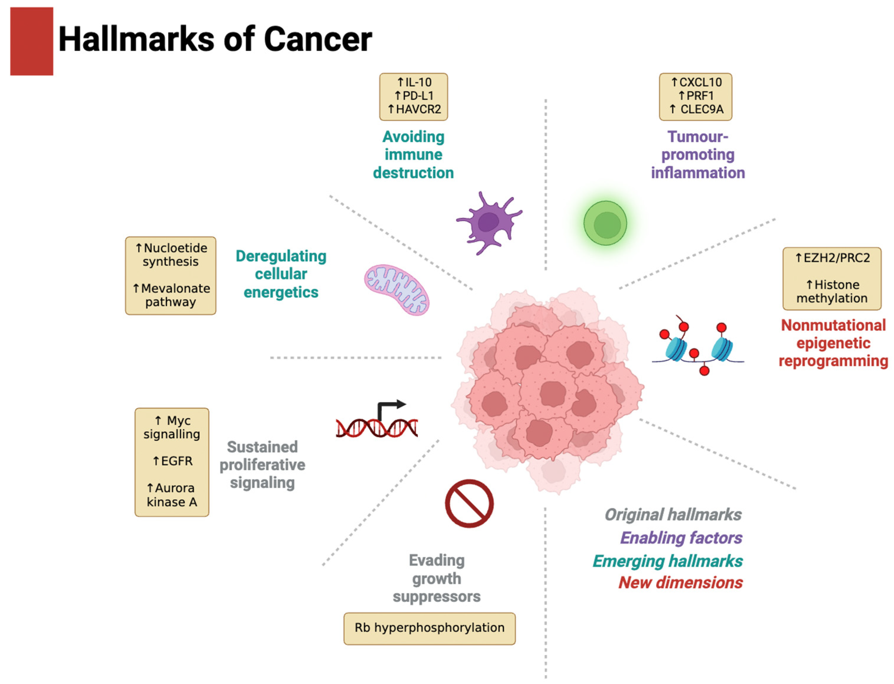
2. Driver of the Hallmarks of Cancer

The hallmarks of cancer were introduced by Hanahan and Weinberg in 2000 and were later expanded in 2011 and 2022 [12,13,14]. They describe a set of capabilities acquired by human cells during the carcinogenic process and provide a systematic structure for studying cancer, designing therapeutic targets, and guiding experimental models. Figure 2 summarises the role of SMARCB1 loss as a driver of the hallmarks of cancer in RTs.

2.1. Evading Growth Suppressors and Sustaining Proliferative Signalling

SMARCB1 has been shown to regulate the p16INK4a cyclin-dependent kinase (CDK) inhibitor which prevents the activation of the CDK4/6-cyclin D1 complex, responsible for Rb protein phosphorylation and hence the transcription of genes associated with G1/S phase progression [15]. SMARCB1 deficiency also enhances the activity of key oncogenic transcription factors, such as Myc, a helix–loop–helix protein that is one of the best-described drivers of cell proliferation [16]. Weissmiller and colleagues found that SNF5 encoded by SMARCB1 inhibits the DNA-binding ability of Myc, impeding its ability to recognise target genes. Further supporting these findings, the reintroduction of SNF5 into SMARCB1-null cells mimicked the primary transcriptional effects of MYC inhibition [17]. As a heterodimer with MAX, it regulates the expression of various genes involved in cell cycle regulation, apoptosis, angiogenesis, and cell metabolism [18,19,20]. OmoMYC (OMO-103) is a drug that targets Myc signalling by binding to c- and n-Myc, Max, and Miz1 and was shown to reduce RT cell viability in in vitro studies [17].
Another implicated oncogene is the epidermal growth factor receptor (EGFR). A phosphoproteomic study highlighted its abnormal expression in RTs and that its inhibition hinders cell proliferation [21]. MRTs have also been shown to exhibit coactivation of the receptor tyrosine kinases (RTKs) platelet-derived growth factor receptor A (PDGFRα) and fibroblast growth factor receptor 2 (FGFR-2) [22]. These are particularly attractive targets, as there exists a vast range of clinically approved RTK inhibitors. Dual targeting of these proteins individually or through the dual inhibitor ponatinib was shown to suppress the Akt and extracellular signal-regulated kinase (ERK1/2) pathways leading to apoptosis [22].
The use of small molecules aimed at recovering the lost function of TSGs in malignancy poses significant challenges, and many tumour suppressors are considered ‘undruggable’ for this reason [23]. Synthetic lethal relationships therefore provide a promising approach for the targeting of such genetic abnormalities [24]. Radko-Juettner et al., showed through a genome-wide CRISPR screen that the DDB1-CUL4-associated factor 5 (DCAF5) gene, which functions as a substrate receptor for E3-ubiquitin ligase, is required for the survival of SMARCB1-mutated RTs. In addition, depletion of DCAF5 allowed SWI/SNF to reaccumulate and restored histone marks such as H3K27ac and H3K4me1 at active enhancers, normalising cellular differentiation and reversing the cancer phenotype. These results suggest that SMARCB1 mutation leads to the destabilisation of the SWI/SNF complexes, which are subsequently cleared by the ubiquitin quality control pathway, in order to keep malignant cells viable [25]. Thus, targeting of DCAF5 may serve as a potential novel therapeutic approach in the treatment of RTs [26].

2.2. Tumour-Promoting Inflammation and Avoiding Immune Destruction

The microenvironment of certain subsets of RTs has been shown to be highly inflammatory and to express a high level of genes involved in CD8+ T cell activation, homing, and tumour infiltration, such as CXCL10, PRF1, and CLEC9A. Yet RTs deploy various survival mechanisms that allow them to thrive in such a hostile environment. Immunohistochemistry of RTs revealed overexpression of IL-10, programmed death ligand 1 (PD-L1), and PD-L1-expressing CD68+ myeloid cells, as well as other immune checkpoint proteins such as HAVCR2 [27]. One study found that in samples of 30 paediatric RTs, 47% stained positive for PD-L1 expression and therefore may have benefited from immune checkpoint inhibition [28]. However, several studies on atypical teratoid/rhabdoid tumours (ATRTs) have revealed the variable expression of PD-L1, ranging from 5.1% to 50%. This variability across studies may reflect differences in thresholds for positivity staining, the inclusion of partial staining, and the use of scoring systems such as the Cologne criteria [29]. Nevertheless, it remains no surprise that various clinical trials are currently examining the efficacy of a multitude of immune checkpoint blockers in the treatment of RTs, discussed in further detail later in this review.

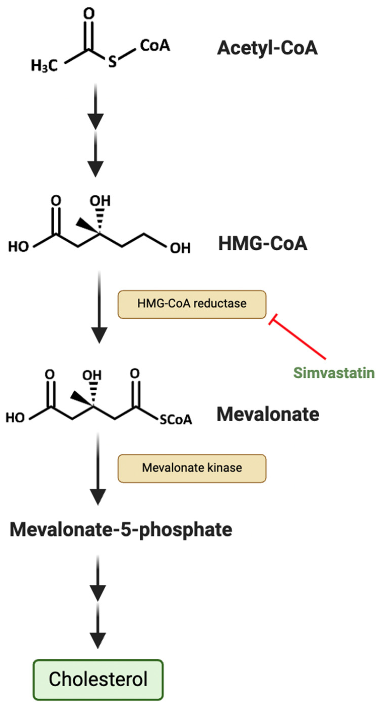
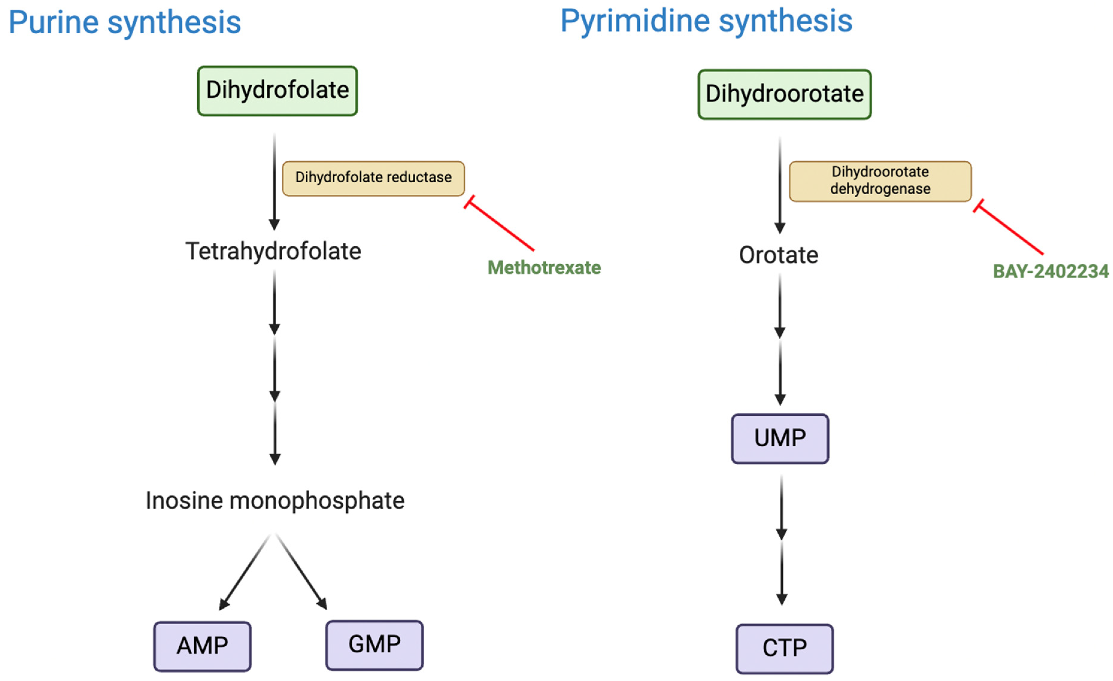
2.3. Deregulating Cellular Energetics

An emerging metabolic target in SMARCB1-deficient tumours is the cholesterol biosynthetic pathway (Figure 3). Cholesterol plays a key role in the tumoural immune response by regulating the function of various immune cells, including dendritic cells, CD8⁺ T lymphocytes, NK cells, and γδ T cells [30]. One metabolite in the cholesterol biosynthetic pathway, isopentenyl pyrophosphate (IPP), regulates the activity of glutathione peroxidase 4 (GPX4), which inhibits ferroptosis to promote cancer cell survival [31]. In addition, cholesterol contributes to the maintenance of cancer stem cells by activating downstream of several developmental signalling pathways, including Hedgehog and Notch [32]. In a newly established RT cell line, it was found that restoration of SMARCB1 significantly downregulated the expression of enzymes such as HMG-CoA reductase (HMGCR) and mevalonate kinase and that the frequently prescribed cholesterol-lowering drug simvastatin induced apoptosis in RT cells [33]. A phase I study (NCT02390843) of simvastatin in combination with topotecan and cyclophosphamide in relapsed and/or refractory paediatric patients with solid and central nervous system (CNS) tumours, including RTs, has since been carried out and found that the combination was well tolerated [34]. It is important to note that cholesterol metabolism involves multiple highly regulated interconnected pathways [32]. Therefore, targeting a single enzyme or receptor within these pathways may not sufficiently inhibit tumour progression. In various solid tumours, including pancreatic cancer, statin treatment has been demonstrated to induce a compensatory increase in HMGCR and exert a partial epithelial-to-mesenchymal transition (EMT) phenotype [35]. EMT is a key process in oncogenesis which confers migratory and metastatic properties to malignant epithelial cells [36]. A potential advantage in the clinical translation of statins in RTs is that they have been increasingly prescribed in paediatric populations with conditions like familial hypercholesterolemia, meaning that their safety and dosing parameters are relatively well defined [37].
A recent study by Kes and colleagues used patient-derived organoids to investigate the metabolic vulnerabilities of RTs [38]. Gene expression analysis and liquid chromatography–mass spectrometry revealed that de novo nucleotide biosynthesis is significantly upregulated in RTs, as demonstrated by the increased levels of metabolites such as uridine monophosphate (UMP) and inosine monophosphate (IMP). Pharmacological inhibition (Figure 4) using methotrexate and BAY-2402234, targeting purine and pyrimidine synthesis pathways, respectively, resulted in significant cytotoxic effects in RT tumoroids while sparing normal kidney organoids. Further in vivo validation in RT xenograft mouse models demonstrated that methotrexate treatment delayed tumour progression [38]. While the direct link between these metabolic aberrations and SMARCB1 loss has not been confirmed, SMARCB1 loss is likely to be indirectly or directly causal given that it is the defining driver of RTs. Further research into the precise role of SMARCB1 deficiency as a driver of nucleotide synthesis may yield novel therapeutic targets and enhance understanding of the pathogenesis of the disease.

2.4. Non-Mutational Epigenetic Reprogramming

Widespread transcriptional dysregulation and aberrant chromatin remodelling is known to occur as a result of SMARCB1 loss in RTs [39]. In mouse embryonic fibroblasts, SMARCB1 deletion resulted in decreased levels of the active histone markers H3K27ac and H3Kme1 at enhancers [40]. The action of the polycomb repressive complex 2 (PRC2), whose catalytic subunit EZH2 exhibits histone methyltransferase activity, is opposed by the SWI/SNF complex. As a result, SMARCB1 mutation is associated with uncontrolled PRC2-mediated inhibition of BRG1/BRM-associated factor (BAF) target genes. Tazemetostat, an inhibitor of EZH2, has received FDA approval for treatment of SMARCB1-deficient epithelioid sarcoma [41], and trials for other malignancies, including RTs, are ongoing. In the absence of SMARCB1, RT cells upregulate GLTSCR1-like-containing BAF complex (GBAF) expression, an SWI/SNF subcomplex which does not contain the SMARCB1 subunit. GBAF’s subunits BRD9 and GLTSCR1 are proposed drug targets, and their degradation or inhibition has been shown to reduce the viability of RT cell lines [42]. SMARCB1 deficiency also prevents the recruitment of cBAF and PBAF to the promoter and enhancer regions of SLC7A11, which plays a crucial role in glutathione metabolism by supplying cysteine for glutathione synthesis. Treatment with eprenetapopt has been shown to further decrease glutathione levels, resulting in oxidative stress and apoptosis [43].
It has recently been demonstrated that in SMARCB1-deficient cells, CBP/p300-mediated H3K27 acetylation promotes the expression of Kringle-containing transmembrane protein 2 (KREMEN2) [44], encoding a protein which complexes with KREMEN1 to regulate the Wnt/β catenin pathway [45] and inhibit apoptosis [46]. SMARCB1-deficient cells subsequently become dependent on KREMEN2 for survival. Dual inhibitors of CBP/p300 have been shown to suppress growth and induce apoptosis in cell lines and xenograft models of SMARCB1-deficient cells but not SMARCB1-expressing cells [44]. This serves as another example of synthetic lethal vulnerabilities in SMARCB-1-deficient RTs, which are highlighted in Figure 5.

2.5. Activating Invasion and Metastasis

While the link between SMARCB1 deficiency and enhanced metastatic potential has been explored in several tumour types, this association has been somewhat understudied in RTs. A recent study in bladder cancer showed that SMARCB1-deficient tumours displayed increased IL6–janus kinase–signal transducer and activator of transcription 3 (IL6-JAK-STAT3) signalling in in vivo models and patient tumours, believed to be associated with metastasis. STAT3 knockdown reduced tumour growth and lung metastasis. In addition, treatment of xenografts with TTI-101, a selective inhibitor of phosphorylated STAT3, reduced metastatic burden as measured by bioluminescence imaging [47]. STAT3 has been shown to promote the expression of Twist and Snail, master regulators of EMT [48]. In addition, a study by Liu and colleagues [49] found that cisplatin-resistant ATRT cells exhibit enhanced invasion, motility, and stem-like properties driven by upregulation of the STAT3–Snail axis. Knockdown of STAT3 was shown to suppress Snail expression, reduce invasiveness, and reverse resistance, while STAT3 inhibition improved survival in ATRT-CisR-transplanted mice. This highlights how STAT3 expression is not only involved in promoting an aggressive disease phenotype but also may be implicated in therapeutic failure. In a case report of poorly differentiated sacrococcygeal chordoma, expression of the premetastatic chemokine receptor CXCR4 was upregulated [50]. This may be explainable by the upregulation of EZH2 in SMARCB1-deficient tumours, which enhances CXCR4 expression through the repression of the tumour-suppressive microRNAs miR-622 and miR-9 [51,52]. However, it is crucial to note that the patient’s tumour also displayed TP53 mutation and RB1 loss, so these premetastatic alterations cannot be attributed with certainty to SMARCB1 loss, particularly in a single patient report.

3. Ongoing Trials and Future Directions

3.1. Immunotherapies

Immune checkpoint proteins (ICPs) are molecules which control the activation of T lymphocytes, a mechanism employed by the body in order to limit the indiscriminate killing of healthy tissues [53]. Various ICPs have been discovered to date, the most notable being PD-1 and cytotoxic T-lymphocyte-associated protein 4 (CTLA-4). PD-1 on T cells binds to PD-L1 expressed on tumour cells and antigen-presenting cells to suppress T cell proliferation, activity, and cytokine production [54]. CTLA-4 competes with CD28 for the binding of co-stimulatory molecules CD80/86 [55]. A more recently discovered ICP is T cell immunoreceptor with Ig and ITIM domains (TIGIT), which inhibits T and NK cell activation through interaction with CD155 [56].
Immune checkpoint inhibitors (ICIs) have been hailed as exciting anti-cancer therapeutics in the last few decades. Single-agent inhibition of PD-1 or PD-L1 in adults has demonstrated promising activity in various solid tumours. However, in the paediatric population, efficacy with mono-blockade has been limited to just a few tumours, particularly lymphomas [57]. Various ongoing clinical trials are therefore investigating the efficacy of combinations of ICIs in the treatment of SMARCB-1-deficient RTs. A phase II trial (NCT04416568) is currently recruiting patients to investigate the combination of nivolumab (anti-PD1) with ipilimumab (anti-CTLA4) in children and young adults with SMARCB1-deficient tumours, including RTs, epithelioid sarcomas, and chordomas [58]. In mouse models of solid tumours, co-blockade of both PD-L1 and TIGIT has been shown to promote the clonal expansion of multipotent anti-tumoural T lymphocytes [59]. Inhibition of PD-L1 and TIGIT through atezolizumab and tiragolumab, respectively, is being examined in a phase I/II clinical trial for the treatment of relapsed or refractory SMARCB1- or SMARCA4-deficient tumours [60]. Another proposed mechanism of enhancing ICI efficacy is through simultaneous inhibition of EZH2, a histone methyltransferase which has been shown to suppress MHC-I presentation and upregulate PD-L1 [61]. TAZNI (NCT05407441) is a phase I/II study recruiting to evaluate the combination of an EZH2 inhibitor, tazemetostat, with nivolumab and ipilimumab for SMARCA4-/SMARCB-1-deficient paediatric malignancies [62]. Table 1 summarises the ongoing clinical trials utilising ICIs in RTs.
Chimeric antigen receptor T (CAR-T) cells are genetically engineered T lymphocytes that express synthetic receptors designed to specifically recognise and bind target antigens on tumour cells [63]. This results in the activation of intracellular signalling domains such as CD3ζ, 4-1BB, and CD28 to elicit cancer cytotoxicity [64]. Multiple CAR-T cell strategies are being investigated for RTs (Table 2). CAR-T cells are often targeted against over-expressed tumour antigens, such as the membrane-bound proteoglycan glypican 3 (GPC3), in the case of solid tumours, including RTs. These trials (NCT05103631, NCT04715191, and NCT04377932) are investigating the use of IL-15/21 to enhance CAR-T cell efficacy and persistence [65,66,67]. CAR-T cells may also serve as ICIs. For example, a couple of trials (NCT05835687 and NCT04897321) are exploring the targeting of B7-H3, a transmembrane immunoregulatory protein, by CAR-T cells for the treatment of solid tumours.
Table 1. Active clinical trials utilising immune checkpoint inhibitors to target RTs.
Trial NumberPhaseSMARCB-1 Targeting TherapiesDisorderTargetStatusSitesPrimary Outcome Measures
NCT04416568 [58]IINivolumab and IpilimumabSMARCB1-negative tumoursPD-1, CTLA-4RecruitingTexas, USAObjective overall response rate
NCT05286801 [60]I/IITiragolumab and AtezolizumabSMARCB1- or SMARCA4-deficient tumours, including RTsTIGIT, PD-L1RecruitingUSA, Canada, and Australia Objective response rate and dose-limiting toxicities
NCT05407441 [62]I/IITazemetostat, nivolumab, ipilimumab SMARCB1-negative or SMARCA4-deficient tumours, including RTsEZH2, PD-1, CTLA-4RecruitingBoston, MA, USA Toxicity and dosing parameters
NCT06622941 [68]IINivolumab (ONO-4538)RTsPD-1Not yet recruitingOsaka and Tokyo, JapanObjective response rate
Table 2. Active clinical trials utilising CAR-T cells and cytotoxic lymphocytes against RTs.
Trial NumberPhaseSMARCB-1-Targeting TherapiesDisorderTargetStatusSitesPrimary Outcome Measures
NCT06193759 [69]ICytotoxic T lymphocytes directed against proteogenomically determined tumour-specific antigens Paediatric brain tumours, including RTsVarious tumour-specific antigensRecruitingWashington, USAVarious adverse events and toxicity parameters
NCT05835687 [70]ILocoregional autologous B7-H3-CAR T cellsPrimary CNS neoplasms, including RTsB7-H3-positive tumoursRecruitingTennessee, USA Maximum tolerated dose
NCT05103631 [65]IGPC3-CART cells and IL-15Solid tumours, including RTsGPC3-positive tumoursRecruitingTexas, USA Dose-limiting toxicities
NCT04897321 [71]IAutologous B7-H3-CAR T cellsSolid tumours, including RTsB7-H3-positive tumoursRecruitingTennessee, USAMaximum tolerated dose
NCT04715191 [66]IGPC3-CART cells and IL-15/21Paediatric solid tumours, including RTsGPC3-positive tumoursRecruitingTexas, USA Dose-limiting toxicities
NCT04377932 [67]IGPC3-CART cells and IL-15Paediatric solid tumours, including RTsGPC3-positive tumoursRecruitingTexas, USA Dose-limiting toxicities
NCT04185038 [72]ILocoregional autologous B7-H3-CAR T cellsPaediatric CNS tumours, including RTsB7-H3-positive tumoursRecruitingWashington, USA Feasibility and adverse event parameters
NCT03618381 [73]IEGFR806 CAR T cell immunotherapyRecurrent/refractory solid tumours in children and young adults, including RTsEGFRRecruitingWashington, USAMaximum tolerated dose, feasibility, and adverse event parameters
NCT04483778 [74]IB7-H3-CAR T cellsRecurrent/refractory solid tumours in children and young adults, including RTsB7-H3-positive tumoursActive, not recruitingWashington, USAVarious safety, tolerability, toxicity, and feasibility parameters

3.2. Targeting Epigenetic Aberrations

The role of epigenetics in cancer development and progression has been spotlighted as a key area of interest in recent years. In 2024, an article by Esteller and colleagues proposed the concept of six potentially targetable epigenetic hallmarks of cancer [75]. Epigenetic therapies in cancer aim to modify aberrant epigenetic patterns, such as DNA methylation, histone modifications, and chromatin remodelling [76]. The phase I study of tazemetostat, which targets the PRC2 catalytic subunit EZH2 (NCT02601937), included paediatric patients with relapsed or refractory RTs, other SMARCB1-deficient tumours, and synovial sarcoma [77]. Objective responses were seen in 14% of the dose-expansion cohort population, with a 24% response in intracerebral RT patients [78].
Vorinostat is an agent which inhibits the activity of histone deacetylases [79]. A phase I trial (NCT01076530) investigated the combination of vorinostat with temozolomide for the treatment of relapsed/refractory paediatric CNS tumours [80]. Nineteen patients were recruited, including two with rhabdoid tumours. Clinical efficacy was modest, with only three patients exhibiting stable disease and one having a partial response. Nevertheless, the combination was well tolerated, and myelosuppression was identified as the dose-limiting toxicity. It was also noted that accumulation of acetylated H3 histone in peripheral blood mononuclear cells occurred following administration of vorinostat [81]. While clinical utility with epigenetic targeting appears limited thus far, it remains a relatively novel field of cancer research. Future studies focused on identifying predictive biomarkers and exploring synergistic combinations may improve outcomes.

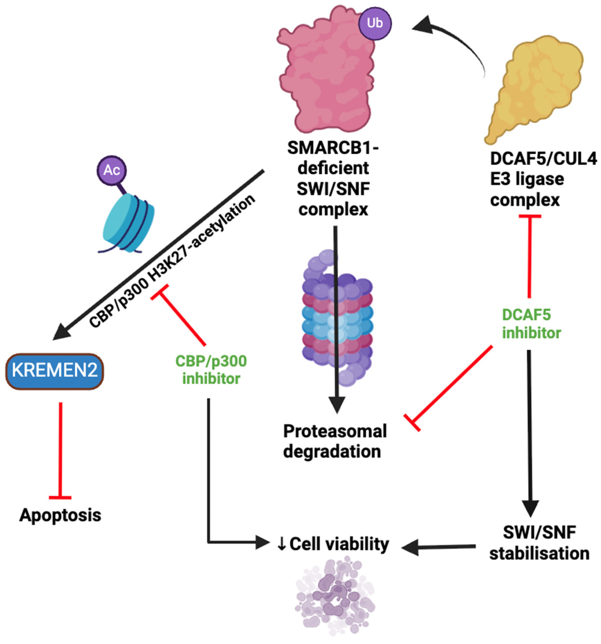
3.3. Targeting Auxiliary Oncogenic Aberrations

Aurora kinase A (AURKA) plays a pivotal role in mitotic spindle formation, and its overexpression has been implicated in various malignancies, including RTs [82,83]. A phase II clinical trial evaluated alisertib, an AURKA inhibitor, as a monotherapy in patients under 22 years with recurrent or progressive ATRTs. The study did not meet its primary efficacy endpoint of ≥10 patients being progression-free at 12 weeks. However, alisertib was found to be well tolerated, with a 6-month progression-free survival (PFS) of 31% ± 8.2%. A third of patients also demonstrated disease stabilisation beyond 6 months [84]. Mutations leading to truncated SMARCB1 can result in its exportin-1-mediated mislocalisation in the cytoplasm and hence loss of tumour-suppressive capacity. A phase I trial of the exportin-1 inhibitor selinexor for paediatric patients with recurrent/refractory solid and CNS tumours established the maximal tolerated dose and recommended a phase II (NCT05985161) starting dose of 35 mg/m2 for patients with recurrent/progressive ATRTs [85]. The proteins CDK4/6 are crucial for cell cycle progression. A phase I trial of the CDK4/6 inhibitor ribociclib in paediatric patients with RTs, neuroblastoma, and other solid tumours showed acceptable safety and prolonged stable disease in a subset of patients [86]. A phase II study is currently recruiting to assess the efficacy of ribociclib with topotecan and temozolomide in paediatric patients with relapsed or refractory neuroblastoma and solid tumours, including RTs [87]. Table 3 provides an overview of the active clinical trials targeting aberrations in the cell cycle (CDK4/6) and the dysregulated nuclear export of SMARCB1 (exportin-1). Future efforts should attempt to identify predictive biomarkers to stratify patients most likely to benefit from inhibitors of these auxiliary oncogenic aberrations.

3.4. Next Frontiers in Rhabdoid Tumour Research and Treatment

Gene therapy holds promise as a potential approach to treating cancers through the modification or restoration of gene expression [90]. A study published by Kim et al. in 2024 explored the use of scL-SMARCB1, a nanomedicine used to deliver wild-type SMARCB1 into ATRT cells via transferrin receptor-mediated endocytosis [91]. scL-SMARCB1 therapy was found to upregulate the expression of MYC-repressed genes such as CDKN1C and CDKN2A, while downregulating MYC-activated genes. AURKA expression was also significantly suppressed. It was also found that transfection with SMARCB1 potentiated cytotoxicity induced by cisplatin and radiation therapy [91]. This highlights how genetic therapy may not only restore biological markers in rhabdoid tumours but also enhance their sensitivity to conventional chemoradiation. This may have the potential to increase therapeutic efficacy and limit toxicity to healthy tissue.
Artificial intelligence (AI) may serve as a powerful tool for analysing complex genomic data in order to identify new therapeutic indications for drugs. One study has described the application of the Response Algorithm for Drug Positioning and Rescue (RADR®) AI platform, combined with the CellMiner Cross Database, in uncovering novel therapeutic insights for the acylfulvene-derivative anti-cancer drugs LP-100 and LP-184 [92]. Gene set enrichment analysis (GSEA) revealed a significant negative correlation between LP-184 sensitivity and SMARCB1 expression. These computational findings were then validated in SMARCB1-deficient ATRT cell lines and through in vivo experiments [92]. This demonstrates the utility of AI-driven platforms in rapidly identifying novel therapeutic indications for drugs. Such an approach may be especially useful for rarer cancers such as SMARCB1-deficient rhabdoid malignancies, which tend to have a relatively limited specific research focus.

4. Conclusions

The discussed trials and future innovations highlight how personalised therapies based on genomic, metabolomic, and immunological profiling have the potential to be the mainstay of RT treatment and disease monitoring. Emerging evidence also highlights the importance of metabolic reprogramming in RTs, with pathways such as cholesterol biosynthesis and nucleotide metabolism presenting novel therapeutic vulnerabilities. Integrating metabolomic profiling into future studies may uncover additional biomarkers and enable the development of metabolism-targeted therapies in SMARCB1-deficient tumours. Identifying subsets of patients who are most likely to respond to the various treatments will be crucial. Pre-clinical models such as xenografts may not fully recapitulate the RT microenvironment or the dynamics of tumour progression. Thus, systems such as patient-derived organoid systems may offer promising avenues to better mimic in vivo conditions and improve translational relevance. The effects, optimal timing, and integration of combinations of SMARCB1-targeted treatments with conventional therapies must be better characterised in order to enhance patient outcomes and allow incorporation into later-phase clinical trials and clinical practice. Additional research and genomic analysis are required to further elucidate the role of SMARCB1 in tumour suppression and to identify additional molecular targets.

Author Contributions

A.L.S.: writing—original draft, software. G.P.P.: conceptualisation, writing—original draft. All authors have read and agreed to the published version of the manuscript.

Funding

This research received no external funding.

Institutional Review Board Statement

Not applicable.

Informed Consent Statement

Not applicable.

Data Availability Statement

No new data has been created outside of this manuscript.

Conflicts of Interest

The authors declare that they have no conflicts of interest regarding the publication of this review.

References

  1. Pawel, B.R. SMARCB1-deficient Tumors of Childhood: A Practical Guide. Pediatr. Dev. Pathol. 2017, 21, 6–28. [Google Scholar] [CrossRef] [PubMed]
  2. Cooper, G.W.; Hong, A.L. SMARCB1-Deficient Cancers: Novel Molecular Insights and Therapeutic Vulnerabilities. Cancers 2022, 14, 3645. [Google Scholar] [CrossRef]
  3. Gupta, N.K.; Godbole, N.; Sanmugananthan, P.; Gunda, S.; Kasula, V.; Baggett, M.; Gajjar, A.; Kouam, R.W.; D’Amico, R.; Rodgers, S. Management of Atypical Teratoid/Rhabdoid Tumors in the Pediatric Population: A Systematic Review and Meta-Analysis. World Neurosurg. 2024, 181, e504–e515. [Google Scholar] [CrossRef]
  4. Nemes, K.; Bens, S.; Bourdeaut, F.; Johann, P.; Kordes, U.; Siebert, R.; Frühwald, M.C. Rhabdoid Tumor Predisposition Syndrome. In GeneReviews®; Adam, M.P., Feldman, J., Mirzaa, G.M., Pagon, R.A., Wallace, S.E., Amemiya, A., Eds.; University of Washington: Seattle, WA, USA, 1993. [Google Scholar]
  5. Sultan, I.; Qaddoumi, I.; Rodríguez-Galindo, C.; Nassan, A.A.; Ghandour, K.; Al-Hussaini, M. Age, stage, and radiotherapy, but not primary tumor site, affects the outcome of patients with malignant rhabdoid tumors. Pediatr. Blood Cancer 2010, 54, 35–40. [Google Scholar] [CrossRef] [PubMed]
  6. Lee, R.S.; Stewart, C.; Carter, S.L.; Ambrogio, L.; Cibulskis, K.; Sougnez, C.; Lawrence, M.S.; Auclair, D.; Mora, J.; Golub, T.R.; et al. A remarkably simple genome underlies highly malignant pediatric rhabdoid cancers. J. Clin. Investig. 2012, 122, 2983–2988. [Google Scholar] [CrossRef]
  7. Nemes, K.; Fruhwald, M.C. Emerging therapeutic targets for the treatment of malignant rhabdoid tumors. Expert Opin. Ther. Targets 2018, 22, 365–379. [Google Scholar] [CrossRef] [PubMed]
  8. Kalimuthu, S.N.; Chetty, R. Gene of the month: SMARCB1. J. Clin. Pathol. 2016, 69, 484–489. [Google Scholar] [CrossRef]
  9. Roberts, C.W.; Leroux, M.M.; Fleming, M.D.; Orkin, S.H. Highly penetrant, rapid tumorigenesis through conditional inversion of the tumor suppressor gene Snf5. Cancer Cell 2002, 2, 415–425. [Google Scholar] [CrossRef]
  10. Han, Z.Y.; Richer, W.; Freneaux, P.; Chauvin, C.; Lucchesi, C.; Guillemot, D.; Grison, C.; Lequin, D.; Pierron, G.; Masliah-Planchon, J.; et al. The occurrence of intracranial rhabdoid tumours in mice depends on temporal control of Smarcb1 inactivation. Nat. Commun. 2016, 7, 10421. [Google Scholar] [CrossRef]
  11. Pathak, R.; Zin, F.; Thomas, C.; Bens, S.; Gayden, T.; Karamchandani, J.; Dudley, R.W.; Nemes, K.; Johann, P.D.; Oyen, F.; et al. Inhibition of nuclear export restores nuclear localization and residual tumor suppressor function of truncated SMARCB1/INI1 protein in a molecular subset of atypical teratoid/rhabdoid tumors. Acta Neuropathol. 2021, 142, 361–374. [Google Scholar] [CrossRef]
  12. Hanahan, D.; Weinberg, R.A. Hallmarks of cancer: The next generation. Cell 2011, 144, 646–674. [Google Scholar] [CrossRef] [PubMed]
  13. Hanahan, D. Hallmarks of Cancer: New Dimensions. Cancer Discov. 2022, 12, 31–46. [Google Scholar] [CrossRef] [PubMed]
  14. Hanahan, D.; Weinberg, R.A. The hallmarks of cancer. Cell 2000, 100, 57–70. [Google Scholar] [CrossRef] [PubMed]
  15. Betz, B.L.; Strobeck, M.W.; Reisman, D.N.; Knudsen, E.S.; Weissman, B.E. Re-expression of hSNF5/INI1/BAF47 in pediatric tumor cells leads to G1 arrest associated with induction of p16ink4a and activation of RB. Oncogene 2002, 21, 5193–5203. [Google Scholar] [CrossRef]
  16. García-Gutiérrez, L.; Delgado, M.D.; León, J. MYC Oncogene Contributions to Release of Cell Cycle Brakes. Genes 2019, 10, 244. [Google Scholar] [CrossRef]
  17. Weissmiller, A.M.; Wang, J.; Lorey, S.L.; Howard, G.C.; Martinez, E.; Liu, Q.; Tansey, W.P. Inhibition of MYC by the SMARCB1 tumor suppressor. Nat. Commun. 2019, 10, 2014. [Google Scholar] [CrossRef]
  18. Amati, B.; Land, H. Myc—Max—Mad: A transcription factor network controlling cell cycle progression, differentiation and death. Curr. Opin. Genet. Dev. 1994, 4, 102–108. [Google Scholar] [CrossRef]
  19. Baudino, T.A.; McKay, C.; Pendeville-Samain, H.; Nilsson, J.A.; Maclean, K.H.; White, E.L.; Davis, A.C.; Ihle, J.N.; Cleveland, J.L. c-Myc is essential for vasculogenesis and angiogenesis during development and tumor progression. Genes Dev. 2002, 16, 2530–2543. [Google Scholar] [CrossRef]
  20. Dong, Y.; Tu, R.; Liu, H.; Qing, G. Regulation of cancer cell metabolism: Oncogenic MYC in the driver’s seat. Signal Transduct. Target. Ther. 2020, 5, 124. [Google Scholar] [CrossRef]
  21. Darr, J.; Klochendler, A.; Isaac, S.; Geiger, T.; Eden, A. Phosphoproteomic analysis reveals Smarcb1 dependent EGFR signaling in Malignant Rhabdoid tumor cells. Mol. Cancer 2015, 14, 167. [Google Scholar] [CrossRef]
  22. Wong, J.P.; Todd, J.R.; Finetti, M.A.; McCarthy, F.; Broncel, M.; Vyse, S.; Luczynski, M.T.; Crosier, S.; Ryall, K.A.; Holmes, K.; et al. Dual Targeting of PDGFRα and FGFR1 Displays Synergistic Efficacy in Malignant Rhabdoid Tumors. Cell Rep. 2016, 17, 1265–1275. [Google Scholar] [CrossRef] [PubMed]
  23. Gregory, G.L.; Copple, I.M. Modulating the expression of tumor suppressor genes using activating oligonucleotide technologies as a therapeutic approach in cancer. Mol. Ther. Nucleic Acids 2023, 31, 211–223. [Google Scholar] [CrossRef]
  24. Ge, M.; Luo, J.; Wu, Y.; Shen, G.; Kuang, X. The biological essence of synthetic lethality: Bringing new opportunities for cancer therapy. MedComm—Oncology 2024, 3, e70. [Google Scholar] [CrossRef]
  25. Radko-Juettner, S.; Yue, H.; Myers, J.A.; Carter, R.D.; Robertson, A.N.; Mittal, P.; Zhu, Z.; Hansen, B.S.; Donovan, K.A.; Hunkeler, M.; et al. Targeting DCAF5 suppresses SMARCB1-mutant cancer by stabilizing SWI/SNF. Nature 2024, 628, 442–449. [Google Scholar] [CrossRef]
  26. Dedes, K.J.; Wilkerson, P.M.; Wetterskog, D.; Weigelt, B.; Ashworth, A.; Reis-Filho, J.S. Synthetic lethality of PARP inhibition in cancers lacking BRCA1 and BRCA2 mutations. Cell Cycle 2011, 10, 1192–1199. [Google Scholar] [CrossRef] [PubMed]
  27. Chun, H.E.; Johann, P.D.; Milne, K.; Zapatka, M.; Buellesbach, A.; Ishaque, N.; Iskar, M.; Erkek, S.; Wei, L.; Tessier-Cloutier, B.; et al. Identification and Analyses of Extra-Cranial and Cranial Rhabdoid Tumor Molecular Subgroups Reveal Tumors with Cytotoxic T Cell Infiltration. Cell Rep. 2019, 29, 2338–2354.e7. [Google Scholar] [CrossRef]
  28. Forrest, S.J.; Al-Ibraheemi, A.; Doan, D.; Ward, A.; Clinton, C.M.; Putra, J.; Pinches, R.S.; Kadoch, C.; Chi, S.N.; DuBois, S.G.; et al. Genomic and Immunologic Characterization of INI1-Deficient Pediatric Cancers. Clin. Cancer Res. 2020, 26, 2882–2890. [Google Scholar] [CrossRef]
  29. Tran, S.; Plant-Fox, A.S.; Chi, S.N.; Narendran, A. Current advances in immunotherapy for atypical teratoid rhabdoid tumor (ATRT). Neurooncol. Pract. 2023, 10, 322–334. [Google Scholar] [CrossRef]
  30. Pecci, F.; Cognigni, V.; Giudice, G.C.; Paoloni, F.; Cantini, L.; Saini, K.S.; Abushukair, H.M.; Naqash, A.R.; Cortellini, A.; Mazzaschi, G.; et al. Unraveling the link between cholesterol and immune system in cancer: From biological mechanistic insights to clinical evidence. A narrative review. Crit. Rev. Oncol./Hematol. 2025, 209, 104654. [Google Scholar] [CrossRef]
  31. Ye, L.; Wen, X.; Qin, J.; Zhang, X.; Wang, Y.; Wang, Z.; Zhou, T.; Di, Y.; He, W. Metabolism-regulated ferroptosis in cancer progression and therapy. Cell Death Dis. 2024, 15, 196. [Google Scholar] [CrossRef]
  32. Xiao, M.; Xu, J.; Wang, W.; Zhang, B.; Liu, J.; Li, J.; Xu, H.; Zhao, Y.; Yu, X.; Shi, S. Functional significance of cholesterol metabolism in cancer: From threat to treatment. Exp. Mol. Med. 2023, 55, 1982–1995. [Google Scholar] [CrossRef] [PubMed]
  33. Matsumoto, F.; Yokogami, K.; Yamada, A.; Moritake, H.; Watanabe, T.; Yamashita, S.; Sato, Y.; Takeshima, H. Targeting cholesterol biosynthesis for AT/RT: Comprehensive expression analysis and validation in newly established AT/RT cell line. Hum. Cell 2024, 37, 523–530. [Google Scholar] [CrossRef] [PubMed]
  34. Cash, T.; Jonus, H.C.; Tsvetkova, M.; Beumer, J.H.; Sadanand, A.; Lee, J.Y.; Henry, C.J.; Aguilera, D.; Harvey, R.D.; Goldsmith, K.C. A phase 1 study of simvastatin in combination with topotecan and cyclophosphamide in pediatric patients with relapsed and/or refractory solid and CNS tumors. Pediatr Blood Cancer 2023, 70, e30405. [Google Scholar] [CrossRef]
  35. Dorsch, M.; Kowalczyk, M.; Planque, M.; Heilmann, G.; Urban, S.; Dujardin, P.; Forster, J.; Ueffing, K.; Nothdurft, S.; Oeck, S.; et al. Statins affect cancer cell plasticity with distinct consequences for tumor progression and metastasis. Cell Rep. 2021, 37, 110056. [Google Scholar] [CrossRef]
  36. Ribatti, D.; Tamma, R.; Annese, T. Epithelial-Mesenchymal Transition in Cancer: A Historical Overview. Transl. Oncol. 2020, 13, 100773. [Google Scholar] [CrossRef]
  37. Fiorentino, R.; Chiarelli, F. Statins in Children, an Update. Int. J. Mol. Sci. 2023, 24, 1366. [Google Scholar] [CrossRef]
  38. Kes, M.M.G.; Morales-Rodriguez, F.; Zaal, E.A.; de Souza, T.; Proost, N.; van de Ven, M.; van den Heuvel-Eibrink, M.M.; Jansen, J.W.A.; Berkers, C.R.; Drost, J. Metabolic profiling of patient-derived organoids reveals nucleotide synthesis as a metabolic vulnerability in malignant rhabdoid tumors. Cell Rep. Med. 2025, 6, 101878. [Google Scholar] [CrossRef]
  39. Kenny, C.; O’Meara, E.; Ulas, M.; Hokamp, K.; O’Sullivan, M.J. Global Chromatin Changes Resulting from Single-Gene Inactivation-The Role of SMARCB1 in Malignant Rhabdoid Tumor. Cancers 2021, 13, 2561. [Google Scholar] [CrossRef] [PubMed]
  40. Wang, X.; Lee, R.S.; Alver, B.H.; Haswell, J.R.; Wang, S.; Mieczkowski, J.; Drier, Y.; Gillespie, S.M.; Archer, T.C.; Wu, J.N.; et al. SMARCB1-mediated SWI/SNF complex function is essential for enhancer regulation. Nat. Genet. 2017, 49, 289–295. [Google Scholar] [CrossRef]
  41. Hoy, S.M. Tazemetostat: First Approval. Drugs 2020, 80, 513–521. [Google Scholar] [CrossRef]
  42. Wang, X.; Wang, S.; Troisi, E.C.; Howard, T.P.; Haswell, J.R.; Wolf, B.K.; Hawk, W.H.; Ramos, P.; Oberlick, E.M.; Tzvetkov, E.P.; et al. BRD9 defines a SWI/SNF sub-complex and constitutes a specific vulnerability in malignant rhabdoid tumors. Nat. Commun. 2019, 10, 1881. [Google Scholar] [CrossRef] [PubMed]
  43. Sasaki, M.; Ogiwara, H. Efficacy of glutathione inhibitor eprenetapopt against the vulnerability of glutathione metabolism in SMARCA4-, SMARCB1- and PBRM1-deficient cancer cells. Sci. Rep. 2024, 14, 31321. [Google Scholar] [CrossRef]
  44. Sasaki, M.; Kato, D.; Murakami, K.; Yoshida, H.; Takase, S.; Otsubo, T.; Ogiwara, H. Targeting dependency on a paralog pair of CBP/p300 against de-repression of KREMEN2 in SMARCB1-deficient cancers. Nat. Commun. 2024, 15, 4770. [Google Scholar] [CrossRef]
  45. Mao, B.; Wu, W.; Davidson, G.; Marhold, J.; Li, M.; Mechler, B.M.; Delius, H.; Hoppe, D.; Stannek, P.; Walter, C.; et al. Kremen proteins are Dickkopf receptors that regulate Wnt/beta-catenin signalling. Nature 2002, 417, 664–667. [Google Scholar] [CrossRef] [PubMed]
  46. Sumia, I.; Pierani, A.; Causeret, F. Kremen1-induced cell death is regulated by homo- and heterodimerization. Cell Death Discov. 2019, 5, 91. [Google Scholar] [CrossRef]
  47. Amara, C.S.; Kami Reddy, K.R.; Yuntao, Y.; Chan, Y.S.; Piyarathna, D.W.B.; Dobrolecki, L.E.; Shih, D.J.H.; Shi, Z.; Xu, J.; Huang, S.; et al. The IL6/JAK/STAT3 signaling axis is a therapeutic vulnerability in SMARCB1-deficient bladder cancer. Nat. Commun. 2024, 15, 1373. [Google Scholar] [CrossRef] [PubMed]
  48. Wendt, M.K.; Balanis, N.; Carlin, C.R.; Schiemann, W.P. STAT3 and epithelial-mesenchymal transitions in carcinomas. Jakstat 2014, 3, e28975. [Google Scholar] [CrossRef]
  49. Liu, W.H.; Chen, M.T.; Wang, M.L.; Lee, Y.Y.; Chiou, G.Y.; Chien, C.S.; Huang, P.I.; Chen, Y.W.; Huang, M.C.; Chiou, S.H.; et al. Cisplatin-selected resistance is associated with increased motility and stem-like properties via activation of STAT3/Snail axis in atypical teratoid/rhabdoid tumor cells. Oncotarget 2015, 6, 1750–1768. [Google Scholar] [CrossRef]
  50. Murzabdillaeva, A.; Elzamly, S.; Brown, R.; Buryanek, J.; Jafri, S.; Rowe, J. Prometastatic CXCR4 and Histone Methyltransferase EZH2 are Upregulated in SMARCB1/INI1-deficient and TP53-mutated Metastatic Poorly Differentiated Chordoma to the Liver. Am. J. Clin. Pathol. 2020, 154 (Suppl. S1), S151. [Google Scholar] [CrossRef]
  51. Liu, H.; Liu, Y.; Liu, W.; Zhang, W.; Xu, J. EZH2-mediated loss of miR-622 determines CXCR4 activation in hepatocellular carcinoma. Nat. Commun. 2015, 6, 8494. [Google Scholar] [CrossRef]
  52. Chien, Y.C.; Chen, J.N.; Chen, Y.H.; Chou, R.H.; Lee, H.C.; Yu, Y.L. Epigenetic Silencing of miR-9 Promotes Migration and Invasion by EZH2 in Glioblastoma Cells. Cancers 2020, 12, 1781. [Google Scholar] [CrossRef] [PubMed]
  53. Marhelava, K.; Pilch, Z.; Bajor, M.; Graczyk-Jarzynka, A.; Zagozdzon, R. Targeting Negative and Positive Immune Checkpoints with Monoclonal Antibodies in Therapy of Cancer. Cancers 2019, 11, 1756. [Google Scholar] [CrossRef]
  54. Jiang, Y.; Chen, M.; Nie, H.; Yuan, Y. PD-1 and PD-L1 in cancer immunotherapy: Clinical implications and future considerations. Hum. Vaccines Immunother. 2019, 15, 1111–1122. [Google Scholar] [CrossRef]
  55. Liu, P.C.; Ssu, C.T.; Tsao, Y.P.; Liou, T.L.; Tsai, C.Y.; Chou, C.T.; Chen, M.H.; Leu, C.M. Cytotoxic T lymphocyte-associated antigen-4-Ig (CTLA-4-Ig) suppresses Staphylococcus aureus-induced CD80, CD86, and pro-inflammatory cytokine expression in human B cells. Arthritis Res. Ther. 2020, 22, 64. [Google Scholar] [CrossRef] [PubMed]
  56. Bolm, L.; Petruch, N.; Sivakumar, S.; Annels, N.E.; Frampton, A.E. Gene of the month: T-cell immunoreceptor with immunoglobulin and ITIM domains (TIGIT). J. Clin. Pathol. 2022, 75, 217–221. [Google Scholar] [CrossRef]
  57. Davis, K.L.; Fox, E.; Isikwei, E.; Reid, J.M.; Liu, X.; Minard, C.G.; Voss, S.; Berg, S.L.; Weigel, B.J.; Mackall, C.L. A Phase I/II Trial of Nivolumab plus Ipilimumab in Children and Young Adults with Relapsed/Refractory Solid Tumors: A Children’s Oncology Group Study ADVL1412. Clin. Cancer Res. 2022, 28, 5088–5097. [Google Scholar] [CrossRef]
  58. Forrest, S.J.; Yi, J.; Kline, C.; Cash, T.; Reddy, A.T.; Cote, G.M.; Merriam, P.; Czaplinski, J.; Bhushan, K.; DuBois, S.G.; et al. Phase II study of nivolumab and ipilimumab in children and young adults with INI1-negative cancers. J. Clin. Oncol. 2021, 39 (Suppl. S15), TPS10055-TPS10055. [Google Scholar] [CrossRef]
  59. Nutsch, K.; Banta, K.L.; Wu, T.D.; Tran, C.W.; Mittman, S.; Duong, E.; Nabet, B.Y.; Qu, Y.; Williams, K.; Muller, S.; et al. TIGIT and PD-L1 co-blockade promotes clonal expansion of multipotent, non-exhausted antitumor T cells by facilitating co-stimulation. Nat. Cancer. 2024, 5, 1834–1851. [Google Scholar] [CrossRef]
  60. A Phase 1/2 Study of Tiragolumab (NSC# 827799) and Atezolizumab (NSC# 783608) in Patients with Relapsed or Refractory SMARCB1 or SMARCA4 Deficient Tumors. 2022. Available online: https://www.dana-farber.org/clinical-trials/22-528 (accessed on 28 April 2025).
  61. Kang, N.; Eccleston, M.; Clermont, P.L.; Latarani, M.; Male, D.K.; Wang, Y.; Crea, F. EZH2 inhibition: A promising strategy to prevent cancer immune editing. Epigenomics 2020, 12, 1457–1476. [Google Scholar] [CrossRef]
  62. Yi, J.S.; Czaplinski, J.; Bhushan, K.; Forrest, S.J.; Shukla, N.; Mack, S.; Hong, A.; Strachan, M.; DuBois, S.G.; London, W.B.; et al. TRLS-15. “TAZNI”: A phase I/II combination trial of tazemetostat with nivolumab and ipilimumab for children with INI1-negative or SMARCA4-deficient tumors (NCT05407441). Neuro-Oncology 2024, 26 (Suppl. S4). [Google Scholar] [CrossRef]
  63. Sterner, R.C.; Sterner, R.M. CAR-T cell therapy: Current limitations and potential strategies. Blood Cancer J. 2021, 11, 69. [Google Scholar] [CrossRef]
  64. Dabas, P.; Danda, A. Revolutionizing cancer treatment: A comprehensive review of CAR-T cell therapy. Med. Oncol. 2023, 40, 275. [Google Scholar] [CrossRef] [PubMed]
  65. ClinicalTrials.gov. Interleukin-15 Armored Glypican 3-Specific Chimeric Antigen Receptor Expressed in Autologous T Cells for Solid Tumors. ClinicalTrials.gov Identifier: NCT05103631. Available online: https://clinicaltrials.gov/study/NCT05103631 (accessed on 28 April 2025).
  66. ClinicalTrials.gov. Interleukin-15 and -21 Armored Glypican-3-Specific Chimeric Antigen Receptor Expressed in T Cells for Pediatric Solid Tumors. ClinicalTrials.gov Identifier: NCT04715191. Available online: https://clinicaltrials.gov/study/NCT04715191 (accessed on 28 April 2025).
  67. ClinicalTrials.gov. Interleukin-15 Armored Glypican 3-Specific Chimeric Antigen Receptor Expressed in T Cells for Pediatric Solid Tumors. ClinicalTrials.gov Identifier: NCT04377932. Available online: https://clinicaltrials.gov/study/NCT04377932 (accessed on 28 April 2025).
  68. ClinicalTrials.gov. Study to ONO-4538 in Patients with Rhabdoid Tumor. ClinicalTrials.gov Identifier: NCT06622941. Available online: https://clinicaltrials.gov/study/NCT06622941 (accessed on 28 April 2025).
  69. ClinicalTrials.gov. Immunotherapy for Malignant Pediatric Brain Tumors Employing Adoptive Cellular Therapy (IMPACT). ClinicalTrials.gov Identifier: NCT06193759. Available online: https://clinicaltrials.gov/study/NCT06193759 (accessed on 28 April 2025).
  70. ClinicalTrials.gov. Loc3CAR: Locoregional Delivery of B7-H3-CAR T Cells for Pediatric Patients With Primary CNS Tumors. ClinicalTrials.gov Identifier: NCT05835687. Available online: https://clinicaltrials.gov/study/NCT05835687 (accessed on 28 April 2025).
  71. ClinicalTrials.gov. B7-H3-Specific Chimeric Antigen Receptor Autologous T-Cell Therapy for Pediatric Patients with Solid Tumors (3CAR). ClinicalTrials.gov Identifier: NCT04897321. Available online: https://clinicaltrials.gov/study/NCT04897321 (accessed on 28 April 2025).
  72. ClinicalTrials.gov. Study of B7-H3-Specific CAR T Cell Locoregional Immunotherapy for Diffuse Intrinsic Pontine Glioma/Diffuse Midline Glioma and Recurrent or Refractory Pediatric Central Nervous System Tumors. ClinicalTrials.gov Identifier: NCT04185038. Available online: https://clinicaltrials.gov/study/NCT04185038 (accessed on 28 April 2025).
  73. ClinicalTrials.gov. EGFR806 CAR T Cell Immunotherapy for Recurrent/Refractory Solid Tumors in Children and Young Adults. ClinicalTrials.gov Identifier: NCT03618381. Available online: https://clinicaltrials.gov/study/NCT03618381 (accessed on 28 April 2025).
  74. ClinicalTrials.gov. B7H3 CAR T Cell Immunotherapy for Recurrent/Refractory Solid Tumors in Children and Young Adults. ClinicalTrials.gov Identifier: NCT04483778. Available online: https://clinicaltrials.gov/study/NCT04483778 (accessed on 28 April 2025).
  75. Esteller, M.; Dawson, M.A.; Kadoch, C.; Rassool, F.V.; Jones, P.A.; Baylin, S.B. The Epigenetic Hallmarks of Cancer. Cancer Discov. 2024, 14, 1783–1809. [Google Scholar] [CrossRef] [PubMed]
  76. Sadida, H.Q.; Abdulla, A.; Marzooqi, S.A.; Hashem, S.; Macha, M.A.; Akil, A.S.A.; Bhat, A.A. Epigenetic modifications: Key players in cancer heterogeneity and drug resistance. Transl. Oncol. 2024, 39, 101821. [Google Scholar] [CrossRef]
  77. ClinicalTrials.gov. EZH2 Inhibitor Tazemetostat in Pediatric Subjects with Relapsed or Refractory INI1-Negative Tumors or Synovial Sarcoma. ClinicalTrials.gov Identifier: NCT02601937. Available online: https://clinicaltrials.gov/study/NCT02601937 (accessed on 28 April 2025).
  78. Chi, S.N.; Bourdeaut, F.; Casanova, M.; Kilburn, L.B.; Hargrave, D.R.; McCowage, G.B.; Pinto, N.R.; Yang, J.; Chadha, R.; Kahali, B.; et al. Update on phase 1 study of tazemetostat, an enhancer of zeste homolog 2 inhibitor, in pediatric patients with relapsed or refractory integrase interactor 1–negative tumors. J. Clin. Oncol. 2022, 40 (Suppl. 16), 10040. [Google Scholar] [CrossRef]
  79. Ge, Z.; Da, Y.; Xue, Z.; Zhang, K.; Zhuang, H.; Peng, M.; Li, Y.; Li, W.; Simard, A.; Hao, J.; et al. Vorinostat, a histone deacetylase inhibitor, suppresses dendritic cell function and ameliorates experimental autoimmune encephalomyelitis. Exp. Neurol. 2013, 241, 56–66. [Google Scholar] [CrossRef] [PubMed]
  80. ClinicalTrials.gov. Vorinostat and Temozolomide in Treating Young Patients with Relapsed or Refractory Primary Brain Tumors or Spinal Cord Tumors. ClinicalTrials.gov Identifier: NCT01076530. Available online: https://clinicaltrials.gov/study/NCT01076530 (accessed on 28 April 2025).
  81. Hummel, T.R.; Wagner, L.; Ahern, C.; Fouladi, M.; Reid, J.M.; McGovern, R.M.; Ames, M.M.; Gilbertson, R.J.; Horton, T.; Ingle, A.M.; et al. A pediatric phase 1 trial of vorinostat and temozolomide in relapsed or refractory primary brain or spinal cord tumors: A Children’s Oncology Group phase 1 consortium study. Pediatr. Blood Cancer 2013, 60, 1452–1457. [Google Scholar] [CrossRef]
  82. Nemes, K.; Johann, P.D.; Tüchert, S.; Melchior, P.; Vokuhl, C.; Siebert, R.; Furtwängler, R.; Frühwald, M.C. Current and Emerging Therapeutic Approaches for Extracranial Malignant Rhabdoid Tumors. Cancer Manag. Res. 2022, 14, 479–498. [Google Scholar] [CrossRef]
  83. Du, R.; Huang, C.; Liu, K.; Li, X.; Dong, Z. Targeting AURKA in Cancer: Molecular mechanisms and opportunities for Cancer therapy. Mol. Cancer 2021, 20, 15. [Google Scholar] [CrossRef]
  84. Upadhyaya, S.; Campagne, O.; Robinson, G.W.; Onar-Thomas, A.; Orr, B.; Billups, C.A.; Tatevossian, R.G.; Broniscer, A.; Kilburn, L.B.; Baxter, P.A.; et al. Phase II study of alisertib as a single agent in recurrent or progressive atypical teratoid rhabdoid tumors. J. Clin. Oncol. 2020, 38 (Suppl. 15), 10542. [Google Scholar] [CrossRef]
  85. Green, A.L.; Minard, C.G.; Liu, X.; Safgren, S.L.; Pinkney, K.; Harris, L.; Link, G.; DeSisto, J.; Voss, S.; Nelson, M.D.; et al. Phase 1 trial of selinexor in pediatric recurrent/refractory solid and CNS tumors (ADVL1414): A Children’s Oncology Group Phase 1 Consortium Trial. Clin. Cancer Res. 2025, 31, 1587–1595. [Google Scholar] [CrossRef] [PubMed]
  86. Geoerger, B.; Bourdeaut, F.; DuBois, S.G.; Fischer, M.; Geller, J.I.; Gottardo, N.G.; Marabelle, A.; Pearson, A.D.J.; Modak, S.; Cash, T.; et al. A Phase I Study of the CDK4/6 Inhibitor Ribociclib (LEE011) in Pediatric Patients with Malignant Rhabdoid Tumors, Neuroblastoma, and Other Solid Tumors. Clin. Cancer Res. 2017, 23, 2433–2441. [Google Scholar] [CrossRef] [PubMed]
  87. ClinicalTrials.gov. Study of Efficacy and Safety of Ribociclib (LEE011) in Combination with Topotecan and Temozolomide (TOTEM) in Pediatric Patients With Relapsed or Refractory Neuroblastoma and Other Solid Tumors. ClinicalTrials.gov Identifier: NCT05429502. Available online: https://clinicaltrials.gov/study/NCT05429502 (accessed on 28 April 2025).
  88. ClinicalTrials.gov. A Study of Selinexor in People with Wilms Tumors and Other Solid Tumors. ClinicalTrials.gov Identifier: NCT05985161. Available online: https://clinicaltrials.gov/study/NCT05985161 (accessed on 28 April 2025).
  89. ClinicalTrials.gov. Study of Palbociclib Combined with Chemotherapy in Pediatric Patients with Recurrent/Refractory Solid Tumors. ClinicalTrials.gov Identifier: NCT03709680. Available online: https://clinicaltrials.gov/study/NCT03709680 (accessed on 28 April 2025).
  90. Cesur-Ergün, B.; Demir-Dora, D. Gene therapy in cancer. J. Gene Med. 2023, 25, e3550. [Google Scholar] [CrossRef] [PubMed]
  91. Kim, S.S.; Moghe, M.; Rait, A.; Donaldson, K.; Harford, J.B.; Chang, E.H. SMARCB1 Gene Therapy Using a Novel Tumor-Targeted Nanomedicine Enhances Anti-Cancer Efficacy in a Mouse Model of Atypical Teratoid Rhabdoid Tumors. Int. J. Nanomed. 2024, 19, 5973–5993. [Google Scholar] [CrossRef]
  92. McDermott, J.; Sturtevant, D.; Kathad, U.; Varma, S.; Zhou, J.; Kulkarni, A.; Biyani, N.; Schimke, C.; Reinhold, W.C.; Elloumi, F.; et al. Artificial intelligence platform, RADR®, aids in the discovery of DNA damaging agent for the ultra-rare cancer Atypical Teratoid Rhabdoid Tumors. Front. Drug Discov. 2022, 2. [Google Scholar] [CrossRef]
Figure 1. Aberrant pathways induced by SMARCB1 deficiency and corresponding therapeutic targets. ‘PRC2’ denotes polycomb repressive complex 2, ‘AURKA’ Aurora Kinase A, ‘RTK’ receptor tyrosine kinase, ‘BRD9’ bromodomain-containing protein 9, ‘GLTSCR1’ glioma tumour suppressor candidate region gene 1, ‘Rb’ retinoblastoma, ‘CDK4/6’ cyclin-dependent kinase 4/6, ‘PD-1’ programmed cell death protein 1, ‘CTLA-4’ cytotoxic T-lymphocyte–associated protein 4; SAM, S-adenosyl methionine, ‘PI3K’ phosphoinositide 3-kinase; ‘Akt’ protein kinase B; ‘mTOR’ mammalian target of rapamycin, and ‘HMGCR’ HMG-CoA reductase. Solid black arrows indicate activation or promotion of a protein or pathway. Solid bars with red cross indicate loss of inhibition. Red bars denote inhibition by corresponding drug or therapy. The symbol “P” in the yellow circle represents phosphorylation. Figure created with BioRender.com.
Metabolites 15 00304 g001
Figure 2. A summary of the role of SMARCB1 deficiency as a driver of the hallmarks of cancer in RTs. SMARCB1 loss promotes sustained proliferative signalling via upregulation of Myc, EGFR, and Aurora kinase A and facilitates evasion of growth suppressors through Rb hyperphosphorylation. Deregulated cellular energetics are marked by enhanced nucleotide synthesis and mevalonate pathway activation. It is important to note that upregulation of nucleotide synthesis has yet to be confirmed as a causal outcome of SMARCB1 deficiency. Tumour-promoting inflammation involves expression of CXCL10, PRF1, and CLEC9A, while immune evasion may be mediated through elevated IL-10, PD-L1, and HAVCR2. Non-mutational epigenetic reprogramming further supports oncogenesis and is driven by increased EZH2/PRC2 activity and histone methylation. Up arrows in figure indicate increased activity of protein or pathway. Figure created with BioRender.com.
Metabolites 15 00304 g002
Figure 3. The mevalonate pathway, highlighting the inhibition of HMG-CoA reductase using simvastatin. ‘HMG-CoA’ denotes β-Hydroxy β-methylglutaryl-CoA. Figure created using BioRender.com.
Metabolites 15 00304 g003
Figure 4. The nucleotide biosynthetic pathway as a potential metabolic vulnerability in RTs. ‘AMP’ denotes adenosine monophosphate, ‘GMP’ guanine monophosphate, ‘UMP’ uridine monophosphate, and ‘CTP’ cytidine triphosphate. Figure created using BioRender.com.
Metabolites 15 00304 g004
Figure 5. Synthetic lethal vulnerabilities in RTs. In SMARCB1-deficient cells, SMARCB1-deficient SWI/SNF complexes are targeted for ubiquitination and proteasomal degradation by the DCAF5/CUL4 E3 ligase complex. Inhibition of DCAF5 stabilises these defective SWI/SNF complexes, leading to toxic accumulation and loss of cell viability. In addition, CBP/p300-mediated H3K27 acetylation upregulates KREMEN2 expression, which suppresses apoptosis and promotes cell survival. Inhibition of CBP/p300 reduces KREMEN2 levels, promoting apoptosis. ‘Ub’ in purple circle indicates ubiquitination, while ‘Ac’ indicates acetylation. Figure created with BioRender.com.
Metabolites 15 00304 g005
Table 3. Clinical trials targeting the cell cycle checkpoint control, the dysregulated nuclear export of SMARCB1, and Aurora kinase A.
Trial NumberPhaseSMARCB-1-Targeting TherapiesDisorderTargetStatusSitesPrimary Outcome Measures
NCT05429502 [87]I/IIRibociclibSolid tumours, including RTsCDK4/6RecruitingUSA, UK, Spain, Singapore, Italy, Germany, France, and AustraliaOverall response rate and dose-limiting toxicities
NCT05985161 [88]IISelinexorSolid tumours, including RTsExportin-1RecruitingVarious USA sitesComplete and partial response
NCT03709680 [89]I/IIPalbociclibRecurrent/refractory solid tumours, including RTsCDK4/6Active, not recruiting>10 countries, including USA, UK, Brazil, and KoreaEvent-free survival, first-cycle dose-limiting toxicities, frequency of adverse events, complete response or partial response
Disclaimer/Publisher’s Note: The statements, opinions and data contained in all publications are solely those of the individual author(s) and contributor(s) and not of MDPI and/or the editor(s). MDPI and/or the editor(s) disclaim responsibility for any injury to people or property resulting from any ideas, methods, instructions or products referred to in the content.

