Article

Metabolome Profiling and Pathway Analysis in Metabolically Healthy and Unhealthy Obesity among Chinese Adolescents Aged 11–18 Years

1
School of Public Health and Management, Ningxia Medical University, Yinchuan 750004, China
2
Key Laboratory of Environmental Factors and Chronic Disease Control, Ningxia Medical University, Yinchuan 750004, China
*
Author to whom correspondence should be addressed.
Metabolites 2023, 13(5), 641; https://doi.org/10.3390/metabo13050641
Submission received: 1 April 2023 / Revised: 4 May 2023 / Accepted: 5 May 2023 / Published: 8 May 2023
(This article belongs to the Section Endocrinology and Clinical Metabolic Research)

Abstract

:
The underlying mechanisms of the development of unhealthy metabolic phenotypes in obese children and adolescents remain unclear. We aimed to screen the metabolomes of individuals with the unhealthy obesity phenotype and identify the potential metabolic pathways that could regulate various metabolic profiles of obesity in Chinese adolescents. A total of 127 adolescents aged 11–18 years old from China were investigated using a cross-sectional study. The participants were classified as having metabolically healthy obesity (MHO) or metabolically unhealthy obesity (MUO) based on the presence/absence of metabolic abnormalities defined by metabolic syndrome (MetS) and body mass index (BMI). Serum-based metabolomic profiling using gas chromatography–mass spectrometry (GC–MS) was undertaken on 67 MHO and 60 MUO individuals. ROC analyses showed that palmitic acid, stearic acid, and phosphate could predict MUO, and that glycolic acid, alanine, 3-hydroxypropionic acid, and 2-hydroxypentanoic acid could predict MHO (all p < 0.05) from selected samples. Five metabolites predicted MUO, 12 metabolites predicted MHO in boys, and only two metabolites predicted MUO in girls. Moreover, several metabolic pathways may be relevant in distinguishing the MHO and MUO groups, including the fatty acid biosynthesis, fatty acid elongation in mitochondria, propanoate metabolism, glyoxylate and dicarboxylate metabolism, and fatty acid metabolism pathways. Similar results were observed for boys except for phenylalanine, tyrosine and tryptophan biosynthesis, which had a high impact [0.098]. The identified metabolites and pathways could be efficacious for investigating the underlying mechanisms of the development of different metabolic phenotypes in obese Chinese adolescents.

1. Introduction

Metabolic disorders, especially obesity in children and adolescents, have increased in frequency and have become a major public health issue throughout the world [1]. Similarly, the standardized prevalence of obesity in Chinese children and adolescents has increased rapidly, from 1.7% in 1991–1995 to 9.6% in 2015–2019 [2].
Studies have shown that obesity can lead to metabolic abnormalities in the body, such as elevated blood pressure, type 2 diabetes, dyslipidemia, and metabolic syndrome [3]. Recent studies have found that obese individuals do not all have the same risk of developing metabolic derangements, and individuals in the same body mass index (BMI) category can also have different metabolic profiles. Some obese individuals, with so-called “metabolically healthy obesity (MHO)”, showed a more beneficial metabolic phenotype than metabolically unhealthy obese (MUO) individuals [4]. Keihani et al. found that individuals with the MUO phenotype showed higher risks of cardiovascular disease (CVD), type-2 diabetes, and all-cause mortality than those with the MHO phenotype [5]. Therefore, the timely classification and identification of obese individuals with different metabolic phenotypes and the early development of cost-effective individualized prevention and intervention programs are of great significance for reducing the risk of metabolic abnormalities in obese children and adolescents.
Currently, studies of the relevant factors and regulatory pathways for distinguishing the MHO and MUO phenotypes are still limited, and their relationships are unclear. Several studies have shown that diets, lifestyles, fat mass distributions, and other behavioral and environmental factors are associated with MHO status. For instance, in our previous study, lower arm fat mass amounts were found in the MHO group than in the MUO group among children and adolescents [6]. However, obesity and its related metabolic abnormalities result from a complex interaction between predisposing genetic and environmental factors. Therefore, these factors may not reveal the causes of the differences between the MHO and MUO phenotypes.
It is worth noting that metabolites, as intermediates or final products of different metabolic pathways, can reflect genetic and environmental exposures and their interactions. Metabolomics measures low-molecular-weight metabolites from biological samples and reveals the dynamic physiological changes that result from behavioral and clinical outcomes. Thus, metabolomics technologies are a more creative and comprehensive screening tool for exploring metabolites. A study of adults revealed key metabolomics profiles and pathways, such as fatty acid biosynthesis, that could distinguish the MHO and MUO phenotypes by using serum-based metabolomics technology [7].
Previous studies have applied metabolomics to investigate the metabolomic profiles and relevant metabolic pathways of obese children and adolescents [8,9]. However, few studies have used metabolomics to characterize the metabolic state of obese children and adolescents. Therefore, we aimed to identify metabolites to distinguish metabolically healthy from unhealthy obese Chinese children and adolescents in our study.

2. Methods

2.1. Study Population

In this cross-sectional study, population-based sampling was used to select children and adolescents aged 11 to 18 years residing in Yinchuan, China, from October 2015 to November 2018. Six schools were randomly selected for different age subgroups. More than 100 children were sampled by a stratified cluster random sampling method per age group (each with half boys and half girls). Of those, 3149 subjects were selected, and 1673 subjects were excluded because they did not meet all the inclusion criteria or due to missing data. The remaining 1716 subjects were enrolled in the study and were classified according to their BMIs into two main groups: the MHO group (n = 67) and the MUO group (n = 60) (Figure 1). All participants completed a questionnaire, anthropometric measurements, blood sample collection, and clinical examination. We calculated the statistical power using G*Power software version 3.1.9.2 (G*Power Software Inc., Kiel, Germanyaccessed on) in a post hoc manner, as follows: power(1 − β) = 0.80, (α = 0.05, effect size = 0.5, n1 = 67, n2 = 60). The exclusion criteria included subjects with diabetes mellitus, hypertension, and renal, thyroid or heart diseases. The study was conducted according to the standards of the Declaration of Helsinki and approved by the Ethics Committee of Ningxia Medical University (2021-G053). All methods were performed in accordance with the relevant guidelines and regulations. Informed consent was provided by all participants and their parents/guardians.

2.2. Anthropometric and Biochemical Analysis

Anthropometric and biochemical data were collected by trained staff using a standardized procedure. Weight, height and waist circumference (WC) were all measured accurately twice to the nearest 0.1 kg and 0.1 cm while the subjects were barefoot and wearing lightweight clothing. The mean values were used to calculate BMIs (calculated as weight per height2 (kg·m−2)).
Systolic and diastolic blood pressures (SBP and DBP) were measured by automatic electronic sphygmomanometers (OMRON HEM-7012; Omron, Kyoto, Japan). These blood pressure measurements were obtained three times for all individuals at 1–2 min intervals, and an appropriate cuff was selected according to the midpoint of the right circumference. The mean values of the last two measurements were recorded as the BP values.
Venous blood samples were collected from all subjects by direct venipuncture after a 12-h overnight fast. Total cholesterol (TC), high-density lipoprotein (HDL-C), triglyceride (TG), low-density lipoprotein (LCL-C) and fasting plasma glucose (glucose oxidase method) levels were directly measured with a 7060C automatic chemistry analyzer (Hitachi, Tokyo, Japan) and were tested by enzymatic methods.

2.3. Definitions

According to the Working Group on Obesity (WGOC) in China, obese children and adolescents were defined by BMI values [10]. Metabolic syndrome (MetS) components were used to define metabolic abnormalities [11] as follows: (1) WC ≥ 90th percentile for age and sex [12]; (2) SBP and/or DBP ≥ 90th percentile for age, sex and height [13]; (3) TG levels ≥ 1.24 mmol·L−1; (4) low serum HDL-C ≤ 1.03 mmol·L−1; and (5) fasting glucose ≥ 5.6 mmol·L−1. Thus, the criterion for MHO were defined as obese with 0–1 abnormalities, and for MUO it was defined as obese with ≥2 abnormalities.

2.4. Metabolome Profile

Plasma samples were collected and stored at −80 °C. The samples were thawed for 30 min at 4 °C. Fifty microliters of a sample was taken and placed in a 1.5 mL EP tube. Next, 5 μL of adonitol (Sigma–Aldrich, St. Louis, MO, USA) was added after adding 200 µL of the methanol (CNW Technologies, Duesseldorf, Germany) extract. The mixture was vortexed for 30 s, sonicated for 10 min in an ice water bath, and then centrifuged at 12,000 rpm for 15 min at 4 °C. A total of 180 µL of supernatant was pipetted into a 1.5 mL EP tube, and 20 μL of each sample to be tested was taken and mixed as a QC (quality control) sample. The metabolites were dried in a vacuum concentrator before being mixed with 80 μL of methoxamine salt reagent (e.g., 20 mg/mL methoxamine hydrochloride (TCI, Tokyo, Japan) dissolved in pyridine (Adamas, Shanghai, China)). The mixture was gently mixed and incubated at 80 °C for 30 min. After that, 100 μL of BSTFA (containing 1% TMCS, v/v) (REGIS Technologies, Morton Grove, IL, USA) was added to each sample, and the mixture was incubated at 70 °C for 1.5 h. The mixture was then cooled to room temperature, and 5 μL of FAMEs (Dr Ehrenstorfer GmbH, Augsburg, Germany) (dissolved in chloroform (Adamas, Shanghai, China)) was added. The machine was tested in a random order.
The metabolomics profile analysis was performed by a gas chromatography mass spectrometer (GC-MS) (GC-MS-QP2010, Shimadzu, Japan). Samples were analyzed by applying the following instrument parameters. Chromatographic separations were performed on an Agilent DB-5MS (30 m × 250 μm × 0.25 μm) column. A volume of 1 µL of derivatized sample solution was injected through a split/splitless injector operating at a temperature of 280 °C, at a split ratio of 20:1, and with a helium carrier gas flow rate of 1 mL·min−1 in constant flow mode. Gas saver flow (25 mL·min−1) was switched on 15 s after sample injection. The temperature program began at 70 °C with a hold time of 4 min, followed by a linear temperature ramp of 8 °C per min up to 300 °C, followed by a hold time of 10 min. The oven temperature was then allowed to cool to 70 °C before the next injection. The transfer line temperature was held at 250 °C. The mass spectrometer source was operated at a temperature of 200 °C in EI mode, with an electron energy of 70 eV. Data were acquired over the range of 35-800 m/z and at an acquisition rate of 20 Hz.

2.5. Statistical Analyses

The original data were obtained from 127 experimental samples, from which 2169 peaks were extracted, and 2168 peaks were retained after preprocessing. All data were normalized to an internal standard (e.g., heptadecanoic acid). Principal component analysis (PCA), as an unsupervised analysis that reduces the dimensionality of the data, was carried out to visualize the distribution and grouping of the samples. The 95% confidence interval in the PCA score plot was used as the threshold to identify potential outliers in the dataset. Partial least square discriminant analysis (PLS-DA) and orthogonal partial least square discriminant analysis (OPLS-DA) established the supervised model to identify those variables with significance, and it is also important to distinguish the MHO from the MUO groups. Metabolite differences were tested by t-tests. The VIP > 1 and p < 0.05 variables contributed greatly to the model, and the data were analyzed with SIMCA Statistics software version 14.0 (Umetrics, Malmo, Sweden). Logistic regression was used to track the optimal metabolic markers with p values of less than 0.05. To further identify the metabolites, we performed ROC curve analyses. All relevant detected metabolites were mapped to metabolic pathways using the Kyoto Encyclopedia of Genes and Genomes (KEGG) database, and 80 human metabolism pathways were selected for further analysis [14]. To clarify the metabolic pathways of differential metabolites, a metabolic pathway analysis was performed with the MetaboAnalyst 3.0 tool (available online at http://www.metaboanalyst.ca/MetaboAnalyst/ (accessed on 10 July 2020)). We performed two different types of pathway analyses, including enrichment and topology analyses. The sex and BMI variables were considered as potential confounding factors and were controlled in the analysis.

3. Results

3.1. Demographic and Clinical Characteristics of Study Participants

The demographic and biomedical data are reported in Table 1. A total of 127 adolescents with obesity were included. The MHO and MUO groups had similar sexes and ages (p > 0.05). Individuals with MHO had significantly lower BMI, SBP, DBP, LDL, TC, and TG values than those with MUO (p < 0.05).

3.2. Serum Metabolic Profiles of MHO and MUO

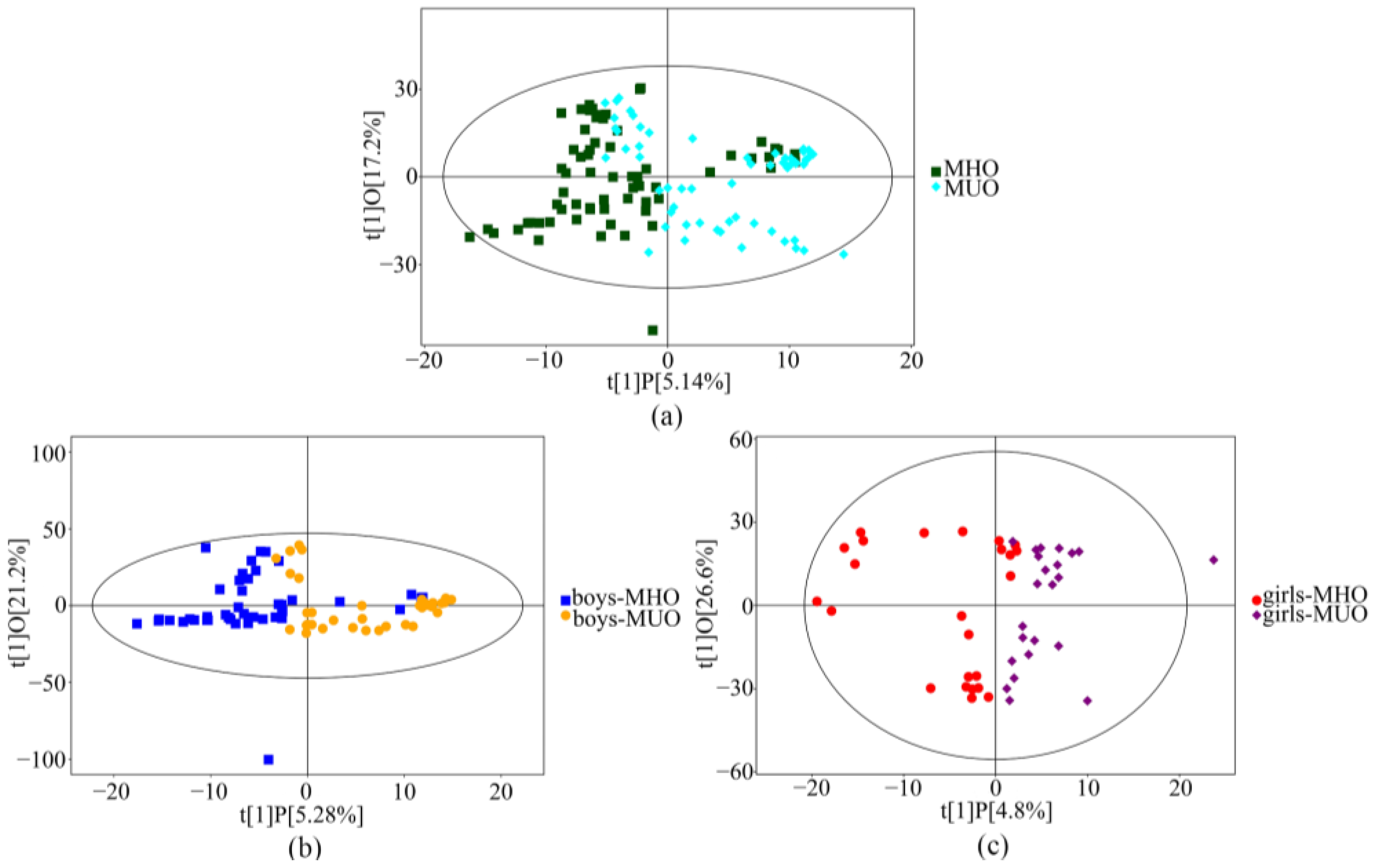
A principal component analysis showed that nearly all samples were within the 95% confidence interval. The OPLS-DA plots showed a clear separation of serum metabolites between the MHO and MUO groups in the total subjects (Figure 2a). The serum metabolic profiles of the MHO and MUO subjects among boys (Figure 2b) and girls (Figure 2c) had similar results to all subjects.

3.3. Differential Metabolites

For all subjects, 20 metabolites with VIP > 0.5 and p < 0.05 were considered differentially expressed between the MHO and MUO groups. We also found differentially expressed metabolites for six (girls) and for 25 (boys) between the MHO and MUO. In all subjects, six metabolites increased and 14 metabolites decreased in the MUO group compared to the MHO group. Among girls, four metabolites decreased and two metabolites increased. Out of 25 metabolites, 17 increased and eight decreased in the MUO group compared to the MHO group in boys. (Table 2).

3.4. Metabolite Pathways of Differential Metabolites

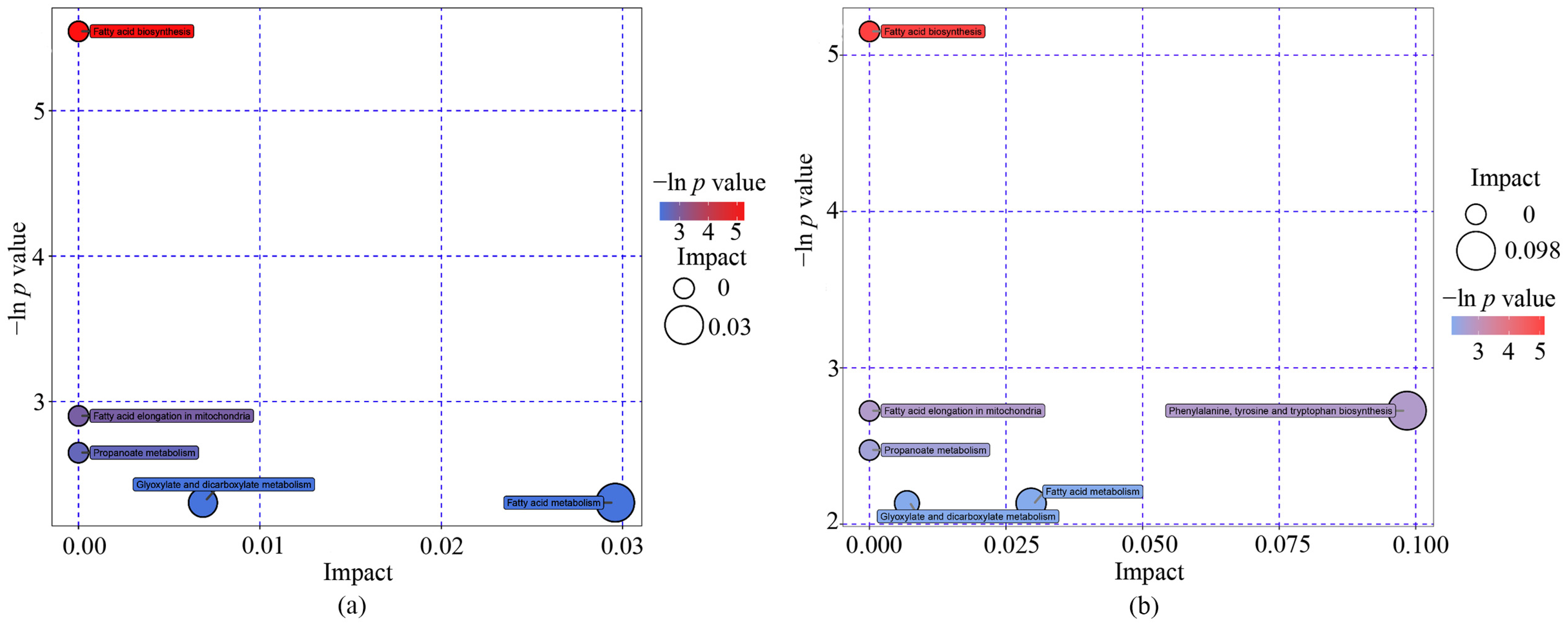
The metabolite pathways of different metabolites are shown in Table 3. Fatty acid biosynthesis pathways were significantly enriched (p < 0.05), and four pathways had low p values (p < 0.1), including the fatty acid elongation in mitochondria, propanoate metabolism, glyoxylate and dicarboxylate metabolism, and fatty acid metabolism pathways. Furthermore, two pathways, including glyoxylate and dicarboxylate metabolism and fatty acid metabolism obtained from topology analysis, had high pathway impacts greater than 0.001 (Table 3 and Figure 3a). Similar results were shown among boys, except for phenylalanine, tyrosine and tryptophan biosynthesis, which had a high impact [0.098] (Table 3 and Figure 3b).

3.5. Further Identification of Metabolites

Multivariable logistic regression revealed that the MUO phenotype was associated with nine metabolites in all participants, and included glycolic acid, palmitic acid, stearic acid, phosphate, asparagine, alanine, 3-hydroxypropionic acid, 2-hydroxypentanoic acid, and isoleucine (all p < 0.05). In girls, the MUO phenotype was associated with two metabolites: glycyl-proline and glucosamine. An MUO status was associated with 19 metabolites, including 2-hydroxybutanoic acid, 2-hydroxypentanoic acid, 3-hydroxypropionic acid, 5-methyluridine, acetophenone, alanine, asparagine, beta-gentiobiose, cyanoalanine, furoylglycine, galactinol, glycerol-alpha-phosphate, glycolic acid, isocitric acid minor, isoleucine, palmitic acid, salicylaldehyde, shikimic acid, and stearic acid (Table 4). The AUCs for these metabolites are presented in Tables S1 and S2. The AUCs for palmitic acid, stearic acid, and phosphate for predicting MUO and glycolic acid, alanine, 3-hydroxypropionic acid, and 2-hydroxypentanoic acid for predicting MHO were all greater than 0.6 (all p < 0.05) in the total population. In boys, five metabolites could predict MUO, 12 metabolites could predict MHO, and only two metabolites from girls could predict MUO.

4. Discussion

In this study, we evaluated metabolome profiling which differentiated the MHO and MUO phenotypes, and several potential metabolic pathways, which modulated the different metabolic profiles of obesity among Chinese adolescents. A panel of metabolites, including palmitic acid, stearic acid, and 2-hydroxybutanoic acid, among others, was chosen to accurately differentiate between subjects with the MHO and MUO. The main metabolic pathways associated with the MUO phenotypes included fatty acid biosynthesis, fatty acid elongation in mitochondria, propanoate metabolism, glyoxylate and dicarboxylate metabolism, fatty acid metabolism and phenylalanine, tyrosine and tryptophan biosynthesis (only in boys).
In this study, we found that stearic acid and palmitic acid might help to initially identify MUO phenotypes in Chinese adolescents independent of age, sex, and BMI. Palmitic acid and stearic acid are the most abundant saturated fatty acids (SFAs) in the diet [15]. In a similar vein, some studies have also shown that patients with type 2 diabetes or hyperlipidemia and mice fed high-fat diets exhibited high plasma SFA levels, including the two acids mentioned above [16,17,18]. Furthermore, some studies have also shown that palmitic acid and stearic acid are more toxic to pancreatic β-cells, resulting in altered pancreatic β-cell function [18,19]. In addition, both can induce inflammation and endoplasmic reticulum stress in in vitro studies and animal studies [20,21]. Thus, these two fatty acids helped us to distinguish obese adolescents who were metabolically unhealthy.
This KEGG ID (C00249) refers to the metabolite palmitic acid, which was involved in both fatty acid elongation in the mitochondria and fatty acid metabolism. The latter had a higher topological analysis impact factor in this study. The pathways mentioned above are related to mitochondrial function: ATP production from mitochondrial β-oxidation depends largely on the catabolism of saturated fatty acids containing 16 carbons and, to a lesser extent, those with 18 carbons such as palmitate and stearate [22]. The synthesis of long-chain saturated fatty acids involves the malonyl-CoA-dependent elongation of fatty acids. An animal study showed that when palmitate was perfused into rat hearts, some of the perfused palmitates underwent a chain extension to form stearate [22]. Stearic acid and palmitic acid also played key roles in the fatty acid biosynthetic pathway, which was identified in our pathway analysis. They elaborated that the elongation of fatty acid chains as described above is localized on the outer surface of the mitochondrial membrane. Malonyl-CoA, the ultimate two-carbon donor for chain elongation, which is present on the outer mitochondrial membrane, is synthesized with mitochondrial β-oxidation-derived acetyl-CoA, and provides malonyl-CoA for fatty acid chain lengthening, which can inhibit the excessive oxidation of mitochondrial fatty acids. In addition, malonyl-CoA induces adipogenesis and stimulates glucose oxidation in response to insulin-activated acetyl-CoA carboxylase. Therefore, these pathway abnormalities may reflect abnormal mitochondrial function in obese children and adolescents. This finding supports the hypothesis proposed by Welsh et al. that SFA is a significant contributor to β-cell dysfunction [23].
Moreover, this KEGG ID (C05984) refers to the metabolite 2-hydroxybutanoate, which was found to identify MHO phenotypes for Chinese adolescents varying in age, sex, and BMI in our study. Another study showed that 2-hydroxybutyric acid participates in the synthesis of substrate branched chain amino acids (BCAAs) [24]. This study suggested that the BCAA and 2-hydroxybutyric concentrations during fasting predicted insulin resistance (IR) in adolescents, which could prevent other diseases in adulthood. 2-hydroxybutyric acid is associated with impaired β-cell function in the human body, and has been recognized as an early indicator of IR and type 2 diabetes in recent years [25]. 2-hydroxybutyric acid was also involved in the propanoate metabolism pathway in this study. Impaired metabolism of the propionic acid pathway not only leads to propionic acidemia but can also induce abnormal blood glucose [26]. When metabolic abnormalities caused by insulin resistance or hyperglycemia occur in obese children and adolescents, more pyruvate and acetyl-CoA are produced by more glucose through the glycolytic pathway, which leads to oxidative stress. To overcome oxidative stress, hepatocytes produce glutathione via cysteine anabolic metabolism, which produces the byproduct α-ketobutyrate, which is then converted to 2-hydroxybutyric acid [25]. In addition, M.Schwab et al. found that the intermediate of propionate metabolism, propionyl-CoA, has a toxic effect on mitochondrial function by inhibiting pyruvate oxidation [27]. These findings suggest that metabolites and related pathways potentially play a role in regulating the metabolic state of early-life obesity through mitochondria.
This KEGG ID (C00160) refers to the metabolite glycolic acid, which was a differential metabolite with p < 0.05 in different metabolic phenotypes and was involved in the glyoxylate and dicarboxylic acid metabolic pathways in this study. Glycolic acid is required for oxalic acid production, which has been found to increase the production of reactive oxygen species from mitochondria [28]. Furthermore, we found that isocitric acid, an intermediate of the tricarboxylic acid cycle, could also identify the MHO phenotype in obese children and adolescents, and it was found to be positively correlated with BMI, abdominal obesity, HOMA-IR, and triglycerides (TG) by Jennifer E et al. [29]. These studies combined suggest that the different metabolic phenotypes of obese children and adolescents are related not only to mitochondrial dysfunction but also to energy metabolic homeostasis.
In our pathway analysis of boys, phenylalanine, tyrosine and tryptophan biosynthesis (C00493) scored highest for their effects (0.098). An animal study showed that apolipoprotein-I (a major component of high-density lipoprotein) knockout mice exhibited abnormalities in the phenylalanine, tyrosine and tryptophan biosynthesis metabolic pathways [30]. Phenylalanine is converted to tyrosine by phenylalanine hydroxylase. This conversion stimulated the synthesis of catecholamines in the brain, which resulted in a decrease in blood pressure [31]. Shikimic acid has been identified as a significant metabolite for distinguishing MHO (Table S2), and was involved in the pathways mentioned above. Shikimic acid is a phenolic compound and an intermediate metabolite in tryptophan synthesis. A small proportion of tryptophan is catalyzed by tryptophan hydroxylase to form 5-hydroxytryptamine, a vasoconstrictor in peripheral tissues. Patients with coronary artery disease have reduced plasma tryptophan concentrations and increased kynurenine/tryptophan levels due to the conversion of the majority of tryptophan to kynurenine [31]. Therefore, the metabolites and pathways may be associated with lipid and blood pressure abnormalities, which are important for the identification and development of different metabolic phenotypes in obese children and adolescents. Consequently, these findings suggest that 2-hydroxybutyric acid, glycolic acid, shikimic acid, alanine, and other metabolites associated with the MHO phenotype indicate that the MHO phenotype has an early-stage cardiovascular disease risk profile.
Notably, our study found that only glycyl-proline and glucosamine were associated with the MUO phenotype in girls, and no relevant metabolic pathways were identified, which suggested that there were sex differences in the metabolites and pathways in identifying the MUO and MHO phenotypes. Several studies based on metabolic disorders and cardiovascular disease have also found that females typically have more beneficial metabolic profiles and lower risks of cardiovascular disease [32,33]. Similarly, a cohort study showed that transitional MHO females had healthier metabolic profiles than their male counterparts. This could explain the sex differences. In addition, the relationship of glycyl-proline and glucosamine with the MUO phenotype has not been studied. Proline is a protective substance for membranes and enzymes in living organisms, and is a free radical scavenger that protects the vascular endothelium from damage [34]. Its plasma levels are often altered by endovascular lesions, but the role of glycyl-proline in cardiovascular metabolism is not yet clear.
To the best of our knowledge, few studies have previously assessed untargeted metabolomics in Chinese children and adolescents with different metabolic phenotypes. However, several limitations of our study should be noted here. First, in a cross-sectional study, we failed to demonstrate the causal association between the identified metabolic pathways or metabolites and different metabolic phenotypes in obese children and adolescents, and more prospective studies are needed for validation. Second, some interpretations may be inaccurate, although previous studies of animals or adults can explain the biological rationality of metabolites found due to the species, age differences, and limited research related to children and adolescents in metabolites. Third, it is noteworthy that the sample size is relatively small. Fourth, in untargeted analyses, it is possible that one peak may correspond to multiple metabolites with similar mass over charge values, leading to potentially less accurate results in pathway analysis. Some metabolites may not be identified in humans and need to be examined manually, even in pathway analysis and with the KEGG dataset. Fifth, we did not adjust for puberty-related variables due to a lack of diet and physical activity data in our study, which may have affected the results of the study. Finally, the definition of MHO may lead to results that are inconsistent with other studies, and different metabolic abnormalities may have different underlying causes.

5. Conclusions

In conclusion, we have successfully identified several related metabolites that distinguish the MUO group from the MHO group, including palmitic acid, stearic acid, glycolic acid, 2-hydroxybutanoic acid, shikimic acid, and others. These metabolites are involved in several important metabolic pathways, including fatty acid biosynthesis, fatty acid elongation in mitochondria, propanoate metabolism, glyoxylate and dicarboxylate metabolism, fatty acid metabolism, and phenylalanine, tyrosine and tryptophan biosynthesis (only in boys). Future studies are needed to validate our findings; that is, abnormal mitochondrial function may be associated with different metabolic states in obese children and adolescents, and more studies are needed to investigate the potential mechanisms of metabolic dysregulation in obesity.

Supplementary Materials

The following supporting information can be downloaded at: https://www.mdpi.com/article/10.3390/metabo13050641/s1, Table S1: ROC curve analysis of metabolites for predicting metabolically unhealthy obesity. Table S2: ROC curve analysis of metabolites for predicting metabolically healthy obesity.

Author Contributions

L.T. analyzed data, wrote and edited the manuscript; M.T. analyzed data, edited the manuscript; X.M. analyzed data, edited the manuscript; L.B. and J.Z. collected data, conceived the experimental design, and performed experiments; W.D. collected data, conceived the experimental design, and reviewed the manuscript. All authors have read and agreed to the published version of the manuscript.

Funding

This research was funded by the National Natural Science Foundation of China, grant number 82160641.

Institutional Review Board Statement

The study was conducted in accordance with the Declaration of Helsinki and approved by the Ethics Committee of Ningxia Medical University (Approval ID: 2021-G053; Approval date: 5 August 2021).

Informed Consent Statement

Informed consent was obtained from all subjects involved in the study.

Data Availability Statement

The data presented in this study are available in the article and in the Supplementary Material.

Acknowledgments

We thank all of the study participants for their contributions.

Conflicts of Interest

The authors declare that they have no conflict of interest.

References

  1. Danese, A.; Tan, M. Childhood maltreatment and obesity: Systematic review and meta-analysis. Mol. Psychiatry 2014, 19, 544–554. [Google Scholar] [CrossRef] [PubMed]
  2. Dong, Y.H.; Chen, L.; Liu, J.Y.; Ma, T.; Zhang, Y.; Chen, M.M.; Zhong, P.L.; Shi, D.; Hu, P.J.; Li, J.; et al. Epidemiology and prediction of overweight and obesity among children and adolescents aged 7–18 years in China from 1985 to 2019. Chin. J. Prev. Med. 2023, 57, 11–19. [Google Scholar]
  3. Smith, G.I.; Mittendorfer, B.; Klein, S. Metabolically healthy obesity: Facts and fantasies. J. Clin. Investig. 2019, 129, 3978–3989. [Google Scholar] [CrossRef]
  4. Vinciguerra, F.; Tumminia, A.; Baratta, R.; Ferro, A.; Alaimo, S.; Hagnäs, M.; Graziano, M.; Vigneri, R.; Frittitta, L. Prevalence and Clinical Characteristics of Children and Adolescents with Metabolically Healthy Obesity: Role of Insulin Sensitivity. Life 2020, 10, 127. [Google Scholar] [CrossRef] [PubMed]
  5. Keihani, S.; Hosseinpanah, F.; Barzin, M.; Serahati, S.; Doustmohamadian, S.; Azizi, F. Abdominal obesity phenotypes and risk of cardiovascular disease in a decade of follow-up: The Tehran Lipid and Glucose Study. Atherosclerosis 2015, 238, 256–263. [Google Scholar] [CrossRef] [PubMed]
  6. Ding, W.Q.; Liu, J.T.; Shang, Y.X.; Gao, B.; Zhao, X.Y.; Zhao, H.P.; Wu, W.J. DXA-measured visceral fat mass and lean body mass reflect abnormal metabolic phenotypes among some obese and nonobese Chinese children and adolescents. Nutr. Metab. Cardiovasc. 2018, 28, 618–628. [Google Scholar] [CrossRef]
  7. Chen, H.-H.; Tseng, Y.J.; Wang, S.-Y.; Tsai, Y.-S.; Chang, C.-S.; Kuo, T.-C.; Yao, W.-J.; Shieh, C.-C.; Wu, C.-H.; Kuo, P.-H. The metabolome profiling and pathway analysis in metabolic healthy and abnormal obesity. Int. J. Obes. 2015, 39, 1241–1248. [Google Scholar] [CrossRef]
  8. Zhao, X.; Gang, X.; Liu, Y.; Sun, C.; Han, Q.; Wang, G. Using Metabolomic Profiles as Biomarkers for Insulin Resistance in Childhood Obesity: A Systematic Review. J. Diabetes Res. 2016, 2016, 8160545. [Google Scholar] [CrossRef]
  9. Mihalik, S.J.; Michaliszyn, S.F.; De Las Heras, J.; Bacha, F.; Lee, S.; Chace, D.H.; DeJesus, J.V.; Arslanian, S.A. Metabolomic profiling of fatty acid and amino acid metabolism in youth with obesity and type 2 diabetes: Evidence for enhanced mitochondrial oxidation. Diabetes Care 2012, 35, 605–611. [Google Scholar] [CrossRef]
  10. Ji, C. Report on childhood obesity in China (1)—Body mass index reference for screening overweight and obesity in Chinese school-age children. Biomed. Environ. Sci. 2005, 18, 390–400. [Google Scholar]
  11. Wan, N.-J.; Mi, J.; Wang, T.-Y.; Duan, J.-L.; Li, M.; Gong, C.-X.; Du, J.-B.; Zhao, X.-Y.; Cheng, H.; Hou, D.-Q.; et al. Metabolic syndrome in overweight and obese schoolchildren in Beijing. Chin. J. Pediatr. 2007, 45, 417–421. [Google Scholar]
  12. Ma, G.-S.; Ji, C.-Y.; Ma, J.; Mi, J.; Sung, R.Y.; Xiong, F.; Yan, W.-L.; Hu, X.-Q.; Li, Y.-P.; DU, S.-M.; et al. Waist Circumference Reference Values for Screening Cardiovascular Risk Factors in Chinese Children and Adolescents. Biomed. Environ. Sci. 2010, 23, 21–31. [Google Scholar] [CrossRef] [PubMed]
  13. Fan, H.; Yan, Y.K.; Mi, J. Updating blood pressure references for Chinese children aged 3–17 years. Chin. J. Hypertens. 2017, 25, 428–435. [Google Scholar]
  14. Kanehisa, M.; Goto, S.; Sato, Y.; Furumichi, M.; Tanabe, M. KEGG for integration and interpretation of large-scale molecular data sets. Nucleic Acids Res. 2012, 40, D109–D114. [Google Scholar] [CrossRef] [PubMed]
  15. Van Rooijen, M.A.; Plat, J.; Zock, P.L.; Blom, W.A.; Mensink, R.P. Effects of two consecutive mixed meals high in palmitic acid or stearic acid on 8-h postprandial lipemia and glycemia in healthy-weight and overweight men and postmenopausal women: A randomized controlled trial. Eur. J. Nutr. 2021, 60, 3659–3667. [Google Scholar] [CrossRef]
  16. Liu, L.; Li, Y.; Guan, C.; Li, K.; Wang, C.; Feng, R.; Sun, C. Free fatty acid metabolic profile and biomarkers of isolated post-challenge diabetes and type 2 diabetes mellitus based on GC–MS and multivariate statistical analysis. J. Chromatogr. B 2010, 878, 2817–2825. [Google Scholar] [CrossRef]
  17. Wang, Y.; Meng, X.; Deng, X.; Okekunle, A.P.; Wang, P.; Zhang, Q.; Ding, L.; Guo, X.; Lv, M.; Sun, C.; et al. Postprandial Saturated Fatty Acids Increase the Risk of Type 2 Diabetes: A Cohort Study in a Chinese Population. J. Clin. Endocrinol. Metab. 2018, 103, 1438–1446. [Google Scholar] [CrossRef]
  18. Lu, H.; Hao, L.; Li, S.; Lin, S.; Lv, L.; Chen, Y.; Cui, H.; Zi, T.; Chu, X.; Na, L.; et al. Elevated circulating stearic acid leads to a major lipotoxic effect on mouse pancreatic beta cells in hyperlipidaemia via a miR-34a-5p-mediated PERK/p53-dependent pathway. Diabetologia 2016, 59, 1247–1257. [Google Scholar] [CrossRef]
  19. Yu, Y.; Guo, R.; Zhang, Y.; Shi, H.; Sun, H.; Chu, X.; Wu, X.; Lu, H.; Sun, C. miRNA-mRNA profile and regulatory network in stearic acid-treated β-cell dysfunction. J. Endocrinol. 2020, 246, 13–27. [Google Scholar] [CrossRef]
  20. Borradaile, N.M.; Han, X.; Harp, J.D.; Gale, S.E.; Ory, D.S.; Schaffer, J.E. Disruption of endoplasmic reticulum structure and integrity in lipotoxic cell death. J. Lipid Res. 2006, 47, 2726–2737. [Google Scholar] [CrossRef]
  21. Schwartz, E.A.; Zhang, W.Y.; Karnik, S.K.; Borwege, S.; Anand, V.R.; Laine, P.S.; Su, Y.; Reaven, P.D. Nutrient modification of the innate immune response: A novel mechanism by which saturated fatty acids greatly amplify monocyte inflammation. Arter. Thromb. Vasc. Biol. 2010, 30, 802–808. [Google Scholar] [CrossRef] [PubMed]
  22. Kerner, J.; Minkler, P.E.; Lesnefsky, E.J.; Hoppel, C.L. Fatty acid chain elongation in palmitate-perfused working rat heart: Mitochondrial acetyl-CoA is the source of two-carbon units for chain elongation. J. Biol. Chem. 2014, 289, 10223–10234. [Google Scholar] [CrossRef] [PubMed]
  23. Welsh, N.; Cnop, M.; Kharroubi, I.; Bugliani, M.; Lupi, R.; Marchetti, P.; Eizirik, D.L. Is there a role for locally produced interleukin-1 in the deleterious effects of high glucose or the type 2 diabetes milieu to human pancreatic islets? Diabetes 2005, 54, 3238–3244. [Google Scholar] [CrossRef] [PubMed]
  24. Tricò, D.; Prinsen, H.; Giannini, C.; De Graaf, R.; Juchem, C.; Li, F.; Caprio, S.; Santoro, N.; Herzog, R.I. Elevated α-Hydroxybutyrate and Branched-Chain Amino Acid Levels Predict Deterioration of Glycemic Control in Adolescents. J. Clin. Endocrinol. Metab. 2017, 102, 2473–2481. [Google Scholar] [CrossRef] [PubMed]
  25. Sousa, A.P.; Cunha, D.M.; Franco, C.; Teixeira, C.; Gojon, F.; Baylina, P.; Fernandes, R. Which Role Plays 2-Hydroxybutyric Acid on Insulin Resistance? Metabolites 2021, 11, 835. [Google Scholar] [CrossRef] [PubMed]
  26. Dweikat, I.M.; Naser, E.N.; Abu Libdeh, A.I.; Naser, O.J.; Abu Gharbieh, N.N.; Maraqa, N.F.; Abu Libdeh, B.Y. Propionic acidemia mimicking diabetic ketoacidosis. Brain Dev. 2011, 33, 428–431. [Google Scholar] [CrossRef]
  27. Schwab, M.A.; Sauer, S.W.; Okun, J.G.; Nijtmans, L.G.J.; Rodenburg, R.; Heuvel, L.P.V.D.; Dröse, S.; Brandt, U.; Hoffmann, G.F.; Ter Laak, H.; et al. Secondary mitochondrial dysfunction in propionic aciduria: A pathogenic role for endogenous mitochondrial toxins. Biochem. J. 2006, 398, 107–112. [Google Scholar] [CrossRef]
  28. Cao, L.-C.; Honeyman, T.W.; Cooney, R.; Kennington, L.; Scheid, C.R.; Jonassen, J.A. Mitochondrial dysfunction is a primary event in renal cell oxalate toxicity. Kidney Int. 2004, 66, 1890–1900. [Google Scholar] [CrossRef]
  29. Ho, J.E.; Larson, M.G.; Ghorbani, A.; Cheng, S.; Chen, M.H.; Keyes, M.; Rhee, E.P.; Clish, C.B.; Vasan, R.S.; Gerszten, R.E.; et al. Metabolomic Profiles of Body Mass Index in the Framingham Heart Study Reveal Distinct Cardiometabolic Phenotypes. PLoS ONE 2016, 11, e0148361. [Google Scholar] [CrossRef]
  30. Liu, J.; Zhao, M.; Zhu, Y.; Wang, X.; Zheng, L.; Yin, Y. LC-MS-Based Metabolomics and Lipidomics Study of High-Density-Lipoprotein-Modulated Glucose Metabolism with an apoA-I Knockout Mouse Model. J. Proteome Res. 2019, 18, 48–56. [Google Scholar] [CrossRef]
  31. Yang, L.J.; Ding, X.X.; Ren, F.D.; Cai, F.; Fu, G.H.; Ren, D.B.; Zhao, Y.L.; Zhang, H. Liquid chromatography-mass spectrometry combined with chemometrics to analyze the metabolic characteristics of patients with coronary heart disease and coronary heart disease with hypertension. Chin. J. Anal. Chem. 2021, 49, 1649–1656. [Google Scholar]
  32. Mosca, L.; Barrett-Connor, E.; Wenger, N.K. Sex/gender differences in cardiovascular disease prevention: What a difference a decade makes. Circulation 2011, 124, 2145–2154. [Google Scholar] [CrossRef] [PubMed]
  33. Krishnan, K.C.; Mehrabian, M.; Lusis, A.J. Sex differences in metabolism and cardiometabolic disorders. Curr. Opin. Infect. Dis. 2018, 29, 404–410. [Google Scholar] [CrossRef] [PubMed]
  34. Gómez, I.M.R.; Moliz, J.N.; Quesada, A.; Montoro-Molina, S.; Vargas-Tendero, P.; Osuna, A.; Wangensteen, R.; Vargas, F. l-Arginine metabolism in cardiovascular and renal tissue from hyper- and hypothyroid rats. Exp. Biol. Med. 2015, 241, 550–556. [Google Scholar] [CrossRef]
Figure 1. Participant screening and enrollment flow chart.
Metabolites 13 00641 g001
Figure 2. The scatter plot of PLS-DA to distinguish the MHO from MUO. The PLS-DA was conducted based on detectable metabolites. ((a): Total, 127; (b): Boys, 80; (c): Girls, 47).
Metabolites 13 00641 g002
Figure 3. The pathway impact in topology analyses and p values in the enrichment analyses conducted by metabolomics pathway analysis. A total of twenty top metabolites for all the participants (a) and twenty-five top metabolites for boys (b) were involved in the pathway analysis. The size of pathway symbols represents significant levels of enrichment analysis, and the color of the pathway symbols represents the impact factors.
Metabolites 13 00641 g003
Table 1. Demographic and clinical characteristics in MHO and MUO participants.
VariablesMHO (n = 67)MUO (n = 60)p Value
Age (years)14.15 ± 1.6614.73 ± 1.670.051
Female, n (%)24 (35.8)23 (38.3)0.700
Height (cm)168.9 ± 8.7170.7 ± 8.60.259
Weight (kg)80.6 ± 11.986.5 ± 13.70.010
BMI (kg/m2)28.1 ± 2.529.6 ± 3.210.005
WC (cm)91.8 ± 8.193.6 ± 10.20.335
SBP (mmHg)119 ± 12124 ± 120.013
DBP (mmHg)69 ± 774 ± 7<0.001
TC (mmol/L)3.68 ± 0.734.29 ± 0.79<0.001
TG (mmol/L) *1.10 (0.92,1.32)2.13 (1.67,2.62)<0.001
HDL-C (mmol/L)1.24 ± 0.191.05 ± 0.21<0.001
LDL-C (mmol/L)2.02 ± 0.662.47 ± 0.840.001
Fasting glucose (mmol/L)4.77 ± 0.364.66 ± 0.400.173
Normally distributed data are expressed as the mean ± SD. The median and interquartile range was used for skewed variables; Abbreviations: MHO, metabolica healthy obesity; MUO, metabolic unhealthy obesity; BMI, body mass index; WC, waist circumstance; SBP, systolic blood pressure; DBP, diastolic blood pressure; TC, total cholesterol; TG, triglyceride; HDL-C, high-density lipoprotein cholesterol; LDL-C, low-density lipopretein cholesterol; *: Skewed distributions were logarithmically transformed for the statistical test.
Table 2. Differential metabolites between the MHO and MUO groups.
MetaboliteMean Relative Quantitative Value in MUO GroupMean Relative Quantitative Value in MHO GroupVIPp ValueFold Change
Total
  glycolic acid0.0220.0131.2430.0101.671
  palmitic acid1.2931.6682.8040.0020.775
  glucose 0.0780.0561.7350.0351.391
  stearic acid1.2431.5822.6670.0030.786
  2-hydroxybutanoic acid0.0540.0301.3830.0061.081
  galactose0.3940.3171.0870.0381.242
  phosphate0.4430.6811.9490.0100.651
  asparagine 0.0350.0171.3990.0031.503
  alanine0.1390.0751.8780.0111.858
  glutamate 0.0080.0132.1170.0340.609
  cyanoalanine0.0300.0202.4330.0291.510
  3-hydroxypropionic acid0.1670.1021.2660.0151.639
  2-hydroxypentanoic acid0.0250.0121.4110.0041.997
  hippuric acid 0.0670.0391.5210.0201.715
  acetophenone0.0760.0461.8770.0151.663
  beta-gentiobiose0.0290.0180.8540.0221.605
  maltotriose 0.0060.0040.8430.0211.540
  isoleucine 0.0310.0191.1870.0211.645
  myristyl myristate0.0000.0011.5110.0460.710
  salicylaldehyde0.0000.0011.3710.0090.770
Girls
  benzylalcohol0.0010.0000.6860.0402.407
  2-ketoadipic acid 0.0000.0001.0740.0281.819
  glycyl-proline0.0000.0003.0200.0090.582
  glucosamine0.0000.0002.5550.0070.552
  pyrophosphate0.0000.0001.1000.0420.575
  2-ketobutyric acid0.0000.0003.1560.0150.583
Boys
  2-hydroxy-2-methylbutanoic acid0.0210.0351.8670.0360.594
  2-hydroxybutanoic acid0.0640.0341.0530.0161.869
  2-hydroxypentanoic acid0.0300.0140.9020.0062.183
  3-hydroxypropionic acid0.1980.1141.2830.0241.743
  5-methyluridine0.0350.0241.1710.0061.452
  acetophenone0.0900.0521.6380.0211.746
  alanine0.1670.0881.4820.0231.898
  asparagine0.0450.0212.0040.0032.105
  beta-gentiobiose0.0360.0190.6030.0111.860
  cyanoalanine0.0390.0253.2860.0171.602
  furoylglycine0.0000.0000.9810.0360.822
  galactinol 0.0470.0300.9040.0201.538
  galactose0.4000.3061.0440.0391.307
  glutamate0.0080.0151.6490.0240.549
  glycerol-alpha-phosphate0.0170.0260.8950.0100.681
  glycolic acid0.0260.0152.0500.0271.674
  isocitric acid0.0900.0661.2180.0251.351
  isogluconic acid0.1130.0651.2300.0311.745
  isoleucine 0.0410.0241.0790.0251.701
  maltotriose 0.0070.0041.4790.0291.631
  palmitic acid1.2051.6672.8540.0020.723
  phosphate0.4330.6901.6750.0320.628
  salicylaldehyde0.0000.0011.6330.0030.731
  shikimic acid0.0500.0280.8070.0131.816
  stearic acid1.1551.5552.7050.0030.743
Table 3. Metabolite pathways of differential metabolites.
Pathway NameTotal Number of Compounds
in the Pathways
Enrichment Analysis p ValueTopology Analysis
Impact Factor
Name of Metabolites and KEGG ID
Total (N = 127)
  Fatty acid biosynthesis490.0040.000Stearic acid cpd:C01530; Palmitic acid cpd:C00249
  Fatty acid elongation in mitochondria270.0550.000Palmitic acid cpd:C00249
  Propanoate metabolism350.0710.0002-Hydroxybutyric acid cpd:C05984
  Glyoxylate and dicarboxylate metabolism500.0990.007Glycolic acid cpd:C00160
  Fatty acid metabolism500.0990.030Palmitic acid cpd:C00249
Boys (n = 80)
  Fatty acid biosynthesis490.0060.000Stearic acid cpd:C01530;Palmitic acid cpd:C00249
  Fatty acid elongation in mitochondria270.0660.000Palmitic acid cpd:C00249
  Propanoate metabolism350.0840.0002-Hydroxybutyric acid cpd:C05984
  Glyoxylate and dicarboxylate metabolism500.1180.007Glycolic acid cpd:C00160
  Fatty acid metabolism500.1180.030Palmitic acid cpd:C00249
  Phenylalanine, tyrosine and tryptophan biosynthesis 270.0670.098Shikimic acid cpd:C00493
Table 4. Results of significant metabolites using logistic regression analysis.
MetaboliteβS.E.Waldp Value
Total a
  glycolic acid−27.82012.4484.9950.025
  palmitic acid0.8560.3605.6570.017
  glucose −7.3863.9973.1450.065
  stearic acid0.8340.3725.0240.025
  2-hydroxybutanoic acid9.0247.5271.4380.231
  galactose−1.3801.1231.5090.219
  phosphate0.7610.3794.0260.045
  asparagine −15.9136.2846.4120.011
  alanine−3.0001.4184.4750.034
  glutamate 22.90516.2951.9760.160
  cyanoalanine−15.1017.7013.8450.050
  3-hydroxypropionic acid−3.2301.4095.2590.022
  2-hydroxypentanoic acid−9.6044.1865.2630.022
  hippuric acid −5.2343.0033.0370.081
  acetophenone−5.5042.8833.6460.056
  beta-gentiobiose−14.4087.8833.3400.068
  maltotriose −89.45848.5073.4010.065
  isoleucine −16.8328.1264.2910.038
  myristyl myristate576.787485.3121.4120.235
  salicylaldehyde1354.748794.3532.9090.088
Girls b
  benzylalcohol−1090.490657.5162.7510.097
  2-ketoadipic acid 9133.5054866.2453.5230.061
  glycyl-proline4800.0512407.6593.9750.046
  glucosamine5353.0602541.7474.4350.035
  pyrophosphate4161.9212452.6392.9440.086
  2-ketobutyric acid9133.5054866.2453.5230.061
Boys b
  2-hydroxy-2-methylbutanoic acid14.9748.6812.9750.085
  2-hydroxybutanoic acid−10.3034.7744.6570.031
  2-hydroxypentanoic acid−25.50010.3656.0530.014
  3-hydroxypropionic acid−3.9641.7165.3370.021
  5-methyluridine−47.93918.5066.7110.010
  acetophenone−7.1103.5284.0610.044
  alanine−3.3981.6984.0040.045
  asparagine −19.8277.4317.1190.008
  beta-gentiobiose−19.1759.5174.0600.044
  cyanoalanine−20.3899.1254.9920.025
  furoylglycine2916.7771479.4923.8870.049
  galactinol −19.1128.6614.8690.027
  galactose−2.5481.5522.6940.101
  glutamate minor38.91420,7593.5140.061
  glycerol-alpha-phosphate38.87417.3725.0070.025
  glycolic acid−32.65615.1204.6640.031
  isocitric acid minor−12.5776.1274.1520.042
  isogluconic acid−4.7722.5913.3910.066
  isoleucine −18.3849.0664.1120.043
  maltotriose −111.40459.6973.4830.062
  palmitic acid1.4080.5346.9520.008
  phosphate0.8680.4663.4760.062
  salicylaldehyde2711.6861206.9275.0480.025
  shikimic acid−14.4797.2443.9950.046
  stearic acid1.4040.5666.1630.013
a Model adjusted for age, sex and BMI; b Model adjusted for age and BMI.
Disclaimer/Publisher’s Note: The statements, opinions and data contained in all publications are solely those of the individual author(s) and contributor(s) and not of MDPI and/or the editor(s). MDPI and/or the editor(s) disclaim responsibility for any injury to people or property resulting from any ideas, methods, instructions or products referred to in the content.