
Prognostic Value of Pretherapeutic Primary Tumor MTV from [18F]FDG PET in Radically Treated Cervical Cancer Patients

 , , ,
, , ,
Abstract
:1. Introduction
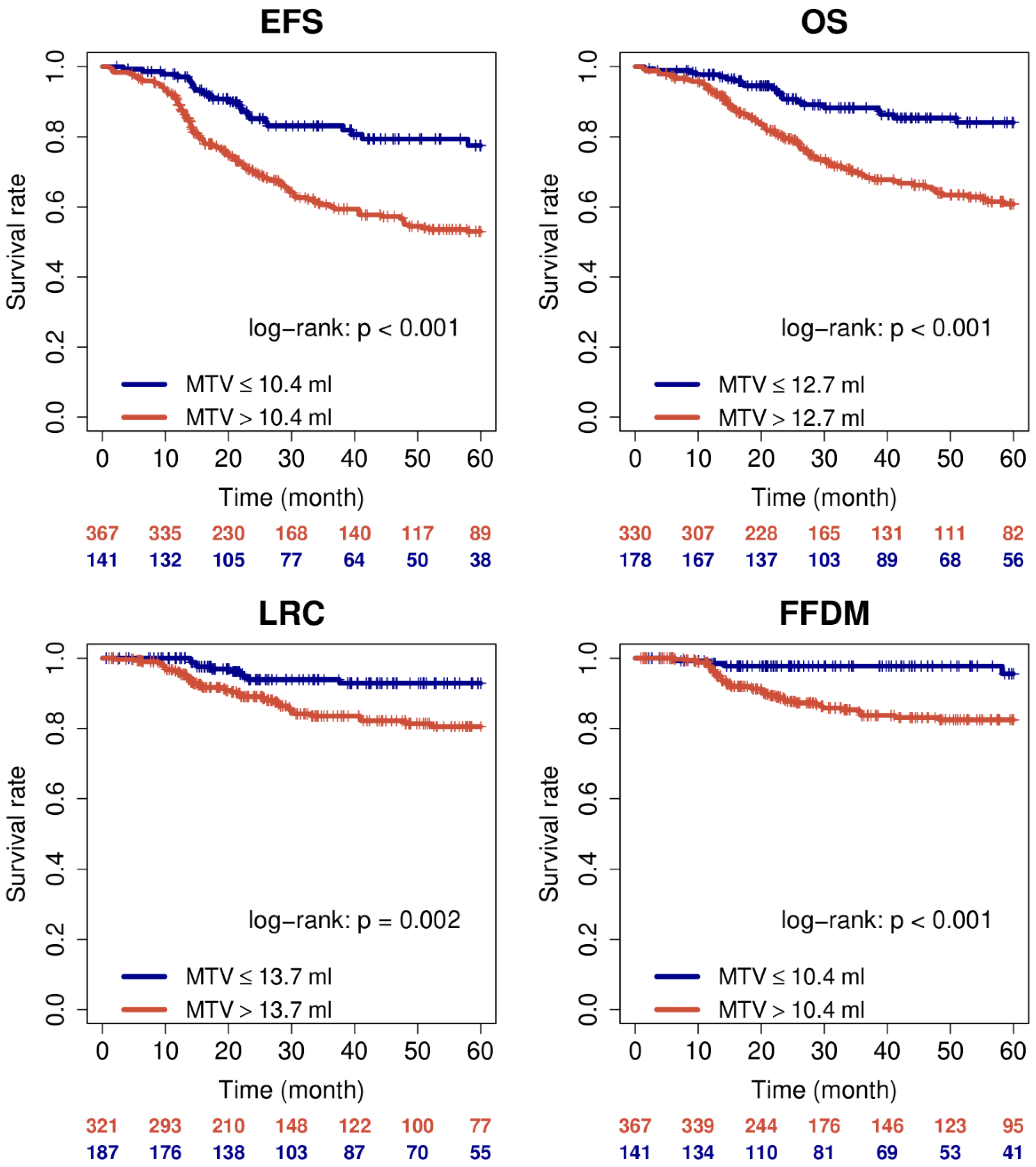
2. Results
3. Discussion
4. Materials and Methods
4.1. Patient Characteristic
4.2. [18F]FDG PET/CT Analysis
4.3. Data Analysis
4.4. Statistical Analysis
5. Conclusions
Author Contributions
Funding
Institutional Review Board Statement
Informed Consent Statement
Data Availability Statement
Conflicts of Interest
References
- Sung, H.; Ferlay, J.; Siegel, R.L.; Laversanne, M.; Soerjomataram, I.; Jemal, A.; Bray, F. Global Cancer Statistics 2020: GLOBOCAN Estimates of Incidence and Mortality Worldwide for 36 Cancers in 185 Countries. CA Cancer J. Clin. 2021, 71, 209–249. [Google Scholar] [CrossRef]
- Tsikouras, P.; Zervoudis, S.; Manav, B.; Tomara, E.; Iatrakis, G.; Romanidis, C.; Bothou, A.; Galazios, G. Cervical cancer: Screening, diagnosis and staging. J. BUON 2016, 21, 320–325. [Google Scholar] [PubMed]
- Haldorsen, I.S.; Lura, N.; Blaakær, J.; Fischerova, D.; Werner, H.M.J. What Is the Role of Imaging at Primary Diagnostic Work-Up in Uterine Cervical Cancer? Curr. Oncol. Rep. 2019, 21, 77. [Google Scholar] [CrossRef] [PubMed] [Green Version]
- Liu, B.; Gao, S.; Li, S. A Comprehensive Comparison of CT, MRI, Positron Emission Tomography or Positron Emission Tomography/CT, and Diffusion Weighted Imaging-MRI for Detecting the Lymph Nodes Metastases in Patients with Cervical Cancer: A Meta-Analysis Based on 67 Studies. Gynecol. Obstet. Invest. 2017, 82, 209–222. [Google Scholar] [CrossRef] [PubMed]
- Sun, Y.; Lu, P.; Yu, L. The Volume-metabolic Combined Parameters from (18)F-FDG PET/CT May Help Predict the Outcomes of Cervical Carcinoma. Acad. Radiol. 2016, 23, 605–610. [Google Scholar] [CrossRef]
- Herrera, F.G.; Breuneval, T.; Prior, J.O.; Bourhis, J.; Ozsahin, M. [(18)F]FDG-PET/CT metabolic parameters as useful prognostic factors in cervical cancer patients treated with chemo-radiotherapy. Radiat. Oncol. 2016, 11, 43. [Google Scholar] [CrossRef] [Green Version]
- Narayanan, P.; Sahdev, A. The role of 18F-FDG PET CT in common gynaecological malignancies. Br. J. Radiol. 2017, 90, 20170283. [Google Scholar] [CrossRef]
- Albano, D.; Bonacina, M.; Savelli, G.; Ferro, P.; Busnardo, E.; Gianolli, L.; Camoni, L.; Giubbini, R.; Bertagna, F. Clinical and prognostic 18F-FDG PET/CT role in recurrent vulvar cancer: A multicentric experience. Jpn. J. Radiol 2021. Epub ahead of print. [Google Scholar] [CrossRef]
- Albano, D.; Zizioli, V.; Treglia, G.; Odicino, F.; Giubbini, R.; Bertagna, F. Role of 18F-FDG PET/CT in restaging and follow-up of patients with uterine sarcomas. Rev. Esp. Med. Nucl. Imagen Mol. Engl. Ed. 2019, 38, 10–16. [Google Scholar] [CrossRef]
- Yilmaz, M.; Adli, M.; Celen, Z.; Zincirkeser, S.; Dirier, A. FDG PET-CT in cervical cancer: Relationship between primary tumor FDG uptake and metastatic potential. Nucl. Med. Commun. 2010, 31, 526–531. [Google Scholar] [CrossRef]
- Kidd, E.A.; Siegel, B.A.; Dehdashti, F.; Grigsby, P.W. The standardized uptake value for F-18 fluorodeoxyglucose is a sensitive predictive biomarker for cervical cancer treatment response and survival. Cancer 2007, 110, 1738–1744. [Google Scholar] [CrossRef]
- Pan, L.; Cheng, J.; Zhou, M.; Yao, Z.; Zhang, J. The SUVmax (maximum standardized uptake value for F-18 fluorodeoxyglucose) and serum squamous cell carcinoma antigen (SCC-ag) function as prognostic biomarkers in patients with primary cervical cancer. J. Cancer Res. Clin. Oncol. 2012, 138, 239–246. [Google Scholar] [CrossRef]
- Chung, H.H.; Kim, J.W.; Han, K.H.; Eo, J.S.; Kang, K.W.; Park, N.H.; Song, Y.S.; Chung, J.K.; Kang, S.B. Prognostic value of metabolic tumor volume measured by FDG-PET/CT in patients with cervical cancer. Gynecol. Oncol. 2011, 120, 270–274. [Google Scholar] [CrossRef] [PubMed]
- Moon, S.H.; Hyun, S.H.; Choi, J.Y. Prognostic significance of volume-based PET parameters in cancer patients. Korean J. Radiol. 2013, 14, 1–12. [Google Scholar] [CrossRef] [PubMed] [Green Version]
- Takagi, H.; Sakamoto, J.; Osaka, Y.; Shibata, T.; Fujita, S.; Sasagawa, T. Usefulness of the maximum standardized uptake value for diagnosis and staging patients with cervical cancer undergoing positron emission tomography/computed tomography. Medicine 2018, 97, e9856. [Google Scholar] [CrossRef] [PubMed]
- Wong, T.Z.; Jones, E.L.; Coleman, R.E. Positron emission tomography with 2-deoxy-2-[(18)F]fluoro-D-glucose for evaluating local and distant disease in patients with cervical cancer. Mol. Imaging Biol. 2004, 6, 55. [Google Scholar] [CrossRef] [PubMed]
- Sturdza, A.; Pötter, R.; Fokdal, L.U.; Haie-Meder, C.; Tan, L.T.; Mazeron, R.; Petric, P.; Šegedin, B.; Jurgenliemk-Schulz, I.M.; Nomden, C.; et al. Image guided brachytherapy in locally advanced cervical cancer: Improved pelvic control and survival in RetroEMBRACE, a multicenter cohort study. Radiother. Oncol. 2016, 120, 428–433. [Google Scholar] [CrossRef]
- Tanderup, K.; Lindegaard, J.C.; Kirisits, C.; Haie-Meder, C.; Kirchheiner, K.; de Leeuw, A.; Jürgenliemk-Schulz, I.; Van Limbergen, E.; Pötter, R. Image Guided Adaptive Brachytherapy in cervix cancer: A new paradigm changing clinical practice and outcome. Radiother. Oncol. 2016, 120, 365–369. [Google Scholar] [CrossRef]
- Wang, D.; Liu, X.; Wang, W.; Huo, L.; Pan, Q.; Ren, X.; Zhang, F.; Hu, K. The role of the metabolic parameters of 18F-FDG PET/CT in patients with locally advanced cervical cancer. Front. Oncol. 2021, 11, 698744. [Google Scholar] [CrossRef]
- Han, S.; Kim, H.; Kim, Y.J.; Suh, C.H.; Woo, S. Prognostic value of volume-based metabolic parameters of 18F-FDG PET/CT In uterine cervical cancer: A systematic review and meta-analysis. Am. J. Roentgenol. 2018, 211, 1112–1121. [Google Scholar] [CrossRef]
- Hofheinz, F.; Pötzsch, C.; Oehme, L.; Beuthien-Baumann, B.; Steinbach, J.; Kotzerke, J.; van den Hoff, J. Automatic volume de- lineation in oncological PET. Evaluation of a dedicated software tool and comparison with manual delineation in clinical data sets. Nuklearmedizin 2012, 51, 9–16. [Google Scholar] [CrossRef] [PubMed]
- Hofheinz, F.; Langner, J.; Petr, J.; Beuthien-Baumann, B.; Steinbach, J.; Kotzerke, J.; van den Hoff, J. An automatic method for accurate volume delineation of heterogeneous tumors in PET. Med. Phys. 2013, 40, 082503. [Google Scholar] [CrossRef] [PubMed]
- Bütof, R.; Hofheinz, F.; Zöphel, K.; Stadelmann, T.; Schmollack, J.; Jentsch, C.; Kotzerke, J.; Baumann, M.; van den Hoff, J. Prognostic Value of Prethera- peutic Tumor-to-Blood Standardized Uptake Ratio in Patients with Esophageal Carcinoma. J. Nucl. Med. 2015, 56, 1150–1156. [Google Scholar] [CrossRef] [PubMed] [Green Version]
- R Core Team. R: A Language and Environment for Statistical Computing. In R Foundation for Statistical Computing; R Core Team: Vienna, Austria, 2021. [Google Scholar]

| EFS | OS | |||||
| Parameter | HR | 95% CI | p-Value | HR | 95% CI | p-Value |
| Age > 57 y | 0.85 | 0.63–1.15 | 0.29 | 0.93 | 0.65–1.34 | 0.71 |
| Histology SCC | 0.96 | 0.58–1.59 | 0.88 | 1.24 | 0.65–2.37 | 0.52 |
| Grading > 2 | 1.19 | 0.81–1.76 | 0.37 | 1.35 | 0.86–2.12 | 0.19 |
| Hyperthermia | 0.6 | 0.44–0.82 | 0.001 | 0.5 | 0.35–0.73 | <0.001 |
| Chemotherapy | 1.12 | 0.69–1.82 | 0.66 | 1.36 | 0.73–2.53 | 0.33 |
| Teleradiotherapy | 0.82 | 0.55–1.23 | 0.34 | 1.01 | 0.61–1.67 | 0.96 |
| Brachytherapy | 0.46 | 0.33–0.64 | <0.001 | 0.45 | 0.31–0.67 | <0.001 |
| Hysterectomy CRT | 0.75 0.79 | 0.43–1.29 0.55–1.13 | 0.3 0.19 | 0.41 0.86 | 0.18–0.94 0.56–1.33 | 0.035 0.5 |
| FIGO stage > II | 2.11 | 1.51–2.96 | <0.001 | 2.33 | 1.54–3.5 | <0.001 |
| MTV | 1.009 | 1.005–1.012 | <0.001 | 1.009 | 1.005–1.013 | <0.001 |
| TLG | 1 | 1–1.001 | 0.0077 | 1 | 1–1.001 | 0.039 |
| SUVmax | 1.004 | 0.979–1.03 | 0.75 | 1 | 0.97–1.031 | 1 |
| SUVmean | 0.997 | 0.957–1.038 | 0.87 | 0.99 | 0.94–1.04 | 0.66 |
| LRC | FFDM | |||||
| Parameter | HR | 95% CI | p-value | HR | 95% CI | p-value |
| Age > 57 y | 0.67 | 0.39–1.16 | 0.15 | 0.68 | 0.39–1.19 | 0.18 |
| Histology SCC | 1.43 | 0.52–3.95 | 0.49 | 0.4 | 0.2–0.78 | 0.0068 |
| Grading > 2 | 0.78 | 0.35–1.72 | 0.54 | 1.8 | 0.96–3.37 | 0.069 |
| Hyperthermia | 0.56 | 0.33–0.96 | 0.037 | 1.02 | 0.59–1.77 | 0.95 |
| Chemotherapy | 1.05 | 0.45–2.46 | 0.9 | 1.28 | 0.51–3.22 | 0.6 |
| Teleradiotherapy | 0.59 | 0.31–1.13 | 0.11 | 1.3 | 0.56–3.06 | 0.54 |
| Brachytherapy | 0.32 | 0.18–0.55 | <0.001 | 0.76 | 0.39–1.48 | 0.42 |
| Hysterectomy CRT | 1.68 0.72 | 0.82–3.43 0.39–1.34 | 0.16 0.3 | 0.32 1.1 | 0.08–1.31 0.54–2.26 | 0.11 0.79 |
| FIGO stage > II | 1.46 | 0.84–2.56 | 0.18 | 1.96 | 1.07–3.58 | 0.029 |
| MTV | 1.008 | 1.001–1.015 | 0.016 | 1.01 | 1–1.02 | <0.001 |
| TLG | 1.001 | 1–1.001 | 0.056 | 1.001 | 1–1.001 | 0.042 |
| SUVmax | 1.03 | 0.99–1.07 | 0.2 | 1.02 | 0.98–1.06 | 0.39 |
| SUVmean | 1.02 | 0.96–1.09 | 0.5 | 1.02 | 0.95–1.09 | 0.55 |
| Parameter | Risk | HR | 95% CI | p Value |
|---|---|---|---|---|
| EFS | ||||
| MTV | >10.4 mL | 2.57 | 1.67–3.97 | <0.001 |
| TLG | >133 mL | 1.85 | 1.35–2.54 | <0.001 |
| OS | ||||
| MTV | >12.7 mL | 2.8 | 1.75–4.48 | <0.001 |
| TLG | >91.9 mL | 2.16 | 1.42–3.3 | <0.001 |
| LRC | ||||
| MTV | >13.7 mL | 2.82 | 1.42–5.61 | 0.003 |
| FFDM | ||||
| MTV | >10.4 mL | 5.04 | 1.82–13.99 | 0.002 |
| TLG | >201 mL | 2.26 | 1.3–3.94 | 0.004 |
| Characteristics | Value |
|---|---|
| Age (years) | |
| Mean ± SD | 55 ± 12 |
| Median | 57 |
| Histology | |
| SCC | 455 (89.6) |
| Adeno Ca | 53 (10.4) |
| Grading | |
| n/a | 140 (27.6) |
| G1 | 28 (5.5) |
| G2 | 251 (49.4) |
| G3 | 89 (17.5) |
| FIGO stage | |
| I | 59 (11.6) |
| II | 150 (29.5) |
| III | 250 (49.2) |
| IV | 49 (9.7) |
| Therapy | |
| Hyperthermia | 264 (52) |
| Chemotherapy | 445 (87.6) |
| Teleradiotherapy | 425 (83.7) |
| Brachytherapy | 396 (78) |
| Hysterectomy CRT | 57 (11.2) 402 (79.1) |
| EFS | OS | |||||
| Parameter | HR | 95% CI | p-Value | HR | 95% CI | p-Value |
| Histology | – | – | ||||
| Hyperthermia | 0.947 | 0.894–1 | 0.063 | 0.84 | 0.764–0.924 | <0.001 |
| Brachytherapy | 0.382 | 0.268–0.543 | <0.001 | 0.432 | 0.286–0.651 | <0.001 |
| FIGO stage | 1.77 | 1.45–2.16 | <0.001 | 1.95 | 1.54–2.46 | <0.001 |
| MTV | 1.01 | 1–1.01 | 0.005 | 1.01 | 1–1.01 | 0.013 |
| LRC | FFDM | |||||
| Parameter | HR | 95% CI | p-value | HR | 95% CI | p-value |
| Histology | - | - | ||||
| Hyperthermia | 1.02 | 0.932–1.11 | 0.68 | 3.07 | 1.56–6.03 | 0.001 |
| Brachytherapy | 0.303 | 0.169–0.546 | <0.001 | 1.73 | – | |
| FIGO stage | - | 1.73 | 1.19–2.53 | 0.004 | ||
| MTV | 1.01 | 1–1.01 | 0.021 | 1.01 | 1–1.02 | 0.009 |
| Bootstrap | Cutoff Range p < 0.05 | |||||
|---|---|---|---|---|---|---|
| Endpoint | Mean HR | Mean p Value | p < 0.05 | Min. Cutoff | Opt. Cutoff | Max. Cutoff |
| EFS | 2.7 | <0.001 | 100% | 3 mL | 10.4 mL | 102.5 mL |
| OS | 2.9 | <0.001 | 100% | 3.2 mL | 12.7 mL | 23.9 mL |
| LRC | 3.2 | 0.021 | 90% | 10.1 mL | 13.7 mL | 23.2 mL |
| FFDM | 6.6 | 0.007 | 98% | 5.8 mL | 10.4 mL | 13.2 mL |
Publisher’s Note: MDPI stays neutral with regard to jurisdictional claims in published maps and institutional affiliations. |
© 2021 by the authors. Licensee MDPI, Basel, Switzerland. This article is an open access article distributed under the terms and conditions of the Creative Commons Attribution (CC BY) license (https://creativecommons.org/licenses/by/4.0/).
Share and Cite
Cegla, P.; Hofheinz, F.; Cholewiński, W.; Czepczyński, R.; Kubiak, A.; van den Hoff, J.; Boś-Liedke, A.; Roszak, A.; Burchardt, E. Prognostic Value of Pretherapeutic Primary Tumor MTV from [18F]FDG PET in Radically Treated Cervical Cancer Patients. Metabolites 2021, 11, 809. https://doi.org/10.3390/metabo11120809
Cegla P, Hofheinz F, Cholewiński W, Czepczyński R, Kubiak A, van den Hoff J, Boś-Liedke A, Roszak A, Burchardt E. Prognostic Value of Pretherapeutic Primary Tumor MTV from [18F]FDG PET in Radically Treated Cervical Cancer Patients. Metabolites. 2021; 11(12):809. https://doi.org/10.3390/metabo11120809
Chicago/Turabian StyleCegla, Paulina, Frank Hofheinz, Witold Cholewiński, Rafał Czepczyński, Anna Kubiak, Jörg van den Hoff, Agnieszka Boś-Liedke, Andrzej Roszak, and Ewa Burchardt. 2021. "Prognostic Value of Pretherapeutic Primary Tumor MTV from [18F]FDG PET in Radically Treated Cervical Cancer Patients" Metabolites 11, no. 12: 809. https://doi.org/10.3390/metabo11120809
APA StyleCegla, P., Hofheinz, F., Cholewiński, W., Czepczyński, R., Kubiak, A., van den Hoff, J., Boś-Liedke, A., Roszak, A., & Burchardt, E. (2021). Prognostic Value of Pretherapeutic Primary Tumor MTV from [18F]FDG PET in Radically Treated Cervical Cancer Patients. Metabolites, 11(12), 809. https://doi.org/10.3390/metabo11120809