Article

Development and Optimization of a Self-Nano-Emulsifying Drug-Delivery System (SNEDDS) of Ibuprofen by Implementing a Box–Behnken Experimental Design


by
María José Jiménez
,
Keyner De La Cruz
and
Reinaldo G. Sotomayor
*
Pharmaceutical Technologies Research Group (GITECFAR), Faculty of Chemistry and Pharmacy, Universidad del Atlántico, Puerto Colombia 081007, Colombia
*
Author to whom correspondence should be addressed.
Sci. Pharm. 2026, 94(3), 82; https://doi.org/10.3390/scipharm94030082 (registering DOI)
Submission received: 29 July 2026 / Revised: 11 September 2026 / Accepted: 14 September 2026 / Published: 20 September 2026

Abstract

Ibuprofen is a widely used non-steroidal anti-inflammatory drug (NSAID) with antipyretic, analgesic, and anti-inflammatory activity; however, its low aqueous solubility limits its dissolution rate and, consequently, its oral bioavailability. This study aimed to develop and physicochemically characterize an ibuprofen-loaded self-nanoemulsifying drug delivery system (SNEDDS) using a Box–Behnken experimental design. Fifteen formulations were prepared and evaluated based on CQAs: cloud point, robustness to dilution, self-emulsification time, droplet size, zeta potential, and polydispersity index (PDI). The experimental responses were subjected to statistical analysis; robustness to dilution as the only response yielding a statistically valid and predictive model within the studied design space, which was used as the sole optimization criterion. The optimal formulation was evaluated and characterized according to previously established CQAs and subjected to thermodynamic stability testing and stress testing over one month. The optimized formulation exhibited rapid self-emulsification, with a self-emulsification time of 37.02 s, a cloud point of 64.87 °C, and high robustness to dilution across different pH conditions and dilution volumes. Moreover, it exhibited a mean droplet size below 157 nm, a zeta potential of −15.43 ± 0.58 mV, and a PDI of 0.251, suggesting adequate colloidal stability and uniformity of the dispersed system. These physicochemical attributes support the potential of the developed system as a platform for further biopharmaceutical evaluation of ibuprofen oral delivery.

1. Introduction

The aqueous solubility of an active ingredient is a critical parameter in the design and performance of pharmaceutical forms, as it determines its dissolution in biological fluids and, consequently, its absorption and systemic bioavailability [1,2]. Currently, low water solubility is a significant challenge in the development of new drugs. This issue arises because medicinal chemistry research often focuses on creating highly lipophilic molecules. Estimates suggest that 70% to 90% of drug candidates in testing and about 40% of approved drugs have poor aqueous solubility [3,4].
Ibuprofen (IBU) serves as a typical example of this problem (BCS II). This nonsteroidal anti-inflammatory drug (NSAID) has high intestinal permeability (log P = 3.68) and a theoretical oral bioavailability nearing 100% after dissolution [5]. However, its solubility depends on pH and is extremely low in acidic environments (≈0.046 mg/mL at pH 1.5 and 25 °C) and improves only in neutral or slightly alkaline conditions (over 0.300 mg/mL at pH 7 and 25 °C) [5,6]. Consequently, dissolution constitutes the rate-limiting step for its absorption, which may lead to variable dissolution rates, delayed drug availability, and the need for relatively high oral doses to achieve therapeutic plasma concentrations. This excessive dosing increases the risk of gastrointestinal adverse effects and causes unwanted variations in how individuals metabolize the drug, which is particularly important for immediate-release formulations [7].
Given this scenario, various technological strategies have been studied aimed at improving the solubility and dissolution rate of poorly soluble active ingredients [8]. Among these, lipid-based drug delivery systems (LBDDS) have shown remarkable potential. This category includes self-emulsifying drug delivery systems (SEDDS) and their subclasses, such as self-microemulsifying (SMEDDS) and self-nanoemulsifying (SNEDDS), which have become especially relevant for optimizing the biopharmaceutical performance of lipophilic drugs [3,9].
The SNEDDS are defined as isotropic mixtures of oils, high hydrophilic–lipophilic balance (HLB) surfactants and cosurfactants or cosolvents, capable of spontaneously forming, after dilution in the gastrointestinal tract, oil-in-water emulsions with droplet sizes typically less than 200 nm [10]. This reduction in droplet size and the consequent high surface area-to-volume ratio explain its ability to accelerate drug dissolution, improve the thermodynamic stability of the system, and enhance oral absorption, even via the lymphatic route, thereby reducing first-pass hepatic metabolism [11]. These systems facilitate rapid drug release while maintaining poorly soluble drugs in a solubilized state during gastrointestinal transit. Consequently, SNEDDS reduce the dependence of drug dissolution on gastrointestinal pH and have emerged as a particularly attractive strategy for improving the oral performance of BCS class II drugs, in which dissolution is the rate-limiting step for absorption.
However, the development of self-nanoemulsifying drug delivery systems requires appropriate selection and optimization of excipients to ensure nanometric droplet formation, homogeneous distribution, and adequate system stability [12]. To achieve this, tools are needed to understand the relationship between formulation variables and critical quality attributes (CQA) [13]. Designs of experiments (DoE) are one of the most commonly used strategies for this purpose.
In this context, the present research aimed to use a Box–Behnken experimental design as a statistical tool to develop and optimize a formulation that compliance the reference values established for CQAs such as cloud point, emulsification time, Robustness to dilution, droplet size, PDI (polydispersity index), zeta potential and thermodynamic stability, to obtain a robust, stable system with improved performance under simulated gastrointestinal environment conditions. The present study constitutes the physicochemical development and optimization stage of an ongoing research program on IBU lipid-based delivery systems initiated by this group.

2. Materials and Methods

2.1. Materials

IBU was used as the active pharmaceutical ingredient. Polysorbate 80 (Tween 80) and PEG-40 hydrogenated castor oil (Cremophor® RH 40) were kindly provided by Croda (Croda International plc; Campinas, Brazil). Peppermint oil was purchased from Handcraft Blends (Austin, TX, USA); Monobasic potassium phosphate (KH2PO4) was purchased from JT Baker (Avantor, Inc.; Phillipsburg, NJ, USA). Hydrochloric acid (HCl), sodium hydroxide (NaOH), and sodium citrate were purchased from Merck (Darmstadt, Germany). Citric acid was purchased from Chemí.

2.2. Methods

This article is based on earlier research conducted by the research group [14,15]. In that phase, the selection of the oil phase, surfactant, and co-surfactant was carried out using a Quality by Design (QbD) approach, considering their impact on critical quality attributes (CQAs) and identifying them as critical material attributes (CMAs). Different surfactants were screened based on their pharmaceutical acceptability, safety profile, reported use in self-emulsifying drug delivery systems, compatibility with the intended route of administration, and their ability to provide the required technological functionality. The oil was selected based on solubility studies with IBU and its acceptance by regulatory entities as GRAS status for oral use in humans and is recognized as an inactive ingredient in FDA and EMA-approved pharmaceutical products [16,17]. Although peppermint, lemon, and anise essential oils exhibited comparable IBU solubilization, peppermint oil was selected because it produced clear and physically stable solutions, whereas lemon and anise oils showed turbidity and sediment formation during the preliminary screening. The high solubilization capacity observed for the essential oil can be attributed to its chemical composition, which is dominated by lipophilic monoterpenes. Peppermint essential oil is mainly composed of menthol (44.39%), menthone (15.36%), menthofuran (10.27%), 1,8-cineole (5.81%), menthyl acetate (4.78%), neoisomenthol (2.37%), and limonene (1.87%). These low-molecular-weight terpenoids possess pronounced hydrophobicity and have been reported to promote favorable van der Waals and hydrophobic interactions with IBU, facilitating its incorporation into the oil phase and reducing its tendency to remain in the crystalline state [18,19]. Its incorporation into SNEDDS for oral NSAIDs has been described in the literature, demonstrating its gastrointestinal safety [20]. Based on these criteria, peppermint essential oil, polysorbate 80 (Tween 80), and PEG-40 hydrogenated castor oil (Cremophor RH 40®) were selected. A 33 full factorial design was applied to evaluate the influence of the CMA proportions on the system’s CQAs. Following self-emulsification testing and pseudo-ternary phase diagram analysis, the preliminary region of the design space was delimited based on optimal formulation characteristics, such as high homogeneity, translucency, and absence of phase separation. Out of the initial 27 runs, eight unique formulations (plus one replicate) fell within this identified design space and were carried forward for further characterization.

2.2.1. Initial Characterization

Drug-Excipient Compatibility Studies (FTIR, Raman, and DSC)
As part of the preliminary excipient compatibility screening, prior to Box–Behnken optimization, ATR-FTIR spectra of unprocessed IBU, peppermint oil, Tween 80, and their binary mixtures with IBU (peppermint oil–IBU and Tween 80–IBU) were recorded using an Anton Paar Lyza 7000 spectrometer (Anton Paar Gmb; Graz, Austria) equipped with a diamond ATR cell module, in transmittance mode over 400–4000 cm−1, with 24 scans, 4.0 cm−1 spectral resolution, and Blackman–Harris apodization. Excipient identity was confirmed against commercial spectral libraries (HQI > 90 for all excipients).
Raman spectroscopy was employed to evaluate the compatibility of the Cremophor RH 40®–IBU pair. Spectra of the pure components and their binary mixture were acquired using a Cora 5001 Raman spectrometer (785 nm excitation, 200 mW laser power, 10 s exposure, three accumulations). Cosmic spike removal, baseline correction, and background subtraction were applied before analysis. IBU identity was confirmed by spectral library matching against reference libraries.
Differential scanning calorimetry (DSC) was performed on pure IBU and the peppermint essential oil–IBU binary mixture using a Mettler Toledo DSC823e calorimeter (Mettler-Toledo International Inc.; Schwerzenbach, Switzerland) over the temperature range of 30–200 °C at a heating rate of 10 °C/min.
Preparation of SEDDS
To prepare the eight formulations selected from the 33 full factorial design based on their adequate emulsification behavior, a specific addition sequence was followed. The order in which components are added influences interface formation, system stability, and final droplet size [21]. Therefore, the addition order is a critical formulation variable. The preparation method was selected based on previously reported SNEDDS formulations and optimized through preliminary experimental screening [21]. Different preparation conditions were assessed, and the final procedure was chosen because it consistently yielded homogeneous formulations with the best visual appearance and no evidence of phase separation or drug precipitation.
The system load was established based on previous solubility studies carried out in earlier stages of this research program, in which IBU exhibited a solubility of approximately 37 mg/mL in peppermint essential oil at 25 °C [14]. The selected drug load of 444.44 mg per 2 g of formulation (22% w/w) remains well below the saturation threshold, ensuring complete dissolution of the drug within the oil phase. Initially, IBU was completely dissolved in the peppermint essential oil by continuous stirring at 20 rpm for 3 min on a magnetic stirring plate (Tecnal, TE-0854-127V; Piracicaba, Brazil). Subsequently, the non-ionic surfactants, Tween 80® and Cremophor RH 40®, were added in the order and proportions corresponding to each formulation (Table 1). The resulting mixture was stirred at 80 rpm for 3.5 min to obtain a homogeneous system. The temperature was maintained at 25 ± 2 °C throughout the preparation procedure. The resulting formulations were tested for self-emulsification efficiency.
Self-Emulsification Efficiency
The self-emulsification efficiency allows for determining the spontaneity of emulsion formation [22]. One milliliter of each formulation was added to 100 mL of phosphate buffer (pH 6.8) at a constant temperature of 25 ± 0.5 °C. The system was maintained under magnetic stirring at 50 rpm. The time required for complete emulsification, defined as the absence of visible droplets and the formation of a homogeneous system, was recorded in seconds. This parameter was considered critical for selecting formulations with adequate functional performance.
This parameter is primarily assessed through visual inspection; however, to reduce the subjectivity inherent in this method, the percentage of transmittance was determined as a complementary quantitative parameter, which is indirectly associated with droplet size. High transmittance values indicate more translucent systems, associated with smaller droplet size and less light scattering [6,23].
The emulsions obtained in the self-emulsification efficiency test were left to stand for 2 h at 25 ± 2 °C to allow the dispersed system to stabilize. The measurements were performed in a UV-Visible 1700 spectrophotometer (Shimadzu Corporation; Kyoto, Japan), properly calibrated, at a wavelength of 638.2 nm [24], using distilled water as a reference blank. Each determination was performed in triplicate (n = 3) to ensure the statistical reproducibility of the results.

2.2.2. Box–Behnken Experimental Design

The formulations that showed optimal performance in the self-emulsification efficiency test were selected as references to establish the levels of each independent variable for the BBD.
The BBD was constructed using Design-Expert® v.10 software (Stat-Ease Inc., Minneapolis, MN, USA). Three independent factors with three levels each were selected: A: percentage of peppermint oil; B: percentage of Tween 80; and C: Cremophor RH 40®, defined from preliminary results. IBU was maintained at a constant amount of 444.44 mg in all formulations. The design required 15 experimental runs, including three center points to estimate experimental error and evaluate model adequacy.
The response variables were cloud point (Y1), dilution robustness (Y2), self-emulsification time (Y3), zeta potential (Y4), droplet size (Y5), and PDI (Y6).
Characterization of BBD-SEDDS
  • Cloud point
The cloud point was defined as the temperature at which the formulation loses transparency as a result of surfactant phase separation [25]. For its determination, 200 mg of each formulation was accurately weighed and diluted in 20 mL of phosphate buffer (pH 6.8). The resulting dispersions were transferred into sealed glass tubes and equilibrated in a thermostatically controlled water bath initially set at 25 °C. The temperature was raised by 5 °C increments, maintaining an equilibrium period of 3–5 min after each increment [26].
  • Robustness to dilution
The stability and self-emulsification capacity of SEDDS must be evaluated under varying in vitro conditions to predict their behavior in vivo, given the significant changes in volume and pH along the gastrointestinal tract [22].
These dilution factors were selected to approximate the progressive dilution encountered by the formulation during gastrointestinal transit. A 1:100 dilution approximates the initial dilution of a single dose upon co-administration with a standard volume of water (250–300 mL), consistent with reported fasted gastric volumes of approximately 28–296 mL following water intake. The 1:250 and 1:1000 dilutions represent the more extensive dilution expected as the formulation transits into the small intestine, where reported fluid volumes range from approximately 30–420 mL in the fasted state (average = 100 mL) and 18–660 mL in the fed state [27]. Evaluating formulation performance across this range therefore provides a more physiologically representative assessment of dilution robustness than testing at a single fixed dilution level.
Different dilution volumes were used to evaluate the effect of each formulation (1:100, 1:250, and 1:1000) in four media: distilled water, phosphate buffer (pH 6.8), 0.1 N HCl, and citrate buffer (pH 4.5). The samples were kept at 25 °C for 24 h and subsequently visually evaluated for phase separation, precipitation, crystallization, or increased turbidity [24].
Each condition was assigned a value of 1 when the formulation remained homogeneous and showed no visible signs of instability after 24 h and 0 when precipitation, turbidity, or phase separation was evident. The final score corresponded to the sum of the values obtained, with a maximum possible score of 12 points, representing the optimal performance of the system. This summative score (0–12) was used as an approximation of a quasi-continuous response for the purpose of linear regression analysis.
Self-Emulsification Time
Self-emulsification time is the time required for the sample to self-emulsify in distilled water. To determine the emulsification time, 1 mL of BBD-SEDDS was dissolved in 250 mL of distilled water at 37 ± 0.5 °C at 50 rpm. The formulation was assessed visually according to the final appearance of the emulsion and the rate of emulsification [22].
Droplet Size, PDI and Zeta Potential
The colloidal properties of the system were evaluated by diluting each BBD-SEDDS at a 1:100 ratio in water and transferring it to an Omega cuvette (Mat. No. 225288) for evaluation. Measurements were performed using dynamic light scattering (DLS) with a particle size analyzer (Anton Paar Litesizer DLS 500; Anton Paar GmbH; Graz, Austria) using Kalliope® software v. 5.6.1 (Anton Paar GmbH; Graz, Austria). The mean droplet size and PDI were determined at an angle of 175° with a stabilization time of 1 min. The zeta potential was measured by electrophoretic mobility under an applied voltage of 200 mV, with a stabilization time of 2 min [10].
Surface Response Analysis of BBD-SEDDS
The statistical model for each response variable was selected following the hierarchical approach recommended in response surface methodology. The relationship between the regression models and the experimental data was evaluated using analysis of variance (ANOVA), applying the following acceptance criteria: a statistically significant overall model (p < 0.05), a non-significant lack of fit (p > 0.05), and reasonable agreement between the adjusted R2 and predicted R2 values. Models were evaluated sequentially, and the one that simultaneously satisfied these criteria was selected, ensuring adequate explanatory and predictive capability within the studied experimental space.
Formulation, Validation, and Point Prediction of Optimized IBU-SNEDDS
Once the statistical validity of the models generated for each response was confirmed, Design-Expert® software proceeded to identify the optimal formulation using the desirability function. This mathematical tool transforms each dependent variable into a dimensionless scale between 0 and 1, where 0 represents a completely undesirable result and 1 represents ideal compliance with the established optimization criterion (maximize, minimize, or maintain within a specific range). Subsequently, an overall desirability is calculated by combining the individual desirabilities, enabling the selection of the formulation that simultaneously maximizes the system’s overall performance.
In addition, the software generated point predictions for each response variable corresponding to the proposed optimal formulation, including the estimated values and their associated confidence intervals. The models’ predictive ability was validated by experimentally preparing and evaluating the optimized IBU-SNEDDS under the same conditions, and comparing the observed values with the predicted values.

2.2.3. Evaluation of Optimized IBU-SNEDDS

The optimized IBU-SNEDDS, in addition to being tested experimentally in the Characterization of SNEDDS section to validate its predictive capability, was also evaluated for the following parameters.
Effect of pH on Droplet Size
The variation in droplet size of optimized IBU-SNEDDS was evaluated at different pH values. 1 mL of each formulation was diluted in 100 mL of different aqueous media: distilled water, citrate buffer (pH 4.5), phosphate buffer (pH 6.8), and 0.1 N HCl solution [24]. The resulting droplet size was quantified using dynamic light scattering (DLS), following the standardized instrumental parameters described in the section on Droplet size, PDI, and Zeta potential.
Effect of Dilution on Droplet Size
The droplet sizes at different dilution volumes were analyzed by adding 1 mL of the formulation to 100 and 1000 mL of distilled water to determine whether they were comparable, and the variation in droplet size [28], as measured by DLS, was evaluated under the same experimental conditions mentioned in section Droplet size, PDI and Zeta potential.
Physical/Kinetic Stability
The test is used as a parameter to assess kinetic stability and compatibility among the components of a dispersion. Inadequate stability can lead to precipitation or phase separation, affecting drug absorption and therapeutic performance [25].
The optimized formulation was subjected to various stress tests, including centrifugation, heating-cooling cycles, and freezing-thawing cycles.
Centrifugation: High centrifugal forces were applied to identify physical instability events, such as phase separation, coalescence, or flocculation. For this purpose, the formulation was diluted 1:100 (v/v) in distilled water and phosphate buffer at pH 6.8. The procedure was performed in triplicate using a Müller Scientific H1850 centrifuge (Hunan Xiangyi Laboratory Equipment Co., Ltd.; Changsha, China). The samples were centrifuged at 5000 rpm (Relative Centrifugal Force, RCF: 2291× g) for 30 min at 25 ± 2 °C. Subsequently, signs of physical instability (phase separation, creaming, or system breakdown) were visually assessed [29].
Heating-cooling: The formulation was subjected to controlled temperature variations to assess its stability against potential phase transitions or precipitation induced by thermal changes. They were subjected to three alternating cycles of 24 h at 4 °C in an ARTIKO LR500 refrigerator (ARTICKO A/S; Esbjerg, Denmark) followed by 24 h at 40 °C in a MEMMERT IN110 incubator (Memmert GmbH + Co. KG; Büchenbach, Germany). After the cycles, they were diluted 1:25 in distilled water to form the nanoemulsion, and any changes, such as precipitation, turbidity, or phase separation were evaluated [29,30].
Freeze–thaw cycles: To determine its resistance to extreme thermal stress and potential system incompatibilities, the formulation was subjected to three 24-h cycles at −20 °C in an ARTIKO LF300 freezer (ARTICKO A/S; Esbjerg, Denmark), followed by 24 h at 25 °C. After the cycles, the samples were centrifuged at 3000 rpm for 5 min and visually inspected for signs of physical instability [29,31].
Stress testing: Accelerated stability was assessed by storing the formulation in sealed glass vials at 40 ± 2 °C and 75 ± 5% RH for 4 weeks [29], according to ICH conditions for climate zone IVb, in a MEMMERT ICH260L climate chamber (Memmert GmbH + Co. KG; Büchenbach, Germany). Visual changes (precipitation, turbidity, phase separation) and spectrophotometric transmittance at 638.5 nm were evaluated weekly in the emulsion system at a 1:100 ratio as indicators of droplet-size changes.
All experiments were performed in triplicate, and the results are reported as mean.

2.3. In Vitro Dissolution Studies

The optimized IBU-SNEDDS formulation was filled into size 00 hard gelatin capsules, each containing an amount equivalent to 200 mg of IBU (this dose was selected to align with the dose commonly used in IBU-SNEDDS dissolution studies and to ensure sink conditions during the dissolution test). Dissolution studies were performed using a dissolution tester (Agilent 708-DS, Agilent Technologies; Cary, NC, USA) following USP recommendations adapted for IBU-loaded lipid-based formulations and previous studies on IBU SEDDS [32]. The capsules were placed individually into vessels containing 900 mL of phosphate buffer (pH 7.2) maintained at 37.0 ± 0.5 °C and stirred using USP Apparatus II (paddle method) at 75 rpm.
Samples were collected at 5, 10, 15, 30, and 45 min. After each sampling, an equal volume of fresh dissolution medium, pre-equilibrated to the test temperature, was added to maintain a constant volume and sink conditions throughout the experiment.
IBU quantification was performed by UV–Vis spectrophotometry (UV-1700, Shimadzu Corporation; Tokyo, Japan) at 222 nm, using phosphate buffer (pH 7.2) as the blank.
For analysis, a 5 mL aliquot withdrawn from each vessel was diluted to 20 mL with the same dissolution medium before measurement.
A five-point calibration curve was prepared from a stock solution of IBU secondary reference standard (purity: 99.8%). The stock solution was prepared by dissolving 45.1 mg of IBU in phosphate buffer (pH 7.2) and diluting to 50 mL, yielding a final concentration of 0.9062 mg/mL.
Dissolution studies were performed in six independent replicates (n = 6), and the results are presented as mean ± standard deviation. Dissolution efficiency (DE) was calculated using the trapezoidal method [33].

3. Results and Discussion

3.1. Initial Characterization

3.1.1. Drug-Excipient Compatibility Studies (FTIR, Raman, and DSC)

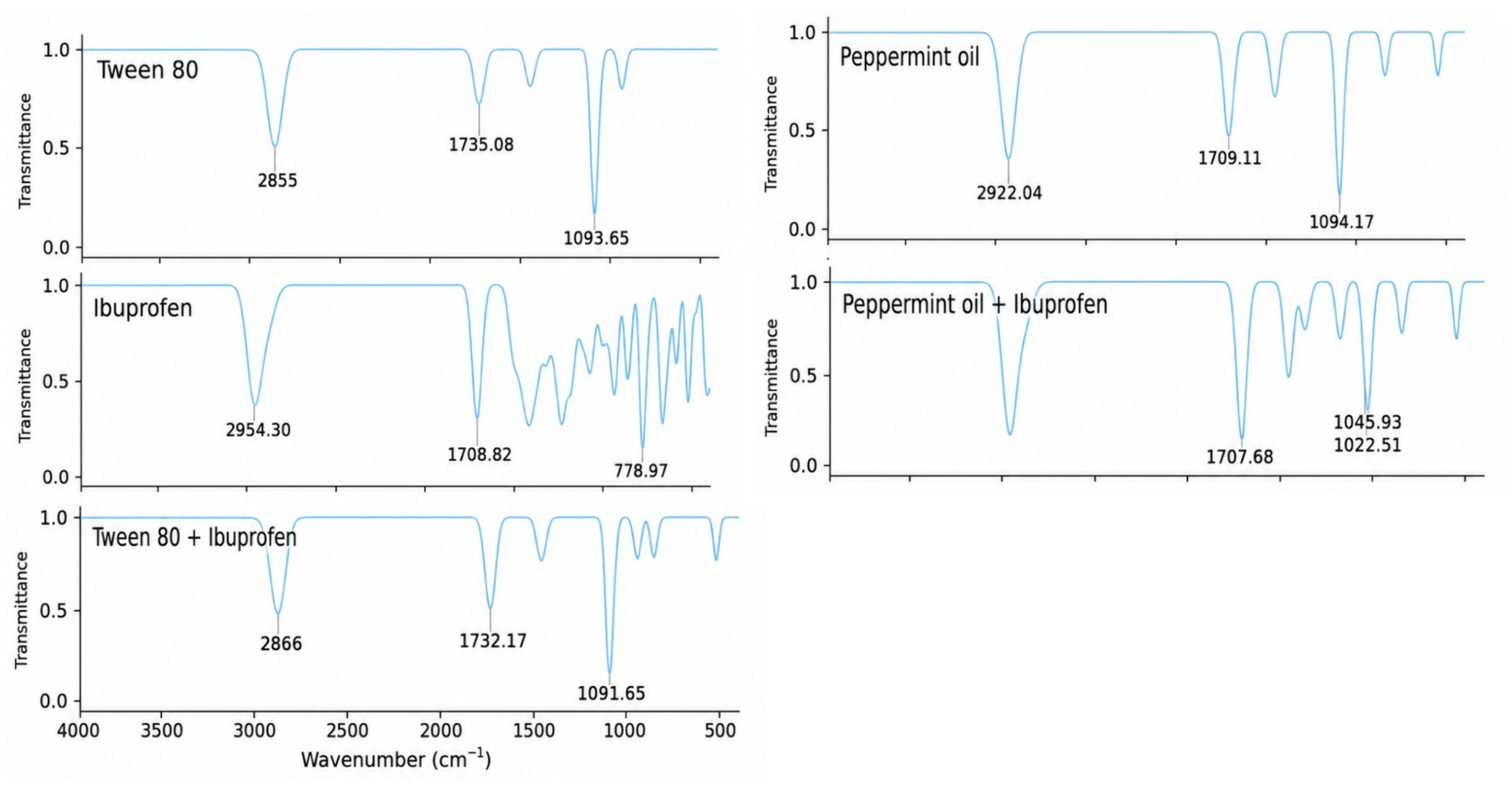
The FTIR spectrum of pure IBU (Figure 1) showed the characteristic carbonyl band of the carboxylic acid dimer at 1708.82 cm−1 and a well-resolved crystalline fingerprint region, including the aromatic out-of-plane bending band at 778.97 cm−1. Tween 80 was unambiguously confirmed by spectral library matching (HQI 99.63), with characteristic ether (1093.65 cm−1) and ester (1735.08 cm−1) bands. Peppermint oil showed a profile dominated by its oxygenated components (menthol and 1,8-cineole, 1094.17 cm−1) and the menthone carbonyl (1709.11 cm−1).
In the peppermint oil–IBU mixture, the drug’s carbonyl band was essentially unshifted (1707.68 cm−1, Δ ≈ 1 cm−1) but markedly intensified, while the oil’s C–O band resolved into two components (1022.51 and 1045.93 cm−1), and the drug’s crystalline fingerprint bands were attenuated to below 15% of their original intensity—jointly indicating molecular-level dispersion without chemical interaction. In the Tween 80–IBU mixture, Tween 80′s own bands remained within 3 cm−1 of their pure-component positions, and only weak residual bands attributable to IBU (520 and 856 cm−1) were detected, consistent with the drug being present but highly diluted within the surfactant matrix.
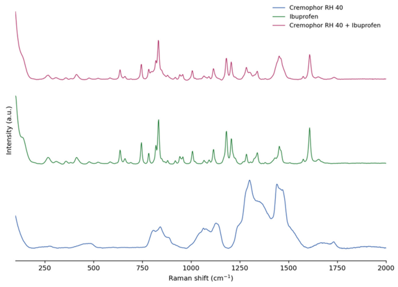
IBU identity in the Cremophor RH 40–IBU mixture was confirmed by spectral library matching (HQI = 86.06) Raman spectroscopy of the Cremophor RH 40–IBU mixture (Figure 2) showed that 14 of 18 detected bands matched pure IBU within ±2 cm−1, including the aromatic ring band (1608 cm−1) and all other major diagnostic bands, indicating no measurable chemical shift. The dominant band of pure Cremophor RH 40 (1300 cm−1, CH2 twisting) was not resolved in the mixture, attributable to the higher Raman scattering cross-section of ibuprofen’s aromatic chromophore relative to the excipient’s aliphatic backbone rather than to a chemical interaction; the excipient’s ester carbonyl band (1732 cm−1) remained detectable and unshifted (1734 cm−1).
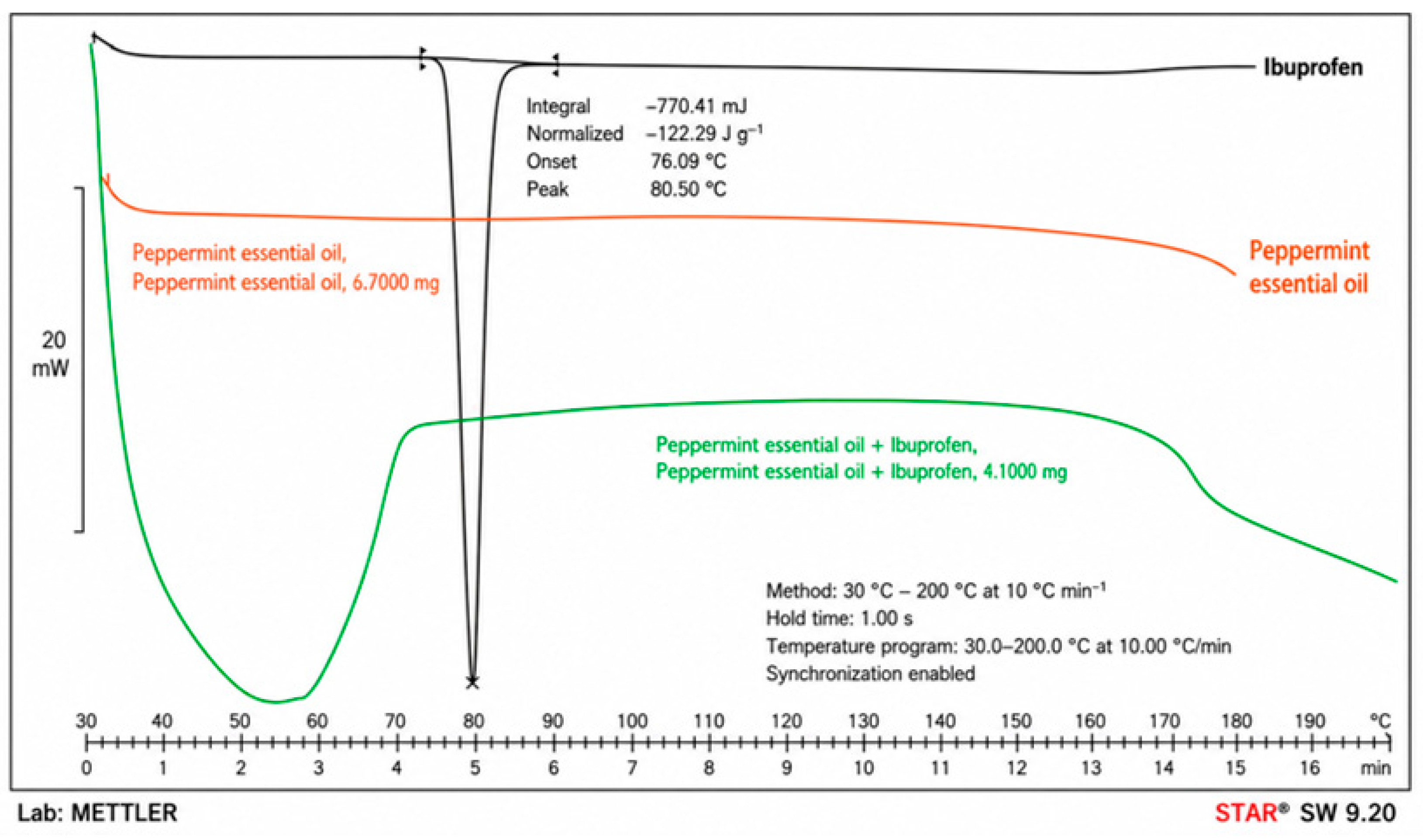
DSC analysis (Figure 3) showed a sharp melting endotherm for pure IBU (onset 76.09 °C, peak 80.50 °C, ΔH = 122.29 J/g), which was completely absent in the peppermint oil–IBU mixture, indicating loss of the drug’s crystalline structure.
Collectively, the FTIR, Raman, and DSC results show no new bands, no loss of characteristic functional groups, and no new thermal events across all evaluated drug–excipient pairs, ruling out chemical incompatibility at the excipient-screening stage. The combined dataset provides converging compatibility evidence for all three excipients (peppermint oil, Tween 80, and Cremophor RH 40) with the active ingredient, using at least one vibrational or thermal technique per pair. These results supported excipient selection at an early formulation stage.

3.1.2. Self-Emulsification Efficiency

All eight formulations exhibited optimal self-emulsification behavior, evidenced by the homogeneity, miscibility, and absence of visible particles in the resulting emulsions. The good optical clarity is confirmed by the transmittance values obtained, which exceed 90%. A transmittance close to 100% suggests the formation of systems with reduced droplet size [24]. However, differences were observed in the efficiency of the self-emulsification process, particularly in the time required to achieve complete emulsification. Four formulations (F1, F2, F5, and F7) showed average self-emulsification times (n = 3) below 2 min (43.40 s, 48.08 s, 107.62 s, and 44.82 s, respectively), reflecting superior self-emulsification efficiency [34]. Therefore, although all formulations were capable of self-emulsification, only those with emulsification times shorter than 2 min (F1, F2, F5, and F7) were selected for defining the concentration limits and for subsequent analyses.

3.2. Box–Behnken Experimental Design

Considering the results obtained in the initial characterization, the levels of each of the independent variables were established (Table 2).

3.2.1. Characterization of BBD-SEDDS

The experimental values obtained for the six responses evaluated in the 15 formulations developed (Table 3) indicate that the BBD-SEDDS system exhibited consistent, controlled behavior within the experimental space studied.
The reproducibility of the experimental process was assessed through the three center-point replicates (formulations 4, 9, and 15). Five of the six evaluated responses showed relative standard deviations below 8% (cloud point, 2.75%; robustness to dilution, 7.53%; self-emulsification time, 0.55%; zeta potential, 7.01%; PDI, 6.27%), indicating good reproducibility of the preparation and measurement process across independent runs. Droplet size showed comparatively higher variability among center points (RSD = 17.55%), consistent with this response’s weaker model fit (Table 4) and its known sensitivity to minor compositional variation within the screened design space; this variability was substantially reduced in the optimized formulation upon confirmatory replication (Section 3.3.4), where droplet size reproducibility improved to approximately 5.6% RSD.
Cloud Point
The cloud point remained within the range of 56–65 °C, with values above the physiological temperature, indicating adequate thermal stability of the system after dilution under simulated gastrointestinal conditions [25,35].
Robustness to Dilution
Robustness to dilution showed greater variability between formulations, with values ranging from 6 to 10, suggesting greater sensitivity of this response to the proportions of the formulation components. From a physicochemical perspective, this behavior can be attributed to the combined roles of the surfactant and the oil phase in stabilizing the emulsion during self-emulsification and subsequent dilution. SNEDDS formulations with appropriate surfactant-to-oil ratios tend to generate more stable systems upon dilution, resulting in more homogeneous emulsions free from visible turbidity or precipitation, as previously reported [36].
Self-Emulsification Time
The self-emulsification time ranged from 38.9 to 65.0 s, reflecting rapid nanoemulsion formation, a desirable characteristic for self-emulsifying orally administered systems [35].
Droplet Size, PDI and Zeta Potential
Regarding colloidal properties, the zeta potential remained within a relatively constant range (−13.7 to −17.0 mV), suggesting moderate and homogeneous electrostatic stability among the formulations [29]. The droplet size was mostly found within the range 127.2–303.3 nm, with the exception of F-14, which exhibited a markedly higher value (548.5 nm), which, characterized by the minimum Tween 80 concentration and maximum Cremophor RH 40 content, represents a boundary condition at the edge of the self-emulsification region.
PDI values ranged from 0.17 to 0.33, indicating generally narrow size distributions, although some formulations approached or slightly exceeded the commonly accepted threshold of 0.30 for highly homogeneous systems [10].

3.2.2. Surface Response Analysis of BBD-SEDDS

According to the results summarized in Table 4, responses Y1, Y3, Y4, and Y6 did not show statistically significant models, despite exhibiting non-significant lack-of-fit tests. The factor levels in the BBD were constrained to the region of the formulation space previously identified as yielding adequate spontaneous self-emulsification, delimited by pseudo-ternary phase diagrams [14]. This QbD-based constraint is methodologically appropriate for SNEDDS development but inherently limits the variability in most CQA responses within the studied space. Negative predicted R2 values observed for Y3, Y4, and Y6 indicate that the mean model predicts better than the fitted model within the studied range, an expected and interpretable result when factors exert negligible influence on a response within the experimental domain [35,37]. Model selection followed the principle of parsimony, retaining only models with p < 0.05 [38]. For Y5 (droplet size), although the linear model did not reach statistical significance (p = 0.1463), the observed range (127.2–548.5 nm) suggests that surfactant concentration may exert an influence on this response at compositional extremes, as evidenced by F-14. A broader experimental domain would likely capture this relationship more completely.
This behavior indicates that, although the models do not exhibit evident structural deficiencies, the evaluated factors exert a limited influence on these responses within the considered experimental range. The experimental design focused on a region of the factor space where the SEDDS system’s performance was already favorable. However, the lack of significance should not be interpreted as a deficiency in the design, but rather as an indication of the system’s robustness to moderate changes in formulation, as reported in previous studies on the optimization of self-emulsifying systems using response surface methodology [37].
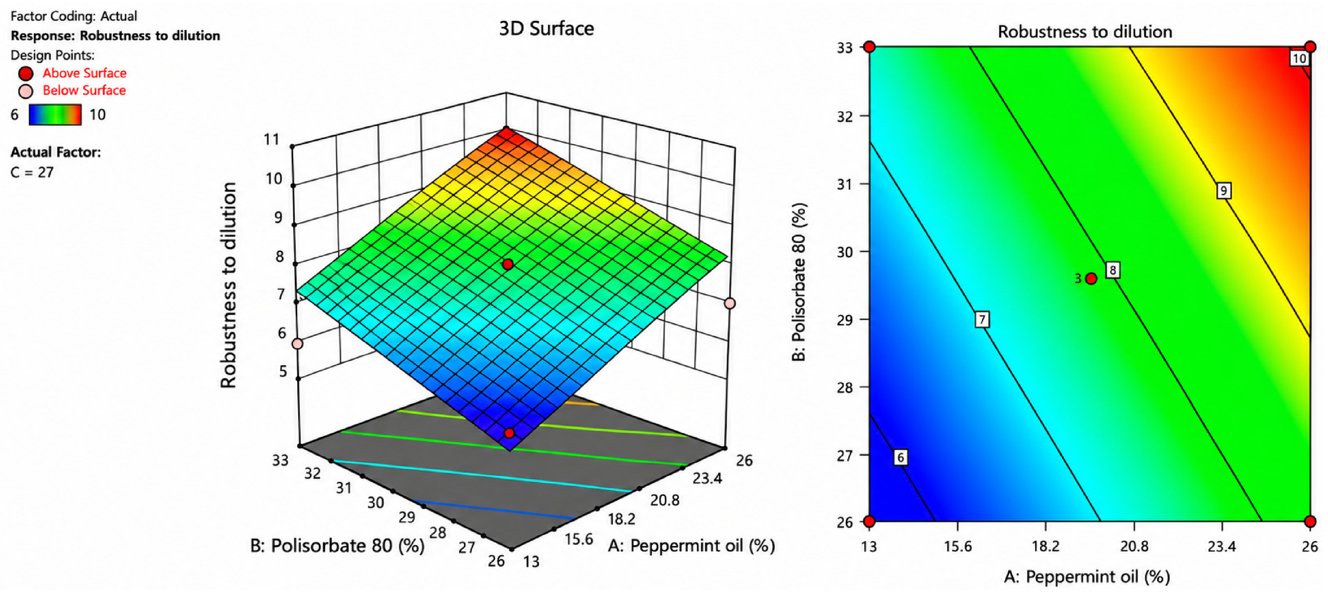
In contrast, only the Y2 response (robustness to dilution) presented a statistically significant and predictive model within the studied experimental space (p < 0.05), with the absence of lack of fit and adjusted R2 and predicted R2 values (Table 4). ANOVA analysis revealed that both the percentage of peppermint oil (A) and polysorbate 80 (B) exerted positive, significant effects on this response, whereas PEG-40 hydrogenated castor oil (C) did not show a significant influence (Table 5). The response surface and contour plots (Figure 4) showed an increasing linear relationship as a function of A and B, consistent with the fitted model, indicating that increasing both components improves the stability of the system after dilution [39].
Three-dimensional response surface (left) and contour plot (right) illustrating the effect of peppermint oil (A) and polysorbate 80 (B) concentrations on the robustness to dilution response of IBU-SEDDS predicted by the Box–Behnken model. Robustness to dilution was expressed as the cumulative stability score after evaluation in four media and three dilution factors. Experimental data were obtained in triplicate (n = 3).

3.2.3. Formulation, Validation and Point Prediction of SNEDDS

A two-tier evaluation framework was applied. In Tier 1, mathematical optimization via desirability function was applied exclusively to responses yielding statistically validated predictive models within the studied design space; only Y2 (robustness to dilution) met this criterion (p = 0.0018, adjusted R2 = 0.6583). The variables of cloud point, self-emulsification time, zeta potential, droplet size, and PDI all had no statistically valid models from which to calculate a desirability function; thus, these variables were not incorporated [40]. They were removed based on the decision-making principle of parsimony, such that nothing should be added to the model unless there is sufficient statistical support for its inclusion to avoid compromising the model’s robustness and/or introducing bias in interpretation. Thus, based on the contemporary model selection criteria, only those models with p < 0.05 were retained for consideration with regard to achieving appropriate relative levels of adequate goodness of fit and structural simplicity [38]. Furthermore, Y1, Y3, Y4, Y5, Y6 were evaluated as predefined acceptance criteria in the predicted optimal formulation to verify that the optimized system met the required physicochemical specifications. This approach is statistically rigorous while ensuring that the complete set of CQAs was assessed in the final formulation.
The optimized composition of the IBU-SNEDDS is presented in Table 6 and yielded a global desirability index of 1. Experimental validation at the predicted optimal point demonstrated marked robustness to dilution, with a mean experimental value of 12 (n = 3 independent replicates), compared with a theoretical prediction of 10.14. The results obtained with the diluted formulation demonstrate the system’s stability across the evaluated dilution conditions, an important factor influencing drug absorption when administered in vivo [23]. Importantly, the observed value remained within the 95% prediction interval (8.90–12.00), supporting both the model’s predictive performance and the adequacy of the selected optimum. In addition, the results showed a low standard deviation across experimental trials, indicating that the optimized system can be replicated very well [35,40].

3.3. Evaluation of Optimized Formulation

To confirm that the optimized formulation maximizes robustness to dilution and complies with the established CQAs for SNEDDSs, its physicochemical characterization was carried out.

3.3.1. Cloud Point

The cloud point reached 64.87 ± 0.15 °C, considerably higher than the physiological temperature, demonstrating the thermal stability of the surfactants used in the system and suggests that phase separation of the surfactants is unlikely to occur at body temperature. A cloud point above 37 °C is generally considered a desirable characteristic for self-emulsifying systems, as it supports the maintenance of their physicochemical properties during dispersion [26].

3.3.2. Self-Emulsification Time

Optimized IBU-SNEDDS exhibited an average self-emulsification time of 37.02 ± 5.65 s, resulting in rapid, complete emulsification upon gentle stirring and heating to physiological temperature. This outcome confirms the spontaneous process and anticipates that effective gastrointestinal environment management will occur [41].

3.3.3. Droplet Size

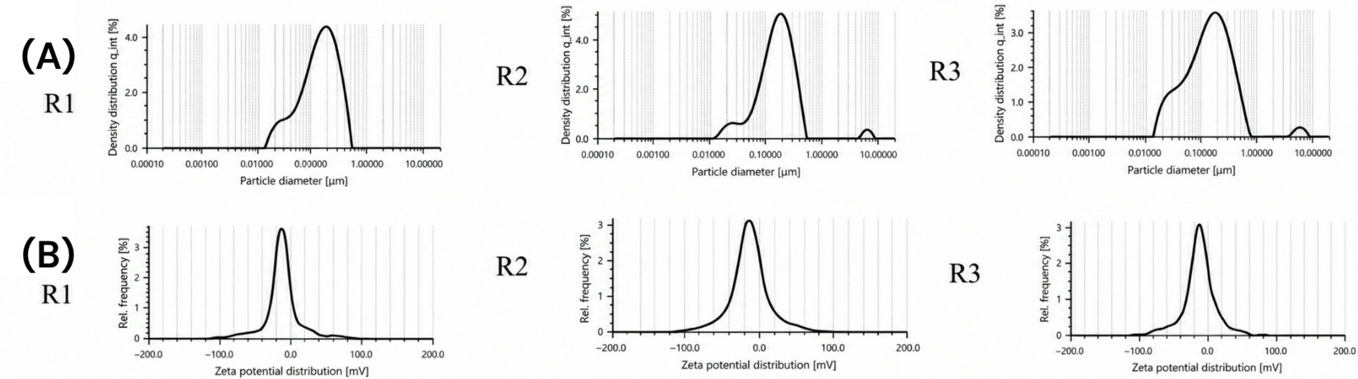
The optimized IBU-SNEDDS exhibited a Z-average hydrodynamic diameter of 157.54 ± 8.9 nm, confirming the formation of droplets within the nanometric range. The intensity-weighted Droplet size distributions (Figure 5A) consistently showed a predominant droplet population centered between approximately 176 and 187 nm across the three independent replicates, representing more than 93% of the total scattering intensity. Minor secondary populations were also detected in two replicates, including a small fraction of droplets around 23 nm and low-intensity peaks at approximately 6 μm. Given their very low contribution (≤5.2% of the total scattering intensity) and the reproducibility of the main population among independent preparations, these secondary peaks are most likely associated with occasional aggregates or dust particles, which are known to disproportionately influence intensity-weighted DLS measurements because scattering intensity increases markedly with Droplet size. The low PDI values obtained for all replicates nevertheless indicate that the formulation maintained an acceptable degree of homogeneity. Scientific literature shows variability in the criteria used to classify a system as SMEDDS or SNEDDS, with no clear uniformity across different authors [21,22,23,42]. Based on a comparative analysis of multiple reported studies, the most widely used criterion was adopted, according to which systems with droplet sizes smaller than 200 nm can be classified as SNEDDS. However, given the discrepancies among the proposed criteria, it is necessary to jointly evaluate other physicochemical properties of the system to comprehensively establish its identity and behavior.

3.3.4. PDI

Optimized IBU-SNEDDS exhibited a PDI of 0.251 ± 0.03, below the commonly accepted reference threshold for self-nanoemulsifying systems, which states that the PDI should be less than 0.3 [30]. This result suggests adequate homogeneity in the droplet size distribution of the colloidal system.
Additionally, SNEDDSs are typically characterized by relatively broader peak-size distributions than self-microemulsifying systems (SMEDDS). This behavior is attributed to the nature of SNEDDS, in which surfactant molecules spontaneously reorganize upon contact with the aqueous medium, resulting in a droplet distribution with nanometric sizes [24]. In accordance with the above, the droplet size distribution observed in Figure 5A shows a broad peak, a characteristic behavior of an SNEDDS.

3.3.5. Zeta Potential

The zeta potential values generally accepted as indicative of high colloidal stability are around ±30 mV, a threshold associated with systems whose stability depends mainly on electrostatic repulsion mechanisms [29]. In this study, optimized IBU-SNEDDS exhibited a zeta potential of −15.43 mV ± 0.58 (Figure 5B), which is below the conventional range. However, unlike colloidal systems stabilized predominantly through electrostatic repulsion, the physical stability of non-ionic surfactant-based SNEDDS cannot be explained solely by the magnitude of the zeta potential, SNEDDS containing non-ionic surfactants rely heavily on steric stabilization mechanisms. Tween 80 and Cremophor RH 40 adsorb at the oil–water interface, where their polyoxyethylene chains extend into the continuous aqueous phase, forming a hydrated steric barrier around the oil droplets. This interfacial layer prevents close droplet–droplet interactions by generating steric hindrance, thereby reducing flocculation and coalescence even when the measured zeta potential is relatively low [29,30]. Consequently, the zeta potential in non-ionic SNEDDS should be interpreted as a complementary parameter rather than the primary determinant of colloidal stability.
This stabilization mechanism has been consistently reported for SNEDDS containing non-ionic surfactants, in which zeta potential values between approximately −15 and −20 mV have been associated with satisfactory physical stability. For example, Pawar et al. (2025) described stable SNEDDS with zeta potential values close to −16 mV and attributed their stability primarily to steric hindrance provided by the non-ionic surfactant layer, with electrostatic repulsion playing only a secondary role [29]. Since the optimized formulation developed in this study contains the same class of surfactants, a similar stabilization mechanism is expected.
These results indicate that the optimized SNEDDS exhibits adequate physical stability, attributable to the combination of electrostatic and steric mechanisms, which supports its performance as an oral delivery system and is discussed in more detail in Section 3.3.8.

3.3.6. Effect of Dilution on Droplet Size

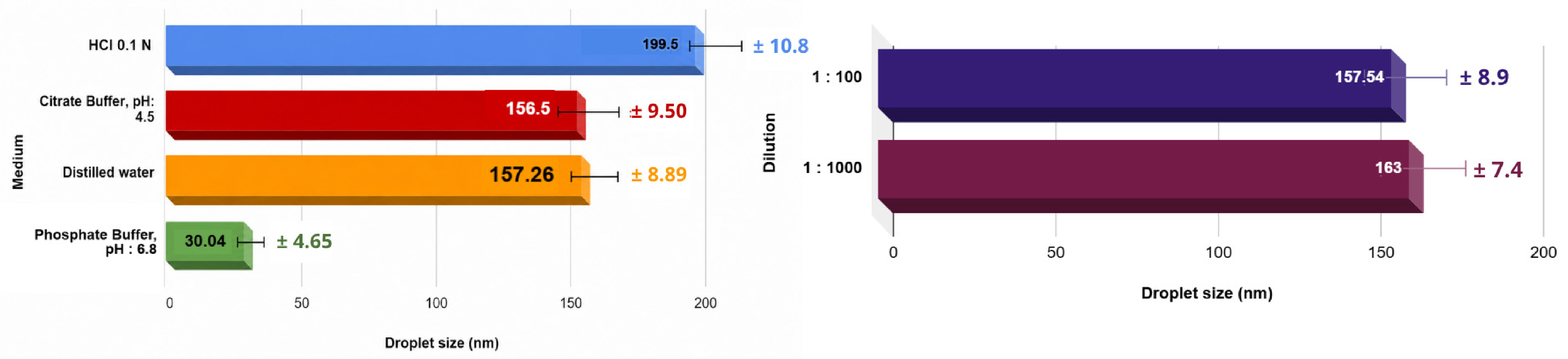
SNEDDS are characterized by maintaining a relatively constant droplet size despite variations in the dilution medium volume. In contrast, SMEDDS typically exhibit greater sensitivity to variations in the volume of the medium because shifts in phase equilibrium can reduce droplet size via surfactant redistribution or, in cases of saturation, destabilize the system due to loss of the interface’s solubilizing capacity. According to the data obtained (Figure 6), the optimized IBU-SNEDDS showed only a modest increase in droplet size. The optimized IBU-SNEDDS maintained a nearly constant mean droplet size after dilution, ranging from 157.54 to 163.0 nm. At the 1:100 dilution level, DLS intensity analysis revealed a single droplet population, indicating a homogeneous nanoemulsion system. At the highest dilution level (1:1000), a predominant population centered around 163 nm represented approximately 95% of the total scattering intensity, while two minor secondary populations accounted collectively for less than 5% of the signal. Despite the appearance of these minor populations at extreme dilution, the mean droplet size remained essentially unchanged, and PDI values remained below 0.30 (0.213–0.276), supporting the overall homogeneity and kinetic stability of the formulation. Hereby classifying the formulation as an SNEDDS [24,43].
The left panel shows the droplet size after dilution in distilled water, 0.1 N HCl, citrate buffer (pH 4.5), and phosphate buffer (pH 6.8), while the right panel compares the droplet size at dilution factors of 1:100 and 1:1000. Values are expressed as the mean ± SD of three independent measurements (n = 3).

3.3.7. Effect of pH on Droplet Size

The droplet size of the optimized IBU-SNEDDS was strongly influenced by the dispersion medium. A marked reduction was observed in phosphate buffer pH 6.8, while larger droplets were formed in 0.1 N HCl. Intermediate sizes were obtained in distilled water and citrate buffer pH 4.5.
Method reproducibility was evaluated in triplicates using distilled water as the reference medium, yielding a mean droplet size of 157.26 ± 8.89 nm with a relative standard deviation (RSD) of 5.65%. The other screening media were likewise measured in triplicate, 681 and their mean droplet sizes were compared with the water. Using the standard deviation of the reference (8.89 nm) to contextualize the observed variability, phosphate Buffer pH 6.8 showed a mean droplet size of 30.04 nm, representing an 81% reduction relative to water. The difference between both means (127.50 nm) far exceeds the experimental error of the methodology (RSD < 6%), confirming a distinct physiological medium effect rather than random measurement variability.
The cause is likely the pH-dependent solubility of IBU (pKa ≈ 4.5) [5,6]. The properties of the aqueous medium in which dilution will take place will greatly affect the final droplet size of a nanoemulsion, such as pH and ionic strength, particularly for drugs with pH-dependent solubility [44]. A marked reduction was observed in the phosphate buffer pH 6.8, which showed a difference of approximately 7 times between the smallest and largest values obtained in the other media, due to the fact that at this pH. IBU is predominantly ionized (>90%), its affinity for the oil–water interface increases, allowing it to interact with the nonionic surfactants. This interaction reduces the interfacial tension and promotes the formation of smaller droplets [44,45,46]. In contrast, in 0.1 N HCl, where IBU remains predominantly in its unionized form (>90%), it preferentially partitions into the oil core, contributing minimally to interfacial stabilization. Similar behavior has been reported for SNEDDS containing ionizable drugs, in which pH-dependent changes in drug partitioning modify the interfacial organization and, consequently, the droplet size [45,46].
The possibility that pH-dependent changes in droplet size and ionization state could influence drug loading capacity or lead to precipitation phenomena during gastrointestinal transit particularly at gastric pH is acknowledged as an important consideration that should be addressed in future in vitro lipolysis and drug precipitation.
Despite the droplet sizes of all systems evaluated differing by pH, they all remained less than 200 nm, making these systems likely to produce stable nanoemulsions and indicating the potential for favourable system performance under different gastrointestinal conditions after oral administration.

3.3.8. Physical/Kinetic Stability and Stress Testing

Optimized IBU-SNEDDS was subjected to various physical stress conditions, including centrifugation, heating, cooling, and freeze–thaw cycles, to evaluate its stability. After these treatments, the system maintained its physical stability, with no signs of instability, such as creaming, emulsion breakdown, or phase separation. This suggests kinetic stability against gravitational separation, temperature-induced phase transitions, and crystallization phenomena.
Stress testing showed that transmittance values remained consistently above 90% in all replicates during the four-week study period. Overall averages ranged from 96.51% to 97.67%, with a standard deviation of less than 2.7. Furthermore, relative standard deviation (RSD) values remained below 3%, confirming the high repeatability of the manufacturing process and the accuracy of the analytical method (Table 7).
A two-way ANOVA was performed to evaluate the effects of batch and storage time on transmittance. No statistically significant differences were observed for batch (F = 0.661, p = 0.542) or time (F = 2.595, p = 0.117) at a significance level of 0.05. These results indicate that transmittance did not vary significantly over time or between batches (p > 0.05). Since transmittance serves as an indirect indicator of droplet size in SNEDDS, the absence of significant variation suggests that no appreciable changes in colloidal structure occurred during the storage period. The absence of a statistically significant effect of both batch and storage time on transmittance therefore confirms the physical stability of the optimized IBU-SNEDDS formulation throughout the four-week accelerated stability study.

3.4. In Vitro Dissolution Studies

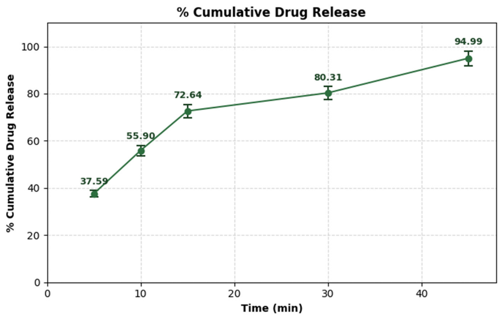
The in vitro dissolution profile (Figure 7) showed a rapid initial release from the optimized IBU-SNEDDS, reaching 37.59 ± 1.31% after 5 min and 72.64 ± 2.83% after 15 min. Drug release increased to 80.31 ± 2.67% at 30 min and reached 94.99 ± 3.08% after 45 min, approaching complete drug release.
This profile is consistent with efficient self-emulsification of the optimized formulation, which promotes the spontaneous formation of a nanoemulsion with a large interfacial area available for drug dissolution. Unlike conventional oral formulations, in which drug dissolution is preceded by crystal dissolution, IBU is already molecularly dispersed within the lipidic formulation prior to administration. Upon contact with the aqueous medium, the spontaneous formation of nanometric droplets provides a markedly increased interfacial surface area, shortening the diffusion path and facilitating rapid drug partitioning into the dissolution medium. Together, these mechanisms contribute to the accelerated dissolution profile observed for the optimized SNEDDS. This behavior was further reflected in a dissolution efficiency over the first 30 min (DE030), calculated by the trapezoidal method, of 59.87%, indicating that nearly 60% of the maximum theoretical dissolution performance was achieved within the first 30 min. This result confirms that drug release occurred rapidly throughout the evaluation period rather than only at the final sampling point. Consistent with this finding, the formulation met the pharmacopeial acceptance criterion for immediate-release IBU dosage forms (Q ≥ 80% at 30 min) by the 30-min sampling point [33], demonstrating that the enhanced solubilization capacity observed during the physicochemical characterization studies translates into in vitro drug release that meets specifications.
Compared with the SNEDDS formulations of 200 mg IBU in hard capsules reported by Mihaylov et al. (2025) based on medium-chain triglycerides, Cremophor RH 40, and PEG 400 or propylene glycol, which achieved drug release above 90% after 60 min, the formulation developed in the present study reached a comparable extent of release within 45 min [5]. This faster dissolution profile may be associated with the optimized formulation composition and the small droplet size obtained after self-emulsification, both of which increase the effective surface area available for drug release. Although these findings suggest the potential for improved oral performance, further studies in biorelevant media and in vivo pharmacokinetic evaluation are required to confirm whether the enhanced dissolution translates into improved bioavailability or a faster onset of action.

4. Conclusions

This study demonstrates that the development of an IBU-loaded self-nanoemulsifying drug delivery system (SNEDDS) using a rational design space approach resulted in an optimized formulation with favorable physicochemical characteristics. Beyond the precise optimization achieved, the system’s high physical stability and rapid emulsification properties suggest its potential to maintain its performance under physiological conditions. The results highlight the achievement of a composition-robust region, supporting manufacturing reproducibility. Consequently, this optimized SNEDDS represents a promising formulation for further investigation as an oral delivery system for IBU. Future studies should evaluate its behavior in biorelevant dissolution media (FaSSGF, FaSSIF, and FeSSIF) and analyze its in vivo pharmacokinetic profile to establish more robust in vitro–in vivo correlations and further corroborate its biopharmaceutical potential.

Supplementary Materials

The following supporting information can be downloaded at: https://www.mdpi.com/article/10.3390/scipharm94030082/s1.

Author Contributions

Conceptualization, funding acquisition, resources, supervision and project administration, R.G.S.; Methodology and writing—review and editing, M.J.J., K.D.L.C. and R.G.S.; Data curation, formal analysis, investigation, visualization, writing—original draft, M.J.J. and K.D.L.C. All authors have read and agreed to the published version of the manuscript.

Funding

This study is part of a university-funded research project supported by the Universidad del Atlántico under the Third internal call for proposals for the strengthening of the institutional network of research seedbeds (REDISIA)-2022 (Project: QYF765-CIS2023).

Data Availability Statement

The original contributions presented in this study are included in the article/Supplementary Materials. Further inquiries can be directed to the corresponding author.

Acknowledgments

The authors would like to thank the Universidad del Atlántico for its technical and institutional support. Special thanks are extended to Anton Paar Colombia for supporting this research by loaning equipment, demonstrating their continued commitment to scientific advancement. Finally, we thank Croda for the donation of excipients.

Conflicts of Interest

The authors declare no conflict of interest.

Abbreviations

The following abbreviations are used in this manuscript:
NSAIDnon-steroidal anti-inflammatory drug
SNEDDSself-nanoemulsifying drug delivery system
SEDDSself-emulsifying drug delivery systems
SMEDDSself-microemulsifying drug delivery systems
CQAcritical quality attributes
PDIpolydispersity index
IBUIbuprofen
DoEDesigns of experiments
BBDBox–Behnken experimental design
BBD-SEDDSSelf-emulsifying formulations developed using a Box–Behnken design.
DLS Dynamic Light Scattering.
QbDQuality by Design.
CMAcritical material attributes
HLBhigh hydrophilic–lipophilic balance
BCSBiopharmaceutics Classification System
GRASGenerally Recognized As Safe

References

  1. Alqahtani, M.S.; Kazi, M.; Alsenaidy, M.A.; Ahmad, M.Z. Advances in Oral Drug Delivery. Front. Pharmacol. 2021, 12, 618411. [Google Scholar] [CrossRef] [Scilit] [PubMed]
  2. Zaslavsky, J.; Allen, C. A dataset of formulation compositions for self-emulsifying drug delivery systems. Sci. Data 2023, 10, 914. [Google Scholar] [CrossRef] [Scilit] [PubMed]
  3. Kalepu, S.; Nekkanti, V. Insoluble drug delivery strategies: Review of recent advances and business prospects. Acta Pharm. Sin. B 2015, 5, 442–453. [Google Scholar] [CrossRef] [Scilit] [PubMed]
  4. Khan, K.U.; Minhas, M.U.; Badshah, S.F.; Suhail, M.; Ahmad, A.; Ijaz, S. Overview of nanoparticulate strategies for solubility enhancement of poorly soluble drugs. Life Sci. 2022, 291, 120301. [Google Scholar] [CrossRef] [Scilit] [PubMed]
  5. Mihaylov, V.; Tosheva, M.; Petrov, V.; Titeva, S. Technological approaches to increase the bioavailability of Ibuprofen. Pharmacia 2025, 72, 1–11. [Google Scholar] [CrossRef] [Scilit]
  6. Ćirić, A.; Medarević, Đ.; Čalija, B.; Dobričić, V.; Rmandić, M.; Barudžija, T.; Malenović, A.; Djekic, L. Effect of ibuprofen entrapment procedure on physicochemical and controlled drug release performances of chitosan/xanthan gum polyelectrolyte complexes. Int. J. Biol. Macromol. 2021, 167, 547–558. [Google Scholar] [CrossRef] [Scilit] [PubMed]
  7. Irvine, J.; Afrose, A.; Islam, N. Formulation and delivery strategies of ibuprofen: Challenges and opportunities. Drug Dev. Ind. Pharm. 2018, 44, 173–183. [Google Scholar] [CrossRef] [Scilit] [PubMed]
  8. Yahaya, Z.S.; Kurfi, F.S.; Mallam, D.; Oloyede, R.B.; Adeleye, O.A.; Okpanachi, G.O. Preliminary research on ibuprofen self-emulsifying formulation. J. Res. Pharm. 2022, 26, 834–841. [Google Scholar] [CrossRef] [Scilit]
  9. Nardin, I.; Köllner, S. Successful development of oral SEDDS: Screening of excipients from the industrial point of view. Adv. Drug Deliv. Rev. 2019, 142, 128–140. [Google Scholar] [CrossRef] [Scilit] [PubMed]
  10. Zafar, A.; Yasir, M.; Alruwaili, N.K.; Imam, S.S.; Alsaidan, O.A.; Alshehri, S.; Ghoneim, M.M.; Alquraini, A.; Rawaf, A.; Ansari, M.J.; et al. Formulation of Self-Nanoemulsifying Drug Delivery System of Cephalexin: Physiochemical Characterization and Antibacterial Evaluation. Polymers 2022, 14, 1055. [Google Scholar] [CrossRef] [Scilit] [PubMed]
  11. Krstić, M.; Medarević, Đ.; Đuriš, J.; Ibrić, S. Chapter 12—Self-nanoemulsifying drug delivery systems (SNEDDS) and self-microemulsifying drug delivery systems (SMEDDS) as lipid nanocarriers for improving dissolution rate and bioavailability of poorly soluble drugs. In Lipid Nanocarriers for Drug Targeting; Grumezescu, A.M., Ed.; William Andrew Publishing: Norwich, NY, USA, 2018; pp. 473–508. Available online: https://www.sciencedirect.com/science/article/pii/B9780128136874000128 (accessed on 19 June 2025).
  12. Vasconcelos, T.; Marques, S.; Sarmento, B. Measuring the emulsification dynamics and stability of self-emulsifying drug delivery systems. Eur. J. Pharm. Biopharm. 2018, 123, 1–8. [Google Scholar] [CrossRef] [Scilit] [PubMed]
  13. Valicherla, G.R.; Dave, K.M.; Syed, A.A.; Riyazuddin, M.; Gupta, A.P.; Singh, A.; Wahajuddin; Mitra, K.; Datta, D.; Gayen, J.R. Formulation optimization of Docetaxel loaded self-emulsifying drug delivery system to enhance bioavailability and anti-tumor activity. Sci. Rep. 2016, 6, 26895. [Google Scholar] [CrossRef] [Scilit] [PubMed]
  14. Sotomayor Pino, R.G.; Salas Ospino, S.C.; Ramirez Castillo, L.; Ospina Garcia, D. Diseño y desarrollo de un sistema de entrega de fármaco autoemulsificable (SEDDS) de ibuprofeno en cápsula dura de gelatina. RESCIFAR Rev. Esp. Cienc. Farm. 2021, 2, 251–252. [Google Scholar]
  15. López, A.; Herazo, K.; Sotomayor, R.G.; López, A.; Herazo, K.; Sotomayor, R.G. Diseño y desarrollo de un sistema de entrega de fármaco autoemulsificable líquido de ibuprofeno incorporado por el método de adsorción por portador en una forma farmacéutica sólida (comprimidos). Rev. Colomb. Cienc. Quím. Farm. 2019, 48, 589–614. [Google Scholar] [CrossRef] [Scilit]
  16. Inactive Ingredient Search for Approved Drug Products. Available online: https://www.accessdata.fda.gov/scripts/cder/iig/index.cfm?event=BasicSearch.page (accessed on 3 June 2026).
  17. Excipients Labelling | European Medicines Agency (EMA). Available online: https://www.ema.europa.eu/en/human-regulatory-overview/marketing-authorisation/product-information-requirements/excipients-labelling (accessed on 4 June 2026).
  18. Vilas-Boas, S.M.; Cordova, I.W.; Abranches, D.O.; Coutinho, J.A.P.; Ferreira, O.; Pinho, S.P. Modeling the Solubility of Monoterpenoids with Hybrid and Predictive Thermodynamic Tools. Ind. Eng. Chem. Res. 2023, 62, 5326–5335. [Google Scholar] [CrossRef] [Scilit] [PubMed]
  19. Abdullah, G.Z.; Abdulkarim, M.F.; Salman, I.M.; Ameer, O.Z.; Yam, M.F.; Mutee, A.F.; Chitneni, M.; Mahdi, E.S.; Basri, M.; Sattar, M.A.; et al. In vitro permeation and in vivo anti-inflammatory and analgesic properties of nanoscaled emulsions containing ibuprofen for topical delivery. Int. J. Nanomed. 2011, 6, 387–396. [Google Scholar] [CrossRef] [Scilit] [PubMed]
  20. Sindi, A.M.; Hosny, K.M.; Alharbi, W.S. Lyophilized Composite Loaded with Meloxicam-Peppermint oil Nanoemulsion for Periodontal Pain. Polymers 2021, 13, 2317. [Google Scholar] [CrossRef] [Scilit] [PubMed] [PubMed Central]
  21. Silberstein, S.; Spierings, E.L.H.; Kunkel, T. Celecoxib Oral Solution and the Benefits of Self-Microemulsifying Drug Delivery Systems (SMEDDS) Technology: A Narrative Review. Pain Ther. 2023, 12, 1109–1119. [Google Scholar] [CrossRef] [Scilit] [PubMed]
  22. Usta, D.Y.; Timur, B.; Teksin, Z.S. Formulation development, optimization by Box-Behnken design, characterization, in vitro, ex-vivo, and in vivo evaluation of bosentan-loaded self-nanoemulsifying drug delivery system: A novel alternative dosage form for pulmonary arterial hypertension treatment. Eur. J. Pharm. Sci. 2022, 174, 106159. [Google Scholar] [CrossRef] [Scilit] [PubMed]
  23. Ali, N.; Ahmed, G.; Hassan, G.; Elsayed, A.; Mamdouh, G. Effect of oils, surfactants and cosurfactants on phase behavior and physicochemical properties of self-nanoemulsifying drug delivery system (Snedds) for irbesartan and olmesartan. Int. J. Appl. Pharm. 2016, 8, 13–24. [Google Scholar]
  24. Rehman, F.U.; Farid, A.; Shah, S.U.; Dar, M.J.; Rehman, A.U.; Ahmed, N.; Rashid, S.A.; Shaukat, I.; Shah, M.; Albadrani, G.M.; et al. Self-Emulsifying Drug Delivery Systems (SEDDS): Measuring Energy Dynamics to Determine Thermodynamic and Kinetic Stability. Pharmaceuticals 2022, 15, 1064. [Google Scholar] [CrossRef] [Scilit] [PubMed] [PubMed Central]
  25. Buya, A.B.; Beloqui, A.; Memvanga, P.B.; Préat, V. Self-Nano-Emulsifying Drug-Delivery Systems: From the Development to the Current Applications and Challenges in Oral Drug Delivery. Pharmaceutics 2020, 12, 1194. [Google Scholar] [CrossRef] [Scilit] [PubMed]
  26. Goméz Palencia, K.; Goméz de la Cruz, L.; Herrera Marquéz, A.; Sotomayor, R.G.; Goméz Palencia, K.; Goméz de la Cruz, L.; Herrera Marquéz, A.; Sotomayor, R.G. Diseño y caracterización de un sistema de entrega de fármacos autoemulsificable (SEDDS) de estradiol por diagramas de fases pseudoternarios y pruebas fisicoquímicas. Rev. Colomb. Cienc. Quím. Farm. 2022, 51, 493–525. [Google Scholar] [CrossRef] [Scilit]
  27. Mudie, D.M.; Amidon, G.L.; Amidon, G.E. Physiological Parameters for Oral Delivery and in Vitro Testing. Mol. Pharm. 2010, 7, 1388–1405. [Google Scholar] [CrossRef] [Scilit] [PubMed]
  28. Jakab, G.; Fülöp, V.; Bozó, T.; Balogh, E.; Kellermayer, M.; Antal, I. Optimization of Quality Attributes and Atomic Force Microscopy Imaging of Reconstituted Nanodroplets in Baicalin Loaded Self-Nanoemulsifying Formulations. Pharmaceutics 2018, 10, 275. [Google Scholar] [CrossRef] [Scilit] [PubMed]
  29. Pawar, A.; Dere, S.; Pandhare, R.; Mohite, P.; Alharbi, H.M.; Subramaniyan, V.; Kumarasamy, V.; Maitra, S.; Ahamed, F.M.M.; Uti, D.E.; et al. Enhancing solubility and dissolution of felodipine using self-nanoemulsifying drug systems through in vitro evaluation. Sci. Rep. 2025, 15, 8900. [Google Scholar] [CrossRef] [Scilit] [PubMed]
  30. Mohite, P.; Sule, S.; Pawar, A.; Alharbi, H.M.; Maitra, S.; Subramaniyan, V.; Kumarasamy, V.; Uti, D.E.; Ogbu, C.O.; Oodo, S.I.; et al. Development and characterization of a self-nano emulsifying drug delivery system (SNEDDS) for Ornidazole to improve solubility and oral bioavailability of BCS class II drugs. Sci. Rep. 2024, 14, 27724. [Google Scholar] [CrossRef] [Scilit] [PubMed]
  31. Jahan, R.N.; Khan, Z.; Akhtar, M.S.; Ansari, M.D.; Solanki, P.; Ahmad, F.J.; Aqil, M.; Sultana, Y. Development of Bedaquiline-Loaded SNEDDS Using Quality by Design (QbD) Approach to Improve Biopharmaceutical Attributes for the Management of Multidrug-Resistant Tuberculosis (MDR-TB). Antibiotics 2023, 12, 1510. [Google Scholar] [CrossRef] [Scilit] [PubMed]
  32. US Food and Drug Administration C for DE and R (CDER). Dissolution Methods Database: Ibuprofen. FDA. 2024. Available online: https://www.fda.gov/drugs/drug-approvals-and-databases/dissolution-methods-database (accessed on 20 February 2026).
  33. Zewail, M.B.; El-Gizawy, S.A.; Osman, M.A.; Haggag, Y.A. Preparation and In vitro characterization of a novel self-nano emulsifying drug delivery system for a fixed-dose combination of candesartan cilexetil and hydrochlorothiazide. J. Drug Deliv. Sci. Technol. 2021, 61, 102320. [Google Scholar] [CrossRef] [Scilit]
  34. Penjuri, S.C.B.; Damineni, S.; Ravouru, N.; Poreddy, S.R. Self-emulsifying drug delivery system (SEDDS) of Ibuprofen: Formulation, in vitro and in vivo evaluation. Čes. Slov. Farm. 2017, 66, 23–34. [Google Scholar] [CrossRef] [Scilit]
  35. Viljoen, J.M.; Cilliers, L.; Plessis, L.H. du Developing Self-Nanoemulsifying Drug Delivery Systems Comprising an Artemether–Lumefantrine Fixed-Dose Combination to Treat Malaria. Front. Biosci.-Elite 2024, 16, 25. [Google Scholar] [CrossRef] [Scilit] [PubMed]
  36. Syukri, Y.; Fitriani, H.; Pandapotan, H.; Nugroho, B.H. Formulation, Characterization and Stability of Ibuprofen-Loaded Self-Nano Emulsifying Drug Delivery System (SNEDDS). Indones. J. Pharm. 2019, 30, 105–113. [Google Scholar] [CrossRef] [Scilit]
  37. Shahba, A.A.-W.; Sherif, A.Y.; Elzayat, E.M.; Kazi, M. Combined Ramipril and Black Seed Oil Dosage Forms Using Bioactive Self-Nanoemulsifying Drug Delivery Systems (BIO-SNEDDSs). Pharmaceuticals 2022, 15, 1120. [Google Scholar] [CrossRef] [Scilit] [PubMed]
  38. Derryberry, D.; Aho, K.; Edwards, J.; Peterson, T. Model Selection and Regression t-Statistics. Am. Stat. 2018, 72, 379–381. [Google Scholar] [CrossRef] [Scilit]
  39. Hsieh, C.-M.; Yang, T.-L.; Putri, A.D.; Chen, C.-T. Application of Design of Experiments in the Development of Self-Microemulsifying Drug Delivery Systems. Pharmaceuticals 2023, 16, 283. [Google Scholar] [CrossRef] [Scilit] [PubMed]
  40. Jha, D.; Sindhu, K.; Kumar, A.; Utwaliya, G.K.; Ranjan, H.; Kumari, N.; Kumari, S.; Pal, R. Optimization of Advance Novel Oral Self-Emulsifying Drug Delivery System (SEDDs) Involving Box-Behnken Design as Statistical Analysis Approach of Nateglinide Drug. J. Neonatal Surg. 2025, 14, 472–490. [Google Scholar]
  41. Balata, G.F.; Essa, E.A.; Shamardl, H.A.; Zaidan, S.H.; Abourehab, M.A. Self-emulsifying drug delivery systems as a tool to improve solubility and bioavailability of resveratrol. Drug Des. Dev. Ther. 2016, 10, 117–128. [Google Scholar] [CrossRef] [Scilit] [PubMed]
  42. Cueto, Y.L.; Ortega, W.L.; Sotomayor, R.G.; Cueto, Y.L.; Ortega, W.L.; Sotomayor, R.G. Sistemas de entrega de fármacos autoemulsificables: Una plataforma de desarrollo alternativa para la industria farmacéutica colombiana. Rev. Colomb. Cienc. Quím. Farm. 2019, 48, 260–313. [Google Scholar] [CrossRef] [Scilit]
  43. Ezike, T.C.; Okpala, U.S.; Onoja, U.L.; Nwike, C.P.; Ezeako, E.C.; Okpara, O.J.; Okoroafor, C.C.; Eze, S.C.; Kalu, O.L.; Odoh, E.C.; et al. Advances in drug delivery systems, challenges and future directions. Heliyon 2023, 9, e17488. [Google Scholar] [CrossRef] [Scilit] [PubMed] [PubMed Central]
  44. Meirinho, S.; Rodrigues, M.; Santos, A.O.; Falcão, A.; Alves, G. Self-Emulsifying Drug Delivery Systems: An Alternative Approach to Improve Brain Bioavailability of Poorly Water-Soluble Drugs through Intranasal Administration. Pharmaceutics 2022, 14, 1487. [Google Scholar] [CrossRef] [Scilit] [PubMed] [PubMed Central]
  45. Zhao, T.; Maniglio, D.; Chen, J.; Chen, B.; Migliaresi, C. Development of pH-sensitive self-nanoemulsifying drug delivery systems for acid-labile lipophilic drugs. Chem. Phys. Lipids 2016, 196, 81–88. [Google Scholar] [CrossRef] [Scilit] [PubMed]
  46. Djekic, L.; Primorac, M.; Filipic, S.; Agbaba, D. Investigation of surfactant/cosurfactant synergism impact on ibuprofen solubilization capacity and drug release characteristics of nonionic microemulsions. Int. J. Pharm. 2012, 433, 25–33. [Google Scholar] [CrossRef] [Scilit] [PubMed]
Figure 1. ATR-FTIR spectra of pure IBU, peppermint essential oil, Tween 80®, and their respective binary mixtures with IBU.
Scipharm 94 00082 g001
Figure 2. RAMAN spectra of pure IBU, cremophor RH 40 and their respective binary mixtures.
Scipharm 94 00082 g002
Figure 3. DSC thermograms of IBU, peppermint essential oil, and their binary mixture.
Scipharm 94 00082 g003
Figure 4. Response surface and contour plot of the response Robustness to dilution.
Scipharm 94 00082 g004
Figure 5. Droplet size distribution and zeta potential of the optimized SNEDDS formulation of IBU. (A) Intensity-weighted droplet size distribution of the optimized IBU-SNEDDS determined by dynamic light scattering and (B) zeta potential distribution of the optimized IBU-SNEDDS formulation for three independent replicates (R1, R2, R3: independent preparation replicates, n = 3).
Scipharm 94 00082 g005
Figure 6. Effect of dilution and pH on Droplet size. Effect of dilution volume (right panel) and dispersion medium pH (left panel) on droplet size of the optimized IBU-SNEDDS formulation.
Scipharm 94 00082 g006
Figure 7. In vitro dissolution profile of the optimized IBU-SNEDDS in phosphate buffer (pH 7.2) Values are expressed as the mean ± SD of six independent measurements (n = 6).
Scipharm 94 00082 g007
Table 1. Quantitative composition of IBU-loaded formulations.
FormulationPeppermint Oil (mg)Tween 80 (mg)Cremophor RH 40® (mg)IBU (mg)
F1518.46518.46518.46444.44
F2444.42666.70444.42444.44
F3388.88583.33583.33444.44
F4345.64864.25345.64444.44
F5259.31648.19648.19444.44
F6478.64598.26478.64444.44
F7444.42555.48555.48444.44
F8311.11777.77466.66444.44
Table 2. Independent variables and responses used for the optimization of BBD-SEDDS.
Independent VariablesLevels
Low (−1)Medium (0)High (+1)
A: Peppermint oil (%w/w)13.019.526.0
B: Polysorbate 80 (% w/w)26.029.533.0
C: PEG-40 Hydrogenated Castor Oil (%w/w)22.027.032.0
Dependent variablesGoals
Y1: Cloud point (°C)
Y2: Robustness to dilution
Maximize
Y3: Self-emulsification time (sec)
Y4: Zeta potential (mV)
Y5: Droplet size (nm) Y6: PDI (%)
Minimize
Table 3. Responses obtained from the 15 formulations evaluated.
FormulationABCCloud Point
(°C)
Robustness to DilutionSelf-Emulsification Time (s)Zeta Potential (mV)Droplet Size (nm)PDI
101160961.193−15.1232.100.260
20−1−164842.877−13.7231.100.300
3−10160645.303−16.9260.600.289
400062853.447−16.8294.400.256
5101651064.990−13.7212.800.217
6−11060651.187−15.6303.300.290
7−10−160758.610−17.0231.900.250
810−156964.213−13.9163.560.256
900065754.027−15.7207.300.250
10110651058.767−14.0176.170.325
111−1057760.427−15.8127.200.284
1201−1601038.863−14.7215.500.274
13−1−1060651.047−14.4271.400.290
140−1161740.260−15.2548.500.255
1500062853.833−14.6245.100.281
Formulations 4, 9, and 15 are center-point replicates. All other values represent single experimental runs per BBD design. IBU content: 444.44 mg in all 15 formulations. The values reported represent the mean of three independent replicates (n = 3).
Table 4. Evaluation of the models for each response.
Independent VariableSuggested
Model
p (Model)p (Lack-of-Fit)Adjusted R2Predicted R2
Cloud PointMean<0.0001---
Robustness to dilutionLinear0.00180.32460.65830.4513
Self-emulsification timeQuadratic0.12480.00140.4864−1.9326
Zeta potentialLinear0.24050.61430.1177−0.3021
Droplet sizeLinear0.14630.19740.2037−0.3102
PDIQuadratic0.15160.29680.2537−2.4978
Table 5. ANOVA of the fitted equation for robustness to dilution of BBD-SEDDS.
SourceSum of SquaresdfMean SquareF-Valuep-Value
Model21.7537.259.990.0018
A-Peppermint oil15.13115.1320.840.0008
B-Tween806.1316.138.440.0143
C-Cremophor RH 400.500010.50000.68890.4242
Residual7.98110.7258--
Lack of Fit7.3290.81302.440.3246
Pure Error0.666720.3333--
Cor Total29.7314---
EquationY2 = 7.87 + 1.38A + 0.875B − 0.25C
Note. p-values less than 0.05 indicate that the terms in the model are significant.
Table 6. Point prediction optimized composition IBU-SNEDDS.
% Oil% Surfactant% Co-SurfactantPredicted MeanObserved MeanStd Dev *nSE Pred **95% PI Low95% PI High
25.78%32.90%25.19%10.14120.85230.687368.901212
IBU content: 444.44 mg. * Std Dev: Standard Deviation. ** SE Pred: Standard Error of Prediction.
Table 7. Statistical analysis of stress testing.
MeanSDRSD (%)
197.6741.5501.587
296.5062.6422.738
397.2001.6171.664
ANOVA analysis
Source of VariationdfFp-value
Batch20.6610.542
Time42.5950.117
Error8--
Total14
Disclaimer/Publisher’s Note: The statements, opinions and data contained in all publications are solely those of the individual author(s) and contributor(s) and not of MDPI and/or the editor(s). MDPI and/or the editor(s) disclaim responsibility for any injury to people or property resulting from any ideas, methods, instructions or products referred to in the content.