1. Introduction
Malaria, a parasitic disease caused by the
Plasmodium genus, continues to pose a major challenge to global public health, particularly in tropical and subtropical regions [
1]. According to the World Health Organization (WHO), malaria affected approximately 263 million people worldwide in 2023, resulting in over 597,000 deaths [
2]. The growing resistance of parasites to existing antimalarial drugs, such as chloroquine and artemisinin, makes it urgent to search for new therapeutic molecules [
3]. Medicinal plants offer considerable therapeutic potential for treating various diseases, including malaria, thanks to their chemical diversity and proven efficacy. They represent a valuable source of bioactive compounds that can inspire the development of new medications. According to several studies, several secondary metabolites isolated from plants, such as alkaloids, terpenoids and saponins, target novel molecular pathways in
Plasmodium, offering potential for more effective and affordable treatments [
4,
5]. Glycolysis is a fundamental metabolic process that allows cells to convert glucose into energy in the form of ATP (adenosine triphosphate).
Plasmodium falciparum rapidly proliferates within red blood cells and therefore requires a continuous energy supply. Among the important molecular targets associated with parasite survival are
P. falciparum lactate dehydrogenase (
PfLDH) and cGMP-dependent protein kinase (
PfPKG).
PfLDH plays a central role in glycolysis and parasite energy metabolism, whereas
PfPKG is involved in signaling pathways regulating parasite development, egress, invasion, and motility [
6,
7]. Therefore, inhibition of these targets may disrupt essential biological functions required for parasite survival.
Mitragyna inermis, a medicinal plant traditionally used in West Africa to treat malaria, has shown interesting antimalarial properties [
8,
9]. Several compounds extracted from this plant demonstrated antimalarial potential [
10]. However, previous phytochemical and pharmacological investigations on
M. inermis and related species (such as
M. speciosa and
M. ciliata) have predominantly focused on crude extracts and indole alkaloids, which are typically considered the main active principles. Consequently, the specific contribution of pure triterpenoids and quinovic acid glycosides, despite being major constituents in
M. inermis stem bark, has been largely overlooked. No comprehensive investigation has yet evaluated certain triterpenes and quinovic acid saponins from
M. inermis using an integrated in silico and in vitro approach targeting
Plasmodium LDH and PKG enzymes. This lack of evaluation represents a gap in understanding their mechanism of action and therapeutic potential against malaria. Accordingly, the present study aims to isolate and characterize major phytoconstituents from the bark of
M. inermis, including those reported for the first time within the genus, to assess their interaction with LDH and PKG through molecular docking and molecular dynamics simulations, and to evaluate in vitro their antiplasmodial effects against chloroquine-sensitive (3D7) and chloroquine-resistant (Dd2)
P. falciparum strains.
2. Materials and Methods
2.1. General Experimental Procedure
NMR spectra of the purified compounds were obtained using Bruker instruments (Bruker BioSpin GmbH, Rheinstetten, Germany) operating at 500/600 MHz (1H) and 125/150 MHz (13C). Tetramethylsilane (TMS; Sigma-Aldrich, St. Louis, MO, USA) was used as the internal reference standard. Chemical shift values (δ) are expressed in parts per million (ppm), and coupling constants (J) are given in hertz (Hz). Electrospray ionization mass spectrometry (ESI-MS) analyses were performed on an Agilent 6220 time-of-flight (TOF) mass spectrometer (Agilent Technologies, Santa Clara, CA, USA). The optical densities of extracts, isolated compounds, and reference standards were measured with an APADA V-1100 spectrophotometer (Shanghai Apada Instruments Co., Ltd., Shanghai, China). Column chromatography was conducted using silica gel (200–425 mesh, Merck KGaA, Darmstadt, Germany), while thin-layer chromatography (TLC) was performed on pre-coated silica gel F254 plates (20 × 20 cm, Merck). Spots corresponding to compounds and fractions were first observed under UV light at 254 and 365 nm, then treated with a 10% H2SO4 solution and heated at 105 °C for 10 min. Solvents from extracts and fractions were removed using a Heidolph rotary evaporator (Heidolph Instruments GmbH & Co. KG, Schwabach, Germany).
2.2. Plant Material Collection and Authentication
The stem bark of Mitragyna inermis (Willd.) K.Schum. was collected in N’Djamena in July 2021. The plant material was authenticated by a qualified botanist at the Department of Botany, M. MELOM Serge. A voucher specimen (No. 03/IUSAE/S043) was prepared and deposited in the herbarium of N’Djamena for future reference. The collected bark was air-dried at room temperature, pulverized into a fine powder, and stored in airtight containers until extraction.
2.3. Extraction, Isolation, and Characterization of Phytoconstituents
The powdered stem bark (1.5 kg) of Mitragyna inermis (Willd.) K.Schum. was macerated in methanol at room temperature for 72 h with occasional stirring. The extract was filtered and concentrated under reduced pressure, using a rotary evaporator to yield a crude methanolic extract. The crude extract (100 g) was subsequently suspended in water and successively partitioned with solvents of increasing polarity, including n-hexane (n-Hex), ethyl acetate (EtOAc), and n-butanol (n-BuOH), to obtain 8.12 g, 28.83 g and 50 g of the respective corresponding fractions. The EtOAc fraction was subjected to column chromatography (CC) over silica gel and eluted with gradient mixtures of increasing polarity of n-Hex–EtOAc (1:0 → 0:1), then EtOAc–MeOH (1:0 → 0:1). One hundred and ten (110) fractions of 200 mL were collected and labelled (F1–F110). Compounds 5 (10 mg), 1 (34.3 mg), 2 (49.8 mg) and 3 (20 mg) were obtained by direct precipitation from fractions F97-F106, F49–F53, F57–F65 and F66–F72, respectively. Compounds 1 and 2 were isolated from fractions eluted with EtOAc, and compound 3 from fractions eluted with EtOAc–MeOH (4.5:0.5, v/v), whereas compound 5 was obtained from fractions eluted with n-hexane–EtOAc (4:1, v/v). Fractions were monitored by TLC, and similar fractions were pooled based on their similar TLC chromatographic profiles. Further purification of fractions F40–F45 using repeated silica gel CC with a gradient of n-Hex–EtOAc led to the isolation of compound 4 (15.7 mg) at the system n-Hex-AcOEt (5:1).
2.4. In Vitro Antiplasmodial Assay
The antiplasmodial activity of the EtOAc fraction and isolated compounds was evaluated against
Plasmodium falciparum strains
Pf3D7 (chloroquine-sensitive) and
PfDd2 (chloroquine-resistant). Parasites were maintained in human O+ erythrocytes at 4% hematocrit in complete RPMI 1640 medium supplemented with 25 mM HEPES, 0.5% Albumax I, 1X hypoxanthine, and 50 µg/mL gentamicin [
11]. For the drug sensitivity assay, sorbitol-synchronized ring-stage parasite cultures [
12] were adjusted to a final 2% parasitemia and 1% hematocrit. In 96-well microplates, 10 µL of pre-diluted test samples (stock solutions dissolved in DMSO, ensuring a non-toxic final solvent concentration < 0.1%
v/
v) were added to 90 µL of the parasite culture across a two-fold serial dilution concentration range spanning from 100 to 0.78 µg/mL. The plates were incubated for 72 h at 37 °C under controlled atmospheric conditions (5% CO
2). Parasite growth inhibition was assessed using the SYBR Green I fluorescence assay [
13]. After incubation, 100 µL of SYBR Green I lysis buffer (comprising 25 mM Tris pH 7.5, 7.5 mM EDTA, 0.012% saponin, and 0.08% Triton X-100) was added to each well. After 1 h of incubation in the dark at 37 °C, fluorescence was recorded using a TECAN Infinite M200 microplate reader at excitation and emission wavelengths of 485 nm and 538 nm, respectively. The IC
50 values were calculated using nonlinear regression analysis (log concentration versus percent inhibition curve) in GraphPad Prism 5.0. Artemisinin and chloroquine were used as positive controls, while untreated cultures served as negative controls. All experiments were performed in triplicate to ensure reproducibility.
2.5. Molecular Docking
The chemical structures of selected phytocompounds were first drawn using ChemDraw Ultra 12.0 and converted to SMILES format. The SMILES strings were then processed using RDKit within a Google Colab environment to generate three-dimensional (3D) structures in SDF format. These SDF files were imported into PyRx (version 0.8) for ligand preparation. Within PyRx, the ligands were energy-minimized using the Universal Force Field (UFF) and the Conjugate Gradient optimization algorithm, applying 1000 steps and an energy convergence threshold of 0.1 kcal/mol per iteration. The minimized ligands were then converted to PDBQT format, with rotatable bonds defined to allow flexibility during docking [
14,
15].
The crystal structures of target
P. falciparum proteins were retrieved from the RCSB Protein Data Bank (PDB). Prior to docking, all crystallographic water molecules, heteroatoms, and co-crystallized ligands were removed using Discovery Studio Visualizer 2021. Polar hydrogens were subsequently added, and Kollman charges were assigned using AutoDock Tools (v1.5.7) to prepare the receptors for docking. Molecular docking was carried out using AutoDock Vina integrated within PyRx, setting the exhaustiveness parameter to 20. The docking grid box was defined to cover the active site region, based on either the co-crystallized ligand coordinates or literature-reported active residues. All ligands were docked flexibly into rigid receptor structures, and binding affinities (kcal/mol) were used as the primary criterion for evaluating ligand-protein interactions [
16,
17,
18]. The docking protocol was validated by re-docking the co-crystallized ligand into the active site of each receptor [
18].
Post-docking analyses were performed using Discovery Studio Visualizer 2021 and PyMOL 2.5 to generate two-dimensional (2D) interaction diagrams and three-dimensional (3D) visualizations of ligand-receptor complexes, allowing detailed assessment of hydrogen bonds, hydrophobic interactions, and binding orientations [
19].
2.6. ADMET and Drug-likeness Screening
The pharmacokinetic behavior and drug-likeness of the selected phytochemicals were evaluated using the
SwissADME and
pkCSM web-based platforms.
SwissADME was employed to predict key physicochemical descriptors, including molecular weight, partition coefficient (LogP), topological polar surface area (TPSA), hydrogen bond donors and acceptors, and rotatable bonds. Lipinski’s Rule of Five and Veber’s criteria were applied to determine oral drug-likeness based on these parameters [
20].
ADMET properties were further predicted using
pkCSM by submitting the SMILES representation of each compound. Parameters related to absorption (water solubility, human intestinal absorption, skin permeability, and Caco-2 cell permeability), distribution (volume of distribution, blood–brain barrier and central nervous system permeability), metabolism (CYP450 enzyme inhibition and substrate prediction), excretion (total clearance and renal OCT2 affinity), and toxicity (hepatotoxicity, skin sensitization, and maximum tolerated dose) were analyzed. All predictions were conducted using default server settings. The combined results were used to identify and prioritize compounds with favorable pharmacokinetic and safety profiles for subsequent experimental evaluation [
21].
2.7. Molecular Dynamics Simulation and Binding Free-Energy Analysis
The predictions from the molecular docking analysis have been confirmed using molecular dynamics simulation employing YASARA version 25.1.13.W.64 Dynamics software (
https://www.yasara.org/index.html; accessed on 27 May 2026). The docked complexes were initially cleaned and optimized, and hydrogen bond networks were oriented [
22]. Following this, we performed Molecular Dynamics (MD) simulations in scene mode, adhering to the default settings of YASARA Structure’s MD run macro through the AMBER14 (Assisted Model Building with Energy Refinement) force field. The field was utilized for this investigation, which is commonly employed to characterize a macromolecular system [
23]. The transferable intermolecular 3-point (TIP3P) water model was utilized, incorporating Cl
− and/or Na
+ ions, resulting in a total of 60,822 (C3_1LDG_complex) and 27,026 (C4_5E16 complex) solvent molecules with a density of 1.001 g/mL. The simulation utilized periodic boundary conditions with a box size of 84.44 × 84.44 × 84.44 Å [
24]. The initial energy minimization for each simulation system was executed using the simulated annealing method, employing the steepest gradient approach over 5000 cycles [
25]. For each amino acid found in the protein, the pKa was measured during solvation [
26]. For each amino acid residue, the SCWRL algorithms were utilized to maintain the proper protonation state. Molecular dynamics simulations were conducted employing PME methods to define long-range electrostatic interactions with a cutoff distance of 8 Å under physiological conditions (298 K, pH 7.4, 0.9% NaCl) [
27]. A multi-time step approach with a simulation time step interval of 2.50 fs was chosen. The simulation trajectories were saved after every 100 ps. Molecular dynamics simulations were conducted for a duration of 100 ns with 401 snapshots under constant pressure using a Berendsen thermostat, with MD trajectories recorded every 10 ps for subsequent analysis [
28,
29]. The simulation trajectories were utilized to calculate the root mean square deviations, the root mean square fluctuations, hydrogen bonds, solvent accessible surface area and radius of gyration.
2.8. Statistical Analysis
All data analyses were conducted using GraphPad Prism version 5.0. The bioassays were performed in triplicate, and the entire experiment was independently repeated three times to ensure reproducibility. For comparisons involving three or more groups with a single independent variable, one-way ANOVA was applied, followed by the Newman-Keuls post hoc test for multiple comparisons. Differences were considered statistically significant at p < 0.05.
3. Results
3.1. Isolation and Structural Identification
The CC investigation of the EtOAc fraction of M. inermis led to the isolation of five compounds (1–5). The structural elucidation of those compounds was achieved through detailed spectroscopic analyses, including 1D and 2D NMR (1H, 13C, COSY, HSQC, HMBC) and ESI-MS, and by comparison with literature data.
Compound
1 was identified as quinovic acid 3-
O-
β-
D-fucopyranoside [
30]. Key diagnostic signals included the olefinic proton at
δH 5.64 (brs) and the anomeric proton at 4.30 (d,
J = 7.8 Hz) corresponding to
β-fucose.
Compound
2 was quinovic acid 3-
O-
β-
D-glucopyranoside [
31,
32], distinguished from
1 by the additional hydroxyl signal of glucose.
Compound
3 was 3
β-
O-
β-
D-fucopyranosyl-28-
O-
β-
D-glucopyranosyl ester [
33], a bidesmosidic glycoside, confirmed by HMBC correlation between H-1′′ (
δH 4.33) and C-28 (
δC 179.3)
Compounds
4 and
5 were identified as olean-12-ene-3
β,19
β,24-triol [
34] and lupeol-3-
O-undecanoate [
35], respectively. Diagnostic signals were three oxygenated carbons for
4 (
δC 65.5, 77.0, 81.3) and an ester carbonyl at
δC 109.6 for
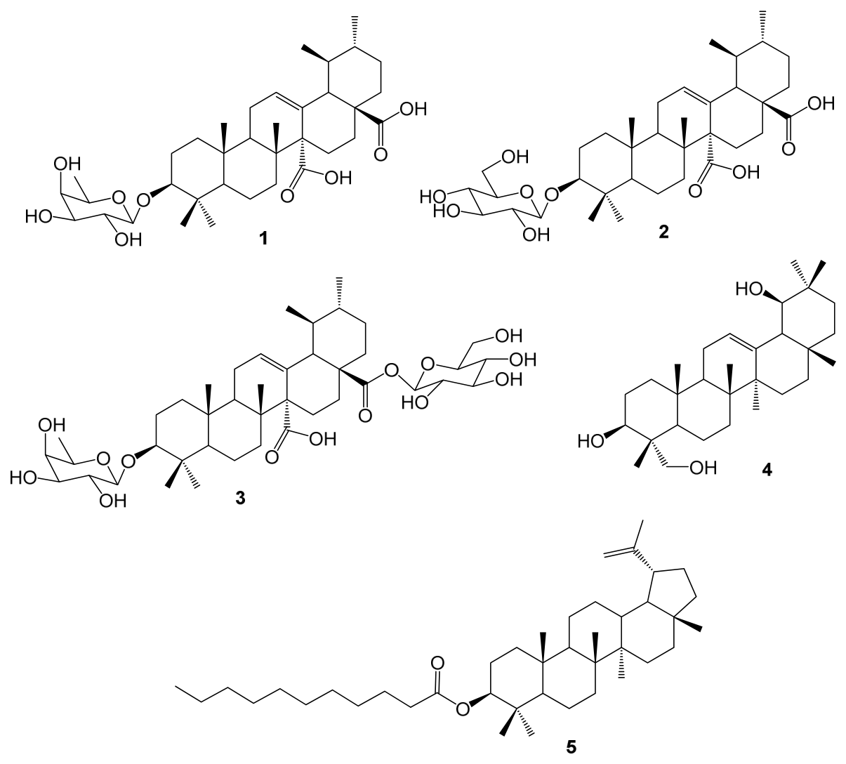
5. Their structures are presented in
Figure 1.
3.2. Antiplasmodial Activity
The EtOAc fraction and the individual compounds quinovic acid 3-
O-
β-
D-fucopyranoside (
1), quinovic acid 3-
O-
β-
D-glucopyranoside (
2), quinovic acid 3-
O-
β-
D-fucopyranosyl-28-
O-
β-
D-glucopyranosyl ester (
3), olean-12-ene-3
β,19
β,24-triol (
4) and lupeol-3-
O-undecanoate (
5) have never been evaluated against
P. falciparum. Therefore, compounds
1–
5 were evaluated in vitro for their antiplasmodial activity against chloroquine-sensitive 3D7 and chloroquine-resistant Dd2
P. falciparum strains. The results are summarized in
Table 1.
3.3. Molecular Docking
To investigate potential protein–ligand interactions associated with the observed antiplasmodial activity, molecular docking simulations were performed against PfLDH and PfPKG, which were selected based on their established roles in parasite energy metabolism and signaling, respectively, for the five isolated phytochemicals (1–5). Two key metabolic enzyme targets from P. falciparum were selected: L-lactate dehydrogenase (PfLDH, PDB ID: 1LDG) and the cGMP-dependent protein kinase N-terminal cGMP-binding domain (PfPKG, PDB ID: 5E16). Artesunate (Art) and chloroquine (CQ) were utilized as standard reference controls.
The reliability and predictive accuracy of the docking protocol were validated by re-docking the co-crystallized NADH ligand of
PfLDH (PDB ID: 1LDG) and the cGMP ligand of the
PfPKG N-terminal domain (PDB ID: 5E16) into their respective experimentally defined binding pockets. The grid box coordinates and dimensions utilized for both receptor targets are summarized in
Table 2. The protocol successfully reproduced the native binding conformations, yielding Root-Mean-Square Deviation (RMSD) values of 0.785 Å for 1LDG and 0.080 Å for 5E16. These values, being well below the acceptable threshold of 2.0 Å, confirmed the robustness of the docking parameters for further virtual screenings. The docking results were considered predictive evidence of potential target interactions with the selected ligands.
The binding affinities calculated by AutoDock Vina for the isolated triterpenoids and standard drugs against both receptor targets are compiled in
Table 3.
Against the PfLDH target (PDB ID: 1LDG), the bidesmosidic saponin, quinovic acid 3β-O-β-D-fucopyranosyl-28-O-β-D-glucopyranosyl ester (3), displayed the strongest binding affinity with a free binding energy of −8.2 kcal/mol, outperforming both artesunate (−6.3 kcal/mol) and chloroquine (−8.0 kcal/mol). Compounds 5 (−7.8 kcal/mol) and 1 (−7.0 kcal/mol) also demonstrated strong binding energy values against this protein target.
Regarding the PfPKG N-terminal cGMP-binding domain (PDB ID: 5E16), artesunate exhibited the lowest binding energy (−7.9 kcal/mol). Among the isolated phytochemicals, the trihydroxylated oleanane aglycone 4 displayed the highest affinity towards this pocket (−6.8 kcal/mol), closely followed by the quinovic acid glycosides 1 (−6.6 kcal/mol) and 3 (−6.5 kcal/mol).
To understand the structural basis of the binding preferences, the specific non-covalent interactions (hydrogen bonding, hydrophobic packing, and pi-mediated interactions) governing the top-scoring complexes were mapped using Discovery Studio (
Table 4).
For the
PfLDH (1LDG) complexes, compound
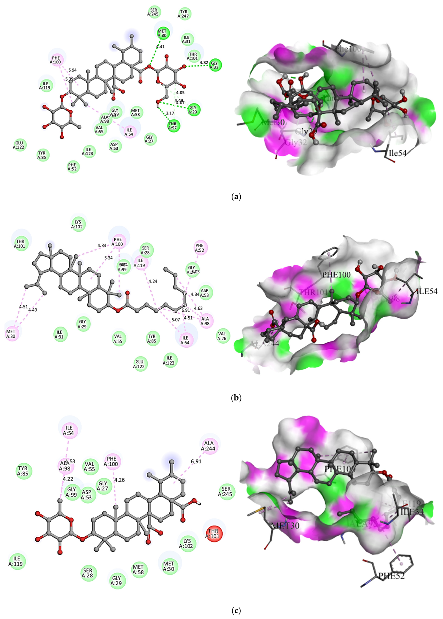
3 established a dense network of four conventional hydrogen bonds involving critical active site residues MET30, GLY32, GLY29, and THR97, along with hydrophobic stabilization driven by ILE54 and PHE100 (
Figure 2a). In contrast, compounds
5 and
1 bound to the
PfLDH pocket exclusively through hydrophobic mechanisms (including pi-alkyl and alkyl interactions) with residues such as PHE100, ILE54, ALA98, and MET30, lacking any conventional hydrogen-bonding stabilization (
Figure 2b,c).
Within the binding pocket of
PfPKG (5E16), compound
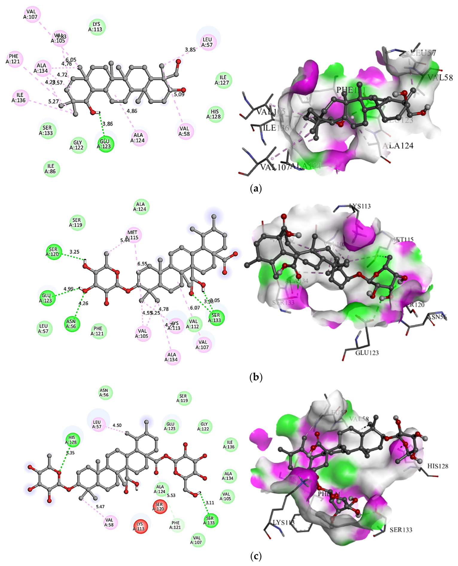
4 formed a key hydrogen bond with GLU123, complemented by an extensive hydrophobic envelope composed of LEU57, VAL58, ALA124, ILE136, ALA134, PHE121, VAL105, and VAL107 (
Figure 3a). Compound
1 established four hydrogen bonds with SER120, GLU123, ASN56, and SER133, stabilized by a hydrophobic network (
Figure 3b). Compound
3 anchored itself to the domain via two hydrogen bonds with HIS128 and SER133, alongside weaker hydrophobic interactions with LEU57 and VAL58 (
Figure 3c).
3.4. ADMET and Drug-likeness Screening
To evaluate the drug-likeness, physicochemical attributes, and full pharmacokinetic behavior of the isolated phytochemicals (1–5), a comprehensive in silico screening was performed using the SwissADME and pkCSM web servers.
The screening revealed that none of the isolated triterpenoids completely adhered to all five criteria, with each molecule exhibiting at least one violation due to the high molecular weights and complex structures characteristic of pentacyclic triterpene scaffolds (
Table 5).
Compounds 1 and 4 each exhibited a single violation: compound 1 exceeded the molecular weight threshold (MW = 632.82 g/mol), while compound 4 exceeded the lipophilicity limit with a calculated LogP (clogP) of 5.58.
Compounds 2 and 5 both triggered two violations. For the monodesmosidic saponin 2, violations were driven by its high molecular weight (648.82 g/mol) and an excessive number of hydrogen bond acceptors (NHA = 10). For the lupane ester 5, violations resulted from its molecular mass (594.99 g/mol) and an extreme lipophilicity profile (clogP = 10.83). The bidesmosidic saponin 3 exhibited three distinct violations, characterized by a high molecular mass (794.97 g/mol), 14 hydrogen bond acceptors, and 8 hydrogen bond donors.
The Absorption, Distribution, Metabolism, and Excretion (ADME) values calculated via the pkCSM predictive server are documented in
Table 6.
Concerning Absorption, clear differences were observed between the glycosylated and aglycone/esterified frameworks. The polyhydroxylated oleanane aglycone 4 and the lupane ester 5 exhibited excellent intestinal tract parameters, displaying high Caco-2 cell permeability (log Papp = 1.32 and 1.29 × 10⁻⁶ cm/s, respectively) and near-complete Human Intestinal Absorption (HIA = 97.23% and 100%). Conversely, the quinovic acid glycosides 1, 2, and 3 showed poor absorption parameters, with HIA values dropping significantly (1: 35.07%; 2: 19.74%; 3: 0%). This low absorption is linked to their low water solubility and large polar molecular surfaces.
The steady-state volume of distribution (VDss) ranged from −0.82 (compound 1) to 0.75 log L/kg (compound 5). The unbound fraction (Fu) in human plasma was zero for the highly lipophilic aglycones 4 and 5, whereas the saponins 1, 2, and 3 maintained free unbound fractions ranging from 0.15 to 0.31. Crucially, none of the five isolates were predicted to cross the Blood–Brain Barrier (log BB < 0.3) or effectively penetrate the Central Nervous System (log PS < −2.0), minimizing the likelihood of neurotoxic side effects.
None of the isolated phytochemicals (1–5) were identified as inhibitors of the major hepatic cytochrome P450 enzymes (CYP1A2, CYP2C19, CYP2C9). Regarding enzymatic substrates, compounds 1 and 5 were predicted to act as substrates for the CYP3A4 isoform, while none of the five molecules interacted with CYP2D6.
Total clearance values varied across a tight range, spanning from −0.007 log mL/min/kg (compound 1) up to 0.20 log mL/min/kg (compound 5). None of the isolated triterpenoids were predicted to act as renal Organic Cation Transporter 2 (OCT2) substrates, indicating standard renal handling paths.
Safety parameters obtained via pkCSM showed promising baseline toxicology targets for the isolates (
Table 6). All five phytochemicals (
1–
5) were predicted to be entirely free of hepatotoxic risks and skin sensitization effects. The maximum tolerated dose in humans was calculated to be highest for compound
1 (0.56 log mg/kg/day) and lowest for the bidesmosidic saponin
3 (−0.44 log mg/kg/day).
3.5. Molecular Dynamics Simulation and MM/PBSA Analysis
To rigorously assess the thermodynamic stability, structural integrity, and conformational behavior of the top-scoring receptor–ligand complexes under physiological conditions, 100 ns explicit solvent molecular dynamics (MD) simulations were executed. Based on the docking results, C3_1LDG was selected because compound 3 showed the most favorable predicted docking score among the isolated compounds against PfLDH, while C4_5E16 was selected because compound 4 showed the most favorable predicted docking score among the isolated compounds against the PfPKG binding pocket. Reference-drug complexes were not subjected to parallel MD simulations; therefore, the MD results are interpreted as characterization of the selected phytochemical complexes rather than as evidence of superior stability relative to the reference drugs.
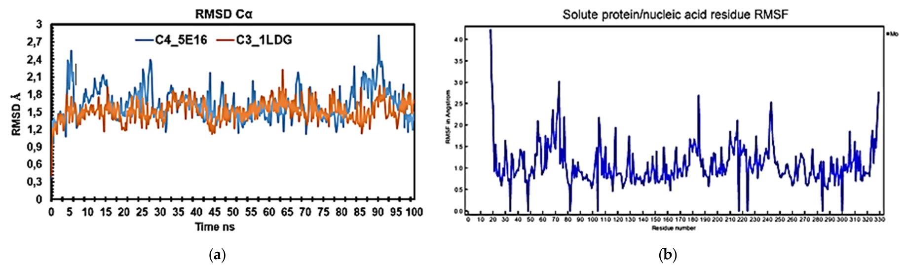
The structural stability of the protein backbones during the 100 ns simulation timeframe was monitored using Root-Mean-Square Deviation (RMSD) calculations. Both complexes achieved steady structural equilibrium within the initial phase of the trajectory and maintained stable, non-fluctuating plateaus until completion. The C3_1LDG complex demonstrated a highly stable binding trajectory with an average backbone RMSD value of 1.514 Å and a maximum recorded deviation of 2.222 Å. In comparison, the C4_5E16 system exhibited slightly higher conformational drift, recording an average backbone RMSD of 1.637 Å and peaking at 2.807 Å (
Figure 4a). The lower deviation profile observed for C3_1LDG indicates a tightly anchored and rigid protein–ligand configuration. To evaluate local macromolecular flexibility, the Root-Mean-Square Fluctuation (RMSF) was mapped across individual amino acid residues (
Figure 4b). Both enzymes retained rigid, stable secondary cores throughout the production run. Fluctuation spikes were predominantly restricted to highly flexible loop architectures and terminal zones. Within the C4_5E16 system, the highest flexibility was captured at residues SER21 (RMSF = 6.63 Å) and LYS155 (RMSF = 5.38 Å). Conversely, the C3_1LDG complex featured notably muted terminal and loop fluctuations, demonstrating that the binding of the bulky saponin ligand
3 imposes a stabilizing structural rigidity onto the
PfLDH global architecture.
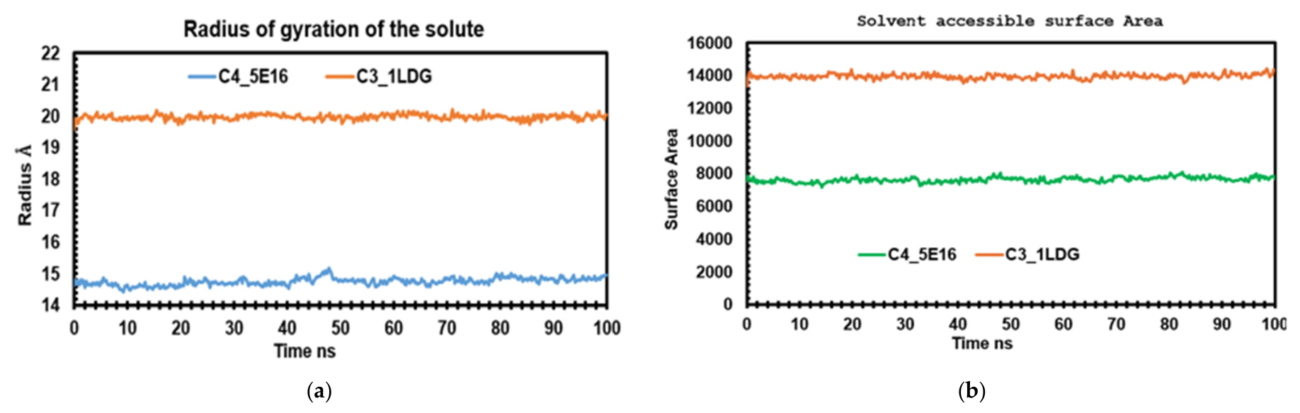
The structural compactness of the complexes was scrutinized via the Radius of Gyration (Rg) parameter over the 100 ns trajectory (
Figure 5a). The C4_5E16 complex maintained a lower average (Rg) value of 14.75 Å, which points to a highly compact, closely folded global architecture. Due to its larger molecular framework, the C3_1LDG complex consistently exhibited a higher average (Rg) value of 19.97 Å. Critically, the steady, flat-line behavior of the (Rg) plots for both systems confirms that no unfolding or significant denaturing events occurred during the 100 ns timescale. This structural equilibrium was further corroborated by the Solvent Accessible Surface Area (SASA) values (
Figure 5b). The average calculated SASA values settled at 13,954 Å
2 for C3_1LDG and 7640 Å
2 for C4_5E16. While the C3_1LDG complex showcased an expectedly larger solvent-exposed surface area due to its expanded structural dimensions, both profiles reached a steady state, confirming that neither pocket underwent major conformational opening or collapse.
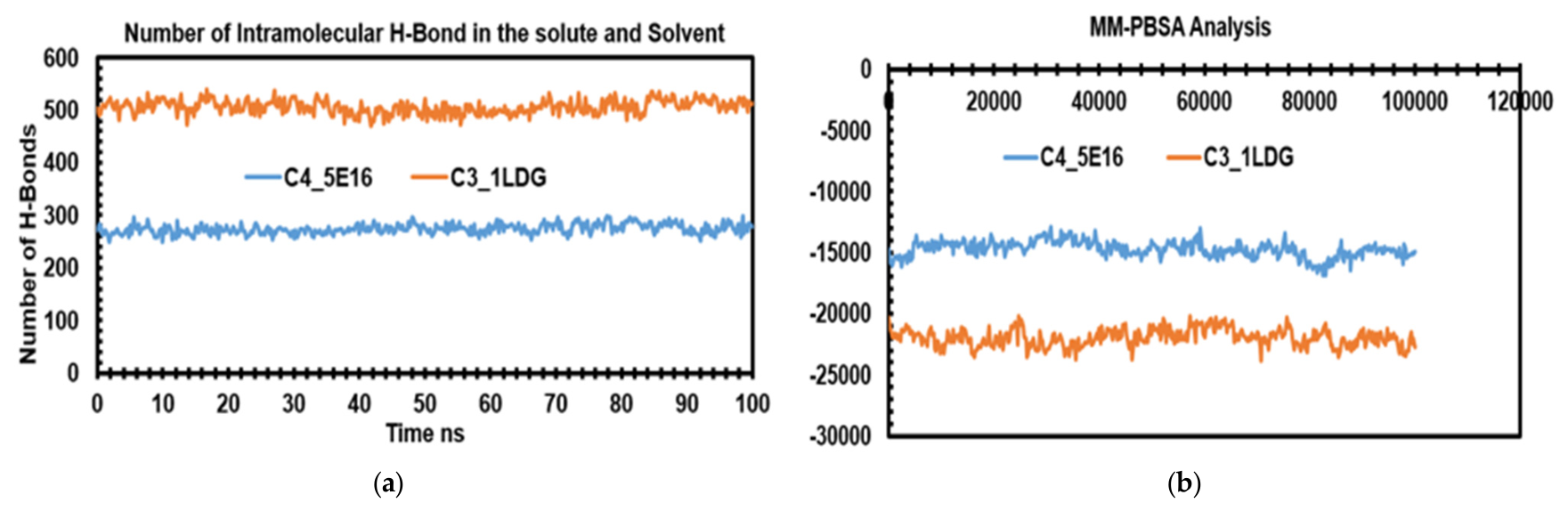
Intermolecular interaction dynamics were detailed via a continuous hydrogen bond count analysis (
Figure 6a). The C3_1LDG complex demonstrated an extraordinarily dense and persistent binding network, maintaining between 13 and 22 simultaneous hydrogen bonds throughout the entire 100 ns production run. In contrast, the C4_5E16 complex maintained a smaller network of 4 to 10 active hydrogen bonds. Furthermore, compound
3 supported a tight web of internal intramolecular hydrogen bonds, adding an extra layer of structural integrity that prevented ligand drifting. To quantify the thermodynamic driving forces behind these associations, MM/PBSA binding free-energy profiles were extracted from the simulation trajectories (
Figure 6b). Consistent with the docking scores and the persistent hydrogen bonding patterns, the C3_1LDG complex demonstrated superior energetic feasibility, maintaining a significantly lower and more favorable binding free energy throughout the trajectory compared to C4_5E16. Collectively, these MD parameters validate that compound
3 establishes a more robust, stable, and energetically prioritized complex with
PfLDH than compound
4 does with
PfPKG, underscoring its therapeutic value as a prominent antiplasmodial lead scaffold.
4. Discussion
4.1. Isolation and Structural Identification
The chromatographic investigation of the ethyl acetate (EtOAc) fraction from Mitragyna inermis led to the successful isolation and structural identification of five major secondary metabolites (1–5). While the genus Mitragyna is extensively recognized for its complex indole alkaloid profile, such as mitragynine or speciophylline, this study underscores the significant presence and diversity of non-alkaloidal constituents, specifically pentacyclic triterpenoids and their glycosylated derivatives, within the species M. inermis.
From a chemotaxonomic perspective, the isolation of compounds
1,
2, and
3 strongly reinforces the status of quinovic acid glycosides as vital taxonomic markers for both the Rubiaceae family and the genus
Mitragyna. The presence of these derivatives bearing specific sugar moieties, such as
D-fucose in quinovic acid 3-
O-
β-
D-fucopyranoside (
1) and
D-glucose in quinovic acid 3-
O-
β-
D-glucopyranoside (
2), aligns precisely with the structural characterizations recently carried out on this genus by Nangmou et al. [
30] and Ouédraogo et al. [
32]. Similarly, the identification of the bidesmosidic saponin
3, elucidated as quinovic acid 3
β-
O-
β-
D-fucopyranosyl-28-
O-
β-
D-glucopyranosyl ester, corroborates foundational literature established by Lamidi et al. [
33] on the complex saponin profiles governing this botanical genus. Typically retrieved in high abundance from the bark of
Mitragyna species, this cluster of triterpenoid glycosides defines a key generic boundary within the family.
In addition to these diagnostic glycosides, the characterization of olean-12-ene-3
β,19
β,24-triol (
4) illustrates the existence of highly driven, specialized oxidation pathways operating in
M. inermis. While rare aglycones of this nature are occasionally mapped across separate families—such as the Leguminosae—their expression here highlights unique enzymatic hydroxylation mechanisms that expand upon the oleanane-type frameworks previously observed in related species by Kouam et al. [
34]. Crucially, while previous literature on the
Mitragyna genus has overwhelmingly focused on profiling its diverse alkaloidal networks, the isolation of compound
4 represents its very first report within the entire genus, revealing an unexpected biosynthetic diversity that distinguishes
M. inermis from closely related species like
M. speciosa.
Finally, the isolation of the long-chain fatty acid ester lupeol-3-
O-undecanoate (
5) provides an original and valuable contribution to mapping the esterification capacity of the plant’s secondary metabolism. Although lupane triterpenes are widely distributed in nature, their long-chain alkanoic acid ester variations are uncommon. The discovery of this specific undecanoate derivative in
M. inermis provides a structural model in full agreement with the structural behaviors described in broader triterpenoid literature by Poumale et al. [
35]. Together, these distinct classes of pentacyclic triterpenoids display an intricate biogenetic intersection of glycosylation, oxidation, and fatty-acid esterification that refines the chemomapping of
M. inermis.
4.2. Antiplasmodial Activity
Malaria remains one of the most devastating infectious diseases in tropical regions, driving cutting-edge research to explore novel molecular scaffolds from plant biodiversity. Among the traditional remedies of West Africa, M. inermis (Willd.) O. Kuntze holds a prominent place in local pharmacopeias, where decoctions of its bark and leaves are widely prescribed to treat fevers and malaria. Although the antimalarial properties of its crude extracts and oxindole alkaloids have been extensively documented, the therapeutic potential of its triterpenoid fractions requires deeper clarification. This study evaluates the in vitro antiplasmodial activity of isolated compounds (1–5) against P. falciparum strains, establishing a scientifically validated link between empirical traditional use and fine chemical composition.
Biological evaluations against chloroquine-sensitive (3D7) and chloroquine-resistant (Dd2) strains of P. falciparum revealed selective inhibition profiles. Although the quinovic acid saponins 1–3 exhibited what is generally classified as moderate overall antiplasmodial activity compared to the highly potent indole alkaloids typically highlighted in Mitragyna literature, they significantly outperformed both the aglycone and the esterified derivatives investigated in this study. While traditional research on Mitragyna species implicitly attributes their antiplasmodial efficacy to alkaloids disrupting the parasite’s food vacuole or DNA replication, the primary therapeutic interest of these three glycosides lies in their proven ability to specifically target P. falciparum lactate dehydrogenase (PfLDH). This targeted enzymatic mechanism highlights quinovic acid scaffolds featuring a C-27 free carboxyl group and optimized glycosylation patterns as superior, valuable templates for antimalarial lead optimization and drug development.
The trihydroxylated oleanane derivative
4 exhibited moderate antiplasmodial potency against the Dd2 strain (IC
50 = 83.69 ± 1.87 μM) and the 3D7 strain (IC
50 = 85.28 ± 2.18 μM), though it remained noticeably less active than the three saponins
1–
3. It is well established that the antiplasmodial performance of polyhydroxylated oleanane triterpenes against
P. falciparum depends heavily on the precise geometric positions, count, and glycosylation status of their hydroxyl groups. Phytochemical investigations on phylogenetically related Combretaceae species support this behavior. For instance, Oluyemi et al. [
36], in their study on
Combretum racemosum, demonstrated that the structural architecture of isolated triterpenes significantly impacts antimalarial profiles, where specific functional distributions modulate lipophilicity and subsequent interaction with parasite targets. Similarly, Baldé et al. [
37] reported that polyhydroxylated triterpenes such as arjunolic acid and arjungenin isolated from the roots of
Terminalia albida display variable in vitro activities against
P. falciparum, highlighting how localized changes in polyhydroxylation and esterification dictate overall potency. For compound
4, which bears its three hydroxyl groups at the 3
β, 19
β, and 24 positions, this specific spatial arrangement and localized polarity distribution may restrict optimal binding interactions with critical parasite structures or cellular membranes. This conformational restriction, paired with the absence of a sugar moiety capable of optimizing amphiphilicity and cellular uptake, clarifies why the free aglycone compound
4 displays significantly limited potency compared to its saponin counterparts.
The total lack of antiplasmodial activity observed for the lupane ester
5 (IC
50 > 200 μM) aligns precisely with established SAR models for lupane-type triterpenoids against 3D7 and Dd2 strains. Previous SAR studies by Fotie et al. [
38] and Bringmann et al. [
39] demonstrate that aliphatic acyl chains spanning between 15 and 22 carbon atoms (C
15–C
22) are essential to maintain antiplasmodial potency. Consequently, the undecyl chain (C
11) located at the C-3 position of compound
5 is too short to establish and maintain optimal hydrophobic interactions within the binding pocket of the parasite target.
Furthermore, the intrinsic pharmacokinetic parameters of the molecule exacerbate this lack of efficacy: the extreme lipophilicity of compound 5, indicated by a calculated log P greater than 10 (clogP > 10), drastically limits its aqueous solubility. This poor solubility reduces its effective concentration at the parasite’s active site during in vitro testing.
Taken together, these pharmacological findings confirm that quinovic acid glycosides (1–3) represent superior structural scaffolds over 3-O-acyl-lupanes (5) and polyhydroxylated oleanane aglycones (4) for antimalarial drug discovery.
It is essential to note that the antimalarial efficacy of compounds 1–4 (IC50 values ranging from 23.76 to 39.12 µg/mL) is significantly lower than that of clinical reference drugs like artemisinins and Chloroquine, which operate at low µM or nM thresholds. This pronounced difference is an expected phenomenon in natural product research. Commercial antimalarial drugs are highly optimized, low-molecular-weight molecules specifically evolved or engineered to target key parasite biological systems with extreme affinity (such as artemisinin-induced radical alkylation or chloroquine-mediated hemozoin inhibition). Conversely, the isolated pentacyclic triterpenoids are bulky molecular frameworks facing steric and lipophilic boundaries that naturally limit rapid accumulation and target disruption within the parasite.
However, when compared to structurally related natural products rather than optimized clinical drugs, the biological performance of the M. inermis isolates aligns perfectly with established standards for triterpene scaffolds. For instance, quinovic acid glycosides purified from
Mitragyna stipulosa [
40] or saponin-rich clusters from
Uncaria tomentosa [
41] routinely yield moderate growth inhibition profiles precisely within the 10–50 µg/mL range. Similarly, the series of polyhydroxylated oleanane-type triterpenes described by Baldé et al. [
37] from the roots of
Terminalia albida—such as arjunolic acid and arjungenin—display comparable in vitro antiplasmodial profiles, with these structural analogs exhibiting moderate activities against
P. falciparum in the micromolar range (IC
50 values between 5 and 15 µM). These cross-literature comparisons confirm that while compounds
1–
4 do not act as immediate therapeutic alternatives, they represent valid, non-alkaloidal chemical scaffolds that establish a solid baseline for future target optimization.
These insights highlight the importance of investigating synergistic interactions within traditional remedies derived from M. inermis. The natural co-occurrence of these distinct secondary metabolite families within the plant may expand or potentiate the overall therapeutic efficacy observed in traditional clinical practices.
4.3. Molecular Docking
Among the docked compounds,
3 displayed the highest binding affinity toward 1LDG (−8.2 kcal/mol), followed by
5 (−7.8 kcal/mol) and
1 (−7.0 kcal/mol). For the 5E16 receptor,
4 exhibited the best binding energy (−6.8 kcal/mol) among the phytocompounds, which was comparable to chloroquine (−5.7 kcal/mol) but slightly lower than artesunate (−7.9 kcal/mol). These results (
Table 3) indicate that several of the selected phytochemicals showed predicted docking scores within a similar range to the reference compounds under the applied docking conditions.
Comprehensive post-docking analysis revealed that all selected phytochemicals established strong and specific interactions with key amino acid residues in the active sites of both receptors. The nature of hydrogen bonding and hydrophobic contacts for each ligand–receptor complex is summarized in
Table 4, while the detailed 2D and 3D visualizations are illustrated in
Figure 2a–c (1LDG complexes) and
Figure 3a–c (5E16 complexes).
In the 1LDG system, compound 3 formed hydrogen bonds with MET30, GLY29, GLY32, and THR97, accompanied by hydrophobic interactions with ILE54 and PHE100. Compound 5 showed strong hydrophobic interactions involving PHE100, ILE199, PHE52, ALA98, ILE54, and MET30, while compound 1 exhibited binding interactions with ILE54, ALA98, PHE100, and ALA244. In the case of 5E16, compound 4 formed a hydrogen bond with GLU123 and hydrophobic interactions with LEU57, VAL58, ALA124, ILE136, ALA134, PHE121, VAL105, and VAL107. Compound 1 established multiple hydrogen bonds with SER120, GLU123, ASN56, and SER133, while forming hydrophobic interactions with MET115, VAL105, LYS113, VAL107, and ALA134. Compound 3 interacted with HIS128 and SER133 through hydrogen bonding and with LEU57 and VAL58 via hydrophobic forces.
Surface representation of the docked complexes revealed that the ligands were well-embedded within the binding pockets of both receptors, forming a stable network of polar and non-polar interactions, which contributes to their overall binding stability.
By specifically targeting lactate dehydrogenase and cGMP-dependent protein kinase, which are involved in parasite energy metabolism and signaling pathways, respectively, this approach offers a reliable strategy for the identification of natural products with therapeutic relevance. While computational and molecular docking studies on the genus Mitragyna have heavily focused on exploring the interaction of major indole alkaloids (such as mitragynine and its derivatives) with human neuroreceptors or cancer targets, their virtual screening against essential P. falciparum metabolic enzymes has remained non-existent.
Pure quinovic acid glycosides have never been evaluated against
P. falciparum strains using such an integrated structural biology viewpoint. Despite the reported antiplasmodial activity of quinovic acid saponin-rich extracts from
Uncaria tomentosa against
P. falciparum 3D7, the individual constituents responsible for this activity remain unidentified [
41]. However, structure-activity studies on quinovic acid derivatives from
M. stipulosa indicate that glycosylation at C-3 modulates cytotoxicity, with the nature and substitution of the sugar moiety influencing both potency and selectivity [
40]. Given that
PfLDH and
PfPKG are essential for parasite survival and possess well-defined hydrophobic and polar binding pockets [
42,
43], it is plausible that the triterpenoid aglycone can occupy the hydrophobic core while the sugar residues engage in hydrogen bonding with surface residues. Therefore, a preliminary structure–activity analysis of the quinovic acid glycosides, combining molecular docking, molecular dynamics and in vitro testing against 3D7 and Dd2 strains, successfully establishes a novel mechanical framework. Unlike traditional
Mitragyna research that focuses on phenotypic growth inhibition, our data explicitly links specific triterpenoid glycosylation patterns to the stabilization of key parasite life-cycle enzymes, generating concrete hypotheses for future optimization.
4.4. ADMET and Drug-likeness
The clinical development of promising antimalarial scaffolds frequently fails due to poor biopharmaceutical and pharmacokinetic properties rather than a lack of intrinsic potency. In this study, the in silico profiling of compounds 1–5 using SwissADME and pkCSM reveals a stark dichotomy between structural lipophilicity, membrane permeability, and experimental antiplasmodial efficacy.
Unlike the well-established, orally compliant clinical profiles of reference drugs such as Chloroquine and Artemisinin, which exhibit low molecular weights, lack Lipinski violations, and display excellent human intestinal absorption (HIA > 89%), the isolated active quinovic acid glycosides (compounds 1–3) present distinct biopharmaceutical profiles. Saponins 1 and 2 violate the molecular weight ceiling (>500 g/mol), while the bidesmosidic saponin 3 triggers three major violations due to its elevated mass (794.97 g/mol) and extensive polar surface area (14 hydrogen bond acceptors and 8 donors).
These structural properties directly dictate their absorption profiles. Compounds 1 and 2 exhibit low human intestinal absorption (HIA = 35.07% and 19.74%, respectively), which drops to 0% for compound 3. The predicted HIA of 0% for compound 3 indicates a potentially substantial limitation to oral absorption, which may be associated with its large molecular size and highly polar glycosylated structure, which limit passive transcellular diffusion through intestinal epithelial barriers, as evidenced by its negative Caco-2 permeability values.
Interestingly, despite its predicted null absorption, compound 3 exhibited strong in vitro antiplasmodial potency. This indicates that while the glycosyl chains are essential to target and anchor the molecule into the PfLDH active site via robust hydrogen bonds (MET30, GLY32, GLY29, THR97), they present a significant challenge for oral formulation. To improve the drug-like properties of these quinovic scaffolds, strategies such as prodrug synthesis, nano-encapsulation, or microemulsion delivery systems may warrant investigation to address the predicted absorption limitation.
This clear dichotomy between high in vitro potency and poor oral biopharmaceutical traits highlights a stark pharmacological divergence from the well-documented alkaloids of the
Mitragyna genus. For instance, the pharmacokinetic profile of mitragynine (the principal alkaloid of
M. speciosa) is characterized by intermediate lipophilicity, strong intestinal absorption, and a propensity to cross lipid barriers effortlessly. However, these favorable parameters are frequently offset by significant CYP enzyme inhibition, metabolic liabilities, and safety alerts [
44,
45,
46]. Conversely, the quinovic acid glycosides (
1–
3) investigated here exhibit a safer baseline systemic safety profile with no predicted hepatotoxicity, shifting the lead optimization challenge from toxicological remediation to formulation engineering (such as nano-encapsulation).
In sharp contrast to the saponins, the polyhydroxylated oleanane aglycone 4 displays an excellent absorption profile, with an HIA of 97.23% and a high Caco-2 cell permeability (log Papp = 1.32 10−6 cm/s). It also strictly complies with Lipinski’s core parameters, except for a marginal overstep in lipophilicity (clogP = 5.58).
However, this high pharmacokinetic availability did not translate into superior in vitro antiplasmodial activity (IC
50 ≈ 39 μg/mL) against both strains. This discrepancy underlines that favorable membrane permeability can maximize the effective intracellular concentration of a drug inside the erythrocyte, but it cannot override unfavorable target-site interactions. As supported by the structural and functional observations of Oluyemi et al. [
36], on antimalarial triterpenoids, the spatial distribution of functional groups and overall oxygenation patterns significantly dictate the antiplasmodial profile and binding potential of pentacyclic triterpenes. The 3
β, 19
β, and 24-trihydroxylation layout of compound
4 presents a distinct architecture that likely lacks the specific spatial orientation necessary for high-affinity interactions with critical parasite targets. Consequently, despite its optimal cellular accumulation, compound
4 fails to establish effective molecular interactions within the parasite’s metabolic pockets or essential subcellular structures, resulting in a plateaued, moderate antiplasmodial response.
The lupane-type ester 5 represents the opposite biopharmaceutical extreme. While it displays a theoretical HIA of 100% and high Caco-2 permeability (log Papp = 1.29), it was completely inactive against P. falciparum (IC50 > 50 μg/mL).
This complete loss of efficacy is perfectly rationalized by its critical physicochemical violations: a molecular weight of 594.99 g/mol and an extreme, out-of-range calculated lipophilicity (clogP = 10.83). This massive lipophilic profile induces a very low water solubility (log mol/L = −4.87), meaning that compound
5 is highly prone to precipitating out of aqueous testing buffers or becoming trapped within non-specific lipid bilayers during in vitro testing. Combined with a short undecyl chain (C
11) that is structurally insufficient to reach the hydrophobic core of the parasite’s active pockets [
38], the high precipitation rate and poor thermodynamic solubility drastically restrict its effective concentration at the target site, rendering it therapeutically inert.
Despite the absorption and solubility challenges identified among specific structural subsets, the overall toxicological and metabolic screening of the five isolates (1–5) provides preliminary computational indicators that may support further hit-to-lead evaluation. Notably, all five isolated phytochemicals were completely free of predicted hepatotoxicity and skin sensitization metrics, highlighting a safer systemic baseline than traditional quinoline standards or artesunate, which frequently trigger hepatotoxic alerts in silico.
Furthermore, none of the molecules acted as inhibitors of key cytochrome P450 metabolizing enzymes (CYP1A2, CYP2C19, CYP2C9). The predicted absence of inhibition of these CYP enzymes may indicate a lower potential for CYP-mediated drug–drug interactions; however, this requires experimental validation. Finally, the negative predictions for Blood–Brain Barrier (BBB) and Central Nervous System (CNS) permeability across all five isolates suggest that these triterpenoids are not predicted to readily penetrate the CNS, steering their pharmacological potential safely away from the central nervous system.
4.5. Molecular Dynamics (MD) Simulations and MM/PBSA Energy Analysis
To understand the macro-temporal behavior of the isolated leads within the biological targets, the structural snapshots provided by molecular docking were extended into 100 ns explicit solvent molecular dynamics (MD) simulations. Static docking scoring functions frequently overlook pocket adaptability and solvent-induced structural strain. Therefore, tracking parameters like RMSD, RMSF, hydrogen-bonding networks, and trajectory-based MM/PBSA binding free energies was used to assess whether the predicted protein–ligand complexes could maintain stable interactions under the simulation conditions.
The 100 ns trajectory for the C3_1LDG complex underscores the exceptional structural stability of the quinovic acid bidesmosidic framework within the PfLDH catalytic site. Achieving a low and tight equilibrium plateau with an average backbone RMSD of 1.514 Å, the complex demonstrates that the ligand remains locked in its primary orientation without significant spatial drift.
This macro-structural stiffness is directly driven by the exceptionally dense intermolecular hydrogen-bonding network recorded over the production run, consistently fluctuating between 13 and 22 active hydrogen bonds. The polar groups of the D-fucopyranosyl and D-glucopyranosyl units establish a comprehensive anchoring web with the active site residues (MET30, GLY32, GLY29, THR97), maintaining the binding pose over time.
Furthermore, the structural rigidity induced by compound 3 is mirrored in the muted RMSF profile of the PfLDH backbone. Triterpenoid saponins often undergo conformational changes due to their flexible sugar moieties. However, compound 3 features an extensive network of internal, intramolecular hydrogen bonds that rigidify its own structure, preventing the ligand drift typically caused by solvent collisions.
This stability is fully supported by the flat-line behavior of the Radius of Gyration (Rg = 19.97 Å) and SASA profiles. Although the bulky architecture of the bidesmoside results in an expectedly larger solvent-exposed surface area compared to smaller aglycones, the system maintains steady conformational equilibrium without causing pocket opening or local denaturation. The MM/PBSA binding free-energy landscape (
Figure 6b) confirms that these cooperative interactions translate into a highly favorable, deep thermodynamic well, supporting the computational stability of the C3_1LDG protein–ligand complex under the simulation conditions.
The dynamic profiling of the C4_5E16 complex reveals a contrasting biophysical mechanism. The PfPKG domain bound to the trihydroxylated oleanane aglycone 4 exhibits a lower average Radius of Gyration (Rg) = 14.75 Å) and a smaller solvent-accessible profile (SASA = 7640 Å2), confirming that the enzyme retains a highly compact, closely folded global architecture when accommodating this smaller, non-sugar triterpene.
However, this structural compactness does not equal superior binding affinity. The C4_5E16 trajectory exhibits a higher average backbone RMSD (1.637 Å) and reaches a larger maximum drift (2.807 Å), indicating a higher degree of structural adjustment within the binding pocket. This higher fluctuation is tied to its smaller hydrogen-bonding profile, which maintains only 4 to 10 active intermolecular interactions.
While the single hydrogen bond formed with GLU123 remains relatively stable, the binding of compound 4 is heavily dependent on hydrophobic packing across a lipophilic envelope (LEU57, VAL58, ALA124, ILE136). Without an extensive polar network to rigidify the site, local residue flexibilities are significantly pronounced. This is evidenced by high RMSF spikes at terminal loops, notably at SER21 (6.63 Å) and LYS155 (5.38 Å).
The lack of specialized polar anchors at the 19β and 24 positions of this specific oleanane aglycone allows for continuous structural adjustment within the PfPKG pocket. This less favorable binding mode is highlighted by its higher, less stable MM/PBSA free-energy profile compared to the saponin target. This dynamic behavior explains why, despite its excellent intestinal absorption and cell permeability, compound 4 yields only a moderate in vitro antiplasmodial response.
When correlated with the experimental in vitro assays, these computational observations clarify the multi-target potential of the M. inermis triterpenoid profile. Saponin 3 acts as a high-affinity, structurally stable anchor against PfLDH, while the polyhydroxylated aglycone 4 operates through a more flexible, membrane-permeable mechanism that targets alternative pathways like PfPKG.
These insights provide a clear strategy for antimalarial lead optimization. To enhance the drug-likeness of the quinovic acid framework (3), structural modifications should aim to reduce molecular weight and polar surface area while preserving the core hydrogen bond acceptors that bind to the MET30 and THR97 residues of PfLDH. Conversely, for the polyhydroxylated oleanane aglycone (4), structural optimization should focus on introducing targeted polar groups at positions identified by SAR models—such as shifting modifications toward the 16β or 22α positions—to mimic the stable hydrogen-bonding networks observed in high-affinity saponin-receptor systems.




 ,
,