Article

Ocular Irritation Potential and Cytotoxicity of Selected Surfactants and Cosurfactants: Identifying Suitable Concentrations for Topical Ophthalmic Formulations


by
Zinah K. Al-Qaysi
1,2,
Ali A. Al-Kinani
2,3 and
Raid G. Alany
2,4,*
1
College of Pharmacy, Imam Ja’afar Al-Sadiq University, Baghdad 10064, Iraq
2
Department of Pharmacy, Kingston University London, London KT1 2EE, UK
3
College of Pharmacy, University of Warith Al-Anbiyaa, Karbala 56001, Iraq
4
School of Pharmacy, The University of Auckland, Auckland 1011, New Zealand
*
Author to whom correspondence should be addressed.
Sci. Pharm. 2026, 94(2), 46; https://doi.org/10.3390/scipharm94020046
Submission received: 16 March 2026 / Revised: 19 May 2026 / Accepted: 21 May 2026 / Published: 5 June 2026
(This article belongs to the Special Issue Innovative Perspectives in Ocular Drug Research)

Abstract

The cornea and conjunctiva are particularly susceptible to injury and adverse effects, either induced by topically applied drugs or excipients used in ophthalmic formulations. Surfactants and cosurfactants are important for producing topical eye formulations of poorly water-soluble drugs, yet they have not been always used in concentrations that are nontoxic and non-irritating to the ocular surface. This study systematically compared the cytotoxicity and ocular irritation potential of commonly used ophthalmic surfactants and cosurfactants under standardized experimental conditions using complementary in vitro and ex vivo ocular safety models. The ocular irritation of Tween 80, Cremophor EL, polyethylene glycol 400 (PEG 400) and propylene glycol (PG) was examined using the HET-CAM (conjunctival) and BCOP (corneal) eye assays. The toxic effect of the four excipients after 24 h on HLE-B3 cell growth was investigated and found to be dose-dependent. The highest tolerable concentrations of Tween 80 and Cremophor EL were 0.25% (w/w), whereas PEG 400 and PG were non-toxic at 5% (w/w). Tween 80 and Cremophor EL at 0.25% (w/w) and PEG 400 and PG at 5% (w/w) were all devoid of conjunctival and corneal irritation. This study systematically compared the cytotoxicity and ocular irritation potential of commonly used ophthalmic surfactants and cosurfactants under standardized experimental conditions using complementary in vitro and ex vivo ocular safety models. Interestingly, there is strong agreement between the results obtained using the HET-CAM and BCOP assays, where both have been successfully used to evaluate the potential for ocular irritation caused by the aforementioned excipients.

1. Introduction

The use of topical ocular dosage forms is associated with adverse effects due to either the loaded drugs or the excipients used. The toxicity and adverse effects of topically applied ophthalmic drugs and excipients are mainly attributed to their effects on the conjunctiva, cornea, iris and beyond [1,2]. For example, topical prostaglandins are typically associated with iris hyperpigmentation and eyelash trichomegaly [3,4]. Corticosteroid eye drops can hinder the healing of corneal lesions, increase the risk of infection, and lead to cataract and glaucoma [5,6]. Voltaren®, a topical nonsteroidal anti-inflammatory eye drop containing diclofenac, has been associated with corneal melting, leading to regulatory restrictions [7,8]. Certain excipients can impact epithelial surface integrity, causing irritation, burning, stinging, excessive tear production, and cytotoxic adverse effects [7]. Certain substances may disturb the precorneal tear film, causing damage to the epithelial surface, leading to dry-eye symptoms. It has been shown that the combination of hydroxyethyl cellulose with benzalkonium chloride may cause ocular epithelial damage [9]. Therefore, it is necessary to assess the possible toxicity and ocular irritation of the ingredients utilized in ophthalmic preparations [10,11].
Surfactants are widely used as pharmaceutical excipients to improve ocular bioavailability by increasing solubility, precorneal retention, and permeability [12,13]. Surfactants affect the barrier properties of the corneal epithelium by perturbing the packing of the comprising lipid bilayer phospholipids and/or opening tight junctions. Non-ionic and Zwitterionic surfactants are preferable (over cationic and anionic surfactants) for ocular use due to their biocompatibility, along with their inherent drug solubilizing and formulation stabilization effects [14,15]. Non-ionic surfactants like Polysorbates, Brij and Cremophor EL and RH are widely used to formulate micelles, emulsions [16], suspensions [17], liposomes [18,19], and niosomes [20], amongst other things.
The physicochemical properties of surfactants and cosurfactants (weakly amphiphilic molecules that are added to a primary surfactant formulation to enhance performance), such as molecular weight, critical micelle concentration (CMC), lipophilicity (log P), hydrophile lipophile balance value (HLB) and dielectric constant (DEC), are important in ocular formulation development. Usually, an increased carbon chain length results in higher log P values, lower DEC, and larger molecular weight (Mwt) [21]. The solubility of a polyethoxylated non-ionic surfactant in water depends on its alkyl chain length and the number of ethylene oxide units in the molecule. For example, surfactants with 12 carbon atoms and over five ethylene oxide units are typically soluble in water at room temperature [22]. According to the HLB system, the higher the percentage weight of polyethylene oxide in a surfactant (i.e., a higher HLB value), the higher the aqueous solution solubility (Table 1) [23]. Surfactant micelles are formed by interactions between hydrophilic and hydrophobic segments and the surrounding medium [24]. Another important characteristic is log P, which measures the lipophilicity of a cosurfactant’s unionized portion at a certain pH range and temperature.
Polysorbate 80 (Tween 80) is a hydrophilic non-ionic surfactant produced by combining oleic acid with polyethoxylated sorbitan [25,26]. The HLB values of Tween 80 and Cremophor EL are 15 and 13, respectively, indicating that they are both hydrophilic in nature. Tween 80 is a common carrier for fat-soluble compounds, pharmaceuticals, cosmetics, and food; it increases solubility and drug absorption [25,27,28,29]. Tween 80 is used in many commercial products as it is non-toxic, biocompatible, cheap, and environmentally friendly. Some of these products are Refresh Endura®, Restasis®, Blephamide®, Viva Ultra Tears®, Durezol®, and Refresh Dry Eye Therapy® [30,31].
Cremophor® EL (polyoxyl 35 castor oil) is a non-ionic surfactant widely employed to solubilize poorly water-soluble drugs. Its use in parenteral formulations such as cyclosporine in Sandimmune® IV, Paclitaxel in Taxol®, and Teniposide in Vumon® (Table 1) highlights its strong solubilizing capacity. Although these products are administered intravenously, the underlying formulation challenges are similar to those encountered with topical ocular delivery, where highly lipophilic APIs benefit from using surfactants to achieve therapeutically relevant concentrations. Notably, cyclosporine is also marketed as eye drop formulations (Restasis® and Ikervis®), where surfactants are used as solubilizers and stabilizers, emphasizing the relevance of evaluating Cremophor EL for ocular tolerance and safety [32].
Cosurfactants are mainly short-chain alcohols often utilized in pharmaceutical formulations to lower surface tension and improve thermodynamic stability. Generally, the chain lengths of cosurfactants vary from C-4 to C-10. Additionally, they include alkane-diols, such as 1,2-propanediol (propylene glycol, PG) and 1,2-butanediol, and alkane–polyols, such as glycerol, glucitol, and low-molecular-weight polyethylene glycol [14,33,34]. Cosurfactants can alter the curvature of the interface due to the virtual significance of their polar groups [35,36]. Cosurfactants like propylene glycol (PG) and polyethylene glycol 400 (PEG 400) are often used in ophthalmic formulations. The physicochemical properties of the investigated surfactants and cosurfactants are shown in Table 1.
Table 1. Physicochemical properties of selected surfactants and cosurfactants and marketed products.
Excipient
Surfactant/Cosurfactant
Molecular FormulaMwt
(g/mol)
Reported
HLB
log PDECMarketed Product
(Ophthalmic and
Intravenous IV)
References
Tween 80C64H124O26131015−4.3928.27Refresh endura®; Restasis®[21,37]
Blephmide®; Viva ultra tears®
Cremophor ELC57H104O9
(CH2 CH2O)n
Where n ≈ 35
1630134.29730.43Taxol IV®, Sandimmune IV®
Vumon IV®
[21,38]
Propylene glycolC3H8O276.19−1.3432Systane Ultra®[21,38]
Systane Balance®
Polyethylene glycol 400C18H38O1040020−4.830.2Systane Altra®[21,38]
Systane Alt®
Systane Ultra®
Systane Balance®
The hydrophile lipophile balance value (HLB), log P; logarithm of partition coefficient in octanol/water, DEC; dielectric constant, Mwt; molecular weight.
PEG 400 is a multifunctional component that may act as an emollient, lubricant, solvent, and emulsifier in personal care products. It acts as a cosurfactant, thickener, opacifier, and humectant. It is often found in shower and bath goods, creams and lotions, shampoos, shaving creams, and liquid soaps [39]. PG is an emollient and emulsifier in cosmetics, pharmaceuticals, and food. As PG becomes more prevalent, it has been linked to allergic contact dermatitis and systemic cutaneous responses [40,41]. As a topical over-the-counter therapy for dry eye, Systane Gel Drops (Alcon Laboratories Inc., Fort Worth, TX, USA) are formulated using propylene glycol (0.3%) and PEG 400 (0.4%) [42,43]. Systane offers a range of topical ocular products for dry-eye patients [42,43,44,45,46]. Normally, the eye’s surface is lubricated and protected by all these products, which enables epithelial cells to heal and replace themselves [44,45,47,48,49]. Although PEG is typically regarded as non-toxic, researchers have identified certain harmful issues with PEG with low molecular weights [50]. Smyth et al. found chronic oral toxicity in PEG oligomer (Mn ~ 200) in rats and monkeys, raising concerns about the safety of these seemingly innocuous compounds [28,29,30].
To reduce the chances of conjunctival and corneal toxicity, the lowest effective amounts of surfactants and cosurfactants must be used [20]. Hence, the aim of this research project was to investigate the cytotoxicity of Tween 80, Cremophor EL, PEG 400, and PG on the Human Lens Epithelial Cell Line (HLEC-B3) using the neutral red uptake assay and to establish their conjunctival and corneal irritation potential using the HET-CAM and BCOP assays. Although these excipients have previously been evaluated individually, direct comparison across multiple ocular safety models under standardized experimental conditions remains limited.

2. Materials and Methods

2.1. Materials

Human Lens Epithelial Cells (HLEC-B3), heat-inactivated fetal bovine serum (FBS), and Eagle’s Minimum Essential Medium (EMEM) were acquired from LGC (London, UK). Dulbecco’s Modified Eagle Medium (DMEM), penicillin–streptomycin (10,000 U/mL), neutral red, and 0.25% trypsin–EDTA were procured from Sigma-Aldrich (Dorset, UK). Industrial Methylated Spirits (IMS), acetic acid, and 100% ethanol were procured from VWR (Leicestershire, UK). Phosphate-buffered saline and sterile phosphate-buffered saline (DPBS) devoid of calcium and magnesium were procured from Fisher Scientific (Loughborough, UK). Hydrogen peroxide solution (30%), monochlorobimane (MCB), and 2′,7′-dichlorofluorescein diacetate (DCFH-DA) were procured from Sigma-Aldrich (Dorset, UK). Freshly excised bovine eyes were procured from a local source (ABP Food Group, Guildford, UK). Sodium hydroxide was acquired from Fisher Scientific (Loughborough, UK). Fluorescein sodium salt and sodium chloride were acquired from Sigma-Aldrich (Dorset, UK). Fertilized white leghorn eggs were obtained from Med Eggs (Norfolk, UK). Industrial methylated spirits (IMS) and acetone were bought from VWR (Leicestershire, UK). Propylene glycols were purchased from Sigma-Aldrich (Dorset, UK). Tween 80 and polyethylene glycol 400 were obtained from ACROS Organics (Fair Lawn, NJ, USA). Cremophor EL 40 was purchased from Sigma-Aldrich (Taufkirchen, Germany).

2.2. Human Lens Epithelial Cell Culture

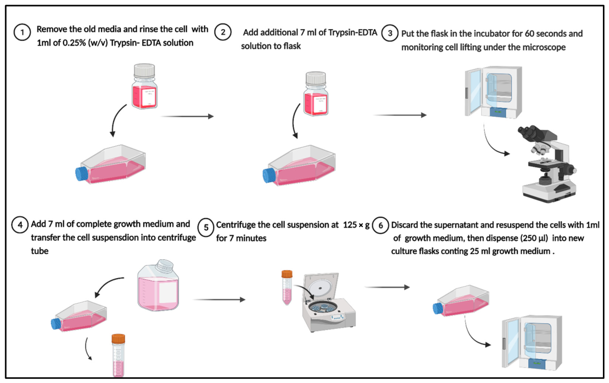
A cryotube containing HLEC-B3 was incubated in a water bath at 37 °C for 1–2 min to reduce contamination. The tubes were then decontaminated with 70% ethanol and transferred to a cell culture flask containing preheated EMEM supplemented with 20% (v/v) FBS and 1% (v/v) penicillin–streptomycin. The flask was maintained at 37 °C with a 5% CO2 and 95% air environment in a Heracell 150i humidified incubator for 48 h. After reaching 70–80% confluence, the flask was removed, the growth media were removed, and the flask was rinsed with 0.25% trypsin–EDTA. The cells were lifted and incubated for 60 s at room temperature to achieve detachment. Fresh, complete EMEM was added, and the flask was gently moved to break down cell aggregation. The solution was centrifuged at 125 g for 5 min, and the supernatant was removed. The resulting cell suspension was mixed with 1 mL of complete medium. The number of cells in the suspension was quantified using a hemacytometer, where 20 µL from the cell suspension was gently mixed with 20 µL trypan blue (Figure 1). The resulting solution was transferred to fast-read counting slides to count the cells using the following equations [51]:
Number of the cell/mL = Cells count average × Sample dilution × 104
C e l l   s u s p e n s i o n   v o l u m e   r e q u i r e d = v o l u m e   n e e d e d   f o r   p l a t e   ×   s e e d i n g   d e n s i t y   ×   1000 v o l u m e   n e e d   p e r   w e l l   ( µ L ) N u m b e r   o f   c e l l s   i n   1   m L   n e e d e d

2.3. Neutral Red Uptake (NRU) Assay

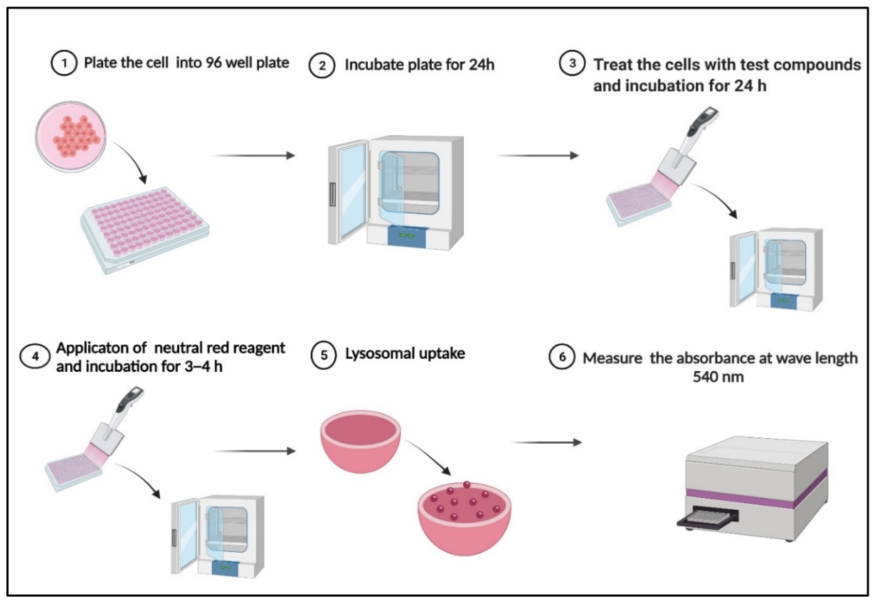
To evaluate the cell cytotoxicity impact of the excipients, the standard method for the NRU test was used [52]. On the first day, a 96-well plate with 200 µL of medium was used to seed HLEC-B3 at a density of around 8000 cells/well. The cells were placed in a Heracell 150i humidified incubator (Thermo Fisher, Loughborough, UK) and kept at 37 °C with 5% CO2 and 95% air, using the growth complete medium. After monitoring cell development using a phase-contrast inverted microscope for two days, the growth medium was removed, and the medication was added. Treatment medium (DMEM + 5% FBS + 1% penicillin–streptomycin) was used to suspend excipients. The cells were then incubated at 37 °C with 5% CO2 and 95% air in a Heracell 150i humidified incubator for 24 h to test the excipient’s impact on the cells. On the third day, a multichannel pipette was used to remove the treatment media from the plates and add 100 µL of neutral red medium (40 µg/mL) to each well. The plate was then put in a Heracell 150i humidified incubator set to 37 °C, 5% CO2, and 95% air for 4 h. Gently tapping the cells to remove any remaining solution was followed by washing them with 150 µL of PBS (pH 7.4) after incubation. The neutral red was then removed. Next, add 150 µL of neutral red destain solution (50% ethanol, 1% acetic acid) in each well. The plate was then vigorously agitated with a microtiter plate shaker for 10 min, or until the neutral red had been removed from the cells and formed a homogenous solution. An Infinite 200 pro plate reader (Tecan, Switzerland) with the Magellan software (Version 7.1) assessed the neutral red extract’s optical density (OD) at 540 nm (Figure 2). The percentage of cell viability was calculated using the following equation: [52].
C e l l   v i a b i l i t y = T r e a t m e n t   a b s o r b a n c e B l a n k   a b s o r b a n c e N e g a t i v e   c o n t r o l   a b s o r b a n c e B l a n k   a b s o r b a n c e × 100
The study involved three experiments with six well replicates of each treatment concentration, using passages (17–22) and blank absorbance as the control.
The NRU assay was performed with a range of concentrations, with H2O2 as the positive control and treatment media as the negative control. Test concentrations were selected based on concentrations commonly reported in ophthalmic formulations, the literature, and preliminary range-finding studies.

2.4. Bovine Corneal Opacity and Permeability (BCOP) Assay

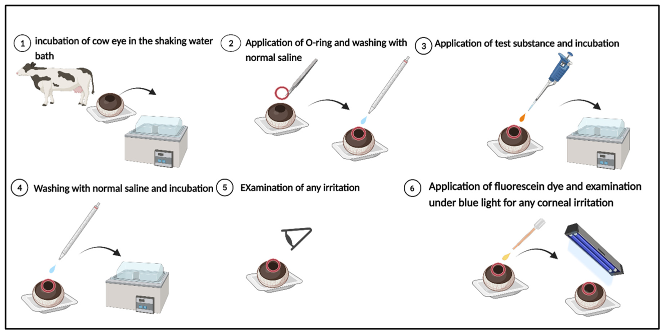
Eyes from bovines were collected and studied for corneal opacity, epithelium separation, and vascularization. The OECD Test Guidelines use a standardized in vitro (BCOP Assay) to discover whether chemicals cause ocular irritation [53]. Damaged or abnormal eyes were discarded following the health and safety regulations for Kingston University London. Four controls were used to assess the irritation response: NaOH (0.5 M), positive control (strong irritant); acetone (moderate irritant); propylene glycol (mild irritant); and normal saline, negative control (non-irritant). The eyes were placed in plastic holding cups and incubated in a humid atmosphere at 37 °C for 10 min. The cornea was covered with a silicon O-ring to provide a consistent application area of the test material. After applying 100 µL of the test substance, the eyes were rinsed and incubated for 10 min. The integrity of the corneal epithelium was assessed using a cobalt blue filter (Figure 3). A visual assessment of corneal injury was conducted using an examination light and 0.5 mL of 2% w/v sodium fluorescein solution. The degree of corneal injury was scored using three criteria: opacity (0–4), epithelial integrity (0–1.5), and epithelial detachment (0–4) [54]. Mean opacity was given a score between 0 and 4 (0, no opacity; 1, slight opacity; 2, marked opacity; 3, severe opacity; 4, opaque opacity). Epithelial integrity staining was given the following scores (0, none; 0.5, diffuse and weak; 1.0, confluent and weak; 1.5, confluent and intense). Epithelial detachment was scored as follows: 0, no gross abnormalities; 2, wrinkling of the corneal surface; 3, loosening of the epithelium; 4, absence of an epithelium. Table 2 shows the corneal irritation potential, based on the mean cumulative scores of the three eyes.

2.5. Hen’s Egg Test on the Chorioallantois Membrane (HET-CAM) Test

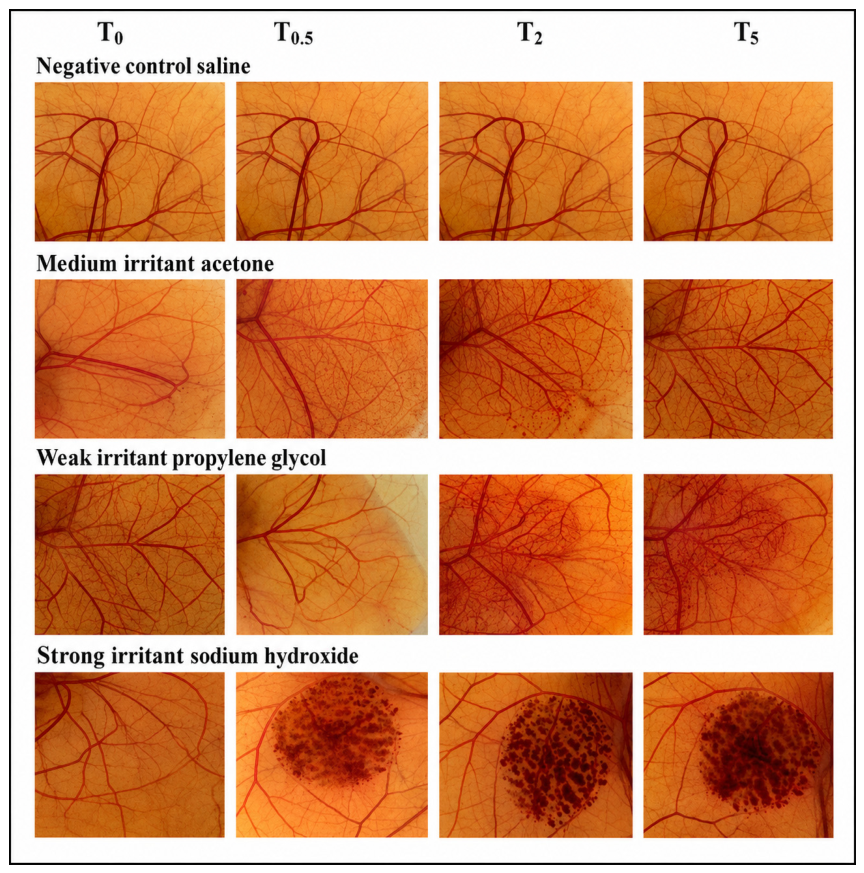
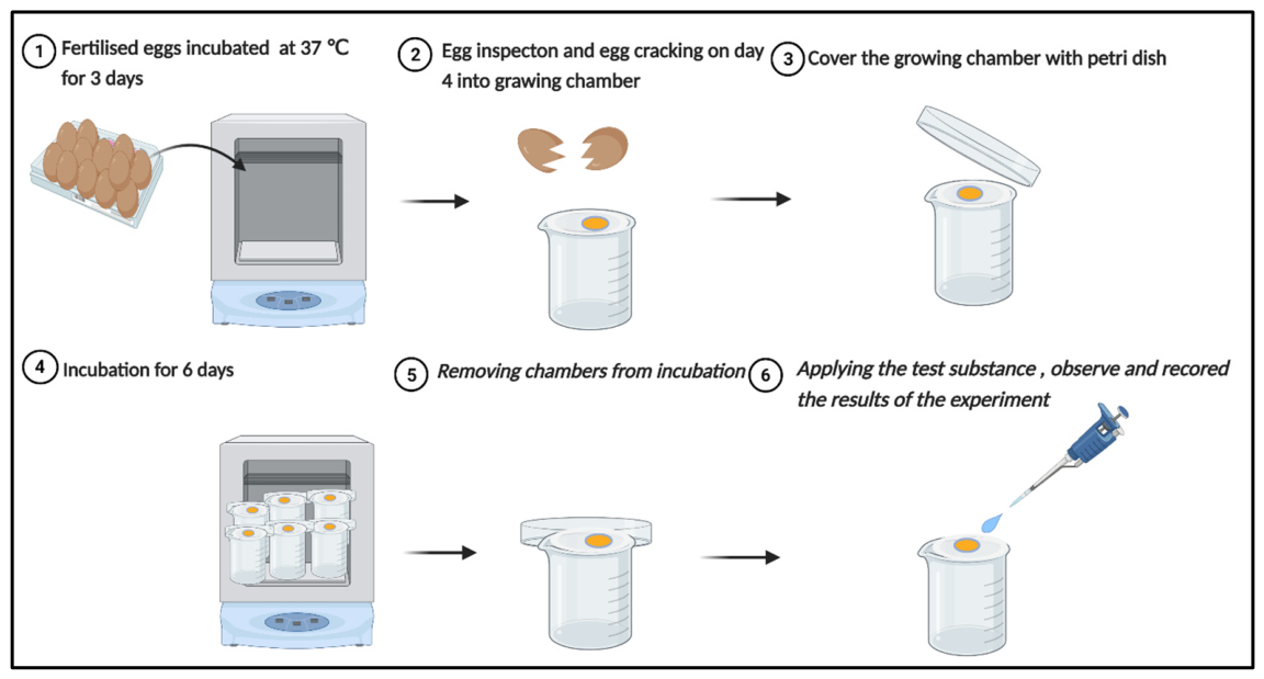
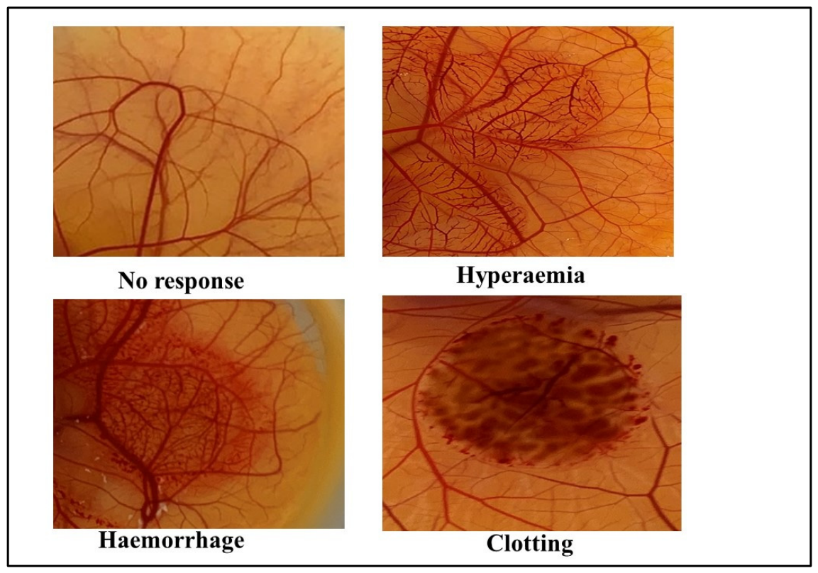
The study involved fertilizing eggs from White Leghorn hens with 70% IMS and incubating them for three days at 37 ± 0.5 °C and 66 ± 5% relative humidity following the HET-CAM protocol outlined by Luepke [57]. A modified procedure was adopted, which involves cultivating the chick embryo in Petri plates, which was further refined using growth chambers built in-house. The growth chambers were lined with a curved, elastic cellophane membrane that resembled an eggshell’s inner membrane, which is thought to aid embryo growth. These modifications imply that embryo survival and the number of CAMs available for testing can be improved [54,58]. The eggs were rotated 3–5 times per day to ensure proper positioning. On the fourth day, the eggs were sprayed with IMS and cracked open to extract their contents. The contents were transferred to a growth chamber, prepared using a modified HET-CAM method. The chamber and Petri dish were sterilized with 70% IMS. The viability of embryos was assessed by assessing the CAM and yolk, discarding eggs with dead embryos or ruptured yolk sacs. The viable embryos were then incubated at 37.5 ± 0.5 °C and 66 ± 5% relative humidity. On day ten, 200 µL of the test solutions was applied to the CAM [37,54]. Figure 4 illustrates the HET-CAM test’s schematic design. Figure 5 shows blood vessels and capillaries being tested for vascular responses used to assess irritation potential, such as coagulation, hyperemia, and bleeding, after 0.5, 2, and 5 min. Three different CAMs were used for each of the test items, and the scores that were accumulated over time were added together to form a single number (Table 3). The irritant potential of the tested chemical was indicated by this value. Table 4 classified the test substance’s irritating effects. The cumulative mean score value of the response allowed for Draize-like irritation potential assessment [21,57,59,60].

2.6. Statistical Analysis

The one-way analysis of variance (ANOVA) and Dunnett’s test for multiple comparisons across groups were executed using GraphPad Prism Software Version 9.3.1.

3. Results and Discussion

3.1. Excipient and Formulations Cytotoxicity Assessment (NRU Assay)

Neutral red uptake (NRU) is a coulometric assay utilized to evaluate the cytotoxicity of various products, including cosmetics, industrial chemicals, medications, and household items [61]. NRU is more sensitive, easier to detect in living cells, less expensive, and requires less equipment to assess cell death. The cytotoxicity of compounds and excipients utilized in formulations is frequently assessed in vitro. The NRU assay is dependent on the capability of live cells to absorb and attach to the NR dye, which is slightly alkaline and can passively permeate the walls of cells [62]. Once inside the cells, the dye accumulates in the lysosomes and interacts with anionic and phosphate groups in the lysosomal matrix [63]. The dye is subsequently removed with an acidified ethanol solution, and its absorbance is measured at 540 nm [64,65]. NR dye uptake is dependent on the preservation of pH gradients by cells, with lysosomal proton gradients keeping a pH lower than the cytoplasm. Changes in these gradients, cell membranes, or cell death might impact dye retention, allowing for differentiation between damaged, viable, and dying cells [66]. The NRU test is a sensitive indication of cell viability and may evaluate cell reproduction, cytotoxicity, or cytostatic effects, depending on the quantity of cells utilized [67].
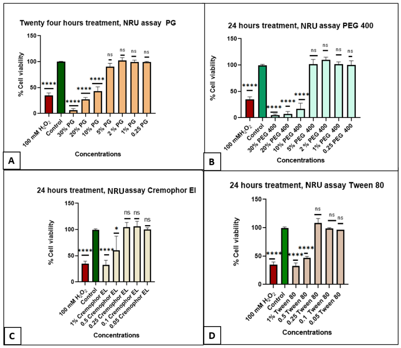
Figure 6 shows the effects of excipient concentrations on HLE-B3 growth after 24 h. Figure 6A,B illustrate that the toxicity of PEG 400 and PG was dose-dependent. After a 24 h treatment with PEG 400 and PG at concentrations of 30, 20 and 10 w/w%, cell growth and cell mortality decreased significantly (p < 0.001), similar to the positive control. On the other hand, treatment with both cosurfactants/cosolvents at concentrations ranging from 0.25 to 5 (w/w%) did not have a significant effect (p > 0.001) on the viability of HLEC-B3 cells. PEG 400 was previously shown to be non-toxic at doses of 20 mg/mL on human cervical cancer cells (HeLa) and mouse fibroblast cell lines [50].
Cremophor EL and Tween 80 showed dose-dependent toxicity (Figure 6C,D). After 24 h treatments at doses of 0.5 and 1 w/w%, there was a statistically significant reduction (p < 0.0001) in HLEC-B3 growth and cell death. Treatment with the lower concentrations of both surfactants (0.05 to 0.25 w/w%) had no significant effect on cell viability (p > 0.0001) when compared to the control group, indicating that the cells were not damaged at these concentrations. Our study demonstrated that the toxic effects of the investigated cosurfactants and surfactants varied depending on their concentrations (Figure 6).
Recent advances in ocular biomaterials and ophthalmic drug delivery systems, including photothermal intraocular platforms, nanocarriers, hydrogels, and stimuli-responsive systems, have highlighted the increasing importance of excipient biocompatibility in ocular formulation design. Lin et al. demonstrated that advanced ophthalmic biomaterial systems may effectively modulate ocular cellular responses while minimizing tissue damage through careful material optimization [68]. However, despite significant progress in functional ocular biomaterials, comparative ocular safety data for commonly used surfactants and cosurfactants under standardized experimental conditions remain limited. The present study addresses this gap by systematically evaluating the cytotoxicity and irritation potential of selected ophthalmic excipients using complementary in vitro and ex vivo ocular safety models.

3.2. Determinations of Conjunctival Biocompatibility Using the Hen’s Egg-Test on the Chorioallantoic Membrane (HET-CAM)

The HET-CAM test is an alternative to animal testing for determining the potential conjunctival irritation of chemicals. It involves applying test chemicals to the chorioallantoic membrane (CAM) of a chicken egg and observing the response of the membrane’s blood vessels. This test is accepted by many regulatory bodies worldwide as an alternative to the Draize rabbit eye test [57]. The CAM exhibits exceptional sensitivity and discrimination toward agricultural pesticides and pharmaceutical excipients with an ocular irritant potential [59,69]. It has been effectively employed to evaluate the irritant potential of a variety of ocular formulations, such as microemulsions, liquid crystals, o/w emulsions, niosomes [54], gels [70], in situ gelling films and inserts [14]. The HET-CAM test is preferred for examining conjunctival irritation surfactants and cosurfactants. The presence of moderate levels of surfactants and cosurfactants in ophthalmic formulations can result in ocular irritation. Therefore, it is critical to screen such formulations to identify suitable concentrations.
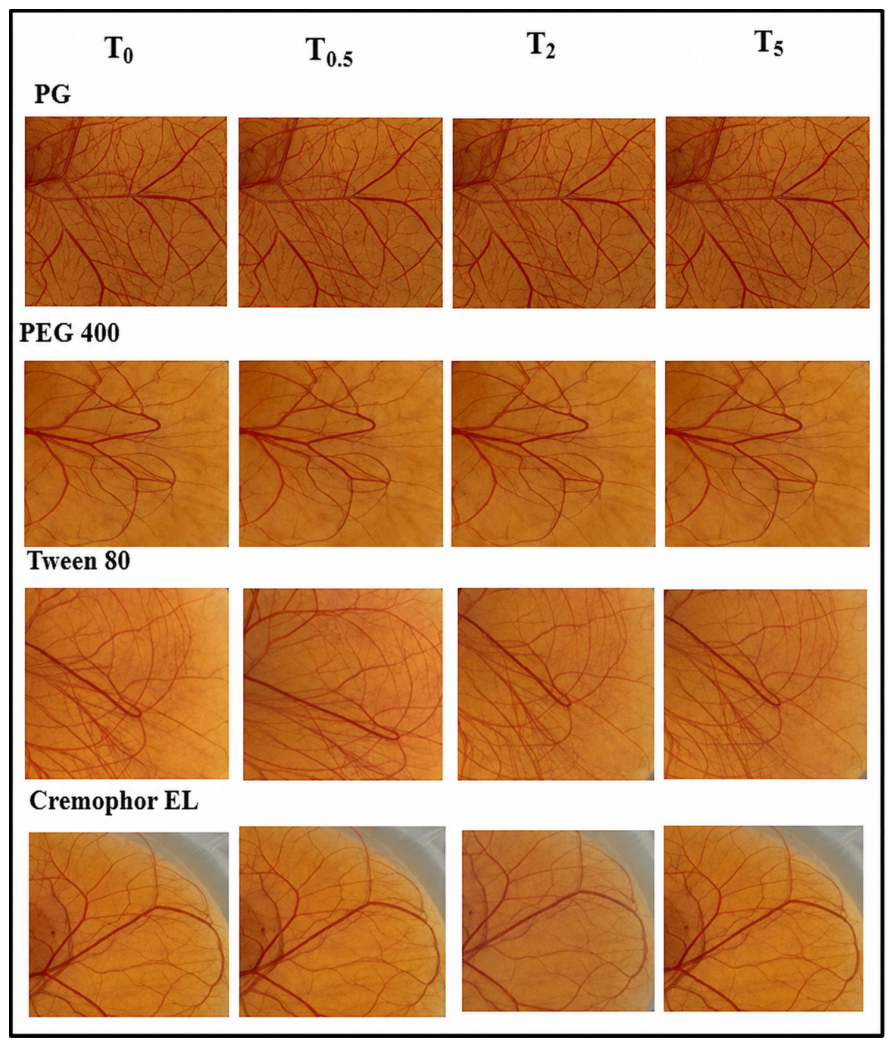
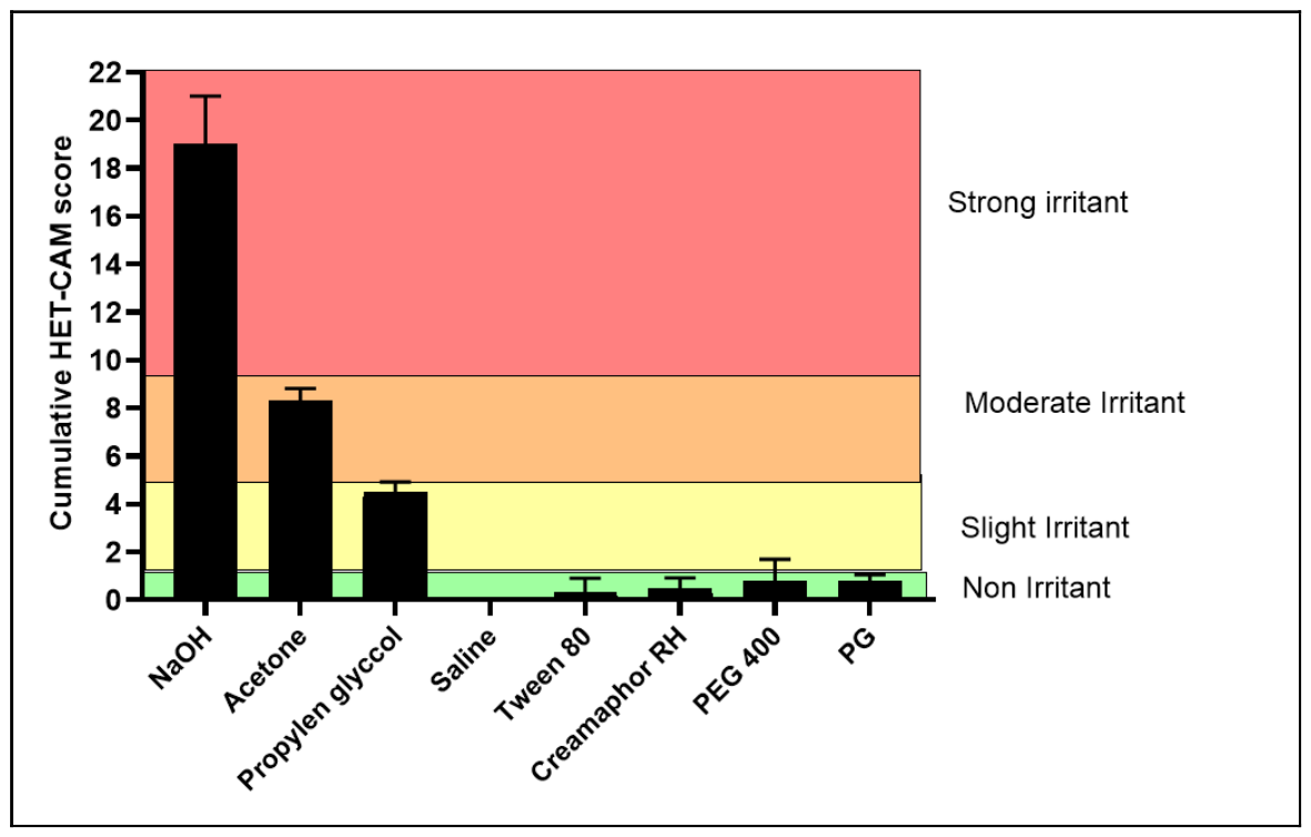
Compared with the negative control (Figure 7) the vascular responses generated by our excipients at 0, 0.5, 2 and 5 min are shown in Figure 8. Neither of the tested cosurfactants (PG and PEG at 5% w/w) showed signs of irritation responses at the concentrations used, with a cumulative score of less than 1. It has been previously reported that the instillation of 50% aqueous solutions of PG does not irritate rabbit eyes [21,71,72].
Tweens 80 and Cremophor EL were shown to be non-irritants at 0.25% w/w with an average cumulative score of less than 1. Eye formulations containing Tweens 80 have been reported to be well tolerated and free of irritation [14,73]. Cremophor EL has also been categorized as safe to the eyes at concentrations of up to 30% (w/w), while a 50% aqueous solution has been shown to produce mild irritation with lacrimation that resolves fast [21,74]. Figure 9 shows the cumulative HET-CAM scores for the all four excipients.

3.3. Excipient Corneal Biocompatibility (BCOP Test)

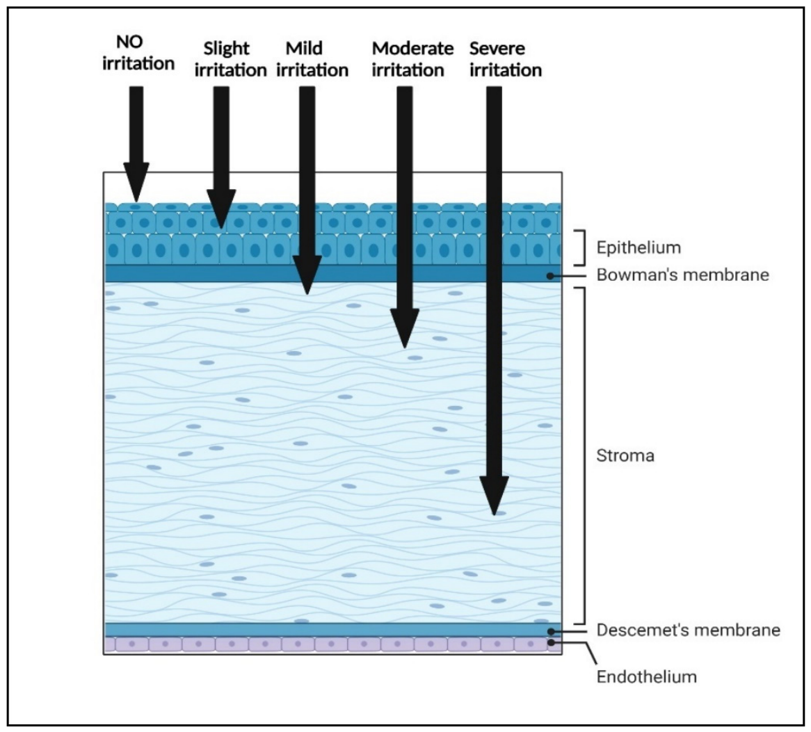
The BCOP test evaluates ocular irritation in chemicals using opacity and permeability scores. The assay evaluates corneal opacity and disruption of the corneal barrier after exposure to the test material, serving as an effective isolated enucleated eye test [75]. Normally, the test evaluates two key endpoints of irritation damage that affect the cornea: opacity and permeability. To measure opacity, the change in light transmission through the cornea is assessed. The measurement of corneal opacity and sodium fluorescein penetration can provide information about protein denaturation, damage to corneal layers, vascularization, swelling, and integrity of the corneal epithelial layer caused by exposure to a test substance [76]. The severity of eye irritation is related to the degree of initial injury caused by the test substance. The degree of irritation is classified based on the depth (extent) of damage caused by the substance. Superficial damage to the corneal epithelial layer is considered slight irritation, while damage to both the epithelium and stroma is considered mild or moderate irritation. Severe irritants cause damage to the corneal endothelial layer after penetrating the epithelium and stroma. Figure 10 illustrates the layers of the cornea and the extent of test substance penetration and subsequent irritation classification [77].
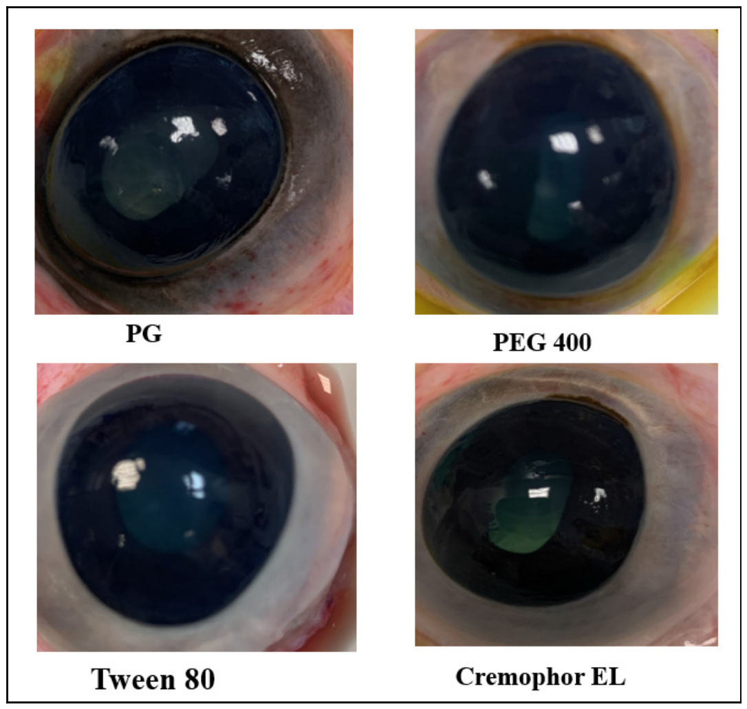
The cumulative scores derived from corneal opacity and fluorescein permeability responses (Figure 11) are translated into BCOP irritation scores (Table 2). Figure 12 presents the cumulative scores for surfactants and cosurfactants tested on bovine eyes. The application of a 5% w/w aqueous PG solution led to no irritating effect on the bovine cornea. It has been previously reported that application of 10% v/v PG causes little or no injury to the bovine cornea [54]. Installing PEG 400 at a concentration of 5% (w/w) was less irritating than the negative control, with an average cumulative of less than two.
The average cumulative scores for Tween 80 and Cremophor EL at 0.25% w/w concentrations were less than 0.5, indicating that they were non-irritants. These excipients exhibited no signs of ocular harm, including no changes in opacity and permeability, nor any signs of corneal epithelial damage. It has been previously reported that neither of these surfactants induces ocular irritation [78]. Overall, the tested excipients did not cause substantial eye irritation and were therefore deemed safe for future testing in live animals.
The physicochemical properties of the tested excipients (Table 1) appear to correlate with their irritation potential. For instance, PEG 400, with low log P (−4.8), exhibited minimal irritation in both HET-CAM and BCOP assays. In contrast, although Tween 80 and Cremophor EL have higher log P values, their relatively moderate HLB values and low concentrations may explain their non-irritant behavior. These findings suggest that higher hydrophilicity and larger molecular size may reduce corneal membrane penetration, thereby limiting ocular irritation potential [79].
Molecular weight (MW) affects membrane permeability. The permeability of molecules across membranes decreases as the size of the molecules increases [41,80]. PEG 400 (380–420 g/mol, hydrophilic) causes mild irritation, yet PG (76.1 g/mol) causes moderate irritation owing to better corneal tissue penetration. Tween 80 and Cremophor EL may induce irritation via surface activity and micellar formation rather than mere diffusion [81,82,83]. That is mostly due to their stronger amphiphilic characteristics, where they can interact with and disrupt the packing of the lipid and phospholipid layers of the corneal epithelium, resulting in concentration-dependent irritation or even cytotoxicity. The CMCs for Tween 80 and Cremophor EL are 0.0016% and 0.02% respectively. Below the CMC, surfactant monomers are more likely to irritate cell membranes. At or above the CMC, surfactants create micelles, which may minimize monomers’ availability to interact with membranes and induce irritation.
A notable advantage of this study is the direct comparison of multiple commonly used ophthalmic excipients using identical experimental conditions across complementary ocular safety models (HET-CAM—conjunctiva, BCOP—cornea and HLEC-B3 cells—lens).
Nevertheless, several limitations should be acknowledged; first, the study evaluated acute exposure conditions and therefore may not fully predict chronic ophthalmic tolerability following repeated administration. Second, cytotoxicity was assessed using HLEC-B3 cells (lens epithelial cells), which provide a robust preliminary screening model (as the human eye lens has an epithelium) but do not accurately reflect corneal or conjunctival epithelial physiology. Third, excipients were evaluated individually rather than within complete ophthalmic formulations, where excipient: excipient interactions may impact ocular safety profiles.

4. Conclusions

We have systematically compared the cytotoxicity and ocular irritation potential of commonly used ophthalmic surfactants and cosurfactants under standardized experimental conditions using complementary in vitro and ex vivo ocular safety models. The ocular irritation of Tween 80, Cremophor EL, polyethylene glycol 400 (PEG 400) and propylene glycol (PG) was examined using the HET-CAM (conjunctival) and BCOP (corneal) eye assays, and their cytotoxicity was tested using HLEC-B3 (lens epithelial cells). Our findings show that PEG 400, PG, Cremophor EL, and Tween 80 exhibit dose-dependent toxicity toward HLEC-B3 cells. While higher concentrations significantly impaired cell growth and increased cell death, lower concentrations, particularly 0.25–5% for cosurfactants and 0.05–0.25% for surfactants, did not affect cell viability, highlighting the importance of careful excipient selection and concentration control in formulation design. The chosen surfactants, Tween 80 with Cremophor EL at 0.25% (w/w), and cosurfactants, PEG 400 and PG at 5% (w/w), were non-irritating according to the HET-CAM and BCOP assays. All four excipients could potentially be used (as per the specified safe concentrations) to develop appropriate formulations that are intended for topical application to the surface of the eye. Finally, it should be noted that numerous eye medications have been evaluated using acute exposure models (≤24 h; cytotoxicity assays on cell lines, HET-CAM, BCOP). These models are commonly used to test cytotoxicity and acute eye irritation; however, they may not reliably assess chronic or recurrent ophthalmic medication safety. Future research using repeated or chronic exposure models may provide a better insight into cumulative effects and long-term ocular safety of these excipients.

Author Contributions

Conceptualization and study design: Z.K.A.-Q., A.A.A.-K. and R.G.A. Methodology, validation, and formal data analysis: Z.K.A.-Q., A.A.A.-K. and R.G.A. Writing—original draft preparation: Z.K.A.-Q. Visualization and supervision: A.A.A.-K. and R.G.A. All authors have read and agreed to the published version of the manuscript.

Funding

This research received no external funding.

Institutional Review Board Statement

Not applicable.

Informed Consent Statement

Not applicable.

Data Availability Statement

All data can be provided by the authors upon request. No publicly accessible archive storage is available.

Acknowledgments

The authors have reviewed and edited the output and take full responsibility for the content of this publication.

Conflicts of Interest

The authors declare no conflicts of interest.

Abbreviations

PGpropylene glycol
PEG 400polyethylene glycol 400
CMCcritical micelle concentration
Log PLipophilicity
HLBhydrophile lipophile balance value
DCdielectric constant
HLBC-B3Human Lens Epithelial Cell Line
ANOVAone-way analysis of variance
HET-CAMHen’s Egg Test on the Chorioallantois Membrane Test
BCOPbovine corneal opacity and permeability

References

  1. Li, J.; Tripathi, R.C.; Tripathi, B.J. Drug-induced ocular disorders. Drug Saf. 2008, 31, 127–141. [Google Scholar] [CrossRef]
  2. Singh, M.; Devi, M.; Kumar, P.; Monika; Singh, R.; Jhawat, V. Adverse effects of antiglaucoma medications: Pathophysiology and novel drug delivery strategies for mitigation. Int. Ophthalmol. 2026, 46, 64. [Google Scholar] [CrossRef]
  3. Johnstone, M.A.; Albert, D.M. Prostaglandin-induced hair growth. Surv. Ophthalmol. 2002, 47, S185–S202. [Google Scholar] [CrossRef] [PubMed]
  4. Stjernschantz, J.W.; Albert, D.M.; Hu, D.-N.; Drago, F.; Wistrand, P.J. Mechanism and clinical significance of prostaglandin-induced iris pigmentation. Surv. Ophthalmol. 2002, 47, S162–S175. [Google Scholar] [CrossRef]
  5. Chu, C.; Chen, X.; Deng, Y.; Zhang, J.; Bi, X.; Ma, Y.; Wei, M.; Zhao, J.; He, H.; Gou, J. MMP-9-responsive contact lens facilitating disease-adaptive and safer dexamethasone delivery for corneal neovascularization therapy. Asian J. Pharm. Sci. 2026, 21, 101146. [Google Scholar] [CrossRef]
  6. Lam, P.D.; Chan, J.W.; Tsui, R.W.; Liu, K.; Chow, V.W.; Chan, J.Y. The evidence-based indications of 0.1% topical tacrolimus in ophthalmology: A scoping review. Ther. Adv. Ophthalmol. 2026, 18, 25158414251405461. [Google Scholar] [CrossRef]
  7. Flach, A. Topically applied nonsteroidal anti-inflammatory drugs and corneal problems: An interim review and comment. Ophthalmology 2000, 107, 1224–1226. [Google Scholar] [CrossRef]
  8. Gaynes, B.I.; Fiscella, R. Topical nonsteroidal anti-inflammatory drugs for ophthalmic use: A safety review. Drug Saf. 2002, 25, 233–250. [Google Scholar] [CrossRef] [PubMed]
  9. Walsh, K.; Jones, L. The use of preservatives in dry eye drops. Clin. Ophthalmol. 2019, 13, 1409–1425. [Google Scholar] [CrossRef] [PubMed]
  10. Budai, P.; Kormos, É.; Buda, I.; Somody, G.; Lehel, J. Comparative evaluation of HET-CAM and ICE methods for objective assessment of ocular irritation caused by selected pesticide products. Toxicol. In Vitro 2021, 74, 105150. [Google Scholar] [CrossRef]
  11. Noecker, R. Ophthalmic preservatives: Considerations for long-term use in patients with dry eye or glaucoma. Rev. Ophthalmol. 2001, 8, 73–79. [Google Scholar]
  12. Wroblewska, K.B.; Jadach, B.; Muszalska-Kolos, I. Progress in drug formulation design and delivery of medicinal substances used in ophthalmology. Int. J. Pharm. 2021, 607, 121012. [Google Scholar] [CrossRef]
  13. Kumar, N.; Kumar, R.; Malhotra, M.; Singh, A.P.; Pal, A. Ophthalmic Ointment & Formulation: A Review. Int. J. Med. Pharm. Drug Res. 2024, 8, 2. [Google Scholar]
  14. Abdelkader, H.; Pierscionek, B.; Alany, R.G. Novel in situ gelling ocular films for the opioid growth factor-receptor antagonist-naltrexone hydrochloride: Fabrication, mechanical properties, mucoadhesion, tolerability and stability studies. Int. J. Pharm. 2014, 477, 631–642. [Google Scholar] [CrossRef]
  15. Sahoo, R.K.; Biswas, N.; Guha, A.; Sahoo, N.; Kuotsu, K. Nonionic surfactant vesicles in ocular delivery: Innovative approaches and perspectives. BioMed Res. Int. 2014, 2014, 263604. [Google Scholar] [CrossRef]
  16. Tamilvanan, S.; Benita, S. The potential of lipid emulsion for ocular delivery of lipophilic drugs. Eur. J. Pharm. Biopharm. 2004, 58, 357–368. [Google Scholar] [CrossRef]
  17. Ali, Y.; Lehmussaari, K. Industrial perspective in ocular drug delivery. Adv. Drug Deliv. Rev. 2006, 58, 1258–1268. [Google Scholar] [CrossRef] [PubMed]
  18. Schaeffer, H.E.; Krohn, D.L. Liposomes in topical drug delivery. Investig. Ophthalmol. Vis. Sci. 1982, 22, 220–227. [Google Scholar]
  19. Meisner, D.; Mezei, M. Liposome ocular delivery systems. Adv. Drug Deliv. Rev. 1995, 16, 75–93. [Google Scholar] [CrossRef]
  20. Perini, G.; Saettone, M.; Carafa, M.; Santucci, E.; Alhaique, F. “Non-ionic surfactant vesicles as ophtalmic carriers for cyclopentolate. A preliminary evaluation”. STP Pharma Sci. 1996, 6, 94–98. [Google Scholar]
  21. Smail, S.S.; Ghareeb, M.M.; Omer, H.K.; Al-Kinani, A.A.; Alany, R.G. Studies on surfactants, cosurfactants, and oils for prospective use in formulation of ketorolac tromethamine ophthalmic nanoemulsions. Pharmaceutics 2021, 13, 467. [Google Scholar] [CrossRef]
  22. Lam, P.H.; Dang, D.M.T.; Dang, C.M. Effects of Co-Surfactants and Processing Conditions on Ocimum gratissimum Essential Oil Microemulsion. J. Indian Chem. Soc. 2026, 103, 102565. [Google Scholar] [CrossRef]
  23. Gadhave, A. Determination of hydrophilic-lipophilic balance value. Int. J. Sci. Res. 2014, 3, 573–575. [Google Scholar]
  24. Adams, M.L.; Lavasanifar, A.; Kwon, G.S. Amphiphilic block copolymers for drug delivery. J. Pharm. Sci. 2003, 92, 1343–1355, Correction in J. Pharm. Sci. 2005, 94, 1160.. [Google Scholar] [CrossRef] [PubMed]
  25. Moiseev, R.V.; Morrison, P.W.; Steele, F.; Khutoryanskiy, V.V. Penetration enhancers in ocular drug delivery. Pharmaceutics 2019, 11, 321. [Google Scholar] [CrossRef]
  26. Ferguson, P.; Cross, R.; Schad, G. Application of SFC for the characterization of formulated drug products. In Separation Science and Technology; Elsevier: Amsterdam, The Netherlands, 2022; pp. 221–255. [Google Scholar]
  27. Davies, J. A quantitative kinetic theory of emulsion type, I. Physical chemistry of the emulsifying agent. In Gas/Liquid and Liquid/Liquid Interface, Proceedings of the International Congress of Surface Activity; Citeseer: University Park, PA, USA, 1957. [Google Scholar]
  28. Griffin, W. Calculation of HLB values of non-ionic surfactants. Am. Perfum. Essent. Oil Rev. 1955, 65, 26–29. [Google Scholar]
  29. Zhang, H.; Yao, M.; Morrison, R.A.; Chong, S. Commonly used surfactant, Tween 80, improves absorption of P-glycoprotein substrate, digoxin, in rats. Arch. Pharmacal Res. 2003, 26, 768–772. [Google Scholar] [CrossRef]
  30. Kimura, M.; Yasueda, S.-I.; Yamaguchi, M.; Inada, K. Compositions Containing Difluprednate. U.S. Patent US-0076124, 12 May 1998. [Google Scholar]
  31. Scaffidi, R.C.; Korb, D.R. Comparison of the efficacy of two lipid emulsion eyedrops in increasing tear film lipid layer thickness. Eye Contact Lens 2007, 33, 38–44. [Google Scholar] [CrossRef]
  32. Lallemand, F.; Felt-Baeyens, O.; Besseghir, K.; Behar-Cohen, F.; Gurny, R. Cyclosporine A delivery to the eye: A pharmaceutical challenge. Eur. J. Pharm. Biopharm. 2003, 56, 307–318. [Google Scholar] [CrossRef]
  33. Vaidya, S.; Ganguli, A.K. 2.01—Microemulsion methods for synthesis of nanostructured materials. In Comprehensive Nanoscience and Nanotechnology, 2nd ed.; Andrews, D.L., Lipson, R.H., Nann , T., Eds.; Academic Press: San Diego, CA, USA, 2019; pp. 1–12. [Google Scholar]
  34. Hegde, R.R.; Verma, A.; Ghosh, A. Microemulsion: New insights into the ocular drug delivery. Int. Sch. Res. Not. 2013, 2013, 826798. [Google Scholar] [CrossRef]
  35. Gautam, N.; Kesavan, K. Development of Microemulsions for Ocular Delivery. Ther. Deliv. 2017, 8, 313–330. [Google Scholar] [CrossRef]
  36. Vandamme, T.F. Microemulsions as ocular drug delivery systems: Recent developments and future challenges. Prog. Retin. Eye Res. 2002, 21, 15–34. [Google Scholar] [CrossRef]
  37. Guo, X.; Rong, Z.; Ying, X. Calculation of hydrophile–lipophile balance for polyethoxylated surfactants by group contribution method. J. Colloid Interface Sci. 2006, 298, 441–450. [Google Scholar] [CrossRef]
  38. Lu, S.; Li, J.; Zhang, S.; Yin, Z.; Xing, T.; Kaplan, D.L. The influence of the hydrophilic–lipophilic environment on the structure of silk fibroin protein. J. Mater. Chem. B 2015, 3, 2599–2606. [Google Scholar] [CrossRef]
  39. Jang, H.-J.; Shin, C.Y.; Kim, K.-B. Safety evaluation of polyethylene glycol (PEG) compounds for cosmetic use. Toxicol. Res. 2015, 31, 105–136. [Google Scholar] [CrossRef]
  40. Jacob, S.E.; Scheman, A.; McGowan, M.A. Propylene glycol. Dermatitis 2018, 29, 3–5. [Google Scholar] [CrossRef] [PubMed]
  41. Labetoulle, M.; Messmer, E.M.; Pisella, P.-J.; Ogundele, A.; Baudouin, C. Safety and efficacy of a hydroxypropyl guar/polyethylene glycol/propylene glycol-based lubricant eye-drop in patients with dry eye. Br. J. Ophthalmol. 2017, 101, 487–492. [Google Scholar] [CrossRef]
  42. Cohen, S.; Martin, A.; Sall, K. Evaluation of clinical outcomes in patients with dry eye disease using lubricant eye drops containing polyethylene glycol or carboxymethylcellulose. Clin. Ophthalmol. 2014, 8, 157. [Google Scholar] [CrossRef]
  43. Srinivasan, S.; Manoj, V. A decade of effective dry eye disease management with systane ultra (polyethylene glycol/propylene glycol with hydroxypropyl guar) lubricant eye drops. Clin. Ophthalmol. 2021, 15, 2421–2435. [Google Scholar] [CrossRef] [PubMed]
  44. Benelli, U. Systane® lubricant eye drops in the management of ocular dryness. Clin. Ophthalmol. 2011, 5, 783–790. [Google Scholar] [CrossRef] [PubMed]
  45. Kading, D. A two-week clinical evaluation of the safety of systane® Ultra in contact lens-wearing patients. Clin. Ophthalmol. 2010, 4, 27–32. [Google Scholar] [CrossRef] [PubMed]
  46. Christensen, M.T. Corneal staining reductions observed after treatment with Systane® Lubricant Eye Drops. Adv. Ther. 2008, 25, 1191–1199. [Google Scholar] [CrossRef]
  47. Lemp, M.A.; Foulks, G.; Devgan, U.; Trattler, W.; Nichols, K. The therapeutic role of lipids: Managing ocular surface disease. Refract. Eyecare Ophthalmol. 2005, 9, 14. [Google Scholar]
  48. Christensen, M.T.; Cohen, S.; Rinehart, J.; Akers, F.; Pemberton, B.; Bloomenstein, M.; Lesher, M.; Kaplan, D.; Meadows, D.; Meuse, P. Clinical evaluation of an HP-guar gellable lubricant eye drop for the relief of dryness of the eye. Curr. Eye Res. 2004, 28, 55–62. [Google Scholar] [CrossRef] [PubMed]
  49. Ubels, J.; Clousing, D.; Van Haitsma, T.; Hong, B.-S.; Stauffer, P.; Asgharian, B.; Meadows, D. Pre-clinical investigation of the efficacy of an artificial tear solution containing hydroxypropyl-guar as a gelling agent. Curr. Eye Res. 2004, 28, 437–444. [Google Scholar] [CrossRef]
  50. Liu, G.; Li, Y.; Yang, L.; Wei, Y.; Wang, X.; Wang, Z.; Tao, L. Cytotoxicity study of polyethylene glycol derivatives. RSC Adv. 2017, 7, 18252–18259. [Google Scholar] [CrossRef]
  51. Al-Kinani, A.A.T. Delivery of Antioxidants to the Lens Using Nanoparticles. Ph.D. Thesis, Kingston University, London, UK, 2016. [Google Scholar]
  52. Cadenas, E. Hydrogen Sulfide in Redox Biology Part B; Academic Press: Cambridge, MA, USA, 2015. [Google Scholar]
  53. Osi, B.; Al-Kinani, A.A.; Al-Qaysi, Z.K.; Khoder, M.; Alany, R.G. Exploring the ocular absorption pathway of fasudil hydrochloride towards developing a nanoparticulate formulation with improved performance. Pharmaceutics 2024, 16, 112. [Google Scholar] [CrossRef]
  54. Abdelkader, H.; Ismail, S.; Hussein, A.; Wu, Z.; Al-Kassas, R.; Alany, R.G. Conjunctival and corneal tolerability assessment of ocular naltrexone niosomes and their ingredients on the hen’s egg chorioallantoic membrane and excised bovine cornea models. Int. J. Pharm. 2012, 432, 1–10. [Google Scholar] [CrossRef] [PubMed]
  55. Churchward, C.P.; Al-Kinani, A.A.; Abdelkader, H.; Swinden, J.; Siwoku, O.; Varnakulasingam, T.; Alany, R.G.; Snyder, L.A. Monocaprin eye drop formulation to combat antibiotic resistant gonococcal blindness. Sci. Rep. 2020, 10, 12010. [Google Scholar] [CrossRef]
  56. Van Erp, Y.; Weterings, P. Eye irritancy screening for classification of chemicals. Toxicol. In Vitro 1990, 4, 267–269. [Google Scholar] [CrossRef]
  57. Luepke, N. Hen’s egg chorioallantoic membrane test for irritation potential. Food Chem. Toxicol. 1985, 23, 287–291. [Google Scholar] [CrossRef]
  58. Alany, R.; Rades, T.; Nicoll, J.; Tucker, I.; Davies, N. W/O microemulsions for ocular delivery: Evaluation of ocular irritation and precorneal retention. J. Control. Reléase 2006, 111, 145–152. [Google Scholar] [CrossRef] [PubMed]
  59. Chan, J.; Maghraby, G.M.; Craig, J.P.; Alany, R.G. Phase transition water-in-oil microemulsions as ocular drug delivery systems: In vitro and in vivo evaluation. Int. J. Pharm. 2007, 328, 65–71. [Google Scholar] [CrossRef] [PubMed]
  60. Rupenthal, I.D.; Green, C.R.; Alany, R.G. Comparison of ion-activated in situ gelling systems for ocular drug delivery. Part 2: Precorneal retention and in vivo pharmacodynamic study. Int. J. Pharm. 2011, 411, 78–85. [Google Scholar] [CrossRef] [PubMed]
  61. Rodrigues, R.M.; Bouhifd, M.; Bories, G.; Sacco, M.-G.; Gribaldo, L.; Fabbri, M.; Coecke, S.; Whelan, M.P. Assessment of an automated in vitro basal cytotoxicity test system based on metabolically-competent cells. Toxicol. In Vitro 2013, 27, 760–767. [Google Scholar] [CrossRef]
  62. Hwang, J.-h.; Hong, J.-h.; Lim, K.-M. Investigation of alternative cell models to BALB/c 3T3 for in vitro neutral red uptake phototoxicity test of pharmaceuticals: NIH 3T3 and HaCaT cells. Toxicol. Res. 2026, 42, 187–197. [Google Scholar] [CrossRef]
  63. Nemes, Z.; Dietz, R.; Lüth, J.; Gomba, S.; Hackenthal, E.; Gross, F. The pharmacological relevance of vital staining with neutral red. Experientia 1979, 35, 1475–1476. [Google Scholar] [CrossRef]
  64. Filman, D.; Brawn, R.; Dandliker, W. Intracellular supravital stain delocalization as an assay for antibody-dependent complement-mediated cell damage. J. Immunol. Methods 1975, 6, 189–207. [Google Scholar] [CrossRef]
  65. Cavanaugh, P.F.; Moskwa, P.S.; Donish, W.H.; Pera, P.J.; Richardson, D.; Andrese, A.P. A semi-automated neutral red based chemosensitivity assay for drug screening. Investig. New Drugs 1990, 8, 347–354. [Google Scholar] [CrossRef]
  66. Kuete, V.; Karaosmanoğlu, O.; Sivas, H. Anticancer activities of African medicinal spices and vegetables. In Medicinal Spices and Vegetables from Africa; Elsevier: Amsterdam, The Netherlands, 2017; pp. 271–297. [Google Scholar]
  67. Gomez-Gutierrez, J.G.; Bhutiani, N.; McNally, M.W.; Chuong, P.; Yin, W.; Jones, M.A.; Zeiderman, M.R.; Grizzle, W.E.; McNally, L.R. The neutral red assay can be used to evaluate cell viability during autophagy or in an acidic microenvironment in vitro. Biotech. Histochem. 2021, 96, 302–310. [Google Scholar] [CrossRef]
  68. Lin, L.; Xu, L.; Sun, W.; Liang, L.; Qi, X.; Zhao, Y.e. Mild photothermal therapy prevents posterior capsule opacification through cytoskeletal remodeling. Adv. Healthc. Mater. 2023, 12, 2300470. [Google Scholar] [CrossRef]
  69. Budai, P.; Lehel, J.; Tavaszi, J.; Kormos, É. HET-CAM test for determining the possible eye irritancy of pesticides. Acta Vet. Hung. 2010, 58, 369–377. [Google Scholar] [CrossRef] [PubMed]
  70. Rupenthal, I.D.; Green, C.R.; Alany, R.G. Comparison of ion-activated in situ gelling systems for ocular drug delivery. Part 1: Physicochemical characterisation and in vitro release. Int. J. Pharm. 2011, 411, 69–77. [Google Scholar] [CrossRef]
  71. Morsi, N.; Mohamed, M.; Refai, H.; El Sorogy, H. Nanoemulsion as a novel ophthalmic delivery system for acetazolamide. Int. J. Pharm. Pharm. Sci. 2014, 6, 227–236. [Google Scholar]
  72. Mahran, A.; Ismail, S.; Allam, A.A. Development of triamcinolone acetonide-loaded microemulsion as a prospective ophthalmic delivery system for treatment of uveitis: In vitro and in vivo evaluation. Pharmaceutics 2021, 13, 444. [Google Scholar] [CrossRef] [PubMed]
  73. Gautheron, P.; Dukik, M.; Alix, D.; Sina, J.F. Bovine corneal opacity and permeability test: An in vitro assay of ocular irritancy. Toxicol. Sci. 1992, 18, 442–449. [Google Scholar] [CrossRef]
  74. Iovino, C.; Mastropasqua, R.; Lupidi, M.; Bacherini, D.; Pellegrini, M.; Bernabei, F.; Borrelli, E.; Sacconi, R.; Carnevali, A.; D’Aloisio, R. Intravitreal dexamethasone implant as a sustained release drug delivery device for the treatment of ocular diseases: A comprehensive review of the literature. Pharmaceutics 2020, 12, 703. [Google Scholar] [CrossRef]
  75. Barile, F.A. Validating and troubleshooting ocular in vitro toxicology tests. J. Pharmacol. Toxicol. Methods 2010, 61, 136–145. [Google Scholar] [CrossRef]
  76. Ferreira, S.M.; Lerner, S.F.; Brunzini, R.; Evelson, P.A.; Llesuy, S.F. Oxidative stress markers in aqueous humor of glaucoma patients. Am. J. Ophthalmol. 2004, 137, 62–69. [Google Scholar] [CrossRef] [PubMed]
  77. Jester, J.; Li, L.; Molai, A.; Maurer, J. Extent of initial corneal injury as a basis for alternative eye irritation tests. Toxicol. In Vitro 2001, 15, 115–130. [Google Scholar] [CrossRef]
  78. Butt, U.; ElShaer, A.; Snyder, L.A.; Al-Kinani, A.A.; Le Gresley, A.; Alany, R.G. Fatty acid based microemulsions to combat ophthalmia neonatorum caused by Neisseria gonorrhoeae and Staphylococcus aureus. Nanomaterials 2018, 8, 51. [Google Scholar] [CrossRef] [PubMed]
  79. Jiao, J. Polyoxyethylated nonionic surfactants and their applications in topical ocular drug delivery. Adv. Drug Deliv. Rev. 2008, 60, 1663–1673. [Google Scholar] [CrossRef]
  80. Czajkowska-Kośnik, A.; Wolska, E.; Chorążewicz, J.; Sznitowska, M. Comparison of cytotoxicity in vitro and irritation in vivo for aqueous and oily solutions of surfactants. Drug Dev. Ind. Pharm. 2015, 41, 1232–1236. [Google Scholar] [CrossRef]
  81. Mochizuki, R.; Shibasaki, K.; Hashizaki, K.; Fujii, M.; Taguchi, H. Skin Permeation of Hydrophilic Middle-to-High Molecular Weight Model Drugs from Decanoic Acid/Arginine Supramolecular Hydrogels. Biol. Pharm. Bull. 2025, 48, 733–743. [Google Scholar] [CrossRef] [PubMed]
  82. Weiszhár, Z.; Czúcz, J.; Révész, C.; Rosivall, L.; Szebeni, J.; Rozsnyay, Z. Complement activation by polyethoxylated pharmaceutical surfactants: Cremophor-EL, Tween-80 and Tween-20. Eur. J. Pharm. Sci. 2012, 45, 492–498. [Google Scholar] [CrossRef] [PubMed]
  83. Bravo González, R.C.; Huwyler, J.; Boess, F.; Walter, I.; Bittner, B. In vitro investigation on the impact of the surface-active excipients Cremophor EL, Tween 80 and Solutol HS 15 on the metabolism of midazolam. Biopharm. Drug Dispos. 2004, 25, 37–49. [Google Scholar] [CrossRef]
Figure 1. Schematic diagram of passaging of HLEC-B3 (BioRender was used for drawing).
Scipharm 94 00046 g001
Figure 2. Schematic diagram of NRU assay (BioRender was used for drawing).
Scipharm 94 00046 g002
Figure 3. Schematic diagram of the BCOP assay (BioRender is used for drawing).
Scipharm 94 00046 g003
Figure 4. Schematic diagram of HET-CAM test [51] (BioRender is used for drawing).
Scipharm 94 00046 g004
Figure 5. Vascular responses used to evaluate and score test materials.
Scipharm 94 00046 g005
Figure 6. The effect of varying excipient concentrations on the proliferation of HLEC after 24 h assessed using the NRU assay. Results are shown as mean values ± SD, n = 16. (A) PG; (B) PEG 400; (C) Cremophor EL; (D) Tween 80. * p < 0.05 and p **** < 0.0001.
Scipharm 94 00046 g006
Figure 7. Vascular responses after application of 4 different controls at 0.5, 2 and 5 min.
Scipharm 94 00046 g007
Figure 8. CAM vascular responses after application of excipients at 0, 0.5, 2 and 5 min.
Scipharm 94 00046 g008
Figure 9. Cumulative HET-CAM scores for the tested excipients. Results are expressed as mean values ± SD (n = 3 independent experiments).
Scipharm 94 00046 g009
Figure 10. Layers of the cornea, along with the degree of test substance penetration, used to determine the resulting irritation classification (BioRender was used for drawing).
Scipharm 94 00046 g010
Figure 11. The degree of corneal opacity for PG, PEG 400, Tween 80 and Cremophor EL.
Scipharm 94 00046 g011
Figure 12. Bovine eye cumulative scores for the controls, tested formulations, surfactants and cosurfactants. Results are expressed as mean values ± SD (n = 3 independent experiments).
Scipharm 94 00046 g012
Table 2. Classification and interpretation of cumulative scores in BCOP irritation test [55,56]. To reduce observer bias, scoring was conducted using predefined OECD criteria and reference controls under standardized experimental conditions.
Cumulative ScoreCorneal Irritation Potential
≤0.3None
0.4–1.9Mild
2.0–3.4Moderate
>3.5Severe
Table 3. HET-CAM’s scoring system [53,57]. To reduce observer bias, scoring was conducted using predefined OECD criteria and reference controls under standardized experimental conditions.
ResponseTimeScore
Hyperaemia0.5 min5
2 min3
5 min1
Haemorrhage0.5 min7
2 min5
5 min3
Clotting/coagulation0.5 min9
2 min7
5 min5
Table 4. Cumulative HET-CAM scores and their translation into conjunctival irritation potential [53,57].
Cumulative ScoreIrritation Potential
0–0.9None
1.0–4.9Mild
5.0–8.9Moderate
9.0–21Strong
Disclaimer/Publisher’s Note: The statements, opinions and data contained in all publications are solely those of the individual author(s) and contributor(s) and not of MDPI and/or the editor(s). MDPI and/or the editor(s) disclaim responsibility for any injury to people or property resulting from any ideas, methods, instructions or products referred to in the content.