Review

Injectable Biostimulator in Adipose Tissue: An Update and Literature Review

by
Kar Wai Alvin Lee
1,
Heesoo Kim
2,
Jong Keun Song
3,
Olena Sydorchuk
4,
Wong Ka Fai
5,
Isabella Rosellini
6,
Hongseok Kim
7,
Kian Hong Lau
8,
Michael H. Gold
9 and
Kyuho Yi
10,*
1
Everkeen Medical Centre, Hong Kong, China
2
Jetema Co., Ltd., Seoul 12834, Republic of Korea
3
Pixelab Plastic Surgery Clinic, Seoul 06593, Republic of Korea
4
ICON Inc., Seoul 06902, Republic of Korea
5
Gaddiel Medical Group Limited, Tsim Sha Tsui, Hong Kong, China
6
Avery Beauty Clinic and Avena Aesthetics, Jakarta 12110, Indonesia
7
VOS Dermatology Clinic, Seoul 06127, Republic of Korea
8
EC Skin Laser Clinic, Perai 13700, Penang, Malaysia
9
The Tennessee Clinical Research Center, Nashville, TN 37203, USA
10
Division of Anatomy and Developmental Biology, Department of Oral Biology, Human Identification Research Institute, BK21 FOUR Project, Yonsei University College of Dentistry, 50-1 Yonsei-ro, Seodaemun-gu, Seoul 03722, Republic of Korea
*
Author to whom correspondence should be addressed.
Sci. Pharm. 2025, 93(4), 62; https://doi.org/10.3390/scipharm93040062
Submission received: 6 October 2025 / Revised: 31 October 2025 / Accepted: 18 November 2025 / Published: 24 November 2025

Abstract

Injectable biostimulatory agents such as poly-L-lactic acid (PLLA), polycaprolactone (PCL), and calcium hydroxyapatite (CaHA) have emerged as key tools in regenerative aesthetics due to their ability to stimulate adipogenesis and adipocyte metabolic activity, enhance collagen production, and improve dermal quality. This review aimed to provide an updated synthesis of the role of these agents in adipocyte stimulation, focusing on their mechanisms of action, clinical efficacy, and therapeutic applications. A comprehensive search of the MEDLINE, PubMed, and Ovid databases was conducted for studies published from 2018 onward, including in vitro and in vivo experiments, randomized controlled trials, and observational studies, which were evaluated according to the Oxford Centre for Evidence-Based Medicine hierarchy. The findings demonstrated that PCL promotes adipose-derived stem cell differentiation and extracellular matrix remodeling, while PLLA exhibits dual effects on collagen synthesis and adipocyte stimulation, with clinical trials such as the SPLASH study confirming significant improvements in dermal thickness and adipogenesis. CaHA provided immediate volumizing benefits with long-term tissue regeneration, and innovative approaches including combination therapies and novel injection protocols expanded clinical applications. Overall, PLLA, PCL, and CaHA represent effective and versatile biostimulatory agents that support natural and durable outcomes in aesthetic practice. Nevertheless, the absence of large-scale trials and standardized protocols highlights the need for further research to optimize safety, efficacy, and long-term treatment strategies.

1. Introduction

Regenerative aesthetics has seen remarkable progress with the advent of injectable biostimulatory agents such as poly-L-lactic acid (PLLA), polycaprolactone (PCL), and calcium hydroxyapatite (CaHA) [1,2,3,4,5]. These substances activate the body’s natural regenerative processes by stimulating fibroblasts, adipocytes, and stem cells to produce collagen and restore tissue structure. Unlike traditional fillers, biostimulatory agents focus not only on immediate volumization but also on the long-term stimulation of collagen synthesis and adipocyte activity [6,7,8,9]. This innovative approach addresses aging-related concerns such as volume loss, skin laxity, and dermal thinning, providing natural and durable results [10].
Adipocytes and adipose-derived stem cells (ADSCs) play a pivotal role in maintaining dermal structure and skin health [11,12,13,14]. As aging or weight loss diminishes adipocyte activity, the dermal layer becomes thinner, leading to visible signs of aging [12,15,16,17]. Injectable biostimulatory agents work by enhancing adipocyte differentiation, promoting adipogenesis, and remodeling the extracellular matrix, ultimately improving dermal elasticity, thickness, and volume [4,18,19].
PLLA, one of the most studied agents, stimulates fibroblasts to produce type I collagen while enhancing adipocyte differentiation and metabolic activity. Clinical studies have demonstrated its effectiveness in improving dermal thickness and elasticity, particularly for facial contouring and body rejuvenation [20,21]. PCL, known for its biocompatibility and slow biodegradation, acts as a scaffold for ADSC proliferation, supporting adipogenesis and collagen synthesis. In clinical and experimental studies, PCL has shown potential for long-term dermal remodeling [22,23]. CaHA, while primarily recognized for its immediate volumizing effects, also supports long-term tissue regeneration by stimulating adipocyte activity and collagen production [24,25].
The versatility of these agents has led to their widespread application in aesthetic medicine, including facial rejuvenation, cellulite reduction, and post-weight-loss skin laxity [26]. Combination therapies, such as superficial enhanced fluid fat injection (SEFFI) with CaHA or protocols involving PLLA for gluteal laxity, have demonstrated synergistic effects in enhancing adipocyte activity and improving overall dermal quality [27,28]. Studies such as the SPLASH randomized trial further confirm the efficacy of biostimulatory agents in promoting dermal elasticity, adipogenesis, and long-lasting volumization [29].
Despite their growing popularity, challenges persist. Many studies lack large-scale, randomized trials or meta-analyses, limiting the generalizability of findings. Variability in methodologies, patient populations, and assessment measures complicates the interpretation of results. Additionally, the long-term safety profiles of biostimulatory agents remain underexplored, highlighting the need for further research to standardize protocols and optimize their clinical application.
This review aims to provide an updated and critical synthesis of the latest evidence on the use of PLLA, PCL, and CaHA in adipocyte stimulation and regenerative aesthetics. It highlights their mechanisms of action, clinical applications, and potential for achieving harmonious, long-lasting outcomes that maintain facial balance. By addressing emerging trends, combination therapies, and innovative techniques, this review provides an evidence-based perspective for clinicians and researchers in aesthetic medicine.
We explicitly set four analytical aims: (1) to delineate adipocyte-targeted mechanisms of major biostimulatory agents (PLLA, PCL, CaHA, HA, and PRP); (2) to critically appraise study quality and follow-up duration across evidence tiers; (3) to identify research gaps and standardization needs; and (4) to translate the synthesized evidence into clinically applicable guidance and algorithms. To structure the appraisal, we used a five-axis framework-covering mechanism, onset versus durability, level of evidence, safety, and indication-specific utility.

2. Summary of Evidence

Each study’s findings are summarized with a brief critical takeaway integrating mechanism, durability, evidence tier, and key limitations, as outlined in Table 1.
Turkevych et al. [30] evaluate the role of PCL in stimulating ADSCs in their experimental study, emphasizing its potential to enhance adipocyte differentiation and regenerative outcomes. The authors conducted both in vitro and in vivo trials to investigate PCL’s biostimulatory effects. The in vitro findings demonstrated that PCL scaffolds promoted the proliferation and differentiation of ADSCs into mature adipocytes, supporting adipogenesis. PCL enhanced collagen synthesis and extracellular matrix deposition, providing structural support for adipocyte function. In vivo histological analysis demonstrated enhanced extracellular matrix and collagen deposition, along with ADSC activation, supporting the regenerative potential of PCL in aesthetic applications. The study underscores PCL’s biocompatibility, slow biodegradation, and ability to sustain long-term adipocyte activity. However, the research lacks large-scale clinical trials and longitudinal data, limiting its immediate translatability to clinical practice. Despite these limitations, it provides compelling preliminary evidence for PCL’s utility in adipocyte stimulation and regenerative medicine (Level IIb).
Overall, current preclinical data indicate that PCL effectively stimulates ADSCs and supports collagen remodeling, showing long-term regenerative potential despite the lack of large-scale clinical validation.
Bota et al. [1] discuss the impact of biostimulatory agents, including PLLA, PCL, and CaHA, on adipocyte-related outcomes in their review. The article highlights the mechanisms by which these agents stimulate adipocyte activity and promote dermal remodeling. PLLA and PCL are shown to induce collagen production and adipogenesis, enhancing dermal thickness and elasticity. CaHA is described as providing immediate volumizing effects while supporting long-term tissue regeneration. The review emphasizes the importance of combining biostimulatory agents with other therapies to enhance adipocyte stimulation and improve clinical outcomes. Although it provides an extensive overview, the absence of a meta-analysis and reliance on heterogeneous studies limit its strength as high-level evidence. Nevertheless, the review serves as a critical resource for understanding the role of biostimulatory substances in adipocyte-related applications (Level IIa).
Radke et al. [31] explore the biostimulatory effects of PLLA in aesthetic medicine, emphasizing its role in adipocyte stimulation and collagen production in their narrative review. The authors discuss how PLLA microspheres act as a stimulant for fibroblasts, promoting the deposition of type I collagen and supporting dermal matrix remodeling. Regarding adipocytes, the review highlights PLLA’s potential to increase adipose tissue volume by enhancing adipocyte differentiation and metabolic activity. The article also discusses the importance of patient education, focusing on realistic outcomes, post-treatment care, and the gradual nature of results. While the review provides clinical insights into the mechanisms and applications of PLLA, it lacks quantitative data and relies heavily on anecdotal evidence from clinical practice. The absence of systematic evaluation or controlled studies reduces the strength of the conclusions. Despite these limitations, the review serves as a practical guide for clinicians, offering a foundation for patient counseling and treatment planning (Level IIIb).
De Paula Barbosa et al. [32] investigate the effects of PLLA on gluteal skin laxity and adipocyte activity in male patients in their prospective clinical study. The study includes a cohort of male participants treated with PLLA injections, with outcomes measured through skin elasticity tests, patient satisfaction surveys, and histological analysis. Findings reveal significant improvements in dermal thickness and elasticity, attributed to increased collagen synthesis and adipocyte stimulation. The authors propose that PLLA enhances adipocyte differentiation and metabolic activity, contributing to improved volume and skin texture. Minimal adverse effects were observed, with high patient satisfaction rates. However, the study is limited by its small sample size and lack of a control group. Despite these limitations, this research provides valuable data on PLLA’s role in stimulating adipocytes and addressing male-specific aesthetic concerns (Level IIb).
Jin et al. [33] investigate the biochemical mechanisms by which PLLA affects dermal adipose tissue in their experimental study. The authors identify lactate, a metabolite of PLLA, as a key agent in modulating adipocytes. In vitro experiments demonstrate that lactate enhances the metabolic activity in adipocytes, leading to the breakdown of dermal fat. In vivo trials confirm a reduction in adipose tissue volume following PLLA treatment, accompanied by improved dermal quality and elasticity. The study provides strong biochemical evidence for PLLA’s ability to influence adipocyte metabolism, making it a potential tool for body contouring and aesthetic enhancement. However, the lack of large-scale clinical trials limits its immediate applicability in clinical practice. The findings are significant for understanding PLLA’s dual role in collagen stimulation and adipocyte modulation, offering insights into its broader applications in aesthetic medicine (Level IIb).
Melfa et al. [27] evaluate the combination of superficial enhanced fluid fat injection (SEFFI) and CaHA for promoting adipocyte activity and improving dermal quality in their retrospective study. The authors analyze patient outcomes to assess the effects of this protocol on tissue regeneration. SEFFI, which involves the injection of autologous fat, provides a rich source of ADSCs that support adipogenesis and tissue repair. CaHA acts as a scaffold, enhancing collagen synthesis and adipocyte differentiation. The results indicate improvements in skin elasticity and volume, with minimal adverse effects reported. While the study highlights the synergistic benefits of combining SEFFI and CaHA, its retrospective design and lack of a control group limit the strength of the evidence. Nonetheless, it provides a foundation for future research into innovative protocols for adipocyte stimulation and regenerative aesthetics (Level IIIb).
Zubair et al. [29] evaluate the impact of PLLA on adipogenesis and volumization in the hip dip in their split-body randomized clinical trial. Participants received PLLA injections on one side of the body and a placebo on the other, allowing for direct within-subject comparisons. Results demonstrated significant increases in dermal thickness, adipocyte activity, and skin elasticity on the PLLA-treated side (ultrasound-measured thickness, blinded photographic ratings). Histological analysis confirmed enhanced adipocyte differentiation and collagen deposition, highlighting PLLA’s dual role in stimulating adipocytes and improving dermal quality. Patient satisfaction rates were high, with minimal adverse effects. The randomized design strengthens the validity of the findings, making this study a robust contribution to evidence-based aesthetic medicine. The authors emphasize PLLA’s potential as a non-surgical solution for body contouring and adipocyte stimulation (Level Ib).
Evidence across studies consistently supports PLLA-induced collagen and adipocyte stimulation with measurable gains in dermal thickness and elasticity, though heterogeneity in the protocols and follow-up duration remains a key limitation.
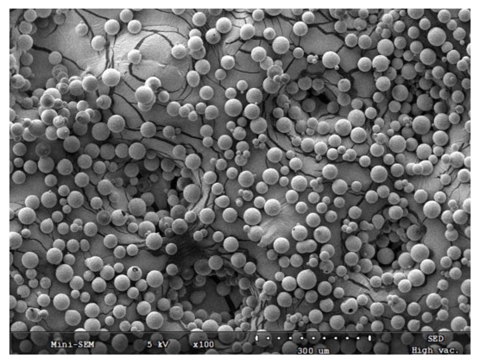
Lee et al. [34] explore the applications of poly-d, l-lactic acid (PDLLA) in dermatology, focusing on its effects on adipocytes and dermal remodeling in their literature review. The authors describe how PDLLA stimulates fibroblasts and adipocytes, enhancing collagen production and adipogenesis. By creating a supportive extracellular matrix, PDLLA promotes adipocyte differentiation and increases dermal thickness. The review also highlights its use in volume restoration, particularly in areas with adipose tissue atrophy. While the article provides a detailed overview of PDLLA’s mechanisms and clinical benefits, it lacks a systematic methodology and quantitative synthesis of data. The absence of meta-analysis limits its utility as high-level evidence. Nonetheless, the review serves as a valuable reference for clinicians and researchers exploring biostimulation in adipocyte-related applications (Level IIa). Overall, PDLLA aligns mechanistically with PLLA, supporting adipocyte differentiation and dermal remodeling within similar biostimulatory frameworks (Figure 1).
Ablon et al. [35] examine the use of platelet-rich plasma (PRP), exosomes, and stem cells in aesthetics, with a section dedicated to their effects on adipocytes in their narrative review. The authors discuss how these biostimulatory agents enhance adipocyte proliferation and differentiation, contributing to improved tissue regeneration and dermal volume. PRP and exosomes are highlighted for their ability to deliver growth factors that activate ADSCs, while stem cell therapies provide direct regenerative effects on adipose tissues. The review includes anecdotal evidence and expert opinions but lacks a critical evaluation of existing studies. The absence of standardized protocols and quantitative data reduces the reliability of the findings. Although the review provides valuable insights into emerging biostimulatory therapies, its limitations make it less robust for clinical decision-making (Level IIIb).
Barbosa et al. [36] introduce the concept of “body harmonization,” focusing on achieving balanced aesthetics through biostimulatory agents such as PLLA and CaHA. The authors discuss how these injectables stimulate adipocytes and collagen production to improve dermal quality and volume. The concept emphasizes using biostimulatory agents to enhance adipocyte activity in areas with volume loss or irregularities, creating visually balanced, anatomically harmonious results. While innovative, the article lacks supporting clinical data or trials to validate its claims. Theoretical frameworks and expert opinions form the basis of the discussion, which limits the strength of the evidence. Despite these shortcomings, the article provides a forward-thinking approach to integrating adipocyte stimulation into aesthetic practice (Level IIIc).
Bezpalko et al. [37] evaluate the effects of non-crosslinked hyaluronic acid-based fillers on dermal adipocytes and dermal quality using clinical and ultrasound assessments in their study. Subdermal injections were administered to improve hydration, dermal thickness, and elasticity. Ultrasound imaging confirmed increased adipose tissue volume and improved dermal structure in treated areas. Patient-reported outcomes indicated high satisfaction with aesthetic results. The study highlights the role of hyaluronic acid in supporting adipocyte activity and dermal remodeling. However, its non-randomized design and small sample size limit the generalizability of the findings. Despite these limitations, the use of objective ultrasound measures adds credibility to the results, making the study a valuable contribution to aesthetic medicine (Level IIb).
Nogueira et al. [38] provide a protocol for using PLLA products (Elleva and Elleva X) to treat skin flaccidity and improve adipocyte activity in their article. The authors outline injection techniques, dosages, and patient selection criteria aimed at enhancing skin tightness and dermal volume. They emphasize PLLA’s biostimulatory effects on adipocytes, leading to improved dermal thickness and elasticity over time. While practical, the article lacks supporting clinical trial data to validate the protocol’s effectiveness. The reliance on expert opinion and anecdotal evidence limits its strength as a scientific resource. Nonetheless, the detailed guidelines serve as a useful reference for clinicians exploring PLLA for body contouring and adipocyte stimulation (Level IIIb).
Dhillon et al. [39] discuss advancements in cellulite therapies, focusing on the synergistic effects of combination treatments, including biostimulatory agents such as PLLA and CaHA, in their review. The authors describe how these injectables stimulate adipocyte activity and enhance collagen production, leading to structural improvements in the dermal and subdermal layers. PLLA is highlighted for its ability to promote adipogenesis, improving volume and dermal thickness, while CaHA provides immediate volumization and long-term biostimulation. The review emphasizes tailoring treatment plans to optimize adipocyte stimulation and reduce cellulite appearance. However, the absence of a systematic approach or meta-analysis limits the robustness of the conclusions. While comprehensive, the review relies on non-randomized evidence and expert opinion, making it a valuable but lower-tier reference for clinical guidance (Level IIa).
Surowiecka et al. [18] investigate the potential of ADSCs in facial rejuvenation, focusing on their role in adipocyte stimulation in their review. The authors explain that ADSCs promote adipogenesis, collagen production, and dermal remodeling, making them an effective tool for addressing volume loss and skin laxity. Clinical and preclinical studies demonstrate improved skin elasticity and dermal thickness following ADSC-based treatments. The review highlights ADSCs’ regenerative effects, including their ability to differentiate into adipocytes and repair damaged tissue. However, most of the supporting evidence comes from small-scale trials and laboratory experiments, and long-term safety data are lacking. While promising, further research is required to establish standardized protocols and verify the clinical efficacy of ADSCs in large populations (Level IIb).
Mazzuco et al. [40] evaluate the effects of PLLA and CaHA on adipocyte activity and dermal quality in the arms in their split-side study. Participants received PLLA on one side and CaHA on the other to allow for direct comparison. Clinical assessments and histological analysis revealed that both agents significantly improved dermal thickness and collagen density. PLLA demonstrated superior adipocyte stimulation, promoting adipogenesis and long-term volumization. CaHA provided immediate volumetric effects but showed less sustained adipocyte activity. Patient satisfaction rates were high for both treatments, with minimal adverse events reported. The randomized design and histological confirmation strengthen the study’s validity, making it a robust contribution to evidence-based aesthetic medicine (Level Ib).
Silveira et al. [41] explore the use of hyperdilute CaHA microspheres for gluteal augmentation, focusing on their biostimulatory effects on adipocytes in their case series. The authors report improvements in volume, contour, and skin texture, attributing these outcomes to CaHA’s ability to enhance collagen synthesis and adipocyte differentiation. While patient satisfaction rates were high, the study lacked a control group and included a small sample size, limiting its generalizability. The findings suggest that hyperdilute CaHA may increase adipocyte activity and dermal volume, offering a non-surgical alternative for body contouring. However, further research is required to validate these preliminary results and establish standardized protocols (Level IIIc).
O’Daniel et al. [42] examine the use of PLLA for maintaining volume after facelift surgery with fat grafting in their observational study. The authors focus on PLLA’s ability to enhance adipocyte activity and support long-term volume retention. Clinical follow-ups revealed sustained improvements in dermal thickness and elasticity, which are attributed to PLLA’s biostimulatory effects on adipocyte differentiation and collagen production. Patients reported high satisfaction rates, with minimal adverse effects. However, the study’s observational design and lack of a control group limit the strength of the evidence. Despite these limitations, the findings provide practical insights into integrating PLLA into post-surgical aesthetic care (Level IIIb).
Sparavigna et al. [43] evaluate the effects of hybrid hyaluronan complexes in a hyaluronan intradermal injection in the neck in their clinical study. Patients received intradermal injections, and outcomes were assessed using subjective and objective measures. The results demonstrated significant improvements in dermal thickness, elasticity, and hydration, which are attributed to hyaluronan’s ability to support adipocyte activity and extracellular matrix remodeling. The study’s strengths include the use of quantitative assessments, such as elasticity tests, but its non-randomized design limits the generalizability of the findings. Overall, the study supports the use of hyaluronan complexes in addressing skin laxity and promoting adipocyte health (Level IIb).
Munia et al. [44] examine the effects of PLLA injections by using a vector technique for facial remodeling in their case series. The authors report that PLLA promotes collagen synthesis and adipocyte differentiation, resulting in improved facial volume and contour. The vector technique ensures even distribution of PLLA, optimizing its biostimulatory effects on the adipocytes and dermal layers. Patients demonstrated increased dermal thickness and elasticity over time, with no significant adverse effects. While the findings highlight PLLA’s potential for adipocyte stimulation and volume restoration, the small sample size and lack of control limit the strength of the conclusions. The study provides a foundation for further research on the vector technique in aesthetic applications (Level IIIc).
Nikolis et al. [45] compare traditional and extended injection techniques of PLLA for enhancing the temporal fossae in their randomized controlled trial. The study demonstrates that PLLA stimulates adipocyte activity, increasing dermal thickness and restoring volume in the treated areas. The extended technique showed superior results in terms of volume uniformity and patient satisfaction. Histological analysis confirmed enhanced adipocyte differentiation and collagen deposition, providing strong evidence for PLLA’s dual biostimulatory effects. Adverse events were minimal and self-limiting. The randomized design and direct comparison strengthen the reliability of the findings, making this a robust contribution to evidence-based facial rejuvenation practices (Level Ib).
Sarlos et al. [46] investigate the combined use of PLLA and hyaluronic acid in addressing fat loss and skin sagging after weight loss from semaglutide therapy in their observational study. The authors report that PLLA enhances adipocyte differentiation and collagen production, while hyaluronic acid provides immediate volume restoration. The combination effectively improved facial contour and dermal elasticity. Patient satisfaction was high, and no significant complications were reported. While the study underscores the synergistic effects of PLLA and hyaluronic acid in adipocyte stimulation and dermal remodeling, its observational nature and small sample size limit the strength of the evidence (Level IIIb).
Thomas et al. [47] provide an overview of non-surgical facial rejuvenation techniques, with a focus on biostimulatory agents such as PLLA and CaHA in that book chapter. The authors highlight the effects of these injectables on adipocytes, emphasizing their role in collagen synthesis, dermal remodeling, and adipocyte stimulation. PLLA and CaHA are presented as effective tools for addressing volume loss and improving skin elasticity. The chapter offers practical guidance on injection techniques and patient selection. However, the information is largely based on expert opinion and lacks supporting clinical trial data, limiting its evidentiary strength (Level V).
Kim et al. [48] explore the use of platelet-rich plasma (PRP) in dermal augmentation, focusing on its effects on adipocytes in their book chapter. PRP is described as a rich source of growth factors that activate ADSCs and promote adipocyte differentiation. Clinical case studies demonstrate improvements in dermal thickness, elasticity, and adipose tissue volume, following PRP treatment. The chapter emphasizes PRP’s versatility and safety but acknowledges the lack of standardized protocols and large-scale evidence. While promising, the findings are based on anecdotal evidence and small studies, limiting their applicability (Level IV).
Xiao et al. [49] evaluate clinical studies on platelet-rich plasma (PRP) for facial rejuvenation, focusing on its effects on adipocytes in their systematic review. The authors report that PRP enhances adipocyte activity and dermal remodeling by delivering growth factors that stimulate ADSCs. Improved dermal thickness, elasticity, and adipose tissue volume were observed across multiple studies. However, the review highlights significant heterogeneity in the study design, protocols, and outcome measures, limiting the ability to draw definitive conclusions. Despite these limitations, the review underscores PRP’s potential for adipocyte stimulation and regenerative aesthetics (Level IIa).
Mazzuco et al. [50] discuss the use of injectable fillers, including biostimulatory agents such as PLLA and CaHA, for treating cellulite in their book chapter. The authors highlight the mechanisms by which these agents stimulate adipocyte activity and enhance collagen production, leading to improvements in skin texture and elasticity. PLLA is noted for its gradual biostimulatory effects, promoting adipocyte differentiation and dermal remodeling over time. CaHA provides immediate volumization while supporting long-term dermal regeneration. The chapter also outlines injection techniques and patient selection criteria. While informative, the content is based on expert opinion and lacks supporting clinical studies, limiting the strength of its conclusions (Level V).
Lin et al. [51] document the use of injectable poly-D, L-lactic acid (PDLLA) for facial rejuvenation in their case report series. The authors describe how PDLLA stimulates adipocytes and fibroblasts, leading to improved dermal elasticity and volume restoration. Over a six-month follow-up, patients exhibited enhanced skin texture and increased dermal thickness, with no significant adverse effects. The study underscores the effectiveness of PDLLA in stimulating adipocyte activity for aesthetic improvements. However, the evidence is limited by the small number of cases and the lack of a control group. The findings provide preliminary insights into PDLLA’s potential but require validation through larger studies (Level IV).
Rovatti et al. [52] evaluate the use of hyperdiluted CaHA for mid- and lower-facial rejuvenation, focusing on its biostimulatory effects on adipocytes in their clinical study. The authors report significant improvements in skin elasticity, dermal thickness, and volume restoration, which are attributed to CaHA’s ability to stimulate adipocyte differentiation and collagen synthesis. Patient satisfaction rates were high, with minimal adverse effects. The study’s strengths include its prospective design and quantitative assessments (e.g., elastography/skin elasticity metrics). However, the small sample size and lack of a control group limit its generalizability. Despite these limitations, the study provides valuable evidence for CaHA’s role in adipocyte stimulation and facial rejuvenation (Level IIb).
Othman et al. [53] examine various approaches to temporal augmentation, including the use of biostimulatory injectables like PLLA and CaHA, in their systematic review. The authors describe how these agents stimulate adipocyte activity and collagen production, leading to improved volume and contour in the temporal region. The review highlights the effectiveness of PLLA in promoting gradual and sustained adipocyte stimulation for long-term results. However, the included studies vary widely in their methodology, and a meta-analysis was not performed. While the review provides a comprehensive overview, the lack of uniformity among the studies limits the strength of its conclusions (Level IIa).
Gil-del Valle et al. [54] investigate the effects of platelet-rich plasma (PRP) activated with ozone on facial lipoatrophy in HIV patients in their study. The authors report that PRP enhances adipocyte activity and restores dermal volume by delivering growth factors that stimulate ADSCs. Improvements in skin texture, elasticity, and volume were observed, along with enhanced cellular redox balance. The treatment also significantly improved patients’ quality of life. While promising, the study’s small sample size and specific patient population limit its generalizability. Nonetheless, it provides important insights into the role of PRP in adipocyte stimulation and dermal regeneration (Level IIb).
Collectively, PRP and related biologics activate ADSCs and enhance adipocyte differentiation, showing regenerative promise with favorable safety, yet high variability in preparation (centrifugation protocols, platelet concentration, activation methods) hampers cross-study comparison.
de Albuquerque et al. [55] explore the use of fillers and collagen stimulators, such as PLLA and CaHA, for body rejuvenation and cellulite treatment in their book chapter. The authors discuss how these injectables stimulate adipocyte activity and collagen synthesis, improving skin elasticity and texture. PLLA is highlighted for its gradual and sustained effects, while CaHA offers immediate results with long-term benefits. The chapter includes practical guidance on injection techniques, but lacks supporting clinical data. The reliance on expert opinion limits its evidentiary strength, though it provides a useful resource for aesthetic practitioners (Level V).
da Cunha et al. [56] evaluate the use of CaHA for treating facial aging, focusing on its effects on adipocytes and dermal remodeling in their study. The authors report significant improvements in skin elasticity, dermal thickness, and facial volume after CaHA injections. These outcomes are attributed to CaHA’s ability to stimulate adipocyte differentiation and collagen production. Patient satisfaction rates were high, and no major complications were reported. The study emphasizes the importance of combining CaHA with lifting techniques for optimal results. However, the lack of a control group and small sample size reduce the strength of the evidence (Level IIb).
CaHA appears to combine immediate volumization with progressive biostimulation; although safety and efficacy are well-documented and crosslinking/dilution and injection plane vary widely, leaving durability uncertain.
Davis et al. [57] discuss combination treatments for cellulite, including biostimulatory agents like PLLA and CaHA, in their review. The authors highlight how these injectables enhance adipocyte activity, collagen production, and dermal remodeling, leading to improvements in skin texture and elasticity. They emphasize the importance of patient selection and treatment customization to achieve optimal results. While the review includes clinical insights and case studies, it lacks systematic methodology and quantitative data, relying instead on expert opinion and anecdotal evidence. Despite these limitations, it offers valuable guidance for clinicians addressing cellulite with biostimulatory agents (Level IV).
Palermo et al. [58] introduce a three-dimensional approach to facial rejuvenation, integrating biostimulatory agents such as PLLA and CaHA, in their book chapter. The authors discuss the role of these injectables in stimulating adipocyte activity and collagen production to restore volume and contour in aging patients. Techniques for achieving natural-looking results are outlined, focusing on individualized treatment plans. While the chapter provides practical insights, it lacks supporting clinical trial data, relying heavily on theoretical frameworks and expert opinion. The approach offers a comprehensive perspective but requires further validation (Level V).
Zarei et al. [59] examine the application of cell therapy, including ADSCs, for facial anti-aging in their review. The authors describe how ADSCs promote adipogenesis and dermal remodeling, improving skin elasticity and volume. Clinical trials show promising results in facial rejuvenation, with increased dermal thickness and reduced signs of aging. However, the review highlights challenges such as variability in protocols and a lack of long-term safety data. While the findings are encouraging, further research is needed to standardize cell therapy for clinical use (Level IIa).
Antonio et al. [60] explore cellular biomodulation as an emerging approach in dermatology, with a focus on ADSCs and platelet-rich plasma (PRP) in their article. The authors highlight the regenerative potential of these therapies, emphasizing their ability to stimulate adipocyte differentiation and dermal remodeling. Case studies demonstrate improvements in skin elasticity, texture, and volume. While the article provides a forward-looking perspective, it relies on theoretical concepts and small-scale studies, limiting its evidence strength. Nonetheless, it offers a vision for the future of dermatology and regenerative aesthetics (Level IIIc).
Aunna Pourang et al. [61] review the use of stem cells and autologous therapies, such as platelet-rich plasma (PRP), in facial rejuvenation in their book chapter. The authors discuss how these treatments enhance adipocyte activity and promote dermal regeneration, leading to improved skin elasticity and volume. Clinical case studies show promising results, but the chapter notes the lack of standardized protocols and large-scale evidence. While informative, the reliance on anecdotal evidence and expert opinion limits its strength as a scientific resource. The chapter underscores the potential of regenerative therapies in aesthetic medicine (Level IV).
Jáñez et al. [62] focus on the applications of platelet-rich plasma (PRP) in aesthetic medicine, particularly its effects on adipocytes, in their book chapter. PRP delivers growth factors that activate ADSCs, promoting adipogenesis and dermal remodeling. Clinical applications demonstrate improvements in skin elasticity, texture, and volume, with minimal adverse effects. The authors emphasize PRP’s safety and versatility but acknowledge the lack of large-scale studies and standardized protocols. While promising, the findings are primarily based on small-scale trials and expert opinion, limiting their generalizability (Level V).
Alessandrini et al. [63] evaluate the effects of auto-cross-linked hyaluronic acid on the décolletage, focusing on its impact on dermal quality and adipocyte activity in their pilot study. The treatment improved skin hydration, elasticity, and volume, with no significant adverse effects reported. While the study highlights hyaluronic acid’s potential for stimulating adipocytes and enhancing dermal remodeling, its small sample size and short follow-up period limit the strength of the conclusions. The findings provide preliminary evidence for hyaluronic acid’s efficacy in rejuvenating the décolletage (Level IIb).
HA-based fillers reliably improve dermal hydration, elasticity, and subdermal structure, but most studies are short-term and non-randomized, and crosslinking/dilution and injection plane vary widely, leaving their durability uncertain.
Overall, the reviewed literature supports the regenerative role of biostimulatory injectables in promoting adipocyte activity and collagen remodeling. PLLA and PCL demonstrate the most consistent histologic and clinical improvements, CaHA provides immediate volumization with sustained biostimulation, and HA and PRP act as complementary or synergistic agents, enhancing hydration and tissue metabolism. Despite encouraging outcomes, small sample sizes, heterogeneous protocols, and short follow-up periods limit definitive conclusions. Standardized methodologies and long-term comparative trials are warranted to establish durability and safety across materials.
Table 1. Summary of the effects of biostimulatory agents on adipocytes.
StudyAgents/MethodsFindingsLimitationsLevel
Turkevych et al. [30]PCLEnhanced ADSC differentiation, collagen synthesis, improved dermal thickness, and elasticity.Lacks large-scale trials and longitudinal data.IIb
Bota et al. [1]PLLA, PCL, CaHAInduced collagen production and adipogenesis; improved dermal remodeling.Lacks meta-analysis, relies on heterogeneous studies.IIa
Radke et al. [31]PLLAPromoted fibroblast stimulation, collagen deposition, and adipocyte metabolic activity.Lacks quantitative data, anecdotal evidence.IIIb
De Paula Barbosa et al. [32]PLLAImproved dermal thickness and skin elasticity; high patient satisfaction.Small sample size, no control group.IIb
Jin et al. [33]PLLALactate enhances adipocyte metabolism; reduced adipose tissue volume, improved dermal quality.Lacks large-scale trials.IIb
Melfa et al. [27]SEFFI and CaHAImproved skin elasticity and volume; supported adipogenesis and tissue repair.Retrospective design, lacks control group.IIIb
Zubair et al. [29]PLLASignificant increases in dermal thickness and patient satisfaction; enhanced adipocyte activity.Focused on one area, potential for bias.Ib
Lee et al. [34]Poly-D, L-lactic acid (PDLLA)Stimulated fibroblasts/adipocytes, enhanced collagen production, and adipogenesis.Lacks systematic methodology, no quantitative synthesis.IIa
Ablon et al. [35]PRP, exosomes, stem cellsEnhanced adipocyte proliferation and differentiation; improved tissue regeneration.Lacks standardized protocols, anecdotal evidence.IIIb
Barbosa et al. [36]PLLA, CaHAImproved dermal quality and volume through adipocyte stimulation.Lacks clinical data to support claims.IIIc
Bezpalko et al. [37]Hyaluronic acid fillersIncreased adipose tissue volume, improved skin hydration and elasticity.Non-randomized design, small sample size.IIb
Nogueira et al. [38]PLLAProtocol outlined for improving skin flaccidity and adipocyte activity; biostimulatory effects noted.Lacks clinical trial data.IIIb
Dhillon et al. [39]PLLA, CaHAEnhanced adipocyte activity and collagen production; tailored treatment plans for cellulite.Lacks systematic approach, relies on non-randomized evidence.IIa
Surowiecka et al. [18]ADSCsPromoted adipogenesis and collagen production, improved skin elasticity and thickness.Most evidence from small-scale trials.IIb
Mazzuco et al. [40]PLLA, CaHAImproved dermal thickness and collagen density; PLLA showed superior adipocyte stimulation.Small sample size, lacks control group.Ib
Silveira et al. [41]Hyperdiluted CaHASignificant improvements in volume and skin texture, attributed to CaHA’s effects.Small sample size, lacks control group.IIIc
O’Daniel et al. [42]PLLAEnhanced volume retention post-facelift; high patient satisfaction.Observational design, lacks control group.IIIb
Sparavigna et al. [43]Hybrid hyaluronan complexesImproved dermal thickness and elasticity; supports adipocyte activity.Non-randomized design, limited generalizability.IIb
Munia et al. [44]PLLAImproved facial volume and contour using vector technique; enhanced adipocyte stimulation.Small sample size, lacks control group.IIIc
Nikolis et al. [45]PLLADemonstrated enhanced adipocyte activity and dermal thickness with extended injection technique.Limited study scope; generalizability may be affected.Ib
Sarlos et al. [46]PLLA, hyaluronic acidImproved facial contour and dermal elasticity; high patient satisfaction.Observational nature, small sample size.IIIb
Thomas et al. [47]PLLA, CaHAOverview of techniques and effects on adipocytes; practical guidance for clinicians.Lacks supporting clinical data; relies on expert opinion.V
Kim et al. [48]PRPEnhances adipocyte activity and dermal volume; promising results from case studies.Lacks large-scale evidence; based on anecdotal evidence.IV
Xiao et al. [49]PRPEnhanced adipocyte activity and dermal remodeling; observed improvements across studies.Significant heterogeneity in study design; limited conclusions.IIa
Mazzuco et al. [50]PLLA, CaHADiscusses mechanisms for cellulite treatment; highlights gradual stimulation effects.Lacks supporting clinical studies; based on expert opinion.V
Lin et al. [51]PDLLAImproved dermal elasticity and volume restoration; significant follow-up outcomes.Limited by small number of cases; lacks control group.IV
Rovatti et al. [52]Hyperdiluted CaHASignificant improvements in facial rejuvenation; high patient satisfaction.Small sample size; lacks control group.IIb
Othman et al. [53]PLLA, CaHAComprehensive overview of biostimulatory effects; highlights effectiveness for temporal augmentation.Lacks uniformity among studies; no meta-analysis performed.IIa
Gil-del Valle et al. [54]PRP with ozoneImproved dermal volume and quality of life in HIV patients; promotes adipocyte activity.Small sample size; specific patient population limits generalizability.IIb
de Albuquerque et al. [55]PLLA, CaHADiscusses body rejuvenation techniques; highlights gradual and immediate effects of injectables.Lacks supporting clinical data; relies on expert opinion.V
da Cunha et al. [56]CaHASignificant improvements in facial aging; high patient satisfaction.Lack of control group; small sample size.IIb
Davis et al. [57]PLLA, CaHAHighlights combination treatments for cellulite; emphasizes treatment customization.Lacks systematic methodology; based on expert opinion.IV
Palermo et al. [58]PLLA, CaHAIntroduces a three-dimensional approach to rejuvenation; discusses individualized treatment plans.Lacks supporting clinical trial data; relies on expert opinion.V
Zarei et al. [59]ADSCsPromotes adipogenesis and dermal remodeling; shows promise in facial rejuvenation.Variability in protocols; lacks long-term safety data.IIa
Antonio et al. [60]ADSCs, PRPHighlights regenerative potential; improves skin elasticity and volume.Relies on theoretical concepts; small-scale studies limit evidence strength.IIIc
Aunna Pourang et al. [61]PRPEnhances adipocyte activity; promising case studies reported.Lacks standardized protocols; relies on anecdotal evidence.IV
Jáñez et al. [62]PRPDelivers growth factors that enhance adipocyte activity; minimal adverse effects noted.Based on small-scale trials; lacks large-scale studies.V
Alessandrini et al. [63]Hyaluronic acidImproved skin hydration and volume; potential for adipocyte stimulation.Small sample size; short follow-up period.IIb

3. Discussion

The use of biostimulatory agents such as PLLA, PCL, and CaHA has become a cornerstone in regenerative aesthetics, driven by their capacity to stimulate collagen production and promote adipocyte activity. This discussion examines the evidence surrounding their mechanisms of action, clinical efficacy, safety, and emerging applications, integrating findings from key studies.

3.1. Mechanisms of Action

Biostimulatory agents rejuvenate the skin by inducing neocollagenesis, extracellular matrix remodeling, and adipocyte stimulation. PLLA, a biodegradable synthetic polymer, generates a subclinical inflammatory response, stimulating fibroblast activity and collagen synthesis via the TGF-β/Smad pathway. Studies by Radke et al. [31] and Jin et al. [33] emphasize that PLLA’s metabolite, lactate, encourages adipocyte differentiation, aiding in dermal volume restoration. Interestingly, while most studies demonstrate PLLA-induced adipogenesis and dermal volumization, Jin et al. [33] reported that its metabolite lactate may, under certain conditions, contribute to adipose tissue reduction. This dual role of PLLA suggests that its biological effects on adipose tissue depend on the concentration, local metabolism, and treatment protocol, highlighting the complexity of its regenerative mechanisms. Over time, PLLA particles degrade, leaving behind newly formed collagen, which maintains its dermal integrity [1].
PCL serves as a biodegradable scaffold for ADSCs, promoting their proliferation and differentiation into mature adipocytes. Turkevych et al. [30] demonstrated in their experimental trials that PCL enhances adipogenesis and extracellular matrix synthesis, supporting long-term dermal remodeling. Its slower degradation rate compared to PLLA ensures sustained collagen production and volumization.
CaHA combines immediate volumization with long-term collagen stimulation. Its calcium microspheres stimulate fibroblast activity and angiogenesis, while acting as a physical scaffold for tissue regeneration [40]. The research by Silveira et al. [41] highlights its role in volumizing and rejuvenating gluteal and facial regions, further validating its efficacy (Figure 2).

3.2. Clinical Efficacy

Several studies have substantiated the clinical efficacy of biostimulatory agents in improving skin laxity, elasticity, and overall dermal quality. In their review, Boța et al. [1] emphasized PLLA’s effectiveness in restoring midface volume and correcting skin laxity. Similarly, the SPLASH randomized trial by Zubair et al. [29] demonstrated the ability of PLLA to enhance adipogenesis and volumization in the hip dell, with significant improvements in patient satisfaction.
PCL also shows promising results in clinical applications. Turkevych et al. [30] observed that PCL significantly improves dermal thickness and elasticity.
CaHA has been widely studied for its dual effects on volumization and dermal remodeling. Melfa et al. [27] explored the combination of SEFFI (superficial enhanced fluid fat injection) with CaHA in a retrospective observational study, reporting substantial improvements in dermal quality and elasticity, particularly in aging patients. Silveira et al. [41] also highlighted CaHA’s efficacy in gluteal augmentation, with excellent patient satisfaction and minimal complications.
Combination therapies can further enhance the outcomes of biostimulatory agents. Barbosa et al. [36] noted that protocols combining CaHA with energy-based devices like high-intensity focused ultrasound (HIFU) and fractional lasers yield superior results in skin tightening and texture improvement. Similarly, Dhillon et al. [39] emphasized the role of PLLA in cellulite reduction when combined with other treatments, such as subcision and energy-based devices.
Agent-level advantages/limitations. PLLA offers gradual, durable neocollagenesis with concurrent adipocyte stimulation; the advantages include longevity and broad indication flexibility, whereas the disadvantages include delayed onset and nodule risk without proper dilution/massage. PCL provides scaffold-driven ADSC support and long-lasting remodeling; the advantages are durability and structural ECM effects, and the disadvantages are technique sensitivity and limited large RCTs. CaHA uniquely bridges immediate volumization with regeneration; the advantages are early visible effect and tissue quality gains, while the disadvantages are plane-specific vascular risk and variability with hyperdilution. HA reliably improves hydration/elasticity and shapes contours; the advantages are reversibility and safety, while the durability is shorter and true biostimulation is limited. PRP/biologics add low-morbidity metabolic support; the advantages are safety and synergy, and the disadvantages are protocol heterogeneity and inconsistent objective endpoints.
Evidence horizon and safety strength by material. PLLA—moderate-to-long clinical follow-up (up to and beyond 12 months in RCTs/split-side and prospective series) with a predictable AE profile when dilution/massage are standardized; PCL—emerging clinical follow-up (6–12 months; strong preclinical mechanistic data), with growing but smaller prospective human datasets; CaHA—consistent 6–12+ month outcomes in multiple prospective studies and consensus guidance, an immediate effect with well-characterized safety when plane/hyperdilution are respected; HA—short-to-mid follow-up but the strongest safety margin and reversibility; PRP/biologics—favorable safety across small trials/series, yet heterogeneous preparation and a shorter follow-up.
Comparability across protocols. Despite encouraging synergy between combined modalities (e.g., PLLA with energy-based devices; CaHA with autologous fat/SEFFI), direct comparability remains limited due to heterogeneity in dilution ratios, injection planes/cannula caliber, device parameters (fluence, pulse width, passes), session spacing, and follow-up intervals. We therefore interpret cross-study differences qualitatively and call for consensus frameworks to harmonize these variables to enable a reproducible and comparable outcome assessment.

3.3. Safety Considerations

Biostimulatory agents are generally safe when administered by trained professionals. However, adverse events, such as nodules, granulomas, and vascular complications, have been reported when injection techniques or patient selection are suboptimal [1]. Radke et al. [31] stressed the importance of proper post-treatment massage in PLLA applications to prevent nodule formation.
Mild side effects like erythema, swelling, and tenderness are common but transient. In rare cases, delayed-onset nodules may develop due to excessive collagen stimulation, as observed in a study by Mazzuco et al. [40]. To mitigate risks, practitioners should adhere to standardized injection protocols and carefully assess patient suitability.
Combination stance and risks. We favor matrix-first sequencing (PLLA/PCL), followed by immediate/contour agents (CaHA/HA) when needed, spacing the sessions to avoid inflammatory overlap. When pairing with energy-based devices, schedule the EBD 2–4 weeks before the initial biostimulator or ≥4 weeks after the final session. Key mitigations include the correct plane (cannula where appropriate), adequate dilution, slow injection with aspiration awareness, and a post-procedure massage for PLLA. Avoid same-day stacking of multiple stimulators in one plane; document product, plane, and dilution to facilitate nodule/granuloma management.

3.4. Standardized Protocols

Existing standardization and complication-avoidance measures are summarized here, including dilution ranges, injection planes, cannula selection, massage protocols for PLLA, vascular safety precautions, and management of nodules or Tyndall-like effects.
However, the absence of universally accepted, evidence-based protocols remains a major challenge in aesthetic medicine.
According to Nogueira et al. [38], optimized protocols for PLLA involve diluting the product adequately and spacing treatments to maximize collagen production while minimizing side effects. Similarly, Turkevych et al. [30] recommend specific dilution ratios and injection techniques for PCL, to ensure consistent outcomes.
For CaHA, Melfa et al. [27] provided detailed guidance on injection techniques for various body areas, including the face, neck, and gluteal regions. They emphasized the importance of hyperdilution for achieving uniform results and minimizing adverse effects.

3.5. Emerging Applications and Combination Therapies

Biostimulatory agents are increasingly integrated into combination protocols to enhance their efficacy. Barbosa et al. [36] described the concept of “body harmonization,” where PLLA, PCL, and CaHA are used in conjunction with dermal fillers, botulinum toxins, and energy-based devices to achieve comprehensive rejuvenation. Dhillon et al. [39] also highlighted the synergistic effects of combining PLLA with subcision and radiofrequency for cellulite reduction.
Innovative techniques, such as SEFFI with CaHA, have gained traction for their ability to enhance adipocyte activity and improve dermal quality. Melfa et al. [27] reported that this combination yields significant improvements in dermal elasticity and volume, particularly in patients with age-related fat loss.
Age-stratified guidance. In younger patients (≤35) with early laxity or texture change, HA (for hydration/contour) and PRP/biologics (metabolic support) predominate; small-volume PLLA can pre-empt laxity in high-movement zones. In midlife (≈35–55) with combined laxity/volume loss, PLLA (durability) or PCL (scaffold-dominant) anchor remodeling; CaHA (often hyperdiluted) addresses neck, arms, or gluteal quality when an immediate effect is desirable. In older patients (≥55) with advanced laxity and deflation, staged PLLA/PCL for baseline matrix + CaHA for early correction + HA for contour refinements yields predictable gains; PRP augments healing in thin, fragile skin. Post-weight-loss or GLP-1–associated deflation benefits from matrix-first (PLLA/PCL) with selective CaHA or HA top-ups (Table 2).

3.6. Limitations and Future Directions

While the efficacy of biostimulatory agents is increasingly supported, several limitations constrain inference. Many studies, such as those by Jin et al. [33] and Mazzuco et al. [40], lack large-scale, randomized designs, limiting the generalizability of their findings. Most available data are small, single-center, and short-term; preparation (e.g., dilution, crosslinking, activation) and injection parameters vary widely; objective endpoints are inconsistently applied; and long-term safety reporting is sparse. The current evidence remains limited by pronounced inter-study heterogeneity, scarcity of randomized controlled trials, and insufficient long-term safety data beyond 12–24 months.
Future research priorities include the following:
(1)
Protocol standardization: Define agent-specific preparation (dilution, particle size/activation), injection planes, volumes, and session spacing to reduce heterogeneity and enable pooling.
To improve cross-study comparability, future reports should also specify a minimum reporting set, including: (i) product preparation (brand, particle size, dilution), (ii) plane and instrument (cannula vs. needle; gauge), (iii) device parameters when applicable (energy type, fluence, pulse width, passes), (iv) session spacing and sequence (matrix-first vs. immediate agents), (v) objective endpoints (ultrasound thickness, elastography, histology) with pre-specified timepoints (e.g., 3/6/12 months), and (vi) AE capture windows (early/delayed). Establishing such a dataset will enhance reproducibility and enable a meaningful comparison across combination protocols.
(2)
Head-to-head randomized trials: Compare PLLA, PCL, and CaHA (±HA/PRP) with prespecified objective endpoints (ultrasound dermal thickness, elastography, histology), blinded assessments, and ≥12–24 month follow-up.
(3)
Mechanistic readouts: Integrate imaging and tissue biomarkers (collagen type I/III ratios, ECM organization) to link dose, kinetics (onset vs. durability), and clinical effect.
(4)
Safety surveillance: Establish prospective registries to quantify delayed nodules/granulomas, vascular events, and mitigation strategies (e.g., massage, cannula use, reversal/management algorithms).
(5)
Patient stratification and indications: Identify responders by phenotype (age, skin laxity, weight-loss status) and optimize combination algorithms (e.g., SEFFI + CaHA, PLLA with energy-based devices) by anatomical site.
(6)
Methodological rigor: Standardize core outcome sets, incorporate allocation concealment/blinding, and report attrition/selective reporting to minimize bias.
(7)
Health economics and QoL: Include cost-effectiveness and validated patient-reported outcomes alongside objective measures.

4. Methodology

Keywords including “Biostimulator”, “Poly L Lactic Acid”, “PLLA”, “Polycaprolactone”, “PCL”, “Calcium Hydroxyapatite”, “CaHA”, “Adipocyte”, “Fat Cell”, “adipose-derived stem cell”, were searched for in the MEDLINE, PubMed and Ovid databases for relevant studies published on clinical trials, diagnosis and treatment from year 2018 onward. Some papers were further reviewed using a double-blinding approach, sample size, control usage, randomization usage, and objective endpoint measurements. All studies were classified according to the Oxford Center for evidence-based medicine evidence hierarchy [64].
Two reviewers independently extracted objective endpoints (e.g., ultrasound dermal thickness, elastography, histology) and safety events, and rated study quality using the Oxford hierarchy together with domain-level risk-of-bias items (randomization, allocation concealment, blinding, attrition, and selective reporting). For synthesis, each study was then mapped onto the five-axis analytical framework (mechanism, onset vs. durability, evidence tier, safety, and indication) to enable consistent cross-study comparisons without altering the underlying study designs.

5. Conclusions

Biostimulatory agents, including PLLA, PCL, and CaHA, represent a major advancement in regenerative aesthetics. Their ability to stimulate collagen production, enhance adipogenesis, and improve dermal quality makes them invaluable tools for addressing aging-related concerns. Within the proposed analytical framework, PLLA demonstrates the strongest balance between durability and biostimulation, PCL provides mechanistic support via ADSC scaffolding, and CaHA bridges immediate volumization with regenerative longevity, while HA and PRP serve as adjunctive modulators, rather than primary stimulators. This analytical approach underscores the comparative strengths and clinical nuances among current biostimulatory agents.
From a clinical perspective, PLLA currently demonstrates the most robust long-term performance, supported by randomized and split-side trials confirming ≥12 month durability and consistent improvements in dermal thickness and elasticity (Zubair et al. [29]; Nikolis et al. [45]). PCL offers emerging value for scaffold-driven remodeling of the jawline and neck, due to its slow biodegradation and ADSC support (Turkevych et al. [30]). CaHA provides immediate volumization and progressive regeneration, which is particularly effective in the neck, arms, and gluteal areas (Mazzuco et al. [40]; Silveira et al. [41]). HA and PRP/biologic adjuncts complement these agents by improving hydration, recovery, and tissue metabolism. Collectively, these distinctions define a tiered, indication-based approach to optimize the treatment selection in regenerative aesthetics.
These clinical insights have been shaped by key research groups whose work established the current evidence base. Zubair et al. [29] conducted randomized and split-side PLLA trials, providing long-term comparative outcomes. Turkevych et al. [30] advanced the mechanistic understanding of PCL and ADSCs scaffolding. Melfa et al. [27] developed and clinically validated SEFFI-with-CaHA techniques for enhanced adipocyte activity. Silveira et al. [41] standardized hyperdiluted CaHA use for facial and gluteal rejuvenation. Mazzuco et al. [40] provided histologic validation in comparative studies of PLLA and CaHA, while Xiao et al. [49] synthesized platelet-rich plasma evidence through a systematic review. Together, these groups represent the most methodologically visible lines of research driving clinical standardization and innovation in regenerative aesthetics.
Despite these advances, important evidence gaps remain. Broader standardization across protocols, direct head-to-head comparisons with objective endpoints, and long-term safety registries are still needed to guide durable, indication-specific algorithms.
Looking ahead, these priorities can be translated into practical clinical guidance. In practice, a matrix-first, age-stratified algorithm (≤35: HA/PRP ± micro-PLLA; 35–55: PLLA/PCL backbone with selective CaHA; ≥55: staged PLLA/PCL plus CaHA, HA for contour) balances durability with safety. Our opinion favors staged combinations over same-day stacking, with documented planes/dilutions and deferred energy devices to minimize adverse events.
While the current evidence supports their efficacy and safety, further research is needed to standardize protocols and optimize treatment outcomes. By continuing to explore the potential of biostimulatory agents, aesthetic practitioners can deliver innovative, anatomically harmonious, and long-lasting outcomes for their patients.

Author Contributions

Conceptualization—K.W.A.L., H.K. (Heesoo Kim), J.K.S., O.S., W.K.F., I.R., H.K. (Hongseok Kim), K.H.L., M.H.G. and K.Y.; Writing—original draft preparation, K.W.A.L., H.K. (Heesoo Kim), J.K.S., O.S., W.K.F. and I.R.; Writing—review and editing, O.S., W.K.F., I.R., H.K. (Hongseok Kim), K.H.L., M.H.G. and K.Y.; Supervision—K.Y. All authors have read and agreed to the published version of the manuscript.

Funding

The current research has received no funding.

Data Availability Statement

The original contributions presented in this study are included in the article. Further inquiries can be directed to the corresponding author.

Conflicts of Interest

All authors were employed by clinical, medical, or commercial institutions at the time of this work, as follows: Kar Wai Alvin Lee (Everkeen Medical Centre, Hong Kong); Heesoo Kim (Jetema Co., Ltd., Seoul, Republic of Korea); Jong Keun Song (Pixelab Plastic Surgery Clinic, Seoul, Republic of Korea); Olena Sydorchuk (ICON Inc., Seoul, Republic of Korea); Wong Ka Fai (Gaddiel Medical Group Limited, Hong Kong); Isabella Rosellini (Avery Beauty Clinic and Avena Aesthetics, Jakarta, Indonesia); Hongseok Kim (VOS Dermatology Clinic, Seoul, Republic of Korea); Kian Hong Lau (EC Skin Laser Clinic, Penang, Malaysia); Michael H. Gold (The Tennessee Clinical Research Center, Nashville, TN, USA); and Kyuho Yi (Yonsei University College of Dentistry, Seoul, Republic of Korea). Although all authors are affiliated with medical or commercial institutions, the authors declare that the research was conducted without any financial, commercial, or institutional influence that could be construed as a potential conflict of interest.

References

  1. Boța, M.; Cristea, A.M.; Vlaia, L.L.; Vlaia, V. The impact of injectable biostimulatory substances on current trends in aesthetic medicine: Focus on poly-L-lactic acid, polycaprolactone, and calcium hydroxyapatite. Med. Evol. 2025, 31, 177–186. [Google Scholar] [CrossRef]
  2. Fisher, S.M.; Borab, Z.; Weir, D.; Rohrich, R.J. The emerging role of biostimulators as an adjunct in facial rejuvenation: A systematic review. J. Plast. Reconstr. Aesthetic Surg. 2024, 92, 118–129. [Google Scholar] [CrossRef] [PubMed]
  3. Pan, Y.; Hao, Y.; Xiao, Y.; Shi, K.; Qu, Y.; Qian, Z. Injectable soft tissue nano/micro fillers for facial reconstruction. J. Biomed. Nanotechnol. 2021, 17, 1–17. [Google Scholar] [CrossRef] [PubMed]
  4. da Cunha, M.G.; Engracia, M.; de Souza, L.G.; Filho, C.D.A.M. Biostimulators and their mechanisms of action. Surg. Cosmet. Dermatol. 2020, 12, 109–117. [Google Scholar]
  5. Wong, T.H.S. A revision and summary of injectable fillers. J. Cosmet. Med. 2020, 4, 7–11. [Google Scholar] [CrossRef]
  6. Haddad, S.; Galadari, H.; Patil, A.; Goldust, M.; Al Salam, S.; Guida, S. Evaluation of the biostimulatory effects and the level of neocollagenesis of dermal fillers: A review. Int. J. Dermatol. 2022, 61, 1284–1288. [Google Scholar] [CrossRef]
  7. Attenello, N.H.; Maas, C.S. Injectable fillers: Review of material and properties. Facial Plast. Surg. 2015, 31, 29–34. [Google Scholar] [CrossRef]
  8. Guo, J.; Fang, W.; Wang, F. Injectable fillers: Current status, physicochemical properties, function mechanism, and perspectives. RSC Adv. 2023, 13, 23841–23858. [Google Scholar] [CrossRef]
  9. Corduff, N.; Goldie, K.; Lin, F.; Lowe, S.; Siew, T.W.; Vachiramon, V.; Chao, Y.Y.; Lesthari, I.; Ong-Amoranto, B.; Lim, T.S.; et al. The evolving field of regenerative aesthetics: A review and case series. Cureus 2025, 17, e87878. [Google Scholar] [CrossRef]
  10. Rho, N.K.; Kim, H.S.; Kim, S.Y.; Lee, W. Injectable “skin boosters” in aging skin rejuvenation: A current overview. Arch. Plast. Surg. 2024, 51, 528–541. [Google Scholar] [CrossRef]
  11. Bellei, B.; Migliano, E.; Picardo, M. Therapeutic potential of adipose tissue derivatives in modern dermatology. Exp. Dermatol. 2022, 31, 1837–1852. [Google Scholar] [CrossRef]
  12. Gaur, M.; Dobke, M.; Lunyak, V.V. Mesenchymal stem cells from adipose tissue in clinical applications for dermatological indications and skin aging. Int. J. Mol. Sci. 2017, 18, 208. [Google Scholar] [CrossRef]
  13. Naderi, N.; Combellack, E.J.; Griffin, M.; Sedaghati, T.; Javed, M.; Findlay, M.W.; Wallace, C.G.; Mosahebi, A.; Butler, P.E.; Seifalian, A.M.; et al. The regenerative role of adipose-derived stem cells (ADSCs) in plastic and reconstructive surgery. Int. Wound J. 2017, 14, 112–124. [Google Scholar] [CrossRef]
  14. Mazini, L.; Rochette, L.; Amine, M.; Malka, G. Regenerative capacity of adipose-derived stem cells (ADSCs) compared with mesenchymal stem cells (MSCs). Int. J. Mol. Sci. 2019, 20, 2523. [Google Scholar] [CrossRef] [PubMed]
  15. Liu, M.; Lu, F.; Feng, J. Aging and homeostasis of the hypodermis in the age-related deterioration of skin function. Cell Death Dis. 2024, 15, 443. [Google Scholar] [CrossRef] [PubMed]
  16. Bonté, F.; Girard, D.; Archambault, J.C.; Desmoulière, A. Skin changes during ageing. In Biochemistry and Cell Biology of Ageing: Part II Clinical Science; Springer: Berlin/Heidelberg, Germany, 2019; pp. 249–280. [Google Scholar]
  17. Wollina, U.; Wetzker, R.; Abdel-Naser, M.B.; Kruglikov, I.L. Role of adipose tissue in facial aging. Clin. Interv. Aging 2017, 12, 2069–2076. [Google Scholar] [CrossRef] [PubMed]
  18. Surowiecka, A.; Strużyna, J. Adipose-derived stem cells for facial rejuvenation. J. Pers. Med. 2022, 12, 117. [Google Scholar] [CrossRef]
  19. Corduff, N. Introducing aesthetic regenerative scaffolds: An immunological perspective. J. Cosmet. Dermatol. 2023, 22, 8–14. [Google Scholar] [CrossRef]
  20. Christen, M.O. Collagen stimulators in body applications: A review focused on poly-L-lactic acid (PLLA). Clin. Cosmet. Investig. Dermatol. 2022, 15, 997–1019. [Google Scholar] [CrossRef]
  21. Oh, S.; Shin, N.; Lee, S.J.; Son, K.H.; Byun, K. Poly-L-lactic acid filler increases adipogenesis and adiponectin in aged subcutaneous tissue. Polymers 2025, 17, 1826. [Google Scholar] [CrossRef]
  22. Kim, J.S. Changes in dermal thickness in biopsy study of histologic findings after a single injection of polycaprolactone-based filler into the dermis. Aesthetic Surg. J. 2019, 39, NP484–NP494. [Google Scholar] [CrossRef]
  23. Christen, M.O.; Vercesi, F. Polycaprolactone: How a well-known and futuristic polymer has become an innovative collagen stimulator in esthetics. Clin. Cosmet. Investig. Dermatol. 2020, 13, 31–48. [Google Scholar] [CrossRef] [PubMed]
  24. Amiri, M.; Meçani, R.; Niehot, C.D.; Phillips, T.; Kolb, J.; Daughtry, H.; Muka, T. Skin regeneration-related mechanisms of calcium hydroxyapatite (CaHA): A systematic review. Front. Med. 2023, 10, 1195934. [Google Scholar] [CrossRef] [PubMed]
  25. Turkevych, A.; Turkevych, D. Influence of calcium hydroxyapatite on soft tissues—A critical viewpoint. J. Appl. Cosmetol. 2022, 40, 19. [Google Scholar]
  26. De Almeida, A.T.; Figueredo, V.; da Cunha, A.L.G.; Casabona, G.; de Faria, J.R.C.; Alves, E.V.; Sato, M.; Branco, A.; Guarnieri, C.; Palermo, E. Consensus recommendations for the use of hyperdiluted calcium hydroxyapatite (Radiesse) as a face and body biostimulatory agent. Plast. Reconstr. Surg.–Glob. Open 2019, 7, e2160. [Google Scholar] [CrossRef]
  27. Melfa, F.; McCarthy, A.; Aguilera, S.B.; van Loghem, J.; Gennai, A. Guided SEFFI and CaHA: A retrospective observational study of an innovative protocol for regenerative aesthetics. J. Clin. Med. 2024, 13, 4381. [Google Scholar] [CrossRef]
  28. Melfa, F.; Gennai, A.; Carfagna, G.; Bovani, B.; Piccolo, D.; Colli, M.; Baldessin, M.; Siragusa, D. Characterization of adipose-derived mesenchymal stem cells from tissue harvested with the guided SEFFI technique and co-cultured with calcium hydroxyapatite. J. Appl. Cosmetol. 2023, 41, 4–19. [Google Scholar] [CrossRef]
  29. Zubair, R.; Ishii, L.; Loyal, J.; Hartman, N.; Fabi, S.G. SPLASH: Split-body randomized clinical trial of poly-L-lactic acid for adipogenesis and volumization of the hip dell. Dermatol. Surg. 2024, 50, 1155–1162. [Google Scholar] [CrossRef]
  30. Turkevych, O.; Turkevych, D. Polycaprolactone (PCL) as an adipose-derived stem cell (ADSC) stimulator—The experimental trial. J. Appl. Cosmetol. 2025, 43, 40–52. [Google Scholar] [CrossRef]
  31. Radke, F.; Wüst, S. PLLA as a biostimulator: What do we know and what should patients be informed about? Cosmet. Med. Aesthetic Surg. 2025, 1, 51. [Google Scholar]
  32. De Paula Barbosa, A.; Ferreira, A.C.M.; Duarte, A.C.; da Silva, R.V. Novel therapeutic approaches with poly-L-lactic acid for treating gluteal skin laxity in male patients. Chin. J. Plast. Reconstr. Surg. 2025, 7, 23–29. [Google Scholar] [CrossRef]
  33. Jin, W.; Chen, G.; Chen, W.; Qiao, G.; Deng, Y.; Li, K.; Cai, W. Poly-L-lactic acid reduces the volume of dermal adipose tissue through its metabolite lactate. Aesthetic Plast. Surg. 2024, 48, 5136–5146. [Google Scholar] [CrossRef] [PubMed]
  34. Lee, K.W.A.; Chan, L.K.W.; Lee, A.W.K.; Lee, C.H.; Wong, S.T.H.; Yi, K.H. Poly-D,L-lactic acid (PDLLA) application in dermatology: A literature review. Polymers 2024, 16, 2583. [Google Scholar] [CrossRef] [PubMed]
  35. Ablon, G.; Smith, Z.I.; Munavalli, G. Applications of plasma-rich plasma, exosomes, and stem cells in aesthetics: A narrative review. Dermatol. Rev. 2024, 5, e250. [Google Scholar] [CrossRef]
  36. Barbosa, A.D.P.; Espasandin, I.; Pinheiro de Lima, L.; de Souza Ribeiro, C.; Raquel Silva, L.; Faria Quintal, T.; Nascimento Lima, E.; Catarina Duarte Vieira, L.; Soares, T.R.; Autran Colaço, A.R. Body harmonization: The definition of a new concept. Clin. Cosmet. Investig. Dermatol. 2023, 16, 3753–3766. [Google Scholar] [CrossRef]
  37. Bezpalko, L.; Filipskiy, A. Clinical and ultrasound evaluation of skin quality after subdermal injection of two non-crosslinked hyaluronic acid-based fillers. Clin. Cosmet. Investig. Dermatol. 2023, 16, 2175–2183. [Google Scholar] [CrossRef]
  38. Nogueira, P.L.; de Morais Teodoro, M.R.F. Protocol for the use of poly-L-lactic acid (Elleva and Elleva X) for skin flaccidity in body areas. J. Clin. Exp. Dermatol. Res. 2023, 14, 629. [Google Scholar]
  39. Dhillon, R.K.; Dayan, S.H.; Hexsel, D.; Shridharani, S.; Chilukuri, S.; LaTowsky, B.; Fabi, S.G. Update: Cellulite therapies and optimizing treatment combinations. Aesthetic Surg. J. 2023, 43, 1508–1520. [Google Scholar] [CrossRef]
  40. Mazzuco, R.; Evangelista, C.; Gobbato, D.O.; de Almeida, L.M. Clinical and histological comparative outcomes after injections of poly-L-lactic acid and calcium hydroxyapatite in arms: A split-side study. J. Cosmet. Dermatol. 2022, 21, 6727–6733. [Google Scholar] [CrossRef]
  41. Silveira, I.; Martinez, B. Bilateral gluteal augmentation with hyperdilute calcium hydroxyapatite microspheres performed using the Bella Vida Instant Brazilian Butt Lift (BBL)TM. Cureus 2022, 14, e26834. [Google Scholar]
  42. O’Daniel, T.G.; Kachare, M.D. The utilization of poly-L-lactic acid as a safe and reliable method for volume maintenance after facelift surgery with fat grafting. Aesthetic Surg. J. Open Forum 2022, 4, ojac014. [Google Scholar] [CrossRef] [PubMed]
  43. Sparavigna, A.; Bombelli, L.; Giori, A.M.; Bellia, G. Efficacy and tolerability of hybrid complexes of high- and low-molecular-weight hyaluronan intradermal injections for the treatment of skin roughness and laxity of the neck. Sci. World J. 2022, 2022, 4497176. [Google Scholar] [CrossRef] [PubMed]
  44. Munia, C.; Parada, M.; de Alvarenga Morais, M.H. Changes in facial morphology using poly-L-lactic acid application according to vector technique: A case series. J. Clin. Aesthetic Dermatol. 2022, 15, 38–42. [Google Scholar]
  45. Nikolis, A.; Rosengaus, F.; Blackburn, G.; Safran, T.; Enright, K.M. A randomized controlled trial evaluating traditional versus extended techniques of poly-L-lactic acid injection for the aesthetic improvement of the temporal fossae. Dermatol. Surg. 2022, 51, 702–709. [Google Scholar] [CrossRef]
  46. Sarlos, P.; Haddad, A.; Avelar, L.E.; Saito, F.L. Facial remodeling addressing fat loss and skin sagging with poly-L-lactic acid SCA and hyaluronic acid filler after semaglutide-associated prescriptive weight loss. Dermatol. Surg. 2022, 51, 1002–1005. [Google Scholar] [CrossRef]
  47. Thomas, M.; Dsilva, J. Newer approaches in non-surgical facial rejuvenation. In Integrated Procedures in Facial Cosmetic Surgery; Springer International Publishing: Cham, Switzerland, 2021; pp. 451–467. [Google Scholar]
  48. Kim, H.J.; González, N.E. Platelet-rich plasma for dermal augmentation of the face and body. In Platelet-Rich Plasma in Dermatologic Practice; Springer International Publishing: Cham, Switzerland, 2021; pp. 93–101. [Google Scholar]
  49. Xiao, H.; Xu, D.; Mao, R.; Xiao, M.; Fang, Y.; Liu, Y. Platelet-rich plasma in facial rejuvenation: A systematic appraisal of the available clinical evidence. Clin. Cosmet. Investig. Dermatol. 2021, 14, 1697–1724. [Google Scholar] [CrossRef]
  50. Mazzuco, R.; Dini, T.D.F. Using fillers to treat cellulite. In Illustrated Manual of Injectable Fillers; CRC Press: Boca Raton, FL, USA, 2020; pp. 133–141. [Google Scholar]
  51. Lin, J.Y.; Lin, C.Y. Injectable poly-D,L-lactic acid in facial rejuvenation: Three case reports. Cosmetol. J. 2020, 4, 000120. [Google Scholar]
  52. Rovatti, P.P.; Pellacani, G.; Guida, S. Hyperdiluted calcium hydroxyapatite 1:2 for mid and lower facial skin rejuvenation: Efficacy and safety. Dermatol. Surg. 2020, 46, e112–e117. [Google Scholar] [CrossRef]
  53. Othman, S.; Cohn, J.E.; Burdett, J.; Daggumati, S.; Bloom, J.D. Temporal augmentation: A systematic review. Facial Plast. Surg. 2020, 36, 217–225. [Google Scholar] [CrossRef]
  54. Gil-del Valle, L.; Suarez, M.A.A.; Rabeiro-Martinez, C.L.; Gravier-Hernández, R.; González-Abreu, M.C.H.; Bermúdez-Alfonso, Y.; Rosa-Font, M.; Campos-Díaz, J.; Hernández-Requejo, D.; Martínez-Sánchez, G.; et al. Facial biostimulation with PRP activated with ozone resounds on cellular redox balance, improves lipoatrophy and quality of life in HIV patients. J. Pharm. Pharmacogn. Res. 2019, 7, 273–287. [Google Scholar] [CrossRef]
  55. de Albuquerque, G.C. Fillers and collagen stimulator for body rejuvenation and cellulitis. In Botulinum Toxins, Fillers and Related Substances; Springer International Publishing: Cham, Switzerland, 2019; pp. 1–7. [Google Scholar]
  56. da Cunha, M.G.; da Cunha, A.L.G.; Gonzaga, M.; da Veiga, G.L.; Alves, B.D.C.A.; Fonseca, F.L.A.; Machado Filho, C.A. Treatment of facial aging with calcium hydroxyapatite–Filling and lifting concept. Eur. J. Biol. Res. 2019, 9, 267–275. [Google Scholar]
  57. Davis, D.S.; Boen, M.; Fabi, S.G. Cellulite: Patient selection and combination treatments for optimal results—A review and our experience. Dermatol. Surg. 2019, 45, 1171–1184. [Google Scholar] [CrossRef]
  58. Palermo, E.C.; Anzai, A.; Jacomo, A.L. Three-dimensional approach of cosmetic patient: Aging gracefully. In Botulinum Toxins, Fillers and Related Substances; Springer International Publishing: Cham, Switzerland, 2019; pp. 199–220. [Google Scholar]
  59. Zarei, F.; Abbaszadeh, A. Application of cell therapy for anti-aging facial skin. Curr. Stem Cell Res. Ther. 2019, 14, 244–248. [Google Scholar] [CrossRef]
  60. Antonio, C.R.; Trídico, L.A. Cells biomodulation: The future of dermatology. Surg. Cosmet. Dermatol. 2019, 11, 11. [Google Scholar] [CrossRef]
  61. Aunna Pourang, M.; Rockwell, H.; Karimi, K. Rejuvenation, including stem cells and autologous. Facial Plast. Surg. Clin. N. Am. 2019, 28, 101–117. [Google Scholar] [CrossRef]
  62. Jáñez, L.; Tejero, P.; Battistella, M. Platelet-rich plasma applications, outcomes and security. In Regenerative Medicine Procedures for Aesthetic Physicians; Springer: Berlin/Heidelberg, Germany, 2019; Volume 8, pp. 139–150. [Google Scholar]
  63. Alessandrini, A.; Tretyakova, K. The rejuvenating effect and tolerability of an auto-cross-linked hyaluronic acid on décolletage: A pilot prospective study. Aesthetic Plast. Surg. 2018, 42, 520–529. [Google Scholar] [CrossRef] [PubMed]
  64. Centre for Evidence-Based Medicine. Oxford Centre for Evidence-Based Medicine: Levels of Evidence (March 2009) Oxford: University of Oxford. Available online: https://www.cebm.ox.ac.uk/resources/levels-of-evidence/oxford-centre-for-evidence-based-medicine-levels-of-evidence-march-2009 (accessed on 5 February 2023).
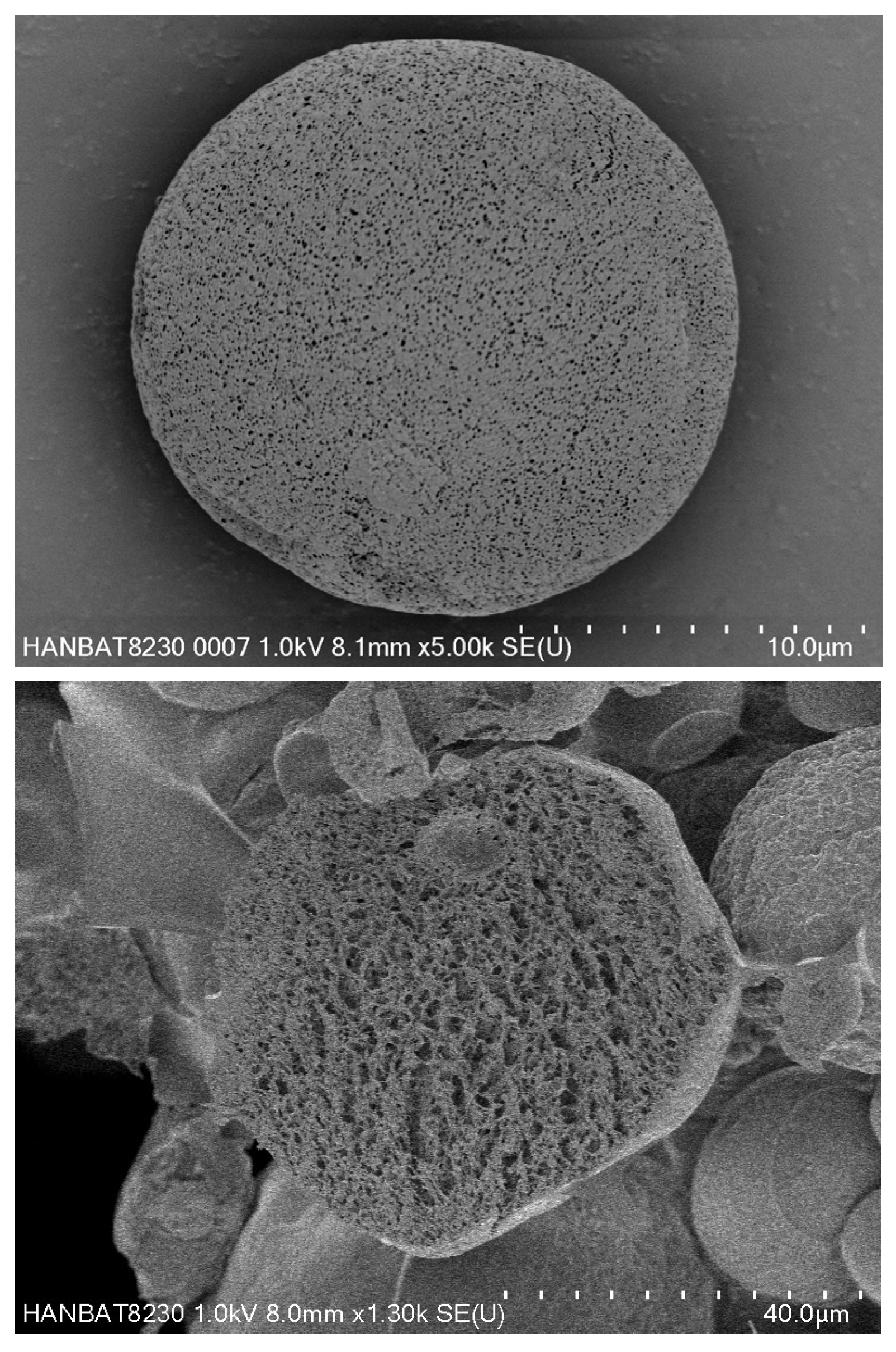
Figure 1. SEM Image of PDLLA in Juvelook.
Scipharm 93 00062 g001
Figure 2. SEM image of CaHA in DClassy (CGBIO Inc., Seoul, Republic of Korea).
Scipharm 93 00062 g002
Table 2. Comparative summary of biostimulatory agents by age and indication.
AgentKey AdvantagesKey DisadvantagesPreferred IndicationsAge-Stratified Notes
PLLADurable neocollagenesis; adipocyte stimulationDelayed onset; nodule risk if dilution/massage inadequateMidface, temple, cheek; body laxity35–55 and ≥55 matrix anchor; micro-dosing ≤35 for prevention
PCLADSC scaffold; long-lasting ECM remodelingTechnique-sensitive; fewer large RCTsGlobal laxity; jawline, neck, arms35–55 and ≥55 durability focus
CaHAImmediate volume + regenerative effectPlane-specific vascular risk; variability with hyper-dilutionNeck, lower face, gluteal, armsAll ages when early effect helpful; caution in thin dermis ≥55
HAReversible; hydration/elasticity; contour refinementShorter durability; limited true biostimulationFine lines, contouring, hydration≤35 for texture; all ages for finishing
PRP/biologicsLow morbidity; metabolic/cellular supportProtocol heterogeneity; variable endpointsTexture improvement, recovery, hair/dermal quality≤35 for prevention; fragile skin ≥55
Disclaimer/Publisher’s Note: The statements, opinions and data contained in all publications are solely those of the individual author(s) and contributor(s) and not of MDPI and/or the editor(s). MDPI and/or the editor(s) disclaim responsibility for any injury to people or property resulting from any ideas, methods, instructions or products referred to in the content.