Communication

A Score-Based Rapid Screening and Network Visualization Method Based on Bioactive Ingredient-Induced Variations in Skin Cell Gene Expression

1
Genesis Institute of Genetic Research, Genesis Healthcare Corporation, Yebisu Garden Place Tower 26F, 4-20-3 Ebisu, Shibuya-ku, Tokyo 150-0013, Japan
2
NODAI Research Institute, Tokyo University of Agriculture, 1-1-1 Sakuragaoka, Setagaya-ku, Tokyo 106-8571, Japan
*
Author to whom correspondence should be addressed.
Sci. Pharm. 2025, 93(4), 56; https://doi.org/10.3390/scipharm93040056
Submission received: 29 September 2025 / Revised: 9 November 2025 / Accepted: 10 November 2025 / Published: 12 November 2025
(This article belongs to the Topic Bioinformatics in Drug Design and Discovery—2nd Edition)

Abstract

Bioactive ingredients are compounds, typically derived from natural sources, that provide specific health benefits or perform certain beneficial functions. Although they can play a role in maintaining good health, their effects can vary widely based on a person’s specific genotype and phenotype, leading to situations where certain ingredients induce beneficial responses for some individuals but not others. Herein, we report a method for the rapid discovery of relationships between genes, bioactive ingredients, and physiological effects. First, RNA-Seq was performed after applying 6 plant-derived ingredients to a three-dimensional skin model. After determining expression changes for each ingredient, these changes were ranked and visualized using score-based prediction models. Based on our analysis, we were able to quickly determine and visualize the effect (or lack thereof) of the ingredients on gene expression. Our findings demonstrate the utility of combining RNA-Seq with score-based models and visualizations for screening bioactive ingredients by gene expression, visualizing their impact based on ingredient or physiological effect, and the applicability of this method to any bioactive ingredient for rapid determination of potential ingredients relevant to maintaining health and wellness.

1. Introduction

Bioactive ingredients are compounds, typically derived from natural sources, that provide specific health benefits or perform certain beneficial functions [1,2,3,4,5]. Although these ingredients can play a role in maintaining good health, the effects of these ingredients on the body can vary widely based on a person’s specific genotype and phenotype. This has led to situations where certain ingredients induce beneficial responses for some individuals but not others [6,7,8,9,10,11]. Thus, the primary obstacle to overcome is screening to ensure ingredients are matched to the proper corresponding phenotype(s).
Personalized healthcare is an approach to medical care that tailors diagnosis, treatment, and prevention to an individual’s unique genotype and/or phenotype [12,13,14]. While bioactive ingredients can play a role in such a customized approach, the complexity of the human genome and its numerous combinations of genetic variants, proteins and compounds, means that the overall effectiveness of any given ingredient may vary greatly depending on the specific genotype and phenotype of an individual [6,7,8,9,10,11]. One potential approach to both accelerating the process of ingredient discovery and evaluating effectiveness is to utilize score-based models, which are already being adopted in pharmaceutical research [15,16,17,18,19]. While few in number, approaches utilizing score-based models and platforms to visualize the relationships between genes and physiological effects of interest to personal health can confirm existing knowledge and also uncover previously unknown interactions and relationships [20,21,22,23]. These computational methods can be combined with RNA-Seq for a more targeted approach than traditional drug discovery methods [24]. Although sequencing time and costs can be higher than traditional microarray for transcriptome analysis, RNA-Seq provides greater precision, more comprehensive results, and most importantly the ability to discover novel transcripts [25,26,27,28,29,30]. Although alternative methods for discovering novel transcripts such as long-read RNA-Seq and whole transcriptome analysis (WTS) can be used, they usually come with higher costs and lower throughputs [31,32,33]. Thus, for initial screening where discovery is more important, RNA-Seq provides a good balance between cost and effectiveness. By combining RNA-Seq and score-based models, the resulting quick and effective screening for potential ingredients and visualization of their effectiveness on a case-by-case basis for each individual customer would be a great boon to both medical professionals and individuals.
In this exploratory study, we established and evaluated a score-based rapid screening and visualization method for bioactive ingredient selection based on gene expression. First, RNA-Seq was performed after applying 6 bioactive ingredients with well-known medicinal, dietary, and/or cosmetic uses to a three-dimensional skin model. After determining expression changes for each ingredient, these changes were ranked and visualized using score-based prediction models. Based on our analysis, we were able to quickly determine and visualize the effect (or lack thereof) of the ingredients on skin gene expression. In addition, the approach also clustered the ingredients into groups related to effects such as wound healing, inflammation, and skin barrier function. These findings demonstrate the utility of score-based prediction models for screening bioactive ingredients by gene expression, visualizing their impact based on ingredient or physiological effect, and the applicability of this method to any bioactive ingredient for rapid determination of potential ingredients relevant to maintaining health and wellness.

2. Materials and Methods

2.1. List of Bioactive Ingredients Used

The following 6 bioactive ingredients were used in this study: Alpinia speciosa Leaf Extract (Maruzen Pharmaceuticals Co., Ltd., Onomichi, Japan), Arnica montana Flower Extract (Maruzen Pharmaceuticals Co., Ltd., Onomichi, Japan), Citrus unshiu Peel Extract (Maruzen Pharmaceuticals Co., Ltd., Onomichi, Japan), Foeniculum vulgare Fruit Extract (Maruzen Pharmaceuticals Co., Ltd., Onomichi, Japan), Colloidal Platinum (INOVEX Co., Ltd., Izu, Japan), and Nahlsgen (NAHLS Co., Ltd., Kyoto, Japan). Each ingredient was diluted in PBS and sterilized by filtration using a DISMIC-25CS (Advantec Toyo Kaisha, Ltd., Tokyo, Japan). Concentrations of each ingredient in solution were the maximum possible before the onset of precipitation and are listed in Table 1.

2.2. Epidermal 3D Model

EpiDermFT Skin Model EFT-400 (Kurabo Industries, Ltd., Osaka, Japan), a multi-layered and highly differentiated 3D structural skin model resembling the structure of human skin (stratum corneum, epidermis, and dermis), was used for all experiments (Figure S1). The model consisted of normal human epidermal keratinocytes (NHEK) derived from neonatal foreskin tissue and normal human dermal fibroblasts (NHDF) derived from neonatal skin. Both NHEK and NHDF cells were derived from multiple donors. After overnight incubation in Dulbecco’s Modified Eagle’s Medium (DMEM), the model was allowed to stand until the addition of substances after the medium was changed.

2.3. RNA-Seq Protocol

An EFT-400 skin model was stabilized overnight in 2.5 mL of DMEM. At the same time, 25 μL solutions of PBS and bioactive ingredients were prepared. After overnight stabilization, each solution was added to the skin model and incubated a second time for 24 h. Negative controls (NC) were incubated in PBS.
After incubation, RNA was extracted from NHEK and NHDF cells in each tissue using ISOGEN reagent according to kit protocols (NIPPON GENE Co., Ltd., Tokyo, Japan). Due to the tissue being in sheet form and thus difficult to dissolve, the skin tissue was cut into small pieces using small sterile scissors. Samples were dissolved in 100 μL of RNase-free H2O. RNA Integrity Number (RIN) values for the RNA samples were confirmed with Agilent RNA 6000 Pico kits (Agilent Technologies Japan, Ltd., Tokyo, Japan).
Libraries were prepared with Illumina Stranded Total RNA Prep kits (Illumina, San Diego, CA, USA) or Ribo-Zero Plus kits (Illumina, San Diego, CA, USA), both according to kit protocols. Libraries were quantified using Bioanalyzer (Agilent 2100 Bioanalyzer, Agilent Technologies Japan, Ltd., Tokyo, Japan) and Qubit (Qubit® 3.0 Fluorometer, Thermo Fisher Scientific, Waltham, MA, USA). RNA-Seq was performed using an Illumina NovaSeq6000 (Illumina, San Diego, CA, USA) at a loading concentration of 100 pM and PE 150.

2.4. Preprocessing of Data

Raw RNA-Seq data was run through DRAGEN (Illumina, San Diego, CA, USA) in order to generate read count data, which was generated in either .sf or .csv format. Python 3.11.4 (Anaconda distribution 2023.07-0) was used to extract the raw data values and load them into a Pandas DataFrame using the Pandas library. Once imported, the data was filtered to remove samples with no read count, samples with read counts of less than 10, and extreme outliers. All genes were cross-referenced with Ensembl through the Ensembl REST API (GrCh38) to ensure validity.

2.5. Determination of Differentially Expressed Genes (DEGs)

DEG analysis was performed on the data using Python 3.11.4 (Rpy2 3.5.12) and R 4.3.0. Normalization was performed using EdgeR’s trimmed mean of M values (TMM). After a second filtering process to remove low/no read count samples and extreme outliers, differentially expressed genes were determined using the Limma-Voom package.

2.6. Calculation of Relationship Score

The relationship score was calculated using a weighted sum model of the form:
A i W S M s c o r e = j = 1 n w j a i j     f o r   i = 1,2 , 3 m
where A = a choice consisting of a set of weights, a = an individual factor, w = weight, n = number of decision criteria, and m = number of alternatives. Individual factors included fold changes, p-values, gene pathways, and physiological effects on the skin.

2.7. Generation of Network Graphs

The results of the relationship score calculations were displayed as network graphs for ease of interpretation. Network graphs were drawn by GenesisGaia, a life sciences platform that can display analyzed data as network graphs, using Python 3.11.4 (NetworkX and PyVis libraries). Bioactive ingredients, differentially expressed genes, calculated relationship scores between the ingredients and genes, and the physiological effects of each gene on the skin were all included in the graphs.

3. Results

We began our study by constructing a list of bioactive ingredients. Within this category, we decided to focus on bioactive skincare and cosmetic ingredients due to the ease of evaluation and extensive pre-existing research on ingredient efficacy [34,35,36,37]. Ingredients were chosen based on their potential antioxidant and anti-inflammatory effects [38,39,40,41]. Initially, naturally derived products were focused on due to their effectiveness on skin condition [42,43,44,45]. After the list was constructed, 4 ingredients were arbitrarily selected: Alpinia speciosa, Arnica montana, Citrus unshiu peel, and Foeniculum vulgare (Fennel) fruit. For comparison, colloidal platinum was chosen because of its claimed ability to act as an antioxidant [46] while Nahlsgen, the tradename for 2-amino-4-[[3-(carboxymethyl)phenoxy]-methoxyphosphoryl]butanoic acid, was added as a synthetic organic product again with proposed antioxidant effects [45].
Once ingredients were selected, epidermal 3D skin models were incubated with 8 different solutions, each containing a different ingredient (Figure 1). The 3D skin models consisted of normal human epidermal keratinocytes (NHEK) and normal human dermal fibroblasts (NHDF). After incubation, tissues were extracted and libraries were prepared. In place of traditional microarray platforms, RNA-Seq was performed to ensure quick and accurate measurement of gene expression changes [47,48]. The resulting raw RNA-Seq data was run through DRAGEN in order to generate read count data. DEGs were determined using Python and R by first normalizing the read count data using EdgeR’s trimmed mean of M values (TMM) and then processing the normalized data with Limma-Voom [49,50,51,52]. For each ingredient solution, a list of differentially expressed genes (DEGs) was generated (Figure 1, Table 2). For 4 of the 6 tested ingredients, 48 DEGs and 113 ingredient-gene relationships were identified, suggesting the organic ingredients produced changes in the skin upon exposure. Curiously, the 2 tested ingredients that did not show any associated DEGs were the non-naturally derived ingredients (Colloidal platinum and Nahlsgen).
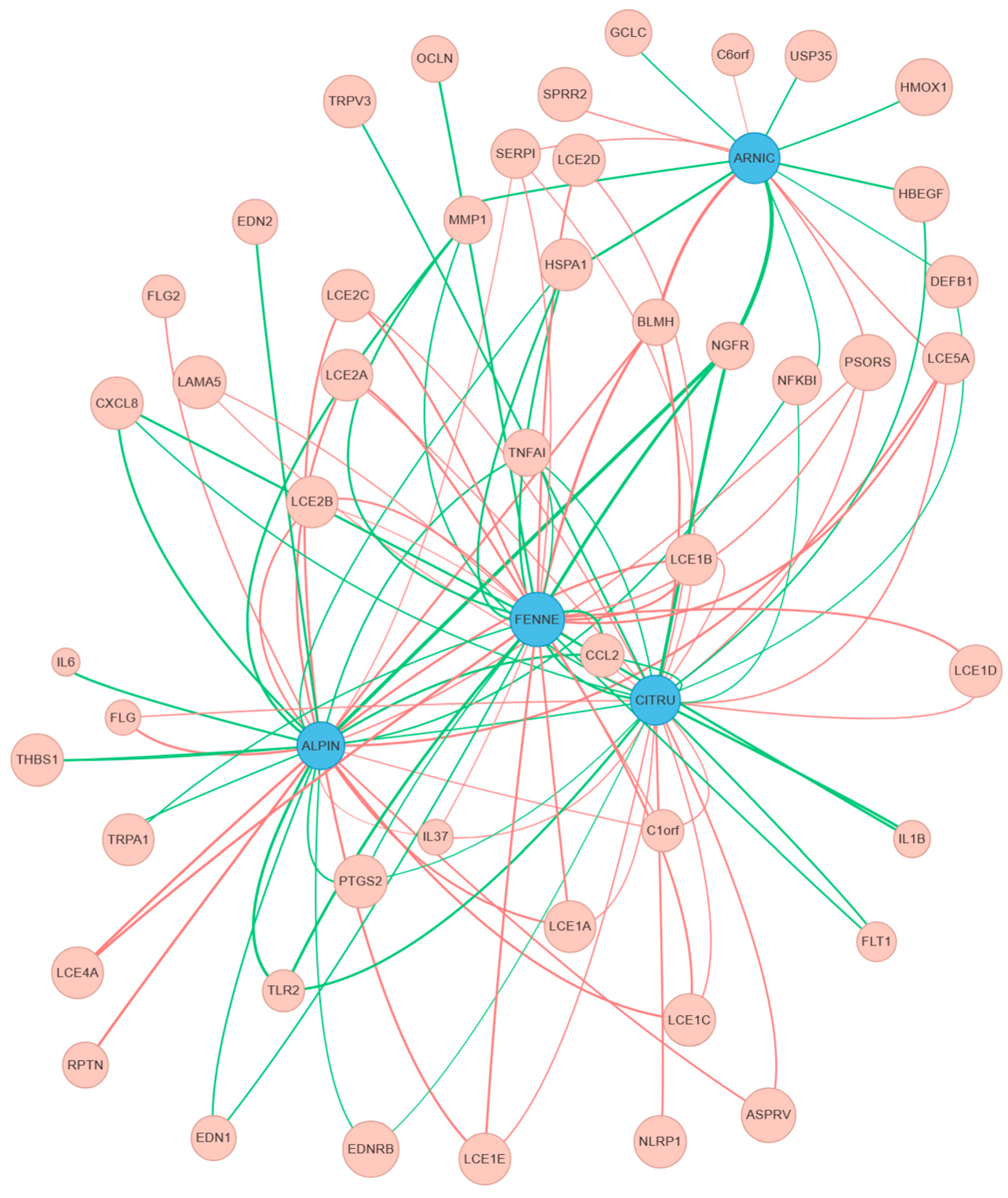
After determining which genes were differentially expressed, the relationships between the DEGs, bioactive ingredients, and physiological skin effects were explored. First, the list of DEGs was imported to GenesisGaia, an R&D platform for life sciences research with features such as peer-reviewed scientific literature amalgamation, a rich genomic database, and the ability to display data as charts and network graphs [53]. After importing, the data was referenced against existing gene and ingredient data in the platform’s database (Figure 1) [54]. In turn, this was referenced against physiological skin effect data from the platform’s database. Once the correlations were found and relationships determined, a “relationship score” was calculated according to a weighted sum model. This model took fold changes, p-values, gene pathways, and physiological effects into account. The weighted sum model was selected due to its simplicity, flexibility, and ability to assign importance to each individual input. After performing calculations, the “relationship score”, representing the likelihood that a gene will be affected by a bioactive ingredient, was visualized in a computer-generated network graph (Figure 2). In these graphs, purple nodes represent bioactive ingredients while peach nodes represent skin genes. The color of the line indicates regulation status: green for upregulation and red for downregulation. The line thickness is representative of the calculated relationship score, where an increasing score correlates to a thicker line (and vice versa). As the relationship score is derived from biologically based parameters, thicker lines generally correlate to higher values for the score’s constituent components. From the network graph, connections between genes and ingredients as well as their predicted relationships could be visualized all at once, making the data more accessible and comprehensible.
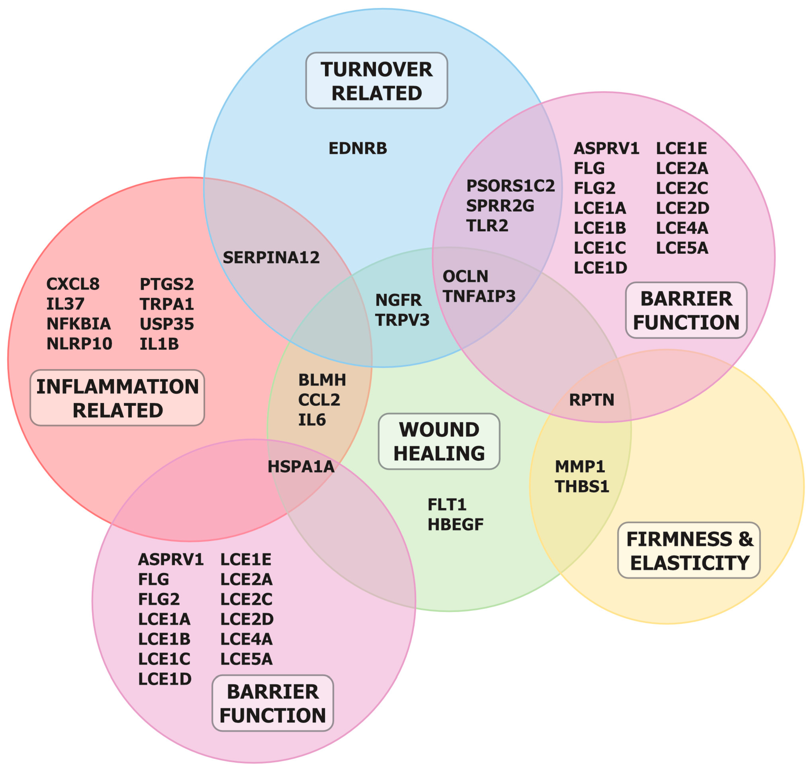
The genes resulting from the DEG analysis are shown in Figure 3 and clustered according to the physiological effects predicted by the gene functions. A number of the identified genes (25 of the 48 identified DEGs) were not present in previous reports of skin-related genes, confirming the validity of the RNA-seq analysis for determining novel genes [55,56]. On the basis of physiological effects commonly seen in skin, 5 major clusters were identified: inflammation-related genes, turnover-related genes, wound healing-related genes, firmness and elasticity-related genes, and barrier function-related genes. Of the 48 identified DEGs, 40 (~83%) corresponded to at least one cluster, implying the relevance of the tested organic ingredients for skincare applications. It should be noted that the output of the model is entirely based on inputs derived from the selected ingredients, DEG analysis results, and existing peer-reviewed research. As such, the final list of genes and the cluster diagram will not change unless additional ingredients are added, changes to the transcriptome occur, and/or relevant new discoveries are added to the scientific literature.

4. Discussion

Growing interest in bioactive ingredients has demonstrated the necessity of understanding the relationships between genes, ingredients, and the physiological effects of both. Unfortunately, genetic datasets necessary to understand these relationships are frequently either incomplete or unavailable. Furthermore, the search process for new bioactive ingredients and confirmation of their physiological effects require large amounts of time, effort, and resources. RNA-Seq paired with AI-driven tools can accelerate the research process by generating the genetic datasets necessary to perform research and investigation. For example, a researcher can generate relationship data, use it to train a traditional statistical or machine learning model, and then deploy and further narrow down potential ingredient candidates. These methods would enable rapid discovery and lead to more effective health outcomes.
As each individual has a unique transcriptome, there is a need for rapid, accurate sequencing and analysis for the eventual realization of personalized health and wellness. Pairing RNA-Seq with predictive algorithms and models is ideal for fulfilling these requirements. Phenotypic changes due to factors like aging and environmental conditions can be accounted for by resampling, resequencing, and reanalysis. This opens up the possibility of assembling a library of phenotype snapshots for an individual, which when combined with predictive models can result in an always up-to-date personalized skincare ingredient list. The inconsistency of a few of the RNA-seq results with previous reports (upregulated expression of some inflammation-related genes and upregulated and/or downregulated expression of typical genes used to evaluate functions) demonstrates the merit of this concept; from a user’s perspective, the ability to track mRNA expression level differences over time on an individual basis and pair specific functional ingredients to those changes ensures that the most effective treatment is always available, ultimately improving the user experience.
Although there are many advantages and opportunities provided by the method described in this paper, there are also a few potential drawbacks. First, because the method described in this paper utilizes RNA-Seq instead of microarray, the amount of data generated is high. Although a personalized library of results can be assembled as described above, this data needs to be properly managed and stored. For large numbers of people over many years, the resources required to do this may be significant. Second, transcriptome changes can be measured on the scale of hours, days, weeks, seasons, or even decades. Thus, the appropriate timescale for an experiment needs to be clearly set in order to properly interpret the results and determine which expression changes are relevant to the study target(s) and which ones are not. Finally, the method described in this paper requires a method for quickly and accurately generating visual representations of the results. While the study used the GenesisGaia platform to handle the scoring and network graph generation, those without access to the platform would need to re-implement the weighted sum model and network graphing separately on their own or with alternative tools.

5. Conclusions

In conclusion, we have designed and implemented a method for screening, predicting, and visualizing the relationships between genes, bioactive ingredients, and their physiological effects. Our findings are significant for a few reasons. First, combining RNA-Seq and DEG analysis data with score-based predictive algorithms can reveal relationships between genes, bioactive ingredients, and their physiological effects. Second, quick and accurate generation of genetic datasets based on quantifiable biological data enables experiments that, until now, were not feasible due to incomplete and/or missing data. Finally, the information gained from the ability to quickly and repeatedly screen genes and bioactive ingredients over any time interval allows the generation of an appropriate list of ingredients for any corresponding transcriptome state. Overall, our findings constitute a foundation for further research into rapid, effective screening methods for bioactive ingredients as well as the development of highly personalized ingredient matching based on an individual’s present situation.

Supplementary Materials

The following supporting information can be downloaded at: https://www.mdpi.com/article/10.3390/scipharm93040056/s1, Figure S1: Diagram detailing the structure of the EpiDermFT Skin Model EFT-400. The stratum corneum equivalent consists of cornified epidermal cells, analogous to those found in vivo. The epidermis equivalent consists of basal cells, spinous layer, and granular layer, also analogous to those found in vivo. Both epidermal layers are derived from normal human epidermal keratinocytes (NHEK) derived from neonatal foreskin tissue. Finally, the dermis equivalent is composed of a collagen matrix containing viable normal human dermal fibroblasts (NHDF) derived from neonatal skin. All layers are mitotically and metabolically active, and exhibit in-vivo like morphological and growth characteristics. Both NHEK and NHDF cells were derived from multiple donors. RNA-Seq was performed on RNA extracted from both the epidermis and the dermis.

Author Contributions

Conceptualization, I.S.-B. and T.M.; methodology, M.O., I.S.-B. and T.M.; software, D.D.O.; validation, M.O., I.S.-B.; formal analysis, M.O., D.D.O.; investigation, M.O. and A.I.; resources, I.S.-B. and T.M.; data curation, M.O. and A.I.; writing—original draft preparation, M.O.; writing—review and editing, C.W.C., I.S.-B., D.D.O.; visualization, D.D.O.; supervision, I.S.-B. and T.M.; project administration, I.S.-B. and T.M.; funding acquisition, I.S.-B. and T.M. All authors have read and agreed to the published version of the manuscript.

Funding

This work was supported by Genesis Healthcare Co. (Program Grant #2024-02).

Data Availability Statement

RNA-Seq data have been submitted to the NIH NCBI GEO database (Accession Number GSE301022).

Acknowledgments

The authors acknowledge Genesis Healthcare, Co. for funding the study and providing unrestricted access to the GenesisGaia R&D platform. The authors thank Hideyuki Aoshima for his support with the laboratory experiments. The authors would like to acknowledge Chun-Chieh Lin for his assistance in proofreading the manuscript. The sponsor had no influence other than providing resources and facilities. The authors thank Maruzen Pharmaceuticals Co., Ltd., INOVEX Co., Ltd., and NAHLS Co., Ltd. for complimentary samples of the bioactive ingredients used in this study. The authors confirm that this contribution had no influence on study design, data analysis, or the conclusions drawn in this work.

Conflicts of Interest

M.O., C.W.C., A.I., and D.D.O. are employees of Genesis Healthcare, Co. T.M. received consulting fees as an advisor to Genesis Healthcare, Co. I.S.-B. and T.M. own stock of Genesis Healthcare, Co. T.M. owns stock options of Genesis Healthcare, Co. I.S.-B., D.D.O., and T.M. (as part of Genesis Healthcare, Co.) have plans to file patents related to this paper but are still undecided. The authors received complimentary samples of the bioactive ingredients from their suppliers (Maruzen Pharmaceuticals Co., Ltd., INOVEX Co., Ltd., and NAHLS Co., Ltd.). The suppliers had no role in study design, data collection, analysis, data interpretation, or writing of the manuscript.

References

  1. National Cancer Institute. NCI Dictionary of Cancer Terms. Available online: https://www.cancer.gov/publications/dictionaries/cancer-terms/def/bioactive-compound (accessed on 1 September 2025).
  2. Weaver, C.M. Bioactive Foods and Ingredients for Health. Adv. Nutr. 2014, 5, 306S–311S. [Google Scholar] [CrossRef]
  3. Samtiya, M.; Aluko, R.E.; Dhewa, T.; Moreno-Reyes, J.M. Potential Health Benefits of Plant Food-Derived Bioactive Components: An Overview. Foods 2021, 10, 839. [Google Scholar] [CrossRef]
  4. Donn, P.; Prieto, M.A.; Mejuto, J.C.; Cao, H.; Simal-Gandara, J. Functional foods based on the recovery of bioactive ingredients from food and algae by-products by emerging extraction technologies and 3D printing. Food Biosci. 2022, 49, 101853. [Google Scholar] [CrossRef]
  5. Kussmann, M.; Abe Cunha, D.H.; Berciano, S. Bioactive compounds for human and planetary health. Front. Nutr. 2023, 10, 1193848. [Google Scholar] [CrossRef]
  6. Farhud, D.D.; Yeganeh, M.Z. Nutrigenomics and Nutrigenetics. Iran. J. Public Health 2010, 39, 1. [Google Scholar] [PubMed]
  7. Liu, J.; Tuvblad, C.; Raine, A.; Baker, L. Genetic and environmental influences on nutrient intake. Genes Nutr. 2012, 8, 241–242. [Google Scholar] [CrossRef] [PubMed]
  8. Pokimica, B.; Garcia-Conesa, M.-T. Critical Evaluation of Gene Expression Changes in Human Tissues in Response to Supplementation with Dietary Bioactive Compounds: Moving Towards Better-Quality Studies. Nutrients 2018, 10, 807. [Google Scholar] [CrossRef]
  9. Niforou, A.; Konstantinidou, V.; Naska, A. Genetic Variants Shaping Inter-individual Differences in Response to Dietary Intakes—A Narrative Review of the Case of Vitamins. Front. Nutr. 2020, 7, 558598. [Google Scholar] [CrossRef]
  10. Mierziak, J.; Kostyn, K.; Boba, A.; Czemplik, M.; Kulma, A.; Wojtasik, W. Influence of the Bioactive Diet Components on the Gene Expression Regulation. Nutrients 2021, 13, 3673. [Google Scholar] [CrossRef] [PubMed]
  11. Chilton, F.H.; Manichaikul, A.; Yang, C.; O’COnnor, T.D.; Johnstone, L.M.; Blomquist, S.; Schembre, S.M.; Sergeant, S.; Zec, M.; Tsai, M.Y.; et al. Interpreting Clinical Trials With Omega-3 Supplements in the Context of Ancestry and FADS Genetic Variation. Front. Nutr. 2021, 8, 808504. [Google Scholar] [CrossRef]
  12. National Institute of Health; National Human Genome Research Institute. Talking Glossary of Genomic and Genetic Terms. Available online: https://www.genome.gov/genetics-glossary/Personalized-Medicine (accessed on 1 September 2025).
  13. Johnson, K.B.; Wei, W.; Weeraratne, D.; Frisse, M.E.; Misulis, K.; Rhee, K.; Zhao, J.; Snowdon, J.L. Precision Medicine, AI, and the Future of Personalized Health Care. Clin. Transl. Sci. 2021, 14, 86–93. [Google Scholar] [CrossRef]
  14. Chen, O.Y.; Roberts, B. Personalized Healthcare and Public Health in the Digital Age. Front. Digit. Health 2021, 3, 595704. [Google Scholar] [CrossRef]
  15. Blanco-Gonzalez, A.; Cabezon, A.; Seco-Gonzalez, A.; Conde-Torres, D.; Antelo-Riveiro, P.; Pineiro, A.; Garcia-Fandino, R. The Role of AI in Drug Discovery: Challenges, Opportunities, and Strategies. Pharmaceuticals 2023, 16, 891. [Google Scholar] [CrossRef] [PubMed]
  16. Zhou, G.; Rusnac, D.-V.; Park, H.; Canzani, D.; Nguyen, H.M.; Stewart, L.; Bush, M.F.; Nguyen, P.T.; Wulff, H.; Yarov-Yarovoy, V.; et al. An artificial intelligence accelerated virtual screening platform for drug discovery. Nat. Commun. 2024, 15, 7761. [Google Scholar] [CrossRef] [PubMed]
  17. Druedahl, L.C.; Price, W.N.; Minssen, T.; Sarpatwari, A. Use of Artificial Intelligence in Drug Development. JAMA Netw. Open. 2024, 7, e2414139. [Google Scholar] [CrossRef]
  18. Pitt, W.R.; Bentley, J.; Boldron, C.; Colliandre, L.; Esposito, C.; Frush, E.H.; Kopec, J.; Labouille, S.; Meneyrol, J.; Pardoe, D.A.; et al. Real-World Applications and Experiences of AI/ML Deployment for Drug Discovery. J. Med. Chem. 2025, 68, 851–859. [Google Scholar] [CrossRef]
  19. Zhang, K.; Yang, X.; Wang, Y.; Yu, Y.; Huang, N.; Li, G.; Li, X.; Wu, J.C.; Yang, S. Artificial intelligence in drug development. Nat. Med. 2025, 31, 45–59. [Google Scholar] [CrossRef] [PubMed]
  20. Meijer, D.; Beniddir, M.A.; Coley, C.W.; Mejri, Y.M.; Öztürk, M.; van der Hooft, J.J.J.; Medema, M.H.; Skiredj, A. Empowering natural product science with AI: Leveraging multimodal data and knowledge graphs. Nat. Prod. Rep. 2024, 42, 654–662. [Google Scholar] [CrossRef]
  21. Chen, H.; King, F.J.; Zhou, B.; Wang, Y.; Canedy, C.J.; Hayashi, J.; Zhong, Y.; Chang, M.W.; Pache, L.; Wong, J.L.; et al. Drug target prediction through deep learning functional representation of gene signatures. Nat. Commun. 2024, 15, 1853. [Google Scholar] [CrossRef]
  22. Singh, P.; Bhat, S.S.; Singh, N.; Venkanna, B.U.; Mohamed, R.; Rao, R.P. Cell-Based Model Systems for Validation of Various Efficacy-Based Claims for Cosmetic Ingredients. Cosmetics 2022, 9, 107. [Google Scholar] [CrossRef]
  23. Markiewicz, E.; Idowu, O.C. Evaluation of Personalized Skincare Through in-silico Gene Interactive Networks and Cellular Responses to UVR and Oxidative Stress. Clin. Cosmet. Investig. Dermatol. 2022, 15, 2221–2243. [Google Scholar] [CrossRef]
  24. Singh, N.; Vayer, P.; Tanwar, S.; Poyet, J.-L.; Tsaioun, K.; Villoutreix, B.O. Drug discovery and development: Introduction to the general public and patient groups. Front. Drug Discov. 2023, 3, 1201419. [Google Scholar] [CrossRef]
  25. Wang, Z.; Gerstein, M.; Snyder, M. RNA-Seq: A revolutionary tool for transcriptomics. Nat. Rev. Genet. 2009, 10, 57–63. [Google Scholar] [CrossRef] [PubMed]
  26. Martin, J.A.; Wang, Z. Next-generation transcriptome assembly. Nat. Rev. Genet. 2011, 12, 671–682. [Google Scholar] [CrossRef] [PubMed]
  27. Khatoon, Z.; Figler, B.; Zhang, H.; Cheng, F. Introduction to RNA-Seq and its Applications to Drug Discovery and Development. Drug Dev. Res. 2014, 75, 324–330. [Google Scholar] [CrossRef]
  28. Ye, C.; Ho, D.J.; Neri, M.; Yang, C.; Kulkarni, T.; Randhawa, R.; Henault, M.; Mostacci, N.; Farmer, P.; Renner, S.; et al. DRUG-seq for miniaturized high-throughput transcriptome profiling in drug discovery. Nat. Commun. 2018, 9, 4307. [Google Scholar] [CrossRef]
  29. Van de Sande, B.; Lee, J.S.; Mutasa-Gottgens, E.; Naughton, B.; Bacon, W.; Manning, J.; Wang, Y.; Pollard, J.; Mendez, M.; Hill, J.; et al. Applications of single-cell RNA sequencing in drug discovery and development. Nat. Rev. Drug Discov. 2023, 22, 496–520. [Google Scholar] [CrossRef] [PubMed]
  30. Aja, P.M.; Agu, P.C.; Ogbu, C.; Alum, E.U.; Fasogbon, I.V.; Musyoka, A.M.; Ngwueche, W.; Egwu, C.O.; Tusubira, D.; Ross, K. RNA research for drug discovery: Recent advances and critical insight. Gene 2025, 947, 149342. [Google Scholar] [CrossRef]
  31. Jobanputra, V.; Wrzeszczynski, K.O.; Buttner, R.; Caldas, C.; Cuppen, E.; Grimmond, S.; Haferlach, T.; Mullighan, C.; Schuh, A.; Elemento, O. Clinical interpretation of whole-genome and whole-transcriptome sequencing for precision oncology. Semin. Cancer Biol. 2022, 84, 23–31. [Google Scholar] [CrossRef]
  32. Fu, X.; Fu, N.; Guo, S.; Yan, Z.; Xu, Y.; Hu, H.; Menzel, C.; Chen, W.; Li, Y.; Zeng, R.; et al. Estimating accuracy of RNA-Seq and microarrays with proteomics. BMC Genom. 2009, 10, 161. [Google Scholar] [CrossRef]
  33. Ruan, M.; Liu, J.; Ren, X.; Li, C.; Zhao, A.Z.; Li, L.; Yang, H.; Dai, Y.; Wang, Y. Whole transcriptome sequencing analyses of DHA treated glioblastoma cells. J. Neurol. Sci. 2019, 396, 247–253. [Google Scholar] [CrossRef]
  34. Hernandez, D.F.; Cervantes, E.L.; Luna-Vital, D.A.; Mojica, L. Food-derived bioactive compounds with anti-aging potential for nutricosmetic and cosmeceutical products. Crit. Rev. Food Sci. Nutr. 2020, 61, 3740–3755. [Google Scholar] [CrossRef]
  35. Michalak, M.; Błońska-Sikora, E.; Dobros, N.; Spałek, O.; Zielińska, A.; Paradowska, K. Bioactive Compounds, Antioxidant Properties, and Cosmetic Applications of Selected Cold-Pressed Plant Oils from Seeds. Cosmetics 2024, 11, 153. [Google Scholar] [CrossRef]
  36. Goyal, A.; Sharma, A.; Kaur, J.; Kumari, S.; Garg, M.; Sindhu, R.K.; Rahman, H.; Akhtar, M.F.; Tagde, P.; Najda, A.; et al. Bioactive-Based Cosmeceuticals: An Update on Emerging Trends. Molecules 2022, 27, 828. [Google Scholar] [CrossRef] [PubMed]
  37. Gimenez Martinez, R.J.; García, F.R.; Cerdá, J.C.M.; Hernández-Ruíz, Á.; Castro, M.I.G.; Valverde-Merino, M.-I.; Camarasa, F.J.H.; Meseguer, F.L.; Gallardo, M.L.-V. Bioactive Substance and Skin Health: An Integrative Review from a Pharmacy and Nutrition Perspective. Pharmaceuticals 2025, 18, 373. [Google Scholar] [CrossRef] [PubMed]
  38. Youn, I.; Han, A.-R.; Piao, D.; Lee, H.; Kwak, H.; Lee, Y.; Nam, J.-W.; Seo, E.K. Phytochemical and pharmacological properties of the genus Alpinia from 2016 to 2023. Nat. Prod. Rep. 2024, 21, 1346–1367. [Google Scholar] [CrossRef] [PubMed]
  39. Min, K.Y.; Lee, K.A.; Kim, H.J.; Kim, K.-T.; Chung, M.-S.; Chang, P.-S.; Park, H.; Paik, H.-D. Antioxidative and anti-inflammatory activities of Citrus unshiu peel extracts using a combined process of subcritical water extraction and acid hydrolysis. Food Sci. Biotechnol. 2014, 23, 1441–1446. [Google Scholar] [CrossRef]
  40. Badgujar, S.B.; Patel, V.V.; Bandivdekar, A.H. Foeniculum vulgare Mill: A Review of Its Botany, Phytochemistry, Pharmacology, Contemporary Application, and Toxicology. Biomed Res. Int. 2014, 2014, 842674. [Google Scholar] [CrossRef]
  41. Kriplani, P.; Guarve, K.; Baghael, U.S. Arnica montana L.—A plant of healing: Review. J. Pharm. Pharmacol. 2017, 69, 925–945. [Google Scholar] [CrossRef]
  42. Hoang, H.T.; Moon, J.-Y.; Lee, Y.-C. Natural Antioxidants from Plant Extracts in Skincare Cosmetics: Recent Applications, Challenges and Perspectives. Cosmetics 2021, 8, 106. [Google Scholar] [CrossRef]
  43. Lin, T.-K.; Zong, L.; Santiago, J.L. Anti-Inflammatory and Skin Barrier Repair Effects of Topical Application of Some Plant Oils. Int. J. Mol. Sci. 2018, 19, 70. [Google Scholar] [CrossRef]
  44. Reddy, B.H.V.; Hussain, S.M.S.; Hussain, M.S.H.; Kumar, R.N.; Gupta, J. Essential oils in cosmetics: Antioxidant properties and advancements through nanoformulations. PRENAP 2025, 6, 100192. [Google Scholar] [CrossRef]
  45. Joyce-Brady, M.; Hiratake, J. Inhibiting Glutathione Metabolism in Lung Lining Fluid as a Strategy to Augment Antioxidant Defense. Curr. Enzym. Inhib. 2011, 7, 71–78. [Google Scholar] [CrossRef] [PubMed]
  46. Aiuchi, T.; Nakajo, S.; Nakaya, K. Reducing Activity of Colloidal Platinum Nanoparticles for Hydrogen Peroxide, 2,2-Diphenyl-1-picrylhydrazyl Radical and 2,6-Dichlorophenol Indophenol. Biol. Pharm. Bull. 2004, 27, 736–738. [Google Scholar] [CrossRef] [PubMed]
  47. Benech, P.D.; Patatian, A. From experimental design to functional gene networks: DNA microarray contribution to skin ageing research. Int. J. Cosmet. Sci. 2014, 36, 516–526. [Google Scholar] [CrossRef]
  48. Rao, M.S.; Van Vleet, T.R.; Ciurlionis, R.; Buck, W.R.; Mittelstadt, S.W.; Blomme, E.A.; Liguori, M.J. Comparison of RNA-Seq and Microarray Gene Expression Platforms for the Toxicogenomic Evaluation of Liver From Short-Term Rat Toxicity Studies. Front. Genet. 2018, 9, 636. [Google Scholar] [CrossRef] [PubMed]
  49. Anaconda, Inc. Conda. Version 3.27. [Computer Software]. Anaconda, Inc.: Austin, TX, USA, 2023. Available online: https://www.anaconda.com/ (accessed on 1 September 2025).
  50. Ritchie, M.E.; Phipson, B.; Wu, D.I.; Hu, Y.; Law, C.W.; Shi, W.; Smyth, G.K. Limma powers differential expression analyses for RNA-sequencing and microarray studies. Nucleic Acids Res. 2015, 43, e47. [Google Scholar] [CrossRef]
  51. Chen, Y.; Lun, A.T.L.; Smyth, G.K. From reads to genes to pathways: Differential expression analysis of RNA-Seq experiments using Rsubread and the edgeR quasi-likelihood pipeline. F1000Research 2016, 5, 1438. [Google Scholar]
  52. Law, C.W.; Alhamdoosh, M.; Su, S.; Dong, X.; Tian, L.; Smyth, G.K.; Ritchie, M.E. RNA-seq analysis is easy as 1-2-3 with limma, Glimma and edgeR. F1000Research 2016, 5, 1408. [Google Scholar] [CrossRef]
  53. Genesis Healthcare, Co. GenesisGaia [Computer Software]. 2024. Available online: https://www.adam-innovations.com/genesisgaia (accessed on 9 November 2025).
  54. Martin, F.J.; Amode, M.R.; Aneja, A.; Austine-Orimoloye, O.; Azov, A.G.; Barnes, I.; Becker, A.; Bennett, R.; Berry, A.; Bhai, J.; et al. Ensembl 2023. Nucleic Acids Res. 2023, 51, D933–D941. [Google Scholar] [CrossRef]
  55. Edqvist, P.-H.D.; Fagerberg, L.; Hallström, B.M.; Danielsson, A.; Edlund, K.; Uhlén, M.; Pontén, F. Expression of Human Skin-Specific Genes Defined by Transcriptomics and Antibody-Based Profiling. J. Histochem. Cytochem. 2014, 63, 129–141. [Google Scholar] [CrossRef] [PubMed]
  56. Gerber, P.A.; Hevezi, P.; Buhren, B.A.; Martinez, C.; Schrumpf, H.; Gasis, M.; Grether-Beck, S.; Krutmann, J.; Homey, B.; Zlotnik, A. Systematic Identification and Characterization of Novel Human Skin-Associated Genes Encoding Membrane and Secreted Proteins. PLoS ONE 2013, 8, e63949. [Google Scholar] [CrossRef] [PubMed]
Figure 1. Summary of process for discovery of skin-related differentially expressed genes (DEGs). First, RNA-Seq is performed on epidermal 3D model. Next, a DEG analysis is performed on the resulting data. The list of determined DEGs is then fed through the score-based screening model, where it is referenced against the existing database. Finally, the data output is analyzed for any novel relationships between genes, bioactive ingredients, and physiological effects.
Scipharm 93 00056 g001
Figure 2. Sample representative ingredient-gene map generated by the GenesisGaia life sciences platform. Purple nodes represent bioactive ingredients, and peach nodes represent skin genes. The color of the line indicates either gene upregulation (green) or downregulation (red). The line thickness represents the predicted relationship strength between an ingredient and a gene; as the line becomes thicker, the predicted relationship becomes stronger.
Scipharm 93 00056 g002
Figure 3. Cluster diagram showing skin genes affected by bioactive ingredients. The genes were able to be clustered into 5 distinct categories: inflammation-related genes, turnover-related genes, wound healing-related genes, firmness and elasticity-related genes, and barrier function-related genes. Novel genes were determined by comparing the predictions obtained from GenesisGaia with known genes from existing ingredient research.
Scipharm 93 00056 g003
Table 1. Table of all 6 bioactive ingredients used in this study. The concentration of each ingredient is the maximum concentration in solution before the onset of precipitation.
Bioactive Ingredient NameConcentration
Alpinia speciosa Leaf Extract0.5%
Arnica montana Flower Extract1.0%
Citrus unshiu Peel Extract0.3%
Colloidal Platinum1.0%
Foeniculum vulgare Fruit Extract2.0%
Nahlsgen1.0%
Table 2. Table of all bioactive ingredient-gene relationships found in this study. Gene expression regulation for each ingredient is indicated by ‘UP’ for upregulation or ‘DOWN’ for downregulation. Log2 fold changes and adjusted p values are included for each ingredient-gene pair.
Ingredient NameGene
Name
Accession
Number
RegulationLog2 Fold ChangeAdj.
p. Val.
Alpinia
speciosa
ASPRV1NM_152792DOWN−1.128.73 × 10−4
BLMHNM_000386DOWN−1.453.61 × 10−4
C1orf68NM_001024679DOWN−1.311.05 × 10−3
CCL2NM_002982UP1.243.10 × 10−2
CXCL8NM_000584UP1.674.26 × 10−3
EDN1NM_001955UP1.124.48 × 10−2
EDN2NM_001956UP2.611.85 × 10−2
EDNRBNM_001122659UP1.181.24 × 10−2
FLGNM_002016DOWN−1.324.33 × 10−3
FLG2NM_001014342DOWN−1.076.71 × 10−3
HSPA1NM_005345UP1.014.27 × 10−3
IL37NM_014439DOWN−1.302.25 × 10−3
IL6NM_000600UP1.231.58 × 10−3
LCE1ANM_178348DOWN−1.087.94 × 10−4
LCE1BNM_178349DOWN−1.121.50 × 10−3
LCE1CNM_178351DOWN−1.158.73 × 10−4
LCE1ENM_178353DOWN−1.328.88 × 10−4
LCE2ANM_178428DOWN−1.041.07 × 10−3
LCE2BNM_014357DOWN−1.246.73 × 10−4
LCE2CNM_178429DOWN−1.081.07 × 10−3
LCE4ANM_001387222DOWN−2.352.40 × 10−2
LCE5ANM_178438DOWN−1.263.93 × 10−3
MMP1NM_002421UP1.271.27 × 10−2
NFKBIANM_020529UP1.141.30 × 10−3
NGFRNM_002507UP3.831.29 × 10−2
PSORS1C2NM_014069DOWN−1.581.00 × 10−3
PTGS2NM_000963UP1.291.81 × 10−3
RPTNNM_001122965DOWN−1.291.44 × 10−2
SERPINA12NM_001382267DOWN−1.773.61 × 10−4
THBS1NM_003246UP1.178.57 × 10−3
TLR2NM_001318789UP1.121.07 × 10−3
TNFAIP3NM_001270508UP1.211.32 × 10−3
TRPA1NM_007332UP1.134.06 × 10−2
Arnica
montana
BLMHNM_000386DOWN−1.443.29 × 10−4
C6orf15NM_014070DOWN−1.097.89 × 10−4
DEFB103ANM_001081551UP1.576.55 × 10−4
GCLCNM_001498UP1.437.50 × 10−3
HBEGFNM_001945UP1.034.85 × 10−3
HMOX1NM_002133UP1.812.37 × 10−3
HSPA1NM_005345UP1.061.12 × 10−2
LCE5ANM_178438DOWN−2.055.71 × 10−3
MMP1NM_002421UP1.331.78 × 10−2
NFKBIANM_020529UP1.089.59 × 10−3
NGFRNM_002507UP4.801.38 × 10−3
PSORS1C2NM_014069DOWN−1.794.52 × 10−3
SERPINA12NM_001382267DOWN−2.116.07 × 10−3
SPRR2GNM_001014291DOWN−1.119.44 × 10−3
USP35NM_020798UP1.247.34 × 10−4
Citrus
unshiu
Peel
ASPRV1NM_152792DOWN−1.363.29 × 10−4
BLMHNM_000386DOWN−1.467.89 × 10−4
C1orf68NM_001024679DOWN−1.826.55 × 10−4
CCL2NM_002982UP1.107.50 × 10−3
CXCL8NM_000584UP1.334.85 × 10−3
DEFB103ANM_001081551UP1.822.37 × 10−3
EDNRBNM_001122659UP1.221.12 × 10−2
FLGNM_002016DOWN−1.195.71 × 10−3
FLT1NM_002019UP1.161.78 × 10−2
HBEGFNM_001945UP1.109.59 × 10−3
HSPA1ANM_005345UP1.221.38 × 10−3
IL1BNM_000576UP3.084.52 × 10−3
IL37NM_014439DOWN−1.446.07 × 10−3
LAMA5NM_005560DOWN−1.169.44 × 10−3
LCE1ANM_178348DOWN−1.077.34 × 10−4
LCE1BNM_178349DOWN−1.181.15 × 10−3
LCE1CNM_178351DOWN−1.444.29 × 10−4
LCE1DNM_178352DOWN−1.012.65 × 10−3
LCE1ENM_178353DOWN−1.344.44 × 10−3
LCE2ANM_178428DOWN−1.305.16 × 10−4
LCE2BNM_014357DOWN−1.426.32 × 10−4
LCE2CNM_178429DOWN−1.286.43 × 10−4
LCE2DNM_178430DOWN−1.251.15 × 10−3
LCE5ANM_178438DOWN−1.915.45 × 10−3
MMP1NM_002421UP1.047.90 × 10−4
NFKBIANM_020529UP1.293.99 × 10−4
NGFRNM_002507UP3.841.31 × 10−2
NLRP10NM_001391958DOWN−1.231.89 × 10−2
PSORS1C2NM_014069DOWN−1.626.49 × 10−4
PTGS2NM_000963UP1.206.49 × 10−4
SERPINA12NM_001382267DOWN−1.754.45 × 10−4
THBS1NM_003246UP1.073.53 × 10−3
TLR2NM_001318789UP1.083.41 × 10−3
TNFAIP3NM_001270508UP1.557.59 × 10−4
TRPV3NM_145068UP1.031.79 × 10−2
Foeniculum
vulgare
(Fennel)
Fruit
BLMHNM_000386DOWN−1.171.46 × 10−3
C1orf68NM_001024679DOWN−1.572.88 × 10−3
CCL2NM_002982UP1.483.08 × 10−3
CXCL8NM_000584UP1.451.15 × 10−2
EDN1NM_001955UP1.132.83 × 10−2
FLT1NM_002019UP1.211.61 × 10−2
HSPA1NM_005345UP1.001.88 × 10−3
IL1BNM_000576UP2.551.69 × 10−2
IL37NM_014439DOWN−1.262.40 × 10−3
LAMA5NM_005560DOWN−1.042.56 × 10−2
LCE1ANM_178348DOWN−1.041.79 × 10−2
LCE1BNM_178349DOWN−1.213.41 × 10−2
LCE1CNM_178351DOWN−1.181.70 × 10−2
LCE1DNM_178352DOWN−1.103.04 × 10−2
LCE1ENM_178353DOWN−1.501.59 × 10−2
LCE2ANM_178428DOWN−1.231.42 × 10−2
LCE2BNM_014357DOWN−1.208.86 × 10−3
LCE2CNM_178429DOWN−1.161.23 × 10−2
LCE2DNM_178430DOWN−1.193.13 × 10−2
LCE4ANM_001387222DOWN−2.183.33 × 10−2
LCE5ANM_178438DOWN−1.575.08 × 10−3
MMP1NM_002421UP1.173.41 × 10−3
NGFRNM_002507UP3.433.54 × 10−2
OCLNNM_001205254UP1.001.32 × 10−2
PSORS1C2NM_014069DOWN−1.311.46 × 10−3
SERPINA12NM_001382267DOWN1.081.57 × 10−2
TLR2NM_001318789UP−1.831.46 × 10−3
TNFAIP3NM_001270508UP1.012.67 × 10−3
TRPA1NM_007332UP1.015.26 × 10−3
Disclaimer/Publisher’s Note: The statements, opinions and data contained in all publications are solely those of the individual author(s) and contributor(s) and not of MDPI and/or the editor(s). MDPI and/or the editor(s) disclaim responsibility for any injury to people or property resulting from any ideas, methods, instructions or products referred to in the content.