Article

Retiplus: Augmented Reality Rehabilitation System to Enhance Autonomy and Quality of Life in Individuals with Low Vision

by
Jonathan José Jiménez
1,*,
Juan Bayón
1,
María Guijarro
1,
Ricardo Bernárdez-Vilaboa
2,
Rafael Cámara
3 and
Joaquín Recas
1
1
Faculty of Computer Science, University Complutense of Madrid, Calle del Prof. José García Santesmases, 9, Moncloa-Aravaca, 28040 Madrid, Spain
2
Faculty of Optics and Optometry, University Complutense of Madrid, C. de Arcos de Jalón, 118, San Blas-Canillejas, 28037 Madrid, Spain
3
Plusindes SL, Calle Faraday, 7, 28049 Madrid, Spain
*
Author to whom correspondence should be addressed.
Electronics 2025, 14(18), 3589; https://doi.org/10.3390/electronics14183589
Submission received: 11 July 2025 / Revised: 2 September 2025 / Accepted: 5 September 2025 / Published: 10 September 2025
(This article belongs to the Special Issue Applications of Virtual, Augmented and Mixed Reality)

Abstract

Augmented reality features, such as overlaying information in real time, modifying the projected scene, or dynamically adjusting parameters like contrast, zoom, and brightness, show promise in addressing the specific challenges faced by people with low vision. These tailored solutions enhance their visual experiences. When combined with mobile technology, these features significantly improve the personalization of visual aids and the monitoring of patients with low vision. Retiplus emerges as a personalized visual aid and rehabilitation system, utilizing smart glasses and augmented reality technology for visual aid functions, along with a mobile app for visual assessment, aid customization, and usage monitoring. This wearable system quickly assesses visual conditions, providing deep insights into the visual perception of patients with low vision. Designed to enhance autonomy and quality of life, Retiplus seamlessly integrates into indoor and outdoor environments, enabling the programming of rehabilitation exercises for both static and ambulatory activities at home. In collaboration with specialists, the system meticulously records patient interaction data for subsequent evaluation and feedback. A clinical study involving 30 patients with low vision assessed the effect of Retiplus, analyzing its impact on visual acuity, contrast sensitivity, visual field, and ambulation. The most notable finding was an average increase of 61% in visual field without compromising ambulation safety. Retiplus introduces a new user-centered approach that emphasizes collaboration among a multidisciplinary team for the customization of visual aids, thereby minimizing the gap between the perceptions of low vision specialists and technologists regarding user needs and the actual requirements of users.

1. Introduction

The global population of visually impaired individuals is estimated to reach approximately 2.2 billion, with projections indicating a moderate increase [1]. This trend is primarily attributed to the rise in life expectancy. Studies [2,3] have demonstrated that advancing age stands out prominently as one of the principal contributors to the prevalence of visual impairments.
In conditions associated with low vision, a substantial increase in patient numbers is anticipated. Specifically, for age-related macular degeneration (AMD), the patient count is projected to surge from 296 million in 2020 to 388 million by 2040 [4]. Similarly, for individuals afflicted with glaucoma (GL), estimates indicate an escalation from 53 million patients in 2020 [5] to 111 million by 2040 [6]. Diabetic retinopathy (DR), a complication of diabetes notorious for inducing vision impairment and blindness [7], underscores a dual predicament: the global incidence of diabetes is forecasted to rise from 451 million in 2017 to 693 million by 2045 [8], while in the United States alone, DR cases are expected to burgeon to 3 million by 2050 [9].
Considerable data and research, including sources such as the World Health Organization (WHO) [1] and studies [10,11,12], indicate a significant prevalence of anxiety and depression among individuals affected by visual impairment. Early detection of these visual pathologies upon the onset of initial symptoms is paramount. Moreover, the global economic impact of visual impairment resulting from diminished productivity amounts to an estimated USD 411 billion [1,11]. Visual impairment in children manifests as delays in motor, language, emotional, social, and cognitive development, potentially leading to lower levels of educational attainment [10]. Among adults, visual impairment heightens the risk of falls [13], fractures, social isolation, and premature admission to nursing homes. The WHO underscores the heterogeneous nature of visual conditions, necessitating tailored treatment approaches for each [1].
For patients afflicted with visual field restrictions, their primary impediments pertain to object visibility and ambulation, particularly in low-light conditions. In this context, ensuring safe ambulation assumes paramount importance, profoundly influencing quality of life, yet the aids tailored to this aspect are relatively scarce [14], mirrored by the paucity of supportive clinical investigations. Moreover, in the majority of patients contending with ophthalmologic pathologies and concomitant visual field impairments, additional anomalies prevail, encompassing diminished maximum contrast visual acuity, decreased sensitivity to luminous contrast, and, in advanced cases, impediments in navigating poorly illuminated environments, colloquially referred to as night blindness or nyctalopia [15].
This article presents a novel head-mounted display (HMD) system featuring smart glasses (SG), as part of the Retiplus augmented reality rehabilitation system based on mobile technology [16]. This system is designed to assist individuals with low vision and support low vision specialists. The SG is an augmented reality (AR) wearable device that captures and processes the physical environment in real time, adding virtual elements to enhance the user’s experience [17].
As alluded to in [18], the conceptualization of augmented reality for the visually impaired (AR4VI) underscores the transformative potential of AR in this demographic. AR facilitates direct intervention within the visual scene perceived by the individual in real time, enabling modification of elements such as zoom, brightness, contrast, or alteration of the visual field size, thereby affording an augmented perception of reality [19].
Retiplus is the result of a user-centered development (UCD) process, created through the collaboration of a multidisciplinary team consisting of engineers, optometrists, and a company specializing in the sale of visual aids for individuals with low vision [20].
The Retiplus system excels in tailoring solutions to meet individual patient needs, aligning with the emphasis on customization for various visual conditions, as highlighted in reference [21]. Additionally, the system incorporates elements outlined in [22], which underscores the necessity for next-generation visual aid systems to integrate pre-designed visual algorithms authored by specialists. This approach optimizes interventions for distinct visual conditions, ensuring tailored products for each patient. Furthermore, Retiplus serves as a valuable rehabilitation tool for low vision specialists, diverging from conventional methods such as those discussed by Scheiman in reference [23] regarding occupational therapy. This system is particularly well-suited for addressing conditions characterized by diminished peripheral visual fields, such as Retinitis Pigmentosa [24], Hemianopsias, Usher syndrome, glaucoma, or diabetic retinopathy [25]. Traditional ophthalmic interventions [26] in this domain typically rely on inverted telescopic systems positioned within the patient’s line of sight or the utilization of sector prisms with various configurations to address hemianopsias-induced restrictions [27]. However, these conventional visual aids suffer from limitations, including diminished visual acuity and perceptual discontinuities [28].

2. Related Work

Head-mounted display (HMD)-based visual aids for individuals with low vision have been under development for over two decades, with the pioneering system being the low-vision enhancement system (LVES) [29], which initially featured black and white imagery. In a study conducted in 1999 [30], an early examination of HMD-based low-vision aids (LVAs) revealed technological limitations as the primary impediment. These limitations included bulky designs, heavy weight, low-resolution displays, portability problems, and battery inefficiencies.
A 2002 study [31] investigated technological aids for individuals with tunnel vision, proposing and evaluating several solutions. Key findings included patient preferences for compact displays, the importance of minification capability, and the need for display transparency or brightness control, particularly for those with night blindness. The study highlighted the necessity for continuous technological advancements to address the evolving needs of individuals with low vision.
A 2004 study [32] evaluated the efficacy of four HMD-based optical low vision aids (Jordy, Flipperport, Maxport, and NuVision) compared to conventional aids for individuals with central field vision deficiencies caused by Age-Related Macular Degeneration (AMD) and Early-Onset Macular Disease (EOMD). Clinical assessments included distance, intermediate, and near visual acuity, contrast sensitivity, and practical tasks such as reading various font sizes, writing checks, and identifying items on shelves.
The study concluded that a multifunctional aid system is essential, with younger individuals particularly benefiting from improved reading of small fonts. However, despite their optical advantages, these aids faced challenges such as high stigmatization due to conspicuous design, weight, battery life, ergonomics, and resolution, which contributed to their underutilization [33].
Nevertheless, the study anticipated that ongoing technological advancements could transform these aids into powerful tools for rehabilitation and assistance for individuals with vision impairments in the future.
The work by [34], investigates the relationship between the functionality and aesthetics of these Optical See-through Displays (OSTs) and HDM solutions and concludes that discretion in the design of the glasses is a critical factor for adoption, for fear of possible stigmatization [35].
In a study by [36], a comprehensive examination of the benefits of this technology is conducted, along with a categorization of these devices from the perspective of visually impaired individuals, aimed at enhancing their quality of life. The study delves into the potential of this technology by analyzing optical, visual, and human parameters pivotal in HMD systems. These parameters encompass stereopsis, head movement tracking, color rendition, resolution, illumination levels, eye dimensions and geometry, as well as the user interface. Through this analysis, the study aims to elucidate the multifaceted aspects contributing to the efficacy and usability of HMD systems for individuals with visual impairments.
Among the HMD solutions on the market, we find Inspire View [37]. This technological solution employs SG and offers features such as zoom, static reading aids with magnification, audio reading, and voice controls. However, a notable drawback is its lack of customization for individual patients [38], an absence of data recording or image positioning functionalities, and the absence of support tools for interaction with low vision specialists.
Similarly, the Orcam [39] system enables reading, facial recognition, and real-time voice feedback but lacks customization and collaboration with specialists.
In contrast, the AceSight system, examined in depth in [40], offers comprehensive functionalities, including measurement of visual field, search ability, real-time obstacle anticipation time, ambulation, and sensations. The study demonstrated that over half of the 57 patients found the aid beneficial, particularly showing improvements in obstacle anticipation and visual field enlargement. However, while younger users found it particularly useful, the system did not significantly enhance object finding and ambulation compared to not wearing the device. Patients also expressed the need for a more portable and discreet design resembling ordinary glasses.
Another LVA available on the market is the eSight system [41], which delivers real-time enhanced images via SG displays. However, similar to other solutions, eSight does not integrate with low-vision specialists, lacks a usage history recording feature. Despite these drawbacks, the study [42] highlights eSight’s effectiveness in improving visual capabilities such as reading, facial recognition, and contrast sensitivity.
In reference [43], a solution for real-time risk detection and subsequent warning in the best vision zone is implemented for individuals with peripheral vision loss. Epson Moveiro BT-200 SG is utilized alongside deep learning algorithms to detect and classify moving objects. Notably, this development introduces motion compensation prior to object detection to mitigate false movements resulting from camera movement. Early experiments with this system using the specified SG model yielded poor performance.
In this section, it has been observed that the solutions do not include the specialists in the rehabilitation process and that they do not focus on knowing the patient’s vision in depth. It is important to know the patient’s vision because even if two patients have the same pathology, their visual condition is different. It has also not been observed that these solutions record the use that patients make of the aids. HDM-based systems have traditionally focused more on technical and technological capabilities rather than addressing real user needs, such as customization [20,44].
The Retiplus system presented in this study presents the following novelties: (1) it is conceived from the design to be a tool that works, evaluates its use and is calibrated together with a low vision specialist; (2) this visual aid records the patient’s use of the aid and stores usage data; (3) it is customizable for each visual condition; (4) it is able to evaluate the visual condition quickly; (5) it can rehabilitate and improve the patient’s functional vision (i.e., the part of their vision that can still be effectively used in daily activities); and (6) it is portable and can be used for ambulation.
The remainder of this article is organized as follows: Section 3 describes the specifications and requirements that a modern HDM-based visual aid and rehabilitation system should include. Section 4 describes the design, architecture, implementation, and functionalities of the resulting Retiplus system; the clinical study and the results of the system evaluation are also described. Section 5 presents the conclusions, and Section 6 presents future directions for further work.

3. Methodology

To bridge the gap between low-vision specialists and technologists, especially in the customization of vision aids [44], and to meet system requirements, a user-centered design (UCD) approach was followed, focusing on the real needs of users to enhance their quality of life. The identified needs comprised ensuring that user interaction with the system is intuitive, enabling the system to quickly assess the user’s visual capabilities and determine their residual vision to improve it. Once the user’s limitations and visual strengths are identified, the next requirement is to create a personalized solution tailored to each user. Additionally, to enhance feedback between the user and specialist, and to improve the UCD process, the system must store usage data, allowing the specialist to better understand how the user interacts with the system.
In the realm of modern vision rehabilitation, customization is paramount [21]. Such systems must be tailored to individual needs, with algorithms specifically designed and prescribed by specialists to optimize treatment for various visual conditions [22]. A proper design is essential for fostering widespread usage and adoption of these systems [34]. A modern system must also collect tool usage data and then use analysis and artificial intelligence technologies to exploit the data to assist the specialist in making decisions for the benefit of the user.
The system must take into account the functional vision of the target patients. AMD affects the central macula and is a major cause of irreversible vision loss that affects high-resolution vision, drastically affects reading and facial recognition [45], and generally affects near vision [46]. The study [4] stresses the importance of establishing strategies to address this problem. Glaucoma affects the optic nerve and visual field, contrast sensitivity, glare, and the time and degree of dark adaptation. Reference [5] describes this pathology and discusses the importance of early diagnosis and treatment. Diabetic retinopathy is a common diabetic complication that affects the loss and quality of central vision, and is the leading cause of blindness in patients with diabetes [7]. Retinitis Pigmentosa (RT) is a disease that progressively causes vision loss affecting more than one million people worldwide. Its first symptoms are loss of night vision, reduction of visual acuity, and narrowing of the visual field [47,48].
The following sections list the desired features and use cases to be considered for this visual aid.

3.1. Patient Interaction with the System

This section introduces bioptic vision and visual scanning. Bioptic vision is defined as the slight tilt of the head so that one can see through the lens or telescope mounted on top of the glasses, as well as outside of it [49,50]. Visual scanning [51,52], is the efficient use of coordinated eye movements to search within the environment, crucial for tasks like navigating obstacles and finding objects. It enables efficient exploration of surroundings and is essential for various daily activities. These two concepts are the main ways patients interact visually with the system for both visual aid and rehabilitation. One of the objectives of Retiplus is that through interaction, habit and practice, the patient naturally adopts the system, just as a driver acquires the natural and unconscious habit of looking in the rear-view mirror.
Two common concepts in low vision, vision aids and vision rehabilitation, are magnification [53] and minification [54,55]; a modern system must implement these concepts. Minification increases the patient’s field in people with peripheral field loss, and magnification acts as a magnifying glass. The positioning of the minified image within the patient’s field of vision must be established in their “tunnel vision”. The minification combined with the positioning of the image will create a bioptimal interaction so that the patient can have the minified image and the real image with just a head movement through this interaction. Once the eye muscles have been trained with the bioptic exercise, the next more advanced step is visual scanning. With visual scanning, patients can adjust their vision without the need to physically move their heads. By simply moving their eyes, they can access both magnified or minified vision as well as their natural vision.
In this paper, we propose a design solution to address this requirement through patient interaction via an HMD system.

3.2. Visual Condition Assessment

To gain an in-depth understanding of each visual condition, it is essential to evaluate it using the same visual aid that the users will be wearing. This approach actively involves the user in the process of continuous improvement and refinement within the UCD framework. Accurately determining the visual condition is also a fundamental aspect of a visual rehabilitation system, so the system must include functionalities that assist in evaluating patients’ visual conditions.

3.2.1. Digitization of Optometric Evaluation Instruments

The resulting system should provide the specialist with the ability to evaluate the visual function, where the recording and visualization of results should be instantaneous. To accomplish this task, the system should be equipped with common measurement tools, such as optotypes [56,57].

3.2.2. Visual Field Measurements

Given the nature of some target patients, determining in which areas within the field of view the patient can see properly is necessary to know the functional vision of the patient and to optimize it, so a modern system must have a functionality that helps to determine this rest and help to understand in which areas the patient sees and in which they do not. So the creation of a tool that determines and evaluates the visual field is essential.

3.2.3. Determine Visual Scanning Capability

It is necessary to know the visual scanning ability of patients because this ability is fundamental to perform activities of daily living, and a modern system must have a training and rehabilitation functionality [52] of this ability.

3.3. Personalization of Visual Aids and Rehabilitation

After performing a visual assessment of the patient, and based on the results obtained, the specialist should use the system to create a customized visual and rehabilitation aid. The implemented system must provide this functionality.

3.4. Patient Usage Data

To monitor the patient’s progress, a modern visual aid must store user usage information, allowing specialists to track the evolution of the patient and understand how the aid is being utilized. The system should store this data securely and provide access to specialists for analysis and informed decision-making.

3.5. Compilation of Key Features

As discussed in this section, the specifications that a modern rehabilitation and visual aid system based on UCD should meet are (1) proper interaction of patients with the system taking into account the visual limitations of these patients; (2) assessing the visual function of patients; (3) customizing the visual aid; and (4) securely collecting the patients’ usage history in the form of confidential data only accessible to authorized specialists.

3.6. Retiplus: A Method to Address the Identified System Requirements

Retiplus was developed as an iterative result of a user-centered design (UCD) process, focused on creating customized solutions for each visual condition in individuals with low vision. This was made possible through the collaboration and expertise of a multidisciplinary team consisting of engineers, opticians, and professionals involved in the commercialization of visual aids for individuals with low vision.
This section describes the development and implementation of the Retiplus [58] system and includes the results obtained. The validity of the Retiplus system was evaluated by conducting a clinical study.

3.6.1. Implementation

The use and adoption of mobile applications has experienced exponential growth in recent years and their adoption is becoming more and more accepted. They are increasingly used in the field of physical and mental health and to help with the adoption of healthy habits [59]. The sensors available in cell phones such as GPS, cameras, proximity, gyroscopes, and the possibility of connecting them to other sensors via Bluetooth offer enormous potential in the field of health.
To meet the specifications and requirements and reach the largest number of patients, the system design was based on the development of a hybrid application for specialists, utilizing mobile technology [16]. For our system, the Ionic framework was used [60] with HTML5, CSS and JavaScript web technologies; this technology allows deploying hybrid native iOS, Android, desktop and web applications with a single code base [61]. For now, the application is only available for the Android platform.
For the part corresponding to the patient, visual aid, and rehabilitation, given the potential of HDM-based systems for rehabilitation and visual aids [36], it is decided to include in the system design an SG and AR applied to low vision. The SG should have sufficient processing power to be able to process the images in real time. When a patient has less than 20° of vision [28,54], they start to have problems with the visual field, so the selected model should have a minimum of 20° of vision.
For data storage, we chose Google’s Firebase [62], a cloud-based NoSQL database specifically designed for mobile development. As a non-relational database, Firebase offers the flexibility needed to store data and iterate on improvements in data integration and processing. It also provides a free tier, called the Spark Plan, which is highly beneficial for development environments. As a cloud-based solution, Firebase supports scalability with a pay-per-use model, should data storage needs increase. Additionally, it has been used in various fields, such as fault detection in power lines [63] and real-time surveillance systems [64].

3.6.2. System Hardware and Functional Scheme

The SG selected for the system, the EPSON MOVERIO BT-350 (Seiko Epson Corporation, Suwa, Nagano, Japan) [65], has two displays, each of which has a field of view of 20° horizontally, 11.3° vertically, and 23° diagonally. The SG has Bluetooth Smart Ready connection and incorporates a controller, which is the physical device integrated with the glasses that enables interaction and control. This controller is where the software corresponding to the visual aid is implemented, allowing the user to manage the system functions directly through the SG. In Figure 1, this is explicitly represented with an arrow denoting the controller, making its role within the system clearer. The camera is located at one end of the glasses and has as a complement of a tilting an external physical solar filter. The displays have a Si OLED display and Intel® Atom™ x5 CPU (Intel Corporation, Santa Clara, CA, USA) with Quad Core 1.44 GHz and 2 GB of RAM. In reference [36], a classification of HDMs is proposed based on, first, how the visual information is transmitted; second, according to its usability; and third, according to its optical design. In addition, the ability to expand the perceived visual field of this model is mentioned, information that helped in the choice of this model. As a hardware tool for the specialist, a LENOVO tablet model TB-x505F (Lenovo Group Limited, Beijing, China) with an Android 10 operating system is used.
Figure 1 presents a schematic representation of the Retiplus system. The login of the specialist in the application with the tablet requires an internet connection. The pairing process between the specialist’s tablet and the SG occurs via Bluetooth and is exclusively required for the specialist’s functionalities. The glasses collect the video that will be sent to the controller, where the processing will be performed in real time to later show this information on the displays.

3.7. System Performance and Real-Time Pipeline

Pipeline stages. The Retiplus system follows a closed-loop pipeline optimized for real-time operation:
1.
Acquisition: frames captured by the smart-glasses camera (Epson Moverio BT-350) (Seiko Epson Corporation, Suwa, Nagano, Japan).
2.
Pre-processing: lightweight resizing/format conversion to reduce computing and memory bandwidth.
3.
Real-time processing: application of the minification/rescaling and overlay rendering on the onboard controller (Intel® Atom™ x5, Quad-Core 1.44 GHz, 2 GB RAM) (Intel Corporation, Santa Clara, CA, USA).
4.
Composition and projection: immediate display of processed frames on the binocular see-through screens.
Latency and frame-rate targets. For HMD AR aids, we set an acceptable end-to-end latency target of ≤50 ms (optimal ≤ 30 ms; tolerable in dynamic use up to ∼80 ms), and a minimum real-time frame rate of ≥30 fps (target 30–60 fps).
Observed performance. Across evaluation sessions, no latency was perceived by users, and no frame-rate drops below 30 fps were observed during algorithm execution. The sustained ≥30 fps implies a per-frame processing budget of ≤33.3 ms, which is consistent with end-to-end latency within the acceptable range for mobility tasks. The system remained robust under head motion and illumination changes, with stable visualization and absence of motion lag reports.
Acceptability with the current controller. Given the sustained ≥30 fps on the Epson Moverio BT-350 (Intel® Atom™ x5, Quad-Core 1.44 GHz, 2 GB RAM) and the lack of perceived latency, the latency/performance profile is acceptable for the intended augmented-vision use. For scenarios demanding higher temporal precision, future iterations may target ≥ 60 fps and further latency reductions through optimization or updated hardware.

3.8. Development and Use of the System

To meet the specifications and needs of a modern rehabilitation and visual aid system mentioned in previous sections, a system with functionalities that meet these requirements was developed and is described below.

3.8.1. Interaction

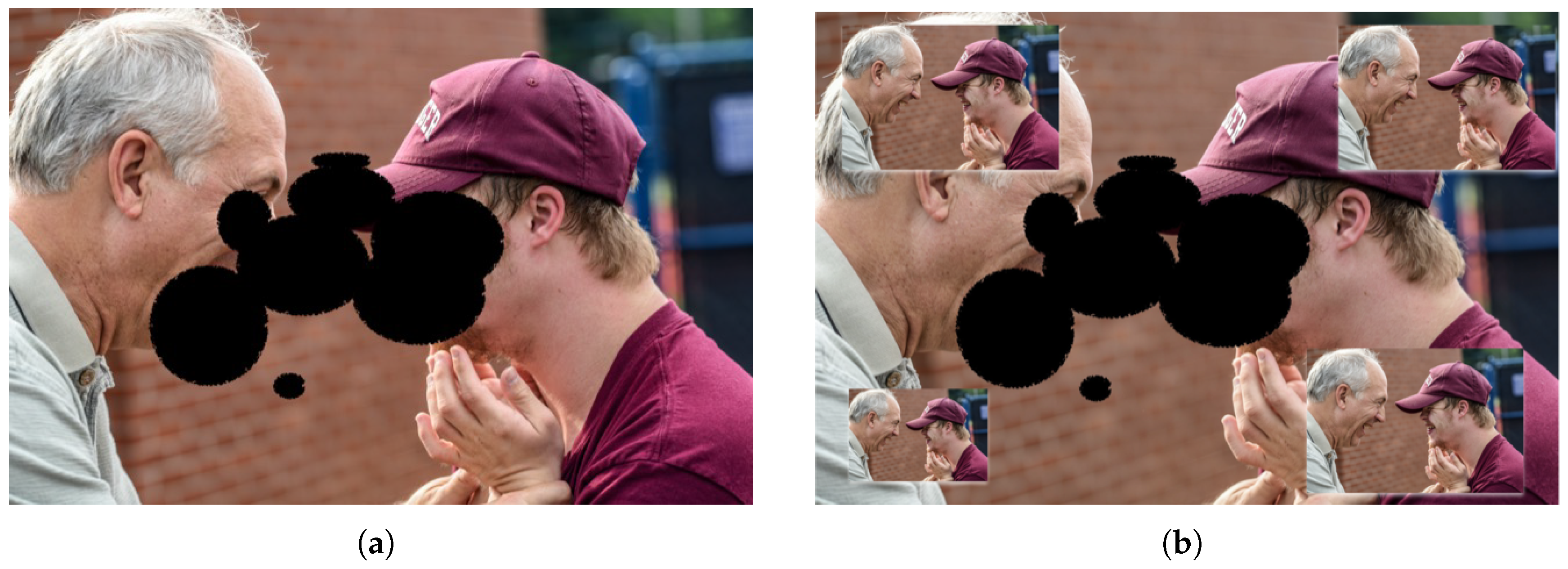
The system, given the characteristics of the SG, allows for bioptic vision and visual scanning. Since the EPSON design is based on near-eye displays that project a transparent image in front of the eye [36], the projected image can be set to the best viewing area of the patient by projecting it onto the required area of the displays. This allows the patient to have a minified image and real vision at the same time. The system displays the image with minification, enabling the patient to perceive the entire figure, albeit at a reduced size. Figure 2a illustrates the vision of a user with central vision loss, while Figure 2b shows the enhanced vision when using Retiplus.
Thanks to the transparency of the displays, the patient’s natural vision remains available in the background, while Retiplus projects the entire minified image in the area where the user desires. In this example, four projections have been placed in each corner of Figure 2b for demonstration purposes, but in practice, the projection is set only in the user’s preferred area. As shown in this example, this functionality can significantly aid in recognizing facial expressions.

3.8.2. Evaluate Mode

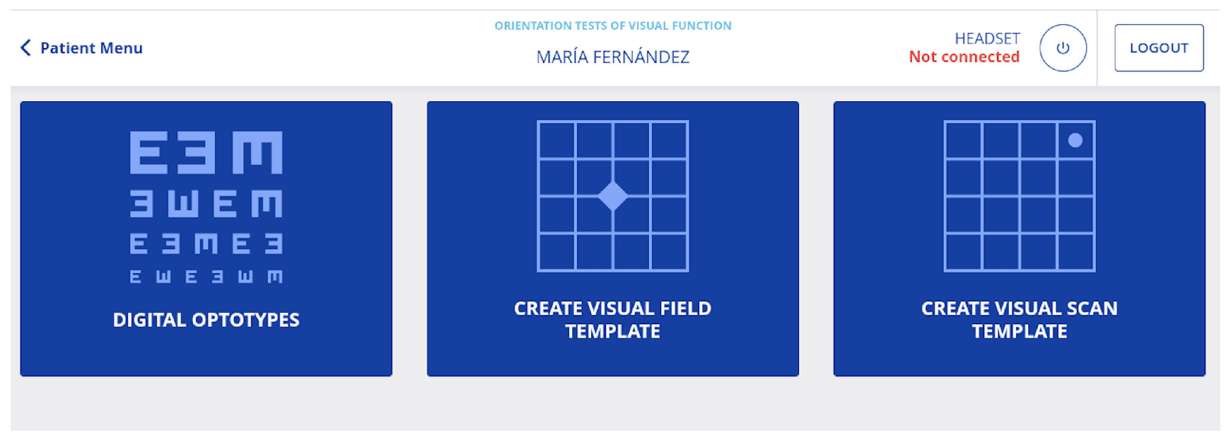
In Retiplus, a visual condition assessment menu has been developed to facilitate the evaluation of visual conditions. These functions are accessed by selecting the desired function shown in Figure 3.
In this menu, visual function is assessed by adding digital optotypes, visual field templates and visual scanning templates.
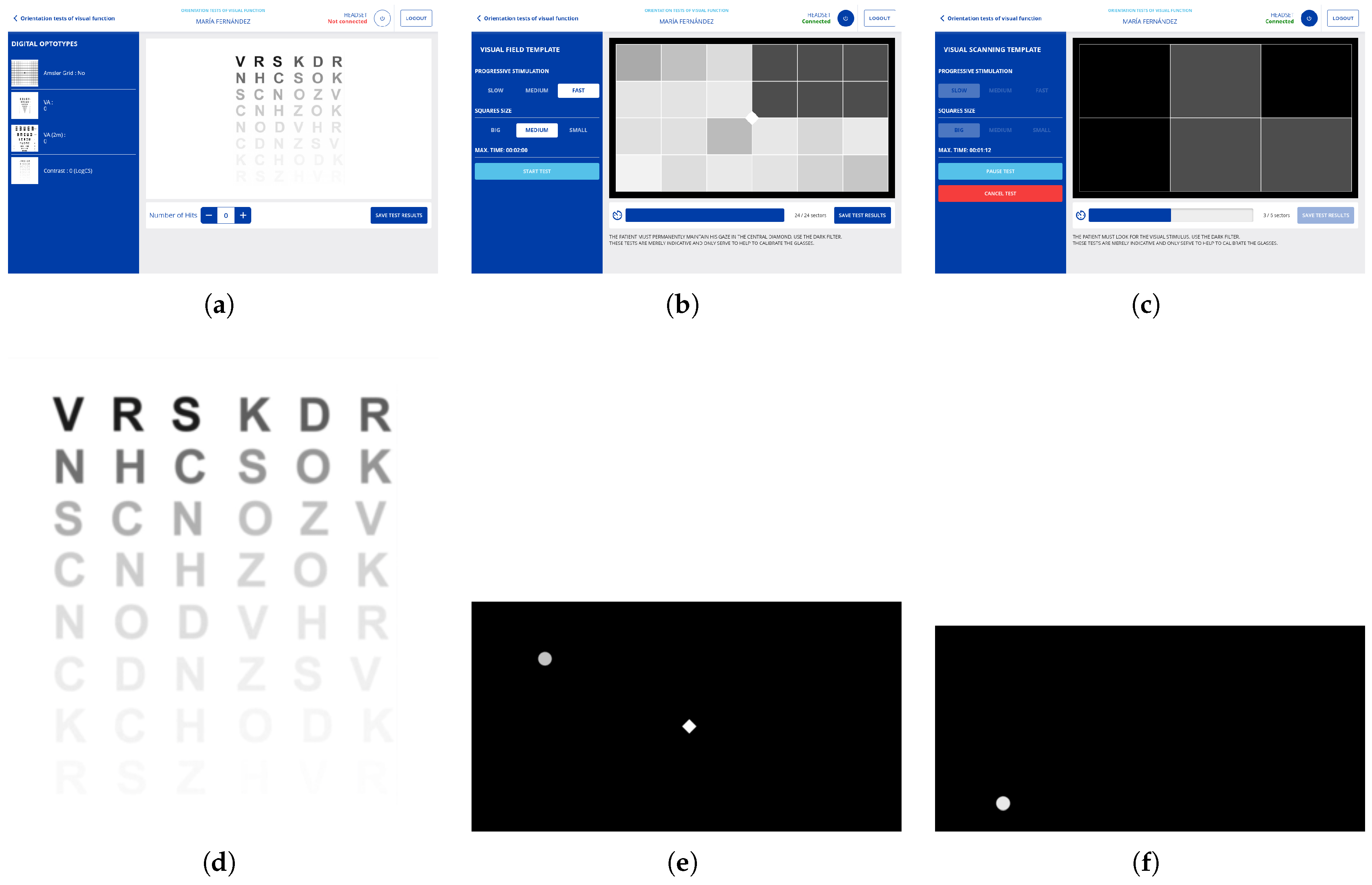
In the digital optotypes, an Amsler Grid [67] has been incorporated to assess potential visual deficiencies. Optotypes were also included for visual acuity equivalent to 4 m or for low vision the one corresponding to 2 m and optotypes for contrast evaluation with the Pelli–Robson test (Figure 4a). Additionally, on the left side of this screen, you can select to evaluate visual acuity at distances of either 2 or 4 m or perform the Amsler grid test. Figure 4d illustrates the virtual execution of the Pelli–Robson test.
To comply with the visual field template specification, the functionality of creating a visual field template is created. In this functionality, the patient must stare at a central white rhombus, which serves as a reference point. Figure 4e displays the functionality for the patient, while Figure 4b demonstrates the corresponding interface for the specialist. Upon viewing the pop-up stimulus while maintaining focus on the rhombus, the patient should press the center button of the controller.
To evaluate the visual scanning ability, a functionality has been developed wherein the patient is instructed to locate the emerging white stimulus and press the central button of the controller upon finding it. Figure 4c depicts the functionality interface for the specialist, while Figure 4f illustrates the test as viewed by the patient.
Both the visual field and visual scanning templates can be adjusted with progressive stimulations with three speeds (slow, medium, or fast) and three frame sizes.

3.8.3. Calibrate Mode

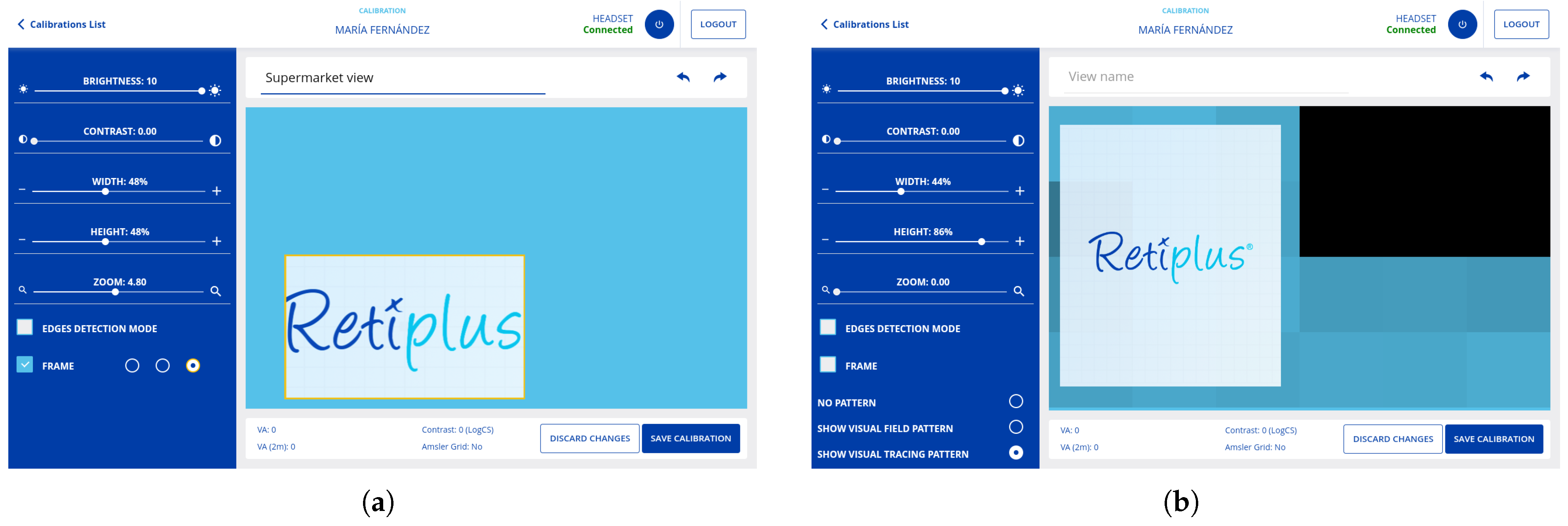
To comply with the customization of the visual aid, it is decided to create views, which we will also call calibrations. The views are real-time screen settings created according to the evaluation. By default, Retiplus has seven predefined views, and to create a new view, the specialist can calibrate in real time, adjusting the view lengthwise and widthwise, placing this view in different areas of the display, adjusting contrast, brightness, and zoom. You can also add contours and name these views. The menu to create a new calibration can be seen in Figure 5a.
In the left part of this menu, the specialist can adjust parameters such as zoom, contrast, or brightness. The width and length of what will be projected on the displays are set. The light blue part shows 100% of the display dimensions shown in the SG, and the square in which the Retiplus logo is framed represents the part that will be projected and in which area of the display. This framed area can be zoomed, contrasted, or brightened. These customized calibrations allow the specialist to program moving or static exercise routines for the patient to perform at home. This functionality can be used, for e.g., rehabilitation only by activating certain zones (right zone, upper part, central part), adding zoom to these zones or adding contrast.
In addition, the visual field and eye-tracking templates generated through the evaluation menu’s functionalities serve as a foundation for creating new calibrations, helping to determine the user’s optimal viewing area. This enables the smart glasses to project images in the user’s best viewing area within the display. Figure 5b demonstrates how a visual field template is used to create a new calibration. The visual field template shown in Figure 4b was used as a reference, indicating that the user did not respond to the visual stimuli presented in the upper-right region of their visual field. The darker boxes in Figure 4b reflect areas where the user failed to detect stimuli, and in the calibration view (Figure 5b), these regions are displayed in black.
Figure 6d shows the views created; these views can be activated or deactivated depending on the patient’s needs, and they can also be exported. In Figure 7, we can see real examples of calibrations that can be used for rehabilitation or for daily use.
It is important to note that all these evaluation and calibration functionalities are displayed on the smart glasses screens and are controlled by the specialist on the tablet. Figure 4 illustrates several functions of the evaluation menu. The images in the top row display the interfaces designed for the specialist, while those in the bottom row depict the perspectives observed by the patient through the smart glasses during the evaluation.

3.8.4. Patient Profile

To track the patient’s progress, a modern visual aid must store information regarding the use of its functionalities. This enables specialists to monitor the patient’s evolution and understand how the system is being utilized. For this purpose, a patient profile menu has been developed within the application, where data pertaining to the usage history of patients is stored. This section shows the statistics of patient use, digital optotype results, visual field templates, and visual scanning, as shown in Figure 6e. This data can be displayed by time bands, all the time or within the last three months, one month, two weeks, or one week. This section helps to know the patient and to be able to eliminate views that are no longer used and to recommend others. This functionality will be helpful for a continuous review and analysis by the specialist.

3.8.5. Use of the System

For specialist use (evaluation mode, calibrate mode, patient, profile), after logging in and selecting the desired language (for now, the application supports English and Spanish) (Figure 6a), the first step is to pair the patient’s SG with the specialist’s tablet from the Retiplus Kit (SG + tablet) via Bluetooth. Once this is completed, the calibrations of the Retiplus Kit tablet can be exported to the SG carried by the patient. After these actions, a list of patients appears, as shown in Figure 6b, and when selecting one, a menu appears, as shown in Figure 6c, with the functionalities that can be applied to the patient. No connection to the tablet is needed to use it as a visual aid; the patient interacts with the aid via the buttons on the controller. For instance, the patient can switch views according to their situation or adjust parameters in real time, such as zoom, brightness, or contrast.
It is important to clarify that the pairing between the specialist’s tablet and the visual aid (smart glasses) is only required during the user’s visit to the specialist. Once new calibrations are generated, the patient can use the visual aid independently, without the tablet. The tablet is solely for use by the low-vision specialist.

3.9. Achievements and Benefits for Users

One of the key achievements of Retiplus is that, through the evaluation and calibration functionalities developed in the tablet application, specialists can create customized visual aids tailored to each specific visual condition.
Once the visual condition is assessed, the specialist can calibrate the visual aid by adjusting it to the user’s specific needs across different contexts, such as at home, outdoors, or at work. These varying situations and contexts will be saved as customizable views or calibrations. The user can then interact with the device through physical buttons to modify settings in real time, such as brightness, contrast, and zoom, and can also switch between saved views according to the situation.
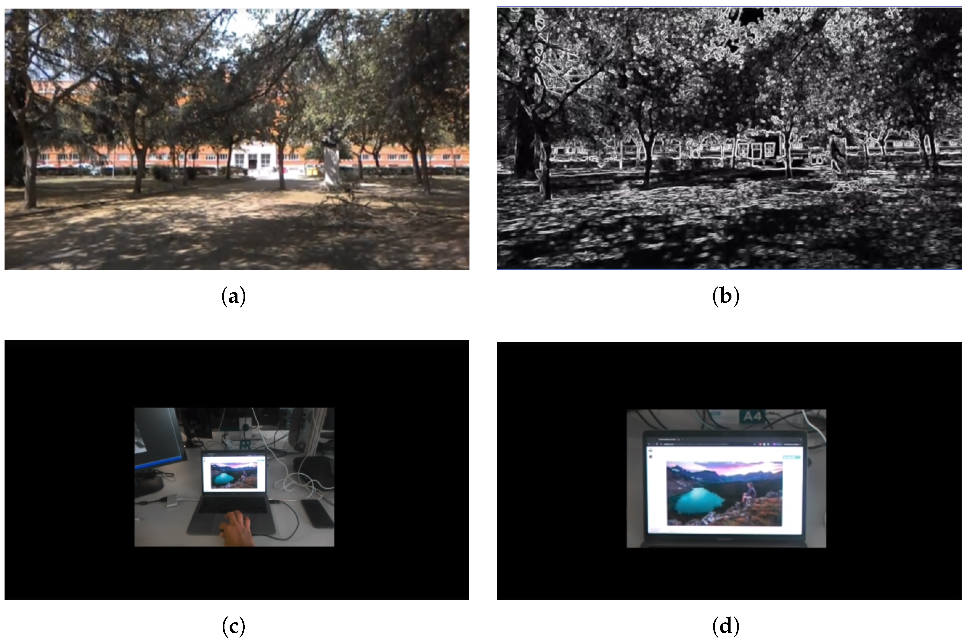
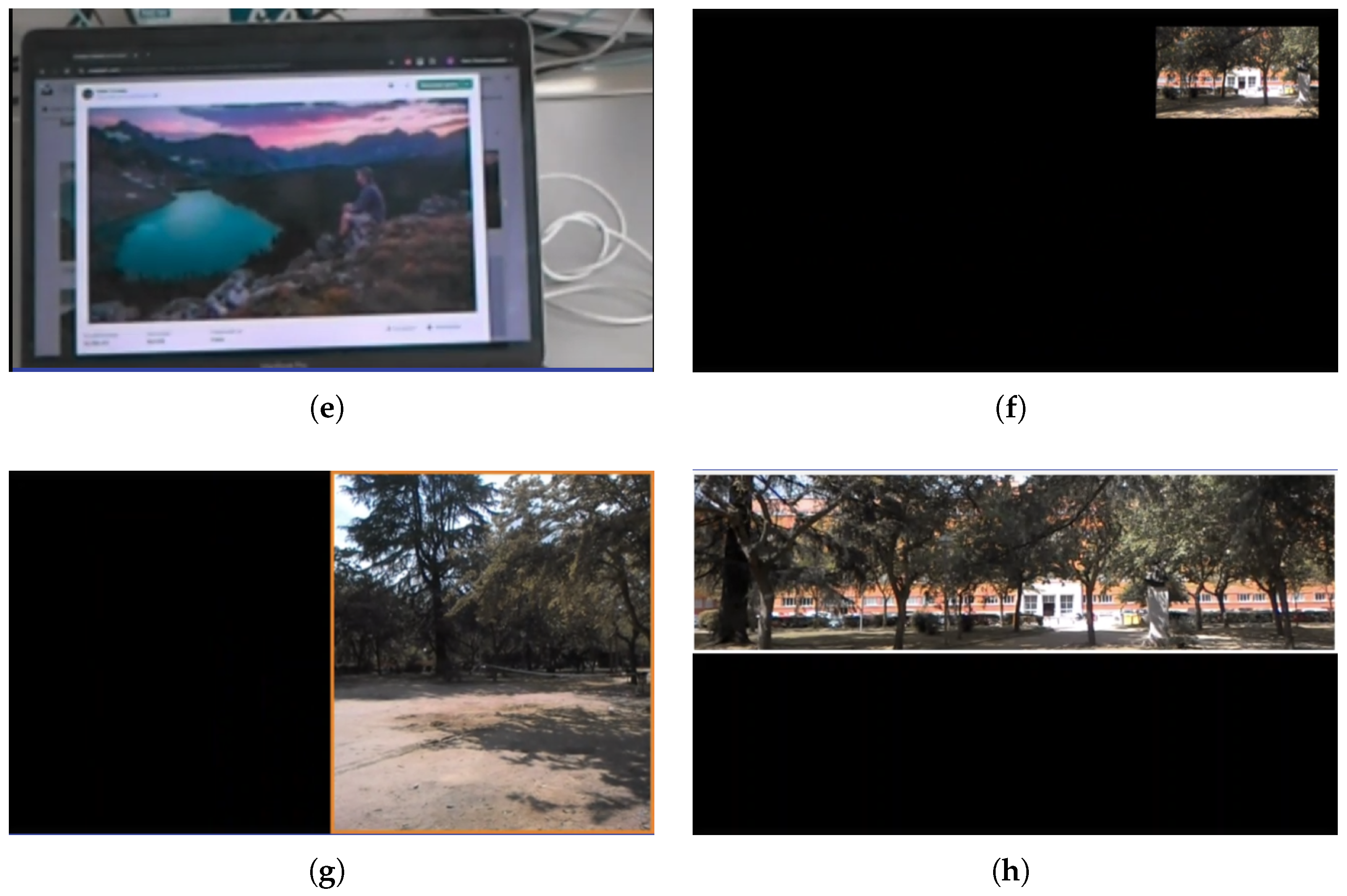
To demonstrate the customization capabilities of Retiplus, several examples of personalized calibrations are shown below. It is important to clarify that the following images, which represent various calibrations, were captured using the system’s smart glasses and recorded on a desktop using the Vysor tool to present them as realistically as possible and at full resolution. In the images, the black background represents the complete projection of the displays. Since the displays are transparent, what users actually see when using the visual aid is their natural vision, with the projection superimposed. In other words, the smart glasses overlay the projected image onto the user’s real-world view. Figure 7a represents the image captured by the smart glasses’ camera, projected to fully occupy the display. This projection can then be customized to suit each user’s visual condition.
For instance, a user with AMD who struggles with near or intermediate vision tasks that require detailed perception [68] can benefit from a customized view with a specific zoom level (Figure 7e), created by their low vision specialist, to aid in tasks requiring precise visual recognition.
For users with Retinitis Pigmentosa, who experience a narrowed visual field and peripheral vision loss [69], the specialist can calibrate a view to fit the user’s remaining functional visual field. By minifying the image and centering it in the user’s better vision zone, Retiplus increases the perceived visual field, helping the user better determine boundaries. Figure 7c illustrates how Retiplus centers the minified image for users with peripheral vision loss. Additionally, this image can be zoomed in or out as needed (Figure 7d).
For users with glaucoma, who often have reduced contrast sensitivity and difficulty distinguishing edges, a view with edge enhancement can be configured for use in ambulation scenarios (Figure 7b).
In cases where a user has better visual acuity in their right visual field, the entire projection can be shifted to this area, with the region highlighted to facilitate its location (Figure 7g). Similarly, for users whose best vision is in the upper part of their visual field, the images from the smart glasses can be projected in that zone (Figure 7h).
These are some examples of calibrations, thus showing the ability of Retiplus to address and propose a solution in the customization of technological visual aids in the population with low vision using mobile development technology and augmented reality. In addition, these calibrations can also be used in visual rehabilitation, enabling or disabling certain areas of vision to stimulate these areas as a visual exercise. Moreover, all these usage statistics will be available to low-vision specialists on their tablet so that they can track which calibrations are being used more, which less, and in which situations. This valuable information will help the specialist to make decisions that will enhance the use of the aid and the functional vision of the users.
Additionally, these calibrations can be employed in visual rehabilitation by enabling or disabling specific areas of the visual field to stimulate those regions as part of a visual exercise.
Moreover, low vision specialists can access usage statistics for all calibrations via their tablet, enabling them to monitor which settings are used most frequently, which are used least, and in which contexts. This valuable data helps specialists make informed decisions to optimize the effectiveness of the vision aid system and enhance users’ visual capabilities.

4. Validation & Results

The clinical study to evaluate the usefulness of the system was conducted by the Optometry University Clinic of Universidad Complutense de Madrid (UCM), and its objective was to evaluate and identify the effect of the Retiplus system on AV visual acuity, SC contrast sensitivity, the binocular visual field, and its effects on ambulation. This study was approved by the Ethics Committee of the Hospital Clínico San Carlos of Madrid (CEIC/CEAC code 22-2018, EudraCT code 665-2017). All study participants signed the informed consent form. The patients recruited comprised 30 people with visual field defects attributed to ocular pathologies due to Retinitis Pigmentosa RP and glaucoma GL. The variables to be analyzed were visual acuity (AV), contrast sensitivity (SC), visual field (VF), and ambulation. The visual assessment included the measurement of visual acuity at best contrast compensation (BVC) using the ETDRS [70] test, which is a test used to measure visual acuity in patients with low vision. The measurement of the contrast sensitivity function was performed with the Pelli–Robson test [71], and the measurement of the visual field with the tangent screen method [72] with the perimetry standard (SAP), which is a standard that establishes parameters such as the size of the stimulus [73]. Specifically, the Goldmann III standard was used [74], which establishes a stimulus size of 4.0 mm2 area and 0.43° diameter of round shape. The tangent screen is a method for measuring the visual field, in which the patient indicates when and how they perceive the emerging stimulus as it moves from the periphery towards the center of the screen. For ambulation, a circuit was designed, which will be explained later. These measurements were taken with and without Retiplus. Variables following a normal distribution will be compared by groups using t-tests [75]. These statistics will be used to observe the effect of the system on patients with and without its use. SPSS [75] (version 29) was used for the statistical analysis. Subjective patient factors such as motivation, adaptability to solutions, physical exercise, and intellectual activity were also analyzed.

4.1. Clinical Study Methodology

Does the use of augmented-reality-based electronic visual aids, combined with mobile technology for personalized assistance, significantly improve the quality of life and functional ability of patients with low vision?
Measurements were performed with optical compensation (glasses) if needed.

4.1.1. Binocular Visual Acuity

Visual acuity measurements were conducted binocularly using the ETDRS optotype at a distance of 4 m from the individual being assessed. If the response did not exceed 15 correct letters at this distance, the distance was reduced to 2 m. Compensation was adjusted accordingly, adding 0.3 logMAR at each change. The number of correctly identified letters was recorded on a logarithmic scale.

4.1.2. Binocular Contrast Sensitivity Function: Measurement with and Without Retiplus

Contrast sensitivity function was measured with the Pelli–Robson test at a distance of 1 m. The illumination conditions of the test were uniform and in accordance with those specified by the manufacturer. The number of letters and triplets seen was recorded on a logarithmic scale Log contrast sensitivity (LogCS).

4.1.3. Binocular Visual Field Measurement: With and Without Retiplus

For the measurement of the binocular functional visual field, the tangent screen method placed at a distance of 1 m was used. With the participant looking directly at the central point marked with a white button and a dynamic stimulus equivalent to the Goldmann standard III to arrange the isopters [76,77] of each person studied. The strategy followed to delimit the visual field was from “unseen to seen” starting from the periphery of the field following the grid drawn on the screen and continuing to the central gaze point. The degrees of the stimulus position up to the central gaze point were noted. The measurements made with the Retiplus system were performed with a minification factor of 1:3 in order to achieve the maximum magnification of the visual field. The visual field was first measured without the device. For the measurement with the Retiplus system, the image of the tangent screen was placed centered on the screen. The limits of the visual field were again noted.

4.1.4. Measurements in Dynamic Ambulation

An indoor obstacle course 20 m long by 2 m wide was designed, where the obstacles (cardboard boxes) were placed at three heights. The course could change its configuration, the basal walking speed, and the walking speed with and without the device, and the number of collisions was measured.

4.2. Results of the Clinical Study

For the interpretation of the results, the following abbreviations are added and shown in the tables: glaucoma (GL), Retinitis Pigmentosa (RP), baseline walking speed (VD0), walking speed without Retiplus in an obstacle course (VDSR), walking speed with Retiplus in an obstacle course (VDCR), visual acuity (VA), contrast sensitivity (CS), visual field without Retiplus (VF), and visual field with Retiplus (VF2).
This is a statistically normalized population with p-values > 0.05 for significance in the Kolmogorov–Smirnov [78] statistic, as shown in Table 1. For each of the variables analyzed, from a sample of 30 patients with no missing data distributed in two visual diseases, there were 21 people with Retinitis Pigmentosa (RP) and 9 with glaucoma (GL). SPSS software generated the p-values automatically for this normality test.
The age of the group is 54.18 ± 15.67 years (min of 19 and max of 86 years). Binocular basal visual acuity (VA) has a mean value of 0.41 ± 0.37 logMAR (min of 1.60 and max of −0.04 logMAR). CS has a value of 1.03 ± 0.63 log (min of 0.15 and max of 1.95) and a value of 1.03 ± 0.56 log with Retiplus. The anterior visual field (VF) without Retiplus has a value of 10.51 ± 4.71° (min of 2.50° and max of 20.13°), and the posterior with Retiplus (VF2) has a value of 16.89 ± 4.68° (min of 8.75° and max of 26.13°).
If we compare the means for three age groups (1 from 19 to 49 years, 2 from 50 to 59 and 3 from 60 to 86)m we observe that there are no differences in any of the dependent variables, neither VA, CS nor VF. It is found that in ANOVA [79] and in post hoc, no such differences are observed either within or outside the group for p > 0.05, as can be seen by disease and by age in Table 2. The same is true for the speed of ambulation in no cases.
Comparing the visual field without and with Retiplus, significant differences were observed across the entire sample. The mean visual field increased from 10.51 ± 4.71° to 16.89 ± 4.68°, demonstrating a high correlation of 0.608 (Table 3). There is a mean increase in the binocular functional visual field of 61%, with no differences by age or disease. In the Table 4, the paired samples test for VF without and with Retiplus is observed. In this table, the t-test showed significant differences between VF and VF2 with a confidence interval of 95%. The results regarding ambulation indicated that the device did not impact the speed or safety of ambulation. Moreover, with prior instructions and regular use, improvements in ambulatory performance were observed. Another finding was that 66% of patients who achieved more than 0.4 Log baseline contrast sensitivity experienced an improvement in contrast sensitivity or maintained the same values by using the system. For the rest of the patients, no statistically significant differences were found between wearing the aid and not wearing it.

4.3. User Experience Evaluation

Within the framework of user-centered [20] design and with the goal of promoting the usability and adoption of visual aids, this section evaluates the system’s usability and user interaction by gathering feedback from users and low-vision specialists. The objective is to identify areas for improvement in future iterations. This effort was made possible through close collaboration between the university, specialty clinics, and low vision associations, where training tests were conducted, and protocols for adapting this augmented reality system were developed. During these tests, usability and interaction surveys were administered, and videos were recorded to document the process.
This section is organized as follows: first, an analysis of a report created and promoted by the National Competence Service for the Deafblind (NKBD) [80] and the National Resource Center for the Deafblind (Eikholt) [81], which are both Norwegian institutions, is presented.
Second, a report from the Faculty of Optics and Optometry at the Complutense University of Madrid is analyzed (UCM). Finally, the section concludes with a compilation of notable user reactions to the system, gathered from various official Retiplus online resources, including the YouTube channel [82] and user testimonials featured on the website [83].
From now on, the first report will be referred to as Eikholt and the second as UCMR (Complutense University of Madrid Report).

4.3.1. Eikholt Report

The objective of the report [84] was to evaluate the validity of the aid for patients with Usher syndrome [85]. To achieve this, a program consisting of several activities was designed to test the aid based on both the users’ experience and the feedback from evaluating specialists. This program included three weekly sessions over three weeks in a group setting.
The participants were four individuals diagnosed with Usher syndrome types 1 and 2. During the first week, the sessions focused on introducing the aid, assessing visual conditions with the system, and performing basic contact exercises, such as identifying objects in a room. In the second week, the initial exercises were repeated, with the addition of ambulation exercises in enclosed spaces. In the third week, the previous week’s exercises were repeated, and outdoor ambulation exercises were introduced.
After each session, the participants were evaluated. One exercise involved seating several people at different distances and asking the participant to identify how many individuals they could see, both with and without the aid. Another exercise required placing everyday objects of various sizes on a table and asking the participant to identify how many objects they could recognize while sitting or standing at distances of 0.5 m, 1 m, and 2 m.
The usability of the controller was also evaluated by reviewing the functionality and layout of the buttons. Participants practiced turning the aid on and off, learned how to charge the device, and trained on switching between saved views. They practiced selecting different views depending on the activity being performed and explored a more advanced mode, where patients had the freedom to adjust parameters such as contrast, brightness, and zoom as required by the environment.
Regarding the hardware, participants expressed a preference for glasses that more closely resemble a regular pair of glasses. They suggested placing the camera at the center of the glasses instead of on the side and requested a camera with higher resolution. Additionally, they preferred a wireless system to eliminate the need for cables, enabling the entire system to operate through wireless communication.
Participants noted that the battery life was somewhat limited but compensated for this using an external battery. However, they would prefer the system to provide an earlier warning when the battery is low. Another concern was that, with prolonged use, the controller becomes too hot, which can cause discomfort.
They also recommended adding a lock button to the controller to prevent accidental pressing of keys. Furthermore, they suggested marking the buttons on the controller with numbers to make it easier to locate and operate the desired button.
Participants found the aid especially useful when an object fell to the ground and they could find it with the contour mode (Figure 7b) of the aid, especially in low-light environments. When one participant discovered this, the others replicated it, suggesting that group training can speed up and improve the utilization of the help. They found it especially useful that default settings can be returned to via a system functionality by pressing a button on the controller. Three of the participants believe they will use the help after the course, while one is hesitant due to the strange appearance of wearing the help. All believe that it is useful in indoor activities in enclosed spaces because it gives a better overview of the room. All believe that the aid works very well in low-light situations, especially for searching for fallen objects on the floor. One participant believed that more outdoor training sessions are needed in ambulation. This study also stresses the important need to check that they have been fitted correctly for patients with combined vision and hearing problems, and for such patients, they also note that it could improve communication, as patients are able to locate people within a room. The testers found that, although many customized “views” can be configured with the system, in practice, four or five are sufficient.

4.3.2. UCMR: Introduction

This report outlines the development of a training protocol designed to evaluate the Retiplus system in patients with peripheral visual field loss. The objective of the training sessions was to assess patients’ adaptation to the aid through a series of structured exercises organized into specific training sessions.
The sample included 28 patients from the Retina España [86] association, of whom 8 were excluded for not meeting the inclusion criteria established by specialists based on their experience with this system. The thresholds for inclusion were VF ≥ 5° and VA ≥ 0.3 decimal. Out of the remaining 20 patients, 16 ultimately completed the training. Regarding low vision pathology, 15 patients were affected by RP, of whom 2 had the Usher type II syndromic form and 1 had glaucoma. The report was structured as follows.
1.
Evaluation of visual function n = 20: Visual field, visual acuity, and contrast sensitivity assessments were conducted in a manner consistent with the methodology described in the previous section of the clinical study. Additionally, a basic method for measuring the visual field was devised to capture patient impressions. This included a test referred to as the “Practical exercise at 3 m,” accompanied by a corresponding questionnaire “Questionnaire for the practical visual field at 3 m exercise” (PVF3MQ).
2.
Training phase n = 16: Five training sessions were conducted to facilitate interaction with the augmented reality aid, with each session evaluated using a questionnaire “Peripheral Vision and Mobility Questionnaire” (PVMQ). The aim of this training was to achieve progressive adaptation to the augmented reality aid across different stages until it could be effectively used by patients in any daily situation—indoors, outdoors, and in controlled or uncontrolled environments. A survey was also provided to the specialists to assess the evolution of the users’ training sessions, which we called the “training evolution questionnaire” (TEQ).
3.
Pre-training and post-training assessment n = 16: The PVMQ questionnaire was administered both before and after each training session, utilizing a Likert scale [87] to measure responses.
Additionally, some training sessions were recorded, but only for participants who had provided explicit informed consent for video recording, capturing patients’ opinions and experiences with the system. The most relevant reactions and feedback from these recordings were transcribed and analyzed as valuable insights for further improvement of the system.

4.3.3. UCMR: Practical Exercise of VF at 3 m

To evaluate the usability of the aid in a practical manner within a context more closely resembling real-world scenarios, and to gather valuable feedback on the initial interaction between the user and the system, a specific test was incorporated, accompanied by a questionnaire. This exercise, designed to measure the VF in a rudimentary manner, involved placing 11 × 12 cm cardboards on a wall at positions corresponding to a 5° angle on both the horizontal and vertical axes, with the patient positioned three meters away. An expert optometrist conducted the test by asking the patient to identify which cardboards they could see in all directions, recording the results. The exercise was performed both with and without the system. To evaluate the binocular visual field during the three-meter practical exercise, patients were asked to complete a questionnaire about their experience using the aid. This questionnaire also provided valuable insights into the potential that users perceive in this type of system during the early stages of use and practical testing, as well as their beliefs regarding its ability to assist them in performing daily life activities. The questionnaire (PVF3MQ) included the following questions:
1.
Have you been able to describe more objects to your right, left, and around you?
2.
Has your overall vision improved?
3.
Has your vision improved in low light?
4.
Do you think your orientation can improve?
5.
Do you think that after your training, the system can help you with ambulation?
Responses were rated on a four-point scale: 1 = not at all, 2 = a little, 3 = moderate, and 4 = a lot.
In Figure 8a, the layout of the practical exercise at 3 m can be seen.

4.3.4. UCMR: Training

The training program consisted of a structured progression of increasing difficulty, designed to help patients adapt to the augmented reality aid in a simple and gradual manner. It was tailored individually according to the degree of vision loss, with the goal of achieving independence and full control of the system. The program was divided into five phases as follows:
Phase 1: Adaptation to the bioptic mode, focusing on the localization of objects and parts of the room.
Phase 2: Static training using visual scanning with an external filter and contour mode, involving tasks such as locating and collecting colored pieces and completing large puzzle exercises.
Phase 3: Static and dynamic training in a controlled environment, including exercises such as navigating circuits of colored cones.
Phase 4: Dynamic exercises in closed spaces, including walking with the aid and identifying stairs.
Phase 5: Outdoor dynamic exercises, which included navigating crowded places, locating specific areas in shopping malls, entering the subway, and independently crossing streets while monitored by an optician–optometrist. This phase also involved walking through bright and shaded areas to assess path perception using the system.
Figure 8b shows a task corresponding to phase 3, in which users have to make a puzzle. To do so, they have to select colored pieces arranged horizontally on a table and place them in a puzzle distributed vertically, so that users practice vision in the vertical and horizontal planes.
A Training Evaluation Questionnaire (TEQ) was conducted to assess improvements in the use of the augmented reality aid throughout the different phases of the program. The survey employed a scale from 1 to 5, where 1 represented the lowest level of usability and 5 indicated the highest level of usability improvement and evolution. This approach enabled an objective evaluation of patients’ progress and adaptation as they advanced through the various training phases.

4.3.5. UCMR: Peripheral Vision and Mobility Questionnaire

This ad hoc survey (PVMQ) was conducted before and after each training session. The questionnaire comprised 11 questions, with possible responses rated on a scale of 1 to 4, where 1 = none, 2 = little, 3 = moderate, and 4 = extreme. For questions [5–9], users were asked to indicate the degree of difficulty in performing specific tasks. The responses were collected in writing.
The survey questions were as follows:
1.
Do you have difficulty getting around in crowded areas?
2.
Do you have difficulty getting around in unfamiliar places?
3.
Do you only take the bus or the subway?
4.
Do you have difficulty at dusk or in low-light conditions?
5.
What degree of difficulty do you have going down steps, stairs, or curbs in low light or at night?
6.
Crossing streets
7.
Locating traffic lights
8.
Locating objects
9.
Seeing billboards, signs, etc.
10.
What degree of difficulty do you have noticing objects to the side while walking?
11.
When you become disoriented, do you have difficulty regaining your bearings?
This survey, referred to as the PVMQ, was designed to capture users’ impressions of the system and evaluate the impact of using the aid versus not using it. Its purpose was to assess the degree of difficulty users experienced when performing tasks with and without the aid. The information gathered is invaluable for both specialists and patients, as it contributes to the iterative process of user-centered design, ensuring that the system aligns with user needs and expectations.
To compare the data, a hypothesis test with a significance level of p = 0.05 was performed following these steps: first, defining the hypothesis; second, evaluating the assumption of normality; and finally, selecting the appropriate statistical test. The null hypothesis ( H 0 ) stated that there were no significant differences between the means, while the alternative hypothesis ( H a ) proposed that the means of the data before and during the use of the system were significantly different.
The Shapiro–Wilk [88] method was used to assess normality. If the assumption of normality was satisfied, parametric statistics, such as the Student’s t-test, were applied. Statistical analysis was performed using SPSS (version 29) software [75].

4.3.6. UCMR: Results of Protocol

The questionnaire accompanying the practical visual field exercise conducted at 3 m (PVF3MQ) yielded the following results: each of the twenty patients answered the five questions, with 60% (48 responses) rated as “very much”, 30% (24 responses) as “quite a lot,” 5% (4 responses) as “normal,” and 5% (4 responses) as “not at all.” These results indicate that 90% of the patients believe that movement, mobility, and orientation can be improved through the aid.
The results of the specialists’ evaluation (TEQ) regarding the progress achieved through training sessions to determine whether they improved the use of the aid were as follows: 56.25% of the participants (n = 45) achieved a higher level, 6.25% (n = 5) reached a high level, 18.75% (n = 15) attained a medium level, 6.35% (n = 5) demonstrated a low level, and 12.5% (n = 10) experienced a decline in performance. In conclusion, more than 60% of the participants considered the training sessions to be highly useful and beneficial.
The means of the Peripheral Vision and Mobility Questionnaire (PVMQ) scores were analyzed using the Student’s t-test to assess the effect of pre-training and post-training sessions. The analysis revealed statistically significant differences (p < 0.05) in all question pairs, except for questions [6, 7, and 11].

4.3.7. Compilation of Video Testimonials: Reactions to the Use of the Aid

To gain a deeper understanding of the context and scope of the aid, video testimonials were conducted throughout the lifecycle of the user-centered development of Retiplus. These testimonials aim to observe and gather feedback on the adaptation of the aid to the specific challenges of users’ daily lives, analyze its usage, and assess users’ impressions—socially, emotionally, and in terms of their daily activities. This approach has also provided valuable insights into potential areas for improvement. These efforts have been made possible through collaboration with clinics, associations, and organizations specializing in low vision, as well as input from patients’ families, low-vision specialists, and the university’s continued commitment to enhancing the quality of life for individuals with low vision.
Patient 1: A 48-year-old patient diagnosed with Retinitis Pigmentosa reported peripheral vision as his primary issue. His main challenges in daily life include walking and navigating his workplace environment, where he often needs to re-scan his surroundings to locate personal objects. Regarding the use of the controller outdoors, he stated that its compact size allows him to keep it in his pocket, enabling him to remain hands-free and mobile. This patient particularly favored a view configured at the top and noted that the functionality of the device was easy to operate using the controller.
Patient 2: A patient diagnosed with Retinitis Pigmentosa (RP) since childhood, at the age of 7–8, with no family history. Her first symptoms were night blindness, and her degeneration has been slow, allowing her to lead a relatively normal life. She previously used a cane at night, a tele magnifying glass to read documents, and selective absorption filters. Upon first using the augmented reality aid, she reported feeling very tired, attributing this to the stimulation of a part of her brain that had been “forgotten.” She noted an improvement in her eye mobility due to the expanded visual field. Regarding autonomy, she expressed feeling more confident, especially in crowded places. However, she uses the aid only up to three sessions per week, as she finds it demanding and tiring. Her most positive experiences with the aid include viewing museum exhibits, particularly paintings, which she can now see in their entirety. She finds the contrast feature particularly useful, as it enhances her perception and differentiation of colors. Interestingly, since using the aid, she no longer feels the need to wear a cap outdoors, as she can now see clouds and trees while walking.
Patient 3: A 21-year-old patient diagnosed with Retinitis Pigmentosa, with a field of vision (FOV) of 7°, reported significant challenges navigating supermarkets due to obstacles like shopping carts and people crossing her path. After using the augmented reality aid, she noted that the enlarged visual field made it much easier to navigate and locate products using the zoom function. Interestingly, after removing the aid, she observed that her eye became more active and alert to external stimuli, allowing her to perceive more information than before.
An event of particular significance observed during the initial use of the augmented reality aid is what specialists have termed the “WOW Test”. This name was inspired by the emotional reactions of excitement and happiness exhibited by users when they first experienced the enhanced capabilities provided by the aid.

4.3.8. Conclusions of User Experience Evaluations

To better understand the results obtained from the compilation of available resources on user experiences—such as video testimonials, reports, and specialist evaluations—we will compare these findings with the User Experience Questionnaire (UEQ) [89,90]. The content of video testimonials and recordings of aid usage are particularly valuable, as they provide spontaneous and natural user reactions. The evaluation will focus on six key dimensions: “Attractiveness,” “Perspicuity,” “Efficiency,” “Dependability,” “Stimulation,” and “Novelty”.
For Perspicuity, the (TEQ) survey showed a positive progression in training task performance, with 62% of users achieving a high level of usability. Additionally, video testimonials indicated that users were able to operate the aid with ease after receiving a few initial guidelines from specialists. Regarding Efficiency, once instructed, users completed tasks effectively in dynamic and controlled environments. However, the (PVMQ) questionnaire revealed that they faced challenges in reorienting themselves in uncontrolled spaces and crossing streets. In terms of Dependability, users did not report any unexpected issues, acknowledging that interaction with the bioptic vision aid requires training. Some even suggested additional training sessions. As for the controller, which operates via physical buttons, no usability concerns were mentioned. Regarding Stimulation, the WOW test demonstrated a strong sense of interest, excitement, and willingness among users to continue exploring and using the aid. Regarding Novelty, testimonials from users and specialists, as well as interest from low-vision clinics, organizations, and the university, have confirmed that the Retiplus system is perceived as both novel and innovative. Finally, regarding Attractiveness, users generally find the aid useful, particularly for ambulation tasks in controlled spaces and for static tasks. This is supported by the (PVMQ) questionnaire, which showed a significant difference in several items between using and not using the aid, indicating that it provides users with a sense of autonomy and safety. However, aesthetics remains one of the main areas for improvement, as some users express concerns about potential social stigma associated with wearing the aid. Additionally, some users are hesitant about using it in crowded outdoor areas, suggesting that a design resembling everyday glasses and featuring wireless communication would encourage wider adoption.
The compilation of user experiences and evaluations revealed that the structured development of training sessions and protocols enhances the utilization of this augmented reality-based technological aid.
Other findings beyond the comparison with the UEQ are presented below. Users indicated that exercises involving bioptic vision require additional training sessions, as do activities related to ambulation in real, uncontrolled environments.
The results from the (PVMQ) questionnaire revealed that the system provides assistance in daily activities related to mobility and peripheral vision. However, no improvement was observed in tasks such as “crossing streets,” “locating traffic lights,” or “reorientation”. Patients did, however, report better adaptation to changes in lighting conditions when transitioning from brightly lit areas to low-light environments and vice versa. It is noteworthy that age did not influence or affect the results obtained.
The findings of the PVF3MQ survey revealed a high level of acceptance and willingness to use the aid, as well as the potential these patients perceive in this type of assistance, with 90% considering that it could help them perform their daily activities.
Regarding the hardware usability of the system, specialists identified the most significant challenges as glare from the tablet when used outdoors and limited battery life. The latter issue was addressed by incorporating an external battery. Regarding the application software, specialists did not report any difficulties in its use.
Another significant observation is that, after using the aid and removing the device, patients exhibited a subjective and unconscious tendency to search for the expanded visual field provided by the aid.

5. Conclusions

The main contributions of this work are the design and development of Retiplus, a customizable visual aid and rehabilitation system for individuals with low vision, which leverages the integration of mobile technology and augmented reality. This work provided positive results in visual field enhancement, where the visual field increased by 61% on average. Contrast sensitivity improved or remained the same in 66% of patients with a baseline sensitivity of >0.4 Log. For use in motion, the system did not negatively affect their ambulation or safety, and the study demonstrated that results improve with proper instructions for use. Thus, this work ratifies HDM-based systems as visual aids and rehabilitation tools [91]. Additionally, this work proposes the use of mobile technology, already integrated into everyday life, along with augmented reality to bridge the gap between the perspectives of technologists and low-vision specialists, and the real needs of low-vision users. By focusing on the personalization of visual aids and collecting system usage data, this approach enhances understanding and feedback between specialists and users.
From the evaluation of user experience, positive qualitative data and areas for improvement were identified. Users found the aid useful, with most indicating that they would use it regularly due to its effectiveness in enhancing daily living tasks, particularly static tasks and ambulation in controlled spaces. The users’ reactions were positive, expressing enthusiasm, happiness, and excitement as they immediately perceived improvements in their visual capabilities. The implementation of well-structured training sessions significantly improved usability, adoption, and overall utilization of the aid. Therefore, such training protocols should be considered a fundamental aspect of usability and should be guided by a low vision specialist during the initial stages of use.
Retiplus provides the low-vision specialist with a tool for evaluation and follow-up through stored usage data. It is also a customizable aid for each visual condition, since, as mentioned, each patient sees differently even if they have the same pathology. It also records the patient’s interaction in a secure and confidential way with the SG, which generates a history of use and relevant patient data. This record of low vision data establishes the basis for the exploitation of the data with the application of artificial intelligence (AI) techniques or, in the future, Big Data, to help the specialist in making decisions. In this way, it aids the specialist and the patient’s caregivers in understanding the patient’s visual perception and their utilization of the smart glasses. It also allows static and mobility home exercises to be programmed. Views can be saved for different activities, for example, for use at night or at sunset. It provides the specialist with tools for the evaluation of the visual condition such as digital optotypes, visual field templates, and visual scanning templates, in a fast and portable way. It also provides digital calibration, positioning the image within the displays, incorporating brightness, contrast, and zoom parameters. This calibration allows us to customize different views to help motivate the elasticity of the brain and create different views that the patient can select according to their environment.
As a unique innovation, Retiplus offers a novel AR-based vision rehabilitation system featuring indoor and outdoor calibration, along with specialist control via a mobile application.

Limitations of Retiplus System

One limitation of this study is the clinical analysis itself. The results and statistical comparisons should be performed on a larger sample to obtain more conclusive evidence and allow differentiation by sex, pathology type, or associations with specific clinical variables.
A key finding was the enlargement of the binocular visual field with Retiplus 1.0, from 10.51° to 16.89°, corresponding to a mean gain of 61%. However, this expansion must be interpreted with caution. The methodology inherently produces a geometric enlargement of the field due to the fixed minification factor (1:3), which reduces three degrees of the scene into one degree on the display. While this effectively enlarges the functional field in patients with very restricted vision (<10°), it does not provide proportional benefits for those with larger residual fields, and no significant improvements were observed in patients with a visual field >10°. Moreover, the current smart glasses hardware presents additional constraints, including limited zoom flexibility, restricted field of view, glare in outdoor environments, and limited battery life (partially mitigated with an external battery).
Another limitation is the absence of visual field maps, raw plots, and spatial visualizations to support the quantitative results. Without these, it is difficult to distinguish between a purely geometric effect of image minification and genuine perceptual gains that might translate into daily-life improvements. Furthermore, no outcome measures related to real-world functional performance (e.g., obstacle avoidance, navigation, or mobility tasks) were included, which limits the clinical interpretability of the findings.
The compilation of user experiences and evaluations revealed that structured training sessions and protocols enhance the effective use of this augmented-reality-based aid. Results from the PVMQ questionnaire indicated improvements in daily activities related to mobility and peripheral vision, but limited impact on tasks such as “crossing streets,” “locating traffic lights,” or “reorientation.” The PVF3MQ survey reflected a high level of acceptance, with 90% of patients considering the aid useful for daily activities. In addition, patients reported subjective adaptation to lighting changes and a tendency to search for the expanded visual field even after removing the device. Nonetheless, specialists highlighted practical limitations, such as glare from the tablet and the need for additional training in uncontrolled outdoor environments.
In this iteration of user-centered development, the hardware was adapted to meet the system’s requirements. However, newer smart glasses models now available provide larger fields of view, higher resolution, improved zoom capabilities, and enhanced usability, which may help address some of the limitations identified in this study.
Another limitation concerns the design and interpretation of the questionnaires employed. While the PVMQ was statistically analyzed and provided quantitative insights into the system’s impact on mobility and daily-life activities, the PVF3MQ and TEQ were primarily conceived as usability-oriented instruments. Their purpose was exploratory, focusing on capturing patients’ subjective impressions, perceived acceptance of the system, and the influence of structured training protocols on usability and adoption. As such, their value lies in offering a general understanding of user experience rather than statistically robust evidence. This methodological distinction should be considered when interpreting the findings. Future studies should address this limitation by incorporating clearer visualizations, complementary statistical analyses, and enhanced data presentation to maximize the scientific value and ensure a more rigorous evaluation of patient-reported outcomes.
Finally, users highlighted aesthetics as the area with the greatest potential for improvement.

6. Future Work

As part of future work within the iterative process of user-centered development (UCD), the next step should involve integrating the system with smart glasses that offer superior technical specifications, particularly in terms of field of view, camera performance, and a more aesthetic design. This integration is expected to further enhance the results obtained. As data collection continues in the current iteration, once a sufficient amount has been gathered, the initial application of artificial intelligence techniques can be explored. Furthermore, sustained data collection could enable the use of big data techniques to further improve system insights and performance.
Research can be performed on improvements such as patient interaction with the system, including interaction through gestures and interaction through audio, to improve the assistive technology [92]. A technological study should be carried out on the familiarity and adaptation of this type of technology according to the age of the patients. The inclusion of a smartphone as part of the system to replace the controller can also be investigated. A smartphone might be a more acceptable solution than the controller due to its convenience and potential to eliminate possible stigmatization when using the custom controller. Research can also be conducted on the inclusion of AI in the system for facial recognition and extraction of information from the environment that can be of help to the patient. The choice of technology is crucial to the solution, since the capacity of the assistance will depend largely on the technological capacity. A natural evolution thanks to the collection of usage data and the multitude of sensors integrated in the SG would be to create a Health-IoT platform that telematically assists the patient, provides the specialist with a real-time usage dashboard, intelligently adjusts certain parameters, or triggers alarms when the system exceeds certain thresholds.

Author Contributions

Conceptualization, J.J.J. and M.G.; methodology, J.R. and M.G.; software, R.C.; validation, R.B.-V., J.J.J. and J.B.; formal analysis, R.C.; investigation, J.J.J. and J.B.; writing—original draft preparation, J.J.J.; writing—review and editing, J.B.; supervision, J.R.; project administration, M.G.; funding acquisition, M.G. All authors have read and agreed to the published version of the manuscript.

Funding

This work has been partially supported by the Ministry of Science and Innovation of Spain (Grant number PID2021-125596OB-I00 and PLEC2022-009261).

Institutional Review Board Statement

This study was approved by the Ethics Committee of the Hospital Clínico San Carlos of Madrid (CEIC/CEAC code 22-2018, EudraCT code 665-2017) code 4156790.

Informed Consent Statement

All study participants signed the informed consent form.

Data Availability Statement

Data are contained within the article.

Conflicts of Interest

Author Rafael Cámara is the Co-founder and CEO of Plusindes, the company responsible for developing and commercializing the visual aid system described in this article. The other authors declare no conflicts of interest. This system originated from a continuous collaboration among university researchers, low vision specialists, patient organizations, and private industry, which collectively contributed to its development and subsequent commercialization.

References

  1. World Health Organization. Blindness and Vision Impairment. 2023. Available online: https://www.who.int/news-room/fact-sheets/detail/blindness-and-visual-impairment (accessed on 13 October 2023).
  2. Chan, T.; Friedman, D.S.; Bradley, C.; Massof, R. Estimates of incidence and prevalence of visual impairment, low vision, and blindness in the United States. JAMA Ophthalmol. 2018, 136, 12–19. [Google Scholar] [CrossRef] [PubMed]
  3. Bourne, R.; Steinmetz, J.D.; Flaxman, S.; Briant, P.S.; Taylor, H.R.; Resnikoff, S.; Casson, R.J.; Abdoli, A.; Abu-Gharbieh, E.; Afshin, A.; et al. Trends in prevalence of blindness and distance and near vision impairment over 30 years: An analysis for the Global Burden of Disease Study. Lancet Glob. Health 2021, 9, e130–e143. [Google Scholar] [CrossRef] [PubMed]
  4. Wong, W.L.; Su, X.; Li, X.; Cheung, C.M.G.; Klein, R.; Cheng, C.Y.; Wong, T.Y. Global prevalence of age-related macular degeneration and disease burden projection for 2020 and 2040: A systematic review and meta-analysis. Lancet Glob. Health 2014, 2, e106–e116. [Google Scholar] [CrossRef] [PubMed]
  5. Enoch, J.; Jones, L.; Taylor, D.J.; Bronze, C.; Kirwan, J.F.; Jones, P.R.; Crabb, D.P. How do different lighting conditions affect the vision and quality of life of people with glaucoma? A systematic review. Eye 2020, 34, 138–154. [Google Scholar] [CrossRef]
  6. Tham, Y.C.; Li, X.; Wong, T.Y.; Quigley, H.A.; Aung, T.; Cheng, C.Y. Global prevalence of glaucoma and projections of glaucoma burden through 2040: A systematic review and meta-analysis. Ophthalmology 2014, 121, 2081–2090. [Google Scholar] [CrossRef]
  7. Teo, Z.L.; Tham, Y.C.; Yu, M.; Cheng, C.Y.; Wong, T.Y.; Sabanayagam, C. Do we have enough ophthalmologists to manage vision-threatening diabetic retinopathy? A global perspective. Eye 2020, 34, 1255–1261. [Google Scholar] [CrossRef]
  8. Cho, N.H.; Shaw, J.; Karuranga, S.; Huang, Y.; da Rocha Fernandes, J.; Ohlrogge, A.; Malanda, B. IDF Diabetes Atlas: Global estimates of diabetes prevalence for 2017 and projections for 2045. Diabetes Res. Clin. Pract. 2018, 138, 271–281. [Google Scholar] [CrossRef]
  9. Khalil, H. Diabetes microvascular complications—A clinical update. Diabetes Metab. Syndr. Clin. Res. Rev. 2017, 11, S133–S139. [Google Scholar] [CrossRef]
  10. Rainey, L.; Elsman, E.B.M.; van Nispen, R.M.A.; van Leeuwen, L.M.; van Rens, G.H.M.B. Comprehending the impact of low vision on the lives of children and adolescents: A qualitative approach. Qual. Life Res. 2016, 25, 2633–2643. [Google Scholar] [CrossRef]
  11. Binns, A.M.; Bunce, C.; Dickinson, C.; Harper, R.; Tudor-Edwards, R.; Woodhouse, M.; Linck, P.; Suttie, A.; Jackson, J.; Lindsay, J.; et al. How effective is low vision service provision? A systematic review. Surv. Ophthalmol. 2012, 57, 34–65. [Google Scholar] [CrossRef]
  12. Demmin, D.L.; Silverstein, S.M. Visual impairment and mental health: Unmet needs and treatment options. Clin. Ophthalmol. 2020, 2020, 4229–4251. [Google Scholar] [CrossRef]
  13. Dhital, A.; Pey, T.; Stanford, M.R. Visual loss and falls: A review. Eye 2010, 24, 1437–1446. [Google Scholar] [CrossRef] [PubMed]
  14. Zhao, Y.; Kupferstein, E.; Tal, D.; Azenkot, S. “It Looks Beautiful but Scary” How Low Vision People Navigate Stairs and Other Surface Level Changes. In Proceedings of the 20th International ACM SIGACCESS Conference on Computers and Accessibility, Galway, Ireland, 22–24 October 2018; pp. 307–320. [Google Scholar]
  15. Mehra, D.; Le, P.H. Physiology, night vision. In StatPearls; StatPearls Publishing: Treasure Island, FL, USA, 2019. [Google Scholar]
  16. Rivera-Romero, O.; Gabarron, E.; Ropero, J.; Denecke, K. Designing personalised mHealth solutions: An overview. J. Biomed. Inform. 2023, 146, 104500. [Google Scholar] [CrossRef]
  17. Ro, Y.K.; Brem, A.; Rauschnabel, P.A. Augmented reality smart glasses: Definition, concepts and impact on firm value creation. In Augmented Reality and Virtual Reality: Empowering Human, Place and Business; Springer: Cham, Switzerland, 2018; pp. 169–181. [Google Scholar]
  18. Coughlan, J.M.; Miele, J. AR4VI: AR as an accessibility tool for people with visual impairments. In Proceedings of the 2017 IEEE International Symposium on Mixed and Augmented Reality (ISMAR-Adjunct), Nantes, France, 9–13 October 2017; pp. 288–292. [Google Scholar]
  19. Chen, Y.; Wang, Q.; Chen, H.; Song, X.; Tang, H.; Tian, M. An overview of augmented reality technology. J. Phys. Conf. Ser. 2019, 1237, 022082. [Google Scholar] [CrossRef]
  20. Ortiz-Escobar, L.M.; Chavarria, M.A.; Schönenberger, K.; Hurst, S.; Stein, M.A.; Mugeere, A.; Rivas Velarde, M. Assessing the implementation of user-centred design standards on assistive technology for persons with visual impairments: A systematic review. Front. Rehabil. Sci. 2023, 4, 1238158. [Google Scholar] [CrossRef] [PubMed]
  21. Ruffieux, S.; Hwang, C.; Junod, V.; Caldara, R.; Lalanne, D.; Ruffieux, N. Tailoring assistive smart glasses according to pathologies of visually impaired individuals: An exploratory investigation on social needs and difficulties experienced by visually impaired individuals. Univers. Access Inf. Soc. 2023, 22, 463–475. [Google Scholar] [CrossRef]
  22. Deemer, A.D.; Bradley, C.K.; Ross, N.C.; Natale, D.M.; Itthipanichpong, R.; Werblin, F.S.; Massof, R.W. Low vision enhancement with head-mounted video display systems: Are we there yet? Optom. Vis. Sci. Off. Publ. Am. Acad. Optom. 2018, 95, 694. [Google Scholar] [CrossRef]
  23. Scheiman, M. Understanding and Managing Vision Deficits: A Guide for Occupational Therapists; Routledge: Oxford, UK, 2011. [Google Scholar]
  24. Altinbay, D.; Taskin, I. Evaluation of vision-related quality of life in Retinitis Pigmentosa patients with low vision. Jpn. J. Ophthalmol. 2021, 65, 777–785. [Google Scholar] [CrossRef]
  25. Brown, M.M.; Brown, G.C.; Sharma, S.; Landy, J.; Bakal, J. Quality of life with visual acuity loss from diabetic retinopathy and age-related macular degeneration. Arch. Ophthalmol. 2002, 120, 481–484. [Google Scholar] [CrossRef]
  26. Minto, H.; Butt, I.A. Low vision devices and training. Community Eye Health 2004, 17, 6. [Google Scholar]
  27. Agarwal, R.; Tripathi, A. Current modalities for low vision rehabilitation. Cureus 2021, 13, e16561. [Google Scholar] [CrossRef]
  28. Apfelbaum, H.; Peli, E. Tunnel vision prismatic field expansion: Challenges and requirements. Transl. Vis. Sci. Technol. 2015, 4, 8. [Google Scholar] [CrossRef] [PubMed]
  29. Massof, R.W.; Rickman, D.L.; Lalle, P.A. Low vision enhancement system. Johns Hopkins APL Tech. Dig. 1994, 15, 120–125. [Google Scholar]
  30. Harper, R.; Culham, L.; Dickinson, C. Head mounted video magnification devices for low vision rehabilitation: A comparison with existing technology. Br. J. Ophthalmol. 1999, 83, 495–500. [Google Scholar] [CrossRef]
  31. Vargas-Martin, F.; Peli, E. Augmented-view for restricted visual field: Multiple device implementations. Optom. Vis. Sci. 2002, 79, 715–723. [Google Scholar] [CrossRef]
  32. Culham, L.E.; Chabra, A.; Rubin, G.S. Clinical performance of electronic, head-mounted, low-vision devices. Ophthalmic Physiol. Opt. 2004, 24, 281–290. [Google Scholar] [CrossRef] [PubMed]
  33. Sivakumar, P.; Vedachalam, R.; Kannusamy, V.; Odayappan, A.; Venkatesh, R.; Dhoble, P.; Moutappa, F.; Narayana, S. Barriers in utilisation of low vision assistive products. Eye 2020, 34, 344–351. [Google Scholar] [CrossRef]
  34. Hoogsteen, K.M.P.; Osinga, S.A.; Steenbekkers, B.L.P.A.; Szpiro, S.F.A. Functionality versus inconspicuousness: Attitudes of people with low vision towards OST smart glasses. In Proceedings of the 22nd International ACM SIGACCESS Conference on Computers and Accessibility, Virtual, 26–28 October 2020; pp. 1–4. [Google Scholar]
  35. Lam, N.; Leat, S.J. Barriers to accessing low-vision care: The patient’s perspective. Can. J. Ophthalmol. 2013, 48, 458–462. [Google Scholar] [CrossRef] [PubMed]
  36. Ehrlich, J.R.; Ojeda, L.V.; Wicker, D.; Day, S.; Howson, A.; Lakshminarayanan, V.; Moroi, S.E. Head-mounted display technology for low-vision rehabilitation and vision enhancement. Am. J. Ophthalmol. 2017, 176, 26–32. [Google Scholar] [CrossRef]
  37. Irisvision. 2024. Available online: https://irisvision.com/irisvision-inspire/ (accessed on 7 June 2024).
  38. Yeo, J.H.; Bae, S.H.; Lee, S.H.; Kim, K.W.; Moon, N.J. Clinical performance of a smartphone-based low vision aid. Sci. Rep. 2022, 12, 10752. [Google Scholar] [CrossRef]
  39. Orcam. 2024. Available online: https://www.orcam.com/es-es/home (accessed on 8 September 2024).
  40. Xu, D.; Yu, M.; Zheng, C.; Ji, S.; Dai, J. The effects of an electronic head-mounted display in vision rehabilitation for patients with tunnel vision. Int. Ophthalmol. 2024, 44, 109. [Google Scholar] [CrossRef]
  41. Esight. 2024. Available online: https://www.esighteyewear.com/how-it-works/ (accessed on 9 November 2024).
  42. Wittich, W.; Lorenzini, M.C.; Markowitz, S.N.; Tolentino, M.; Gartner, S.A.; Goldstein, J.E.; Dagnelie, G. The effect of a head-mounted low vision device on visual function. Optom. Vis. Sci. 2018, 95, 774. [Google Scholar] [CrossRef]
  43. Younis, O.; Al-Nuaimy, W.; Alomari, M.H.; Rowe, F. A hazard detection and tracking system for people with peripheral vision loss using smart glasses and augmented reality. Int. J. Adv. Comput. Sci. Appl. 2019, 10, 1–9. [Google Scholar] [CrossRef]
  44. Parker, A.T.; Swobodzinski, M.; Wright, J.D.; Hansen, K.; Morton, B.; Schaller, E. Wayfinding tools for people with visual impairments in real-world settings: A literature review of recent studies. Front. Educ. 2021, 6, 723816. [Google Scholar] [CrossRef]
  45. Palanker, D.; Le Mer, Y.; Mohand-Said, S.; Sahel, J.A. Simultaneous perception of prosthetic and natural vision in AMD patients. Nat. Commun. 2022, 13, 513. [Google Scholar] [CrossRef] [PubMed]
  46. Stelmack, J. Quality of life of low-vision patients and outcomes of low-vision rehabilitation. Optom. Vis. Sci. 2001, 78, 335–342. [Google Scholar] [CrossRef] [PubMed]
  47. Cross, N.; van Steen, C.; Zegaoui, Y.; Satherley, A.; Angelillo, L. Current and future treatment of Retinitis Pigmentosa. Clin. Ophthalmol. 2022, 16, 2909. [Google Scholar] [CrossRef]
  48. Verbakel, S.K.; van Huet, R.A.; Boon, C.J.; den Hollander, A.I.; Collin, R.W.; Klaver, C.C.; Hoyng, C.B.; Roepman, R.; Klevering, B.J. Non-syndromic Retinitis Pigmentosa. Prog. Retin. Eye Res. 2018, 66, 157–186. [Google Scholar] [CrossRef]
  49. Chun, R.; Cucuras, M.; Jay, W.M. Current perspectives of bioptic driving in low vision. Neuro-Ophthalmology 2016, 40, 53–58. [Google Scholar] [CrossRef]
  50. Wang, S.; Moharrer, M.; Baliutaviciute, V.; Dougherty, B.E.; Cybis, W.; Bowers, A.R.; Luo, G. Bioptic telescope use in naturalistic driving by people with visual impairment. Transl. Vis. Sci. Technol. 2020, 9, 11. [Google Scholar] [CrossRef]
  51. Berger, S. 5 Activities to Improve Visual Scanning and Tracking. 2024. Available online: https://napacenter.org/visual-scanning-activities/ (accessed on 17 March 2024).
  52. Kasneci, E.; Black, A.A.; Wood, J.M. Eye-tracking as a tool to evaluate functional ability in everyday tasks in glaucoma. J. Ophthalmol. 2017, 2017, 6425913. [Google Scholar] [CrossRef] [PubMed]
  53. Deemer, A.D.; Swenor, B.K.; Fujiwara, K.; Deremeik, J.T.; Ross, N.C.; Natale, D.M.; Bradley, C.K.; Werblin, F.S.; Massof, R.W. Preliminary evaluation of two digital image processing strategies for head-mounted magnification for low vision patients. Transl. Vis. Sci. Technol. 2019, 8, 23. [Google Scholar] [CrossRef] [PubMed]
  54. Vargas-Martín, F.; Peli, E. Eye movements of patients with tunnel vision while walking. Investig. Ophthalmol. Vis. Sci. 2006, 47, 5295–5302. [Google Scholar] [CrossRef]
  55. Luo, G.; Woods, R.L.; Peli, E. Collision judgment when using an augmented-vision head-mounted display device. Investig. Ophthalmol. Vis. Sci. 2009, 50, 4509–4515. [Google Scholar] [CrossRef]
  56. de Jong, P.T. A history of visual acuity testing and optotypes. Eye 2022, 38, 13–24. [Google Scholar] [CrossRef]
  57. PrecisionVison. Snellen Eye Chart—A Description and Explanation. 2025. Available online: https://precision-vision.com/snellen-eye-chart-a-description-and-explanation/?srsltid=AfmBOorg1f9pd0gl-nPgPTvExsVmzGT7JtCZ7722LN000zZrFOG5k-6G, (accessed on 3 February 2025).
  58. Retiplus. Sistema Retiplus. 2023. Available online: https://retiplus.com/ (accessed on 25 July 2023).
  59. Sharma, S.; Soni, S.; Kaushik, S.; Kalaivani, M.; Dadhwal, V.; Sharma, K.A.; Sharma, D. SwasthGarbh: A smartphone App for improving the quality of antenatal care and ameliorating maternal-fetal health. IEEE J. Biomed. Health Inform. 2022, 27, 2729–2738. [Google Scholar] [CrossRef] [PubMed]
  60. Ionic. Ionicframework. 2024. Available online: https://ionicframework.com/ (accessed on 7 March 2024).
  61. Waranashiwar, J.; Ukey, M. Ionic framework with angular for hybrid app development. Int. J. New Technol. Res. 2018, 4, 263068. [Google Scholar]
  62. Firebase Google. Firebase Real Time Database. 2024. Available online: https://firebase.google.com/docs/database?hl=es-419 (accessed on 15 June 2024).
  63. Goswami, L.; Agrawal, P. Iot based diagnosing of fault detection in power line transmission through google firebase database. In Proceedings of the 2020 4th International Conference on Trends in Electronics and Informatics (ICOEI) (48184), Tirunelveli, India, 15–17 June 2020; pp. 415–420. [Google Scholar]
  64. Ahmed, T.; Nuruddin, A.T.B.; Latif, A.B.; Arnob, S.S.; Rahman, R. A real-time controlled closed loop IoT based home surveillance system for Android using Firebase. In Proceedings of the 2020 6th International Conference on Control, Automation and Robotics (ICCAR), Singapore, 20–23 April 2020; pp. 601–606. [Google Scholar]
  65. Epson. Moverio BT 350 Specifications. 2023. Available online: https://neoverio.com/wp-content/uploads/2019/10/EPSON-MOVERIO-RANGE-BRO1119-MIDRES.pdf (accessed on 6 September 2023).
  66. Unsplash; Anderson, N. Dos Hombres Riendose el Uno del Otro. 2024. Available online: https://unsplash.com/es/fotos/dos-hombres-riendose-el-uno-del-otro-FHiJWoBodrs (accessed on 9 October 2024).
  67. Bjerager, J.; Schneider, M.; Potapenko, I.; van Dijk, E.H.; Faber, C.; Grauslund, J.; Pfau, K.; Huemer, J.; Muttuvelu, D.V.; Rasmussen, M.L.; et al. Diagnostic accuracy of the Amsler grid test for detecting neovascular age-related macular degeneration: A systematic review and meta-analysis. JAMA Ophthalmol. 2023, 141, 315–323. [Google Scholar] [CrossRef]
  68. Rubin, G.S. Measuring reading performance. Vis. Res. 2013, 90, 43–51. [Google Scholar] [CrossRef]
  69. Hu, C.X.; Zangalli, C.; Hsieh, M.; Gupta, L.; Williams, A.L.; Richman, J.; Spaeth, G.L. What do patients with glaucoma see? Visual symptoms reported by patients with glaucoma. Am. J. Med. Sci. 2014, 348, 403–409. [Google Scholar] [CrossRef]
  70. Kaiser, P.K. Prospective evaluation of visual acuity assessment: A comparison of snellen versus ETDRS charts in clinical practice (An AOS Thesis). Trans. Am. Ophthalmol. Soc. 2009, 107, 311. [Google Scholar] [PubMed]
  71. Njeru, S.M.; Osman, M.; Brown, A.M. The effect of test distance on visual contrast sensitivity measured using the Pelli–Robson chart. Transl. Vis. Sci. Technol. 2021, 10, 32. [Google Scholar] [CrossRef]
  72. Fuller, M.L.; Briceño, C.A.; Nelson, C.C.; Bradley, E.A. Tangent screen perimetry in the evaluation of visual field defects associated with ptosis and dermatochalasis. PLoS ONE 2017, 12, e0174607. [Google Scholar] [CrossRef]
  73. Goñi, F.J.; Maja, K. Standard Automated Perimetry. In Glaucoma Imaging; Springer: Cham, Switzerland, 2016; pp. 1–26. [Google Scholar]
  74. Phu, J.; Khuu, S.K.; Zangerl, B.; Kalloniatis, M. A comparison of Goldmann III, V and spatially equated test stimuli in visual field testing: The importance of complete and partial spatial summation. Ophthalmic Physiol. Opt. 2017, 37, 160–176. [Google Scholar] [CrossRef]
  75. Field, A. Discovering Statistics Using IBM SPSS Statistics; Sage Publications Limited: London, UK, 2024. [Google Scholar]
  76. Optician Certification. Isopter. 2024. Available online: https://opticiancertification.org/isopter/ (accessed on 14 December 2024).
  77. Sakai, D.; Maeda, T.; Yamamoto, M.; Yokota, S.; Maeda, A.; Hirami, Y.; Nakamura, M.; Kurimoto, Y.; Mandai, M. Relationship between residual visual field and full-field stimulus testing in patients with late-stage retinal degenerative diseases. Sci. Rep. 2024, 14, 2793. [Google Scholar] [CrossRef]
  78. Lilliefors, H.W. On the Kolmogorov–Smirnov test for normality with mean and variance unknown. J. Am. Stat. Assoc. 1967, 62, 399–402. [Google Scholar] [CrossRef]
  79. Miller, R.G., Jr. Beyond ANOVA: Basics of Applied Statistics; CRC Press: Boca Raton, FL, USA, 1997. [Google Scholar]
  80. NKBD. National Competence Service for the Deafblind. 2025. Available online: https://www.dovblindhet.no/ (accessed on 28 January 2025).
  81. Eikholt. National Resource Center for the Deafblind. 2025. Available online: https://www.eikholt.no/ (accessed on 28 January 2025).
  82. Retiplus. YouTube Retiplus Channel. 2025. Available online: https://www.youtube.com/@retiplus1103 (accessed on 5 February 2025).
  83. Retiplus. Retiplus Testimonials. 2025. Available online: https://retiplus.com/en/testimonios/ (accessed on 6 January 2025).
  84. Johansson, A.-B.; Lund, R.; Retiplus. An Innovation Project. 2025. Available online: https://www.eikholt.no/app/uploads/2023/06/Eikholt-rapport-02_22-web.pdf (accessed on 22 January 2025).
  85. Edwards, A.; Fishman, G.A.; Anderson, R.J.; Grover, S.; Derlacki, D.J. Visual acuity and visual field impairment in Usher syndrome. Arch. Ophthalmol. 1998, 116, 165–168. [Google Scholar] [CrossRef] [PubMed]
  86. Fundación Retina España. 2025. Available online: https://www.retina.es/ (accessed on 3 February 2025).
  87. Nemoto, T.; Beglar, D. Likert-scale questionnaires. In Proceedings of the JALT 2013 Conference Proceedings, Kobe, Japan, 25–29 October 2013; Volume 108, pp. 1–6. [Google Scholar]
  88. Shapiro, S.S.; Wilk, M.B. An analysis of variance test for normality (complete samples). Biometrika 1965, 52, 591–611. [Google Scholar] [CrossRef]
  89. UEQ Org. User Experience Questionnaire. 2025. Available online: https://www.ueq-online.org/ (accessed on 29 January 2025).
  90. Laugwitz, B.; Held, T.; Schrepp, M. Construction and evaluation of a user experience questionnaire. In Proceedings of the HCI and Usability for Education and Work: 4th Symposium of the Workgroup Human-Computer Interaction and Usability Engineering of the Austrian Computer Society, USAB 2008, Graz, Austria, 20–21 November 2008; Proceedings 4. Springer: Berlin/Heidelberg, Germany, 2008; pp. 63–76. [Google Scholar]
  91. Pur, D.R.; Lee-Wing, N.; Bona, M.D. The use of augmented reality and virtual reality for visual field expansion and visual acuity improvement in low vision rehabilitation: A systematic review. Graefe’s Arch. Clin. Exp. Ophthalmol. 2023, 261, 1743–1755. [Google Scholar] [CrossRef]
  92. Csapó, Á.; Wersényi, G.; Nagy, H.; Stockman, T. A survey of assistive technologies and applications for blind users on mobile platforms: A review and foundation for research. J. Multimodal User Interfaces 2015, 9, 275–286. [Google Scholar] [CrossRef]
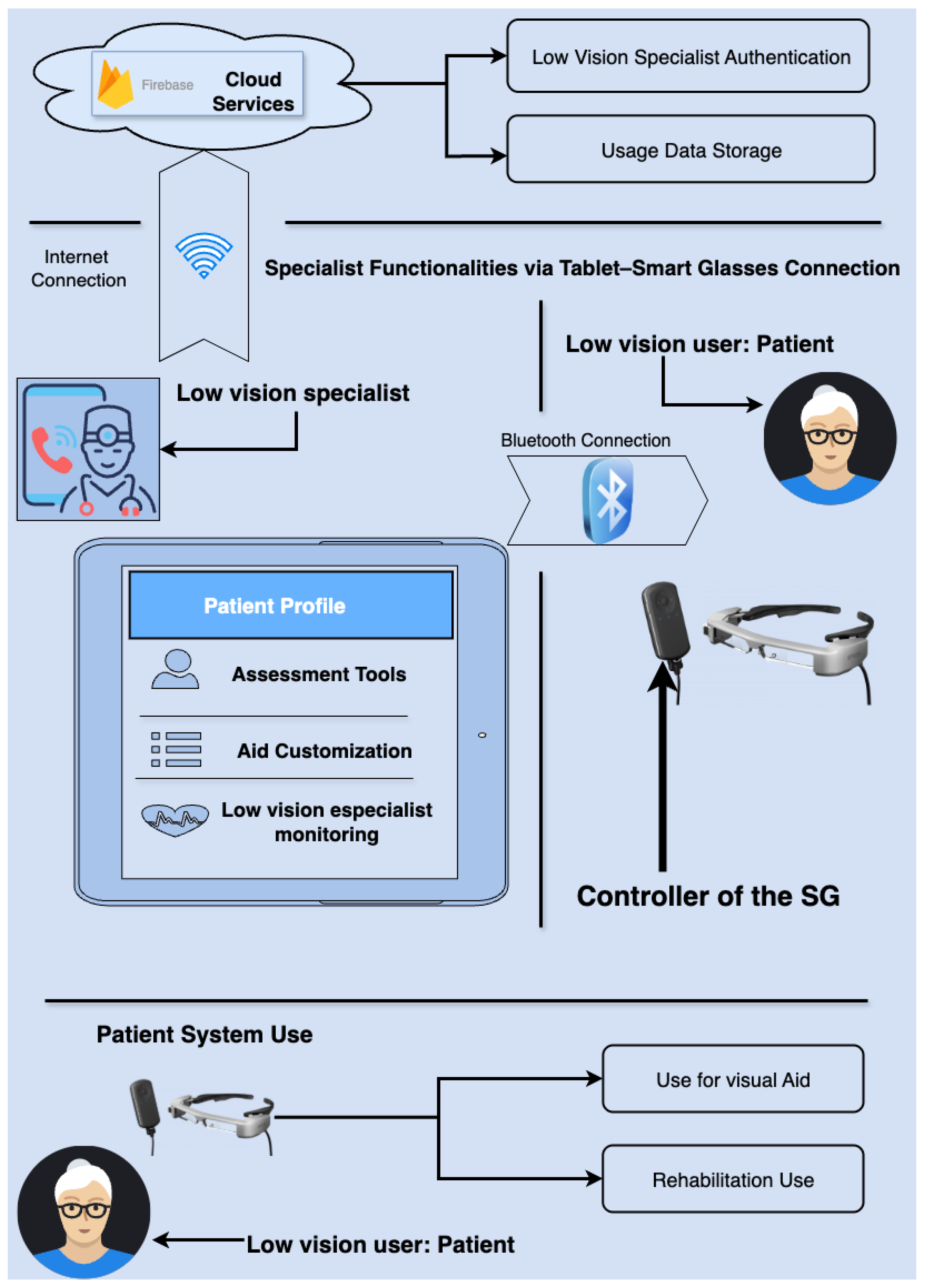
Figure 1. Schematic Representation of the Retiplus System.
Electronics 14 03589 g001
Figure 2. Example illustrating the vision [66] (a) without Retiplus and (b) with Retiplus.
Electronics 14 03589 g002
Figure 3. Specialist evaluation menu.
Electronics 14 03589 g003
Figure 4. Evaluation functionalities represent the options on the screen (Figure 3). (a) Pelli–Robson contrast test evaluation menu for specialist. (b) Display for the visual field specialist during the test and small squares. (c) Display for the visual scan specialist during the test. (d) Pelli–Robson test for patients. (e) Visual field test for patients. (f) Visual scanning test for patients.
Electronics 14 03589 g004
Figure 5. Calibration views. (a) Screen for creating a customized calibration by the specialist; (b) calibration using field of view template.
Electronics 14 03589 g005
Figure 6. Screenshots of the application. (a) Login page. (b) patient list (c) patient menu. (d) calibration list, where we can see the views created by the specialist and also the default views; and (e) patient profile data.
Electronics 14 03589 g006
Figure 7. Examples of calibration. (a) Calibration occupying the entire display. (b) Calibration contour mode. (c) Calibration for users with peripheral vision loss. (d) Calibration for users with peripheral vision loss with zoom. (e) Calibration with zoom whose projection occupies the entire display of the smart glasses. (f) Projection of the image onto the patient’s upper right viewing area, which may represent, for example, their better visual scan. (g) The image is projected onto the user’s right viewing area. (h) The image is projected onto the user’s upper viewing area.
Electronics 14 03589 g007aElectronics 14 03589 g007b
Figure 8. Specific Task for users [58]: (a) The setup for a practical exercise conducted at a distance of 3 m to measure the visual field in a rudimentary manner. (b) A specific task from phase 3 of the training, which involves assembling a puzzle.
Electronics 14 03589 g008
Table 1. Normality tests Kolmogorov–Smirnov.
REF.StatisticianglSig p-Value
VD00.133290.183
VDSR0.152290.075
VDCR0.103290.200 *
AGE0.100290.200 *
VA0.149290.086
CS0.133290.183
VF0.159290.051
VF20.156290.059
REF: variables. *: Lower limit of true significance. gl: Degrees of freedom. Sig p-value: signification level.
Table 2. Analysis of significant differences by disease and age group with ANOVA.
Sum of SquaresglMean SquareFSig
VA by DiseaseBetween groups0.02810.0280.1980.660
Within groups3.936280.141--
Total3.96429---
CS by DiseaseBetween groups0.11210.1120.3480.560
Within groups8.993280.321--
Total9.15029---
VF by DiseaseBetween groups2.02312.0230.0880.769
Within groups642.3862822.942--
Total644.40929---
VF2 by DiseaseBetween groups31.788131.7881.4730.235
Within groups604.2282821.580--
Total636.01629---
VA by AgeBetween groups0.43420.2171.6610.209
Within groups3.530270.131--
Total3.96429---
CS by AgeBetween groups0.06620.0330.0980.907
Within groups0.039270.335--
Total9.15029---
VF by AgeBetween groups31.380215.6900.6910.510
Within groups613.0282722.705--
Total644.40929---
VF2 by AgeBetween groups2.06621.0330.0440.957
Within groups633.9502723.480--
Total636.01629---
gl: Degrees of Freedom; F: ANOVA coefficient; Sig: p-value signification level.
Table 3. Paired sample correlations for the field of view without and with Retiplus.
NCorrelationSig p FactorSig p of Two Factors
Pair VF and VF2300.608<0.001<0.001
Sig: signification.
Table 4. Paired sample test for visual field without and with Retiplus (Pair VF and VF2).
MeasureValue
Mean (m)−6.37815
Standard deviation (SD)4.16194
Standard error of the mean (SEM)0.75986
95% CI Lower bound−7.93224
95% CI Upper bound−4.82405
t statistic (t)−8.394
Degrees of freedom (gl)29
Significance one factor (Sig of OF)<0.001
Significance factors (Sig of F)<0.001
Disclaimer/Publisher’s Note: The statements, opinions and data contained in all publications are solely those of the individual author(s) and contributor(s) and not of MDPI and/or the editor(s). MDPI and/or the editor(s) disclaim responsibility for any injury to people or property resulting from any ideas, methods, instructions or products referred to in the content.