High-Precision Detection of Cells and Amyloid-β Using Multi-Frame Brightfield Imaging and Quantitative Analysis
Abstract
1. Introduction
2. Related Works
- Difficulty in Direct Visualization: A aggregates, whether intra- or extracellular, exhibit extremely low contrast in brightfield images, making them inherently difficult to visualize and distinguish from the background.
- Lack of Distinct Morphological Features: Unlike cells, A aggregates do not display clear or consistent morphological patterns. This absence of distinctive visual features complicates the training of AI models, as the features necessary for accurate detection are often ambiguous or absent in brightfield modalities.
- In this study, we proposed a detection method of A in time-lapse brightfield microscopy 72 that does not utilize deep learning models.
3. Preprocessing for Time-Lapse Brightfield Microscopy
3.1. Challenges in A Detection
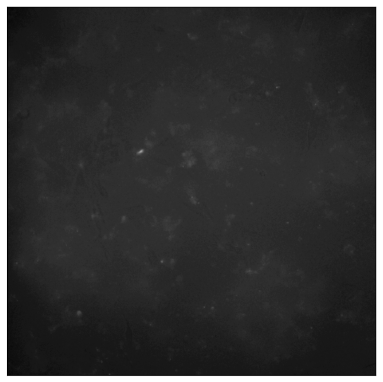
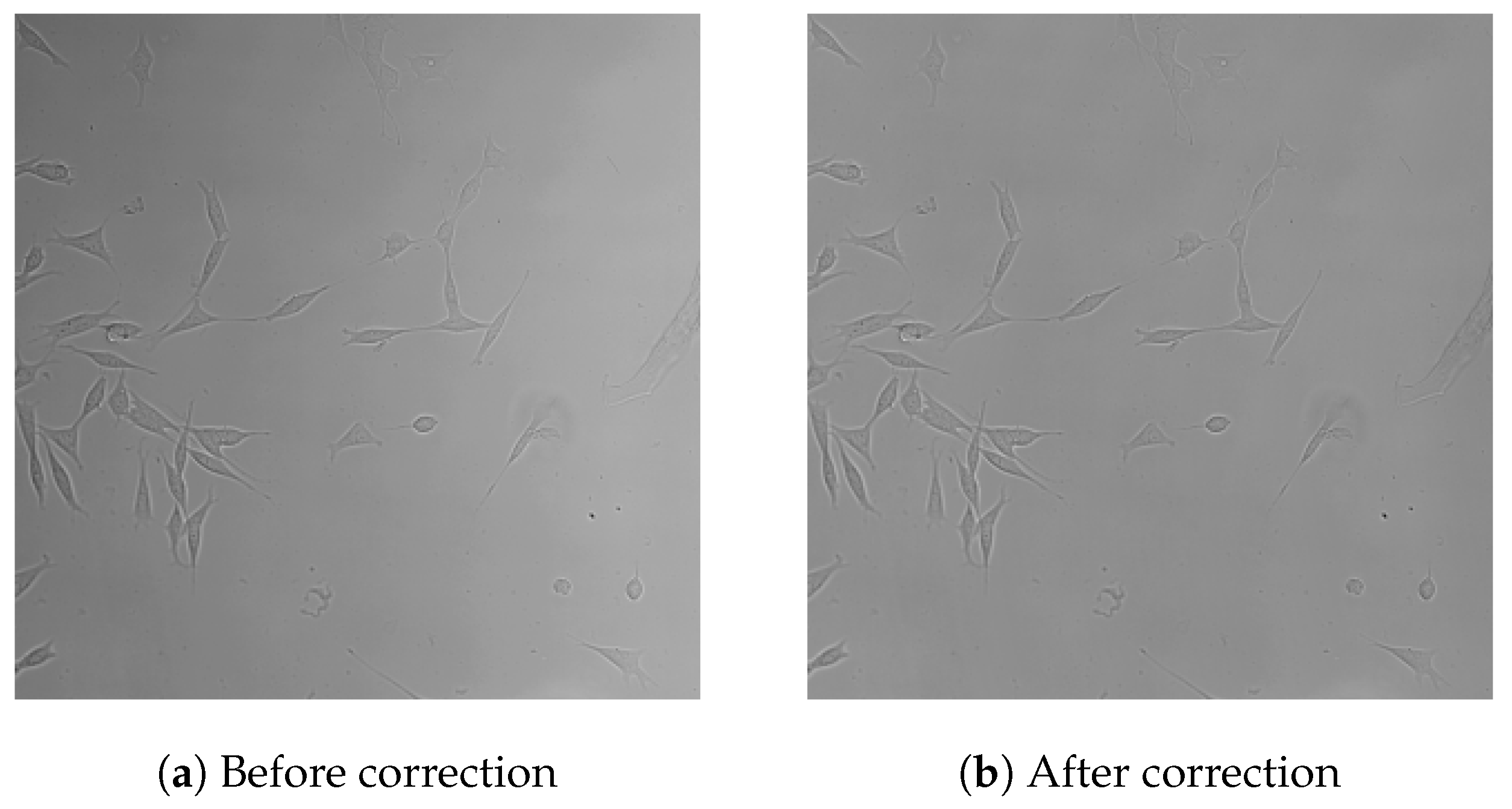
- Vignetting artifacts: Radial brightness decay caused by lens flare and uneven illumination, which obscure object boundaries. An example of this effect is shown in Figure 1.
- Temporal flickering: Frame-to-frame brightness fluctuations due to unstable illumination, unrelated to biological signals.
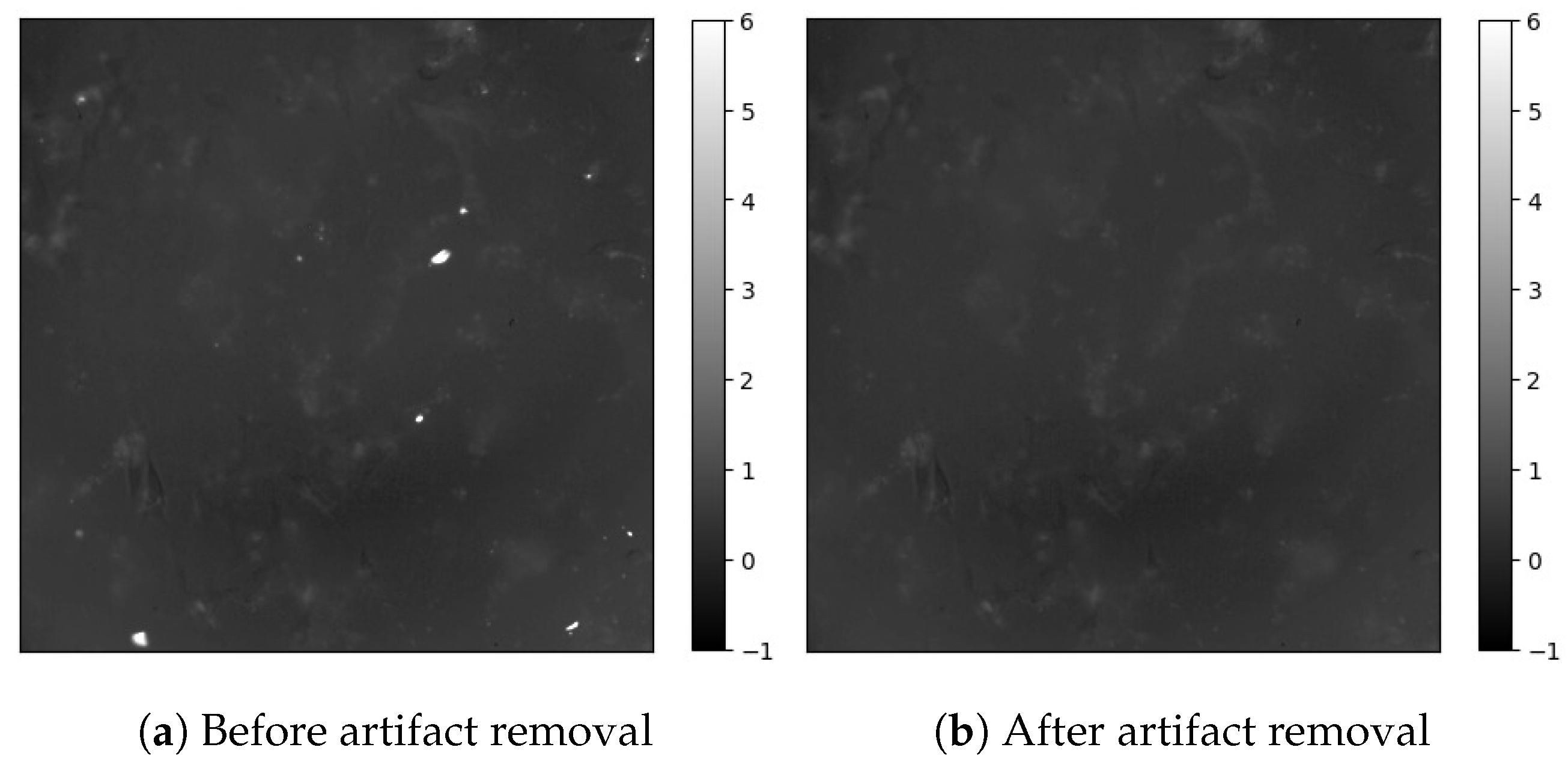
- Small bright debris: Foreign particles that mimic A aggregates, leading to incorrect detection if unfiltered.
3.2. A Image Preprocessing
- (1)
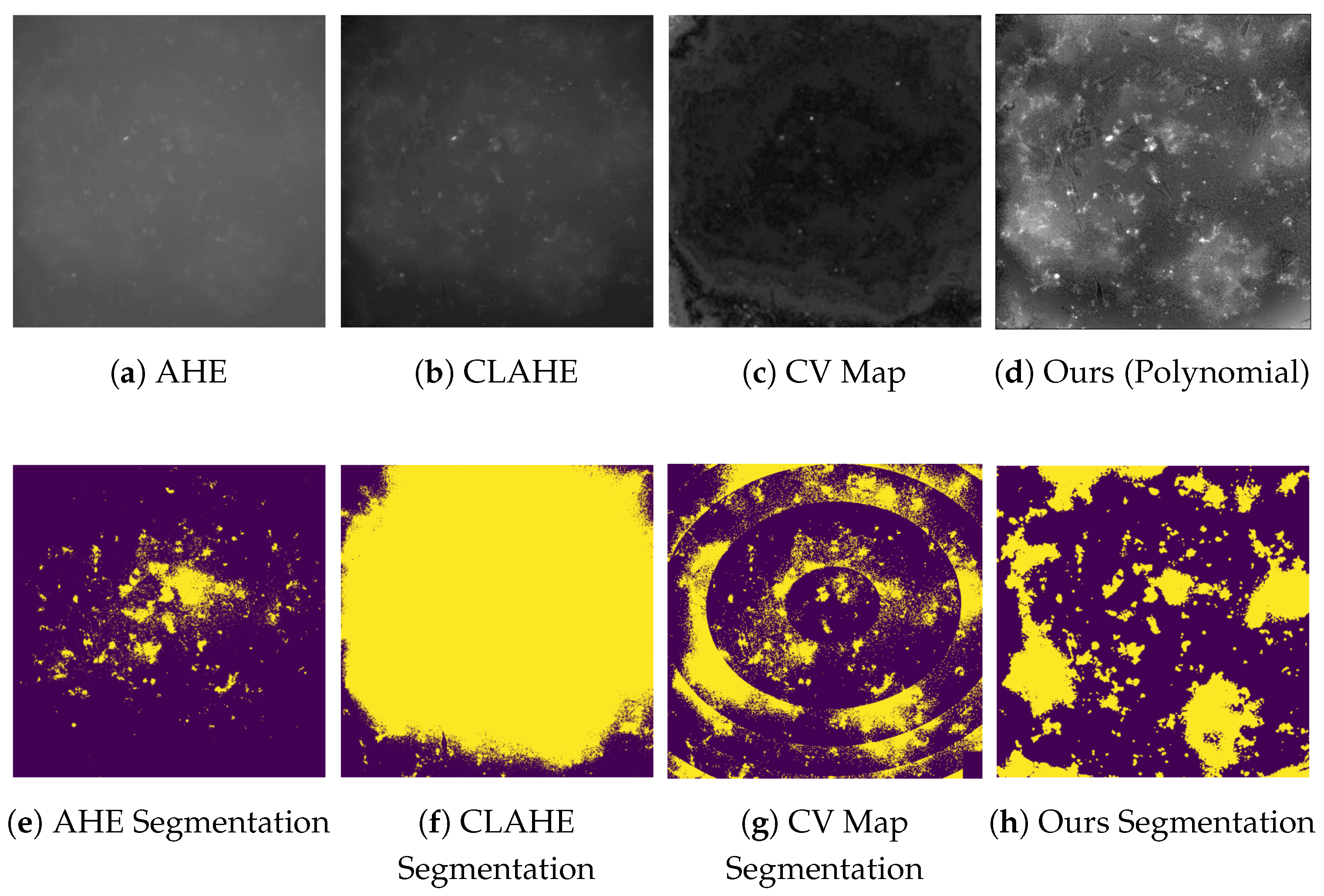
- Polynomial-Based Background Correction
- (2)
- Temporal Brightness Normalization
- (3)
- Flicker Smoothing via Exponential Moving Average (EWMA)
- (4)
- Tone Stabilization through Percentile Scaling and Histogram Matching
- (5)
- Artifact and Debris Removal via Brightness Profiling and Inpainting
- (6)
- Comparison with Conventional Background Correction Methods
- Adaptive Histogram Equalization (AHE): Enhances local contrast by redistributing pixel intensities within small regions. While effective in improving local details, it is prone to noise amplification and over-enhancement in homogeneous areas [12].
- Contrast-Limited Adaptive Histogram Equalization (CLAHE): An extension of AHE that limits histogram amplification to reduce noise. Commonly used in biomedical imaging to enhance tissue visibility while avoiding over-saturation [13].
3.3. Cell Image Preprocessing
Local Variance Filtering Based on Temporal Brightness Fluctuations
3.4. Summary
4. Detection of A and Cells, and Quantification of A Aggregation Around Cells
4.1. Segmentation Methodology
4.1.1. A Segmentation
- For low-concentration samples (≤5 μM): In these cases, A aggregates are sparse and typically appear as small, isolated structures. After background correction, the intensity distribution becomes sufficiently distinct to allow for global thresholding. We empirically selected a fixed threshold for each concentration group based on representative brightness histograms. This approach minimizes false positives and ensures high specificity under relatively uniform background conditions.
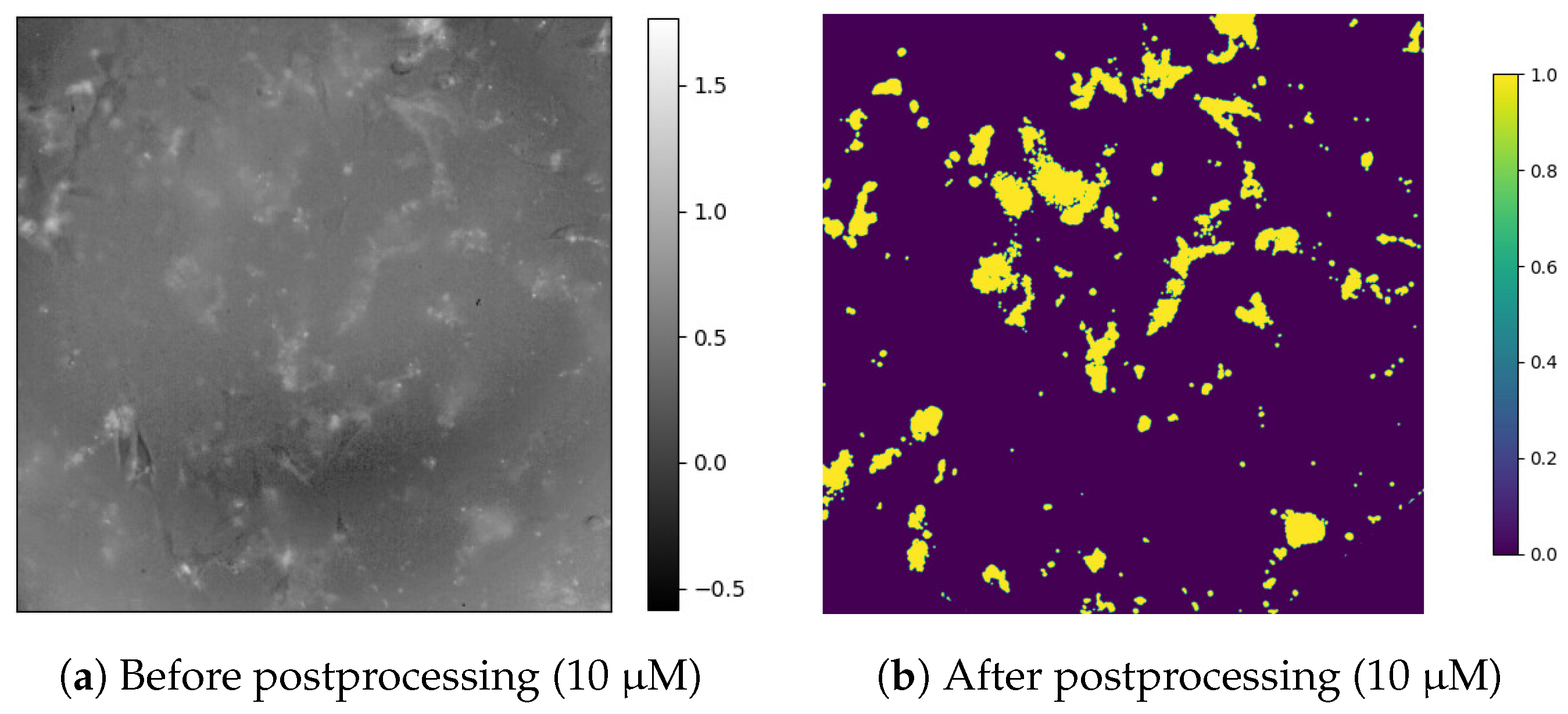
- For high-concentration samples (≥10 μM): At higher concentrations, aggregation is more widespread, with considerable variability in brightness and density across time and space. Therefore, we employed a semi-automatic, dynamic thresholding strategy:
- Step 1: Manual inspection was performed on several keyframes (e.g., frames 0, 30, and 60), and optimal threshold values were empirically determined for each.
- Step 2: Threshold values for intermediate frames were obtained via linear interpolation. To suppress abrupt transitions caused by local brightness anomalies, the frame-to-frame change in threshold was constrained to ≤0.02.
- Step 3: A binary mask was generated for each frame using the interpolated threshold.
- Step 4: To enhance temporal consistency and reduce transient noise, a five-frame temporal voting filter was applied: a pixel is retained as foreground (A) only if it is labeled as foreground in at least 3 of the 5 adjacent frames (two preceding, current, and two following). This majority-vote filter helps preserve persistent structures while discarding flickering artifacts.
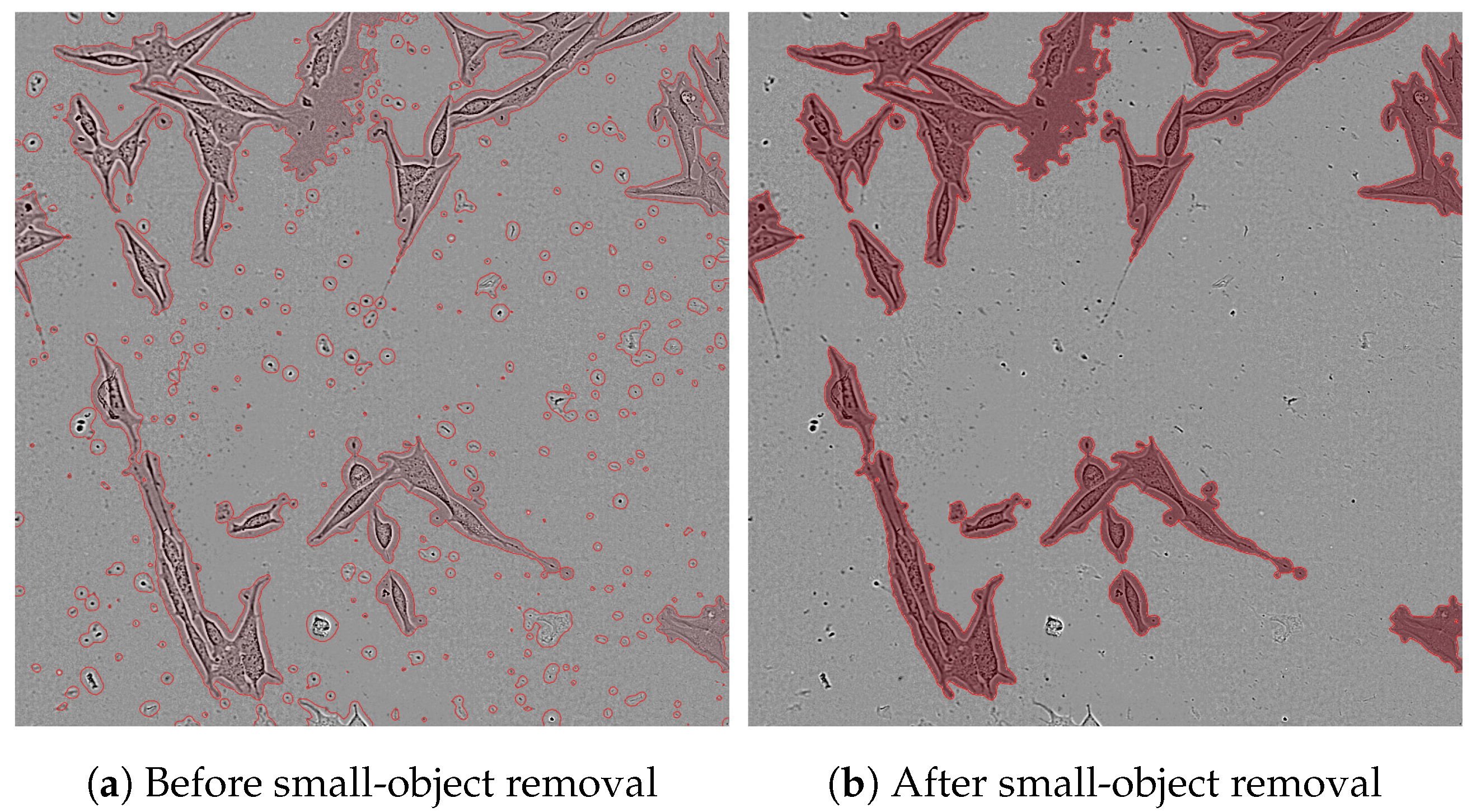
- Step 5: To further refine the segmentation result, we removed small objects with fewer than 500 pixels (which are typically noise or debris) and applied a morphological closing operation to connect fragmented A clusters, filling small gaps and smoothing boundaries.
4.1.2. Cell Segmentation
- Step 1: A block-wise local variance map was computed to enhance structures with temporal brightness changes—mainly corresponding to moving or morphologically active cells.
- Step 2: The variance map was transformed into a confidence map using a sigmoid activation function. This step assigns higher confidence to regions more likely to be cells [18].
- Step 3: A fixed threshold was applied to the confidence map to obtain a binary cell mask.
- Step 4: Small connected components were removed to eliminate debris or noise.
4.2. Metrics for A–Cell Relationship Analysis
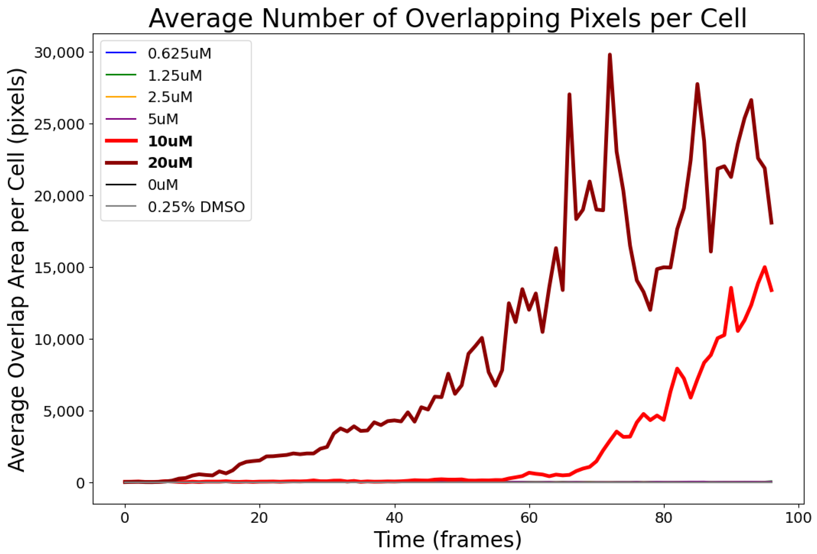
- Average Number of Overlapping Pixels per Cell: This metric calculates the mean number of pixels where A overlaps with each detected cell at each time point. It provides a direct measure of per-cell A burden and is sensitive to how A selectively adheres to or accumulates on certain cells. The relationship between A aggregates and individual cells is visually illustrated in Figure 6.
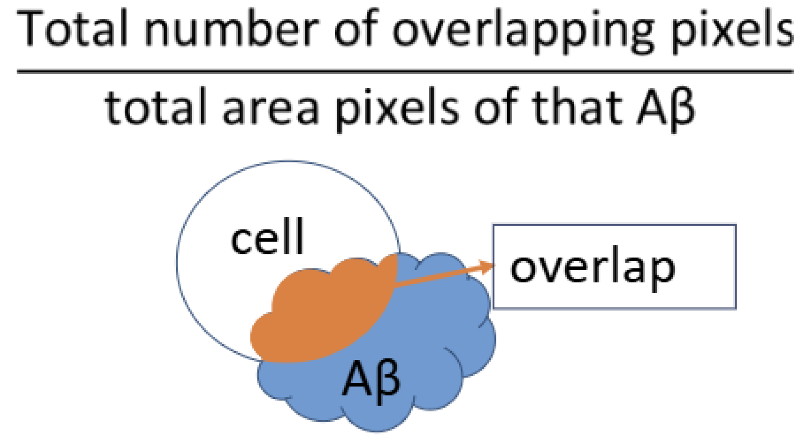
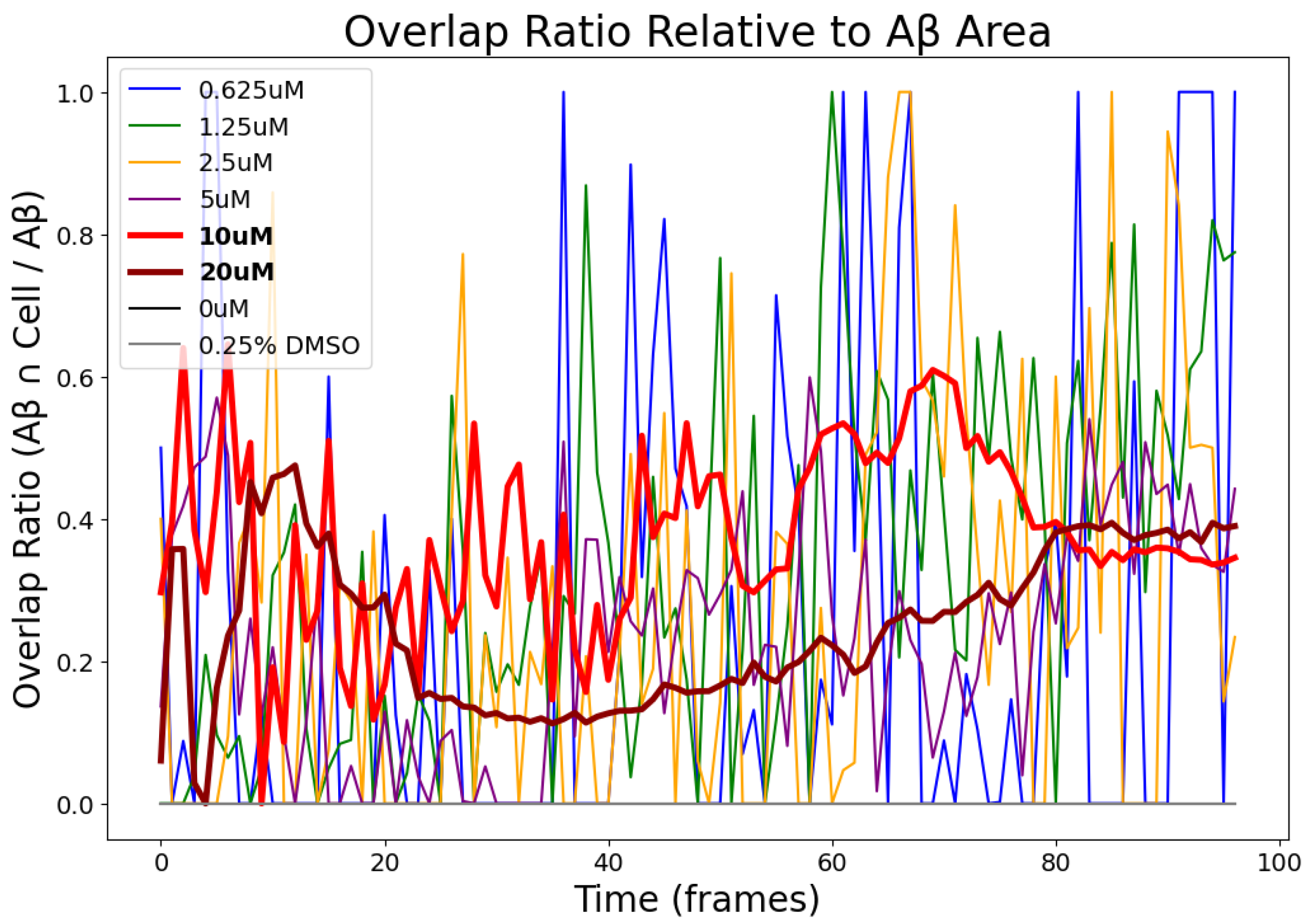
- Overlap Ratio Relative to A Area: This metric calculates the proportion of A pixels that are co-localized with cell regions. A high value implies preferential A clustering near cells, while a low value suggests diffuse or cell-independent aggregation. This relationship is visually illustrated in Figure 7.
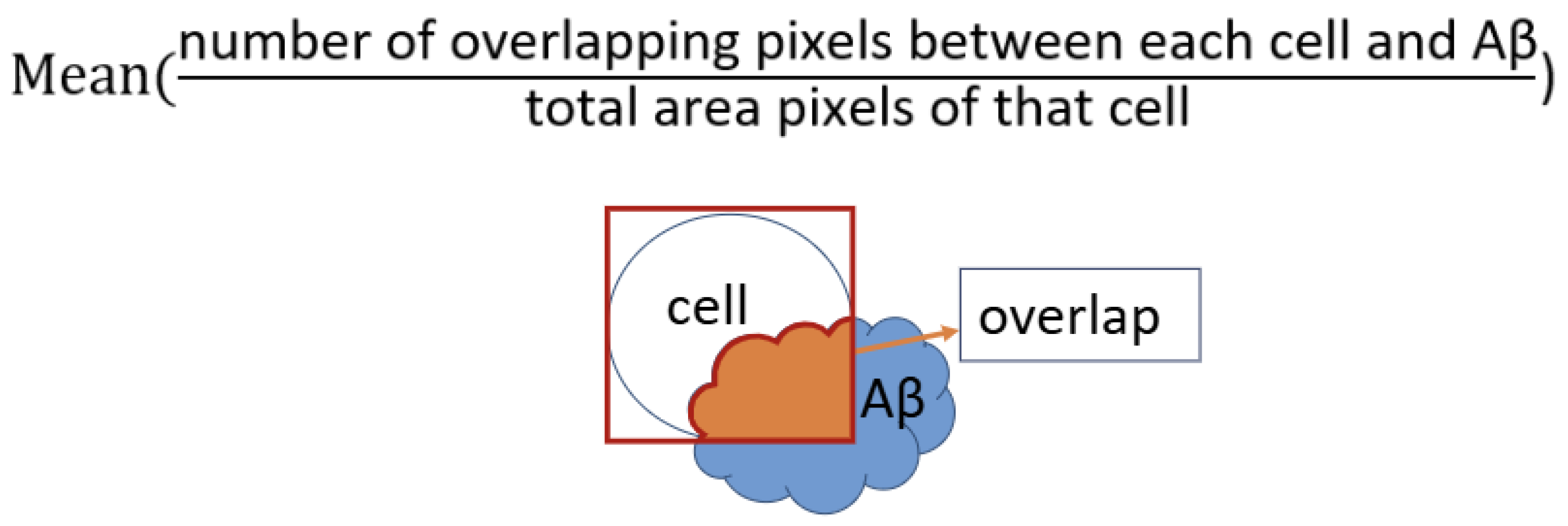
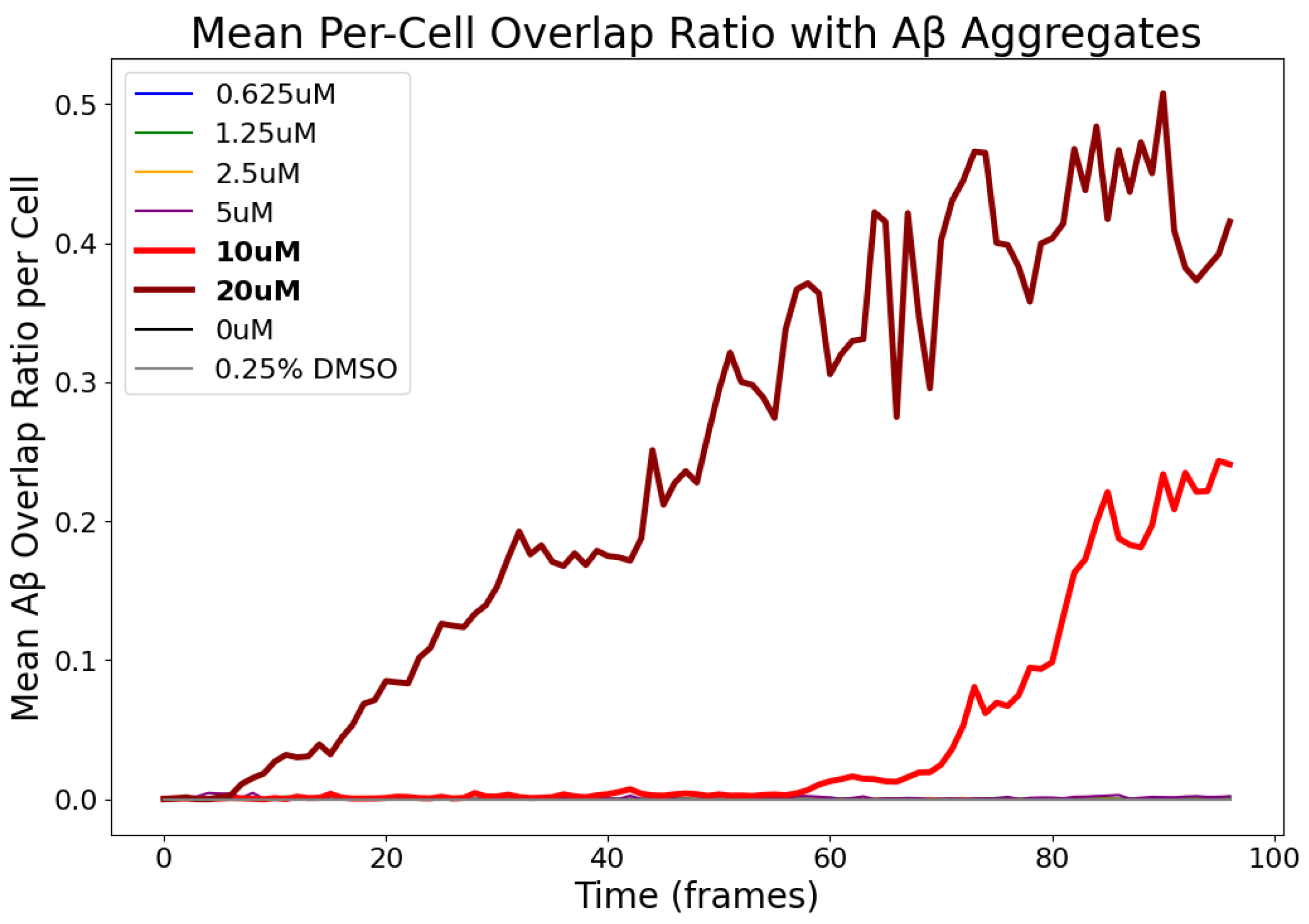
- Overlap Ratio Relative to Cell Area: This indicator computes the ratio between the overlapping pixels and the total cell area. It reflects how much of each cell is physically covered by A, accounting for size variations across different cell morphologies. This relationship is visually illustrated in Figure 8.
- Number of A Aggregates: We count the number of distinct A clusters per frame, defined as connected components above a minimum size threshold (excluding artifacts). This metric reflects overall aggregation dynamics and how they vary with concentration and over time. This relationship is visually illustrated in Figure 9.
5. Experimental Results
5.1. Experimental Setup
- Day 1: Plate Coating
- Day 2: Cell Seeding
- Day 3: A Treatment
- Day 4: Microscopy Observation
5.2. Segmentation Results
Validation via Area-Time Curves
- At high concentrations (e.g., 20 μM), the segmented A area increases over time and then stabilizes, matching the expected tendency of A to accumulate progressively under such conditions.
- At low concentrations (e.g., 2.5 μM), the segmented area remains low and exhibits irregular fluctuations, likely due to weak A presence and sensitivity to noise.
5.3. Quantitative Indicators of A–Cell Interaction
- Average Overlapping Pixels per Cell: measures how much A contacts individual cells.
- Overlap Ratio Relative to A Area: evaluates how concentrated A is around cell regions.
- Overlap Ratio Relative to Cell Area: captures the percentage of each cell’s surface that is in contact with A.
- Number of A Aggregates: counts cluster structures to estimate aggregation extent.
5.4. Temporal Trends and Concentration-Dependent Behavior
5.4.1. Average Overlapping Pixels per Cell
5.4.2. Overlap Ratio Relative to A Area
5.4.3. Overlap Ratio Relative to Cell Area
5.4.4. Number of A Aggregates
6. Analysis
6.1. Dose-Dependent A Accumulation on Cell Surfaces
6.2. Membrane Binding Efficiency and Selectivity
6.3. Heterogeneity in Aggregation Dynamics
6.4. Implications for the A Toxicity Hypothesis
- A surface accumulation initiates when concentrations exceed 5–10 μM.
- Aggregation accelerates beyond frame 50 (approx. 12 h), indicating a transition from oligomer formation to extensive clustering.
- Cell surface coverage by A surpasses 30% under 20 μM conditions, a level reported in prior studies to impair membrane integrity and receptor function [22].
6.5. Limitations and Future Directions
- Incorporating deep learning-based segmentation tools such as Cellpose [3] to enable accurate instance segmentation of individual cells in brightfield images;
- Analyzing temporal changes in cell activity, including protrusion formation, shrinkage, and motility under different A concentrations;
- Implementing single-cell tracking to monitor dynamic behaviors over time and quantify responses to A exposure at the individual-cell level;
- Quantifying morphological indicators such as circularity or aspect ratio to capture structural alterations related to cell stress or death;
- Estimating instantaneous velocities to assess changes in cell migration dynamics in response to A accumulation.
7. Conclusions
Author Contributions
Funding
Data Availability Statement
Acknowledgments
Conflicts of Interest
Abbreviations
| AD | Alzheimer’s Disease |
| A | Amyloid Beta |
| SH-SY5Y | Human neuroblastoma cell line |
References
- Hardy, J.; Selkoe, D.J. The amyloid hypothesis of Alzheimer’s disease: Progress and problems on the road to therapeutics. Science 2022, 297, 353–356. [Google Scholar] [CrossRef] [PubMed]
- Čepa, M. Segmentation of Total Cell Area in Brightfield Microscopy Images. Methods Protoc. 2018, 1, 43. [Google Scholar] [CrossRef] [PubMed]
- Stringer, C.; Wang, T.; Michaelos, M.; Pachitariu, M. Cellpose: A generalist algorithm for cellular segmentation. Nat. Methods 2020, 18, 100–106. [Google Scholar] [CrossRef] [PubMed]
- Schindelin, J.; Arganda-Carreras, I.; Frise, E.; Kaynig, V.; Longair, M.; Pietzsch, T.; Preibisch, S.; Rueden, C.; Saalfeld, S.; Schmid, B.; et al. Fiji: An open-source platform for biological-image analysis. Nat. Methods 2012, 9, 676–682. [Google Scholar] [CrossRef] [PubMed]
- Magnusson, K.E.G.; Jelinek, R. Automated analysis of cell migration and proliferation in time-lapse microscopy. Methods Mol. Biol. 2020, 2199, 175–191. [Google Scholar]
- Klunk, W.E.; Engler, H.; Nordberg, A.; Wang, Y.; Blomqvist, G.; Holt, D.P.; Bergström, M.; Savitcheva, I.; Huang, G.; Estrada, S.; et al. Imaging brain amyloid in Alzheimer’s disease with Pittsburgh Compound-B. Ann. Neurol. 2004, 55, 306–319. [Google Scholar] [CrossRef] [PubMed]
- Jack, C.R., Jr.; Bernstein, M.A.; Fox, N.C.; Thompson, P.M.; Alexander, B.D.; Harvey, D.; Borowski, B.; Britson, P.J.; Whitwell, J.L.; Ward, C.; et al. The Alzheimer’s Disease Neuroimaging Initiative (ADNI): MRI methods. J. Magn. Reson. Imaging 2008, 27, 685–691. [Google Scholar] [CrossRef] [PubMed]
- Lee, S.S.; Pelet, S.; Peter, M.; Dechant, R. A rapid and effective vignetting correction for quantitative microscopy. RSC Adv. 2014, 4, 52727–52733. [Google Scholar] [CrossRef]
- Willmott, C.J.; Matsuura, K. Advantages of the mean absolute error (MAE) over the root mean square error (RMSE) in assessing average model performance. Clim. Res. 2005, 30, 79–82. [Google Scholar] [CrossRef]
- Trigka, M.; Dritsas, E.; Moustakas, K. Joint Power and Contrast Shrinking in RGB Images with Exponential Smoothing. In Proceedings of the 2022 IEEE 14th Image, Video, and Multidimensional Signal Processing Workshop (IVMSP), Nafplio, Greece, 26–29 June 2022; pp. 1–5. [Google Scholar] [CrossRef]
- Bottenus, N.; Byram, B.C.; Hyun, D. Histogram Matching for Visual Ultrasound Image Comparison. IEEE Trans. Ultrason. Ferroelectr. Freq. Control 2021, 68, 1487–1495. [Google Scholar] [CrossRef] [PubMed]
- Noor, N.M.; Khalid, N.E.A.; Ali, M.H.; Numpang, A.D.A. Fish Bone Impaction Using Adaptive Histogram Equalization. In Proceedings of the 2010 Second International Conference on Computer Research and Development, Kuala Lumpur, Malaysia, 7–10 May 2010; pp. 163–167. [Google Scholar]
- Pizer, S.; Johnston, R.; Ericksen, J.; Yankaskas, B.; Muller, K. Contrast-limited adaptive histogram equalization: Speed and effectiveness. In Proceedings of the First Conference on Visualization in Biomedical Computing, Atlanta, GA, USA, 22–25 May 1990; pp. 337–345. [Google Scholar]
- Joris, P.; Develter, W.; Van De Voorde, W.; Suetens, P.; Maes, F.; Vandermeulen, D.; Claes, P. Preprocessing of Heteroscedastic Medical Images. IEEE Access 2018, 6, 26047–26058. [Google Scholar] [CrossRef]
- Arachchige, C.N.P.G.; Prendergast, L.A.; Staudte, R.G. Robust analogs to the coefficient of variation. J. Appl. Stat. 2020, 49, 268–290. [Google Scholar] [CrossRef] [PubMed]
- Piccardi, M. Background subtraction techniques: A review. In Proceedings of the 2004 IEEE International Conference on Systems, Man and Cybernetics, The Hague, The Netherlands, 10–13 October 2004; Volume 4, pp. 3099–3104. [Google Scholar] [CrossRef]
- Barron, A.E.; Guo, Z. Thermodynamic limits of Aβ aggregation: Mapping metastable and nucleation zones in supersaturated solutions. Chem. Sci. 2024, 15, 1234–1247. [Google Scholar] [CrossRef]
- Yang, P.; Song, W.; Zhao, X.; Zheng, R.; Qingge, L. An improved Otsu threshold segmentation algorithm. Int. J. Comput. Sci. Eng. 2020, 22, 146–153. [Google Scholar] [CrossRef]
- Tokuraku, K.; Marquardt, M.; Ikezu, T.; Buckle, A.M. Real-Time Imaging and Quantification of Amyloid-β Peptide Aggregates by Novel Quantum-Dot Nanoprobes. PLoS ONE 2009, 4, e8492. [Google Scholar] [CrossRef] [PubMed]
- Haass, C.; Selkoe, D.J. Soluble protein oligomers in neurodegeneration: Lessons from the Alzheimer’s amyloid β-peptide. Nat. Rev. Mol. Cell Biol. 2007, 8, 101–112. [Google Scholar] [CrossRef] [PubMed]
- Benilova, I.; Karran, E.; De Strooper, B. The toxic Aβ oligomer and Alzheimer’s disease: An emperor in need of clothes. Nat. Neurosci. 2012, 15, 349–357. [Google Scholar] [CrossRef] [PubMed]
- Lambert, M.P.; Barlow, A.K.; Chromy, B.A.; Edwards, C.; Freed, R.; Liosatos, M.; Rozovsky, I.; Trommer, B.; Viola, K.L.; Wals, P.; et al. Diffusible, nonfibrillar ligands derived from Aβ1-42 are potent central nervous system neurotoxins. Proc. Natl. Acad. Sci. USA 1998, 95, 6448–6453. [Google Scholar] [CrossRef] [PubMed]






Disclaimer/Publisher’s Note: The statements, opinions and data contained in all publications are solely those of the individual author(s) and contributor(s) and not of MDPI and/or the editor(s). MDPI and/or the editor(s) disclaim responsibility for any injury to people or property resulting from any ideas, methods, instructions or products referred to in the content. |
© 2025 by the authors. Licensee MDPI, Basel, Switzerland. This article is an open access article distributed under the terms and conditions of the Creative Commons Attribution (CC BY) license (https://creativecommons.org/licenses/by/4.0/).
Share and Cite
Li, M.; Kuragano, M.; Baar, S.; Endo, M.; Tokuraku, K.; Watanabe, S. High-Precision Detection of Cells and Amyloid-β Using Multi-Frame Brightfield Imaging and Quantitative Analysis. Electronics 2025, 14, 3418. https://doi.org/10.3390/electronics14173418
Li M, Kuragano M, Baar S, Endo M, Tokuraku K, Watanabe S. High-Precision Detection of Cells and Amyloid-β Using Multi-Frame Brightfield Imaging and Quantitative Analysis. Electronics. 2025; 14(17):3418. https://doi.org/10.3390/electronics14173418
Chicago/Turabian StyleLi, Mengyu, Masahiro Kuragano, Stefan Baar, Mana Endo, Kiyotaka Tokuraku, and Shinya Watanabe. 2025. "High-Precision Detection of Cells and Amyloid-β Using Multi-Frame Brightfield Imaging and Quantitative Analysis" Electronics 14, no. 17: 3418. https://doi.org/10.3390/electronics14173418
APA StyleLi, M., Kuragano, M., Baar, S., Endo, M., Tokuraku, K., & Watanabe, S. (2025). High-Precision Detection of Cells and Amyloid-β Using Multi-Frame Brightfield Imaging and Quantitative Analysis. Electronics, 14(17), 3418. https://doi.org/10.3390/electronics14173418


