Article

Bioassay-Guided Isolation of Chemical Constituents from Lycopodiastrum casuarinoides and Targeted Evaluation of Their Potential Efficacy in Cosmetics

1
School of Chemistry and Chemical Engineering, Central South University of Forestry and Technology, 498 South Shaoshan Road, Changsha 410004, China
2
School of Forestry, Central South University of Forestry and Technology, 498 South Shaoshan Road, Changsha 410004, China
*
Author to whom correspondence should be addressed.
These authors contributed equally to this work.
Cosmetics 2025, 12(4), 174; https://doi.org/10.3390/cosmetics12040174 (registering DOI)
Submission received: 5 May 2025 / Revised: 7 August 2025 / Accepted: 12 August 2025 / Published: 16 August 2025

Abstract

Natural tyrosinase inhibitors are currently a hot research topic due to their potential application in cosmetic and medicinal products. For the plant Lycopodiastrum casuarinoides, the chemical constituents with a tyrosinase inhibitory effect have not been investigated yet. Bioassay-guided isolation was conducted on the aboveground parts, resulting in the isolation of 10 compounds (110). Their chemical structures were confirmed by their spectral data and comparison with literature data. It might be worth pointing out that compounds 39 were isolated from the genus Lycopodiastrum for the first time. The bioassay revealed that compounds 6 and 7 displayed moderate mushroom tyrosinase inhibitory activity (IC50 = 1.90 and 2.43 mM, respectively), which was close to the positive control kojic acid (IC50 = 0.17 mM). Moreover, the in silico experiments disclosed that Lys180, His178 and other amino residues played key roles in the binding modes between compounds 6 and 7 and mushroom tyrosinase (PDB: 2Y9X). These findings suggested potential for further investigation on this species as a source of cosmetic ingredients.

1. Introduction

Tyrosinase is a multifunctional enzyme that is a catalyst for the oxidations of phenolic compounds including monophenols and o-diphenols [1]. This enzyme is widely distributed in plants and animals [2]. Among the different tyrosinases from different organisms, the one from the mushroom Agaricus bisporus has high similarity and homology with human tyrosinase [3]. For humans, this enzyme plays a key physiological role, because its dysregulated expression can lead to various skin disorders such as melanoma, freckles, chloasma, and vitiligo [4,5]. Considering its crucial role in melanogenesis, tyrosinase inhibitors appear promising for application in both cosmetic and medicinal products for skin tone enhancement and depigmentation [1,6].
Many researchers have devoted themselves to finding natural tyrosinase inhibitors from diverse natural sources including plants, fungi, and bacteria [1,6,7,8,9]. Among them, plant extracts and phytochemicals are research hotspots. A vast array of species have been subjected to tyrosinase inhibitory bioassay, such as Citrus limon [10], Launaea intybacea [11], Artemisia scoparia [12], Aquilaria malaccensis [13], Myrtus communis [14], Cotoneaster frigidus [15], Euryale ferox [16], and Ophiorrhiza puffii [17]. Interestingly, phytochemicals with different carbon frameworks were found as tyrosinase inhibitors, including flavonoids [18], alkaloids [19], steroids [20,21], and terpenes [22].
Lycopodiastrum casuarinoides is an evergreen fern of the family Lycopodiaceae, and is mainly distributed in tropical and subtropical regions of Asia [23]. In China, this plant has been used as a traditional medicine for centuries. Phytochemical investigations of L. casuarinoides have disclosed various types of chemical constituents [23], including alkaloids [24,25], terpenes [26,27], lipids [28], and miscellaneous others [29]. Interestingly, these chemicals exhibited a wide spectrum of pharmaceutical efforts [23], including cholinesterase inhibitory activity [28,30], acid-sensing ion channel 1a inhibitory activity [24], Cav3.1 channel inhibitory activity [25], anti-inflammatory activity [27], cytotoxic activity [31], and neuroprotective activity [32]. However, the tyrosinase inhibitory effect for the plant has not been investigated yet.
Therefore, this study aimed to explore the phytochemical constituents of L. casuarinoides by bioassay-guided isolation and evaluate their mushroom tyrosinase inhibitory activity in vitro and in silico.

2. Materials and Methods

2.1. Instruments and Chemicals

The Bruker DRX-400 and 600 spectrometers (Bruker Biospin AG, Fällanden, Germany) were employed to measure NMR spectra. CDCl3 was used as the NMR solvent (Shanghai Aladdin Biochemical Technology Co., Ltd., Shanghai, China). ESIMS spectra were recorded on a Q-TOF Micro LC-MS spectrometer (Waters Technologies (Shanghai) Co., Ltd., Shanghai, China). Column chromatography was conducted using commercial silica gel (200–300 and 300–400 mesh, Qingdao Haiyang Chemical Group Co., Ltd., Qingdao, China) and Sephadex LH-20 gel (Amersham Biosciences, Amersham, UK). Analytical TLC was performed using precoated silica gel plates (GF-254, Yan Tai Zi Fu Chemical Group Co., Yantai, China). All analytical solvents for column chromatography were purchased from Shanghai Chemical Reagents Co., Ltd., Shanghai, China.

2.2. Biomaterials

The aboveground parts of the plants L. casuarinoides were sampled from Baiyunxian Mountain, Hunan, China, in January 2023. A voucher specimen (No. P2023HN1) was kept in the Lab of Natural Product Chemistry of Central South University of Forestry and Technology (CSUFT). Dr. L. Wu of CSUFT conducted the taxonomy work on the samples.

2.3. Extraction and Bioassay-Guided Isolation

The aboveground parts of the plants were air-dried (dried weight 1.6 kg), and then were powdered followed by extraction with 90% ethanol using ultrasonication (40 KHz/200 W) at ambient temperature (2 h × 3). The plant-to-solvent ratio during the extraction was 1:5 g/mL. The collected ethanol extracts were combined and then concentrated with a rotary evaporator to result in a dark residue (301.6 g) after removal of the solvent. Then, petroleum ether (P) and chloroform (C) extracts were yielded after successive partition of P and C with the suspension of the residue in H2O.
These two extracts were tested for their mushroom tyrosinase inhibitory activity at 10 mg/mL. The results (Figure 1) showed that the C extract displayed a potent mushroom tyrosinase inhibitory effect with an inhibition rate of 85.3%, and was selected for subsequent isolation.
The C extract was column chromatographed over silica gel (200–300 mesh) by eluting it with P/ethyl acetate (E) at a gradient of 20:1 to 0:100 to produce five fractions, Fr. C1–C5.
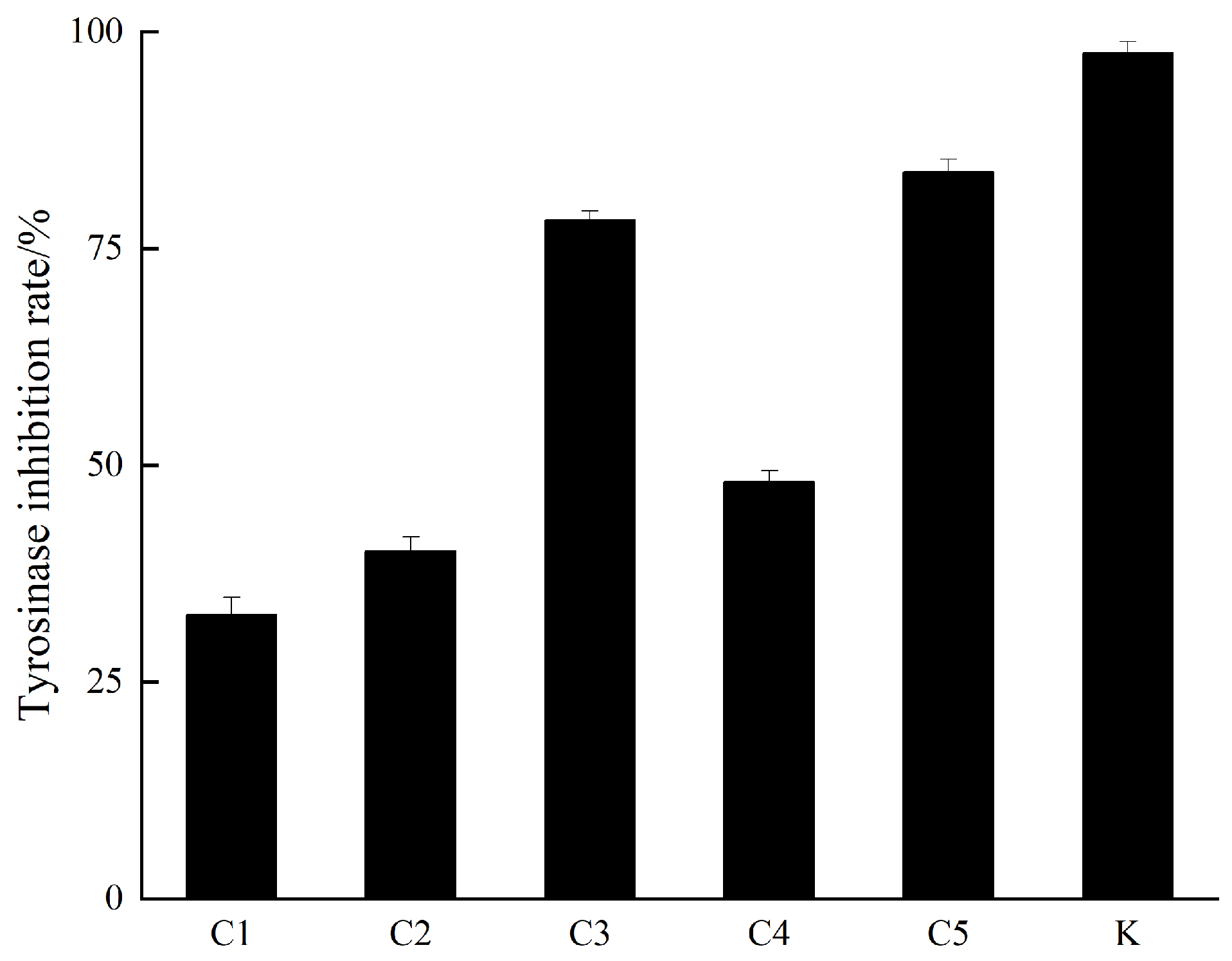
These five fractions were tested for their mushroom tyrosinase inhibitory activity at 10 mg/mL. The results (Figure 2) showed that two fractions (Fr. C3 and Fr. C5) displayed potent mushroom tyrosinase inhibitory effects with inhibition rates of 78.3% and 83.8%, respectively. Meanwhile, the other three fractions exhibited mushroom tyrosinase inhibition rates of less than 50%. Subsequently, two fractions (Fr. C3 and Fr. C5) were selected for further purification.
The Fr. C3 fraction was divided into five subfractions (Fr. C3A–C3E) by column chromatography (CC) over silica gel (300–400 mesh) with P/E 10:1 → 2:1. Compound 3 (10.2 mg) was isolated from the Fr. C3C subfraction by silica gel CC (P/E 6:1). Compounds 4 (3.3 mg) and 10 (5.1 mg) were obtained from the Fr. C3E subfraction by CC over silica gel eluting with CH2Cl2 (D)/CH3OH (M) with a gradient from 1:0 to 20:1 and the subsequent Sephadex LH-20 gel with elution of P/D/M with a ratio of 2:1:1.
Three subfractions (Fr. C5A–C5C) were obtained after the silica gel CC of the Fr. C fraction (P/E 5:1 → 0:1). Purified by silica gel CC (P/E 5:1 → 2:1), the Fr.C5A subfraction was divided into three subfractions (Fr. C5A1–C5A3). Compound 5 (3.8 mg) was given by the Fr. C5A1 subfraction after silica gel CC (P/E 4:1). Sephadex LH-20 gel CC of the Fr. C5A2 subfraction (P/D/M 2:1:1) yielded compound 9 (5.3 mg). Compounds 7 (3.1 mg) and 8 (4.1 mg) were isolated from the Fr. C5A3 subfraction by silica gel CC (P/E 3:1). By silica gel column CC (P/E 2:1), Compounds 1 (1.1 g) and 2 (30.0 mg) were isolated from the Fr. C5B subfraction. Compound 6 (7.5 mg) was obtained from the Fr. C5C subfraction by CC over silica gel eluting with D/M at a ratio of 30:1 and the subsequent Sephadex LH-20 gel with elution of P/D/M at a ratio of 2:1:1.
Figure 3 is an overview of the aforementioned experimental protocol of chemical compositions 110.

2.4. Characteristic 1H and 13C NMR and MS Spectral Data of Isolated Compounds

  • Compound 1: 1H NMR (CDCl3, 600 MHz): δH 4.83 (2H, s, H-26α, H-27α), 4.56 (2H, s, H-26β, H-27β), 3.25 (2H, d, J = 11.7 Hz, H-3, H-21), 0.99 (6H, s, H-23, H-30), 0.76 (6H, s, H-24, H-29), 0.64 (6H, s, H-25, H-28); 13C NMR (CDCl3, 150 MHz): δC 37.2 (C-1, C-19), 28.1 (C-2, C-20), 79.1 (C-3, C-21), 39.4 (C-4, C-22), 54.8 (C-5, C-17), 24.2 (C-6, C-16), 38.4 (C-7, C-15), 148.6 (C-8, C-14), 57.7 (C-9, C-13), 39.3 (C-10, C-18), 22.8 (C-11, C-12), 28.5 (C-23, C-29), 15.5 (C-24, C-30), 14.7 (C-25, C-28), 106.9 (C-26, C-27). ESI-MS m/z: 465.1 [M + Na]+.
  • Compound 2: 1H NMR (CDCl3, 400 MHz): δH 4.90 (2H, s, H-27), 3.33 (1H, d, J = 11.6 Hz, H-3), 3.23 (1H, d, J = 11.6 Hz, H-21), 1.08 (3H, s, H-25), 0.99 (3H, s, H-28), 0.81 (3H, s, H-24), 0.76 (3H, s, H-30), 0.69 (3H, s, H-23), 0.63 (3H, s, H-29); 13C NMR (CDCl3, 100 MHz): δC 37.1 (C-1), 28.5 (C-2), 78.8 (C-3), 39.3 (C-4), 54.9 (C-5), 27.7 (C-6), 42.5 (C-7), 212.0 (C-8), 64.9 (C-9), 42.5 (C-10), 21.2 (C-11), 23.7 (C-12), 57.4 (C-13), 147.5 (C-14), 37.3 (C-15), 23.7 (C-16), 53.7 (C-17), 38.4 (C-18), 37.1 (C-19), 28.1 (C-20), 79.1 (C-21), 39.4 (C-22), 27.7 (C-23), 15.5 (C-24), 14.6 (C-25), 108.0 (C-27), 14.9 (C-28), 28.5 (C-29), 15.5 (C-30). ESI-MS m/z: 467.2 [M + Na]+.
  • Compound 3: 1H NMR (CDCl3, 600 MHz): δH 5.35 (1H, m, H-6), 3.52 (1H, tt, J = 11.1, 4.6 Hz, H-3), 1.01 (3H, s, H-19), 0.92 (3H, d, J = 6.6 Hz, H-21), 0.84 (3H, t, J = 7.7 Hz, H-29), 0.83 (3H, d, J = 6.7 Hz, H-26), 0.81 (3H, d, J = 6.8 Hz, H-27), 0.68 (3H, s, H-18); 13C NMR (CDCl3, 150 MHz): δC 37.4 (C-1), 32.1 (C-2), 72.0 (C-3), 42.4 (C-4), 140.9 (C-5), 121.9 (C-6), 32.1 (C-7), 31.8 (C-8), 50.3 (C-9), 36.7 (C-10), 21.2 (C-11), 39.9 (C-12), 42.5 (C-13), 56.9 (C-14), 24.5 (C-15), 28.4 (C-16), 56.2 (C-17), 12.0 (C-18), 19.2 (C-19), 36.3 (C-20), 18.9 (C-21), 34.1 (C-22), 26.2 (C-23), 46.0 (C-24), 29.3 (C-25), 19.5 (C-26), 20.0 (C-27), 23.2 (C-28), 12.1 (C-29). ESI-MS m/z: 415.2 [M + H]+.
  • Compound 4: 1H NMR (CDCl3, 400 MHz): δH 12.03 (1H, s, 5-OH), 6.95 (3H, m, H-2′, H-5′, H-6′), 6.08 (1H, d, J = 2.4 Hz, H-6), 6.06 (1H, d, J = 2.3 Hz, H-8), 5.71 (1H, s, 4′-OH), 5.34 (1H, dd, J = 13.1, 3.0 Hz, H-2), 3.94 (3H, s, 3′-OCH3), 3.81 (3H, s, 7-OCH3), 3.10 (1H, dd, J = 17.2, 13.1 Hz, H-3α), 2.79 (1H, dd, J = 17.2, 3.0 Hz, H-3β); 13C NMR (CDCl3, 100 MHz): δC 79.5 (C-2), 43.6 (C-3), 196.1 (C-4), 164.3 (C-5), 95.3 (C-6), 168.1 (C-7), 94.4 (C-8), 163.0 (C-9), 103.3 (C-10), 130.4 (C-1′), 108.9 (C-2′), 146.9 (C-3′), 146.4 (C-4′), 114.7 (C-5′), 119.8 (C-6′), 56.2 (3′-OCH3), 55.8 (7-OCH3). ESI-MS m/z: 339.0 [M + Na]+.
  • Compound 5: 1H NMR (CDCl3, 400 MHz): δH 12.79 (1H, s, 5-OH), 7.49 (1H, dd, J = 8.4, 2.0 Hz, H-6′), 7.33 (1H, d, J = 2.1 Hz, H-2′), 7.04 (1H, d, J = 8.4 Hz, H-5′), 6.57 (1H, s, H-3), 6.49 (1H, d, J = 2.2 Hz, H-8), 6.37 (1H, d, J = 2.2 Hz, H-6), 4.01 (3H, s, 3′-OCH3), 3.89 (3H, s, 7-OCH3); 13C NMR (CDCl3, 100 MHz): δC 164.2 (C-2), 104.7 (C-3), 182.6 (C-4), 162.4 (C-5), 98.2 (C-6), 165.7 (C-7), 92.8 (C-8), 157.9 (C-9), 105.7 (C-10), 123.6 (C-1′), 108.6 (C-2′), 149.4 (C-3′), 147.1 (C-4′), 115.2 (C-5′), 120.9 (C-6′), 56.3 (3′-OCH3), 56.0 (7-OCH3). ESI-MS m/z: 313.1 [M − H].
  • Compound 6: 1H NMR (CDCl3, 400 MHz): δH 8.18 (1H, dd, J = 7.9, 1.6 Hz, H-5), 7.65 (1H, td, J = 7.8, 1.7 Hz, H-7), 7.44 (1H, d, J = 8.4 Hz, H-8), 7.39 (1H, t, J = 7.6 Hz, H-6), 7.12 (2H, d, J = 8.5 Hz, H-2′, H-6′), 6.83 (2H, d, J = 8.5 Hz, H-3′, H-5′), 6.14 (1H, s, H-3), 3.78 (3H, s, 4′-OCH3), 3.01 (2H, m, H-7′), 2.90 (2H, m, H-8′); 13C NMR (CDCl3, 100 MHz): δC 168.7 (C-2), 110.4 (C-3), 178.5 (C-4), 125.9 (C-5), 125.1 (C-6), 133.7 (C-7), 118.0 (C-8), 156.6 (C-9), 123.9 (C-10), 131.9 (C-1′), 129.4 (C-2′, C-6′), 114.2 (C-3′, C-5′), 158.4 (C-4′), 32.3 (C-7′), 36.6 (C-8′), 55.4 (4′-OCH3). ESI-MS m/z: 281.2 [M + H]+.
  • Compound 7: 1H NMR (CDCl3, 600 MHz): δH 8.10 (4H, s, H-2, H-3, H-5, H-6), 4.26 (4H, m, H-2′, H-11′), 1.73 (2H, m, H-3′, H-12′), 1.46 (4H, m, H-4′, H-13′), 1.40 (4H, m, H-5′, H-14′), 1.34 (4H, m, H-6′, H-15′), 0.90 (6H, t, J = 7.0 Hz, H-7′, H-16′), 1.32 (4H, m, H-8′, H-17′), 0.95 (6H, t, J = 7.5 Hz, H-9′, H-18′); 13C NMR (CDCl3, 150 MHz): δC 134.4 (C-1, C-4), 129.6 (C-2, C-3, C-5, C-6), 166.1 (C-1′, C-10′), 67.9 (C-2′, C-11′), 39.1 (C-3′, C-12′), 30.7 (C-4′, C-13′), 29.1 (C-5′, C-14′), 23.1 (C-6′, C-15′), 14.2 (C-7′, C-16′), 24.1 (C-8′, C-17′), 11.2 (C-9′, C-18′). ESI-MS m/z: 391.3 [M + H]+.
  • Compound 8: 1H NMR (CDCl3, 600 MHz): δH 7.61 (1H, d, J = 15.9 Hz, H-1′), 7.07 (1H, dd, J = 8.2, 1.9 Hz, H-6), 7.03 (1H, d, J = 1.9 Hz, H-2), 6.91 (1H, d, J = 8.2 Hz, H-5), 6.29 (1H, d, J = 15.9 Hz, H-2′), 5.86 (1H, s, 4-OH), 4.25 (2H, q, J = 7.1 Hz, H-4′), 3.93 (3H, s, 3-OCH3), 1.33 (3H, t, J = 7.1 Hz, H-5′); 13C NMR (CDCl3, 150 MHz): δC 127.2 (C-1), 109.4 (C-2), 148.0 (C-3), 146.9 (C-4), 114.8 (C-5), 123.2 (C-6), 144.8 (C-1′), 115.8 (C-2′), 167.4 (C-3′), 60.5 (C-4′), 14.5 (C-5′), 56.1 (3-OCH3). ESI-MS m/z: 245.1 [M + Na]+.
  • Compound 9: 1H NMR (CDCl3, 400 MHz): δH 7.96 (2H, m, H-2, H-6), 6.87 (2H, m, H-3, H-5), 4.35 (2H, q, J = 7.1 Hz, H-2′), 1.38 (3H, t, J = 7.1 Hz, H-3′); 13C NMR (CDCl3, 100 MHz): δC 123.0 (C-1), 132.0 (C-2, C-6), 115.3 (C-3, C-5), 160.2 (C-4), 166.9 (C-1′), 61.0 (C-2′) 14.5 (C-3′). ESI-MS m/z: 189.0 [M + Na]+.
  • Compound 10: 1H NMR (CDCl3, 600 MHz): δH 9.87 (1H, s, H-1′), 7.81 (2H, d, J = 8.7 Hz, H-2, H-6), 6.96 (2H, d, J = 8.6 Hz, H-3, H-5); 13C NMR (CDCl3, 150 MHz): δC 130.2 (C-1), 132.5, (C-2, C-6), 116.1 (C-3, C-5), 161.5 (C-4), 191.1 (C-1′). ESI-MS m/z: 123.0 [M + H]+.

2.5. In Vitro Mushroom Tyrosinase Inhibitory Activity Assay

A total of 100 μL L-tyrosine (1 mg/mL, Shanghai Macklin Biochemical Co., Ltd., Shanghai, China), 25 μL of mushroom tyrosinase (100 U/mL, Shanghai Aladdin Biochemical Technology Co., Ltd., Shanghai, China), 50 μL of Phosphate buffer (pH 6.8, 0.01 M, Phygene Biotechnology Co., Ltd., Fuzhou, China), and 25 μL of sample solutions were added to a 96-well plate. The mixture solutions were incubated at 37 °C for 30 min. The sample concentrations were increased from 0.2 mg/mL to 1.0 mg/mL with an interval of 0.2 mg/mL. Each concentration was tested three times in parallel. A microplate reader was employed to measure the absorbance of each well at 475 nm. The inhibition rate of the test compound against mushroom tyrosinase was calculated as the following formula: inhibition rate (%) = [1 − (A1 − A2)/(A3 − A0)] × 100, where A0–A1 represent the absorbances of blank control, sample solution, sample solution control, and blank, respectively. IC50 was derived from the fitting curve of inhibition rates vs. different concentrations of the test compound using SPSS (v27.0) software. Kojic acid (Shanghai Macklin Biochemical Technology Co., Ltd., Shanghai, China) was used as the positive control.

2.6. In Silico Tyrosinase-Compound Binding Mechanism Analysis

Molecular docking was used for the in silico tyrosinase-compound binding mechanism analysis. The crystal structure of mushroom tyrosinase (PDB ID: 2Y9X) with the ligand tropolone [33] was retrieved from the RCSB Protein Data Bank. The optimizations of structures 6 and 7 were subjected to docking calculations with mushroom tyrosinase using AutoDock Vina (v4.2) software. The removal of the ligand tropolone and water molecules was processed by Pymol (v2.4) software. Then AutoDock Vina (v4.2) was used for the analysis of the results, with the sets of coordinates at (−7.40 −23.55 −32.51) and the grid box size at (70.36 Å × 70.36 Å × 70.36 Å). The visualization of the results employed Discovery Studio Visualizer (v19.1) software.

3. Results and Discussion

3.1. Identification of Phytochemicals

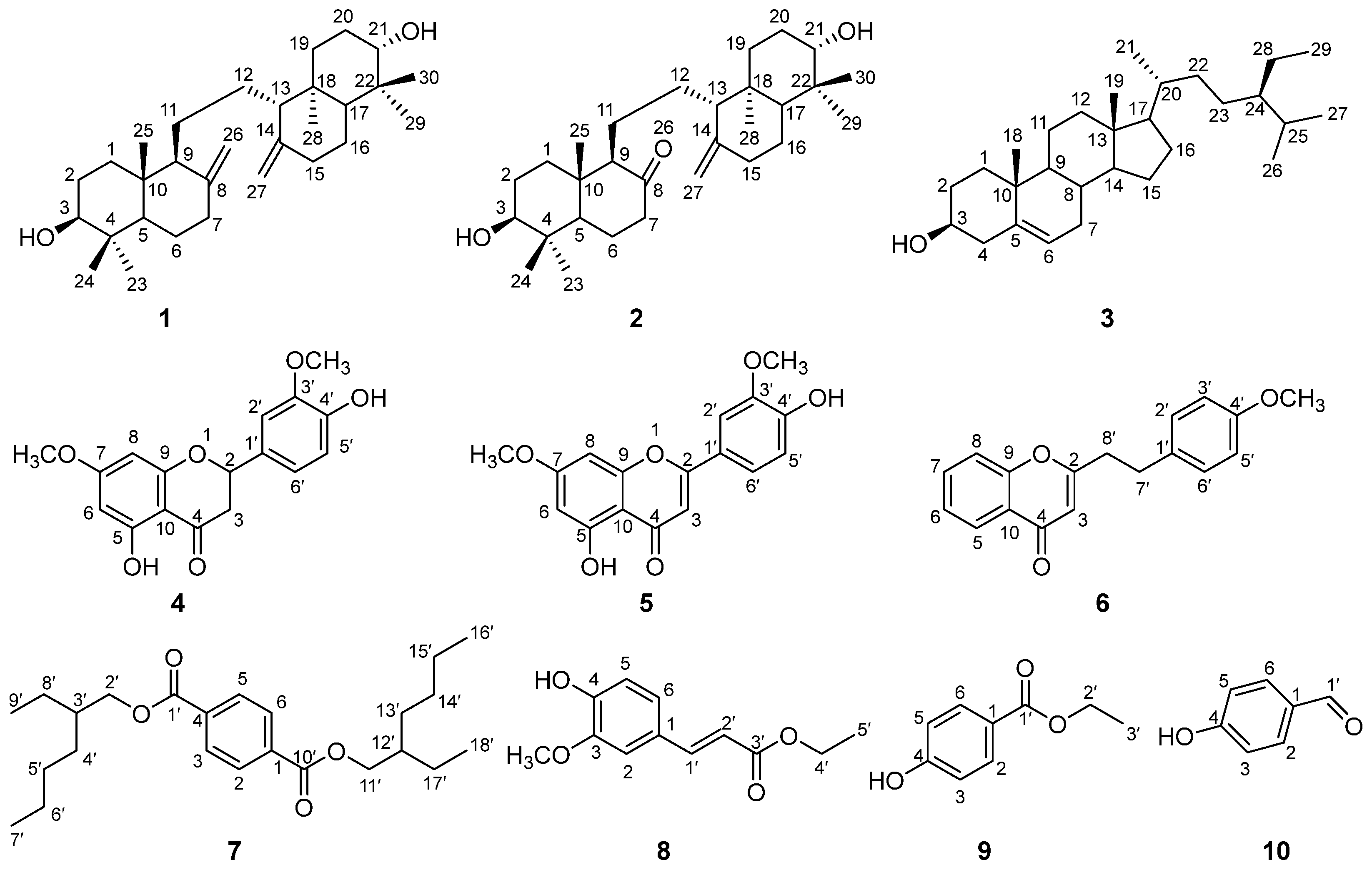
The chemical constituents 110 (Figure 4) were α-onocerin (1) [34], 26-nor-8-oxo-α-onocerin (2) [35], β-sitosterol (3) [20], eriodictyol-7,3′-dimethyl ether (4) [36], 4′,5-dihydroxy-3′,7-dimethoxyflavone (5) [37], 2-[2-(4-methoxyphenyl)ethyl]chromone (6) [38], terephthalic acid bis (2-ethyl-hexyl) ester (7) [39], ethyl ferulate (8) [40], ethylparaben (9) [41], and 4-hydroxybenzaldehyde (10) [29], confirmed by 1H and 13C NMR and MS data, and compared with the literature data.
In the 1H and 13C NMR spectra of compound 1, the signals at δH 4.83 (1H, s), 4.56 (1H, s) and δC 148.6 (s), and 106.9 (t) revealed the presence of the exo-methylene group. The chemical shifts of δH 3.25 (1H, d, J = 11.7 Hz) and δC 79.1 (d) were characteristic for the oxygen-bearing methine. The methyls resonated at δH 0.99 (3H, s), 0.76 (3H, s), and 0.64 (3H, s) and δC 28.5 (q), 15.5 (q), 14.7 (q). A literature survey revealed that the overall 1H and 13C NMR spectral data of 1 displayed a high degree of resemblance to those of the serratane-type triterpene α-onocerin [34]. Consequently, compound 1 was confirmed as α-onocerin, which is depicted in Figure 4.
Interestingly, the 1D NMR (1H and 13C) spectra of compound 2 (26-nor-8-oxo-α-onocerin) exhibited close similarity to those of 1, including two oxygenated methines [δH 3.33 (1H, d, J = 11.6 Hz), 3.23 (1H, d, J = 11.6 Hz); δC 78.8 (d), 79.1 (d)] and six singlet methyls [δH 0.69 (3H, s), 0.81 (3H, s), 1.08 (3H, s), 0.99 (3H, s), 0.63 (3H, s), 0.76 (3H, s); δC 27.7 (q), 15.5 (q), 14.6 (q), 14.9 (q), 28.5 (q), 15.5 (q)]. Their difference was in the appearance of a carbonyl group [δC 212.0 (s)] in 2 instead of one exo-methylene group in 1. Accordingly, compound 2 was confirmed as 26-nor-8-oxo-α-onocerin, whose data matched well with the literature data [35].
In the 1D NMR spectra of compound 3, one double bond [δH 5.35 (1H, m); δC 140.9 (s), 121.9 (d)], one oxygenated methine [δH 3.52 (1H, tt, J = 11.1, 4.6 Hz); δC 72.0 (d)], and six methyls [δH 0.68 (3H, s), 0.81 (3H, d, J = 6.8 Hz), 0.83 (3H, d, J = 6.7 Hz), 0.84 (3H, t, J = 7.7 Hz), 0.92 (3H, d, J = 6.6 Hz), 1.01 (3H, s); δC 12.0 (q), 19.2 (q), 18.9 (q), 19.5 (q), 20.0 (q), 12.1 (q)] were identified. It was realized that the 1D NMR spectral data of 3 were identical to those of β-sitosterol [20]. Consequently, the structure of 3 was assigned as that shown in Figure 4.
There was one ketone [δC 196.1 (s)], one tetra-substituted benzene [δH 6.08 (1H, d, J = 2.4 Hz), 6.06 (1H, d, J = 2.3 Hz); δC 164.3 (s), 95.3 (d), 168.1 (s), 94.4 (d), 163.0 (s), 103.3 (s)], one tri-substituted benzene [δH 6.95 (3H, m); δC 130.4 (s), 108.9 (d), 146.9 (s), 146.4 (s), 114.7 (d), 119.8 (d)], and two methoxyls [δH 3.94 (3H, s), 3.81 (3H, s); δC 56.2 (q), 55.8 (q)] observed in the 1D NMR spectra of compound 4. A literature survey disclosed that the1D NMR spectral data of 4 resembled those of 4′,5-dihydroxy-3′,7-dimethoxyflavone [36]. Hereto, the structure of 4 was determined as that in Figure 4.
Inspection of 1H and 13C NMR spectra of compound 5 revealed a high structural similarity with 4, including one tetra-substituted benzene [δH 6.37 (1H, d, J = 2.2 Hz), 6.49 (1H, d, J = 2.2 Hz); δC 162.4 (s), 98.2 (d), 165.7 (s), 92.8 (d), 157.9 (s), 105.7 (s)], one tri-substituted benzene ring [δH 7.49 (1H, dd, J = 8.4, 2.0 Hz), 7.33 (1H, d, J = 2.1 Hz), 7.04 (1H, d, J = 8.4 Hz); δC 123.6 (s), 108.6 (d), 149.4 (d), 147.1 (s), 115.2 (d), 120.9 (d)], and two methoxyls [δH 4.01 (3H, s), 3.89 (3H, s); δC 56.3 (q), 56.0 (q)]. However, the typical 13C NMR data of an α,β-unsaturated ketone [δH 6.57 (1H, s); δC 164.2 (s), 104.7 (d), 182.6 (s)] in the 1H and 13C NMR spectra of 5 were observed. This observation suggested that the difference between 5 and 4 lay in the presence of a double bond conjugated with the ketone group at C-2–C-4. As a result, compound 5 was established as 4′,5-dihydroxy-3′,7-dimethoxyflavone, whose data were consistent with those recorded in the literature [37].
The group of signals that resonated at δH 8.18 (1H, dd, J = 7.9, 1.6 Hz), 7.65 (1H, td, J = 8.3, 1.7 Hz), 7.44 (1H, d, J = 8.4 Hz), 7.39 (1H, t, J = 7.6 Hz) and δC 125.9 (d), 125.1 (d), 133.7 (d), 118.0 (d), 156.6 (s), 123.9 (s) in the 1H and 13C NMR spectrum of chemical constituent 6 suggested that an ortho-disubstituted benzene was present in the chemical structure of 6. Meanwhile, the group of peaks with the chemical shifts δH 7.12 (2H, d, J = 8.5 Hz), 6.83 (2H, d, J = 8.5 Hz) and δC 131.9 (s), 129.4 (d, ×2), 114.2 (d, ×2), 158.4 (s) indicated there was a para-disubstituted benzene ring as a partial structure. The up-field chemical shifts at δH 3.78 (3H, s) and δC 55.4 (q) corresponded to a methoxyl group. Moreover, the chemical shifts attributable to an α,β-unsaturated ketone [δH 6.14 (1H, s); δC 178.5 (s), 168.7 (s), 110.4 (d)] were recognized. These data were indicative of the spectral features of 2-[2-(4-methoxyphenyl)ethyl]chromone [38]. Based on a comparison with literature data [38], the chemical structure of 6 was validated, as shown in Figure 4.
In the 1D NMR spectra of compound 7, one para-disubstituted benzene [δH 8.10 (4H, s); δC 134.4 (s, ×2), 129.6 (d, ×4)], and two oxymethylenes [δH 4.26 (4H, m); δC 67.9 (t, ×2), 129.6 (d, ×4)], four methyls [δH 0.91 (6H, t, J = 7.0 Hz), 0.95 (6H, t, J = 7.5 Hz); δC 14.2 (q, ×2), 11.2 (q, ×2)] were observed. These NMR spectral data of 7 resembled those of terephthalic acid bis (2-ethyl-hexyl) ester [39]. As a consequence, chemical composition 7 was determined as terephthalic acid bis (2-ethyl-hexyl) ester (Figure 4).
A tri-substituted benzene [δH 7.03 (1H, d, J = 1.9 Hz), 6.91 (1H, d, J = 8.2 Hz), 7.07 (1H, dd, J = 8.2, 1.9 Hz); δC 127.2 (s), 109.4 (d), 148.0 (s), 146.9 (s), 114.8 (d), 123.2 (d)], an α,β-conjugated ethyl ester [δH 7.61 (1H, d, J = 15.9 Hz), 6.29 (1H, d, J = 15.9 Hz), 4.25 (2H, q, J = 7.1 Hz), 1.33 (3H, t, J = 7.1 Hz); δC 144.8 (d), 115.8 (d), 167.4 (s), 60.5 (t), 14.5 (q)], along with a methoxyl [δH 3.93 (3H, s); δC 56.1 (q)] were recognized in the 1D NMR spectra of chemical constituent 8. It was found that the 1D NMR spectral data of 8 were identical to those of ethyl ferulate [40]. Hereto, 8 was established as ethyl ferulate (Figure 4).
As shown in the 1H and 13C NMR spectra of compound 9, one carbonyl carbon [δC 166.9 (s)], one para-disubstituted benzene [δH 7.96 (2H, m), 6.87 (2H, m); δC 123.0 (s), 132.0 (d, ×2), 115.3 (d, ×2), 160.2 (s)], and one oxyethyl [δH 4.35 (2H, q, J = 7.1 Hz), 1.38 (3H, t, J = 7.1 Hz); δC 61.0 (t), 14.5 (q)] were recognized. It was found that the 1D NMR spectral data of 9 were identical to those of ethylparaben [41]. Accordingly, 9 was assigned as ethylparaben (Figure 4).
The presence of one aldehyde carbon [δH 9.87 (1H, s); δC 191.1 (d)], and one para-disubstituted benzene [δH 7.81 (2H, d, J = 8.7 Hz), 6.96 (2H, d, J = 8.6 Hz); δC 130.2 (s), 132.5, (d, ×2), 116.1 (d, ×2), 161.5 (s)] were recognized in the 1D NMR spectra of 10. The literature survey revealed that the overall 1H and 13C NMR spectral data of 10 were identical to literature data of 4-hydroxybenzaldehyde [29]. Consequently, the chemical structure of 10 was established as that observed in Figure 4.

3.2. Mushroom Tyrosinase Inhibitory Activity

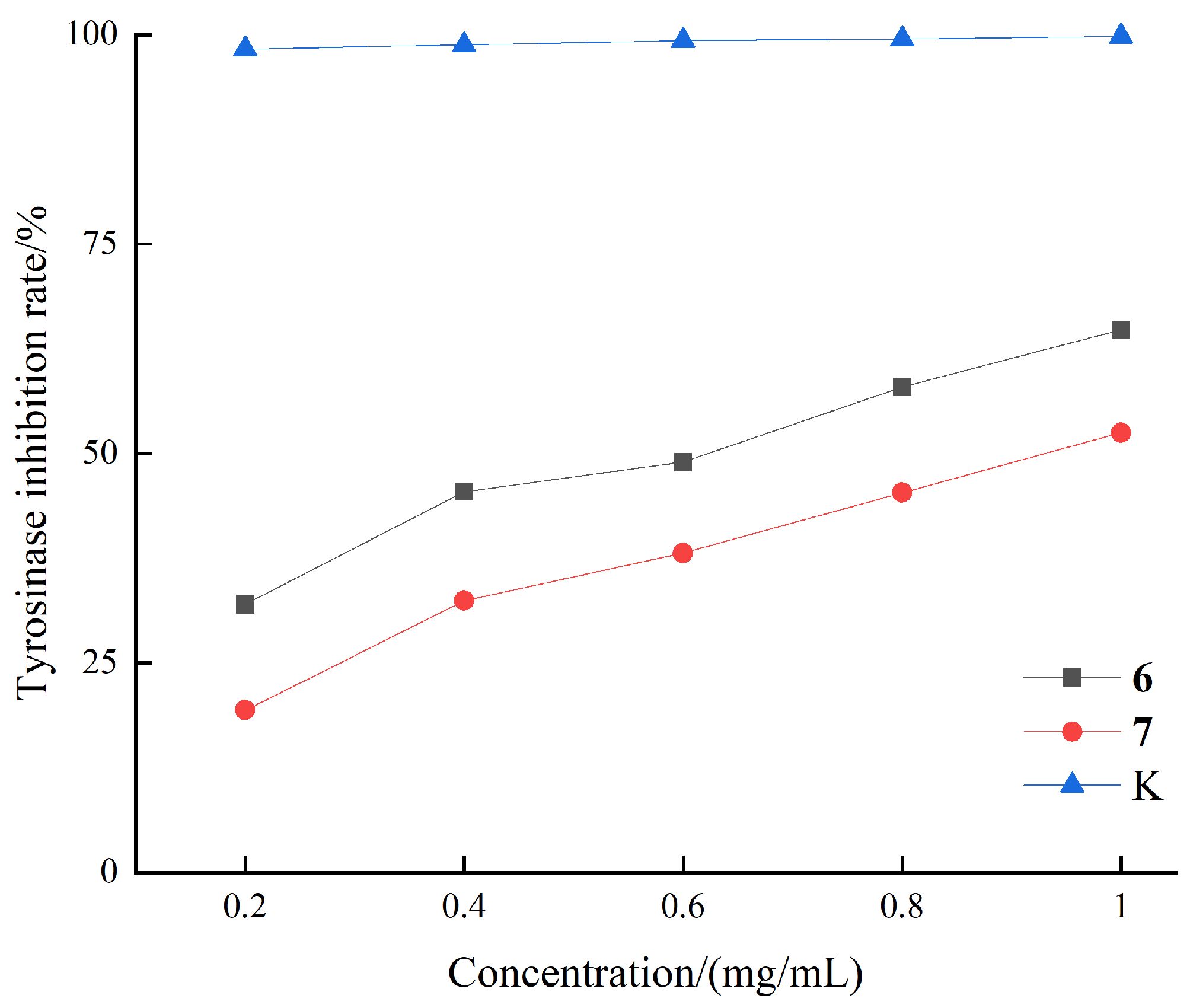
All of these isolates except compound 3 were subjected to the mushroom tyrosinase inhibitory bioassay. In our previous work [20], compound 3 displayed potent mushroom tyrosinase inhibitory activity (IC50 = 35.5 μM). In this biotest, only compounds 6 and 7 displayed promising mushroom tyrosinase inhibitory effects with inhibition rates of 64.9% and 51.5% at 1 mg/mL, respectively (Figure 5). Further experiments indicated that the IC50 values of their mushroom tyrosinase inhibitory activity were 1.90 and 2.43 mM (Table 1), respectively. These data revealed that compounds 6 and 7 exhibited moderate mushroom tyrosinase inhibitory activity—close to acceptable ranges, though less potent than the positive control kojic acid (IC50 = 0.17 mM).

3.3. Mushroom Tyrosinase-Compound Binding Mechanism

The decipherment of the binding modes between bioactive compounds 6 and 7 and mushroom tyrosinase (PDB ID: 2Y9X) [33,42] was investigated by molecular docking. As displayed in Table 2, the binding energy scores for compounds 6 and 7 were −6.60 and −5.75 kcal/mol, respectively, which were close to that of the native ligand tropolone. It is known that a score of <−5 kcal/mol indicates strong binding activity [43]. The lower the score, the stronger the binding affinity. Therefore, these binding energy scores confirmed the good docking quality of compounds 6 and 7 binding to mushroom tyrosinase.
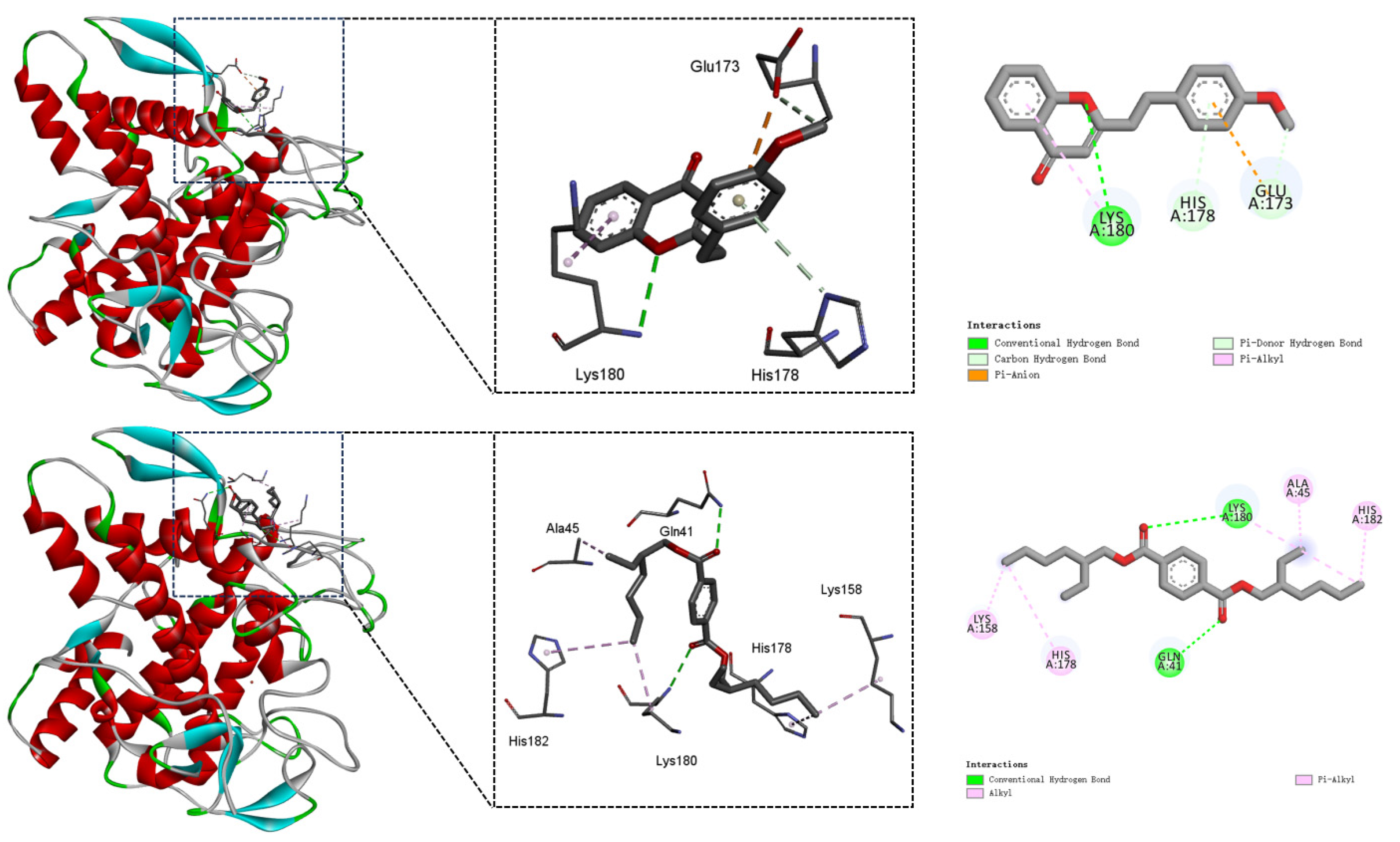
Figure 6 showed the 2D and 3D interactive images for chemical constituents 6 and 7 with mushroom tyrosinase (PDB ID: 2Y9X), namely the plots of docked complexes and molecular surfaces.
As deciphered in Figure 6, chemical composition 6 engaged in a hydrogen bond with amino acid residue Lys180, and a π–alkyl stacking interaction was formed with Lys180 in the mushroom tyrosinase [33]. The benzene ring of compound 6 induced a π–donor bond with the amino acid residue His178 and a π–anion bond with the amino acid residue Glu173. Furthermore, the amino acid residue Glu173 involved a carbon–hydrogen bond with the methoxy group of 6. In short, compound 6 interacted via three hydrogen bonds and two π–involving interactions primarily involving Lys180, His178 and Glu173 in the mushroom tyrosinase.
As for compound 7, its two keto groups induced two hydrogen bonds with the amino acid residues Gln41 and Lys180, respectively. Additionally, compound 7 established two π–alkyl interactions with amino acid residues His182 and His178, as well as three alkyl stacking interactions with amino acid residues Lys180, Ala45, and Lys158, all of which are located in the active site (Figure 6). In brief, compound 7 interacted via two hydrogen bonds and five alkyl-involving interactions primarily involving Gln41, Lys180, His182, His178, Ala45, and Lys158 in the mushroom tyrosinase.

4. Conclusions

In summary, an array of chemical constituents featuring diverse skeletons were afforded in a bioassay-guided investigation of the plant L. casuarinoides. These chemical compositions (110) could be divided into two triterpenes (1 and 2), one steroid (3), three flavonoids (46), and four benzene derivatives (710). Notably, compounds 39 were isolated from the genus Lycopodiastrum for the first time. The mushroom tyrosinase inhibitory activity was assessed for all the isolates. As a result, 6 and 7 showed moderate inhibition (IC50 = 1.90 and 2.43 mM, respectively) that was close to acceptable ranges, though less potent than the positive control kojic acid (IC50 = 0.17 mM). Moreover, in silico experiments disclosed that Lys180, His178, and other amino residues played key roles in the binding modes between compounds 6 and 7 and mushroom tyrosinase. This investigation not only enriched the chemical and biological diversities of the plant L. casuarinoides, but also suggested the utilization of this species as a source of anti-hyperpigmentation formulations in future.
It might be useful to point out that there were some limitations associated with this study. First, only mushroom tyrosinase was used in our bioassays. Although mushroom tyrosinase has high similarity and homology with human tyrosinase and has been extensively used in the bioassays, it is better to perform inhibitory experiments with human tyrosinase. Second, the mushroom tyrosinase inhibitory bioactivities of compounds 6 and 7 were not strong in this study. Work will be conducted to modify or derivatize compounds 6 and 7 to improve their inhibitory potency. Third, the pharmacological profiles of compounds 6 and 7, including general cytotoxicity, absorption, distribution, metabolism, and excretion, were not measured. Fourth, the solubility, permeability, and formulation feasibility of compounds 6 and 7 and their derivatives in cosmetic products should be investigated in future.

Author Contributions

Conceptualization, L.-F.L.; methodology, Z.-Y.G., Q.-B.Y., L.W. and X.-Y.J.; software, Z.-Y.G., Q.-B.Y. and C.-F.J.; validation, Z.-Y.G., Q.-B.Y. and L.W.; formal analysis, Z.-Y.G., Q.-B.Y., L.W. and X.-Y.J.; investigation, J.-Y.Z., Z.-Y.G., Q.-B.Y. and C.-F.J.; resources, L.W.; data curation, Z.-Y.G.; writing—original draft preparation, J.-Y.Z. and Z.-Y.G.; writing—review and editing, L.-F.L.; visualization, J.-Y.Z. and Z.-Y.G.; supervision, L.-F.L.; funding acquisition, L.-F.L.; project administration, L.-F.L. All authors have read and agreed to the published version of the manuscript.

Funding

This research was sponsored by the Human Provincial Natural Science Foundation of China (Nos. 2025JJ50098 and 2025JJ50122), the Changsha Municipal Natural Science Foundation (No. kq2402254), the National Natural Science Foundation of China (No. 41876194), National Innovative Entrepreneurship Training Program for Undergraduates (No. S202410538027).

Institutional Review Board Statement

Not applicable.

Informed Consent Statement

Not applicable.

Data Availability Statement

Data is contained within the article.

Conflicts of Interest

The authors declare no conflicts of interest.

Abbreviations

The following abbreviations are used in this manuscript:
Cchloroform
CCcolumn chromatography
Ddichloromethane
Eethyl acetate
Kkojic acid
Mmethanol
Ppetroleum ether

References

  1. Chang, T.-S. An updated review of tyrosinase inhibitors. Int. J. Mol. Sci. 2009, 10, 2440–2475. [Google Scholar] [CrossRef]
  2. Yang, S.; Jiang, J.; Zhou, A.; Zhou, Y.; Ye, W.; Cao, D.-S.; Yang, R. Substrate-photocaged enzymatic fluorogenic probe enabling sequential activation for light-controllable monitoring of intracellular tyrosinase activity. Anal. Chem. 2020, 92, 7194–7199. [Google Scholar] [CrossRef]
  3. Vanitha, V.; Soundhari, S. Isolation and characterisation of mushroom tyrosinase and screening of herbal extracts for anti-tyrosinase activity. Int. J. ChemTech Res. 2017, 10, 1156–1167. [Google Scholar]
  4. Arrowitz, C.; Schoelermann, A.M.; Mann, T.; Jiang, L.I.; Weber, T.; Kolbe, L. Effective tyrosinase inhibition by Thiamidol results in significant improvement of mild to moderate melasma. J. Investig. Dermatol. 2019, 139, 1691–1698. [Google Scholar] [CrossRef]
  5. Godoy-Gallardo, M.; Labay, C.; Hosta-Rigau, L. Tyrosinase-loaded multicompartment microreactor toward melanoma depletion. ACS Appl. Mater. Interfaces 2019, 11, 5862–5876. [Google Scholar] [CrossRef]
  6. Zolghadri, S.; Bahrami, A.; Hassan Khan, M.T.; Munoz-Munoz, J.; Garcia-Molina, F.; Garcia-Canovas, F.; Saboury, A.A. A comprehensive review on tyrosinase inhibitors. J. Enzyme Inhib. Med. Chem. 2019, 34, 279–309. [Google Scholar] [CrossRef]
  7. Bonesi, M.; Xiao, J.; Tundis, R.; Aiello, F.; Sicari, V.; Loizzo, R.M. Advances in the tyrosinase inhibitors from plant source. Curr. Med. Chem. 2019, 26, 3279–3299. [Google Scholar] [CrossRef] [PubMed]
  8. Parvez, S.; Kang, M.; Chung, H.-S.; Bae, H. Naturally occurring tyrosinase inhibitors: Mechanism and applications in skin health, cosmetics and agriculture industries. Phytother. Res. 2007, 21, 805–816. [Google Scholar] [CrossRef]
  9. Burlando, B.; Clericuzio, M.; Cornara, L. Moraceae plants with tyrosinase inhibitory activity: A review. Mini-Rev. Med. Chem. 2017, 17, 108–121. [Google Scholar] [CrossRef] [PubMed]
  10. Huang, J.; Xu, X.; Liu, C.; Liu, L. Exploring the potential of lemon peel extracts in cosmetics: Chemical composition and bioactive properties. J. Microbiol. Biotechnol. 2025, 35, e2412042. [Google Scholar] [CrossRef] [PubMed]
  11. Qurat ul, A.; Saleem, M.; Nazir, M.; Riaz, N.; Tousif, M.I.; Tauseef, S.; Hassan, L.; Zengin, G.; Sharifi-Rad, M.; Shah, S.A.A. Secondary metabolite profiling, antioxidant capacity, enzyme inhibitory potential and in silico studies of Launaea intybacea (Jacq.) Beauverd: A multifunctional approach to probe into the new nutraceuticals. J. Mol. Struct. 2023, 1294, 136480. [Google Scholar] [CrossRef]
  12. Kuerban, G.; Turak, A.; Begmatov, N.B.; Zhao, J.; Aisa, H.A. Chemical composition of Artemisia scoparia and their bioactivities. Chem. Biodivers. 2024, 21, e202400414. [Google Scholar] [CrossRef]
  13. Gogoi, R.; Sarma, N.; Begum, T.; Chanda, S.K.; Lekhak, H.; Sastry, G.N.; Lal, M. Agarwood (Aquilaria malaccensis L.) a quality fragrant and medicinally significant plant based essential oil with pharmacological potentials and genotoxicity. Ind. Crops Prod. 2023, 197, 116535. [Google Scholar] [CrossRef]
  14. Al-Najjar, B.O.; Elshibani, F.; Sharkasi, M.A.; Shintiri, N.E.; Naili, E.E.; Abdulsayid, M.; Abozayed, R.S.; Mohammed, H.A. Phytochemical analysis, bioactivity, and molecular docking studies of Myrtus communis L. seeds and fruit peel extracts demonstrating antioxidant and anti-tyrosinase properties. Sci. Rep. 2025, 15, 5634. [Google Scholar] [CrossRef]
  15. Cakmak, U.; Tuncay, F.O.; Kolcuoglu, Y. Comparative analysis of LC-HRMS profiling, ATR FT-IR spectroscopy, antioxidant, anti-diabetic, and anti-tyrosinase activities of extracts from Cotoneaster frigidus Wall. Ex Lindl. “Cornubia” fruit. Food Res. Int. 2025, 201, 115535. [Google Scholar] [CrossRef] [PubMed]
  16. Wu, L.; Song, W.; Jiang, Y.; Dai, Y.; Qin, Z.; Liu, L.; Wei, S.; Chen, H. Structural characterization of complex tannins from Euryale ferox fruit peels and their inhibitory mechanisms against tyrosinase activity and melanogenesis. Int. J. Biol. Macromol. 2025, 298, 139909. [Google Scholar] [CrossRef] [PubMed]
  17. Bu, Q.; Yang, Q.-B.; Ge, Z.-Y.; Shi, Z.-X.; Liang, L.-F. Phytochemicals and health-promoting evaluations of Ophiorrhiza puffii, a Chinese endemic plant. Nat. Prod. Res. 2025, in press. [CrossRef] [PubMed]
  18. Joo, Y.; Seo, Y.H.; Lee, S.; Shin, E.; Yeon, S.W.; Kim, S.B.; Lee, M.K. Antioxidant and tyrosinase-inhibitory activities and biological bioactivities of flavonoid derivatives from Quercus mongolica pollen. Molecules 2025, 30, 794. [Google Scholar] [CrossRef]
  19. Xu, Z.-Y.; Hu, Z.; La, C.-S.; Du, N.-N.; Bai, M.; Hao, J.; Lin, B.; Huang, X.-X.; Song, S.-J. Hydroxyl-amide alkaloids from pepper roots: Potential sources of natural antioxidants and tyrosinase inhibitors. J. Agric. Food Chem. 2024, 72, 19800–19811. [Google Scholar] [CrossRef]
  20. Bu, Q.; Ge, Z.-Y.; Liang, L.-F. Chemical and biological investigation on the potential ornamental plant Ophiorrhiza chinensis. Agronomy 2024, 14, 1872. [Google Scholar] [CrossRef]
  21. Watti, O.I.; Yalo, M.; Sharma, R.; Makhaba, M.; Hussein, A.A.; Mabusela, W.T. Phytochemistry, anti-tyrosinase, and anti-diabetes studies of extracts and chemical constituents of Dicerothamnus rhinocerotis leaves. Chemistry 2024, 6, 546–554. [Google Scholar] [CrossRef]
  22. Khan, M.T.H.; Khan, S.B.; Ather, A. Tyrosinase inhibitory cycloartane type triterpenoids from the methanol extract of the whole plant of Amberboa ramosa Jafri and their structure–activity relationship. Bioorg. Med. Chem. 2006, 14, 938–943. [Google Scholar] [CrossRef]
  23. Liu, Y.; Wang, Q.; Xie, Z.; Zheng, D.-K.; Li, J.; Tan, G.-S. Lycopodiastrum casuarinoides: An overview of their phytochemicals, biological activities, structure-activity relationship, biosynthetic pathway and 13C NMR data. Fitoterapia 2023, 165, 105425. [Google Scholar] [CrossRef] [PubMed]
  24. Jiang, S.; Li, W.-Y.; Gao, B.-B.; Zhao, Q.-S. Lycocasine A, a Lycopodium alkaloid from Lycopodiastrum casuarinoides and its acid-sensing ion channel 1a inhibitory activity. Molecules 2024, 29, 1581. [Google Scholar] [CrossRef]
  25. Jiang, S.; Li, W.-Y.; Gao, B.-B.; Ou, Y.-F.; Yuan, Z.-F.; Zhao, Q.-S. Casuattimines A–N, fourteen new Lycopodium alkaloids from Lycopodiastrum casuarinoides with Cav3.1 channel inhibitory activity. Bioorg. Chem. 2024, 142, 106962. [Google Scholar] [CrossRef] [PubMed]
  26. Liu, Y.; Li, J.; Li, D.; Li, X.-M.; Li, D.; Zhou, G.; Xu, K.-P.; Kang, F.-H.; Zou, Z.-X.; Xu, P.-S.; et al. Anti-cholinesterase activities of constituents isolated from Lycopodiastrum casuarinoides. Fitoterapia 2019, 139, 104366. [Google Scholar] [CrossRef] [PubMed]
  27. Nguyen, C.N.; Thi Hong Nguyen, H.; Thuong Thi Trinh, D.; Hong Van Le, T.; Nguyen, T.D.; Vinh, D.; To, D.C.; Huynh, L.; Tran, M.H. Anti-inflammatory activity via NO production inhibition of compounds from Vietnamese Lycopodium casuarinoides Spring. Phytochem. Lett. 2023, 58, 42–48. [Google Scholar] [CrossRef]
  28. Liu, Y.; Yao, X.-C.; Li, J.; Zou, Z.-X.; Xi, C.; Xu, K.-P.; Kang, F.-H.; Xu, P.-S.; Tan, G.-S. New unsaturated fatty acids from the aerial parts of Lycopodiastrum casuarinoides. Phytochem. Lett. 2021, 41, 55–60. [Google Scholar] [CrossRef]
  29. Jiang, M.-x.; Yao, X.-c.; Liu, Y.; Tan, G.-s.; Li, J. Chemical constituents from Lycopodiastrum casuarinoides. Chin. Tradit. Pat. Med. 2022, 44, 821–824. [Google Scholar] [CrossRef]
  30. Wang, L.-L.; Hao, L.-J.; Zhou, Z.-B.; Zhu, X.-L.; Shi, Z.-H.; Miyamoto, T.; Pan, K. Lycodine-type alkaloids and their glycosides from Lycopodiastrum casuarinoides. Phytochemistry 2018, 154, 63–72. [Google Scholar] [CrossRef]
  31. Zhang, H.-B.; Hu, J.; Li, J.-X.; Hao, S.-H. Cytotoxic lycodine alkaloids from the aerial parts of Lycopodiastrum casuarinoides. J. Asian Nat. Prod. Res. 2020, 22, 217–224. [Google Scholar] [CrossRef]
  32. Tang, Y.; Fu, Y.; Xiong, J.; Li, M.; Ma, G.-L.; Yang, G.-X.; Wei, B.-G.; Zhao, Y.; Zhang, H.-Y.; Hu, J.-F. Casuarinines A–J, lycodine-type alkaloids from Lycopodiastrum casuarinoides. J. Nat. Prod. 2013, 76, 1475–1484. [Google Scholar] [CrossRef]
  33. Ismaya, W.T.; Rozeboom, H.J.; Weijn, A.; Mes, J.J.; Fusetti, F.; Wichers, H.J.; Dijkstra, B.W. Crystal structure of Agaricus bisporus mushroom tyrosinase: Identity of the tetramer subunits and interaction with tropolone. Biochemistry 2011, 50, 5477–5486. [Google Scholar] [CrossRef]
  34. Zhu, M.; Wang, W.-Y.; Chen, W.-J.; Wei, J.-H.; Liang, C.-Y.; Feng, X.; Lu, C.-S. Chemical constituents and antioxidant activity in vitro of Zhuang medicine Dalbergia rimosa. J. Chin. Med. Mater. 2024, 47, 1147–1152. [Google Scholar] [CrossRef]
  35. Teng, X.-F.; Liu, Y.-L.; Yang, F.; Wang, H.-J.; He, L. Chemical constituents from Lycopodium fargesii and their anti-osteoporotic activity. Nat. Prod. Res. Dev. 2022, 34, 226–231. [Google Scholar] [CrossRef]
  36. Vasconcelos, J.M.J.; Silva, A.M.S.; Cavaleiro, J.A.S. Chromones and flavanones from Artemisia campestris subsp. maritima. Phytochemistry 1998, 49, 1421–1424. [Google Scholar] [CrossRef]
  37. Peng, K.; Mei, W.-L.; Wu, J.; Dai, H.-F. Flavones from the stem of Aquilaria sinensis. J. Trop. Suptrop. Bot. 2010, 18, 97–100. Available online: http://jtsb.ijournals.cn/jtsb_en/article/abstract/2415 (accessed on 11 August 2025).
  38. Nakanishi, T.; Inada, A.; Nishi, M.; Yamagata, E.; Yoneda, K. A new and a known derivatives of 2-(2-phenylethyl) chromone from a kind of agarwood (“Kanankoh,” in Japanese) originating from Aquilaria agallocha. J. Nat. Prod. 1986, 49, 1106–1108. [Google Scholar] [CrossRef]
  39. Ma, Z.-Y.; Lu, Q.-X.; Nian, H.-F.; Teng, H.-D.; Yang, G.-Z.; Chen, Y. Chemical constituents from Garcinia multiflora fruits. Chin. Tradit. Herb. Drugs 2019, 50, 17–21. [Google Scholar] [CrossRef]
  40. Pal, P.P.; Begum, S.A.; Basha, A.S.; Araya, H.; Fujimoto, Y. A new lignan (polonilignan) and inhibitors of nitric oxide production from Penicillium polonicum, an endophytic fungi of Piper nigrum. Chem. Biodivers. 2023, 20, e202200840. [Google Scholar] [CrossRef]
  41. Wang, X.-N.; Du, J.-C.; Tan, R.-X.; Liu, J.-K. Chemical constituents of basidiomycete Hydnum repandum. Chin. Tradit. Herb. Drugs 2005, 36, 1126–1130. [Google Scholar] [CrossRef]
  42. Ashraf, Z.; Rafiq, M.; Nadeem, H.; Hassan, M.; Afzal, S.; Waseem, M.; Afzal, K.; Latip, J. Carvacrol derivatives as mushroom tyrosinase inhibitors; synthesis, kinetics mechanism and molecular docking studies. PLoS ONE 2017, 12, e0178069. [Google Scholar] [CrossRef] [PubMed]
  43. Wang, J.; Zhang, Z.; Li, Q.; Hu, Z.; Chen, Y.; Chen, H.; Cai, W.; Du, Q.; Zhang, P.; Xiong, D.; et al. Network pharmacology and molecular docking reveal the mechanisms of curcumin activity against esophageal squamous cell carcinoma. Front. Pharmacol. 2024, 15, 1282361. [Google Scholar] [CrossRef] [PubMed]
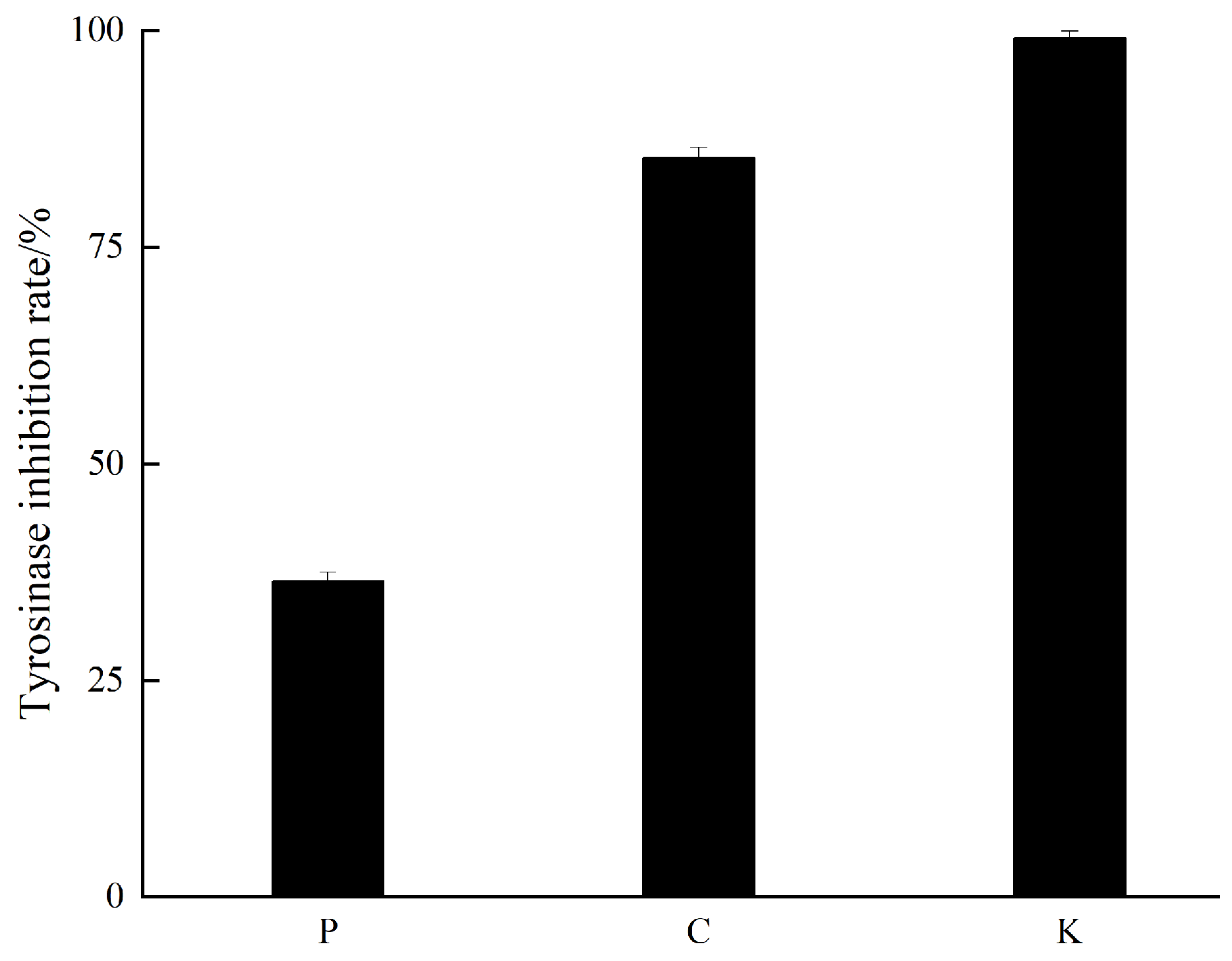
Figure 1. The results of mushroom tyrosinase inhibitory bioassay for the petroleum ether (P) and chloroform (C) extracts and the positive control kojic acid (K).
Cosmetics 12 00174 g001
Figure 2. The results of mushroom tyrosinase inhibitory bioassay for the C1–C5 fractions and the positive control kojic acid (K).
Cosmetics 12 00174 g002
Figure 3. Summary of key experimental procedures of compounds 110.
Cosmetics 12 00174 g003
Figure 4. Compounds 110 from L. casuarinoides.
Cosmetics 12 00174 g004
Figure 5. The results of mushroom tyrosinase inhibitory bioassay for compounds 6 and 7 and the positive control kojic acid (K).
Cosmetics 12 00174 g005
Figure 6. The binding mechanisms between compounds 6 (upper) and 7 (lower) and the mushroom tyrosinase (PDB: 2Y9X).
Cosmetics 12 00174 g006
Table 1. The mushroom tyrosinase inhibitory results of compounds 6 and 7 and the positive control.
CompoundsIC50 (mM)
61.90 ± 0.03
72.43 ± 0.03
Kojic acid 10.17 ± 0.03
1 Positive control.
Table 2. The binding energy scores of compounds 6 and 7 and tropolone.
CompoundsBinding Energy (kcal/mol)
6−6.60
7−5.75
tropolone 1−6.07
1 The native ligand in the crystal (PDB ID: 2Y9X).
Disclaimer/Publisher’s Note: The statements, opinions and data contained in all publications are solely those of the individual author(s) and contributor(s) and not of MDPI and/or the editor(s). MDPI and/or the editor(s) disclaim responsibility for any injury to people or property resulting from any ideas, methods, instructions or products referred to in the content.