Article

Safety Profile and Efficacy of Biosea® Revive Serum for Hair Growth Through In Vitro Assessment and Clinical Evaluation

1
Department of Fragrance and Cosmetic Science, College of Pharmacy, Kaohsiung Medical University, Kaohsiung City 807, Taiwan
2
Department of Pharmacy and Master Program, College of Pharmacy and Health Care, Tajen University, Pingtung County 907, Taiwan
3
School of Pharmacy, College of Pharmacy, Kaohsiung Medical University, Kaohsiung City 807, Taiwan
4
Shiny Brands Research & Innovation Center, Shiny Brands Group Co., Ltd., Taipei City 105, Taiwan
5
Drug Development and Value Creation Research Center, Kaohsiung Medical University, Kaohsiung City 807, Taiwan
6
Department of Medical Research, Kaohsiung Medical University Hospital, Kaohsiung City 807, Taiwan
7
Institute of Biomedical Sciences, National Sun Yat-Sen University, Kaohsiung City 804, Taiwan
8
College of Professional Studies, National Pingtung University of Science and Technology, Pingtung City 912, Taiwan
*
Authors to whom correspondence should be addressed.
These authors contributed equally to this work.
Cosmetics 2025, 12(4), 139; https://doi.org/10.3390/cosmetics12040139
Submission received: 31 March 2025 / Revised: 5 June 2025 / Accepted: 11 June 2025 / Published: 1 July 2025
(This article belongs to the Section Cosmetic Formulations)

Abstract

Excessive hair loss can negatively impact psychological well-being and personal appearance. Providing effective hair growth products containing natural ingredients to people with hair loss can solve this problem. This study investigates Biosea® Revive serum (BRS), a novel hair care product containing biotinoyl tripeptide-1 and Phyllanthus emblica fruit extract as the main ingredients, as a natural intervention for hair growth. Results from the in vitro study demonstrates that BRS not only increased human hair dermal papilla cell (HHDPC) cell proliferation, but also reduced reactive oxygen species generation and 5α-reductase expression when compared to the control group, with BRS showing similar effect to the positive control, minoxidil. In addition, a 90-day clinical trial with 40 participants (KMUHIRB-F(I)-20230125; approval date: 18 August 2023) was conducted to assess the effectiveness and safety of BRS. The results revealed that BRS can improve hair density and quality in both men and women participants, with a significant reduction in transepidermal water loss (TEWL) in women (p < 0.05). Moreover, there were no adverse effects on blood parameters or scalp irritation reported after BRS treatment. In conclusion, we suggest that BRS offers a safe and effective solution for improving hair follicle health and is suitable for long-term use.

1. Introduction

The societal perception of beauty significantly influences our aesthetic preferences. While the perception of beauty varies among individuals, it profoundly impacts personal appearance, social interactions, and psychological well-being. Among various aspects of appearance, hair is a critical factor [1,2]. When individuals lose more than the typical daily range of 100 to 150 hairs lost daily, it can visibly affect their appearance. Hair loss can result from various factors, including the conversion of testosterone into dihydrotestosterone (DHT) via 5α-reductase, which exacerbates hair follicle shrinkage and death [3]. Additionally, excessive accumulation of reactive oxygen species (ROS) in scalp follicle cells contributes to oxidative stress, ultimately increasing hair loss. Evaluating the potential of active ingredients to mitigate hair loss can be achieved by assessing their ability to inhibit DHT production, reduce excess ROS formation, and promote the growth of human hair dermal papilla cells (HHDPC) [2].
In recent years, natural compounds and peptides have been extensively studied for their hair growth-promoting properties. Among them, biotinoyl tripeptide-1 [4], acetyl tetrapeptide-3, Trifolium pratense (Clover) flower extract [5], Phyllanthus emblica fruit extract [6], and Wasabi japonica leaf extract [7], have demonstrated potential. It is well known that nanotechnology has garnered attention for enhancing the bioavailability of active compounds. Studies have shown that nano-systems can improve the efficacy of active ingredients through sustained release and targeted delivery to hair follicles, thereby enhancing their effectiveness while reducing scalp irritation [8,9]. A clinical trial on androgenetic alopecia reported that, compared to conventional formulations, minoxidil processed with nanotechnology led to a greater increase in hair count among participants [10].
This study aims to evaluate the safety profile and efficacy of a composite formulation, called Biosea® Revive serum (BRS) with the benefits of multiple hair growth-promoting active ingredients. The research design includes three in vitro assays: (1) hair follicle cell safety and proliferation test, (2) inhibition of ROS generation assay, and (3) 5α-reductase protein expression induced by DHT in HHDPC. In addition, a clinical trial involving 40 participants (20 males and 20 females) was conducted to evaluate the efficacy of BRS in promoting hair growth.

2. Materials and Methods

2.1. Preparation of BRS and Particle Size Analysis

The composition of BRS consists of phases A, B, and C. Phase A is an alcohol-water solution. Phase B, consisting of propylene glycol, butylene glycol, hydroxyacetophenone, 1,2-hexanediol, PEG-40 hydrogenated castor oil, tocopheryl acetate, Citrus bergamia peel oil expressed, Mentha piperita oil, Eucalyptus radiata flower/leaf/stem oil, fragrance, linalool, limonene, menthol, and menthyl lactate, were heated at 75 °C and homogenized with an IKA mixer (OST 20 D S1, Staufen, Germany) to obtain a clear solution. Phase C, including water, disodium EDTA, butylene glycol, water, dextran, acetyl tetrapeptide-3, Trifolium pratense (clover) flower extract, sebaryl FL BC10142, panthenol, propanediol, glycerin, Phyllanthus emblica fruit extract, butylene glycol, Wasabi japonica leaf extract, biotinoyl tripeptide-1, glycerin, water, Sargassum hemiphyllum extract, and Chlorella vulgaris extracellular vesicles, were stirred at room temperature until fully dissolved. Subsequently, Phases B and C were incorporated into Phase A in succession and blended using an IKA mixer to achieve BRS.
Dynamic light scattering (DLS) was employed to characterize the particle size, distribution profile, and aggregation behavior of Biosea® Revive Serum (BRS). The polydispersity index (PDI) serves as a key parameter for assessing particle size distribution. A PDI approaching 0.01 signifies a highly uniform, monodisperse system, whereas values near 1.0 indicate a broader distribution of particle sizes. When the PDI exceeds 1.0, it suggests the presence of multiple distinct particle populations within the formulation. In the context of polymer-based nanomaterials, a PDI below 0.3 is typically regarded as indicative of a stable and homogeneously dispersed system [11].

2.2. In Vitro Assays

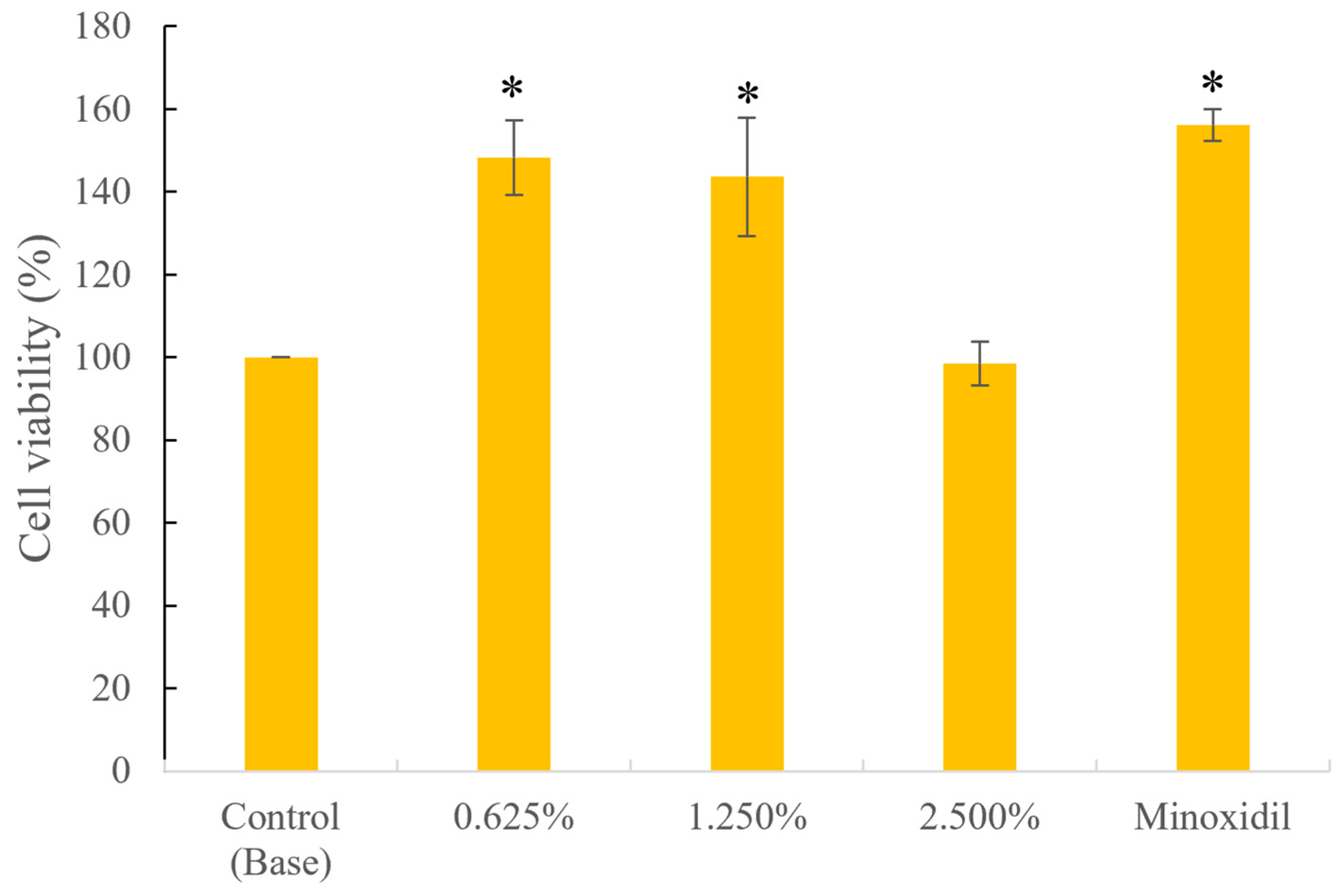
2.2.1. Hair Follicle Cell Safety and Proliferation Test

The study included five groups: 0.625%,1.25%, and 2.5% BRS solution as test sample group, negative control group (which consisted of 2.5% formulation base alone), and the positive control group, where minoxidil was prepared in dimethyl sulfoxide (DMSO) at an initial concentration of 4.8 mM and diluted with mesenchymal stem cell medium (MSCM) to a final concentration of 20 μM (0.0004%). The rationale for using minoxidil at this concentration is based on prior literature employing a similar in vitro model system [12,13,14]. Human hair dermal papilla cells (HHDPC, ScienCell, Catalog #2400) at a density of 2 × 10³ cells/well were seeded into 96-well plates using MSCM. The plates were incubated at 37 °C in a 5% CO2 incubator for 18–24 h to allow cell adhesion. After removing the medium, 100 μL of the prepared test samples at various concentrations in MSCM were added, and the plates were incubated for 72 h. The medium was replaced with freshly prepared test solutions of the same concentration every 24 h. After 72 h, 20 μL of dimethylthiazol-carboxymethoxyphenyl-sulfophenyl-tetrazolium (MTS) reagent was added to each well, followed by a 3 h incubation. The absorbance at 490 nm was measured using a microplate reader. Cell viability and proliferation rates were calculated using Microsoft Excel with the following formula:
Cell viability (%) = (OD490 of test sample/OD490 of control) × 100%

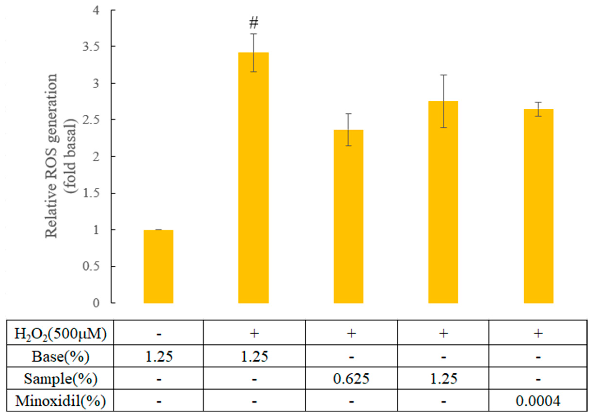
2.2.2. Inhibition of Reactive Oxygen Species (ROS) Generation Assay

A fluorescent probe, 2′,7′-dichlorodihydrofluorescein diacetate (DCF-DA), was prepared in ethanol at an initial concentration of 2 mg/mL. It was subsequently diluted 1:1 with Dulbecco’s phosphate buffered saline (DPBS) and further diluted 10-fold twice to achieve a final concentration of 10 μg/mL. Hydrogen peroxide (H2O2, 14.7 M) was diluted with MSCM medium to a final concentration of 2.167 mM. HHDPC at a density of 2 × 10³ cells/well were seeded into 96-well plates using MSCM medium. The plates were incubated at 37 °C in a 5% CO2 incubator for 18–24 h to allow cell adhesion. After removing the medium, 100 μL of BRS (0.625% and 1.25%), positive control (minoxidil at 0.0004%), or negative control (which consisted of formulation base alone) in MSCM were added, and the plates were incubated for 72 h. The medium was replaced with freshly prepared solutions of the same concentration every 24 h. After 72 h, 10 μg/mL of DCF-DA reagent (100 μL/well) was added, and the cells were incubated for 30 min. The cells were washed twice with DPBS and then treated with 100 μL/well of DPBS and 30 μL/well of 2.167 mM hydrogen peroxide for 1 h. The fluorescence intensity was measured at an excitation wavelength of 485 nm and an emission wavelength of 535 nm using a fluorescence microplate reader. The intracellular ROS generation and inhibition were calculated using Microsoft Excel.
The formula for ROS generation and inhibition is as follows:
ROS generation = (Fluorescence intensity of test sample/Fluorescence intensity of control)

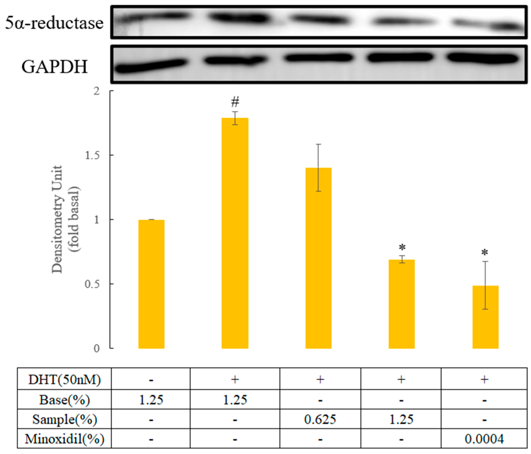
2.2.3. 5α-Reductase Protein Expression Induced by Dihydrotestosterone (DHT) in Human Hair Follicle Dermal Papilla Cells

Dihydrotestosterone (DHT), used as the inducing agent, was prepared in DMSO at an initial concentration of 5 mM, diluted 5-fold with DMSO, and subsequently diluted with MSCM medium to a final concentration of 300 nM. HHDPC at a density of 4 × 10⁴ cells/well were seeded into 24-well plates and incubated at 37 °C in a 5% CO2 incubator for 18–24 h to allow cell adhesion. After removing the culture medium, 0.5 mL of test samples at different concentrations (0.625% and 1.25% BRS, positive control (minoxidil at 0.0004%), or negative control (which consisted of formulation base alone)) were added to the wells. After 48 h of incubation, 100 μL of 300 nM DHT solution was added to induce the cells for 24 h. The cells were then washed twice with DPBS, and 20 μL of 2× sample buffer was added to lyse the cells. The cell lysate was scraped using a pipette tip, transferred to microcentrifuge tubes, and diluted with 20 μL of RIPA buffer (1× final dilution). The mixture was sonicated for 10 min and heated at 95 °C on a heating block to denature the proteins. The protein samples were stored at −20 °C for further analysis. For protein separation, 8–12% SDS-PAGE was assembled in an electrophoresis chamber filled with running buffer. The denatured protein samples were loaded into the wells, and electrophoresis was conducted at 300 V and 30 mA until the proteins were fully separated. The proteins were then transferred onto a polyvinylidene difluoride (PVDF) membrane under transfer conditions of 300 V and 300 mA for 60 min. The PVDF membrane was blocked with blocking buffer at room temperature for 30–60 min, followed by three washes with Tris-buffered saline with Tween-20 (TTBS) (5 min each). The membrane was incubated overnight at 4 °C with diluted primary antibodies targeting 5α-reductase and Glyceraldehyde-3-phosphate dehydrogenase (GAPDH). The next day, the membrane was washed three times with phosphate-buffered saline with Tween 20 (PBS-T) for 5 min each, then incubated with diluted secondary antibodies (anti-Mouse and anti-Rabbit) at room temperature for 1 h. After three washes with TTBS (5 min each), ECL chemiluminescence reagent was added, and the membrane was exposed to the imaging system for 20–30 s to capture the results.
The resulting images were analyzed using the Image Lab software (version 6.1) to perform semi-quantitative analysis of the protein expression levels. The quantitative results were statistically analyzed using SPSS 20 to determine their significance.

2.3. Clinical Trial

2.3.1. Study Design

This study comprises two groups in an open-label, parallel clinical trial design. This study planned to recruit 40 participants aged 25 to 55 years who were experiencing hair loss, including 20 women and 20 men, divided into two gender-based groups (20 participants per group). The exclusion criteria were as follows: (1) Participants who had received hair growth treatments within three months prior to the study. (2) Those diagnosed with any active scalp diseases, such as scalp psoriasis (confirmed by a dermatologist). (3) Individuals who had undergone cancer chemotherapy within six months before the study or planned to receive such treatment during the study. (4) Those with a history of alcohol abuse and/or mental disorders, including trichotillomania. (5) Participants who had previously undergone hair transplantation surgery. (6) Individuals currently taking medications that could cause hypertrichosis or undergoing treatment for androgenetic alopecia. (7) Those with known allergies or hypersensitivity to any cosmetic ingredients used in this study. (8) Pregnant or breastfeeding women. (9) Participants who had undergone chemical hair treatments, such as straightening, perming, or dyeing, within three months prior to the study. (10) Women confirmed as postmenopausal based on their medical history. (11) Individuals with a history of thyroid diseases. (12) Participants with any uncontrolled medical conditions, including HIV, hepatitis, severe cardiac or respiratory diseases, or other serious medical illnesses. As the primary objective of this study was to develop a cosmetic-grade skincare product, the clinical trial was designed to recruit generally healthy volunteers to better simulate the actual target users in real-world cosmetic applications. Since cosmetic products are typically intended for the general population rather than individuals with specific pathological hair loss conditions, recruitment was not restricted to participants with a specific type of alopecia. This approach also allowed the evaluation of the product’s potential benefits on overall scalp and hair condition in a routine care setting. Recruitment was carried out using word of mouth, onsite, and online advertisement posters within the Kaohsiung Medical University campus.
The clinical trial utilized a split-scalp design where each participant served as their own control. Two symmetrical 20-mm diameter areas were shaved on the scalp: one designated for treatment (right side) and one as an untreated control (left side). The participants were instructed to use 100% BRS twice daily, applying one spray (approximately 0.1 g) each time to the shaved area on the right side, gently massaging it until fully absorbed. After application, washing the scalp again was to be avoided. The left side remained untreated to serve as an internal control. The trial was conducted from 18 August 2023, to 31 July 2024. This study adhered to the ethical principles outlined in the Declaration of Helsinki and was approved by the Institutional Review Board of Kaohsiung Medical University Chung-Ho Memorial Hospital (Approval Number: KMUHIRB-F(I)-20230125). All participants were fully informed about the details of the clinical study and voluntarily signed the informed consent form to participate.

2.3.2. Safety Evaluation

During the study, researchers scheduled follow-up assessments with participants on Day 0 (before product use) and on Days 15, 30, 45, and 90 (after product use). At each follow-up, hair in two designated test areas on the participants’ scalps were trimmed. Using a VapoMeter(Delfin, Kuopio, Eastern Finland, Finland), a non-invasive probe, scalp transepidermal water loss (TEWL) was measured to evaluate scalp safety before and after product use. Additionally, on Day 0 and Day 90, blood samples were collected by a certified medical laboratory to assess any potential impact of the product on blood parameters. The blood tests included liver function markers (GOT, GPT, Total protein, Albumin, Globulin, Albumin/Globulin Ratio, Total Bilirubin, Direct Bilirubin, Gamma-Glutamyl Transferase), kidney function markers (Blood Urea Nitrogen, Creatinine, Uric Acid), hematologic parameters (White Blood Cell Count, Red Blood Cell Count, Hemoglobin, Hematocrit, Mean Corpuscular Volume, Mean Corpuscular Hemoglobin, Mean Corpuscular Hemoglobin Concentration, Platelet Count), and lipid profile indicators (Total Cholesterol, Triglycerides, HDL-Cholesterol, Total Cholesterol/HDL Cholesterol Ratio, LDL-Cholesterol, LDL/HDL Ratio). These comprehensive evaluations ensured a thorough assessment of product safety.

2.3.3. Hair Analysis Measurement

During the study, researchers scheduled follow-up appointments with participants on Day 0 (before product application) and on Days 15, 30, 45, and 90 after starting product use. At each visit, the hair in two designated test areas on the scalp was trimmed. Participants applied the product to one of these areas, designated as the treatment area, while the other served as an untreated control area. This study utilized the TrichoScan HD 4.0 system (Tricholog GmbH, Freiburg, Germany) for hair analysis, which combines ultra-high-definition imaging with software analysis (developed by DatInf GmbH, Tübingen, Germany) to evaluate the effects of BRS on various hair parameters. These include the density calculated based on the detected number of hairs (hairs/cm2), hair mass (mm/cm2): the cumulative thickness (diameter) of hairs within a standardized 1 cm2 area, representing the sum of hair diameters, median hair thickness (μm): the median value of the hair diameter distribution, mean hair thickness (μm): the average value of the hair diameter distribution, vellus hair density (1/cm2): the density of vellus hairs, defined as hairs with a thickness of less than 40 μm, terminal hair density (1/cm2): the density of terminal hairs, defined as hairs with a thickness greater than 40 μm, vellus hair ratio (%): the percentage of vellus hairs relative to the total number of hairs, and terminal hair ratio (%): the percentage of terminal hairs relative to the total number of hairs. These parameters were assessed to measure improvements.

3. Results

3.1. In Vitro Study

3.1.1. Determination of Particle Size by DLS

As summarized in Table 1 BRS exhibited an average particle size of 102.63 ± 64.87 nm, with a PDI of 0.19 ± 0.05. These findings confirm that the formulation predominantly falls within the nanometer range and maintains a well-dispersed, uniform particle distribution, meeting the standards for monodispersity.

3.1.2. Hair Follicle Cell Safety and Proliferation Activity

In the human hair dermal papilla cells proliferation assay, treatment with 0.625% and 1.25% concentrations of BRS for 72 h resulted in cell growth percentages of 148.24% and 143.59%, respectively. These results demonstrate that the BRS at concentrations of 0.625% and 1.25% exhibits significant proliferative effects compared to the control group (p < 0.05) and achieves results comparable to the positive control group (Minoxidil), with no statistically significant difference (p > 0.05; Figure 1).

3.1.3. Antioxidant Activity

In the ROS generation assay, the induced group, exposed to H2O2 alone, resulted in 3.42 times higher ROS production in HHDPC compared to the negative control group (which consisted of formulation base alone). After 72 h of treatment with 0.625% and 1.25% of BRS, ROS generation levels were 2.37 times and 2.75 times that of the negative control group, respectively (Figure 2). These results indicate a trend toward inhibition of ROS generation by the BRS at 0.625% and 1.25% compared to the induced group, though the differences were not statistically significant. When converted to inhibition percentages, the data show that the 0.625% BRS achieved a 43.7% inhibitory effect, exhibiting better activity than the 1.25% formulation (Table 2). In addition, the effects at both concentrations were statistically comparable to the positive control group (Minoxidil) (p > 0.05). These findings suggest that the ROS generation inhibitory ability of BRS is equivalent to that of Minoxidil.

3.1.4. In Vitro Hair Growth Activity

The induced group, exposed to DHT alone, resulted in 1.79 times higher 5α-reductase protein expression levels in HHDPC compared to the negative control group (which consisted of formulation base alone). In the experiment evaluating the effect of the BRS on 5α-reductase protein expression in HHDPC induced by dihydrotestosterone (DHT). After 72 h of treatment with 0.625% and 1.25% of BRS, the 5α-reductase protein expression levels were 1.40 times and 0.69 times that of the negative control group, respectively (Figure 3). These results demonstrate that the BRS at 0.625% and 1.25% significantly inhibited 5α-reductase protein expression compared to the DHT group (p < 0.05) and exhibited similar effect to the positive control group (Minoxidil) (p > 0.05). When converted to inhibition percentages, the BRS at 0.625% and 1.25% showed inhibition rates of 50.16% and 139.36%, respectively (Table 3). At a concentration of 1.25%, the BRS exhibited no statistically significant difference in effect compared to Minoxidil (p > 0.05). These findings indicate that the 1.25% BRS has an inhibitory effect on 5α-reductase protein expression equivalent to that of Minoxidil.

3.2. Safety Profile and Efficacy of Biosea® Revive Serum for Hair Growth

3.2.1. Safety Profile

In the female group, the relative percentage of TEWL (transepidermal water loss) in the BRS application site significantly decreased after 90 days of serum application. These results demonstrate a statistically significant reduction in TEWL in the application site compared to baseline after 90 days of serum application. No statistically significant differences were observed in the male group (Table 4). The comprehensive blood test results for both male and female participants were analyzed. In the male group, although some indicators, such as albumin, albumin/globulin ratio, and total bilirubin, showed statistically significant differences (p < 0.05) in the independent samples t-test, all values remained within the normal physiological range (Table S1). Similarly, in the female group, albumin and fasting blood glucose also exhibited statistically significant differences (p < 0.05), but all values were within the normal range. Additionally, no statistically significant differences were observed in other indicators for the female group (Table S2). These findings indicate that the use of the BRS does not affect liver or kidney function or other related health indicators in either male or female participants. Furthermore, the serum application did not cause any scalp irritation, ensuring its safety for prolonged use.

3.2.2. Hair Growth Efficacy

All hair-related parameters were measured using TrichoScan HD 4.0 and calculated using a relative percentage formula. Values above 100% indicate an increase compared to baseline (Day 0), while values below 100% represent a decrease. In the male group, the hair mass after BRS treatment significantly increased at Days 15, 30, and 45 (p < 0.05). Similarly, hair density showed significant increases at Days 30 and 45 (p < 0.05). Terminal hair density also exhibited significant increases at Days 15, 30, and 45 (p < 0.05). Moreover, vellus hair density showed a marked improvement by Day 90 (p < 0.05; Table 5).
In the female group, total hair density significantly increased at Day 15 (p < 0.05). Likewise, vellus hair density showed a significant increase at Day 30 (p < 0.05; Table 6).

4. Discussion

The causes of hair loss can be categorized into two major types: intrinsic and extrinsic factors. Intrinsic factors mainly include the sensitivity of hair follicles to androgens, particularly the adverse effects of dihydrotestosterone (DHT) produced under the catalysis of 5α-reductase, leading to follicle atrophy and impaired hair growth. Additionally, psychological stress such as depression and anxiety may trigger or exacerbate hair loss. Extrinsic factors include irregular lifestyles, ultraviolet (UV) radiation, and pollutants, which induce elevated oxidative stress and excessive accumulation of reactive oxygen species (ROS), further impairing hair follicle function and worsening hair loss [15]. To address hair loss caused by intrinsic and extrinsic factors, this study developed a hair care product Biosea® Revive serum (BRS) aimed at regulating internal factors and reducing external damage, thereby comprehensively improving hair loss conditions and promoting hair follicle health and hair growth.
To target these mechanisms, this study designed BRS nanoparticles formulation combining multiple peptides and natural active ingredients to achieve a multi-mechanistic approach to mitigating hair loss. Research indicates that biotinoyl tripeptide-1 effectively inhibits 5α-reductase activity, reducing DHT production and thereby improving hair loss [16]. The combination of acetyl tetrapeptide-3 and red clover extract suppresses DHT production, reduces microinflammation, and promotes the synthesis of extracellular matrix (ECM) proteins, stabilizing follicle structure and enhancing hair growth [5]. Moreover, both Phyllanthus emblica fruit extract and wasabi leaves have demonstrated potential in promoting dermal papilla cell proliferation, prolonging the anagen phase of hair follicles, and alleviating oxidative stress [6,7].
In this study, human hair dermal papilla cells were used to evaluate cell proliferation and 5α-reductase inhibition. Results showed that after 72 h of treatment with the BRS at concentrations of 0.625% and 1.25%, cell proliferation increased to 148.24% and 143.59%, respectively, significantly higher than the control group (p < 0.05) and comparable to the positive control minoxidil (p > 0.05). Additionally, in experiments exploring the effect of the BRS on DHT-induced 5α-reductase protein expression, it was found that DHT alone increased 5α-reductase expression to 1.79-fold. However, after 72 h of treatment with 0.625% and 1.25%BRS, 5α-reductase expression decreased to 1.40-fold and 0.69-fold, respectively (p < 0.05). Notably, the inhibitory effect of 1.25% BRS was comparable to minoxidil (p > 0.05). These results confirm that the BRS not only effectively promotes dermal papilla cell proliferation and hair growth, but also significantly inhibits 5α-reductase protein expression, reducing excessive DHT production and alleviating the progression of hair loss. Excessive ROS can damage the DNA, lipids, and proteins of hair follicle cells, inducing apoptosis and inflammatory responses, thereby exacerbating follicle atrophy. Studies have shown that the inhibitory effect of DHT on hair growth is mediated by the release of the hair growth inhibitory factor TGF-β1, a process driven by ROS or free radical accumulation in androgen-sensitive cells [17,18]. Additionally, other research has demonstrated that ROS scavengers and antioxidants can successfully block TGF-β1 release, effectively preventing hair growth inhibition [19]. In the ROS generation test induced by H2O2, the BRS nanoparticles formulation exhibited significant inhibitory effects, particularly at concentrations of 0.625% and 1.25%, with inhibition rates of 43.7% and 28.1%, respectively, comparable to minoxidil (p > 0.05). These results suggest the BRS has considerable potential for reducing oxidative stress, further supporting its value in promoting hair health. Scalp safety and efficacy are critical considerations in the development of hair care products. This study employed transepidermal water loss (TEWL) as an indicator for assessing skin barrier function [20]. TEWL measures the rate of water evaporation through the skin barrier, with significant increases indicating barrier damage. One study showed that individuals with atopic dermatitis exhibited significant differences in scalp surface morphology compared to healthy individuals, with severe damage to the stratum corneum, rough hair surfaces, and reduced water content, reflecting impaired barrier function [21]. This highlights the correlation between scalp barrier health and hair health, emphasizing the importance of maintaining barrier function in hair care product development. This study demonstrated that the BRS significantly reduced TEWL in female subjects (p < 0.05), indicating that it not only preserved, but also enhanced the scalp barrier function. In addition, a study investigating gender-based behaviors and attitudes toward cosmetic treatments indicated that women show a significantly higher concern for their appearance than men and are more likely to use skincare products to maintain or improve skin condition [22]. In the same way, the lower TEWL values observed in female participants may be attributed to their more proactive scalp care practices, resulting in better scalp barrier function and hydration status.
In the development of treatments for hair loss, blood tests are a common and necessary assessment to monitor physiological indicators such as liver and kidney function, hematological parameters, and lipid levels. For example, oral medications like finasteride, metabolized by the liver, may impact liver function, making blood tests essential for safety evaluations [23]. This study conducted detailed blood tests to assess the safety of the product. Results showed that all participants exhibited no abnormalities in liver function, kidney function, or hematological parameters after using the product. These findings further confirmed the product’s safety. Additionally, after long-term use for 90 days, the product caused no burden on liver or kidney function, demonstrating its suitability for individuals with different physiological conditions and providing robust scientific support for its safety evaluation. Quantitative analyses of hair density and quality provide an objective method free from subjective judgment and are widely applied in diagnosing hair loss and evaluating the effects of various treatments, including medications and laser therapies [24].
For instance, a study involving 79 patients with androgenetic alopecia (AGA) investigated the effects of nutritional supplements, lotions, and their combination on reducing hair loss by measuring hair density [25]. Another study on platelet-rich plasma (PRP) treatment for AGA also used hair density and quality assessments to evaluate treatment efficacy [26]. Hair density and quality measurements have thus become important and reliable parameters for evaluating improvements in hair loss. Moreover, a randomized, controlled, double-blind clinical trial recruited 90 male patients with AGA, revealing that a topical nonionic vesicle (niosomes) formulation of minoxidil significantly increased hair count compared to traditional minoxidil solutions, highlighting the importance of formulation design in enhancing the efficacy of active ingredients [10]. Other studies have indicated that nanotechnology formulations can effectively promote the targeted delivery of active ingredients, enhance local bioavailability, and reduce side effects [8,9]. The results of this study showed significant improvements in hair density and quality for both male and female groups at different time points. Notably, hair density in males increased significantly by day 30 and 45, while the female group exhibited significant improvements as early as day 15. These findings demonstrate the significant potential of the BRS nanoparticles formulation in promoting hair growth, with efficacy observed across genders.

5. Conclusions

In conclusion, the BRS nanoparticles formulation significantly improved hair density and quality in individuals of both genders. Its mechanisms involve promoting dermal papilla cell proliferation, reducing oxidative stress, lowering 5α-reductase protein expression, and enhancing skin barrier function, showing that BRS is a safe and non-irritating product, that can promote hair growth.

Supplementary Materials

The following supporting information can be downloaded at: https://www.mdpi.com/article/10.3390/cosmetics12040139/s1, Figure S1: Original unprocessed images of blots for 5α-reductase; Table S1: Comprehensive Health Blood Test Results for Male Group; Table S2: Comprehensive Health Blood Test Results for Female Group.

Author Contributions

Conceptualization and methodology were conducted by H.-Y.H., S.-H.Y., C.-J.W., C.-Y.Y., T.-H.W. and F.-L.Y. Data curation, validation, formal analysis, and investigation were conducted by C.-J.W., C.-Y.Y., P.B.S., T.-H.W. and F.-L.Y. Resources were provided by F.-L.Y. Writing—original draft preparation was completed by C.-J.W. Writing—review and editing was performed by F.-L.Y., P.B.S. and H.-M.H. Supervision and funding acquisition were managed by F.-L.Y. All authors have read and agreed to the published version of the manuscript.

Funding

The financial and technical support provided by Shiny Brands Group Co., Ltd. through the industry-academia collaboration project with Kaohsiung Medical University (grant number: S111032).

Institutional Review Board Statement

The study was conducted in accordance with the Declaration of Helsinki, and approved by the Institutional Review Board of Kaohsiung Medical University Chung-Ho Memorial Hospital (protocol code KMUHIRB-F(I)-20230125 and date of approval 18 August 2023).

Informed Consent Statement

Informed consent was obtained from all subjects involved in the study. All subject data were anonymized and coded prior to analysis.

Data Availability Statement

All data presented in the study are available on request from the corresponding author (flyen@kmu.edu.tw).

Acknowledgments

The authors acknowledge Shiny Brands Group Co., Ltd. for providing the samples used in this study. We also express our sincere gratitude to Chu-Sung Hu, attending dermatologist at the Department of Dermatology, Kaohsiung Medical University Chung-Ho Memorial Hospital, for his valuable assistance in evaluating the scalp and hair conditions of the study participants.

Conflicts of Interest

The study was supported by Shiny Brands Group Co., Ltd. through an industry-academia collaboration project with Kaohsiung Medical University. Three authors, Hui-Yu Hu, Shang-Hsuan Yang and Hsiang-Ming Hsueh, are affiliated with the funding company and contributed technical input during the conceptualization and methodology development stages. However, the funder was not involved in the study design, collection, analysis, interpretation of data, the writing of this article or the decision to submit it for publication. Other authors declare that there is no conflict of interest related to this publication.

References

  1. Clinic, A.; Asper, A.; Mittal, A.; Shome, D.; Parbhoo, D.; Thanzama, J.; Doshi, K.; Sachde, N.; Gaunkar, R.; Kapoor, R.; et al. Evaluation of the Safety and Effectiveness of Intradermal Administration of QR678 Neo® Hair Growth Factor Formulation: A Phase-IV, Open-Label, Single-Arm Multi-Ethnicity Clinical Trial. J. Cosmet. Dermatol. 2022, 21, 580–589. [Google Scholar] [CrossRef] [PubMed]
  2. Bae, W.Y.; Jung, W.H.; Shin, S.L.; Kim, T.R.; Sohn, M.; Suk, J.; Jung, I.; Lee, Y.I.; Lee, J.H. Heat-Treated Limosilactobacillus Fermentum LM1020 with Menthol, Salicylic Acid, and Panthenol Promotes Hair Growth and Regulates Hair Scalp Microbiome Balance in Androgenetic Alopecia: A Double-Blind, Randomized and Placebo-Controlled Clinical Trial. J. Cosmet. Dermatol. 2024, 23, 2943–2955. [Google Scholar] [CrossRef] [PubMed]
  3. Nestor, M.S.; Ablon, G.; Gade, A.; Han, H.; Fischer, D.L. Treatment Options for Androgenetic Alopecia: Efficacy, Side Effects, Compliance, Financial Considerations, and Ethics. J. Cosmet. Dermatol. 2021, 20, 3759–3781. [Google Scholar] [CrossRef] [PubMed]
  4. Ledwoń, P.; Errante, F.; Papini, A.M.; Rovero, P.; Latajka, R. Peptides as Active Ingredients: A Challenge for Cosmeceutical Industry. Chem. Biodivers. 2021, 18, e2000833. [Google Scholar] [CrossRef]
  5. Loing, E.; Lachance, R.; Ollier, V.; Hocquaux, M. A New Strategy to Modulate Alopecia Using A Combination of Two Specific and Unique Ingredients. J. Cosmet. Sci. 2013, 64, 45–58. [Google Scholar]
  6. Wongrakpanich, A.; Leanpolchareanchai, J.; Morakul, B.; Parichatikanond, W.; Teeranachaideekul, V. Phyllanthus emblica Extract-loaded Transfersomes for Hair Follicle Targeting: Phytoconstituents, Characterization, and Hair Growth Promotion. J. Oleo Sci. 2022, 71, 1085–1096. [Google Scholar] [CrossRef]
  7. Okunishi, I. Improvement of Cognitive Function by Wasabi Component “Hexaraphane”. In Herbs and Spices-New Perspectives in Human. Health and Food Industry: New Perspectives in Human. Health and Food Industry; IntechOpen: London, UK, 2024; p. 227. [Google Scholar]
  8. Singh, S.; Sonia; Sindhu, R.K.; Alsayegh, A.A.; Batiha, G.E.; Alotaibi, S.S.; Albogami, S.M.; Conte-Junior, C.A. Formulation Development and Investigations on Therapeutic Potential of Nanogel from Beta vulgaris L. Extract in Testosterone-Induced Alopecia. Biomed. Res. Int. 2023, 2023, 1777631. [Google Scholar] [CrossRef]
  9. Naeini, A.H.; Mahdavipour, K.; Rastegari, A.; Aghsami, M.; Montazeri, H.; Faghihi, H.; Mohammadi, Z. Chitosan and Its Amphiphilic Derivative Nanoparticles Loaded with Minoxidil for Induction of Hair Growth: In vitro and in vivo Evaluation. Int. J. Biol. Macromol. 2024, 259, 129122. [Google Scholar] [CrossRef]
  10. Meymandi, S.S.; Amiri, R.; Aflatunian, M.; Pardakhti, A. Comparison of the Efficacy of Niosomal Minoxidil with Conventional Minoxidil in the Treatment of Androgenetic Alopecia: A Randomized, Controlled, Double-Blind Clinical Trial. J. Dermatol. Cosmet. 2014, 5, 53–60. [Google Scholar]
  11. Danaei, M.; Dehghankhold, M.; Ataei, S.; Hasanzadeh Davarani, F.; Javanmard, R.; Dokhani, A.; Khorasani, S.; Mozafari, M.R. Impact of Particle Size and Polydispersity Index on the Clinical Applications of Lipidic Nanocarrier Systems. Pharmaceutics 2018, 10, 57. [Google Scholar] [CrossRef]
  12. Kang, J.I.; Yoon, H.S.; Kim, S.M.; Park, J.E.; Hyun, Y.J.; Ko, A.; Ahn, Y.S.; Koh, Y.S.; Hyun, J.W.; Yoo, E.S.; et al. Mackerel-Derived Fermented Fish Oil Promotes Hair Growth by Anagen-Stimulating Pathways. Int. J. Mol. Sci. 2018, 19, 2770. [Google Scholar] [CrossRef] [PubMed]
  13. Baek, Y.H.; Lee, J.H.; Chang, S.J.; Chae, Y.; Lee, M.H.; Kim, S.H.; Han, K.I.; Kim, T.J. Heat-Killed Enterococcus faecalis EF-2001 Induces Human Dermal Papilla Cell Proliferation and Hair Regrowth in C57BL/6 Mice. Int. J. Mol. Sci. 2022, 23, 5413. [Google Scholar] [CrossRef]
  14. Wen, T.-C.; Li, Y.-S.; Rajamani, K.; Harn, H.-J.; Lin, S.-Z.; Chiou, T.-W. Effect of Cinnamomum osmophloeum Kanehira Leaf Aqueous Extract on Dermal Papilla Cell Proliferation and Hair Growth. Cell. Transplant. 2018, 27, 256–263. [Google Scholar] [CrossRef] [PubMed]
  15. Sadick, N.S.; Callender, V.D.; Kircik, L.H.; Kogan, S. New Insight into the Pathophysiology of Hair Loss Trigger A Paradigm Shift in the Treatment Approach. J. Drugs. Dermatol. 2017, 16, s135–s140. [Google Scholar] [PubMed]
  16. Garre, A.; Piquero, J.; Trullas, C.; Martinez, G. Efficacy and Safety of A New Topical Hair Loss-Lotion Containing Oleanolic Acid, Apigenin, Biotinyl Tripeptide-1, Diaminopyrimidine Oxide, Adenosine, Biotin and Ginkgo Biloba in Patients with Androgenetic Alopecia and Telogen Effluvium: A Six-Month Open-Label Prospective Clinical Study. J. Cosmetol. Trichol. 2018, 4, 1000132. [Google Scholar] [CrossRef]
  17. Inui, S.; Fukuzato, Y.; Nakajima, T.; Yoshikawa, K.; Itami, S. Androgen-Inducible TGF-Beta1 from Balding Dermal Papilla Cells Inhibits Epithelial Cell Growth: A Clue to Understand Paradoxical Effects of Androgen on Human Hair Growth. FASEB J. 2002, 16, 1967–1969. [Google Scholar] [CrossRef]
  18. Shin, H.; Yoo, H.G.; Inui, S.; Itami, S.; Kim, I.G.; Cho, A.R.; Lee, D.H.; Park, W.S.; Kwon, O.; Cho, K.H.; et al. Induction of Transforming Growth Factor-Beta 1 by Androgen Is Mediated by Reactive Oxygen Species in Hair Follicle Dermal Papilla Cells. BMB Rep. 2013, 46, 460–464. [Google Scholar] [CrossRef]
  19. Rajput, R. A Scientific Hypothesis on the Role of Nutritional Supplements for Effective Management of Hair Loss and Promoting Hair Regrowth. J. Nutrition. Health. Food. Sci. 2018, 6, 1–11. [Google Scholar] [CrossRef]
  20. Verdier-Sévrain, S.; Bonté, F. Skin Hydration: A Review on Its Molecular Mechanisms. J. Cosmet. Dermatol. 2007, 6, 75–82. [Google Scholar] [CrossRef]
  21. Kim, K.S.; Shin, M.K.; Kim, J.H.; Kim, M.H.; Haw, C.R.; Park, H.K. Effects of Atopic Dermatitis on the Morphology and Water Content of Scalp Hair. Microsc. Res. Tech. 2012, 75, 620–625. [Google Scholar] [CrossRef]
  22. Girdwichai, N.; Chanprapaph, K.; Vachiramon, V. Behaviors and Attitudes Toward Cosmetic Treatments Among Men. J. Clin. Aesthet. Dermatol. 2018, 11, 42–48. [Google Scholar] [PubMed]
  23. Basaria, S.; Jasuja, R.; Huang, G.; Wharton, W.; Pan, H.; Pencina, K.; Li, Z.; Travison, T.G.; Bhawan, J.; Gonthier, R.; et al. Characteristics of Men Who Report Persistent Sexual Symptoms After Finasteride Use for Hair Loss. J. Clin. Endocrinol. Metab. 2016, 101, 4669–4680. [Google Scholar] [CrossRef] [PubMed]
  24. Kolasinski, J. TrichoScan®: A Useful Method For Measuring Hair Growth Parameters and Enhancing Patient Selection for Hair Restoration Surgery. Hair Transplant. Forum Int. 2009, 19, 56–59. [Google Scholar] [CrossRef]
  25. Bayer, M.; Gahrtz, M.; Voss, W.; Schlippe, G.; Whitfield, T. The Effect of A Food Supplement and A Hair Lotion on the Progression of Androgenetic Alopecia. J. Cosmet. Dermatol. Sci. Appl. 2019, 9, 292–304. [Google Scholar] [CrossRef]
  26. Sasaki, G.H. The Effects of Lower vs. Higher Cell Number of Platelet-Rich Plasma (PRP) on Hair Density and Diameter in Androgenetic Alopecia (AGA): A Randomized, Double-Blinded, Placebo, Parallel-Group Half-Scalp IRB-Approved Study. Aesthet. Surg. J. 2021, 41, NP1659–NP1672. [Google Scholar] [CrossRef]
Figure 1. The growth of human hair dermal papilla cells after 72 h of treatment with the BRS. The data represents the results of n = 4 and is expressed as mean ± standard deviation. * indicates a significant difference (p < 0.05) between the test substance or positive control group (Minoxidil) and the control (Base) group.
Cosmetics 12 00139 g001
Figure 2. The effect of the nano essence on ROS generation in HHDPC induced by H2O2. The data represents the results of two replicates and is expressed as mean ± standard deviation. # indicates a significant difference (p < 0.05) between the induced group and the control group. ‘-‘: without treatment; ‘+’: H2O2 treatment.
Cosmetics 12 00139 g002
Figure 3. The effect of the nano essence on 5α-reductase protein expression in HHDPC induced by dihydrotestosterone (DHT). The data represents the results of two replicates and is expressed as mean ± standard deviation. # indicates a significant difference (p < 0.05) between the control group and the DHT-induced group, while * indicates a significant difference (p < 0.05) between the test substance or positive control group (Minoxidil) and the DHT-induced group. ‘-‘: without treatment; ‘+’: DHT treatment.
Cosmetics 12 00139 g003
Table 1. Particle size of Biosea® Revive serum (BRS) (diluted 10×) by laser particle size analyzer.
Particle Size (nm) PDI
102.63 ± 64.870.19 ± 0.05
The experiment was performed in triplicate, and the results are expressed as the mean ± standard deviation (SD).
Table 2. The inhibition percentage of ROS generation in HHDPC induced by H2O2 with the BRS.
SampleInhibitory Percentage (%) &
0.625% BRS43.66 ± 2.98
1.250% BRS28.12 ± 7.13
Minoxidil 0.0004%31.46 ± 3.53
& The inhibition percentage calculation formula is as follows: Inhibitory percentage (%) = [1 − [(Sample − Control)/(Induced − Control)]] × 100.
Table 3. The inhibition percentage of 5α-reductase protein expression in HHDPC induced by dihydrotestosterone (DHT) with the nano essence.
SampleInhibition Percentage (%) &
0.625% BRS50.16 ± 19.71 *
1.250% BRS139.36 ± 1.01
Minoxidil 0.0004%163.83 ± 19.57
& The inhibition percentage calculation formula is as follows: Inhibitory percentage (%) = [1 − [(Sample − Control)/(Induced − Control)]] × 100; * indicates a significant difference (p < 0.05) compared to the positive control group (Minoxidil).
Table 4. The relative percentages of TEWL (g/m2h) before and after BRS treatment in both groups.
MaleFemale
DayApplication SiteControl AreaApplication SiteControl Area
0100 ± 0100 ± 0100 ± 0100 ± 0
15110.7 ± 81.9130.6 ± 8086.9 ± 54.3113.4 ± 67.5
30113 ± 88113.4 ± 48.5103.2 ± 93.1132.8 ± 107.9
4598.9 ± 52.5125.2 ± 56.385.8 ± 55.1123.2 ± 64.7
90111.6 ± 74.1122.2 ± 54.170.2 ± 41.9 *100.4 ± 68.6
* p-values < 0.05. Significant changes were observed after use compared to day 0 (baseline). The relative percentage was calculated using the formula: (post-use value ÷ pre-use value on day 0) × 100%. Values are presented as mean ± standard deviation.
Table 5. The relative percentages of hair parameters before and after BRS treatment in the male group.
DayHair Density $
(%)
Hair Mass #
(%)
Median Hair Thickness a
(%)
Mean Hair Thickness b
(%)
0100 ± 0100 ± 0100 ± 0100 ± 0
15109.7 ± 22.4110.5 ± 22.6 *101.8 ± 17.2101.3 ± 10.7
30110.1 ± 16.7 *111.8 ± 16.9 *102.2 ± 14.3101.8 ± 6.8
45114.6 ± 21.5 *114.3 ± 18.2 *101.9 ± 18.6100.5 ± 8.4
90107.2 ± 20.5105.7 ± 20.297.9 ± 15.699.2 ± 8.6
DayVellus Hair Density@
(%)
Terminal Hair Density&
(%)
Vellus Hair Ratioc
(%)
Terminal Hair Ratiod
(%)
0100 ± 0100 ± 0100 ± 0100 ± 0
15124.1 ± 85.9110.9 ± 21.4107.9 ± 56.6102.1 ± 14.4
30120 ± 52.4109.1 ± 17.8108.1 ± 37.399.3 ± 8.9
45146.3 ± 102.8111.8 ± 20.4121.4 ± 63.798.6 ± 14.1
90129.3 ± 63.2100.7 ± 24.4118.8 ± 45.894.2 ± 14.7
* p-values < 0.05. Significant changes were observed after use compared to day 0 (baseline). The relative percentage was calculated using the formula: (post-use value ÷ pre-use value on day 0) × 100%. Values are presented as mean ± standard deviation. $ Hair density (1/cm2), # Hair mass (mm/cm2), a Median hair thickness (μm), b Mean hair thickness (μm), c Vellus Hair Ratio (%), and d Terminal Hair Ratio (%) @,Vellus Hair Density (1/cm2), & Terminal Hair Density (1/cm2).
Table 6. The relative percentages of hair parameters before and after BRS treatment in the female group.
DayHair Density $
(%)
Hair Mass #
(%)
Median Hair Thickness a
(%)
Mean Hair Thickness b
(%)
0100 ± 0100 ± 0100 ± 0100 ± 0
15116.1 ± 34.9 *114.5 ± 35.999 ± 13.998.5 ± 8.8
30119.5 ± 45.6117.1 ± 43.399 ± 7.698.6 ± 6.9
45117.9 ± 41113.5 ± 37.798.6 ± 12.296.8 ± 9.9
9097 ± 29.996.2 ± 32.499.3 ± 6.898.3 ± 11.5
DayVellus Hair Density @
(%)
Terminal Hair Density &
(%)
Vellus Hair Ratio c
(%)
Terminal Hair Ratio d
(%)
0100 ± 0100 ± 0100 ± 0100 ± 0
15144.3 ± 111.8114.5 ± 33.5116 ± 56.399 ± 12.6
30146.4 ± 98.1 *118.2 ± 45.2123.5 ± 84.899.4 ± 10
45187.4 ± 216.5115.1 ± 39.7143.9 ± 107.198.4 ± 19
90167.7 ± 212.493.6 ± 31.5163.4 ± 178.595.9 ± 17.1
* p-values < 0.05. Significant changes were observed after use compared to day 0 (baseline). The relative percentage was calculated using the formula: (post-use value ÷ pre-use value on day 0) × 100. Values are presented as mean ± standard deviation. $ Hair density (1/cm2), # Hair mass (mm/cm2), a Median hair thickness (μm), b Mean hair thickness (μm), c Vellus Hair Ratio (%), and d Terminal Hair Ratio (%) @, Vellus Hair Density (1/cm2), & Terminal Hair Density (1/cm2).
Disclaimer/Publisher’s Note: The statements, opinions and data contained in all publications are solely those of the individual author(s) and contributor(s) and not of MDPI and/or the editor(s). MDPI and/or the editor(s) disclaim responsibility for any injury to people or property resulting from any ideas, methods, instructions or products referred to in the content.

