Article

Bacteriostasis and Anti-Inflammation of Staphylococcus epidermidis Fermentation Broth to Propionibacterium acnes

School of Chemical and Material Engineering, Jiangnan University, Wuxi 214122, China
*
Author to whom correspondence should be addressed.
Cosmetics 2025, 12(2), 33; https://doi.org/10.3390/cosmetics12020033
Submission received: 24 December 2024 / Revised: 15 February 2025 / Accepted: 17 February 2025 / Published: 21 February 2025

Abstract

:
In this work, Staphylococcus epidermidis (S. epidermidis) fermentation broth with inhibitory effects on the growth of Propionibacterium acnes (P. acnes) and inflammation caused by P. acnes. was obtained. Three kinds of S. epidermidis fermentation broth, cultivated in beef protein medium, and glycerol or glucose added into the beef protein medium, referred to as SFB, Gly-SFB, and Glu-SFB, had different performances on bacteriostasis and anti-inflammation. Upon adding SFB, Gly-SFB or Glu-SFB to the P. acnes culture medium at a concentration of 10%, the CFUs of P. acnes decreased by 44. 6%, 85.9%, and 82.1%, respectively. As the concentration of the fermentation broth increased, the CFUs lowered accordingly. Thermal inactivation of P. acnes (TI.P) induces RAW264.7 cells to produce inflammatory factors and leads to inflammation. Following intervention with Gly-SFB and Glu-SFB, the pro-inflammatory cytokine IL-1 β expression levels diminished by 51.3% and 24.5%, respectively, while the expression levels of IL-6 decreased by 75.4% and 66.7%, respectively. Conversely, after treatment with SFB, the expression level of IL-1 β escalated by 88.7%, whereas the expression level of IL-6 decreased by 19.3%. Western blot revealed that the S. epidermidis fermentation broth exhibits anti-inflammatory activities by inhibiting the production of inflammation-related proteins in the NF-κB signaling pathway. LC-MS/MS integrated with untargeted metabolomics was employed to examine the metabolic variations among SFB, Gly-SFB, and Glu-SFB. Six oligopeptides exhibited elevated concentrations in Gly-SFB and Glu-SFB compared to SFB. The anti-inflammatory efficacy of these six oligopeptides was assessed, and only three oligopeptides, EQIW, HGYK, and WFYL, significantly diminished the expression of pro-inflammatory factors IL-1β and IL-6 induced by TI.P. Therefore, Gly-SFB and Glu-SFB enhance the inhibitory effect on the proliferation of P. acnes and exhibit significant anti-inflammatory properties against TI.P-induced inflammation.

1. Introduction

Gram-positive P. acnes belongs to the Propionibacterium genus. P. acnes is a primary etiological agent of acne, characterized by its prevalence on the skin and its distribution predominantly in sebaceous glands and hair follicles. The incursion of acne bacteria into sebaceous glands can initiate the secretion of hydrolytic enzymes and other inflammatory mediators. These hydrolytic enzymes and inflammatory mediators recruit phagocytic cells to the locus of bacterial parasites, eliciting localized inflammation. Ultimately, this culminates in the obliteration of sebaceous glands, leading to acne [1,2]. Acne frequently manifests in teens and typically presents as pimples. In extreme instances, it may progress to inflammatory papules, pustules, nodules, and cysts, and even result in scarring, which has a negative influence on adolescents’ mental health and social interactions. CDK9 (cyclin-dependent kinase 9) is crucial in various clinical situations, including HIV-1 infection and cancer. The link between CDK9 and cyclin T1 is essential for sustaining the active state of kinases. Peptides represent a promising approach for targeting protein–protein interactions to inhibit CDK9. Consequently, peptides may serve as therapeutic agents in medicine [3]. It was revealed that peptide sequences including proline and phenylalanine, isoleucine and alanine, alanine and leucine, along with FLPPVTSMG and PPYLSP, exhibit anti-inflammatory properties [4,5]. Isoleucine-alanine and alanine-leucine can mitigate the excessive production of nitric oxide induced by lipopolysaccharide activation, and exhibit anti-inflammatory properties. Thus, besides antibacterial agents and antibiotics employed in treating acne, peptide sequences containing particular amino acids may be used to treat acne inflammation.
The skin serves as a vast repository of microorganisms, containing a wealth of microbial nutrients. S. epidermidis is a common microorganism found in the bacterial communities that inhabit the skin and mucous membranes of humans [6,7,8,9]. In general, S. epidermidis does not pose a risk to human health, and numerous studies have demonstrated its beneficial functions in maintaining the health of the human skin barrier. According to the literature [10,11], when S. epidermidis and P. acnes are co-cultured, lipoteichoic acid released by S. epidermidis activates TLR2 and induces the expression of miR-143 in keratinocytes. miR-143 directly targets 30 UTRs of TLR2, reduces the stability of TLR2 mRNA and subsequently decreases the expression of TLR2 protein. Therefore, it inhibits the expression of pro-inflammatory cytokines induced by P. acnes. Further confirmation was obtained by applying a miR-143 antagonist to mice ears to abolish the inhibitory effect of lipoteichoic acid on skin inflammation generated by P. acnes. Glycerol fermentation by S. epidermidis yields short-chain fatty acids (SCFAs) such as acetic acid, butyric acid, lactic acid, and succinic acid, which suppress the growth of P. acnes. The development of P. acnes significantly decreased when sucrose was used in place of glycerol during the co-culture of S. epidermidis and P. acnes. SCFAs are frequently employed as antibacterial agents for treating P. acnes [12].
Nevertheless, bacteria are strictly forbidden in practical applications, particularly in cosmetics. Fermentation technology not only offers the benefits of a gentle experimental condition, synergistic effects and the absence of external contaminants, but also efficiently eliminates the pollution produced by experimental microorganisms. Therefore, it is possible to establish a viable route for practical applications. Nevertheless, there are few reports on the inhibition effect of S. epidermidis fermentation broth on the proliferation and progression of acne. A fermentation broth where bacteria are removed has the potential to decrease the emergence of antibiotic resistance strains and minimize the usage and adverse effects of antibiotics. In the present work, three kinds of S. epidermidis fermentation broth, cultivated in a beef protein medium, and glycerol or glucose added to the beef protein medium, referred to as SFB, Gly-SFB, and Glu-SFB, were prepared. Reports suggest that the secretion of P. acnes may trigger more severe inflammation. In this work, an inflammation model was established by thermally inactivating P. acnes (TI.P), and the response of three types of S. epidermidis fermentation broth to inflammation caused by P. acnes secretion was investigated [13,14,15]. The plate-counting method was used to evaluate the bacteriostasis of S. epidermidis fermentation broth to P. acnes. TI.P was used to activate RAW264.7 cells, and pro-inflammatory factor expression was compared to assess the anti-inflammatory properties of S. epidermidis fermentation broth. LC-MS/MS combined with non-targeted metabolomics techniques was employed to screen the compounds in the fermentation broth that might possess antibacterial and anti-inflammatory properties. Scanning electron microscopy (SEM) was utilized to observe the morphology of P. acnes treated with the S. epidermidis fermentation broth, thereby elucidating the antibacterial mechanism of the S. epidermidis culture broth. Western blot analysis was conducted to confirm the expression of inflammation-related proteins in the NF-κB signaling pathway and to investigate the anti-inflammatory mechanism of the S. epidermidis fermentation broth. This work aimed to offer novel insights and methodologies for acne treatment and to substantiate the application of S. epidermidis in cosmetics.

2. Experimental Procedure

2.1. Materials

S. epidermidis (ATCC12228) and P.acnes (BNCC336649) were obtained from Bena Chuanglian Biotechnology Co., Ltd. (Henan, China). China National Pharmaceutical Group Chemical Reagent Co., Ltd. (Beijing, China), provided beef extract, peptone, casein peptone (trypsin hydrolysis), yeast paste, sodium chloride, sodium thioglycolate, glucose, glycerol, potassium dihydrogen phosphate, l-cysteine, resazurin, and acetonitrile (HPLC).
DMEM and sterile PBS were purchased from Hyclone Laboratories, LLC (Logan, UT, USA), and fetal bovine serum was from purchased Beijing Lanjieke Technology Co., Ltd. (Beijing, China). Penicillin and streptomycin were purchased from Beijing Solaibao Technology Co., Ltd. (Beijing, China), and trypsin was purchased from Gibco. IL-1β and IL-6 ELISA kits were procured from Abcam Corporation (Cambridge, UK). RIPA Lysis Buffer and CCK-8 were acquired from Biyun Tian Biotechnology Co., Ltd. (Shanghai, China). Oligopeptides QIGP, VRFI, YIR, EQIW, HGYK, and WFYL (98%) were custom-tailored by Qiangyao Biotechnology Co., Ltd. (Shanghai, China).

2.2. Bacterial and Cell Cultivation

S. epidermidis was cultured in a beef protein medium containing 3.0 g beef extract, 15.0 g peptone, 5.0 g sodium chloride, and 20.0 g agar (without in the liquid medium) dissolved in 1.0 L distilled water at pH 7.0. The P. acnes strain was cultured in a liquid medium, composed of 15.0 g casein hydrolysate, 5.0 g yeast extract, 5.0 g glucose, 0.5 g sodium thioglycolate, 0.5 g l-cysteine, 2.5 g sodium chloride, 0.001 g resazurin, and 0.75 g agar (without in the liquid medium) in 1.0 L distilled water at pH 7.1 ± 0.2. The two types of culture media were sterilized at 121 °C for 20 min. Each culture medium was fixed at 100 mL, and a 5% bacterial inoculum of the strains was employed for the whole experiment unless specified differently. The cultivation temperature for both bacteria was 37 °C, while P. acnes was required to be placed in an anaerobic environment.
RAW264.7 cell complete culture medium was prepared using 90% DMEM and 10% FBS and stored at 4 °C for future use. The cryovial containing RAW264.7 cells was removed from liquid nitrogen or −80 °C and immediately immersed in a 37 °C water bath. The cryovial was vigorously agitated to facilitate thawing. The dissolved cell solution was transferred into a centrifuge tube containing 9 mL of complete culture medium on a sterile bench, and centrifuged at 800 rpm for 5 min. After removing the supernatant, the cells were resuspended in a T25 culture flask with 6 mL of complete culture medium and incubated at 37 °C in a 5% CO2 incubator. Upon reaching a cell density over 80%, the cells were blown using a sterile pipette and centrifuged. The supernatant was discarded, and the cells were washed with PBS. The cells were resuspended in two T25 flasks using 6 mL of complete culture medium, and then 6 mL of complete culture medium was added to each T25 culture flask. All cells utilized in the work were in excellent condition, ranging from the 4th to the 10th generation.

2.3. Preparation of S. epidermidis Fermentation Broth

The colonies of S. epidermidis were grown in a culture medium containing beef protein and incubated at 37 °C for 24 h in a biochemical incubator to produce seed bottles. A 5% S. epidermidis bacteria solution was dispersed into the beef protein culture medium. The initial pH of the culture medium was set at pH 7.0. The content of glycerol or glucose as an additional carbon source in the culture medium was 2% for each. After a 48-h interval, the S. epidermidis fermentation broth was collected and centrifuged at 8000 rpm for 5 min at 4 °C. The supernatant was filtered through a 0.22 μm membrane to eliminate bacterial cells. After rotational evaporation, a 16-fold concentrated fermentation broth was obtained and preserved at 4 °C. The fermentation broth, cultivated at 37 °C for 48 h in a beef protein culture medium, was referred to as SFB. To the fermentation broth, cultivated in beef protein culture medium, glycerol or glucose were added as extra carbon sources, denoted as Gly-SFB and Glu-SFB, respectively.

2.4. Assessment of Antibacterial Efficacy

P. acnes in logarithmic growth mode was centrifugated at 8000 rpm for 4 min at 4 °C to obtain P. acnes bacteria. P. acnes bacteria were resuspended in phosphate-buffered saline (PBS) and PBS supplemented with S. epidermidis fermentation broth, respectively. Then, 16-fold concentrated S. epidermidis fermentation broth was diluted with PBS to achieve final concentrations of 4-fold, 2-fold, 1-fold, and 0.5-fold of S. epidermidis fermentation broth, respectively. Then, the resuspended bacterial solution was diluted with PBS to obtain dilutions multiple of 1.0 × 10⁴ and 1.0 × 10⁵. Thereafter, 100 μL of each dilution was applied onto plates and cultured in an anaerobic atmosphere at 37 °C for 24 h. A triplicate replication of each sample was conducted, and the number of P. acnes strains of each sample was documented [16].

2.5. LC-MS/MS

The unenriched S. epidermidis fermentation broth from Section 2.3 was mixed with acetonitrile (HPLC grade) in a 1:3 volumetric ratio. After centrifugation, the supernatant was collected and filtered through a 0.22 μm membrane to remove biomacromolecules. The filtrate was injected directly for LC-MS/MS analysis. LC-MS/MS analysis of S. epidermidis fermentation broth was conducted on Waters MALDI SYNAPT Q-TOF MS. HPLC conditions: BEH C18 column (2.1 × 150 mm2, 1.7 μm); flow rate: 0.3 mL/min; column temperature: 45 °C; injection volume: 5 μL; mobile phase: 0.1% formic acid (phase A) and acetonitrile (phase B); gradient elution protocol: 0–24 min 30% B, 24–27 min 80% B, 27–30 min 100% B. MS parameters: positive ion scanning mode; spray voltage: 3.50 kV; cone voltage: 30 V; cone gas flow rate: 50 L/h; collision energy: 6 to 20 volts; scan range: 20 to 2000 m/z. A data matrix encompassing retention time and ion peak intensity was input into SIMCA 14.1 software for multivariate statistical analysis [17].

2.6. SEM

P. acnes at a concentration of 5% of the initial inoculation was introduced onto a cell culture plate inserted with cell slides and cultivated for 24 h. The cell slide with attached P. acnes was then incubated in PBS or PBS containing S. epidermidis fermentation broth for another 24 h. After treatment with PBS or S. epidermidis fermentation broth, P. acnes adhered on the cell slides was fixed using a 2.5% glutaraldehyde solution within 4 h. Following that, the cells were dehydrated using a gradient of ethanol ranging from 30% to 100%. At last, the cells were affixed to the conductive sealant. The morphologies of P. acnes before and after treatment with the S. epidermidis fermentation broth were analyzed using a scanning electron microscope (SEM) (SU1510, Hitachi, Japan) at an acceleration voltage of 3.0 kV [18].

2.7. Cell Viability

The fermentation broth of S. epidermidis and customized oligopeptides were filtered using a sterile 0.22 μ m filter. Next, DMEM was employed to dilute the S. epidermidis fermentation broth and tailored oligopeptides in varying ratios. A population of RAW264 cells exhibiting robust development from the 4th to the 10th generation was introduced into a 96-well plate at a concentration of 2 × 10⁵/mL. The plate was transferred to a 37 °C, 5% CO2 incubator and incubated overnight. The initial culture medium was removed from the culture plate and suitable samples at 100 μL per well were added. The wells containing DMEM were designated as the model group, those oligopeptides or fermentation broth diluted with DMEM as the sample group, and the wells with intact cell culture medium as the normal group. After incubation at 37 °C with 5% CO2 for 24 h, 10 μL of CCK-8 solution was added to each well and incubated for 30 min. Then, the absorbance of each well at a wavelength of 450 nm was determined. The cellular survival rate was determined using the following formula:
Cell survival rate (%) = OD of model group or sample group/OD of normal group × 100%

2.8. Preparation of Thermal Inactivation of P. acnes (TI.P)

P. acnes in logarithmic growth mode was centrifugated at 8000 rpm for 4 min at 4 °C to obtain P. acnes bacteria. P. acnes bacteria were washed with sterile PBS three times and resuspended in 1/4 volume of sterile PBS. The bacteria were incubated with sterile PBS in an 80 °C water bath for 30 min, then cooled down to room temperature and stored at −20 °C for future use.

2.9. Assessment of Anti-Inflammatory Efficacy

RAW264.7 cells were seeded in a 12-well plate at a density of 1 × 10⁶ mL and incubated at 37 °C with 5% CO2 for 24 h. After removing the initial culture medium, the culture medium, TI.P, and TI.P with fermentation broth and oligopeptide samples were introduced as the blank group, control group, and experimental group, respectively, and incubated at 37 °C with 5% CO2 for 24 h. Each sample was centrifuged at 8000 rpm for 5 min at 4 °C to obtain the supernatant. The amounts of IL-6 and IL-1β in the cell supernatant were determined following the ELISA kit instructions.

2.10. Western Blot

RAW264.7 cells were cultivated under optimal growth conditions in a 6-well plate at a density of 5 × 10⁵ cells/mL, with 1.5 mL each well, for 24 h. The cells cultivated in the original medium, medium with 10% TI.P, and medium with 10% TI.P and 10% fermentation broth were designated as the blank, control, and experimental groups, respectively. The cells were rinsed 2–3 times with sterile PBS to acquire a cell pellet. A sufficient volume of RIPA Lysis Buffer was added to the cell pellet and incubated in an ice bath for 30 min to facilitate cell lysis. The lysed cell fluid was centrifuged at 12,000 rpm and 4 °C for 10 min to collect the supernatant. A portion of the supernatant was taken and protein concentration was measured using the BCA assay kit. Another portion of the supernatant was mixed with a 5-fold protein loading buffer in a 4:1 ratio to prepare a protein solution. The protein solution was denatured in a metal bath at 100 °C for 5 min and subsequently kept in a refrigerator at −20 °C for future use. A 20 μg protein solution was introduced into each well of the SDS-PAGE gel, followed by electrophoresis and membrane transfer. After the incubation of the primary and secondary antibodies, the mixture was transferred to the gel imager for imaging and archival purposes. This work employed the ImageJ software (ImageJ 1.52P) processing system to analyze the quantification of optical density within a defined wavelength range [19].

3. Results and Discussion

3.1. Bacteriostasis of S. epidermidis Fermentation Broth to P. acnes

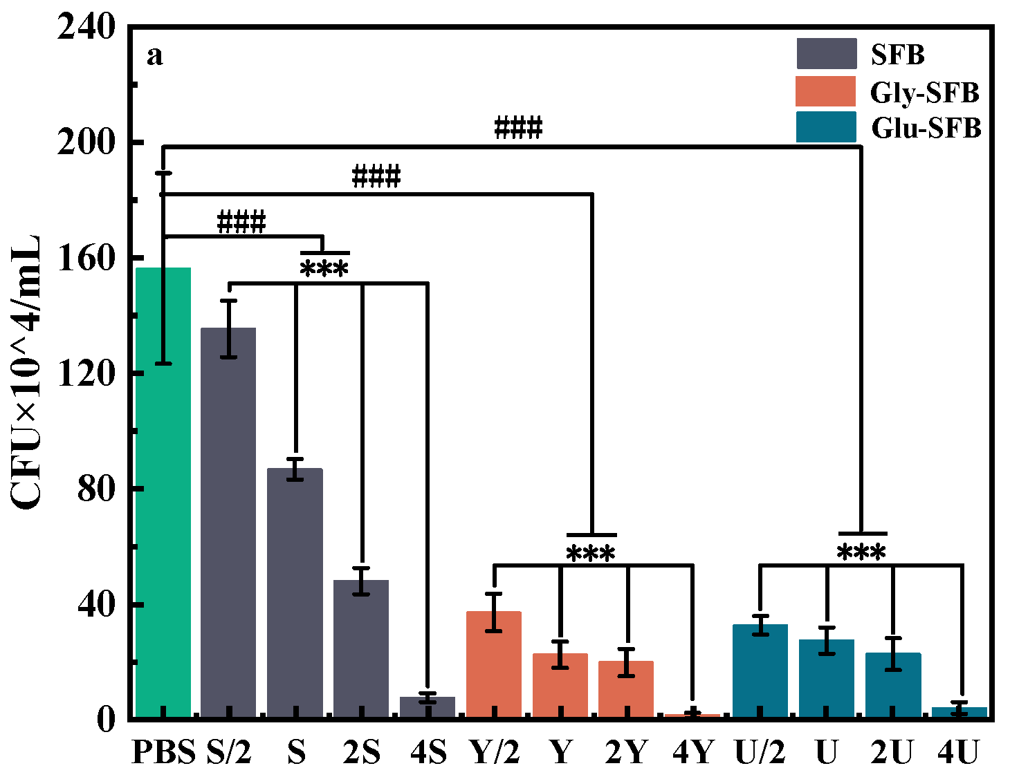
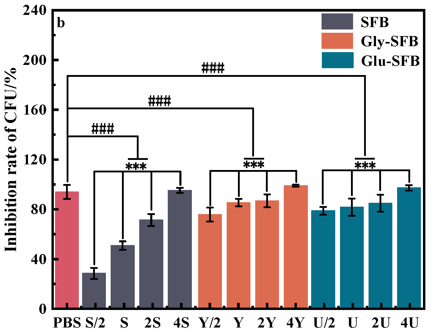
3.1.1. Inhibitory Effect of Three Kinds of S. epidermidis Fermentation Broth on P. acnes

Colony-forming units (CFUs) of P.acnes in PBS as a blank control and in 1/2-fold, 1-fold, 2-fold, and 4-fold SFB (S), Gly-SFB (Y), and Glu-SFB (U), respectively, are shown in Figure 1, and the colony images of P. acnes are presented in Figure S1. Figure 1 shows that SFB, Gly-SFB, and Glu-SFB exhibited differing growth inhibitory effects on P. acnes. Upon exposure to the 1-fold S. epidermidis fermentation broth, the CFUs of P. acnes treated with SFB, Gly-SFB, and Glu-SFB were 86.67 × 10⁴, 22 × 104, and 28 × 104, and the inhibition rates were 44.6%, 85.9%, and 82.1%, respectively. Even when 1/2-fold Gly-SFB or Glu-SFB was used, the inhibiting rate was 76.5% and 78.7%. At the 4-fold level, Gly-SFB demonstrated the highest bacteriostasis, followed by Glu-SFB, while SFB ranked last. However, the inhibiting rate of SFB reached 95%. This showed that Gly-SFB and Glu-SFB exhibited higher inhibitory effects on P. acnes in comparison to SFB. The bacteriostasis of S. epidermidis fermentation broth to P. acnes was ascribed to the metabolites generated by S. epidermidis. Glycerol or glucose as additional carbon sources increased the quantity and diversity of antibacterial substances in the S. epidermidis fermentation broth and enhanced the effectiveness of Gly-SFB and Glu-SFB in inhibiting P. acnes. Consequently, under identical conditions, Gly-SFB and Glu-SFB demonstrated more significant inhibitory effects on the growth of P. acnes than SFB, and all three kinds of S. epidermidis fermentation broth exhibited a clear dose–response relationship.

3.1.2. pH of S. epidermidis Fermentation Broth

The pH of the three kinds of S. epidermidis fermentation broth was determined, and the results are shown in Table 1. All culture media initially have a pH value of 7.0. Throughout the fermentation process, the pH value of SFB exhibited a slight increase, whereas the pH values of Gly-SFB and Glu-SFB demonstrated a substantial reduction. This demonstrated that glucose or glycerol added to the beef protein culture medium produced acidic compounds by S. epidermidis. It was reported that S. epidermidis metabolizes glucose via glycolysis to produce pyruvate. Some pyruvic acids undergo aerobic hydrolysis to provide acetyl CoA, whereas others are transformed anaerobically to lactic acid. Acetyl CoA into the tricarboxylic acid cycle provides succinic acid [20,21,22,23]. Glycerol is firstly phosphorylated by glycerol kinase to produce 3-phosphoglycerate. 3-phosphoglycerate is converted into dihydroxyacetone phosphate by phosphoglycerate dehydrogenase before glycolysis. Consequently, the variation in glycolysis between S. epidermidis utilizing glycerol or glucose accounts for the differing pH values observed in Gly-SFB and Glu-SFB. Lactic acid, succinic acid, and other short-chain fatty acids produced during glycerol or glucose fermentation by S. epidermidis played an important role in antibacterial activities [10,11]. Thus, the production of organic acids led to Gly-SFB and Glu-SFB presenting better inhibitory effects on the growth of P. acnes than those of SFB.

3.1.3. Oligopeptides Analysis with LC-MS/MS

SFB, Gly-SFB, and Glu-SFB were repeatedly prepared six times for parallel experiments. These S. epidermidis fermentation broth were centrifuged and the supernatant was collected for LC-MS/MS analysis [24,25,26]. The oligopeptides with a higher content in Gly-SFB and Glu-SFB than in SFB were determined and identified using mass spectrometry, and the results are shown in Table 2.

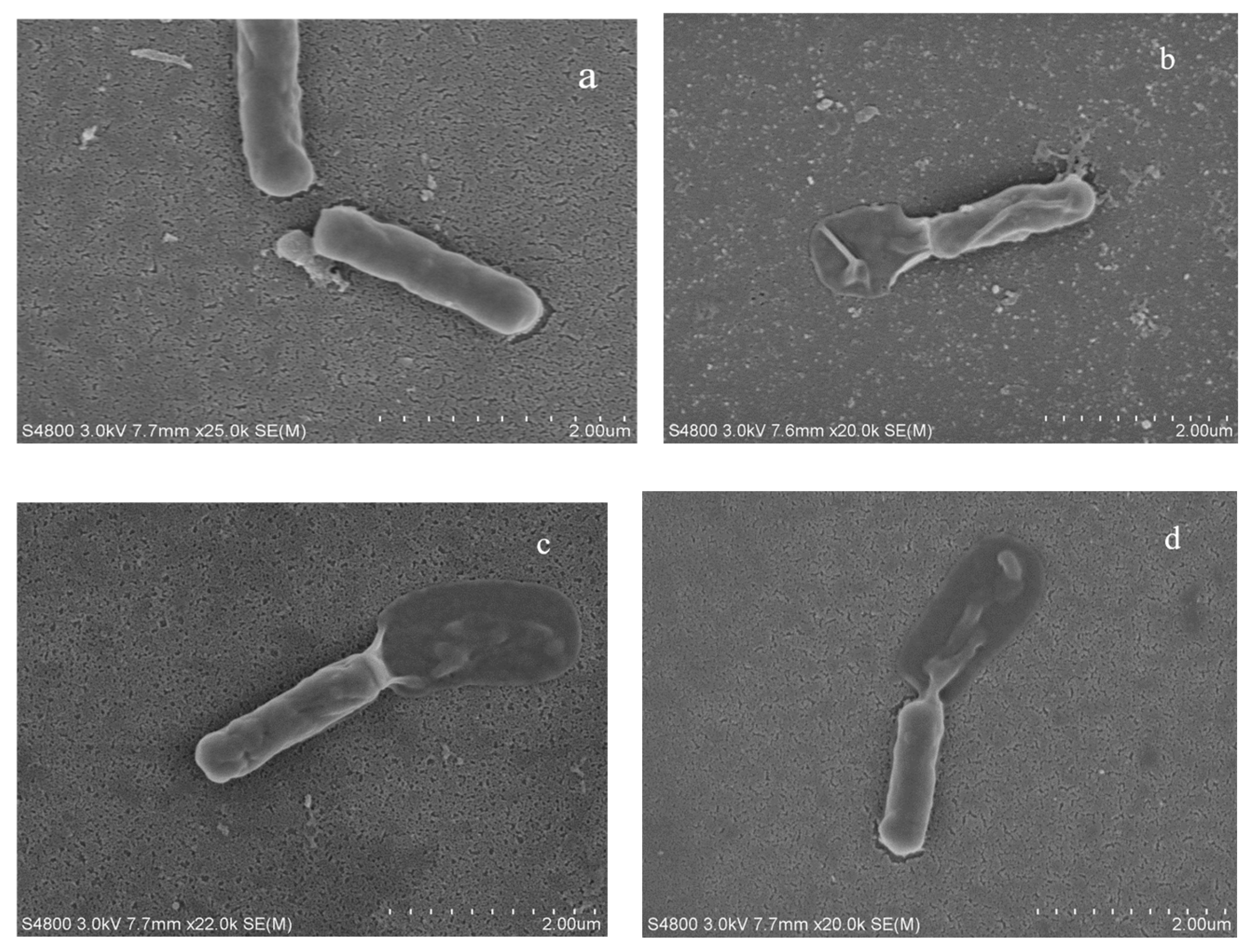
3.1.4. The Impact of Fermentation Broth on Bacterial Morphology

Figure 2 shows the morphology of P. acnes subjected to sterile PBS, SFB, Gly-SFB, and Glu-SFB, respectively. Under normal conditions, P. acnes exhibited a smooth, elongated rod morphology. After treatment with the S. epidermidis fermentation broth, the bacterial cell surface contracted, resulting in the leakage of cellular contents and subsequent bacterial mortality. According to the literature, oligopeptides are amphiphilic substances. The electronegativity of bacterial cell membranes facilitates electrostatic interactions between oligopeptides and membrane surfaces. Subsequently, the bacterial surface is coated with hydrophobic oligopeptides which adhere to lipid molecules, infiltrate the cell membrane, and create circular apertures. Upon decomposition of the bacterial structure, the bacteria expel cellular contents, resulting in their eventual demise [20,21,27]. Simultaneously, glycerol and glucose fermentation by S. epidermidis generates short-chain fatty acids, which can permeate the bacterial cell membrane, increase intracellular osmotic pressure, and induce bacterial lysis and death [10,11]. Consequently, the fermentation broth of S. epidermidis produced the antibacterial substances that inhibit the proliferation of P. acnes, while the fermentation broth prepared by adding glycerol or glucose enhances the inhibitory effect.

3.2. Anti-Inflammation of S. epidermidis Fermentation Broth

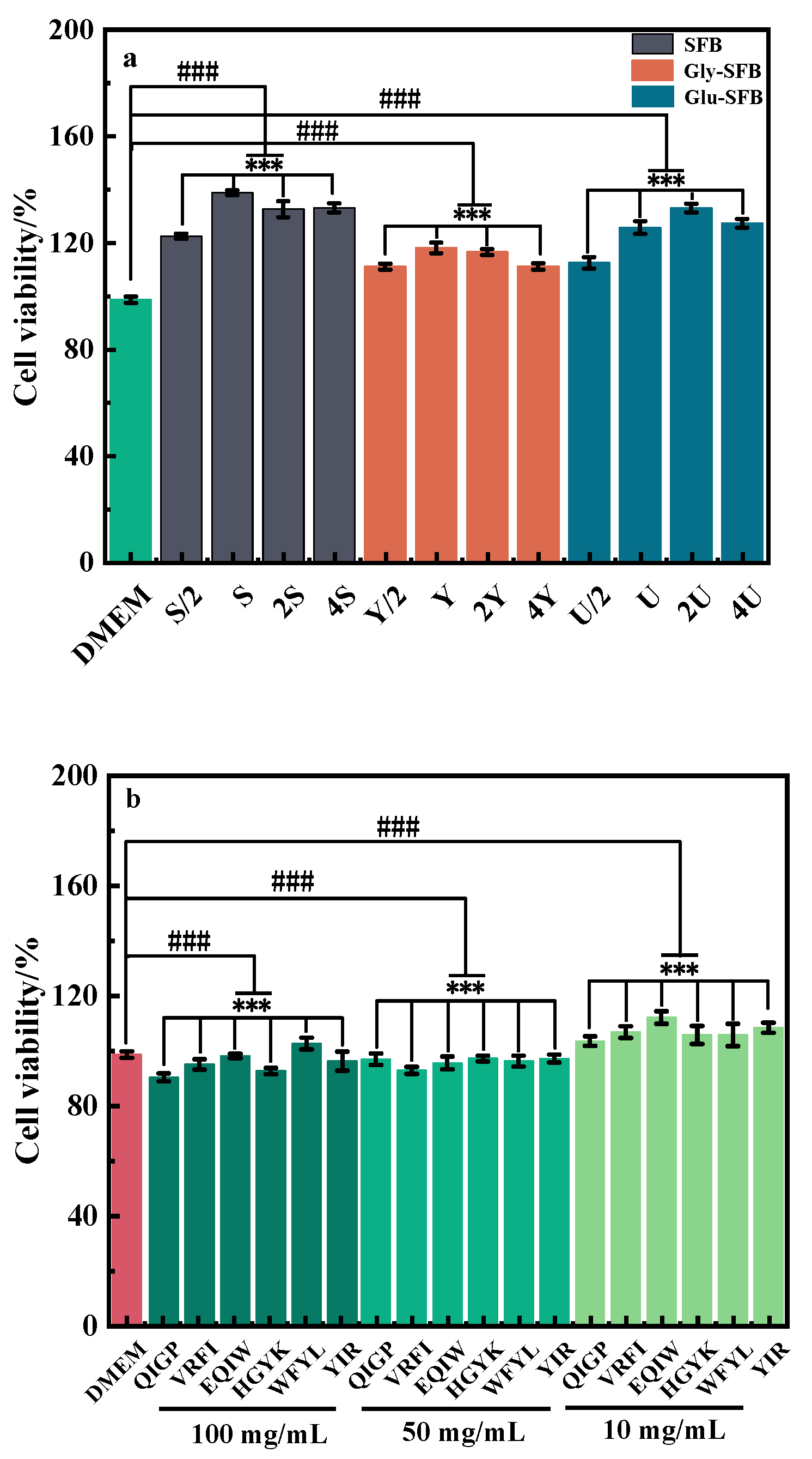
3.2.1. The Influence of Fermentation Broth or Oligopeptides on Cellular Viability
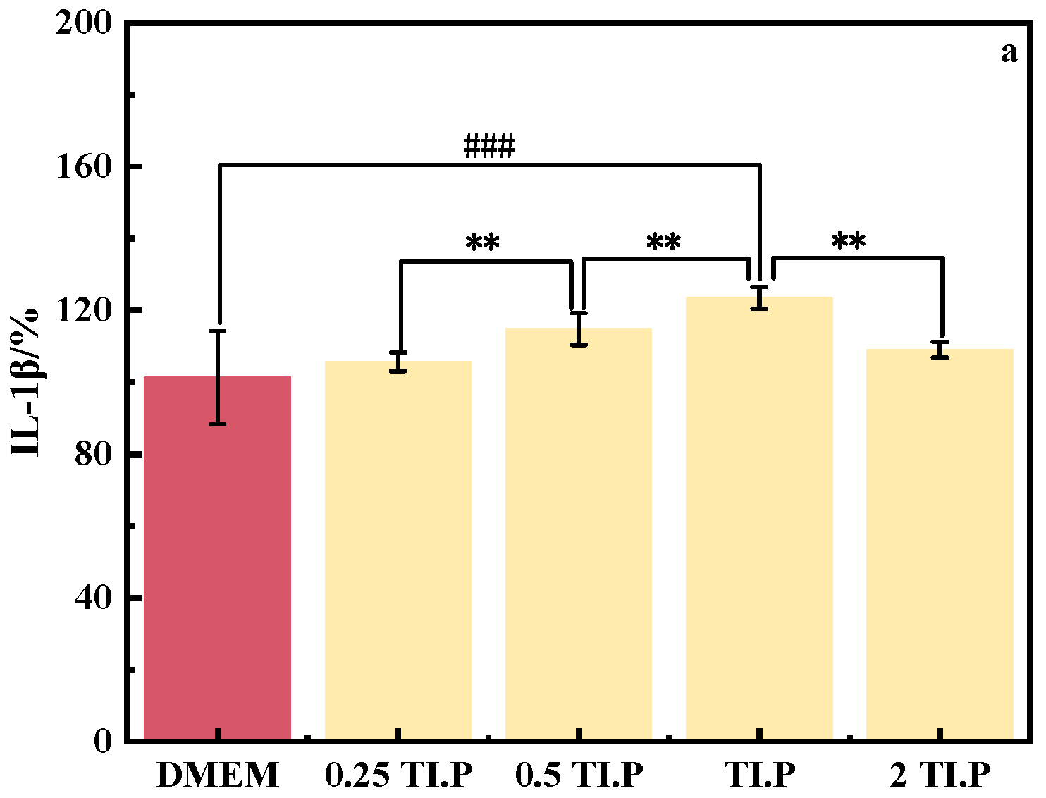
The primary causative agent of the inflammation linked to facial acne is P. acnes. Given their role in inflammation and substantial impact on the formation of anti-inflammatory effects, macrophages are frequently employed as a model to assess the effectiveness of anti-inflammatory therapies. Using the CCK-8 method, the survival rate of RAW264.7 cells in the S. epidermidis fermentation broth was determined to select the appropriate concentration in the anti-inflammatory model. It is widely accepted that a cell survival rate of 80% or more indicates that the additional sample does not influence cell activity and that the sample concentration remains within the permissible limit. Figure 3 demonstrates that, with DMEM serving as a blank control, no types of the fermentation broth reduced cell viability; on the contrary, these even enhanced it. The activity of RAW264.7 cells in SFB was maximized when the fermentation broth was unconcentrated (S). With SFB diluted to a half (1/2S), or concentrated by 2-fold (2S) and 4-fold (4S), cell viability exceeded that of the blank control but was inferior to that in S. The same trend in cell viability was observed in the Gly-SFB treatment. Treated with Glu-SFB, cell viability reached its zenith at 2U. This indicates that three distinct doses of S. epidermidis fermentation broth did not cause any harm to RAW264.7 cells. Following treatment with various doses and types of oligopeptides, the viability of RAW264.7 cells exceeded 80%. At 10 ppm, the cell viability of RAW264.7 exceeded 100% for all eight oligopeptides. Consequently, oligopeptides at this concentration range have no detrimental effects on RAW264.7 cells.

3.2.2. TI.P

Following the protocol detailed in Section 2.8, P. acnes cells were resuspended in different volumes of sterile PBS and subjected to an 80 °C inactivation treatment to attain 0.25 TI.P, 0.5 TI.P, TI.P, and 2 TI.P, respectively. TI.P was obtained by resuspending P. acnes cells in sterile PBS at a 1:4 volume ratio to prepare the sample. Different concentrations of TI.P were added to RAW264.7 cell culture media to evaluate the effect of TI.P on RAW264.7 cell viability, as shown in Figure 4. The release rates of the inflammatory mediators IL-1β and IL-6 were evaluated after stimulation with different quantities of TI.P, as shown in Figure 5. Images of RAW264.7 cells subjected to various amounts of TI.P are included in Figure S2 of the Supplementary Materials. Figure 4 demonstrates that cells treated with 2 TI.P showed a significant decrease in cell viability, which did not meet the requirements. Following treatment with other concentrations of TI.P on RAW264.7, cell viability remained higher than 80%. As the concentration of thermal inactivation increased, the release of inflammatory components by RAW264.7 grew progressively. Consequently, TI.P was selected as the ideal stimulus concentration for the cellular inflammation model.

3.2.3. Anti-Inflammatory Characteristics of S. epidermidis Fermentation Broth

The overgrowth of P. acnes in sebaceous glands is a pathogenic contributor to acne inflammation. Sebum secretion nourishes P. acnes proliferation, which subsequently affects sebaceous glands, leading to keratinization of gland ducts, obstructing sebum excretion, and worsening acne inflammation. The key elements that control the homeostasis of sebaceous glands include hormones, paracrine factors, transcription factors, and signaling pathways, all of which play important roles in the etiology of acne. Therefore, an inflammatory model was created by stimulating RAW264.7 cells with TI.P to induce the production of pro-inflammatory cytokines and elicit an inflammatory response. Figure 6 demonstrates that introducing S/2 and S increased pro-inflammatory cytokine IL-1β produced by RAW264.7 cells by 215 % and 189 %, respectively. The introduction of 2S reduced the release of the pro-inflammatory cytokine IL-1β by 17.2%. A probable explanation was that such low anti-inflammatory substances in SFB were inadequate to suppress the synthesis of IL-1β. As the concentration increased, the anti-inflammatory substances increased to reduce the release of IL-1β. Gly-SFB and Glu-SFB reduced the release of IL-1β. The secretion of IL-1β decreased progressively by 30.1 %, 51.3 %, and 72.0 %, respectively, with increasing Gly-SFB concentration from Y/2, Y, and 2Y. After the injection of Glu-SFB at the concentration of U/2, U, and 2U, the release of IL-1β decreased by roughly 24%, but without a dose-dependent decline. All three kinds of fermentation broths reduced the release of IL-6 at all three concentration levels. As the concentration of SFB increased, IL-6 diminished progressively. Nevertheless, IL-6 subsequently increased as the Gly-SFB and Glu-SFB concentrations increased. Without concentration or dilution, SFB, Gly-SFB, and Glu-SFB reduced the release of IL-6 by 19.4%, 75.4%, and 66.7%, respectively, and Gly-SFB and Glu-SFB presented a better inhibition of IL-6 release than SFB. Therefore, adding glycerol or glucose to the culture medium enriches the variety and abundance of metabolites by S. epidermidis, considerably enhancing the anti-inflammatory activity of S. epidermidis fermentation broth. Glycerol can diminish the release of inflammatory mediators and the migration of leukocytes, hence producing anti-inflammatory actions. Consequently, the anti-inflammatory efficacy of Gly-SFB surpasses that of Glu-SFB.

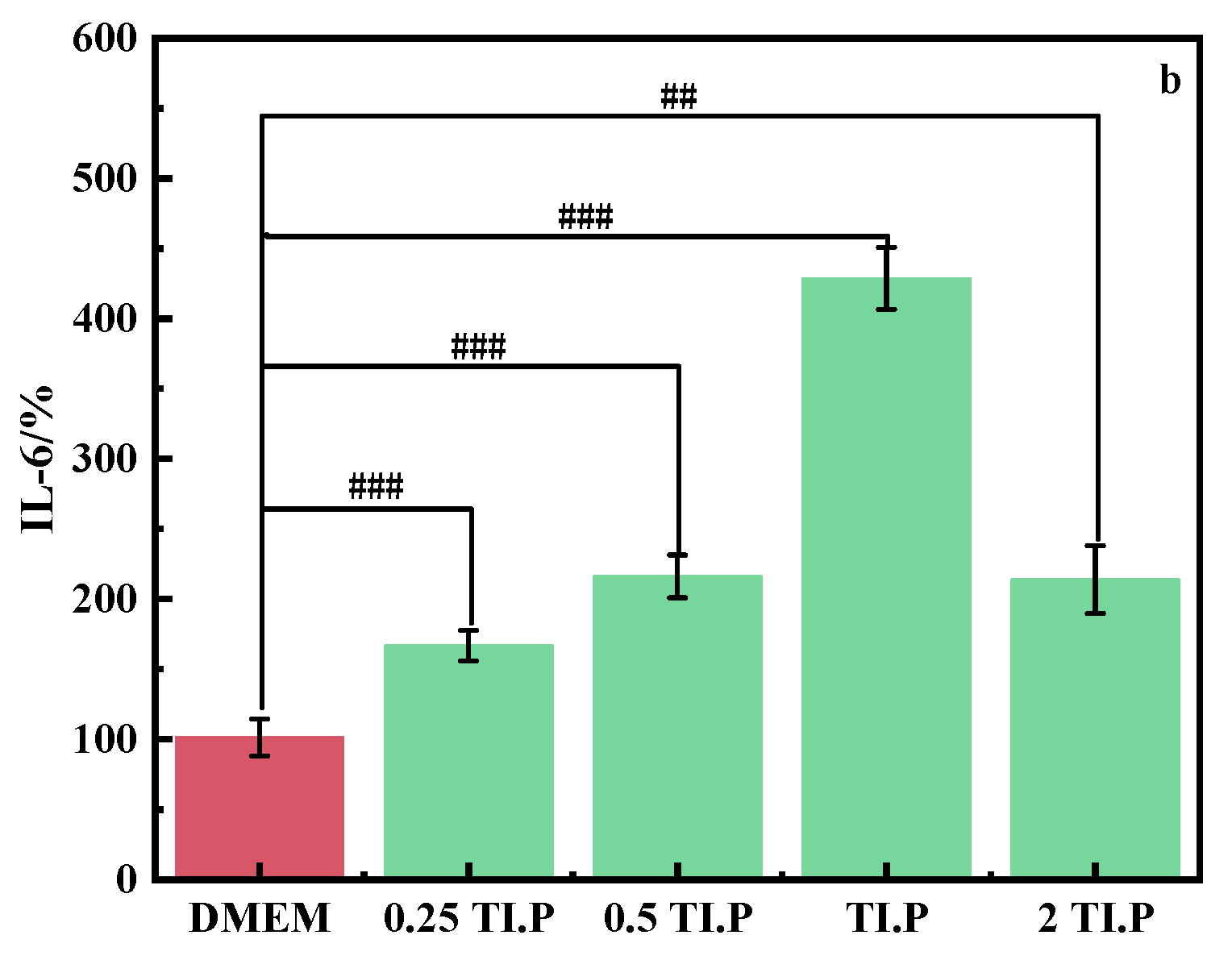
3.2.4. Anti-Inflammatory Characteristics of the Oligopeptides from S. epidermidis Fermentation Broth

LC-MS/MS was employed to investigate oligopeptides in SFB, Gly-SFB, and Gly-SFB, and the total ion chromatograms of three types of fermentation broth are shown in Figure S3. Six oligopeptides, QIGP, VRFI, YIR, EQIW, HGYK, and WFYL, with a higher content in Gly-SFB and Gly-SFB than in SFB were obtained. The experimental findings demonstrate that only three oligopeptides can suppress the release of pro-inflammatory cytokines IL-1 β and IL-6 from RAW264.7 cells induced by TI.P. Figure 7 shows that at doses of 50 ppm and 100 ppm, WFYL, EQIW, and HGYK markedly decreased the expression of inflammatory factors IL-1 β and IL-6. Previous reports [28,29] indicated that peptide sequences comprising proline and phenylalanine residues exhibited anti-inflammatory effects. Isoleucine, alanine, and alanine-leucine possess anti-inflammatory effects and can suppress the overproduction of nitric oxide triggered by lipopolysaccharide activation. FLPPVTSMG and PPYLSP exhibit anti-inflammatory therapeutic properties. In comparison to SFB, Gly-SFB and Glu-SFB exhibited better anti-inflammatory effects, potentially due to the presence of these oligopeptides. In addition, glycerol, which was not entirely consumed during fermentation, facilitated anti-inflammatory responses by inhibiting the movement of leukocytes and the production of inflammatory enzymes. Figure S4 illustrates the consumption rates of glucose and glycerol throughout the fermentation process. The release rates of inflammatory factors following treatment with the other three oligopeptides are illustrated in Figure S4.

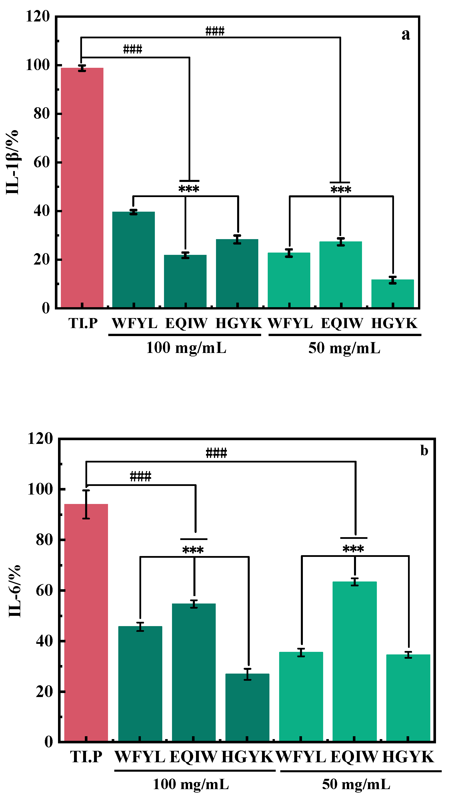
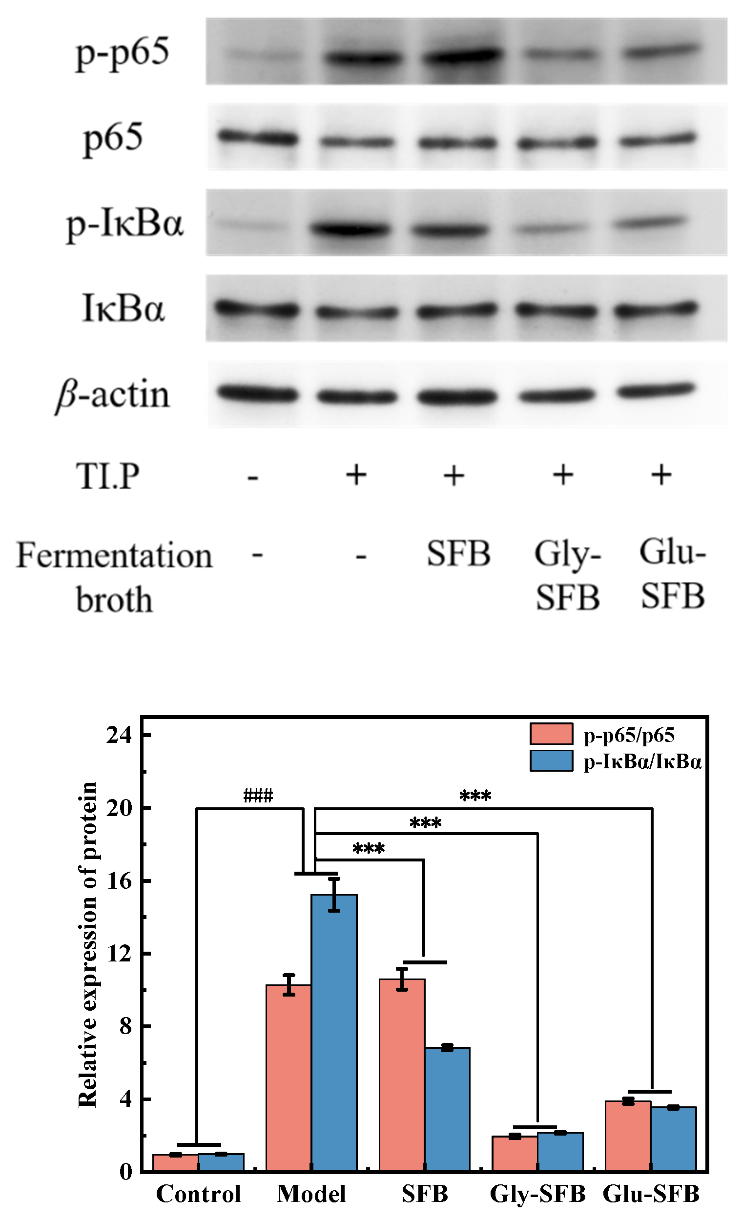
3.2.5. Investigation of Signaling Pathways Related with Inflammation

NF-κB directly interacts with IκB family inhibitory proteins, such as IκBα, in the cytoplasm. Stimulation of cells by inflammatory factors triggers the activation of NF-κB, phosphorylation, degradation of IκB proteins, and the subsequent translocation of phosphorylated NF-κB dimers (p65 and p50) to the nucleus. This translocation triggers the transcription of the target genes, producing various pro-inflammatory stimuli [22,23,30]. In Figure 8, it is shown that TI.P stimulated RAW264.7, resulting in elevated phosphorylation levels of IκBα and p65 in the cells and promoting the synthesis of several inflammatory factors. Furthermore, the addition of SFB led to an increase in p65 phosphorylation and a decrease in IκBα phosphorylation. In contrast, a significant decrease in the phosphorylation levels of both IκBα and p65 occurred after the addition of Gly-SFB and Glu-SFB. These findings indicate that Gly-SFB and Glu-SFB can efficiently decrease the expression of proteins linked to inflammatory factors triggered by TI.P. Consequently, this reduces the production of pro-inflammatory factors and minimizes the occurrence and progression of inflammation. These results align with the outcomes of the previous inflammatory factor assay. Thus, Gly-SFB and Glu-SFB can effectively mitigate inflammation resulting from heat inactivation of P. acnes and lower the expression of proteins associated with pro-inflammatory factors.

4. Conclusions

SFB, Gly-SFB, and Glu-SFB were demonstrated to have dose-dependent inhibitory effects on the proliferation of P. acnes. The inhibitory effect of half of Gly-SFB and Glu-SFB on the growth of P. acnes was stronger than that of 2-fold SFB. Thus, the fermentation broth of S. epidermidis using glycerol or glucose as additional carbon sources in the culture medium enhanced the antibacterial effectiveness against the growth of P. acnes. Gly-SFB and Glu-SFB, as well as 2-fold SFB, markedly reduce the release of pro-inflammatory cytokines IL-1β and IL-6 from RAW264.7 cells induced by TI.P. and exhibited anti-inflammatory properties. The introduction of glycerol or glucose resulted in the production of acidic substances, which exhibit antibacterial characteristics. Six oligopeptides were discovered in Gly-SFB and Glu-SFB with a relatively higher content compared to SFB. Among the six oligopeptides, WFYL, EQIW, and HGYK exhibited notable anti-inflammatory properties. Therefore, the fermentation broth of S. epidermidis prepared by adding glycerol or glucose possessed superior antibacterial properties and markedly enhanced anti-inflammatory effects. Substances with antibacterial activities in Gly-SFB and Glu-SFB might include short-chain fatty acids and oligopeptides, and the latter in the fermentation broth were proved to have effective anti-inflammatory characteristics against inflammation induced by TI.P.

Supplementary Materials

The following supporting information can be downloaded at: https://www.mdpi.com/article/10.3390/cosmetics12020033/s1, Figure S1. The P. acnes strain count with treated by initial culture medium or S. epidermidis fermentation broth, which were diluted to 1.0 × 10⁴ (a) and 1.0 × 10⁵ (b), respectively; Figure S2. The survival status of RAW264.7 cells in the normal condition (a), and following treatment with 0.25 TI.P (b), 0.5 TI.P (c), TI.P (d), and 2 TI.P (e), respectively; Figure S3. The total ion chromatogram of SFB, Gly-SFB and Glu-SFB; Figure S4. Effect of oligopeptides on the protein expressions of NF-κB signaling pathway of RAW 264.7 cells.

Author Contributions

W.G.: Conceptualization, Methodology, Software, Experiments, Data Analysis, Writing—Original Draft. Y.Z.: Investigation: Validation. Y.C.: Conceptualization, Resources, Supervision, Writing—Review & Editing. All authors have read and agreed to the published version of the manuscript.

Funding

This research received no external funding.

Data Availability Statement

Data will be made available on request.

Acknowledgments

This research did not receive any specific grant from funding agencies in the public, commercial, or not-for-profit sectors.

Conflicts of Interest

The authors declare that they have no known competing financial interests or personal relationships that could have appeared to influence the work reported in this paper.

Abbreviation

Staphylococcus epidermidisS. epidermidis
Propionibacterium acnesP. acnes
Thermal inactivation of P. acnesTI.P
The fermentation broth of S. epidermidis cultivated in a beef protein mediumSFB
The fermentation broth of S. epidermidis cultivated in a beef protein medium with adding glycerolGly-SFB
The fermentation broth of S. epidermidis cultivated in a beef protein medium with adding glucoseGlu-SFB
Gas-phase high-throughput time-of-flight mass spectrometerGC-MS
High-performance liquid chromatographyHPLC
Ultra-High-performance liquid chromatography–tandem quadrupole time of flight mass spectrometerHPLC-MS/MS
Field emission scanning electron microscopeSEM

References

  1. Habeshian, K.A.; Cohen, B.A. Current issues in the treatment of acne vulgaris. Pediatrics 2020, 145 (Suppl. 2), 225–230. [Google Scholar] [CrossRef] [PubMed]
  2. Clayton, R.W.; Göbel, K.; Niessen, C.M.; Paus, R.; Van Steensel, M.A.; Lim, X. Homeostasis of the sebaceous gland and mechanisms of acne pathogenesis. Br. J. Dermatol. 2019, 181, 677–690. [Google Scholar] [CrossRef] [PubMed]
  3. Taghizadeh, M.S.; Taherishirazi, M.; Niazi, A.; Afsharifar, A.; Moghadam, A. Structureguided design and cloning of peptide inhibitors targeting CDK9/cyclin T1 proteinprotein interaction. Front. Pharmacol. 2024, 15, 1327820. [Google Scholar] [CrossRef] [PubMed]
  4. Callender, V.D.; Baldwin, H.; Cook-Bolden, F.E.; Alexis, A.F.; Gold, L.S.; Guenin, E. Effects of topical retinoids on acne and post-inflammatory hyperpigmentation in patients with skin of color: A clinical review and implications for practice. Am. J. Clin. Dermatol. 2022, 23, 69–81. [Google Scholar] [CrossRef] [PubMed]
  5. April, W.A.; Joshua, H.; Leon, H.K. Oral Tetracyclines and acne: A systematic review for dermatologists. J. Drugs Dermatol. 2020, 19, 6–13. [Google Scholar]
  6. Loomis, K.H.; Wu, S.K.; Ernlund, A.; Zudock, K.; Reno, A.; Blount, K.; Karig, D.K. A mixed community of skin microbiome representatives influences cutaneous processes more than individual members. Microbiome 2021, 9, 22. [Google Scholar] [CrossRef]
  7. Segre, J.A. Epidermal barrier formation and recovery in skin disorders. Clin. Investig. 2006, 116, 1150–1158. [Google Scholar] [CrossRef]
  8. Di Meglio, P.; Perera, G.K.; Nestle, F.O. The multitasking organ: Recent insights into skin immune function. Immunity 2011, 35, 857–869. [Google Scholar] [CrossRef]
  9. Proksch, E.; Brandner, J.M.; Jensen, J.-M. The skin: An indispensable barrier. Exp. Dermatol. 2008, 17, 1063–1072. [Google Scholar] [CrossRef]
  10. Xia, X.; Li, Z.; Liu, K.; Wu, Y.; Jiang, D.; Lai, Y. Staphylococcal LTA-Induced miR-143 Inhibits P. acnes Mediated Inflammatory Response in Skin. J. Investig. Dermatol. 2016, 136, 621–630. [Google Scholar] [CrossRef]
  11. Geyfman, M.; Debabov, D.; Poloso, N.; Alvandi, N. Mechanistic insight into the activity of a sulfone compound dapsone on Propionibacterium (Newly Reclassified as Cutibacterium) Acnes-mediated cytokine production. J. Exp. Dermatol. 2019, 28, 190–197. [Google Scholar] [CrossRef] [PubMed]
  12. Wang, Y.; Kao, M.-S.; Yu, J.; Huang, S.-H.; Marito, S.; Gallo, R.L.; Huang, C.-M. A Precision Microbiome Approach Using Sucrose for Selective Augmentation of S. epidermidis Fermentation against P. acnes. Int. J. Mol. Sci. 2016, 17, 1870. [Google Scholar] [CrossRef]
  13. Wei, J. Study on the Combination of Traditional Chinese Medicine Extracts for Inhibiting Acne Inflammation with Multi-Targets. Ph.D. Dissertation, Jiangnan University, Wuxi, China, 2020. [Google Scholar]
  14. Jan, A.T. Outer Membrane Vesicles (OMVs) of Gram-negative Bacteria: A Perspective Update. Front. Microbiol. 2017, 8, 1053. [Google Scholar] [CrossRef]
  15. Choi, E.J.; Lee, H.G.; Bae, I.H.; Kim, W.; Park, J.; Lee, T.R.; Cho, E.-G. Propionibacterium acnes-Derived Extracellular Vesicles Promote Acne-Like Phenotypes in Human Epidermis. J. Investig. Dermatol. 2018, 138, 1371–1379. [Google Scholar] [CrossRef]
  16. Zhao, Y.; Su, R.; Zhang, W.; Yao, G.-L.; Chen, J. Antibacterial activity of tea saponin from Camellia oleifera shell by novel extraction method. Ind. Crops Prod. 2020, 153, 112604. [Google Scholar] [CrossRef]
  17. Yu, Q.; Pan, H.; Shao, H.; Qian, C.; Han, J.; Li, Y.; Lou, Y. UPLC/MS-based untargeted metabolomics reveals the changes in muscle metabolism of electron beam irradiated Solenocera melancholy during refrigerated storage. Food Chem. 2022, 367, 130713. [Google Scholar] [CrossRef]
  18. Xiao, Y.; Pan, Z.P.; Yin, C.X.; Su, J.; Hu, X.; Zhu, X.R. Antifungal activity and mechanism of ε-polylysine against Geotrichum citriaurantii. Food Sci. 2020, 41, 221–229. [Google Scholar]
  19. Lim, H.-J.; Kang, S.-H.; Song, Y.-J.; Jeon, Y.D.; Jin, J.S. Inhibitory Effect of Quercetin on Propionibacterium acnes-induced Skin Inflammation. Int. Immunopharmacol. 2021, 96, 107557. [Google Scholar] [CrossRef]
  20. Ladram, A. Antimicrobial peptides from frog skin biodiversity and therapeutic promises. Front. Biosci. 2013, 21, 1341–1371. [Google Scholar] [CrossRef]
  21. Lei, J.; Sun, L.; Huang, S.; Zhu, C.; Li, P.; He, J. The antimicrobial peptides and their potential clinical applications. Am. J. Transl. Res. 2019, 11, 3919–3931. [Google Scholar]
  22. Jeong, H.-Y.; Choi, Y.-S.; Lee, J.-K.; Lee, B.J.; Kim, W.K.; Kang, H. Anti-inflammatory activity of citric acid-treated wheat germ extract in lipopolysaccharide-stimulated macrophages. Nutrients 2017, 9, 730. [Google Scholar] [CrossRef] [PubMed]
  23. Baldwin, S.A. The NF-κB and IκB proteins: Discoveries and insights. Annu. Rev. Immunol. 1996, 14, 649–681. [Google Scholar] [CrossRef] [PubMed]
  24. Aminov, R. Metabolomics in antimicrobial drug discovery. Expert Opin. Drug Discov. 2022, 17, 1047–1059. [Google Scholar] [CrossRef] [PubMed]
  25. Zeki, O.C.; Eylem, C.C.; Recber, T.; Kır, S.; Nemutlu, E. Integration of GC-MS and LC-MS for untargeted metabolomics profiling. J. Pharm. Biomed. Anal. 2020, 190, 113509. [Google Scholar] [CrossRef]
  26. Chen, C.; Gonzalez, F.J.; Idle, J.R. LC-MS-based metabolomics in drug metabolism. Drug Metab. Rev. 2007, 39, 581–597. [Google Scholar] [CrossRef]
  27. Morave, J.H.; Morave, J.Z.; Yazdanparast, Z.; Heiat, M.; Mirhosseini, A.; Moosazadeh Moghaddam, M.; Mirnejad, R. Antimicrobial peptides: Features, action, and their resistance mechanisms in bacteria. Microb. Drug Resist. 2018, 24, 747–767. [Google Scholar] [CrossRef]
  28. Hu, F.X. Extraction, Purification, and Identification of Polyphenols from Chaenomeles Speciosa (Sweet)Nakai and Its Antioxidant and Anti-Inflammatory Activities. Ph.D. Dissertation, Shandong Agricultural University, Tai’an, China, 2022. [Google Scholar]
  29. Ji, Z.W. Study on the Preparation of Foxtail Millet Prolamins Peptide and Its Antioxidant and Anti-Inflammatory Activities. Ph.D. Dissertation, Jiangnan University, Wuxi, China, 2020. [Google Scholar]
  30. Ratan, Z.A.; Jeong, D.; Sung, N.Y.; Shim, Y.Y.; Cho, J.Y. Lomix, a mixture of flaxseed linusorbs, exerts anti-inflammatory effects through src and sky in the NF-κB pathway. Biomolecules 2020, 10, 843–859. [Google Scholar] [CrossRef]
Figure 1. CFUs of P. acnes (a) and inhibitory rate (b) in PBS and S. epidermidis fermentation broths. Note: Compared with control, ### p < 0.001; compared in sample group: *** p < 0.001.
Cosmetics 12 00033 g001aCosmetics 12 00033 g001b
Figure 2. SEM of P. acnes in sterile PBS (a), treated with SFB (b), Gly-SFB (c), and Glu-SFB (d).
Cosmetics 12 00033 g002
Figure 3. The survival rate of RAW264.7 cells under treatments by 10% S. epidermidis fermentation broths (a) or oligopeptide (b) for an incubation time of 24 h. Note: Compared with control, ### p < 0.001; compared in sample group: *** p < 0.001.
Cosmetics 12 00033 g003
Figure 4. The survival rate of RAW264.7 cells under treatments by different concentrations of thermal inactivation of P. acnes. ** p < 0.05.
Cosmetics 12 00033 g004
Figure 5. Expression of inflammatory factors IL-1β (a) and IL-6 (b) treated with varying doses of thermal inactivation of P. acnes. Note: compared with control, ### p < 0.001, ## p < 0.05; compared in sample group: ** p < 0.05.
Cosmetics 12 00033 g005aCosmetics 12 00033 g005b
Figure 6. Expression of inflammatory factors IL-1β (a) and IL-6 (b) treated with varying types and doses of S. epidermidis fermentation broth. Note: Compared with control, ### p < 0.001; Compared in sample group: ** p < 0.05, *** p < 0.001.
Cosmetics 12 00033 g006
Figure 7. Expression of inflammatory factors IL-1β (a) and IL-6 (b) treated with varying types and doses of oligopeptides. Note: Compared with control, ### p < 0.001; Compared in sample group: *** p < 0.001.
Cosmetics 12 00033 g007
Figure 8. Effect of S. epidermidis fermentation broth on the protein expressions of NF-κB signaling pathway of RAW 264.7 cells. Note: Compared with control, ### p < 0.001; Compared with model group, *** p < 0.001.
Cosmetics 12 00033 g008
Table 1. pH of S. epidermidis fermentation broths.
SFBGly-SFBGlu-SFB
pH8.044.914.49
Table 2. Identification of oligopeptides in SFB, Glu-SFB, and Gly-SFB with LC-MS/MS.
No.Retention Time (min)m/zIdentificationNormalized Abundance
SFBGly-SFBGlu-SFB
15.16650.2939TRP-PHE-TYR-LEU003909.597
25.22414.2388GLN-ILE-GLY-PRO2951.953510.194259.72
35.72556.3145VAL-ARG-PHE-ILE1665.332154.252634.44
45.86783.4417TYR-ILE-ARG4696.135595.516950.33
58.011149.56GLU-GLN-ILE-TRP9097.6012,044.5713,739.04
68.71007.52HIS-GLY-TYR-LYS10,670.7011,213.5814,142.54
Disclaimer/Publisher’s Note: The statements, opinions and data contained in all publications are solely those of the individual author(s) and contributor(s) and not of MDPI and/or the editor(s). MDPI and/or the editor(s) disclaim responsibility for any injury to people or property resulting from any ideas, methods, instructions or products referred to in the content.

