Article

Nicotinamide Riboside Ameliorates Hyperpigmentation on Photo-Irradiated Skin

Joonghun Pharmaceutical Co., Ltd., 15 Gukhoe-daero 62-gil, Yeongdeungpo-gu, Seoul 07236, Republic of Korea
*
Author to whom correspondence should be addressed.
Cosmetics 2024, 11(3), 73; https://doi.org/10.3390/cosmetics11030073
Submission received: 18 March 2024 / Revised: 16 April 2024 / Accepted: 1 May 2024 / Published: 5 May 2024

Abstract

:
Nicotinamide adenine dinucleotide (NAD) is one of the most important and essential components within an organism. Extensive ongoing research is aimed at harnessing its potential in managing diverse diseases by supplying various forms of NAD in its oxidized state, NAD+. Ultraviolet radiation (UVR) is the most common environmental exposure factor, but also carries many risks. UVR affects the epidermis and contributes to sunburn, photo-allergy, DNA damage, and certain cancers, notably melanoma. Research has shown that NAD+ precursors, including nicotinamide riboside (NR), reduce melanogenesis in aged melanocytes. In this study, we used NR to determine whether melanin hyperpigmentation was suppressed after light stimulation. We found that melanogenesis was inhibited when B16F10 cells treated with α-melanocyte-stimulating hormone were exposed to specific doses of NR. Additionally, tyrosinase activity (a key step in melanin production) was suppressed. However, there was no difference in the expression level of melanogenic genes. Ultraviolet B light directly stimulated HaCaT cells, inducing the RNA expression of metalloproteinases. Treatment with NR suppressed the corresponding gene expression and reduced cytotoxicity. This study demonstrates the possibility of using NR as a new skin-whitening ingredient due to its inhibitory effect on hyperpigmentation and ability to maintain skin layers affected by UVR.

1. Introduction

With the progression of aging or exposure to ultraviolet radiation (UVR), the skin becomes increasingly damaged and signs of aging appear. Skin hyperpigmentation may develop in such individuals. Melanin plays an important role in protecting the skin from UVR-induced damage [1]. However, severe pigmentation resulting in conditions such as age spots, melasma, and solar lentigines signifies an abnormal condition [2]. Therefore, people often seek remedies to ameliorate pigmentation concerns through approaches such as cosmetics, medication, and laser therapy.
Excessive exposure to UVR, particularly ultraviolet B (UVB) light, adversely affects the epidermal layer, especially keratinocytes, which secrete signaling factors such as α-melanocyte-stimulating hormone (α-MSH) [3]. These secreted α-MSH molecules initiate the melanogenic pathway. In humans, melanin exists in two primary forms: eumelanin, which produces black and brown pigmentation; and pheomelanin, which produces red and yellow pigmentation [4,5].
Hyperpigmentation is an example of a condition caused by the excessive secretion or deposition of melanin. Hyperpigmentation refers to the phenomenon of making the skin relatively dark compared to its normal state. Hyperpigmentation may occur due to a deficiency of vitamin B12 or folic acid, as well as photo-stimulation, or be caused by hereditary, hormonal changes such as pregnancy, inflammation, skin injuries, age, or the effects of some medications [6,7,8].
There are two important control points in the regulation of melanogenesis. The first is stimulation-induced melanogenic gene regulation. Several important genes are involved in this process: microphthalmia-associated transcription factor (mitf), tyrosinase-related protein 1 (trp1), tyrosinase-related protein 2 (trp2), and tyrosinase (tyr). The mitf gene regulates the transcription of melanogenic genes such as trp1, trp2, and tyr through activation of the α-MSH-induced cyclic adenosine monophosphate (cAMP) response element-binding protein (CREB) [9,10]. The second control point in melanogenesis regulation involves tyrosinase. Tyrosinase is a key enzyme that initiates melanin production, catalyzing the oxidation of L-tyrosine to L-dihydroxyphenylalanine (L-Dopa) and L-Dopa to L-dopaquinone [4,11]. Therefore, controlling tyrosinase is an essential target in controlling pigmentation, but few compounds are known to safely inhibit this enzyme [12].
Nicotinamide riboside (NR) is a natural nutrient present in human foods such as milk. After the discovery of NR as an NAD+ precursor (Bieganowski and Brenner, 2004), Brenner conducted research on NAD+ and clinical trials for various diseases [13]. No serious side effects such as flushing, pruritus, hyperglycemia, hyperuricemia, or elevations of liver or muscle enzymes, as seen with similar doses of niacin, have been reported [14]. NR is nontoxic (up to at least 2000 mg daily in adult humans) and is also expected to exhibit low passive permeability across the human intestinal mucosa [15,16]. According to another study by Brenner (2014), when the levels of metabolites were checked through nicotinamide (Nam) or NR gavage, oral NR showed significantly distinct hepatic pharmacokinetics compared to oral Nam. Additionally, more NAD+ and NADP+ were produced in oral NR than in oral Nam [17]. Nowadays, people use it as a health supplement [18]. NR has been found to ameliorate tissue nicotinamide adenine dinucleotide (NAD+) depletion induced by acute kidney injury via increases in autophagy and SIRT1. The induction of NAD+ has protective effects against numerous conditions, including diabetes, obesity [19], neurodegenerative diseases, and noise-induced hearing loss [20,21,22]. Lapato et al. reported that NR supplementation enhances muscle mitochondrial biogenesis [23]. However, while numerous studies have investigated the bio-maintenance effects of NR supplementation, only a few have focused on direct pigmentation. Unlike other precursors, NR has no side effects, such as skin redness. In a study by Martens et al., symptoms reported during the placebo treatment period included nausea, flushing, leg cramps, bruising, headache, skin rash, flushing, syncope, and drowsiness; in contrast, no adverse reactions occurred during NR treatment [24]. These results demonstrate the safety of NR as a source of NAD+.
This study was conducted to determine whether NR can improve melanin production in skin damaged by photo-stimulation. To create conditions similar to the skin environment damaged by photo-stimulation, we treated the B16F10 cell line, a melanocyte, with a-MSH and examined whether NR inhibits melanin production produced by this stimulation. In addition, we directly irradiated the HaCaT cell line, a keratinocyte, with UVB, and looked at the effect of NR on the cells. Through these results, we hope that these results will show the potential of NR as a new and safe treatment for skin damage caused by photo-stimulation.

2. Materials and Methods

2.1. Chemicals

NR was purchased from Selleck Chemicals (Houston, TX, USA). Arbutin, α-MSH, mushroom tyrosinase, L-tyrosine, and hydrogen peroxide were purchased from Sigma (St. Louis, MO, USA).

2.2. Cell Culture and Chemical Treatments

The mouse melanoma cell line B16F10 (ATCC, CRL-6475) was maintained in Dulbecco’s modified Eagle’s medium (DMEM) with 10% fetal bovine serum and 1% penicillin and streptomycin at 37 °C with 5% CO2. The cells were serially subcultured at 70–80% confluence. During all experiments with B16F10 cells, the cells were seeded at a density of 7 × 10⁴ cells/dish, in 35 mm dishes. At 24 h after seeding, NR was added daily for indicated incubation times. Arbutin (100 µM) as a positive control and α-MSH (100 nM) were added only once, at 24 h after seeding.
The human keratinocyte cell line HaCaT was kindly provided by Professor Gi-Ryang Kweon of Chungnam National University. HaCaT cells were maintained in DMEM with 10% fetal bovine serum and 1% antibiotics at 37 °C with 5% CO2. The cells were passaged every 2–3 days. They were seeded at a density of 3 × 10⁵ cells/well, in 6-well plates for viability testing and 12-well plates for quantitative polymerase chain reaction (QPCR).
The HaCaT cell line was only used in some supplementary figure parts (Figures S2 and S3): the cells used in all other experiments were the B16F10 cell line.

2.3. UVB Exposure

Twenty-four hours after seeding, as previously mentioned, the HaCaT cell plate was washed twice with 1× Dulbecco’s phosphate-buffered saline (DPBS), replaced with 1 mL of 1× DPBS, and exposed to the indicated amounts of UVB. UVB exposure experiments were conducted using a Bio-Link (VILBER, Marne-la-vallée, France) UV irradiation machine. The intensity of UVB was set to 34 mJ, and UVB irradiation was performed by opening the lid of the cell plate containing 1 mL of 1× DPBS based on a 6-well plate. The distance between the lamp and the plate was about 13 cm. After UVB irradiation, the 1× DPBS solution was replaced with culture medium with or without other treatments and cultured for 24 h.

2.4. Cell Viability Assay (CCK-8)

The cck8 experiments were performed in supplementary sections using B16F10 cells (Figure S1) and HaCaT cells (Figure S2). After the experimental time, CCK8 solution was diluted 10-fold with new culture medium. The diluted medium was placed into a culture plate and incubated for 1 h at 37 °C. The absorbance was then measured at 450 nm using a microplate reader.

2.5. Cell Sulforhodamine B (SRB) Assay

Upon completing the experiment, the media were discarded and the cells were washed twice with cold 1× DPBS. The cells were fixed with 1 volume of cold 10% trichloroacetic acid solution for 20 min at room temperature. The solution was discarded, and the cells were washed three times with distilled water. The solution was then changed to 1 volume of 0.4% SRB for 20 min at room temperature. After incubation, the cells were washed twice with 1% acetic acid solution and dried. Next, 500 µL of 10 mM unbuffered Tris solution was added, and the absorbance was read at 564 nm with a FLUOstar Omega microplate reader (BMG Labtech, Ortenberg, Germany).

2.6. Melanin Contents

For confirmation of extracellular melanin, the cells were cultured with phenol-red-free medium. After the experiment, the culture medium was centrifuged at 330× g for 5 min to remove debris. For confirmation of intracellular melanin, the cells were washed twice with 1× DPBS. After discarding the solution, the cells were lysed in 1 N NaOH for 60 min at 60 °C. Absorbance was measured at 490 nm using a microplate reader (BMG Labtech, Ortenberg, Germany) and normalized based on the protein concentration quantified with a bicinchoninic acid (BCA) assay.

2.7. Mushroom Tyrosinase Inhibition Assay

Mushroom tyrosinase activity was assessed using L-tyrosine. In a 96-well plate (SPL Life Sciences, Pocheon, Korea), a mixture containing 110 µL of 0.1 M phosphate buffer (pH 6.5), 10 µL of 1.5 mM L-tyrosine, 20 µL of 1500 U/mL mushroom tyrosinase, and 10 µL of NR (or water) was prepared, totaling 300 µL. After incubation for 15 min at 37 °C, the absorbances were checked at 490 nm. The tyrosinase inhibition rate was calculated using the equation shown below.
tyrosinase inhibitory activity (%) = 100 − ((ODb-b’/ODa-a’) × 100)
a = vehicle with tyrosinase, a’ = vehicle with buffer
b = chemical with tyrosinase, b’ = chemical with buffer

2.8. Quantitative Polymerase Chain Reaction (QPCR)

The expressions of melanogenic genes such as tyr, trp-1, trp-2, and mitf genes (B16F10 cells) and metalloproteinases, mmp1 and mmp9 (HaCaT cells) were determined by real-time PCR using 18s rRNA as an internal positive control. Cells were seeded at a density of 7 × 10⁴ cells/dish in 35 mm dishes with the B16B10 cell line and 2 × 10⁵ cells/well, in 12-well plates with the HaCaT cell line. After 24 h of seeding, cells were treated with indicated conditions (incubation times were 7 h for B16F10 cells and 24 h for HaCaT cells). The cells were lysed using Trizol reagent (Invitrogen, Carlsbad, CA, USA), followed by total RNA extraction using chloroform, isopropanol, and 75% ethanol. The concentration of total RNA was measured using UV–VIS spectrophotometry (BioDrop). cDNA was synthesized from 2 µg of RNA using M-MLV reverse transcriptase (Invitrogen Life technologies). Real-time quantitative polymerase chain reaction was carried out with an Exicycler™ 96 Real-Time Quantitative Thermal Block (Bioneer Corporation, Daejeon, South Korea) using TB Green® Premix Ex Taq™ II (Tli RNaseH Plus) and ROX Plus (Takara Bio, Shiga, Japan). Primers were purchased from Bioneer Corporation, and the sequences were as follows: m-mitf forward: 5′-ctgatggacgatgccctctc-3′ and reverse: 5′-tccgtttcttctgcgctcat-3′; m-tyr forward: 5′- ccattttcctcgagcctgtg-3′ and reverse: 5′-agatggtgcactggacagaa-3′; m-trp1 forward: 5′-ggtggaccaatcaggaga-3′ and reverse: 5′-cgaaaactgtctgtagaattgg-3′; m-trp2 forward: 5′-gctgaacaaggaatgctgcc-3′ and reverse: 5′-tcacaggtcatcctggtttcg-3′; h-mmp1 forward: 5′-aggtctctgagggtcaagca-3′ and reverse: 5′-ctggttgaaaagcatgagca-3′; h-mmp9 forward: 5′-catcgtcatccagtttggtgt-3′ and reverse: 5′-agggaccacaactcgtcatc-3′; 18s ribosomal RNA forward: 5′-ctggttgatcctgccagtag-3′; reverse: 5′-cgaccaaaggaaccataact-3′.

2.9. Statistical Analysis

All statistical analyses were performed using GraphPad Prism 8 (GraphPad Software, La Jolla, CA, USA), and values are expressed as the mean ± standard error of triplicate experiments. Experimental groups were compared using the unpaired t-test and one-way analysis of variance. Mean differences are expressed as * p < 0.05, ** p < 0.01, *** p < 0.001, and **** p < 0.0001.

3. Results

3.1. Effect of NR on Cell Survival

To determine the concentration of NR to use for the experiment, we performed a Cell Counting Kit 8 assay (Dojindo Laboratories, Kumamoto, Japan) and found that 0.5–1.0 mM NR had no effect on B16F10 cell viability (Figure S1).
In subsequent experiments, NR affected NAD+ utilization, prompting us to verify its effects using the sulforhodamine B (SRB) method instead of the CCK8 assay, which relies on NAD+. NR is well known enough to be easily converted to nicotinamide (NAM) [25,26]; therefore, it was administered daily for specific durations. Following 48 h of treatment, cytotoxicity was assessed using the SRB assay [27] at various concentrations of NR.
The concentration of a-MSH used in all B16F10 cell experiments ranged from 100 nM to 500 nM in preliminary experiments; 100 nM was used as the minimum concentration that showed an increase in melanin both inside and outside the cells, and it is the concentration used in many studies [28,29,30,31]. In preliminary experiments, in order to use a concentration that shows melanin synthesis inhibition effects without showing cytotoxicity, arbutin was treated from 100 µM to 1 mM to check cytotoxicity and melanin amounts, and it was confirmed that the concentration of 100 µM arbutin was appropriate (data not shown). Additionally, it was confirmed that this concentration was widely used in several studies [30,32,33]. We found no significant differences in NR treatment at the selected doses in α-MSH-treated B16F10 cells (Figure 1), confirming the concentration to be used in subsequent experiments.

3.2. Decrease in α-MSH-Induced Melanin Content by NR

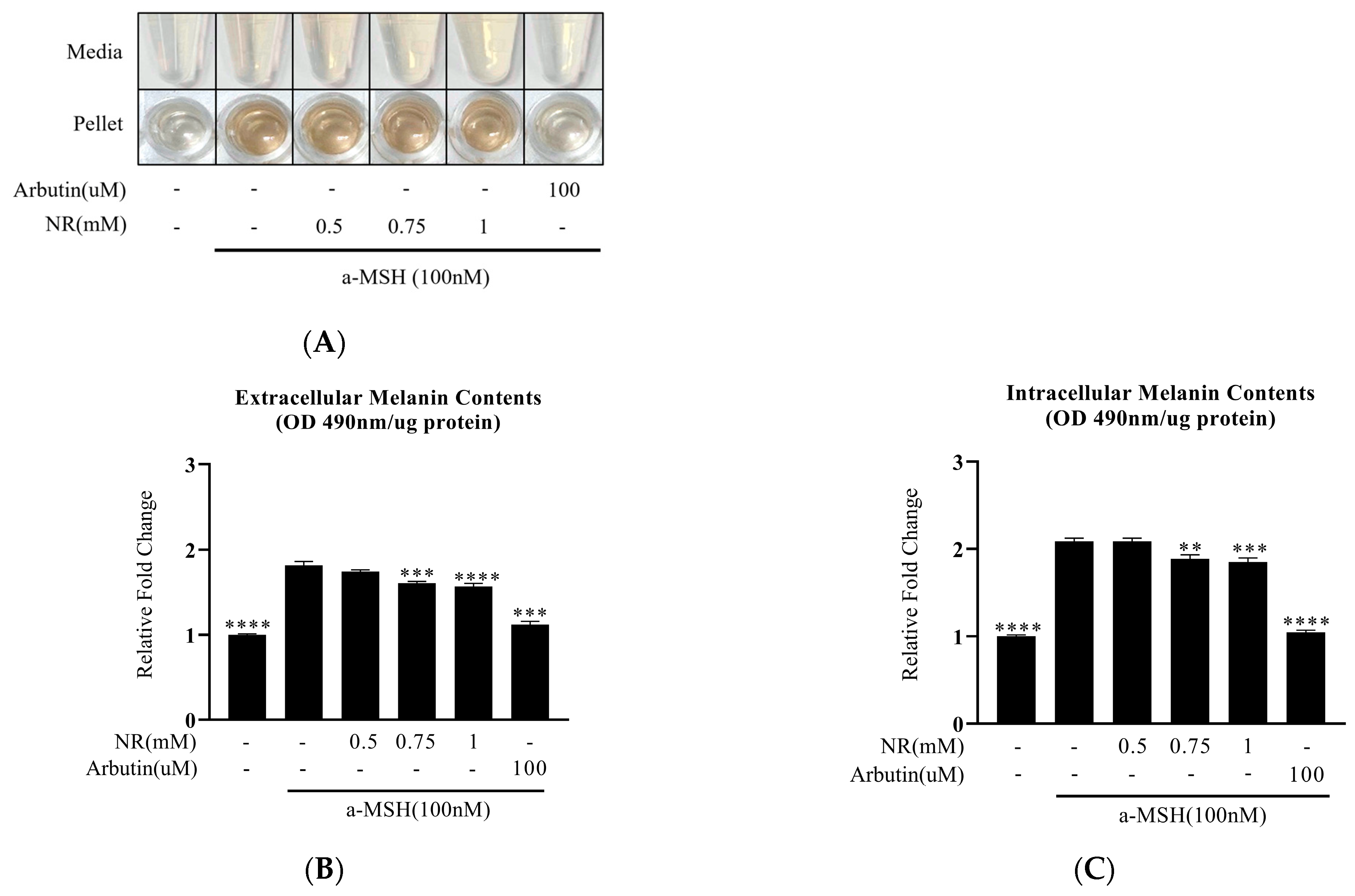
Many previous studies have shown that UVR-damaged epidermal keratinocytes secrete specific hormones such as α-MSH, which are recognized for inducing melanogenic signals through activation of the cAMP/protein kinase A (PKA)/CREB pathway [34,35]. Building on this concept, we treated B16F10 melanocytes with α-MSH to determine whether NR inhibits melanogenesis under conditions that induce it. Our results showed that NR decreased α-MSH-induced melanin content both extracellularly and intracellularly after 48 h (Figure 2A–C).
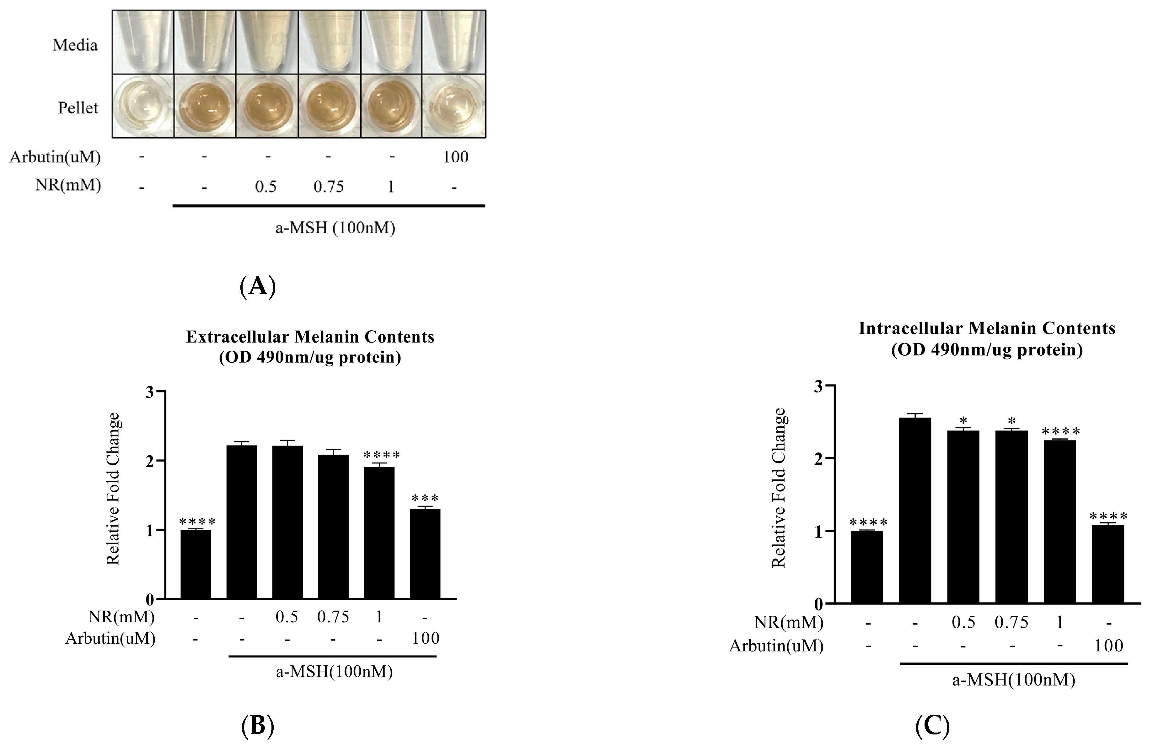
Extracellular melanin secretion induced by α-MSH was higher after 72 than 48 h of incubation (2.2-fold vs. 1.8-fold, respectively) (Figure 3). The intracellular melanin content was also significantly higher after 72 than 48 h of incubation (2.5-fold vs. 2.0-fold, respectively). Although we could not ascertain whether this increase was due to a higher basal level of melanogenesis, the 72 h results showed that only the 1 mM dose of NR showed a significant reduction in melanin secretion. The intracellular melanin content exhibited a significant inhibitory effect at every experimental dose (Figure 3C). This suggests that NR inhibits the progression of melanogenesis rather than just melanin secretion.

3.3. Direct Inhibition of Tyrosinase Activity by NR

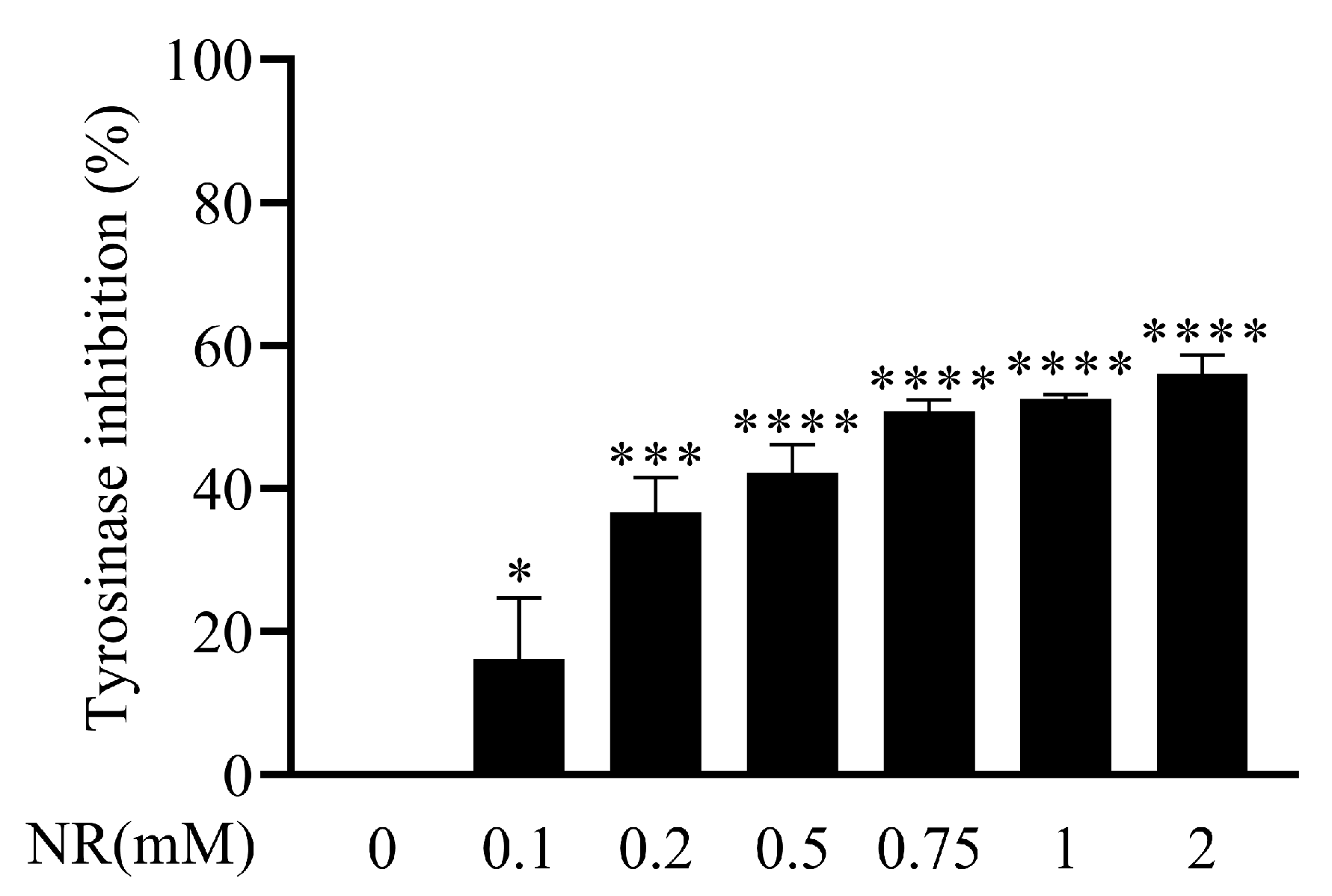
To confirm the mechanism underlying melanogenesis, we performed a mushroom tyrosinase activity assay using L-tyrosine, the main substrate for the initiation of melanogenesis. The experiment showed that NR directly inhibits tyrosinase activity (Figure 4).

3.4. Regulation of Melanogenic Genes by NR

Numerous reports have indicated that α-MSH can induce the expression of mitf and regulate downstream genes such as trp1, trp2, and tyr [36,37]. Therefore, we assessed the levels of melanogenic genes (Figure 5). While the induction of α-MSH was significant, NR treatment exhibited no effect on the expression of these genes (Figure 5A–D). This confirmed that NR inhibits melanogenesis by directly regulating tyrosinase activity, independently of the regulation of melanogenic gene expression (Figure 4 and Figure 5).

4. Discussion and Conclusions

Symptoms related to pigmentation may be caused by disease, aging, or persistent or temporary irritation. There are various causes of pigmentation, but depending on the degree of pigmentation, it may cause abnormalities in the body or, conversely, may protect cells from irritation [1,38]. Among existing whitening ingredients, there are many effective ingredients; however, discovering new ingredients is important. Additionally, since these ingredients are used in the human body, their safety is very important. We studied pigmentation induced by ultraviolet rays among various pigmentation-related situations, and used nicotinamide riboside (NR) to improve it.
The global market for skin-whitening products reached USD 9.96 billion in 2021, and is expected to grow at a compound annual growth rate of 5.5% through 2030 [39]. There is a crucial need to identify ingredients with potent whitening effects and high stability to ensure their safe use in various applications. We believe that NR can fulfill this role. Thus, the purpose of this study was to identify novel therapeutic ingredients, with an emphasis on safety, to mitigate excessive melanin production induced by skin damage.
One of the most widely used ingredients for melanin suppression is arbutin, which consists of D-glucose and hydroquinone (HQ). Arbutin is a commonly utilized compound in whitening cosmetics worldwide and is known for its skin-whitening effects, with concentrations reaching up to 7% [40]. It is a derivate of HQ; therefore, arbutin can undergo conversion to HQ when exposed to certain conditions, such as acidic pH, UVR, or high temperatures [41,42]. HQ itself is a very effective whitening agent and is 100 times more effective than arbutin [43]. The potential toxicity of HQ, which is generated from the breakdown of arbutin, has been extensively studied in numerous papers [44,45,46]. Particularly, hydroquinone is known to induce erythema, stinging, irritation, allergic contact dermatitis, melanin regeneration promotion, and white patches [47,48,49,50,51]. Additionally, arbutin has been reported to cause skin irritation and allergic reactions in some users, with more pronounced side effects observed in individuals with sensitive skin [43]. Several studies have highlighted the ability of HQ to induce intracellular oxidative stress, abnormal DNA damage, and genotoxicity. However, the consensus on its benefits remains uncertain [45,46]. Therefore, there is an emphasized need to develop skin-whitening agents that minimize side effects and ensure safety and efficacy.
NAD+ is a substance naturally present in living organisms, and the side effects of NAD+ are virtually unknown. This safety profile underscores the importance of actively researching various precursors capable of increasing NAD+ levels, and thus determining its beneficial functions within the body. NAD+ regulates numerous biological processes, including glycolysis, the respiratory chain, DNA repair, and antioxidant defenses. Numerous precursors contribute to NAD+ production, including nicotinamide mononucleotide (NMN), nicotinic acid (NA), nicotinamide, tryptophan, and NR. Research on using NAD+ induction to improve pigmentation disorders is under way, with nicotinamide representing a precursor well known for its skin-whitening ability. In this study, we explored the skin-whitening effects of other NAD+ precursors. Nicotinamide and niacin, among other NAD+ precursors, are essential components of biological systems and are abundantly found in natural sources such as eggs, vegetables, fruits, and fish. They are known to be effective in skin whitening and maintaining cellular homeostasis [44,52,53]. Benavente et al. found that extremely low NAD+ levels, such as those seen in patients with niacin deficiency, adversely affect cell growth and survival [54]. In niacin-deficient HaCaT cells, nicotinamide can regulate melanogenesis via the reduction in melanosome secretion [55]. NMN downregulates melanogenesis in aged melanocytes by inhibiting cAMP/Wnt signaling and, in the same way, the direct elevation of NAD+ reduces both melanin production and cAMP/Wnt signaling [56].
NR is converted in vivo to NMN by NR kinase, which is then converted to NAD+ by NMN adenylyltransferase (NMNAT), increasing NAD+ levels in the body [57]. NR originated from studies on pellagra treatment, a disease caused by niacin deficiency [58]. Additionally, it has demonstrated weight loss and improved insulin sensitivity effects in mice with high-fat-diet-induced obesity. Moreover, it enhances glucose homeostasis, increases adiponectin levels, and reduces hepatic cholesterol in mice with type 2 diabetes by regulating the NLRP3 inflammasome [19,59]. NR is also being studied for its potential to increase NAD+ levels in the treatment of infections, including SARS-CoV-2, which caused the recent coronavirus pandemic [60]. NR is currently in the spotlight because of its high bioavailability, safety, and superior ability to increase NAD+ levels compared with other precursors. Among the various NAD+ precursors, NR has higher oral bioavailability than nicotinamide (and consequently, a higher oral bioavailability than nicotinic acid) [17].
Tyrosinase is the most important regulator of melanogenesis. As a dicopper oxidase, it interacts with histidine residues at their active site, and two copper ions are important for the catalytic activity of this enzyme [61,62]. Tyrosinase converts L-tyrosine and L-Dopa to melanin through oxidation [63]. Therefore, many studies have focused on the direct control of tyrosinase. The approaches used in such studies include degrading the expression level of tyrosinase and directly inhibiting its activity [61]. Many ingredients with tyrosinase inhibitory functions have been discovered, some of which are well known, including kojic acid [64], arbutin [65], salicylhydroxamic acid, and hydroquinone [66].
The present study began with the goal of discovering new therapeutic agents, emphasizing safety, to alleviate excessive melanin production resulting from skin damage. NR was found to directly inhibit enzymes involved in melanin production, thereby preventing skin hyperpigmentation. Additionally, it was demonstrated that NR may support skin layer maintenance by suppressing the expression of degradation enzymes in damaged skin.
This study showed that NR effectively inhibited α-MSH-induced melanogenesis (Figure 2 and Figure 3). We also confirmed that this action occurs through the direct inhibition of tyrosinase activity (Figure 4). However, further research on the regulation of cAMP/PKA, RAS/RAF, and GSK-3β/β-catenin signaling is required [61].
Maeda et al. reported that the melanin production activity in melanocytes induced by arbutin treatment is not attributed to differences in gene expression levels [44]. Our results also showed that NR had an inhibitory effect on melanin production through inhibition of enzyme activity (Figure 4), independently of the gene expression level of tyrosinase (Figure 5).
Additionally, in supplementary experiments, we assessed the cytotoxicity of UVB-induced HaCaT cells, mimicking damaged skin (Figures S2 and S3). These data showed that NR had a protective effect against UVB-induced cytotoxicity at specific doses (Figure S3). We also confirmed that NR reduced the expression of matrix metalloproteinases, namely, collagenase (mmp-1) and gelatinase (mmp-9), which can degrade certain extracellular matrix proteins [67,68,69,70,71].
In conclusion, NR is an effective therapeutic ingredient that inhibits melanin production at certain concentrations and protects skin by restoring the viability of keratinocytes and suppressing matrix metalloproteinases expression. Therefore, NR is a potential skin-whitening ingredient and is significant as a safe treatment for serious diseases caused by photo-stimulation; further research on more specific mechanisms is needed in the future.

Supplementary Materials

The following supporting information can be downloaded at: https://www.mdpi.com/article/10.3390/cosmetics11030073/s1, Figure S1: NR has no effect on B16F10 cell growth at concentrations prior to 2 mM; Figure S2: NR protect UVB-induced cell death; Figure S3: NR decrease the gene expression of UVB-induced matrix metalloproteinase.

Author Contributions

Conceptualization, S.H.C.; methodology, S.H.C. and Y.J.L.; validation, Y.J.L., S.H.C. and S.G.J.; formal analysis, Y.J.L. and S.G.J.; investigation, Y.J.L., S.H.C., S.G.J. and M.J.R.; resources, S.H.C. and M.J.R.; data curation, S.H.C.; writing—original draft preparation, S.H.C.; writing—review and editing, S.H.C.; visualization, S.H.C. and Y.J.L.; supervision, S.H.C.; project administration, S.H.C.; funding acquisition, not applicable. All authors have read and agreed to the published version of the manuscript.

Funding

This research received no external funding.

Institutional Review Board Statement

Not applicable.

Informed Consent Statement

Not applicable.

Data Availability Statement

All data are contained within the manuscript.

Acknowledgments

All authors would like to thank Gi-Ryang Kwon (Chungnam National University, school of medicine) for providing the HaCaT cell line.

Conflicts of Interest

Authors Yeon Jae Lee, Seul Gi Jang, Min Jeong Ryu, and Seung Hee Choi were employed by the Joonghun Pharmaceutical Co., Ltd. The authors declare that the research was conducted in the absence of any commercial or financial relationships that could be construed as a potential conflicts of interest.

References

  1. Brenner, M.; Hearing, V.J. The protective role of melanin against UV damage in human skin. Photochem. Photobiol. 2008, 84, 539–549. [Google Scholar] [CrossRef] [PubMed]
  2. Plensdorf, S.; Livieratos, M.; Dada, N. Pigmentation Disorders: Diagnosis and Management. Am. Fam. Physician 2017, 96, 797–804. [Google Scholar] [PubMed]
  3. Bolognia, J.; Murray, M.; Pawelek, J. UVB-induced melanogenesis may be mediated through the MSH-receptor system. J. Investig. Dermatol. 1989, 92, 651–656. [Google Scholar] [CrossRef] [PubMed]
  4. Wakamatsu, K.; Ito, S. Advanced chemical methods in melanin determination. Pigment Cell Res. 2002, 15, 174–183. [Google Scholar] [CrossRef] [PubMed]
  5. Prota, G. Recent advances in the chemistry of melanogenesis in mammals. J. Investig. Dermatol. 1980, 75, 122–127. [Google Scholar] [CrossRef] [PubMed]
  6. Jangda, A.; Voloshyna, D.; Ramesh, K.; Bseiso, A.; Shaik, T.A.; Al Barznji, S.; Usama, M.; Saleem, F.; Ghaffari, M.A.Z. Hyperpigmentation as a Primary Symptom of Vitamin B12 Deficiency: A Case Report. Cureus 2022, 14, e29008. [Google Scholar] [CrossRef] [PubMed]
  7. Kannan, R.; Ng, M.J. Cutaneous lesions and vitamin B12 deficiency: An often-forgotten link. Can. Fam. Physician 2008, 54, 529–532. [Google Scholar] [PubMed]
  8. Cohen, P.R. Linea Nigra: Case Report of a Woman With a Pregnancy-Associated Linear Streak of Cutaneous Hyperpigmentation on Her Abdomen From the Umbilicus to the Pubic Symphysis. Cureus 2023, 15, e48408. [Google Scholar] [CrossRef]
  9. Schepsky, A.; Bruser, K.; Gunnarsson, G.J.; Goodall, J.; Hallsson, J.H.; Goding, C.R.; Steingrimsson, E.; Hecht, A. The microphthalmia-associated transcription factor Mitf interacts with beta-catenin to determine target gene expression. Mol. Cell. Biol. 2006, 26, 8914–8927. [Google Scholar] [CrossRef]
  10. Shibahara, S.; Takeda, K.; Yasumoto, K.; Udono, T.; Watanabe, K.; Saito, H.; Takahashi, K. Microphthalmia-associated transcription factor (MITF): Multiplicity in structure, function, and regulation. J. Investig. Dermatol. Symp. Proc. 2001, 6, 99–104. [Google Scholar] [CrossRef]
  11. Hearing, V.J.; Jiménez, M. Mammalian tyrosinase--the critical regulatory control point in melanocyte pigmentation. Int. J. Biochem. 1987, 19, 1141–1147. [Google Scholar] [CrossRef] [PubMed]
  12. Zolghadri, S.; Bahrami, A.; Hassan Khan, M.T.; Munoz-Munoz, J.; Garcia-Molina, F.; Garcia-Canovas, F.; Saboury, A.A. A comprehensive review on tyrosinase inhibitors. J. Enzym. Inhib. Med. Chem. 2019, 34, 279–309. [Google Scholar] [CrossRef]
  13. Bieganowski, P.; Brenner, C. Discoveries of nicotinamide riboside as a nutrient and conserved NRK genes establish a Preiss-Handler independent route to NAD+ in fungi and humans. Cell 2004, 117, 495–502. [Google Scholar] [CrossRef]
  14. Guyton, J.R.; Bays, H.E. Safety considerations with niacin therapy. Am. J. Cardiol. 2007, 99, 22C–31C. [Google Scholar] [CrossRef] [PubMed]
  15. Airhart, S.E.; Shireman, L.M.; Risler, L.J.; Anderson, G.D.; Nagana Gowda, G.A.; Raftery, D.; Tian, R.; Shen, D.D.; O’Brien, K.D. An open-label, non-randomized study of the pharmacokinetics of the nutritional supplement nicotinamide riboside (NR) and its effects on blood NAD+ levels in healthy volunteers. PLoS ONE 2017, 12, e0186459. [Google Scholar] [CrossRef]
  16. Dollerup, O.L.; Christensen, B.; Svart, M.; Schmidt, M.S.; Sulek, K.; Ringgaard, S.; Stødkilde-Jørgensen, H.; Møller, N.; Brenner, C.; Treebak, J.T.; et al. A randomized placebo-controlled clinical trial of nicotinamide riboside in obese men: Safety, insulin-sensitivity, and lipid-mobilizing effects. Am. J. Clin. Nutr. 2018, 108, 343–353. [Google Scholar] [CrossRef]
  17. Trammell, S.A.; Schmidt, M.S.; Weidemann, B.J.; Redpath, P.; Jaksch, F.; Dellinger, R.W.; Li, Z.; Abel, E.D.; Migaud, M.E.; Brenner, C. Nicotinamide riboside is uniquely and orally bioavailable in mice and humans. Nat. Commun. 2016, 7, 12948. [Google Scholar] [CrossRef] [PubMed]
  18. Gilmour, B.C.; Gudmundsrud, R.; Frank, J.; Hov, A.; Lautrup, S.; Aman, Y.; Røsjø, H.; Brenner, C.; Ziegler, M.; Tysnes, O.B.; et al. Targeting NAD. Mech. Ageing Dev. 2020, 186, 111208. [Google Scholar] [CrossRef]
  19. Cantó, C.; Houtkooper, R.H.; Pirinen, E.; Youn, D.Y.; Oosterveer, M.H.; Cen, Y.; Fernandez-Marcos, P.J.; Yamamoto, H.; Andreux, P.A.; Cettour-Rose, P.; et al. The NAD(+) precursor nicotinamide riboside enhances oxidative metabolism and protects against high-fat diet-induced obesity. Cell Metab. 2012, 15, 838–847. [Google Scholar] [CrossRef]
  20. Sharma, C.; Donu, D.; Cen, Y. Emerging Role of Nicotinamide Riboside in Health and Diseases. Nutrients 2022, 14, 3889. [Google Scholar] [CrossRef]
  21. Li, F.; Wu, C.; Wang, G. Targeting NAD Metabolism for the Therapy of Age-Related Neurodegenerative Diseases. Neurosci. Bull. 2024, 40, 218–240. [Google Scholar] [CrossRef] [PubMed]
  22. Han, S.; Du, Z.; Liu, K.; Gong, S. Nicotinamide riboside protects noise-induced hearing loss by recovering the hair cell ribbon synapses. Neurosci. Lett. 2020, 725, 134910. [Google Scholar] [CrossRef] [PubMed]
  23. Lapatto, H.A.K.; Kuusela, M.; Heikkinen, A.; Muniandy, M.; van der Kolk, B.W.; Gopalakrishnan, S.; Pöllänen, N.; Sandvik, M.; Schmidt, M.S.; Heinonen, S.; et al. Nicotinamide riboside improves muscle mitochondrial biogenesis, satellite cell differentiation, and gut microbiota in a twin study. Sci. Adv. 2023, 9, eadd5163. [Google Scholar] [CrossRef] [PubMed]
  24. Martens, C.R.; Denman, B.A.; Mazzo, M.R.; Armstrong, M.L.; Reisdorph, N.; McQueen, M.B.; Chonchol, M.; Seals, D.R. Chronic nicotinamide riboside supplementation is well-tolerated and elevates NAD. Nat. Commun. 2018, 9, 1286. [Google Scholar] [CrossRef] [PubMed]
  25. Damgaard, M.V.; Treebak, J.T. What is really known about the effects of nicotinamide riboside supplementation in humans. Sci. Adv. 2023, 9, eadi4862. [Google Scholar] [CrossRef] [PubMed]
  26. Liu, L.; Su, X.; Quinn, W.J.; Hui, S.; Krukenberg, K.; Frederick, D.W.; Redpath, P.; Zhan, L.; Chellappa, K.; White, E.; et al. Quantitative Analysis of NAD Synthesis-Breakdown Fluxes. Cell Metab. 2018, 27, 1067–1080.e5. [Google Scholar] [CrossRef]
  27. Vichai, V.; Kirtikara, K. Sulforhodamine B colorimetric assay for cytotoxicity screening. Nat. Protoc. 2006, 1, 1112–1116. [Google Scholar] [CrossRef] [PubMed]
  28. Lorz, L.R.; Yoo, B.C.; Kim, M.Y.; Cho, J.Y. Anti-Wrinkling and Anti-Melanogenic Effect of. Int. J. Mol. Sci. 2019, 20, 1043. [Google Scholar] [CrossRef] [PubMed]
  29. Jeong, D.; Park, S.H.; Kim, M.H.; Lee, S.; Cho, Y.K.; Kim, Y.A.; Park, B.J.; Lee, J.; Kang, H.; Cho, J.Y. Anti-Melanogenic Effects of Ethanol Extracts of the Leaves and Roots of. Molecules 2020, 25, 5375. [Google Scholar] [CrossRef]
  30. Kim, D.E.; Chang, B.Y.; Ham, S.O.; Kim, Y.C.; Kim, S.Y. Neobavaisoflavone Inhibits Melanogenesis through the Regulation of Akt/GSK-3β and MEK/ERK Pathways in B16F10 Cells and a Reconstructed Human 3D Skin Model. Molecules 2020, 25, 2683. [Google Scholar] [CrossRef]
  31. Lee, H.J.; An, S.; Bae, S.; Lee, J.H. Diarylpropionitrile inhibits melanogenesis via protein kinase A/cAMP-response element-binding protein/microphthalmia-associated transcription factor signaling pathway in α-MSH-stimulated B16F10 melanoma cells. Korean J. Physiol. Pharmacol. 2022, 26, 113–123. [Google Scholar] [CrossRef] [PubMed]
  32. Peng, L.H.; Liu, S.; Xu, S.Y.; Chen, L.; Shan, Y.H.; Wei, W.; Liang, W.Q.; Gao, J.Q. Inhibitory effects of salidroside and paeonol on tyrosinase activity and melanin synthesis in mouse B16F10 melanoma cells and ultraviolet B-induced pigmentation in guinea pig skin. Phytomedicine 2013, 20, 1082–1087. [Google Scholar] [CrossRef] [PubMed]
  33. Li, X.; Yang, H.W.; Jiang, Y.; Oh, J.Y.; Jeon, Y.J.; Ryu, B. Ishophloroglucin A Isolated from. Mar. Drugs 2020, 18, 470. [Google Scholar] [CrossRef] [PubMed]
  34. Oh, T.I.; Yun, J.M.; Park, E.J.; Kim, Y.S.; Lee, Y.M.; Lim, J.H. Plumbagin Suppresses α-MSH-Induced Melanogenesis in B16F10 Mouse Melanoma Cells by Inhibiting Tyrosinase Activity. Int. J. Mol. Sci. 2017, 18, 320. [Google Scholar] [CrossRef] [PubMed]
  35. D’Mello, S.A.; Finlay, G.J.; Baguley, B.C.; Askarian-Amiri, M.E. Signaling Pathways in Melanogenesis. Int. J. Mol. Sci. 2016, 17, 1144. [Google Scholar] [CrossRef] [PubMed]
  36. Shibahara, S.; Yasumoto, K.; Amae, S.; Udono, T.; Watanabe, K.; Saito, H.; Takeda, K. Regulation of pigment cell-specific gene expression by MITF. Pigment Cell Res. 2000, 13 (Suppl. S8), 98–102. [Google Scholar] [CrossRef] [PubMed]
  37. Yoon, Y.; Bae, S.; Kim, T.J.; An, S.; Lee, J.H. Nodakenin Inhibits Melanogenesis Via the ERK/MSK1 Signaling Pathway. Pharmazie 2023, 78, 6–12. [Google Scholar] [CrossRef] [PubMed]
  38. Kobayashi, N.; Muramatsu, T.; Yamashina, Y.; Shirai, T.; Ohnishi, T.; Mori, T. Melanin reduces ultraviolet-induced DNA damage formation and killing rate in cultured human melanoma cells. J. Investig. Dermatol. 1993, 101, 685–689. [Google Scholar] [CrossRef] [PubMed]
  39. GVR Report Cover Skin Lightening Products Market Size, Share & Trends Analysis Report by Product (Creams, Cleanser, Mask), by Nature, by Region, and Segment Forecasts, 2022–2030. Available online: https://www.grandviewresearch.com/industry-analysis/skin-lightening-products-market (accessed on 1 March 2024).
  40. Degen, G.H.; SCCS. Opinion of the Scientific Committee on Consumer Safety (SCCS)--Opinion on the safety of the use of β-arbutin in cosmetic products. Regul. Toxicol. Pharmacol. 2015, 73, 866–867. [Google Scholar] [CrossRef]
  41. Jeon, J.S.; Kim, B.H.; Lee, S.H.; Kwon, H.J.; Bae, H.J.; Kim, S.K.; Park, J.A.; Shim, J.H.; Abd El-Aty, A.M.; Shin, H.C. Simultaneous determination of arbutin and its decomposed product hydroquinone in whitening creams using high-performance liquid chromatography with photodiode array detection: Effect of temperature and pH on decomposition. Int. J. Cosmet. Sci. 2015, 37, 567–573. [Google Scholar] [CrossRef]
  42. Yang, C.H.; Chang, N.F.; Chen, Y.S.; Lee, S.M.; Lin, P.J.; Lin, C.C. Comparative study on the photostability of arbutin and deoxy arbutin: Sensitivity to ultraviolet radiation and enhanced photostability by the water-soluble sunscreen, benzophenone-4. Biosci. Biotechnol. Biochem. 2013, 77, 1127–1130. [Google Scholar] [CrossRef] [PubMed]
  43. Maeda, K.; Fukuda, M. Arbutin: Mechanism of its depigmenting action in human melanocyte culture. J. Pharmacol. Exp. Ther. 1996, 276, 765–769. [Google Scholar] [PubMed]
  44. Boo, Y.C. Mechanistic Basis and Clinical Evidence for the Applications of Nicotinamide (Niacinamide) to Control Skin Aging and Pigmentation. Antioxidants 2021, 10, 1315. [Google Scholar] [CrossRef] [PubMed]
  45. Tsutsui, T.; Hayashi, N.; Maizumi, H.; Huff, J.; Barrett, J.C. Benzene-, catechol-, hydroquinone- and phenol-induced cell transformation, gene mutations, chromosome aberrations, aneuploidy, sister chromatid exchanges and unscheduled DNA synthesis in Syrian hamster embryo cells. Mutat. Res. 1997, 373, 113–123. [Google Scholar] [CrossRef] [PubMed]
  46. Marrazzini, A.; Chelotti, L.; Barrai, I.; Loprieno, N.; Barale, R. In vivo genotoxic interactions among three phenolic benzene metabolites. Mutat. Res. 1994, 341, 29–46. [Google Scholar] [CrossRef]
  47. Tse, T.W. Hydroquinone for skin lightening: Safety profile, duration of use and when should we stop? J. Dermatolog. Treat. 2010, 21, 272–275. [Google Scholar] [CrossRef] [PubMed]
  48. Parvez, S.; Kang, M.; Chung, H.S.; Cho, C.; Hong, M.C.; Shin, M.K.; Bae, H. Survey and mechanism of skin depigmenting and lightening agents. Phytother. Res. 2006, 20, 921–934. [Google Scholar] [CrossRef] [PubMed]
  49. Luo, L.; Jiang, L.; Geng, C.; Cao, J.; Zhong, L. Hydroquinone-induced genotoxicity and oxidative DNA damage in HepG2 cells. Chem. Biol. Interact. 2008, 173, 1–8. [Google Scholar] [CrossRef] [PubMed]
  50. Yoshimura, M.; Watanabe, Y.; Kasai, K.; Yamakoshi, J.; Koga, T. Inhibitory effect of an ellagic acid-rich pomegranate extract on tyrosinase activity and ultraviolet-induced pigmentation. Biosci. Biotechnol. Biochem. 2005, 69, 2368–2373. [Google Scholar] [CrossRef]
  51. Draelos, Z.D. Skin lightening preparations and the hydroquinone controversy. Dermatol. Ther. 2007, 20, 308–313. [Google Scholar] [CrossRef]
  52. Shariff, R.; Du, Y.; Dutta, M.; Kumar, S.; Thimmaiah, S.; Doraiswamy, C.; Kumari, A.; Kale, V.; Nair, N.; Zhang, S.; et al. Superior even skin tone and anti-ageing benefit of a combination of 4-hexylresorcinol and niacinamide. Int. J. Cosmet. Sci. 2022, 44, 103–117. [Google Scholar] [CrossRef] [PubMed]
  53. Kim, H.M.; Byun, K.A.; Oh, S.; Yang, J.Y.; Park, H.J.; Chung, M.S.; Son, K.H.; Byun, K. A Mixture of Topical Forms of Polydeoxyribonucleotide, Vitamin C, and Niacinamide Attenuated Skin Pigmentation and Increased Skin Elasticity by Modulating Nuclear Factor Erythroid 2-like 2. Molecules 2022, 27, 1276. [Google Scholar] [CrossRef] [PubMed]
  54. Benavente, C.A.; Jacobson, E.L. Niacin restriction upregulates NADPH oxidase and reactive oxygen species (ROS) in human keratinocytes. Free Radic. Biol. Med. 2008, 44, 527–537. [Google Scholar] [CrossRef] [PubMed]
  55. Hakozaki, T.; Minwalla, L.; Zhuang, J.; Chhoa, M.; Matsubara, A.; Miyamoto, K.; Greatens, A.; Hillebrand, G.G.; Bissett, D.L.; Boissy, R.E. The effect of niacinamide on reducing cutaneous pigmentation and suppression of melanosome transfer. Br. J. Dermatol. 2002, 147, 20–31. [Google Scholar] [CrossRef] [PubMed]
  56. Brito, S.; Baek, J.M.; Cha, B.; Heo, H.; Lee, S.H.; Lei, L.; Jung, S.Y.; Lee, S.M.; Kwak, B.M.; Chae, S.; et al. Nicotinamide mononucleotide reduces melanin production in aged melanocytes by inhibiting cAMP/Wnt signaling. J. Dermatol. Sci. 2022, 106, 159–169. [Google Scholar] [CrossRef] [PubMed]
  57. Yoshino, J.; Baur, J.A.; Imai, S.I. NAD. Cell Metab. 2018, 27, 513–528. [Google Scholar] [CrossRef] [PubMed]
  58. Cercillieux, A.; Ciarlo, E.; Canto, C. Balancing NAD. Cell. Mol. Life Sci. 2022, 79, 463. [Google Scholar] [CrossRef] [PubMed]
  59. Lee, H.J.; Hong, Y.S.; Jun, W.; Yang, S.J. Nicotinamide Riboside Ameliorates Hepatic Metaflammation by Modulating NLRP3 Inflammasome in a Rodent Model of Type 2 Diabetes. J. Med. Food 2015, 18, 1207–1213. [Google Scholar] [CrossRef]
  60. Heer, C.D.; Sanderson, D.J.; Voth, L.S.; Alhammad, Y.M.O.; Schmidt, M.S.; Trammell, S.A.J.; Perlman, S.; Cohen, M.S.; Fehr, A.R.; Brenner, C. Coronavirus infection and PARP expression dysregulate the NAD metabolome: An actionable component of innate immunity. J. Biol. Chem. 2020, 295, 17986–17996. [Google Scholar] [CrossRef]
  61. Pillaiyar, T.; Namasivayam, V.; Manickam, M.; Jung, S.H. Inhibitors of Melanogenesis: An Updated Review. J. Med. Chem. 2018, 61, 7395–7418. [Google Scholar] [CrossRef]
  62. Cabanes, J.; García-Cánovas, F.; Lozano, J.A.; García-Carmona, F. A kinetic study of the melanization pathway between L-tyrosine and dopachrome. Biochim. Biophys. Acta 1987, 923, 187–195. [Google Scholar] [CrossRef] [PubMed]
  63. Alam, M.B.; Bajpai, V.K.; Lee, J.; Zhao, P.; Byeon, J.H.; Ra, J.S.; Majumder, R.; Lee, J.S.; Yoon, J.I.; Rather, I.A.; et al. Inhibition of melanogenesis by jineol from Scolopendra subspinipes mutilans via MAP-Kinase mediated MITF downregulation and the proteasomal degradation of tyrosinase. Sci. Rep. 2017, 7, 45858. [Google Scholar] [CrossRef] [PubMed]
  64. Gonçalez, M.L.; Corrêa, M.A.; Chorilli, M. Skin delivery of kojic acid-loaded nanotechnology-based drug delivery systems for the treatment of skin aging. Biomed. Res. Int. 2013, 2013, 271276. [Google Scholar] [CrossRef] [PubMed]
  65. Chakraborty, A.K.; Funasaka, Y.; Komoto, M.; Ichihashi, M. Effect of arbutin on melanogenic proteins in human melanocytes. Pigment. Cell Res. 1998, 11, 206–212. [Google Scholar] [CrossRef] [PubMed]
  66. Palumbo, A.; d’Ischia, M.; Misuraca, G.; Prota, G. Mechanism of inhibition of melanogenesis by hydroquinone. Biochim. Biophys. Acta 1991, 1073, 85–90. [Google Scholar] [CrossRef] [PubMed]
  67. Pittayapruek, P.; Meephansan, J.; Prapapan, O.; Komine, M.; Ohtsuki, M. Role of Matrix Metalloproteinases in Photoaging and Photocarcinogenesis. Int. J. Mol. Sci. 2016, 17, 868. [Google Scholar] [CrossRef] [PubMed]
  68. Kim, H.H.; Shin, C.M.; Park, C.H.; Kim, K.H.; Cho, K.H.; Eun, H.C.; Chung, J.H. Eicosapentaenoic acid inhibits UV-induced MMP-1 expression in human dermal fibroblasts. J. Lipid Res. 2005, 46, 1712–1720. [Google Scholar] [CrossRef] [PubMed]
  69. Kim, M.K.; Shin, J.M.; Eun, H.C.; Chung, J.H. The role of p300 histone acetyltransferase in UV-induced histone modifications and MMP-1 gene transcription. PLoS ONE 2009, 4, e4864. [Google Scholar] [CrossRef] [PubMed]
  70. Koźák, I.; Klisenbauer, D.; Juhás, T. UV-B induced production of MMP-2 and MMP-9 in human corneal cells. Physiol. Res. 2003, 52, 229–234. [Google Scholar] [CrossRef]
  71. Kim, D.J.; Iwasaki, A.; Chien, A.L.; Kang, S. UVB-mediated DNA damage induces matrix metalloproteinases to promote photoaging in an AhR- and SP1-dependent manner. JCI Insight 2022, 7, e156344. [Google Scholar] [CrossRef]
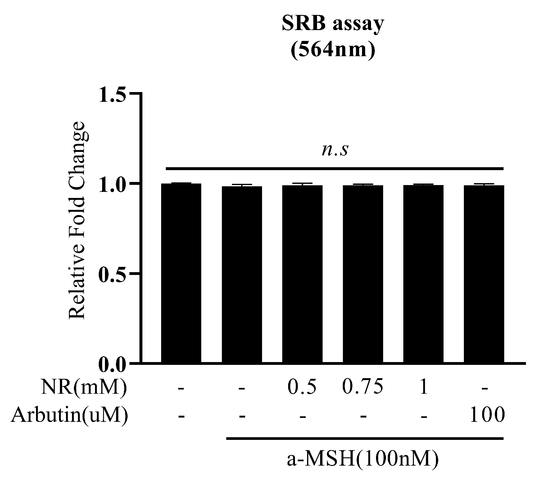
Figure 1. NR does not induce cell death. Indicated doses of chemical were added on the B16F10 cell line after 24 h a-MSH induction for 48 h. Relative to the α-MSH control. n.s: not significant. (unpaired t-test). The data are expressed as the mean ± SE (standard error).
Cosmetics 11 00073 g001
Figure 2. NR decreases melanin contents. NR treatment extracellular ((A) upper layer, (B)) and intracellular ((A) lower layer, (C)) melanin contents in 48 h. Relative to α-MSH control: ** p < 0.01, *** p < 0.001, and **** p < 0.0001 (unpaired t-test). The data are expressed as the mean ± SE.
Cosmetics 11 00073 g002
Figure 3. NR decrease melanin contents. NR treatment extracellular ((A)-upper layer, (B)) and intracellular ((A)-lower layer, (C)) melanin contents in 72 h. Relative to α-MSH control: * p < 0.05, *** p < 0.001, **** p < 0.0001 (unpaired t-test). The data are expressed as the mean ± SE.
Cosmetics 11 00073 g003
Figure 4. NR inhibit mushroom tyrosinase activity. NR directly inhibits mushroom tyrosinase activity using L-tyrosine in a dose-dependent manner. Relative to vehicle control (NR 0 mM): * p < 0.05, *** p < 0.001, and **** p < 0.0001 (unpaired t-test). The data are expressed as the mean ± SE.
Cosmetics 11 00073 g004
Figure 5. NR does not regulate the melanogenic gene. Expression of melanogenic genes: mitf (A), tyrosinase (B), trp1 (C), and trp2 (D). Relative to α-MSH control: n.s (not significant), * p < 0.05, ** p < 0.01, and *** p < 0.001 (unpaired t-test). The data are expressed as the mean ± SE.
Cosmetics 11 00073 g005
Disclaimer/Publisher’s Note: The statements, opinions and data contained in all publications are solely those of the individual author(s) and contributor(s) and not of MDPI and/or the editor(s). MDPI and/or the editor(s) disclaim responsibility for any injury to people or property resulting from any ideas, methods, instructions or products referred to in the content.