Article

Microtopography and Barrier Function in Healthy Skin: Differences between Forearm, Cheek and Palm

by
Raquel Sanabria-de la Torre
1,2,3,4,
María Ceres-Muñoz
4,
Carlota Pretel-Lara
4,
Trinidad Montero-Vílchez
1,3,* and
Salvador Arias-Santiago
1,3,4
1
Biosanitary Research Institute of Granada (ibs. GRANADA), 18014 Granada, Spain
2
Department of Biochemistry and Molecular Biology III and Immunology, University of Granada, 18071 Granada, Spain
3
Dermatology Department, Virgen de las Nieves University Hospital, 18014 Granada, Spain
4
Dermatology Department, School of Medicine, University of Granada, 18016 Granada, Spain
*
Author to whom correspondence should be addressed.
Cosmetics 2024, 11(1), 5; https://doi.org/10.3390/cosmetics11010005
Submission received: 31 October 2023 / Revised: 7 December 2023 / Accepted: 27 December 2023 / Published: 29 December 2023

Abstract

:
(1) Background: Skin barrier function resides mostly in the stratum corneum, which consists of a protein component, the corneocyte (bricks), which provides a scaffold for the second component, the extracellular matrix, consisting of multilayers of lipids (mortar). These two components closely interact and this could be the basis for the differences in the biophysical properties of the skin between anatomical regions. So, the aim of this study was to compare skin microstructural properties between body sites. (2) Methods: A comparative study was conducted that included healthy individuals without previous skin diseases. Skin barrier function parameters and microtopography parameters (smoothness, roughness, desquamation, wrinkles, surface, volume, contrast, variance, homogeneity, anisotropy, total cell count, flaking index, skin surface hardness, brightness, deformability and friction) were measured on the forearm, cheek and palm. (3) Results: 44 participants were included in this study, with a mean age of 38.8 ± 15.0 years. Significant differences were found between body sites for 14 of the 15 parameters evaluated. Smoothness was higher on the forearm than on the cheek and palm (240.02 Sems vs. 348.16 vs. 408.19 Sems, p < 0.05). Hardness was higher on the palm than on the forearm and cheek (13.22 AU vs. 9.44 AU vs. 7.94 AU, p < 0.05). Moreover, we observed that sociodemographic characteristics such as age, sex, tobacco and/or alcohol use, influenced the parameters evaluated. (4) Conclusions: The differences in skin barrier function and microtopography between anatomical regions reflects the different structure of skin in each body part and could help to understand the influence of the sociodemographic characteristics on theses parameters. This information could be useful for comparison with pathological skin characteristics and for targeting new treatments.

1. Introduction

The skin is the largest organ of the human body, responsible for forming a barrier between the “inside” and the “outside” of the body, preventing the invasion of pathogens and repelling chemical attacks, as well as preventing the unregulated loss of water and solutes [1]. The physical barrier is mainly located in the stratum corneum, which consists of protein-enriched cells and lipid-enriched intercellular domains [2]. Any modification in epidermal differentiation and lipid composition results in an alteration of the barrier function, a central event in various skin disorders and diseases [2].
Assessment of epidermal barrier function usually involves the measurement of several parameters [3]. Increased Trans-Epidermal Water Loss (TEWL), increased temperature and erythema [4,5] and decreased Stratum Corneum Hydration (SCH) are associated with impairments in barrier function [5]. The Surface Evaluation of Living Skin (SELS) provides reliable and valid values for skin microtopography [6,7] and comprises the fine and interwoven lines and ridges of the skin surface [8]. The cutaneous microtopography grade has been considered an objective measure of sun exposure [9], aging [8,10] and skin cancer [8,9].
Knowledge about the structure and function of human skin is of great interest for the dermatology, cosmetic and healthcare disciplines. The parameters of epidermal barrier function are altered in various inflammatory skin pathologies such as psoriasis [11] and atopic dermatitis [12]. Likewise, external damage such as the use of personal protective equipment [13] or sun exposure [14] affects these skin biophysical parameters. Also, the excipients used for topical treatments have a distinct impact on the barrier function of the skin [15]. However, there is significant variation in skin properties for different ages, genders and body regions due to the differences in the structure and morphology of the skin tissues [16]. Regional differences in TEWL, SCH, pH, erythema and melanin are well known [16,17]. However, there is little information on the normal values of certain less-widespread parameters related to skin function and structure, such as brightness, friction, smoothness, roughness, desquamation or contrast, among others [6]. Measuring these data in healthy subjects provides a baseline for the natural variability in the skin [3], which is crucial for identifying significant changes that could indicate abnormal conditions and allows for the early detection of subtle changes. Research into less-known parameters of skin barrier function in healthy subjects is essential for better understanding the normal physiology of the skin by providing valuable insights into the structure and function of the skin and how it responds to its environment [3]. In addition, measuring parameters in healthy subjects allows for the validation of methods and technologies used to ensure that measurement tools are reliable and accurate before applying them in clinical contexts.
Using non-invasive methods, it is possible to map the biophysical properties of the skin and generate normal ranges for healthy volunteers [16,18]. The main objective of the present study was to compare skin microtopography and epidermal barrier function on the forearm, cheek and palm of subjects with healthy skin. Some of the parameters measured were smoothness, roughness, desquamation, wrinkles, surface, volume, contrast, variance, homogeneity, anisotropy, total cell count, flaking index, skin surface hardness, brightness, deformability and friction.

2. Materials and Methods

2.1. Study Design

A cross-sectional study was conducted to compare skin microtopography and epidermal barrier function in different body sites of healthy volunteers between December 2022 and March 2023 in the Dermatology and Venereology Department of the Virgen de las Nieves University Hospital, Granada, Spain. Participants were adults with healthy skin and no previous inflammatory skin pathology, who signed the informed consent form.
Exclusion criteria were having a previous personal history of any inflammatory skin disease, clinical infection of the area under evaluation, receiving topical treatment that could alter barrier function (retinoids, corticoids, phototherapy, chemical peels), known or suspected incapacity to comply with the study protocol or having no signature on the informed consent form.

2.2. Skin Microtopography and Epidermal Barrier Function Parameters

Measurements were performed on the forearm, cheek and palm after staying for at least 30 min in a room with controlled ambient air temperature and humidity, which were measured with the TFA Lab Thermometer IP65 LT-101 (Wertheim, Germany; average air temperature 22 ± 1 °C; ambient air humidity 45% ± 5%).
A total of 16 parameters related to skin structural properties and epidermal barrier function were measured. Visioscan VC 20plus (Courage + Khazaka electronic GmbH, Bilbao, Spain) was employed to assess smoothness (Sesm: lower Sesm, higher smoothness), roughness (Ser: lower Ser, higher roughness), desquamation (Sesc: lower Sesc, less desquamation), wrinkles (Sew: higher Sew, more wrinkles), surface (%, elevated surface area means poor skin quality), volume (mm3, high volume indicates moisturized skin), contrast (arbitrary units (AU), low contrast indicates good skin quality), variance (AU, high variance states rough skin), homogeneity (AU, high homogeneity indicates moisturized skin), anisotropy (AU, high anisotropy means old skin) and total cell count (AU, high total cell count states young skin). Corneofix (Courage + Khazaka electronic GmbH) was used to evaluate the flaking index (%). Durometer (PCE-DDO 10) was used to measure skin surface hardness (AU).
Skin-glossymeter GL-200 (Courage + Khazaka electronic GmbH), Indentometer IDM 800 (Courage + Khazaka electronic GmbH) and Frictiometer (Courage + Khazaka electronic GmbH) connected to a Multi Probe Adapter (Courage + Khazaka electronic GmbH) were employed to measure brightness (AU), deformability (mm) and friction (AU), respectively.
All parameters were measured 10 times in each anatomical location and the average was used for statistical analysis.

2.3. Other Variables

Sociodemographic data including sex, age, weight, height, occupation, marital status, level of education, sun exposure and toxic habits were recorded in a clinical interview. The phototype was assessed by a dermatologist using Fitzpatrick grading. The frequency of body and facial moisturization and the use of sunscreen were self-reported by each participant.

2.4. Statistical Analysis

For descriptive analyses, continuous variables were expressed as mean ± standard deviation (SD) and qualitative variables as absolute and relative frequency distributions. The Shapiro–Wilk test was used to check the normality of data distributions and Levene test to check the homogeneity of variance. The Student’s t-test for paired samples was used to compare differences in parameters between body sites. The Student’s t-test for independent samples was used to compare differences in parameters between gender and age groups. Pearson’s correlation coefficient was calculated to assess the association between continuous variables and was expressed as (r, p value). Statistical significance was defined by a two-tailed p < 0.05. Statistical analyses were performed using the SPSS package (SPSS for Windows, version 24.0; SPSS Inc., Chicago, IL, USA).
Accepting an alpha risk of 0.05 and a beta risk of 0.2 in a two-sided test, 31 subjects are necessary to recognize as statistically significant a difference greater than or equal to 0.05 units. The standard deviation is assumed to be 6. A dropout rate of 5% was expected. G*Power 3.1.9.2., from Heinrich-Heine-Universität Düsseldorf, was used to calculate the sample size.

2.5. Ethics

This study was conducted in accordance with the guidelines of the Declaration of Helsinki and was approved by the Ethics Committee of Virgen de las Nieves University Hospital on 19 May 2019 (HC01/0442-N-20). The nature of the study was explained to all participants, who agreed to participate by verbal and written consent. All measurements were non-invasive and participant data were kept confidential.

3. Results

3.1. Sample Characteristics

A total of 44 healthy individuals were included in this study, comprising 65.9% women (29/44) and 34.1% men (15/44). The mean age was 38.8 ± 15.0 years old and 31.8% of participants were smokers (14/44). Detailed sociodemographic characteristics of the participants are shown in Table 1.

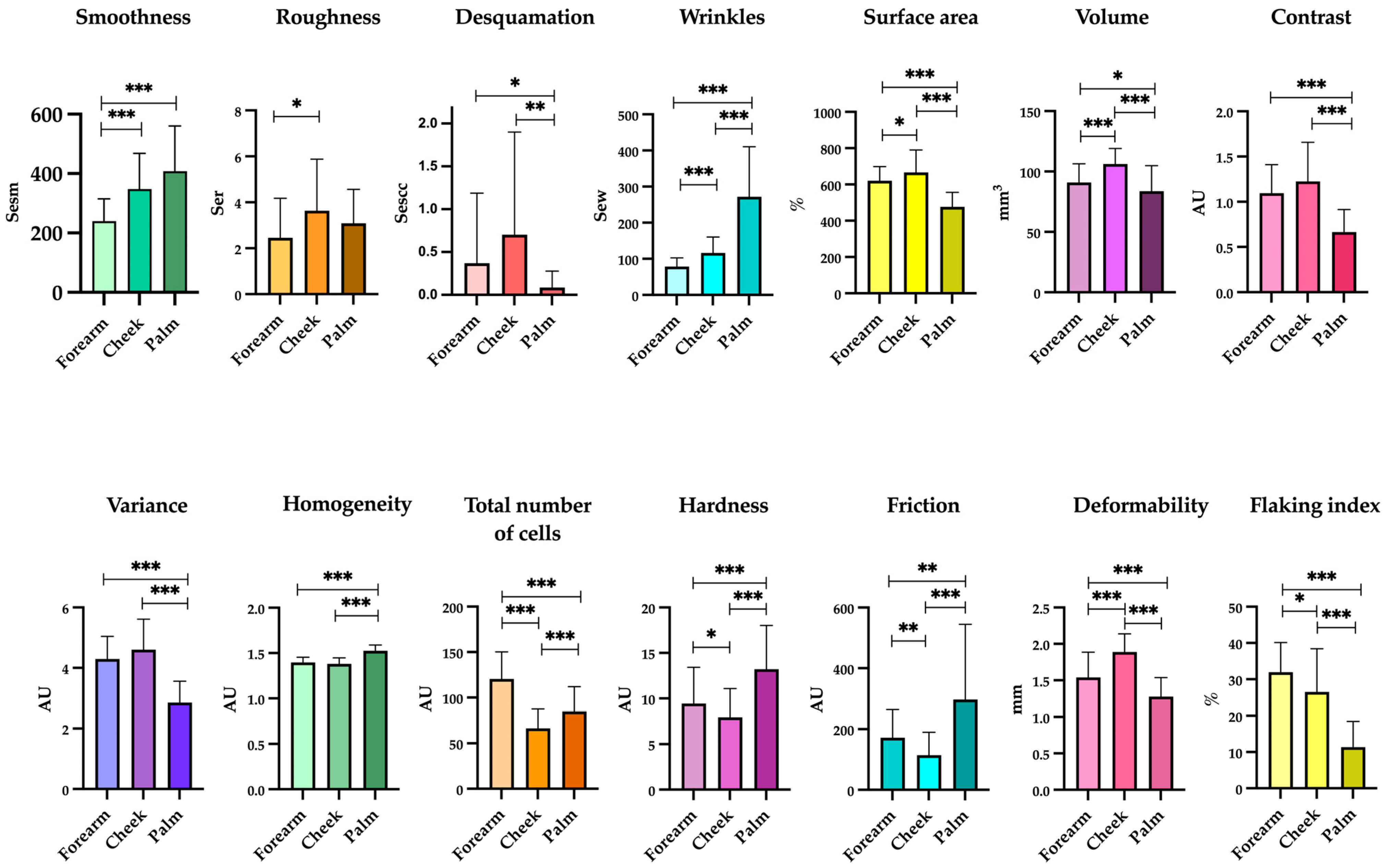
3.2. Skin Homeostasis and Epidermal Barrier Function Changed between Body Sites

Barrier function parameters and skin microtopography from three body sites (i) forearm, (ii) cheek and (iii) palm were compared, Table 2.
Smoothness was higher on the forearm than on the cheek and palm (240.02 Sems vs. 348.16 vs. 408.19 Sems, p < 0.05). Roughness was higher on the forearm than on the cheek (2.45 Ser vs. 3.64 Ser, p = 0.005) with no differences observed between the forearm and palm or the forearm and cheek. Desquamation was higher on the cheek and forearm than on the palm (0.70 Sesc vs. 0.09 Sesc, p = 0.002; 0.37 Sesc vs. 0.09 Sesc, p = 0.022, respectively) with no differences observed between the forearm and cheek. Wrinkles were higher on the palm than on the cheek and forearm (271.96 Sew vs. 116.32 vs. 78.61 Sew, p < 0.05). Surface area was higher on the cheek than on the forearm and palm (666.69% vs. 620.62% vs. 477.05%, p < 0.05). Volume was higher on the cheek than on the forearm and palm (106.16 mm3 vs. 90.88 mm3 vs. 83.64 mm3, p < 0.05). The contrast parameter was higher on the cheek and forearm than on the palm (1.23 AU vs. 0.67 AU, p < 0.001; 1.09 AU vs. 0.67 AU, p < 0.001), with no significant differences between the forearm and cheek. The variance was higher on the cheek and forearm than on the palm (4.60 AU vs. 2.25 AU, p < 0.001; 4.29 AU vs. 2.85 AU, p < 0.001), with no significant differences between the forearm and cheek. Homogeneity was higher on the palm than on the forearm and cheek (1.52 AU vs. 1.40 AU, p < 0.001; 1.52 AU vs. 1.38 AU, p < 0.001) with no significant differences between the forearm and cheek. The total number of cells was higher on the forearm than on the palm and cheek (120.48 AU vs. 85.07 AU vs. 66.36 AU, p < 0.001). Hardness was higher on the palm than on the forearm and cheek (13.22 AU vs. 9.44 AU vs. 7.94 AU, p < 0.05). Friction was higher on the palm than on the forearm and cheek (269.95 AU vs. 172.21 AU vs. 114.23 AU, p < 0.05). Deformability was higher on the cheek than on the forearm and palm (1.89 mm vs. 1.54 mm vs. 1.28 mm, p < 0.001). The flaking index was higher on the forearm than on the cheek and palm (31.93% vs. 26.54% vs. 11.33%, p < 0.05). No significant differences in brightness were observed between body sites. Figure 1 represents the statistically significant differences in these parameters.

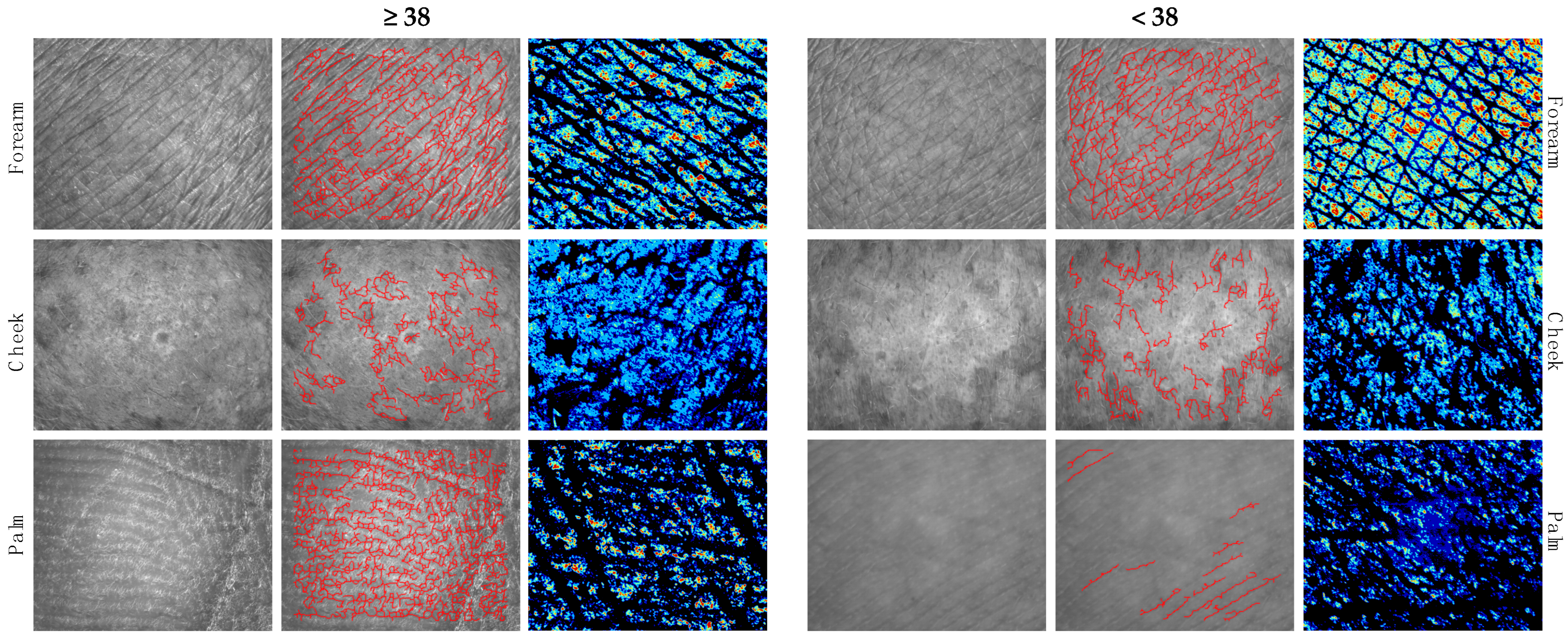
3.3. Differences in Skin Microtopography and Epidermal Barrier Function in Subjects ≥38 and <38 Years

Comparison of skin microtopography and barrier function between subjects ≥38 vs. <38 years are shown in Figure 2 and Table S1. The surface area on the palm was higher in subjects ≥38 years than in those <38 years (506.21% vs. 452.76%, p = 0.035). The contrast in the palm was higher in subjects ≥38 years than in those <38 years (0.76 AU vs. 0.58 AU, p = 0.031) (Figure 2, left). The variance in the palm was higher in subjects ≥38 years than in those <38 years (3.11 AU vs. 2.63 AU, p = 0.037). Homogeneity in the palm was higher in subjects <38 years than in 38 years (1.55 AU vs. 1.5 AU, p = 0.011) (Figure 2, middle). Palm friction was higher in subjects <38 years than in those ≥38 years (392.31 AU vs. 181.88 AU, p = 0.003). Palm sheen was higher in subjects ≥38 years than in subjects <38 years (8.19 AU vs. 6.93 AU, p = 0.002). The flaking index on the forearm was higher in subjects ≥38 years than in those <38 years (34.99% vs. 29.39%, p = 0.022) whereas the flaking index on the cheek was higher in subjects <38 years than in those ≥38 years (30.27% vs. 22.05%, p = 0.015) (Figure 2, right). No differences were observed for the rest of the parameters.

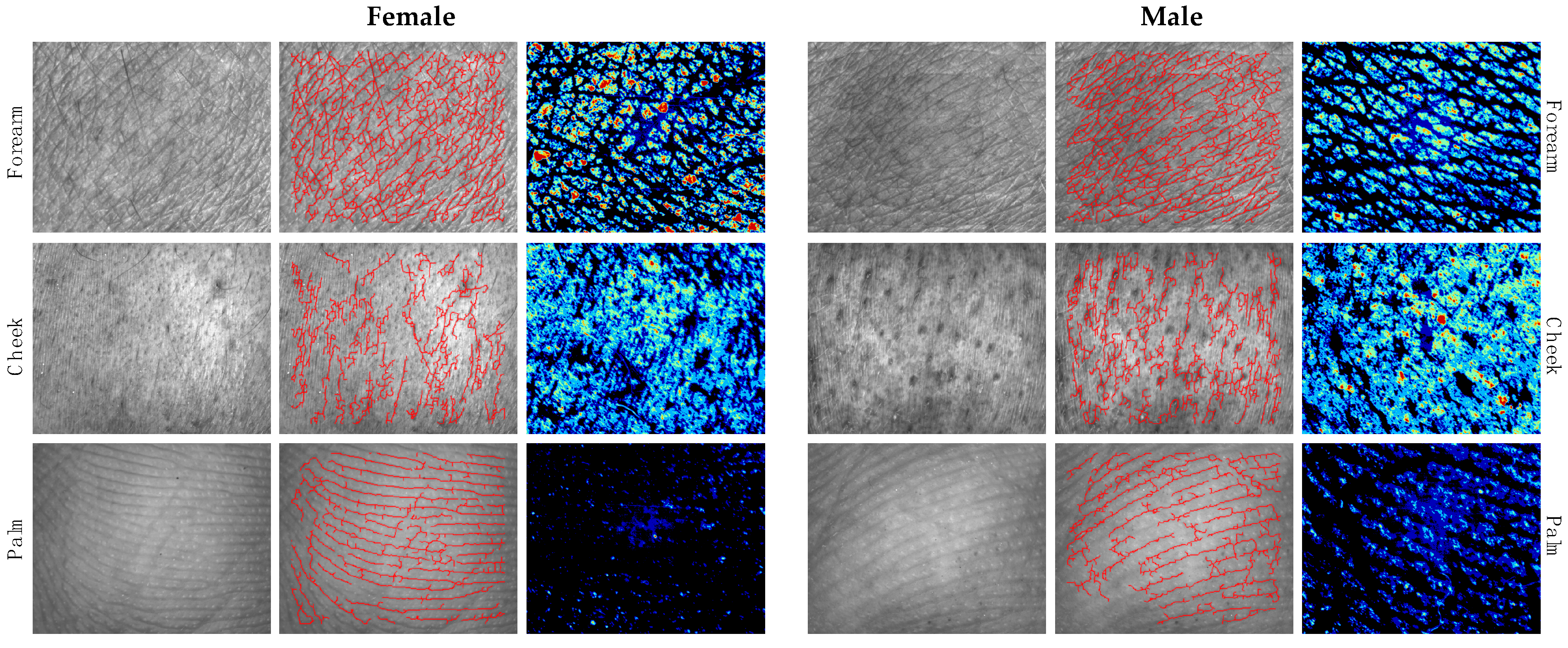
3.4. Differences in Skin Microtopography and Epidermal Barrier Function between Genders

Differences in skin microtopography and barrier function between the sexes are shown in Figure 3 and Table S2. Women had greater smoothness on the forearm than men (255.88 Sesm vs. 209.31 Sesm, p = 0.022). Men showed greater roughness on the forearm than women (3.63 Ser vs. 1.84 Ser, p = 0.010) but women showed greater roughness on the palm than men (3.42 Ser vs. 2.40 Ser, p = 0.016) (Figure 3, left). Females had more desquamation on the forearm than males (0.5 Sesc vs. 0.11 Sesc, p = 0.045) (Figure 3, right). Females had higher palm brightness than males (7.93 AU vs. 6.67 AU, p = 0.003). No differences were observed for the rest of the parameters (Figure 3, middle).

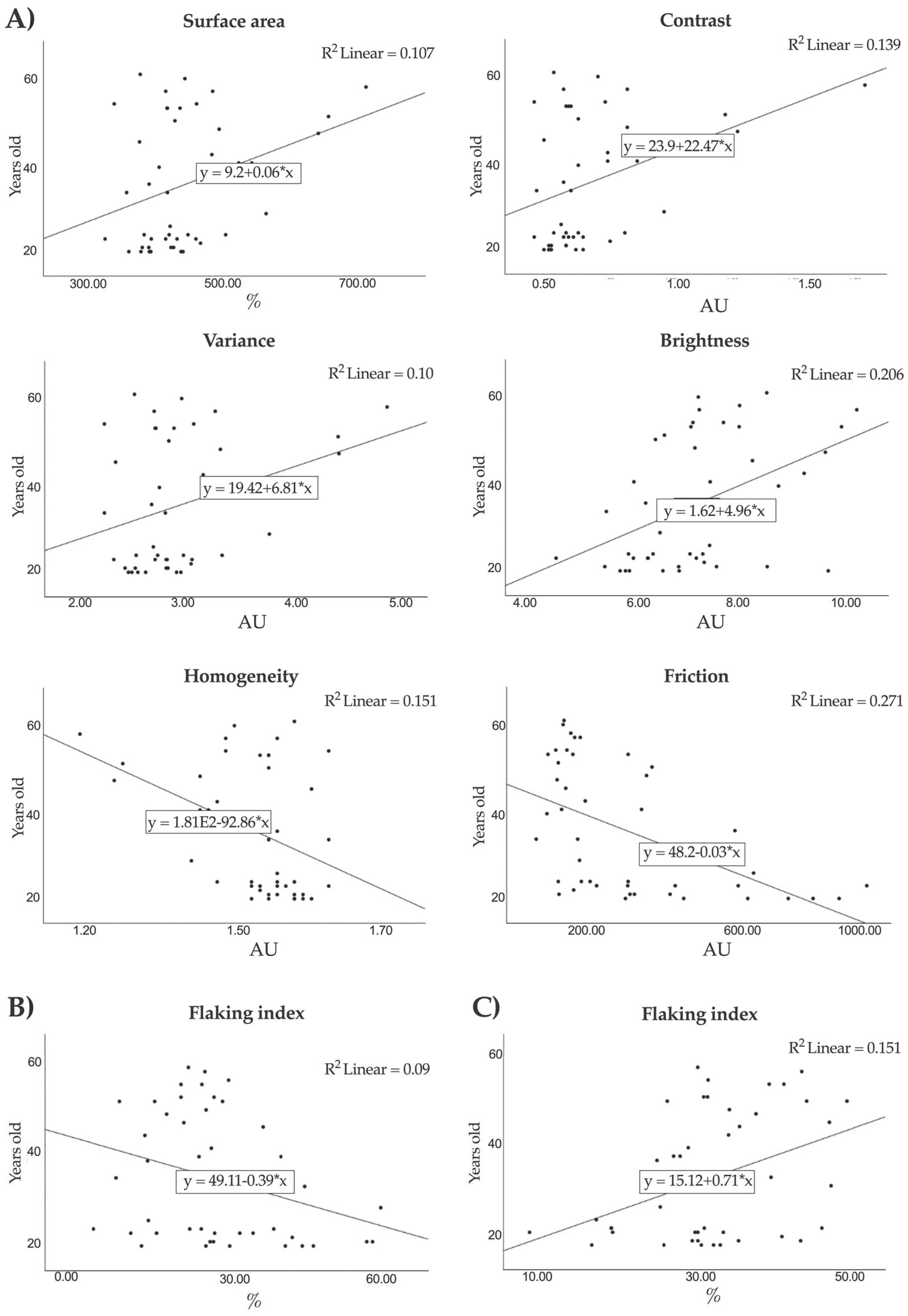
3.5. Correlation between Skin Biophysical Parameters and Age

The relationship between age and epidermal barrier function parameters was assessed (Table S3). On the forearm, a positive correlation was observed between subject age and flaking index (r = 0.388; p = 0.009); this was in contrast to the cheek, where a negative correlation was found between subject age and flaking index (r = −0.305; p = 0.044). On the palm, a positive correlation was observed between subject age and surface area (r = −0.327, p = 0.030), contrast (r = 0.373, p = 0.013), variance (r = 0.321, p = 0.034) and gloss (r = 0.454, p = 0.002). In contrast, also on the palm, a negative correlation was found between age and homogeneity (r = −0.389, p = 0.009) and also friction (r = −0.520, p < 0.001). Figure 4 shows the significant correlations between barrier function parameters and age.

3.6. Influences of Toxic Habits (Tobacco and Alcohol Use) on Skin Microtopography and Epidermal Barrier Function

Differences in skin microtopography and barrier function between smokers and non-smokers, as well as between alcohol and non-alcohol users, are shown in Table S4 and Table S5, respectively. On the palm, non-smokers showed higher smoothness (405.45 Sesm vs. 414.07 Sesm, p = 0.012), lower contrast (0.62 AU vs. 0.77 AU, p = 0.047), lower variance (2.69 AU vs. 3.20 AU, p = 0.028) and greater homogeneity (1.54 AU vs. 1.50 AU, p = 0.037). On the arm, non-smokers had higher brightness (11.33 AU vs. 6.98 AU, p = 0.007) and lower flaking index (29.32% vs. 23.75%, p = 0.041). On the cheek, there were no differences in the parameters assessed between smokers and non-smokers. Regarding alcohol use, the friction and flaking index were lower on the cheek of non-alcohol users (92.48 AU vs. 135.97 AU, p = 0.002) (23.75% vs. 29.32%, p = 0.039). In addition, alcohol users showed more friction in the palm (407.65 AU vs. 185.67 AU, p < 0.001). On the forearm, there were no differences in the parameters assessed between alcohol users vs. non-alcohol users.

4. Discussion

The present study shows the differences in skin microtopography and epidermal barrier function between cheek, forearm and palm, and proposes normal values for the parameters assessed at each anatomical location. Specifically, 15 parameters related to skin microstructure and homeostasis were evaluated, including smoothness, roughness, desquamation, wrinkles, surface area, volume, contrast, variance, homogeneity, total cell count, hardness, friction, brightness, deformability and flaking index. 10 of the 15 parameters evaluated were significantly different between forearm and cheek; 13 of the 15 parameters evaluated were different between forearm and palm and 12 of the 15 parameters evaluated were different between cheek and palm. The parameters that showed statistically significant differences in all of the comparisons analyzed (forearm vs. cheek; forearm vs. palm; cheek vs. palm) were wrinkles, surface area, volume, total number of cells, hardness, friction, deformability and flaking index. The parameters that showed differences between two of the comparisons evaluated (forearm vs. palm; cheek vs. palm, or forearm vs. palm; forearm vs. cheek) were desquamation, homogeneity, variance and contrast for the first comparison, and smoothness for the second. Roughness differed in the forearm vs. cheek comparison. Brightness was the only parameter that did not differ significantly between body sites. Finally, the forearm area showed the least variability in most parameters, followed by the cheek and finally the palm.
The differences in skin biophysical parameters between the forearm, cheek and palm can be explained by regional differences in the number of cell layers of the stratum corneum that characterize each body region [19]. Similarly, it can also be explained by variations in skin composition such as the amount of lipids, natural moisturizing factors or the number of sebaceous glands [20,21,22]. Ceramides are the major lipid component (along with cholesterol, free fatty acid, and other minor components) of the intercellular spaces of stratum corneum that forms the epidermal permeability barrier [23]. Variations of ceramide molecular profiles are characteristic at different body sites, which could be the basis for the differences in both the microstructural properties and epidermal barrier function in distinct anatomical locations. Ceramides promote water retention and maintain a healthy barrier function and hydrated skin. Therefore, regional variations in the ceramide profile could explain the differences in the various biophysical parameters of the skin. On the other hand, sociodemographic characteristics including exposome factors such as lifestyle habits (smoking and alcohol use, sun exposure, moisturizers or sunscreen use) may also have contributed to these distinctions, which are higher smoothness, brightness and homogeneity in non-smokers; higher contrast, variance and flaking index in smokers; and lower friction and flaking index in non-alcohol users [24,25,26]. Because of these variations, it is especially important to be certain about these when selecting participants for in vivo assays for cosmetic attributes [27]. Similar to the parameters assessed in the present study, other studies have found differences in barrier function parameters such as corneocyte turnover [19], TEWL [4] and SCH [28] between body sites.
Skin properties vary among individuals of different age, gender, ethnicity and skin types. Previous studies found that TEWL is higher for men, which is likely explained by them spending more time outdoors and their skin is more damaged [29]. However, measurements have shown no clear and statistically significant difference between the elastic properties, SCH, sebum or pH of the skin in females versus males at the same anatomical sites [29,30,31,32]. In our study, by comparing the different biophysical parameters of skin barrier function between sexes, significant differences were observed in parameters such as desquamation, smoothness and roughness of the forearm, as well as in roughness and brightness of the palm, which are probably either due to factors of the subjects’ exposome [25,33,34] or to sex hormones [22,29]. On the other hand, when comparing the skin barrier function parameters between ages, significant differences were observed in a greater number of parameters than in the comparison of the sexes. These parameters were surface area, contrast, variance, homogeneity, friction and brightness in the palm and flaking index on the forearm and cheek. Specifically, a positive correlation was observed between the flaking index on the forearm and cheek with age. In line with our results, barrier function as assessed by TEWL was found to increase with age [12]. This is consistent with the fact that the composition and architecture of the stratum corneum changes with aging, resulting in dry, less elastic and more pruritic skin, with a slower recovery from irritation and a greater predisposition to inflammation and infections [35]. Corneocyte desquamation, which is regulated by proteases, can be related to a thinner stratum corneum and enhanced permeability; thus, corneocyte size and organization is closely related to the skin barrier function. We also observed a positive correlation between surface area, contrast and variance with age, which is consistent with previous studies which found that the surface area should decrease after successful treatment [36]. On the other hand, we observed a negative correlation between palm homogeneity and age. Homogeneity indicates the uniformity of the image and a moisturized skin should show a higher homogeneity value than dry skin [6], just as an aged skin will be drier and less homogeneous.
These results suggest that skin differs more by age than by sex. This is supported by the fact that as we age, the skin structure changes with thinning of the epidermis and dermis, increased TEWL, fragmentation of collagen and elastin and decreased skin barrier immunity [37,38,39]. The loss of thickness can be caused by decreased cell proliferation and by significant changes that occur with age to dermal components. The extracellular matrix constituents (collagens, elastin, glycosaminoglycans, among others) are significantly reduced with intrinsic skin aging [40,41]. Moreover, oxidative stress contributes to intrinsic skin aging, not only by the increase in reactive oxygen species (ROS) generation (by mitochondrial leakage, inflammation, etc.), but also by age-related decreases in cellular repair capacity [40]. Therefore, aging results in an increased incidence of cancer and skin infections [39]. Variations in the biophysical properties of the skin may be involved in individual susceptibility to skin diseases [39]. In addition, in the present study it was observed that toxic habits affected the structural properties of the skin, including an alteration in smoothness, contrast, variance, homogeneity, brightness and flaking index in the skin of smokers and in the flaking index and friction in the skin of alcohol users. These results support the development and reliability of studies to determine the anti-aging capacity of different compounds with equipment similar to that used in the present work [42,43,44]. These future studies should be conducted while controlling for sex, age and toxic habits, to avoid biases related to the correlations defined in the present study.
This study was subject to the following limitations: (1) Its cross-sectional design. (2) The variability of skin homeostasis parameters is reliant on external conditions; however, to maximize outcome accuracy, all participants were measured in the same room with stable temperature and humidity. (3) Other body sites such as the skin of the back, abdomen or leg remain to be assessed. It would be interesting to conduct further studies to determine the normal values of microtopography and skin barrier function in the above-mentioned body sites. The main strength of our study is the novelty of the objective assessment of skin microtopography by processing and evaluating the images with the evaluation method SELS of the visioscan VC 20 plus software, inter alia with respect to the skin parameters of roughness, desquamation, wrinkles and smoothness.

5. Conclusions

This study on individuals with healthy skin describes the differences in skin mi-crotopography and epidermal barrier function between forearm, cheek and palm and shows the influence of sex and age on these parameters. The development of standardized biophysical profiles of the skin barrier in the stratum corneum would provide insight into damage factors and improve the understanding of skin diseases in the future. These findings are useful for comparison with pathological features of the skin. A deeper understanding of skin diseases will allow better targeting of medical decisions and treatments.

Supplementary Materials

The following supporting information can be downloaded at: https://www.mdpi.com/article/10.3390/cosmetics11010005/s1, Table S1: Skin biophysical parameters in subjects ≥38 vs. <38 years; Table S2: Skin biophysical parameters in males vs. females; Table S3: Correlation between skin biophysical parameters and age at different body sites; Table S4: Skin biophysical parameters in smokers vs. non-smokers; Table S5: Skin biophysical parameters in alcohol users vs. non-alcohol users.

Author Contributions

Conceptualization, T.M.-V. and S.A.-S.; methodology, M.C.-M., C.P.-L. and R.S.-d.l.T.; software, M.C.-M., C.P.-L. and T.M.-V.; validation, T.M.-V. and M.C.-M.; formal analysis, T.M.-V.; investigation, R.S.-d.l.T.; resources, T.M.-V.; data curation, R.S.-d.l.T.; writing—original draft preparation, R.S.-d.l.T.; writing—review and editing, R.S.-d.l.T., T.M.-V. and S.A.-S.; visualization, R.S.-d.l.T., T.M.-V. and S.A.-S.; supervision, T.M.-V.; project administration, S.A.-S.; funding acquisition, S.A.-S. All authors have read and agreed to the published version of the manuscript.

Funding

This research was funded by Instituto de Salud Carlos III (ISCIII) through the project PI23/01875. T.M.V. was supported by a postdoctoral fellowship from the ISCIII (CM22/00083) and R.S.-d.l.T. was supported by a predoctoral fellowship from Ministry of Universities (FPU21/00833).

Institutional Review Board Statement

This study was conducted in accordance with the Declaration of Helsinki, and approved by the Ethics Committee of Granada (HC01/0442-N-20, 08/05/2020).

Informed Consent Statement

Informed consent was obtained from all subjects involved in this study.

Data Availability Statement

The data supporting reported results are available on request from the corresponding author. The data are not publicly available due to ethical restrictions.

Acknowledgments

This research is a part of the doctoral thesis of Raquel Sanabria de la Torre in the Clinical Medicine and Public Health program of the University of Granada. This paper can only be used for the defense of the mentioned doctoral thesis.

Conflicts of Interest

The authors declare no conflicts of interest.

References

  1. Dorado, J.G.; Fraile, P.A. Anatomía y Fisiología de La Piel. Pediatr. Integral 2021, 24, 156.e1–156.e13. [Google Scholar]
  2. Baroni, A.; Buommino, E.; De Gregorio, V.; Ruocco, E.; Ruocco, V.; Wolf, R. Structure and Function of the Epidermis Related to Barrier Properties. Clin. Dermatol. 2012, 30, 257–262. [Google Scholar] [CrossRef]
  3. Espinosa-Rueda, M.I.; Montero-Vilchez, T.; Martinez-Lopez, A.; Molina-Leyva, A.; Sierra-Sánchez, A.; Arias-Santiago, S.; Buendia-Eisman, A. Cutaneous Homeostasis and Epidermal Barrier Function in a Young Healthy Caucasian Population. Eur. J. Dermatol. 2021, 31, 176–182. [Google Scholar] [CrossRef] [PubMed]
  4. Alexander, H.; Brown, S.; Danby, S.; Flohr, C. Research Techniques Made Simple: Transepidermal Water Loss Measurement as a Research Tool. J. Investig. Dermatol. 2018, 138, 2295–2300.e1. [Google Scholar] [CrossRef] [PubMed]
  5. Maroto-Morales, D.; Montero-Vilchez, T.; Arias-Santiago, S. Study of Skin Barrier Function in Psoriasis: The Impact of Emollients. Life 2021, 11, 651. [Google Scholar] [CrossRef]
  6. Trojahn, C.; Schario, M.; Dobos, G.; Blume-Peytavi, U.; Kottner, J. Reliability and Validity of Two in Vivo Measurements for Skin Surface Topography in Aged Adults. Skin Res. Technol. 2015, 21, 54–60. [Google Scholar] [CrossRef]
  7. Theek, C.; Tronnier, H.; Heinrich, U.; Braun, N. Surface Evaluation of Living Skin (SELS) Parameter Correlation Analysis Using Data Taken from Astronauts Working under Extreme Conditions of Microgravity. Ski. Res. Technol. 2020, 26, 105–111. [Google Scholar] [CrossRef]
  8. Kuklinski, L.F.; Zens, M.S.; Perry, A.E.; Green, A.C.; Karagas, M.R. Skin Microtopography as a Measure of Photoaging and Risk of Squamous Cell Carcinoma of the Skin in a US Population. Photodermatol. Photoimmunol. Photomed. 2017, 33, 41. [Google Scholar] [CrossRef]
  9. Holman, C.D.J.; Armstrong, B.K.; Evans, P.R.; Lumsden, G.J.; Dallimore, K.J.; Meehan, C.J.; Beagley, J.; Gibson, I.M. Relationship of Solar Keratosis and History of Skin Cancer to Objective Measures of Actinic Skin Damage. Br. J. Dermatol. 1984, 110, 129–138. [Google Scholar] [CrossRef]
  10. Nanzadsuren, T.; Myatav, T.; Dorjkhuu, A.; Ganbat, M.; Batbold, C.; Batsuuri, B.; Byamba, K. Skin Aging Risk Factors: A Nationwide Population Study in Mongolia Risk Factors of Skin Aging. PLoS ONE 2022, 17, e249506. [Google Scholar] [CrossRef]
  11. Montero-Vilchez, T.; Martinez-Lopez, A.; Sierra-Sanchez, A.; Soler-Gongora, M.; Jimenez-Mejias, E.; Molina-Leyva, A.; Buendia-Eisman, A.; Arias-Santiago, S. Erythema Increase Predicts Psoriasis Improvement after Phototherapy. J. Clin. Med. 2021, 10, 3897. [Google Scholar] [CrossRef] [PubMed]
  12. Epidermal, S.; Monteiro Rodrigues, L.; Querleux, B.; Gyulai, R.; Montero-Vilchez, T.; Cuenca-Barrales, C.; Rodriguez-Pozo, J.-A.; Diaz-Calvillo, P.; Tercedor-Sanchez, J.; Martinez-Lopez, A.; et al. Epidermal Barrier Function and Skin Homeostasis in Atopic Dermatitis: The Impact of Age. Life 2022, 12, 132. [Google Scholar] [CrossRef]
  13. Montero-Vilchez, T.; Martinez-Lopez, A.; Cuenca-Barrales, C.; Rodriguez-Tejero, A.; Molina-Leyva, A.; Arias-Santiago, S. Impact of Gloves and Mask Use on Epidermal Barrier Function in Health Care Workers. Dermatitis 2021, 32, 57–62. [Google Scholar] [CrossRef] [PubMed]
  14. Xiao, P.; Chen, D. The Effect of Sun Tan Lotion on Skin by Using Skin TEWL and Skin Water Content Measurements. Sensors 2022, 22, 3595. [Google Scholar] [CrossRef] [PubMed]
  15. Ordoñez-Toro, A.; Montero-Vilchez, T.; Muñoz-Baeza, J.; Sanabria-De-la-Torre, R.; Buendia-Eisman, A.; Arias-Santiago, S. The Assessment of Skin Homeostasis Changes after Using Different Types of Excipients in Healthy Individuals. Int. J. Environ. Res. Public Health 2022, 19, 16678. [Google Scholar] [CrossRef] [PubMed]
  16. John, A.J.U.K.; Galdo, F.D.; Gush, R.; Worsley, P.R. An Evaluation of Mechanical and Biophysical Skin Parameters at Different Body Locations. Skin Res. Technol. 2023, 29, e13292. [Google Scholar] [CrossRef] [PubMed]
  17. Zhou, M.; Gan, Y.; Yang, M.; He, C.; Jia, Y. Lipidomics Analysis of Facial Skin Surface Lipids between Forehead and Cheek: Association between Lipidome, TEWL, and PH. J. Cosmet. Dermatol. 2020, 19, 2752–2758. [Google Scholar] [CrossRef]
  18. Seo, J.I.; Ham, H.I.; Baek, J.H.; Shin, M.K. An Objective Skin-Type Classification Based on Non-Invasive Biophysical Parameters. J. Eur. Acad. Dermatol. Venereol. 2022, 36, 444–452. [Google Scholar] [CrossRef]
  19. Tagami, H. Location-Related Differences in Structure and Function of the Stratum Corneum with Special Emphasis on Those of the Facial Skin. Int. J. Cosmet. Sci. 2008, 30, 413–434. [Google Scholar] [CrossRef]
  20. Prost-Squarcioni, C. Histology of Skin and Hair Follicle. Med. Sci. 2006, 22, 131–137. [Google Scholar] [CrossRef]
  21. Van Smeden, J.; Bouwstra, J.A. Stratum Corneum Lipids: Their Role for the Skin Barrier Function in Healthy Subjects and Atopic Dermatitis Patients. Curr. Probl. Dermatol. 2016, 49, 8–26. [Google Scholar] [CrossRef]
  22. Dąbrowska, A.K.; Spano, F.; Derler, S.; Adlhart, C.; Spencer, N.D.; Rossi, R.M. The Relationship between Skin Function, Barrier Properties, and Body-Dependent Factors. Skin Res. Technol. 2018, 24, 165–174. [Google Scholar] [CrossRef] [PubMed]
  23. Uchida, Y.; Park, K. Ceramides in Skin Health and Disease: An Update. Am. J. Clin. Dermatol. 2021, 22, 853–866. [Google Scholar] [CrossRef] [PubMed]
  24. Nam, G.W.; Baek, J.H.; Koh, J.S.; Hwang, J.K. The Seasonal Variation in Skin Hydration, Sebum, Scaliness, Brightness and Elasticity in Korean Females. Skin Res. Technol. 2015, 21, 1–8. [Google Scholar] [CrossRef] [PubMed]
  25. Krutmann, J.; Schikowski, T.; Morita, A.; Berneburg, M. Environmentally-Induced (Extrinsic) Skin Aging: Exposomal Factors and Underlying Mechanisms. J. Investig. Dermatol. 2021, 141, 1096–1103. [Google Scholar] [CrossRef] [PubMed]
  26. Celebi Sozener, Z.; Ozdel Ozturk, B.; Cerci, P.; Turk, M.; Gorgulu Akin, B.; Akdis, M.; Altiner, S.; Ozbey, U.; Ogulur, I.; Mitamura, Y.; et al. Epithelial Barrier Hypothesis: Effect of the External Exposome on the Microbiome and Epithelial Barriers in Allergic Disease. Allergy 2022, 77, 1418–1449. [Google Scholar] [CrossRef] [PubMed]
  27. Marques, G.d.A.; Hiraishi, C.F.; Macedo, P.I.d.S.; Pinto, C.A.S.d.O.; Gregório, J.; Rosado, C.; Velasco, M.V.R.; Baby, A.R. HPLC-TBARS-EVSC (High-Performance Liquid Chromatography–Thiobarbituric Acid Reactive Substances—Ex Vivo Stratum Corneum) Protocol: Selection of the Subjects and Approach to Present the Results. Int. J. Cosmet. Sci. 2023, 45, 647–654. [Google Scholar] [CrossRef] [PubMed]
  28. Du Plessis, J.; Stefaniak, A.; Eloff, F.; John, S.; Agner, T.; Chou, T.C.; Nixon, R.; Steiner, M.; Franken, A.; Kudla, I.; et al. International Guidelines for the in Vivo Assessment of Skin Properties in Non-Clinical Settings: Part 2. Transepidermal Water Loss and Skin Hydration. Skin Res. Technol. 2013, 19, 265–278. [Google Scholar] [CrossRef]
  29. Firooz, A.; Sadr, B.; Babakoohi, S.; Sarraf-Yazdy, M.; Fanian, F.; Kazerouni-Timsar, A.; Nassiri-Kashani, M.; Naghizadeh, M.M.; Dowlati, Y. Variation of Biophysical Parameters of the Skin with Age, Gender, and Body Region. Sci. World J. 2012, 2012, 386936. [Google Scholar] [CrossRef]
  30. Wilhelm, K.P.; Cua, A.B.; Maibach, H.I. Skin Aging. Effect on Transepidermal Water Loss, Stratum Corneum Hydration, Skin Surface PH, and Casual Sebum Content. Arch. Dermatol. 1991, 127, 1806–1809. [Google Scholar] [CrossRef]
  31. Humbert, P.; Maibach, H.I.; Fanian, F.; Agache, P. Agache’s Measuring the Skin: Non-Invasive Investigations, Physiology, Normal Constants: Second Edition; Springer: Cham, Switzerland, 2017; pp. 1–1651. [Google Scholar] [CrossRef]
  32. Cua, A.B.; Wilhelm, K.P.; Maibach, H.I. Elastic Properties of Human Skin: Relation to Age, Sex, and Anatomical Region. Arch. Dermatol. Res. 1990, 282, 283–288. [Google Scholar] [CrossRef] [PubMed]
  33. Ya-Xian, Z.; Suetake, T.; Tagami, H. Number of Cell Layers of the Stratum Corneum in Normal Skin—Relationship to the Anatomical Location on the Body, Age, Sex and Physical Parameters. Arch. Dermatol. Res. 1999, 291, 555–559. [Google Scholar] [CrossRef]
  34. Dąbrowska, M.; Mielcarek, A.; Nowak, I. Evaluation of Sex-Related Changes in Skin Topography and Structure Using Innovative Skin Testing Equipment. Skin Res. Technol. 2018, 24, 614–620. [Google Scholar] [CrossRef] [PubMed]
  35. Papaccio, F.; D’arino, A.; Caputo, S.; Bellei, B. Focus on the Contribution of Oxidative Stress in Skin Aging. Antioxidants 2022, 11, 1121. [Google Scholar] [CrossRef] [PubMed]
  36. Bhat, B.B.; Kamath, P.P.; Chatterjee, S.; Bhattacherjee, R.; Nayak, U.Y. Recent Updates on Nanocosmeceutical Skin Care and Anti-Aging Products. Curr. Pharm. Des. 2022, 28, 1258–1271. [Google Scholar] [CrossRef] [PubMed]
  37. Williams, I.R.; Kupper, T.S. Immunity at the Surface: Homeostatic Mechanisms of the Skin Immune System. Life Sci. 1996, 58, 1485–1507. [Google Scholar] [CrossRef] [PubMed]
  38. Sunderkötter, C. Aging and the Skin Immune System. Arch. Dermatol. 1997, 133, 1256. [Google Scholar] [CrossRef] [PubMed]
  39. Chambers, E.S.; Vukmanovic-Stejic, M. Skin Barrier Immunity and Ageing. Immunology 2020, 160, 116–125. [Google Scholar] [CrossRef]
  40. Shin, J.W.; Kwon, S.H.; Choi, J.Y.; Na, J.I.; Huh, C.H.; Choi, H.R.; Park, K.C. Molecular Mechanisms of Dermal Aging and Antiaging Approaches. Int. J. Mol. Sci. 2019, 20, 2126. [Google Scholar] [CrossRef]
  41. Franco, A.C.; Aveleira, C.; Cavadas, C. Skin Senescence: Mechanisms and Impact on Whole-Body Aging Skin Aging and Senescence. Trends Mol. Med. 2021, 28, 97–109. [Google Scholar] [CrossRef]
  42. Alonso-Bartolomé, R.; Rodríguez, D.P.; Rodríguez-Jiménez, P.; Ruiz-Rodríguez, R. Method R: Efficacy of a Cosmetic Routine Comprising Six Topical Lipid-Encapsulated Products. J. Cosmet. Dermatol. 2022, 21, 4422–4432. [Google Scholar] [CrossRef] [PubMed]
  43. Waqas, M.K.; Khan, B.A.; Akhtar, N.; Chowdhry, F.; Khan, H.; Bakhsh, S.; Khan, S.; Rasul, A. Fabrication of Tamarindus Indica Seeds Extract Loaded-Cream for Photo-Aged Skin: Visioscan® Studies. Postepy Dermatol. Alergol. 2017, 34, 339–345. [Google Scholar] [CrossRef] [PubMed]
  44. Rattanawiwatpong, P.; Wanitphakdeedecha, R.; Bumrungpert, A.; Maiprasert, M. Anti-Aging and Brightening Effects of a Topical Treatment Containing Vitamin C, Vitamin E, and Raspberry Leaf Cell Culture Extract: A Split-Face, Randomized Controlled Trial. J. Cosmet. Dermatol. 2020, 19, 671–676. [Google Scholar] [CrossRef] [PubMed]
Figure 1. Comparison of biophysical parameters of forearm, cheek and palm skin in healthy individuals. * p < 0.05; ** p < 0.01; *** p < 0.001.
Cosmetics 11 00005 g001
Figure 2. Skin topography imaging with Visioscan VC 20plus in subjects ≥38 vs. subjects <38 years. Left: Topographic measurements on images of skin. The images show a variety of grey levels, the darker ones representing lines and wrinkles, the extremely bright ones representing plateaus of the skin microrelief. Middle: Topographic measurements on images of the skin with lines taken into account for the calculation of anisotropy. Graph of the “rose of directions’’ supporting the calculation of the anisotropy index. The distribution of the red lines in the image is calculated by the Visioscan VC 20 plus software. Directions and cells surrounded by lines (polygons) are typical parameters to characterize skin aging. Right: Desquamation measurement using corneofix. The corneocytes are analyzed by thickness in five different layers. Very thick (red), still bright (orange), medium thickness (green), least thick (light and dark blue) and the background (black).
Cosmetics 11 00005 g002
Figure 3. Skin topography imaging with Visioscan VC 20 plus in female subjects vs. male subjects. Left: Topographic measurements on images of the skin. The images show a variety of grey levels, the darker ones representing lines and wrinkles, the extremely bright ones representing plateaus of the skin microrelief. Middle: Topographic measurements on images of the skin with lines taken into account for the calculation of anisotropy. Graph of the “rose of directions” supporting the calculation of the anisotropy index. The distribution of the red lines in the image is calculated by the Visioscan VC 20 plus software. Directions and cells surrounded by lines (polygons) are typical parameters to characterize skin aging. Right: Desquamation measurement using corneofix. The corneocytes are analyzed by thickness in five different layers. Very thick (red), still bright (orange), medium thickness (green), least thick (light and dark blue) and the background (black).
Cosmetics 11 00005 g003
Figure 4. (A) Palm: Correlation between age and surface area (r = 0.327; p = 0.030), contrast (r = 0.373; p = 0.013), homogeneity (r = −0.389; p = 0.009), variance (r = 0.321; p = 0.034), brightness (r = 0.454; p = 0.002) and friction (r = −0.520; p < 0.001). (B) Cheek: Correlation between age and flaking index (r = −0.305; p = 0.044). (C) Forearm: Correlation between age and flaking index (r = 0.388; p = 0.009).
Cosmetics 11 00005 g004
Table 1. Sociodemographic characteristics.
Sociodemographic CharacteristicsParticipants (n = 44)
Age (years)38.8 ± 15.0
Sex
Female29 (65.9%)
Male15 (39.1%)
Weight (kg)67.5 ± 12.9
Height (cm)169.1 ± 10.0
Phototype
II10 (22.7%)
III27 (61.4%)
IV7 (15.9%)
Marital status
Single25 (56.8%)
Married16 (36.4%)
Divorced2 (4.5%)
Widowed1 (2.3%)
Level of education
Basic9 (20.5%)
Higher35 (79.5%)
Occupation
Physician10 (22.7)
Student11 (25.0)
Administrative6 (13.6)
Miscellaneous17 (38.7)
Smoking habit (yes)14 (31.8%)
Cigarettes/day2.1 ± 4.5
Alcohol use (yes)22 (50.0%)
Units/week0.9 ± 1.2
Moisturizer use (>3 days/week)
Body28 (63.6%)
Face33 (75.0%)
Sun exposure (hours/week)5 ± 4.6
Sunscreen use
Never3 (6.8%)
Sometimes9 (20.5%)
Always32 (72.7%)
For quantitative measures, mean and standard deviation (±) were calculated whereas for quantitative measures the absolute frequencies (%) and n are shown.
Table 2. Skin biophysical parameters according to body site in the study population.
ParameterForearmCheekPalmp *p **p ***
Smoothness (Sesm)240.02 ± 74.67348.16 ± 119.99408.19 ± 152.33<0.001<0.0010.050
Roughness (Ser)2.45 ± 1.723.64 ± 2.243.08 ± 1.480.0050.0510.188
Desquamation (Sesc)0.37 ± 0.820.70 ± 1.980.09 ± 0.190.1280.0220.002
Wrinkles (Sew)78.61 ± 23.33116.32 ± 44.04271.96 ± 138.55<0.001<0.001<0.001
Surface area (%)620.62 ± 77.51666.69 ± 123.18477.05 ± 79.130.025<0.001<0.001
Volume (mm3)90.88 ± 15.49106.16 ± 12.8383.64 ± 21.2<0.0010.041<0.001
Contrast (AU)1.09 ± 0.311.23 ± 0.430.67 ± 0.250.088<0.001<0.001
Variance (AU)* 4.29 ± 0.754.60 ± 1.012.85 ± 0.700.064<0.001<0.001
Homogeneity (AU)1.40 ± 0.061.38 ± 0.071.52 ± 0.060.155<0.001<0.001
Total number of cells (AU)120.48 ± 29.7566.36 ± 21.1785.07 ± 26.04<0.001<0.001<0.001
Hardness (AU)9.44 ± 3.987.94 ± 3.1413.22 ± 4.790.038<0.001<0.001
Friction (AU)172.21 ± 91.80114.23 ± 75.79269.95 ± 248.040.0020.002<0.001
Brightness (AU)8.36 ± 8.347.21 ± 1.37.50 ± 1.370.3770.5140.274
Deformability (mm)1.54 ± 0.351.89 ± 0.251.28 ± 0.26<0.001<0.001<0.001
Flaking index (%)31.93 ± 8.226.54 ± 11.8611.33 ± 7.10.019<0.001<0.001
p * value based on Student’s t-test for paired samples comparing the corresponding values of biophysical skin parameters between forearm and cheek. p **—value based on Student’s t-test for paired samples comparing the corresponding values of biophysical skin parameters between forearm and palm. p ***—value based on Student’s t-test for paired samples comparing the corresponding values of biophysical skin parameters between cheek and palm. p significant < 0.05.
Disclaimer/Publisher’s Note: The statements, opinions and data contained in all publications are solely those of the individual author(s) and contributor(s) and not of MDPI and/or the editor(s). MDPI and/or the editor(s) disclaim responsibility for any injury to people or property resulting from any ideas, methods, instructions or products referred to in the content.

