Article

Skin Anti-Aging Effect of Oral Vitamin A Supplementation in Combination with Topical Retinoic Acid Treatment in Comparison with Topical Treatment Alone: A Randomized, Prospective, Assessor-Blinded, Parallel Trial

by
Massimo Milani
and
Francesca Colombo
*,† on behalf of the To-Re Trial Study Group
Medical Department, Cantabria Labs Difa Cooper, 21042 Caronno Pertusella, VA, Italy
*
Author to whom correspondence should be addressed.
On behalf of the To-Re Trial Study Group: M. Meneghini (Genova), E. Rossi (Modena), F. Satolli (Parma), S. Scrivani (Parma).
Cosmetics 2023, 10(5), 144; https://doi.org/10.3390/cosmetics10050144
Submission received: 7 September 2023 / Revised: 4 October 2023 / Accepted: 12 October 2023 / Published: 18 October 2023
(This article belongs to the Collection Feature Papers in Cosmetics in 2023)

Abstract

:
Topical retinoid treatment is considered a standard therapeutic approach for chrono and photo skin aging. Retinol (vitamin A) is the precursor of endogenous retinoids. A prospective, 12-week, randomized, parallel-group trial comparing the combination of vitamins’ oral supplementation (one capsule daily, 50.000 UI vitamin A and 50 mg vitamin E) and a 0.02% retinoic acid topical gel formulation (RG) applied in the evening (Group B) in comparison with the topical RG treatment alone (Group A) was conducted. A total of 60 subjects (men and women, aged >50 years, mean age 60 ± 8 years) with moderate-severe facial skin aging (Glogau score > 2) were enrolled after their written informed consent. Thirty participants were randomly assigned to Group A and 30 to Group B. The primary endpoint was the clinical evaluation of a Skin Aging Global Score (SAGS), at baseline, and after 6 and 12 weeks. A VISIA® (Canfield Scientific, Parsippany, NJ, USA)face sculptor analysis was performed in a subgroup of 20 subjects. Skin tolerability was evaluated in both groups at weeks 6 and 12. In comparison with the baseline, SAGS scores in both groups were reduced by 13% (Group A) and by 14% (Group B) after 6 weeks and by 22% (Group A) and by 27% (Group B) at week 12. At the end of the study, SAGS score absolute reduction in Group B was significantly greater (p < 0.01) in comparison with the absolute reduction in Group A. Both treatment regimens were well tolerated. The combination of medium-high doses of oral retinol supplementation (Vitamin A) and topical retinoic acid gel showed superior efficacy in terms of clinical improvement in comparison with the topical treatment alone in subjects with moderate/severe skin aging.

1. Introduction

1.1. Skin Aging

Skin aging is a complex biological process caused by both intrinsic (chrono-aging) and extrinsic factors (photo-aging). Intrinsic skin aging occurs inevitably, as a natural consequence of physiological changes over time [1]. Different factors contribute to chrono-aging: physiological variation with anatomical modification, lifestyle, hormonal changes, and ethnicity [1]. The different ethnicities are related to different pigmentations. High pigmentation is generally protective and is able to provide a 500-fold level of protection from ultraviolet (UV) radiation [2]. The main clinical features of chrono-aging include dryness, homogeneous color, atrophy, and fine wrinkling [3]. Extrinsic factors are, to varying degrees, partially controllable. Several studies have demonstrated the role of ultraviolet radiation exposure, air pollution, tobacco smoking, and unhealthy diet in the skin aging process [4,5]. It is well known that solar exposure plays the most relevant role in extrinsic aging processes (the so-called photo aging). Long-term exposure to UV rays causes unwanted and deleterious effects on the skin and represents the major risk factor for the development of both non-melanoma and melanoma skin cancer. The clinical signs of photo aging are coarse wrinkles, dark spots, skin xerosis, reduced skin elasticity, and sallowness of the skin [6]. Permanent visible changes of the skin could have a significant emotional and psychological impact on the quality of life [7]. In addition, skin aging is associated with medical implications, such as xerosis, pruritus, and skin damage (dermatoporosis), which represent common and problematic conditions associated with skin aging [3]. Skin aging is a highly complex and not fully understood process, and different mechanisms are involved in its development.

1.1.1. Oxidative Stress and Skin Aging

Reactive oxygen species (ROS) are a class of reactive substances containing oxygen. The main ROS in the body are: superoxide anion (O2−), hydrogen peroxide (H2O2), hydroxyl radical (·OH), singlet oxygen (1O2), lipid peroxides, and nitrogen oxides. They mainly derive from cellular oxidative metabolism and UV radiation [8,9]. Under normal conditions, ROS production and clearance are in a delicate dynamic balance, in which antioxidants play a key role [9]. Generally speaking, the concentration of antioxidants is higher in the epidermal layer than the dermal layer [10].
According to the free radical or oxidative stress theory of aging, oxidative cellular damage represents one of the main contributors to the aging process. Excessive ROS can directly damage cells, causing lipid, protein, nucleic acid, and mitochondrial DNA damage [11]. ROS, through the stimulation of mitogen-activated protein kinase (MAPK) and the activation of the heterodimer activator protein 1 (AP-1), induces the synthesis of matrix metalloproteinases (MMPs) [12] responsible for collagen degradation. A study by Fisher et al. confirmed the role of MMPs as mediators of collagen damage: the UV radiation determined an elevation of MMPs that degraded skin collagen contributing to photo aging [13]. The skin is well-equipped with several weapons to deal with oxidative stress, such as physical (skin barrier) and biological components (antioxidant molecules). However excessive sunlight exposure and other free radicals generated (e.g., from pollution, smoking, unhealthy diet) can overwhelm these protective systems inducing oxidative damage.

1.1.2. Autophagy and Skin Aging

Autophagy is a mechanism involved in cellular homeostasis; it represents one of the survival mechanisms of cells in the presence of intrinsic and extrinsic stress. Autophagy is a process of cellular self-digestion in which waste material is delivered to lysosomes for breakdown [14] Autophagy includes three main pathways: macro autophagy, micro autophagy, and chaperone-mediated autophagy [15].
Autophagy is involved in cellular senescence and skin aging, although their relationship is still unclear [9]. Recent studies have underlined that skin aging could be partially caused by defects of autophagy [14]. The autophagy process has a key role in homeostasis, intracellular remodeling, and suppressing the accumulation of dangerous compounds. Therefore, the alteration of this process leads to a decrease in the protective function of skin epithelia [14]. Although UV radiation is able to activate autophagy in epidermal keratinocyte, melanocytes, dermal fibroblasts, and sebaceous gland cells’ excessive UV irradiation interfere with this process. Chronic UVA exposure determined lysosomal dysfunction in skin fibroblasts, while excessive UVB irradiation also leads to decreased autophagy function in fibroblasts in vitro [15].
Considering that autophagy plays a potentially crucial role in counteracting aging, strategies aimed at its modulation could be promising for the prevention of skin aging [16,17].

1.2. Topical Retinoids and Skin Aging

Different anti-aging approaches have been developed to rejuvenate aged skin and prevent age-related skin diseases. Among anti-aging substances, retinoids and alpha-hydroxy acids are the actives commonly used at the topical level [7] and retinoic acid represents the gold standard treatment [3]. Retinoic acid and its metabolic precursor, retinol, increase collagen formation, preventing collagen degradation in the skin and leading to a clinical improvement in wrinkles. In addition, retinoic acid also determined an improvement in dyspigmentation [3]. Recently, a clinical trial showed the clinical efficacy and tolerability of a medical device containing retinoic acid (0.02%) and glycolic acid (4%) in a polyvinyl gel vehicle (RG) [7] as an anti-skin aging strategy. The RG significantly reduced dark spots (−40%) and severity of wrinkles (−12%) after 4 weeks compared to the baseline [7]. Retinoids have a well-established role in cellular differentiation [18]. Recent studies underlined that retinoic acid is also able to promote autophagosome maturation [19]. The pharmacological modulation of autophagic activity has been suggested as a strategy for therapy that involves different clinical situations, including premature aging [19]. Therefore, retinoids can favor the process of autophagy; this could be an additional anti-aging mechanism of these molecules.

1.3. Vitamins and Skin Aging

Vitamin A is a non-esterified free form of retinoid and has to be converted by the body into the active form (retinoic acid) by two conversion steps [8]. Retinoids are involved in many biological processes, such as inflammation, differentiation, proliferation, vision, growth, and apoptosis [9]. Retinoids, including retinol, are the actives commonly used at the topical level to counteract skin aging. Is demonstrated that retinol is able to stimulate new collagen deposition, reducing MMPs expression. However, levels of retinoids in the skin decline when exposed to ultraviolet irradiation [20]. Adequate assumption of Vitamin A (retinol) is therefore necessary for the growth of healthy skin and hair [20].
Vitamin E (tocopherols) is a vitamin with well-known antioxidant properties. It is able to counteract radicals and protect the skin from solar radiation [10]. Indeed, the use of antioxidants is an effective strategy to counteract the skin-aging processes [21]. Among the activities of tocopherols, Vitamin E stimulates glutathione (GSH) synthesis in HaCaT keratinocytes [22], suppresses UVB-induced edema, erythema, and lipid peroxidation, and reduces some UVA negative effects [23].
In recent years, the connection between nutrition and skin condition has been underlined. Different nutrients such as carotenoids, tocopherols, flavonoids, vitamins (A, C, D and E), essential omega-3 fatty acids, some proteins, and probiotics have been referred to be agents capable of promoting skin health and beauty [24,25]. These substances have been widely used in the skincare industry, both topically and as oral supplements to improve skin appearance [26].
Both Vitamins A and E must be regularly consumed in our diet [27]. Recently, a drug containing Vitamin A (50000 UI) and Vitamin E (50 mg) has been proposed as an adjuvant anti-aging treatment. However, no clinical data are available regarding the anti-aging effect of the association between oral Vitamin A and Vitamin E (“In” strategy) and the topic utilization of a gel (RG) containing retinoic acid (0.02%) and glycolic acid (4%) (“Out” strategy). This “In&Out” strategy, which combines antioxidants (Vitamin E) and retinoids (Vitamin A and retinoic acid), could represent an interesting approach to counteract the oxidative stress and the defects of autophagy involved in skin aging.
In this context, a prospective, randomized, controlled, 3-month, assessor-blinded, trial was conducted to assess the clinical efficacy and the tolerability of this “In&Out” strategy.

2. Materials and Methods

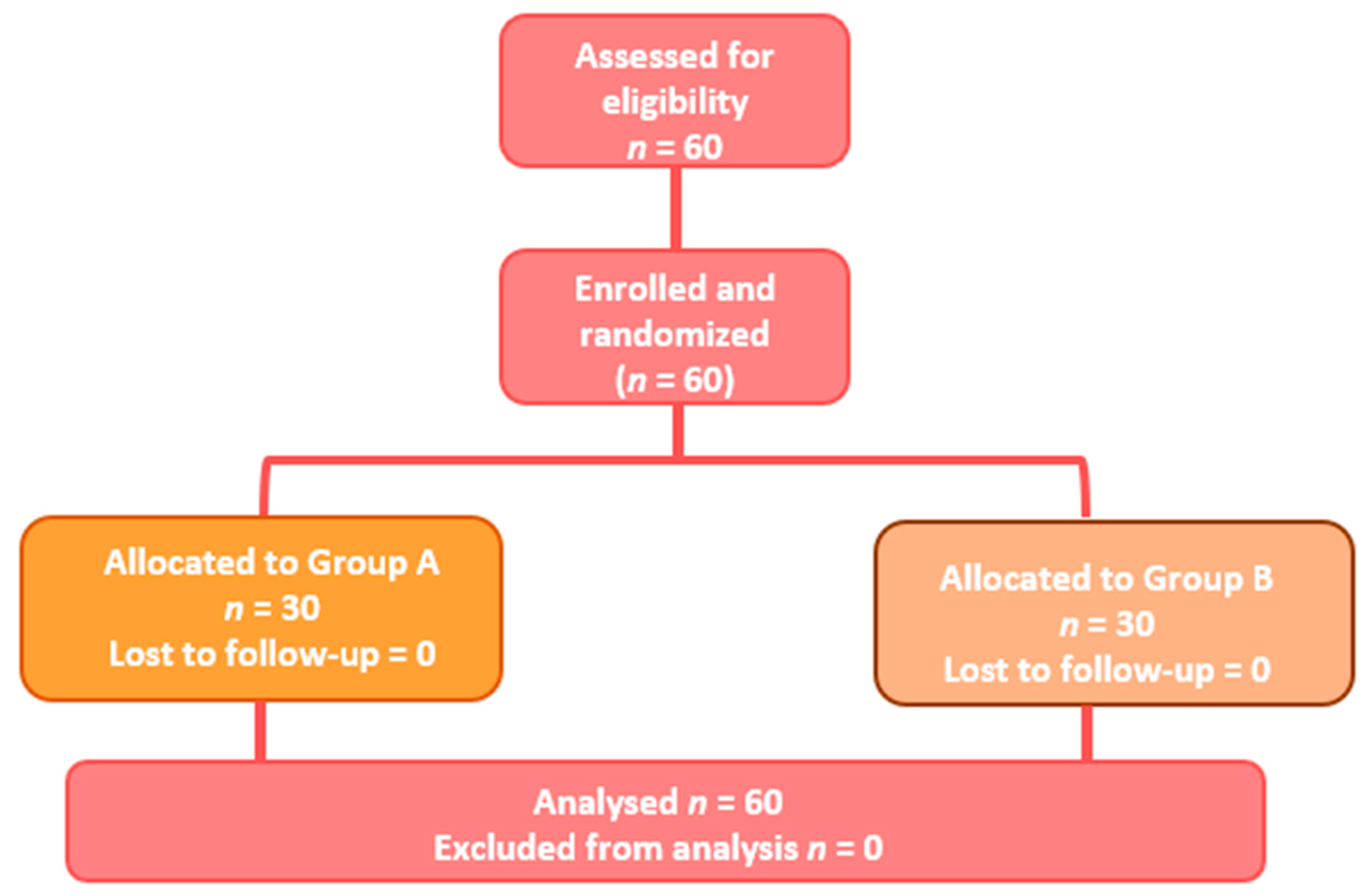
2.1. Population and Study Design
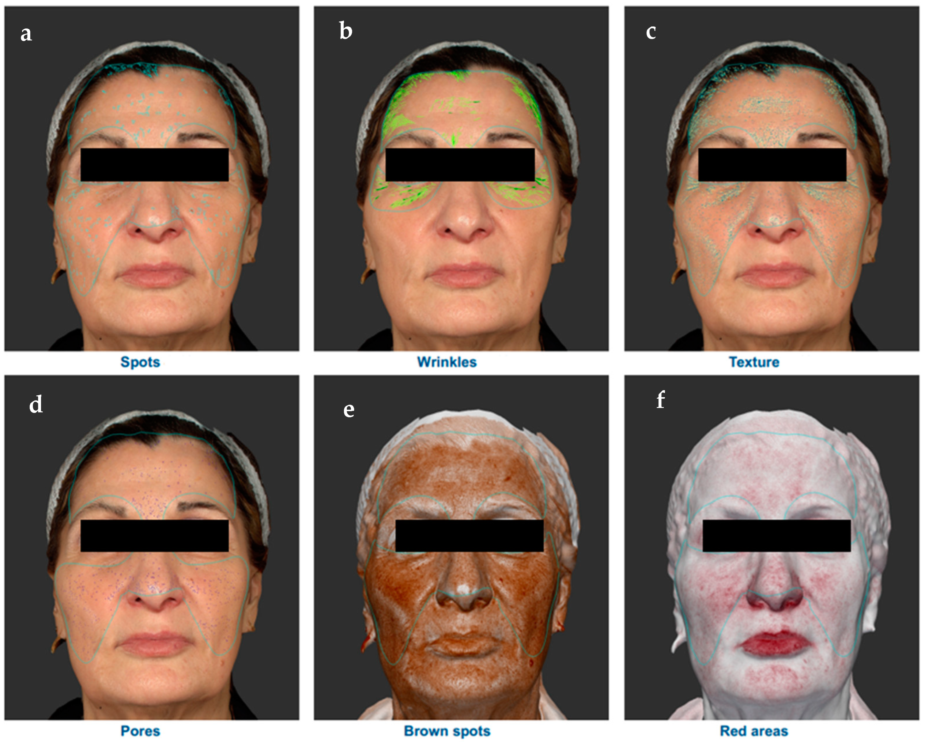
Between September 2022 and June 2023, a total of 60 participants (56 women and 4 men, median age 60 ± 8 years), were enrolled and randomized in a 1:1 allocation ratio and instructed to apply the medical device containing retinoic acid (0.02%) and glycolic acid (4%) once a day every two days (Group A) or to apply the medical device and orally consume the drug containing Vitamin A (50000 UI) and Vitamin E (50 mg), one capsule daily (Group B). The randomization list was generated by a dedicated computer program. Both products were commercially available (Cantabria Labs Difa Cooper, Caronno Pertusella, Italy).
The main inclusion criteria were men and women with age > 50 years, with moderate-severe skin aging (Glogau Score > 2). The main exclusion criteria were pregnancy or breastfeeding, clinically relevant liver disease, untreated dyslipidemia, and acute skin diseases. The study was conducted over 12 weeks, and participants were evaluated at the baseline and after 6 and 12 weeks. The trial took place in four different dermatology clinics in Italy. The trial was conducted according to Good Clinical Practice Guidelines and Helsinki Declaration. All subjects provided a signed informed consent.

2.2. Study Outcomes

The primary efficacy outcome was evaluated in an assessor-blinded fashion by a panel of four dermatologists (To-Re Trial Study Group) at the baseline and after 6 and 12 weeks through the Skin Aging Global Score (SAGS), a subjective score that asses six parameters (elasticity, wrinkle, roughness, pigmentation, erythema, and skin pores) with a 5-point score for each item (from 0 to 4; maximum score that indicated the highest skin-aging condition: 24). A VISIA® objective face sculptor analysis was performed in a subgroup of 20 subjects.
The VISIA Complexion Analysis Camera (Canfield Scientific, Parsippany, New Jersey, United States) is a camera system that uses specific lighting (standardized and cross-polarized lighting) to evaluate different skin surface characteristics: (1) spots on the skin surface (they occur when melanin coagulates below the skin surface as a result of sun damage and are generally invisible under light conditions); (2) wrinkles; (3) skin texture; (4) pores; (5) brown spots (occur from an excess of melanin, they are lesions on the skin such as hyper-pigmentation, freckles, lentigines, and melasma); (6) red areas (blood vessels and hemoglobin) (Figure 1).
The results are expressed as percentile scores using, as a comparison, a database of skin features of a group of people of the same age and skin type. The 50% percentile represents the average of the reference group, higher scores are associated with an improvement in terms of skin condition [28].
In addition, a final evaluation of efficacy was conducted by investigators with a 4-point subjective score from 0 (none) to 3 (excellent) after 12 weeks, considering different final effects (firming effect, volume effect, nourishing effect, or illuminating effect). The nourishing effect refers to a subjective evaluation of the improvement of the skin appearance in terms of tone and texture, as a possible consequence of a skin barrier function recovery through the reconstitution of the hydrophilic film. The illuminating effect refers to an improvement in the glow and radiance of the skin.
The tolerability of products was self-reported and evaluated after 6 and 12 weeks with a 3-point score from 0 (scarce) to 2 (excellent).

2.3. Statistical Analysis
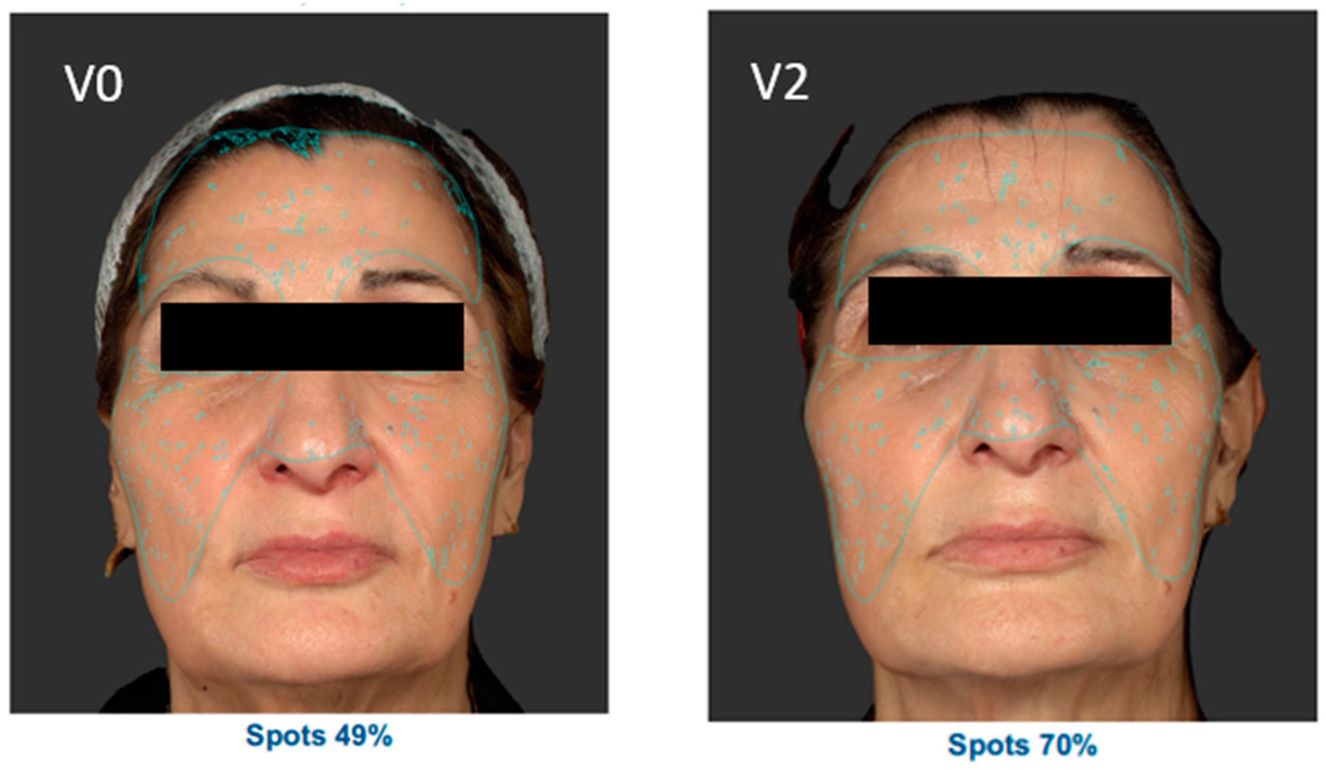
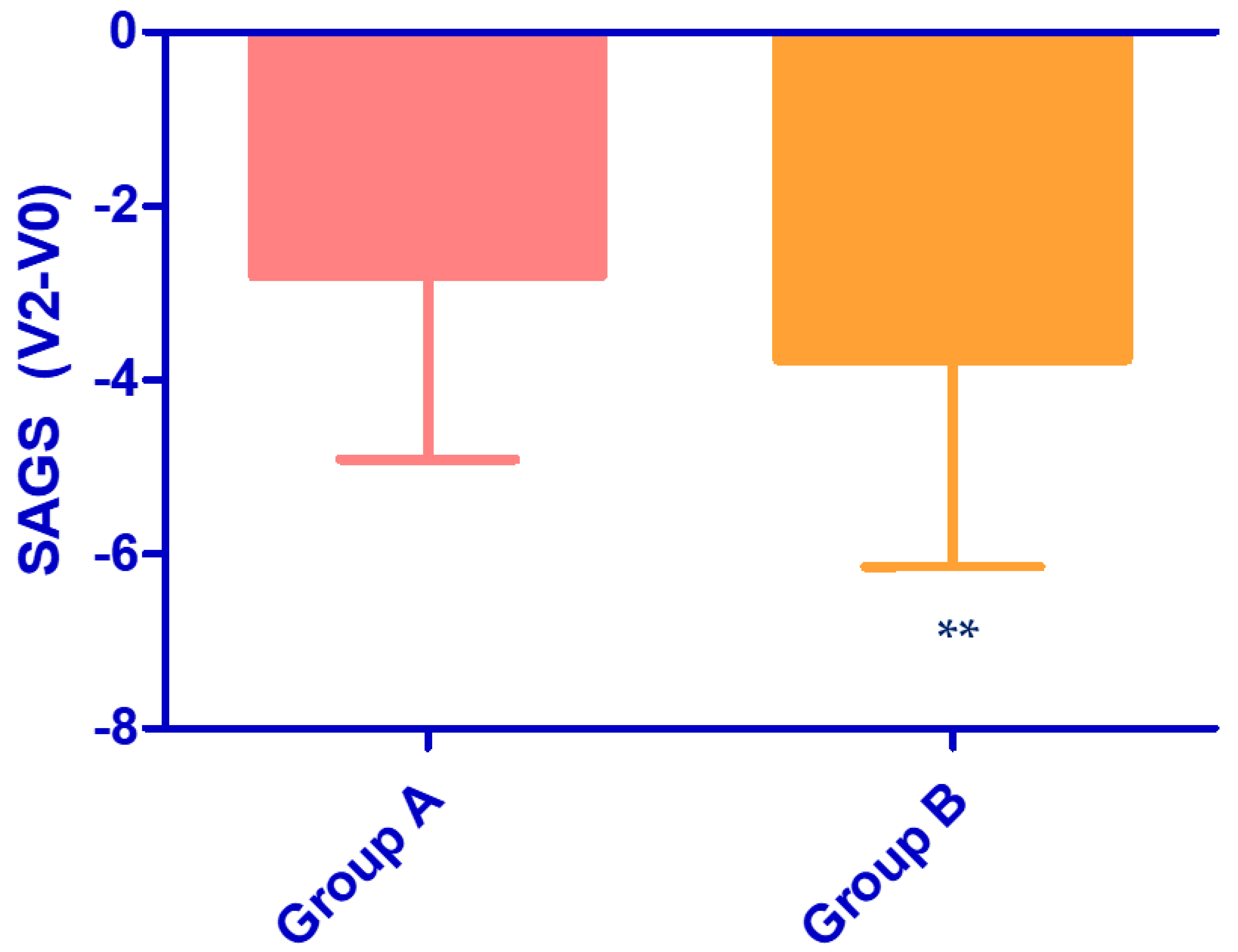
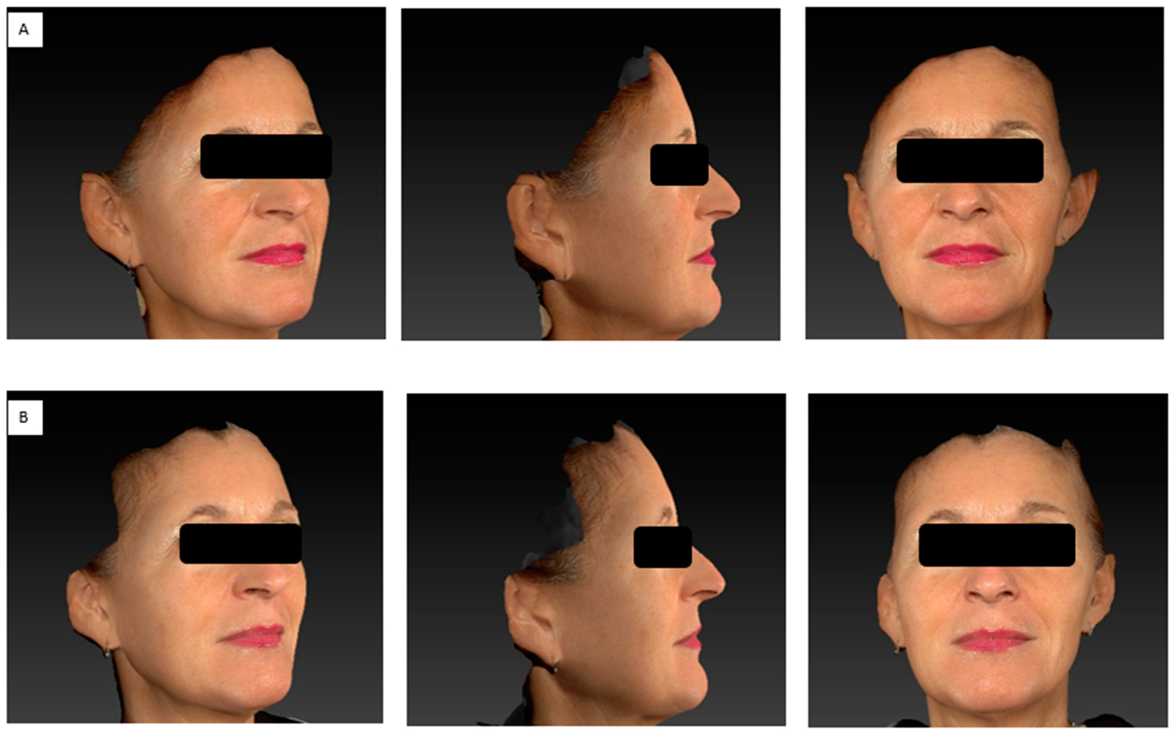
A total of 60 participants were enrolled and randomized in a 1:1 allocation ratio. The sample size calculation was performed considering the data of Campione et al. [7], which reported a reduction of −12% in wrinkle severity after the topical application of a gel containing retinoic acid (0.02%) and glycolic acid (4%), and a hypothesis to find an additional effect in SGAS reduction of 25% from the combination of the In&Out strategy. With an effect size (Choen’s d value) of 0.75, an alpha value of 0.05, and a power of 85%, a total of at least 54 subjects should be enrolled. The sample size was calculated using G*Power statistical software version 3.1.9.4 (G*Power, Heinrich Heine University, Kiel, Germany). Statistical analyses were conducted using GraphPad statistical software version 5.0 (GraphPad Software Inc., La Jolla, CA, USA). A non-parametric unpaired test was used to compare the different groups, while the ANOVA test was applied to compare data at baseline and after 6 and 12 weeks. Data are expressed as mean ± standard deviation (SD), and a p-value < 0.05 was considered significant.

3. Results

The trial was carried out between September 2022 and June 2023. A total of 60 participants were enrolled and completed the study. Subjects were randomized into two groups: Group A (n = 30, mean age 59.9 ± 8.5 years, mean SAGS at baseline 12.8 ± 4.0) and Group B (n = 30, mean age 60.3 ± 8.3 years, mean SAGS at baseline 13.8 ± 4.6). Demographic and baseline characteristics were similar between groups (p > 0.05). The flow diagram of the study is illustrated in Figure 2.
The Skin Aging Global Score (SAGS), based on the sum of different six parameters (elasticity, wrinkle, roughness, pigmentation, erythema, and skin pores) decreased in both groups (Table 1): The reduction was statistically significant after 12 weeks in Group A (−22%, p < 0.05) and in Group B (−27%, p < 0.01). As illustrated in Figure 3, the reduction in SAGS after 12 weeks compared to the baseline was significantly higher in Group B compared to Group A (−3.8 ± 2.4 and −2.8 ± 2.1, respectively, p < 0.01).
Regarding the score of the single parameters considered, it generally decreased in both groups, resulting in an improvement in skin aging. The skin elasticity statistically improved after 12 weeks in Group A (score reduction −25%, p < 0.01) and both after 6 and 12 weeks in Group B (score reduction −21%, p < 0.05, and −32%, p < 0.01, respectively). The roughness (p < 0.001) and pigmentation (p < 0.05) statistically improved in both groups after 12 weeks. A statistical improvement in skin pores was observed only in Group B after 12 weeks (p < 0.05).
The In&out strategy allowed us to obtain a better global efficacy for all the parameters considered (Figure 4). In particular, a statistically better firing and volume effect (p < 0.05) was observed in Group B.
A VISIA® face sculptor analysis was performed in a subgroup of 20 subjects. Figure 5 reports the VISIA® analysis of a subject in Group B.
Regarding the VISIA® face sculptor analysis, no statistical differences were observed between V0 (baseline) and V2 (12 weeks) for all the parameters considered. However, a positive trend was observed in Group B (oral Vitamin A+ topical retinoic acid gel) for different parameters: spots, pores, and red areas.
Regarding the spots on the skin surface, a positive result in terms of percentile was observed in 80% of subjects in Group B compared to 40% of subjects in Group A (p = 0.067).
Figure 6 reports the result on the spots parameter evaluated on V0 (baseline) and V2 (12 weeks) in a subject of Group B (oral Vitamin A+ topical retinoic acid gel).
Both treatment regimens were well tolerated. The tolerability of the topical treatment containing retinoic acid (0.02%) and glycolic acid (4%) was judged to be excellent by 90% of subjects in Group A and 93.3% of subjects in Group B. The tolerability of the oral treatment was considered excellent by 96.7% of participants in Group B.

4. Discussion

The present study suggested that a Vitamin A oral supplementation provided an additional effect in improving skin appearance in subjects with mild to moderate skin aging treated with a retinol-based topical product, supporting the value of this “In&Out” strategy. The combination of oral and topical treatments determined an absolute reduction in SAGS (Skin Aging Global Score) significantly greater (p < 0.01) in comparison with the group that used only the topical approach (absolute difference between Group A and Group B—0.97, 95% CI: from 0.33 to 1.61). In addition, both treatment regimens were well tolerated. No statistical differences were observed between V0 (baseline) and V2 (12 weeks) for all the parameters considered with VISIA® analysis. This could be due to the relatively short treatment period considered and the limited subjects evaluated (20 subjects compared to 60 participants enrolled in the study). However, a positive trend was observed in Group B (oral Vitamin A+ topical retinoic acid gel) for different parameters (spots, pores, and red areas).
Skin aging is determined by well-known cellular pathways. Oxidative stress, induced by reactive oxygen species (ROS), is one of the main mechanisms involved in skin aging, leading to lipid, protein, and DNA damage [9]. Autophagy is an intracellular stress response enhanced when cellular components are damaged. Therefore, this mechanism could play a crucial role in counteracting skin aging [17]. Retinoids, including retinol, represent reference anti-aging substances and are widely used in topical products. In recent years, the connection between nutrition and skin condition has been underlined. The diet is closely related to aging, and nutrients, such as vitamins, can directly affect the skin [11]. Retinol, or Vitamin A, is a fat-soluble vitamin essential for vision, but also for healthy skin and mucous membrane surface, reproduction, and the integrity of the immune system [29]. Retinol is able to regulate the growth, differentiation, and maintenance of epithelial tissues [26]; inhibit the UV-induced, MMP-mediated breakdown of collagen; and protect against UV-induced decreases in procollagen expression [13,30,31]. In addition, recent studies underlined that retinoic acid is also able to promote autophagosome maturation [19]. On the other hand, Vitamin E represents the physiological antioxidant of lipid structures, contributing to stabilizing the cell membranes and acting also at the sebum level [32]. The role of Vitamin A supplementation as a chemopreventive agent was previously observed. In a large Phase III trial, the oral administration of retinol (25,000 IU/day for up to 5 years) in 2297 randomized participants with evidence of moderate to severe actinic keratosis, was associated with a 32% reduction in the risk of squamous cell carcinoma [33,34]. Another trial evaluated the safety and efficacy of retinyl palmitate supplementation (doses range of 25,000–75,000 IU/day for 1 year) in the treatment of patients with sun-damaged skin. The study underlined that the doses of 50,000 and 75,000 IU/day were more efficacious, showing a similar safety profile of the low dose considered (25,000 IU/day) [35]. Despite the possible role of nutrients in modulating some skin conditions, the consensus is lacking on the use of oral supplements and systemic treatments for skin aging and photo aging. To date, numerous clinical trials evaluated the role of oral hydrolyzed collagen and oral hyaluronic acid on skin aging. Evidence on other nutritional supplementation (such as vitamins, flavonoids, and plant extracts) is lacking [36].
The supplementation of antioxidants and oral retinoids might be a protective strategy to counteract the skin-aging process associated with skin oxidative damage and reduce autophagy activity. Until now, no clinical data are available on the anti-aging effect of oral treatment with retinol or retinal derivatives. For this reason, this prospective, randomized, controlled, 3-month, assessor-blinded, trial was conducted. Our data underlined the potential role of nutritional supplementation in skin-aging prevention.
Some limitations should be considered in evaluating our study results. The main limitation was that this study is an open trial, however, to increase the internal validity of our results, we adopted an assessor-blinded evaluation of the primary outcome. Another limitation is the lack of an oral placebo in Group A. It is well known that placebo is able to increase the internal validity of a study. However, considering the complexity of designing a well-conducted placebo control trial, studies with cosmetics generally do not adopt this strategy. Vitamin A and E levels were not considered before starting the study, and this could represent a selection bias. However, hypovitaminosis A and E is a rare condition in industrialized countries; the randomization procedure is a useful tool to reduce the risk of this kind of bias.

5. Conclusions

This study underlined the potential role of nutritional supplementation in improving skin health. The results of the present trial confirmed that the combination of an “In & Out” strategy of oral retinol supplementation and topical retinoic acid gel shows superior efficacy in terms of clinical improvement in comparison with topical retinoic acid treatment alone in subjects with moderate/severe skin aging.

Author Contributions

M.M. and F.C. participated in the study design and drafting of the manuscript. All authors contributed to the review and final approval of the manuscript. All authors have read and agreed to the published version of the manuscript.

Funding

The present trial was supported by an unrestricted grant of Cantabria Labs Difa Cooper.

Institutional Review Board Statement

No ethical approval was required since our study was based on the application of a topical gel which is not a drug and the consumption of a drug over the counter which did not require a doctor’s prescription.

Informed Consent Statement

All participants provided written informed consent and a photo consent statement before starting the study. Written informed consent has been obtained from the patients to publish this paper.

Data Availability Statement

The data that support the findings of this study are available on request from the corresponding author. The data are not publicly available due to privacy or ethical restrictions.

Conflicts of Interest

M.M. and F.C. are employees of Difa Cooper Cantabria Labs which commercialized the products used in the trial. The authors declare that the research was conducted in the absence of any commercial or financial relationships that could be construed as a potential conflict of interest. The funders had no role in the design of the study; in the collection, analyses or interpretation of data; in the writing of the manuscript; or in the decision to publish the results.

References

  1. Farage, M.A.; Miller, K.W.; Elsner, P.; Maibach, H.I. Intrinsic and extrinsic factors in skin ageing: A review. Int. J. Cosmet. Sci. 2008, 30, 87–95. [Google Scholar] [CrossRef]
  2. Rees, J.L. The Genetics of Sun Sensitivity in Humans. Am. J. Hum. Genet. 2004, 75, 739–751. [Google Scholar] [CrossRef]
  3. Sachs, D.L.; Fisher, G.; Voorhees, J.J. Skin Ageing. Rook’s Textbook of Dermatology, 9th ed.; Wiley: Hoboken, NJ, USA, 2016; pp. 1–11. [Google Scholar] [CrossRef]
  4. Fisher, G.J.; Kang, S.; Varani, J.; Bata-Csorgo, Z.; Wan, Y.; Datta, S.; Voorhees, J.J. Mechanisms of Photoaging and Chronological Skin Aging. Arch. Dermatol. 2002, 138, 1462–1470. [Google Scholar] [CrossRef]
  5. Krutmann, J.; Bouloc, A.; Sore, G.; Bernard, B.A.; Passeron, T. The skin aging exposome. J. Dermatol. Sci. 2017, 85, 152–161. [Google Scholar] [CrossRef]
  6. Quan, T.; Fisher, G.J. Role of Age-Associated Alterations of the Dermal Extracellular Matrix Microenvironment in Human Skin Aging: A Mini-Review. Gerontology 2015, 61, 427–434. [Google Scholar] [CrossRef]
  7. Campione, E.; Cosio, T.; Lanna, C.; Mazzilli, S.; Dika, E.; Bianchi, L. Clinical efficacy and reflectance confocal microscopy monitoring in moderate-severe skin aging treated with a polyvinyl gel containing retinoic and glycolic acid: An assessor-blinded 1-month study proof-of-concept trial. J. Cosmet. Dermatol. 2021, 20, 310–315. [Google Scholar] [CrossRef]
  8. Silva, A.R.; Menezes, P.F.C.; Martinello, T.; Novakovich, G.F.L.; Praes, C.E.O.; Feferman, I.H.S. Antioxidant kinetics of plant-derived substances and extracts. Int. J. Cosmet. Sci. 2010, 32, 73–80. [Google Scholar] [CrossRef]
  9. Gu, Y.; Han, J.; Jiang, C.; Zhang, Y. Biomarkers, oxidative stress and autophagy in skin aging. Ageing Res. Rev. 2020, 59, 101036. [Google Scholar] [CrossRef]
  10. Rinnerthaler, M.; Bischof, J.; Streubel, M.; Trost, A.; Richter, K. Oxidative Stress in Aging Human Skin. Biomolecules 2015, 5, 545–589. [Google Scholar] [CrossRef]
  11. Gragnani, A.; Cornick SMac Chominski, V.; Ribeiro de Noronha, S.M.; Alves Corrêa de Noronha, S.A.; Ferreira, L.M. Review of Major Theories of Skin Aging. Adv. Aging Res. 2014, 3, 265–284. [Google Scholar] [CrossRef]
  12. Chen, T.; Hou, H.; Fan, Y.; Wang, S.; Chen, Q.; Si, L.; Li, B. Protective effect of gelatin peptides from pacific cod skin against photoaging by inhibiting the expression of MMPs via MAPK signaling pathway. J. Photochem. Photobiol. B 2016, 165, 34–41. [Google Scholar] [CrossRef] [PubMed]
  13. Fisher, G.J.; Wang, Z.; Datta, S.C.; Varani, J.; Kang, S.; Voorhees, J.J. Pathophysiology of Premature Skin Aging Induced by Ultraviolet Light. N. Engl. J. Med. 1997, 337, 1419–1429. [Google Scholar] [CrossRef] [PubMed]
  14. Eckhart, L.; Tschachler, E.; Gruber, F. Autophagic Control of Skin Aging. Front. Cell Dev. Biol. 2019, 7, 143. [Google Scholar] [CrossRef]
  15. Ma, J.; Teng, Y.; Huang, Y.; Tao, X.; Fan, Y. Autophagy plays an essential role in ultraviolet radiation-driven skin photoaging. Front. Pharmacol. 2022, 13, 864331. [Google Scholar] [CrossRef] [PubMed]
  16. Wang, M.; Charareh, P.; Lei, X.; Zhong, J.L. Autophagy: Multiple Mechanisms to Protect Skin from Ultraviolet Radiation-Driven Photoaging. Oxid. Med. Cell. Longev. 2019, 2019, 8135985. [Google Scholar] [CrossRef] [PubMed]
  17. Kim, H.; Park, S.-Y.; Moon, S.; Lee, J.; Kim, S. Autophagy in Human Skin Fibroblasts: Impact of Age. Int. J. Mol. Sci. 2018, 19, 2254. [Google Scholar] [CrossRef]
  18. Orfali, N.; McKenna, S.L.; Cahill, M.R.; Gudas, L.J.; Mongan, N.P. Retinoid receptor signaling and autophagy in acute promyelocytic leukemia. Exp. Cell Res. 2014, 324, 1–12. [Google Scholar] [CrossRef]
  19. Rajawat, Y.; Hilioti, Z.; Bossis, I. Autophagy: A target for retinoic acids. Autophagy 2010, 6, 1224–1226. [Google Scholar] [CrossRef]
  20. VanBuren, C.A.; Everts, H.B. Vitamin A in Skin and Hair: An Update. Nutrients 2022, 14, 2952. [Google Scholar] [CrossRef]
  21. Masaki, H. Role of antioxidants in the skin: Anti-aging effects. J. Dermatol. Sci. 2010, 58, 85–90. [Google Scholar] [CrossRef]
  22. Masaki, H.; Okano, Y.; Ochiai, Y.; Obayashi, K.; Akamatsu, H.; Sakurai, H. α-Tocopherol Increases the Intracellular Glutathione Level in HaCaT Keratinocytes. Free Radic. Res. 2002, 36, 705–709. [Google Scholar] [CrossRef] [PubMed]
  23. Wu, S.; Gao, J.; Dinh, Q.T.; Chen, C.; Fimmel, S. IL-8 production and AP-1 transactivation induced by UVA in human keratinocytes: Roles of d-α-tocopherol. Mol. Immunol. 2008, 45, 2288–2296. [Google Scholar] [CrossRef] [PubMed]
  24. Ristow, M.; Zarse, K.; Oberbach, A.; Klöting, N.; Birringer, M.; Kiehntopf, M.; Stumvoll, M.; Kahn, C.R.; Blüher, M. Antioxidants prevent health-promoting effects of physical exercise in humans. Proc. Natl. Acad. Sci. USA 2009, 106, 8665–8670. [Google Scholar] [CrossRef] [PubMed]
  25. Ristow, M.; Schmeisser, S. Extending life span by increasing oxidative stress. Free Radic. Biol. Med. 2011, 51, 327–336. [Google Scholar] [CrossRef] [PubMed]
  26. Schagen, S.K.; Zampeli, V.A.; Makrantonaki, E.; Zouboulis, C.C. Discovering the link between nutrition and skin aging. Dermatoendocrinology 2012, 4, 298–307. [Google Scholar] [CrossRef] [PubMed]
  27. Praça, F.G.; Viegas, J.S.R.; Peh, H.Y.; Garbin, T.N.; Medina, W.S.G.; Bentley, M.V.L.B. Microemulsion co-delivering vitamin A and vitamin E as a new platform for topical treatment of acute skin inflammation. Mater. Sci. Eng. C 2020, 110, 110639. [Google Scholar] [CrossRef]
  28. Henseler, H. Investigation of the precision of the Visia® complexion analysis camera system in the assessment of skin surface features. GMS Interdiscip Plast. Reconstr. Surg DGPW 2022, 11, Doc08. [Google Scholar] [CrossRef]
  29. Navarra, T. The Encyclopedia of Vitamins, Minerals and Supplements, 2nd ed.; Facts on File Inc.: New York, NY, USA, 2004. [Google Scholar]
  30. Fisher, G.J.; Datta, S.; Wang, Z.; Li, X.Y.; Quan, T.; Chung, J.H.; Kang, S.; Voorhees, J.J. c-Jun–dependent inhibition of cutaneous procollagen transcription following ultraviolet irradiation is reversed by all-trans retinoic acid. J. Clin. Investig. 2000, 106, 663–670. [Google Scholar] [CrossRef]
  31. Lee, S.-J.; Cho, S.-A.; An, S.-S.; Na, Y.-J.; Park, N.-H.; Kim, H.-S.; Lee, C.-W.; Kim, H.-K.; Kim, E.-K.; Jang, Y.-P.; et al. Significantly Inhibits Retinoid-Induced Skin Irritation In Vitro and In Vivo. Evid.-Based Complement. Altern. Med. 2012, 2012, 190370. [Google Scholar] [CrossRef]
  32. Packer, L.; Weber, S.U.; Thiele, J.J. Sebaceous Gland Secretion is a Major Physiologic Route of Vitamin E Delivery to Skin. J. Investig. Dermatol. 1999, 113, 1006–1010. [Google Scholar] [CrossRef]
  33. Cartmel, B.; Moon, T.E.; Levine, N. Effects of long-term intake of retinol on selected clinical and laboratory indexes. Am. J. Clin. Nutr. 1999, 69, 937–943. [Google Scholar] [CrossRef] [PubMed]
  34. Moon, T.E.; Levine, N.; Cartmel, B.; Bangert, J.L.; Rodney, S.; Dong, Q.; Peng, Y.M.; Alberts, D.S. Effect of retinol in preventing squamous cell skin cancer in moderate-risk subjects: A randomized, double-blind, controlled trial. Southwest Skin Cancer Prevention Study Group. Cancer Epidemiol. Biomark. Prev. 1997, 6, 949–956. [Google Scholar]
  35. Alberts, D.; Ranger-Moore, J.; Einspahr, J.; Saboda, K.; Bozzo, P.; Liu, Y.; Xu, X.-C.; Lotan, R.; Warneke, J.; Salasche, S.; et al. Safety and Efficacy of Dose-Intensive Oral Vitamin A in Subjects with Sun-Damaged Skin. Clin. Cancer Res. 2004, 10, 1875–1880. [Google Scholar] [CrossRef] [PubMed]
  36. Morgado-Carrasco, D.; Gil-Lianes, J.; Jourdain, E.; Piquero-Casals, J. Oral Supplementation and Systemic Drugs for Skin Aging: A Narrative Review. Actas Dermosifiliogr. 2023, 114, T114–T124. [Google Scholar] [CrossRef]
Figure 1. Skin surface characteristics evaluated with VISIA® Complexion Analysis Camera: (a) spots on the skin surface; (b) wrinkles; (c) skin texture; (d) pores; (e) brown spots; (f) red areas.
Cosmetics 10 00144 g001
Figure 2. Flow diagram of the study. In total, 60 subjects were enrolled and randomized for treatment order (Group A = topical retinoic acid gel; Group B = oral Vitamin A + topical retinoic acid gel). During the study, no participants dropped out.
Cosmetics 10 00144 g002
Figure 3. Reduction in SAGS from baseline (V0) to 12 weeks (V2). Group A (topical treatment) and Group B (oral + topical treatments). ** p < 0.01.
Cosmetics 10 00144 g003
Figure 4. Global efficacy evaluated considering different effects (firming effect, volume effect, nourishing effect, and illuminating effect). Group A (topical retinoic acid gel) and Group B (oral Vitamin A+ topical retinoic acid gel). * p < 0.05.
Cosmetics 10 00144 g004
Figure 5. VISIA® analysis of a subject in Group B (oral Vitamin A+ topical retinoic acid gel) taken at baseline (A) and after 12 weeks (B).
Cosmetics 10 00144 g005
Figure 6. Percentile improvement in spots parameter evaluated with VISIA® on V0 (baseline) and V2 (12 weeks) in a subject of Group B (oral Vitamin A+ topical retinoic acid gel). The 50% percentile represents the average of the reference group, and higher scores are associated with an improvement in terms of skin conditions.
Cosmetics 10 00144 g006
Table 1. Evaluation of elasticity, wrinkle, roughness, pigmentation, erythema, and skin pore parameters and Skin Aging Global Score (SAGS) in Group A (topical retinoic acid gel) and Group B (oral Vitamin A+ topical retinoic acid gel). Negative numbers reflect a clinical improvement in the parameters.
Group A
Mean ± SD
(% Variation)
Group B
Mean ± SD
(% Variation)
V0—
Baseline
V1—
6 Weeks
V2—
12 Weeks
V0—
Baseline
V1—
6 Weeks
V2—
12 Weeks
Elasticity2.4 ± 0.82.0 ± 0.6
(−16%)
1.8 ± 0.6 **
(−25%)
2.7 ± 0.92.2 ± 0.7 *
(−21%)
1.9 ± 0.7 **
(−32%)
Wrinkle2.4 ± 0.92.2 ± 0.9
(−7%)
2.1 ± 0.9
(−14%)
2.8 ± 0.82.7 ± 0.7
(−5%)
2.4 ± 0.7
(−13%)
Roughness2.3 ± 0.81.8 ± 0.6
(−20%)
1.5 ± 0.8 ***
(−33%)
2.4 ± 0.91.8 ± 0.9
(−23%)
1.5 ± 0.8 ***
(−37%)
Pigmentation2.5 ± 1.02.2 ± 1.0
(−11%)
1.8 ± 0.9 *
(−26%)
2.6 ± 1.02.2 ± 0.8
(−16%)
1.8 ± 1.0 *
(−29%)
Erythema1.5 ± 1.11.5 ± 1.0
(−2%)
1.4 ± 0.9
(−7%)
1.5 ± 0.91.5 ± 1.0
(+2%)
1.2 ± 0.9
(−18%)
Skin pores1.7 ± 0.91.4 ± 1.0
(−16%)
1.3 ± 1.0
(−22%)
1.8 ± 0.91.5 ± 1.0
(−18%)
1.2 ± 0.9 *
(−36%)
SAGS12.8 ± 4.011.2 ± 4.0
(−13%)
10.0 ± 4.0 *
(−22%)
13.8 ± 4.611.9 ± 4.2
(−14%)
10.0 ± 3.9 **
(−27%)
Statistical analyses comparing results with baseline data, * p < 0.05, ** p < 0.01; *** p < 0.001.
Disclaimer/Publisher’s Note: The statements, opinions and data contained in all publications are solely those of the individual author(s) and contributor(s) and not of MDPI and/or the editor(s). MDPI and/or the editor(s) disclaim responsibility for any injury to people or property resulting from any ideas, methods, instructions or products referred to in the content.

