Article

Improvement of Human Epidermal Barrier Structure and Lipid Profile in Xerotic- and Atopic-Prone Skin via Application of a Plant-Oil and Urea Containing pH 4.5 Emulsion

by
Jürgen Blaak
1,*,
Dorothee Dähnhardt
2,
Stephan Bielfeldt
3,
Christiane Theiss
1,
Isabel Simon
1,
Klaus-Peter Wilhelm
3,
Stephan Dähnhardt-Pfeiffer
2 and
Peter Staib
1,*
1
Research & Development and Regulatory Affairs, Kneipp GmbH, 97084 Wuerzburg, Germany
2
Microscopy Service Dähnhardt GmbH, 24220 Flintbek, Germany
3
SGS Proderm GmbH, 22869 Schenefeld/Hamburg, Germany
*
Authors to whom correspondence should be addressed.
Cosmetics 2023, 10(4), 95; https://doi.org/10.3390/cosmetics10040095
Submission received: 25 May 2023 / Revised: 19 June 2023 / Accepted: 20 June 2023 / Published: 25 June 2023
(This article belongs to the Special Issue Feature Papers in Cosmetics in 2023)

Abstract

:
Epidermal barrier dysfunction can lead to xerotic skin and promote skin disorders like atopic dermatitis. Atopic skin is characterized by reduced water-retaining compounds, altered lipid composition and elevated skin pH. Against this background, a study was conducted to investigate the impact of a specific skin care product on epidermal barrier function in dry and atopic-prone skin. A marketed pH 4.5 cosmetic formulation containing 10% urea and specific plant oils was evaluated on 25 subjects with dry and atopic-prone skin. Measurements of skin hydration, pH, and barrier function were performed before and after 3 weeks of product usage. Additionally, visual scoring and stratum corneum lipid analysis using electron microscopy were conducted to investigate lipid composition. An improved skin hydration compared to the untreated area and a tendency to decrease the baseline elevated skin surface pH were observed. The visual scoring showed reduced dryness, roughness, and tension through the application. Furthermore, the stratum corneum lipid matrix was improved in terms of lipid content and organization. The combination of an acidic product’s pH, a relevant urea content and effective plant oils is shown to be beneficial in terms of improving the skin barrier function, structure and appearance and is recommended for dry and atopic-prone skin.

1. Introduction

The epidermal permeability barrier (EPB) protects the human body from external influences and is formed by the stratum corneum (SC). This outermost part of the epidermis consists of protein-rich corneocytes embedded in a highly organized lipid matrix, which is essential for several skin barrier properties. Among other functions, it serves as a permeability and moisture barrier, antimicrobial and immune barrier or as the first layer for mechanical and physical skin resistance [1,2,3].
Dysfunctions and alterations regarding the EPB lead to dry skin and xerosis, which, in turn, is characterized by an imbalanced epidermal differentiation and desquamation, altered stratum corneum lipid levels and abnormal water balance in combination with reduced natural moisturizing factor levels [4]. Additionally, dry skin can be an initial symptom of several skin conditions and disorders, including atopic-prone skin or established atopic dermatitis (AD). Referring to atopic skin, here, the EPB dysfunction is characterized by reduced amounts of water-retaining compounds and structural proteins, as well as an altered composition of stratum corneum lipids along with a decrease in ceramide levels (CER) [5].
In addition, for certain xerotic skin conditions [6,7,8], as well as for AD [9,10,11], elevation in skin surface pH (ss-pH) is described. Hence, developing skin care products for xerotic- and atopic-prone skin should specifically address the described (patho-)physiological background and etiology. In detail, topical preparations were formulated with hydrating agents and humectants in combination with (re-)lipidating ingredients [4] and additionally accompanied by lower formula pH, described as one element of the “next-generation barrier repair” [12] or as “targeted skin acidification” [13] to support the acidic character of the SC and the related processes.
Against this background, we initiated a previous study to investigate a plant-oil-containing cosmetic skin care lotion with 10% urea and a given acidic product pH of 4.5, applied by subjects with dry skin and atopic diatheses [13]. In the former study, the test product was shown to improve and stabilize the acidic nature of the skin surface over a 4-week application period. In addition, EPB recovery was accelerated on the treated test site compared to the reference product. Moreover, EPB integrity and cohesion were improved via product application.
The present study and product evaluation were conducted to further increase the knowledge of the impact of this marketed skin care product on dry and atopic-prone skin and to understand the underlying mechanisms and effects. Therefore, further instrumental measurements and skin analysis tools were utilized to evaluate and highlight specific relevant endpoints, particularly epidermal lipid lamellae organization and lipid composition. In conclusion, the present work examined the impact of a slightly acidic topical skin care product containing 10% urea and plant oils on skin hydration and pH, epidermal barrier function, lipid lamella organization and lipid composition in subjects with xerotic- and atopic-prone skin.

2. Materials and Methods

2.1. Study Design, Ethical Considerations, and Subjects

The present prospective, randomized, intra-individual single-center human intervention study has been performed in accordance with the Revised Declaration of Helsinki, local laws and regulations, in line with the European Community Good Clinical Practice (EC-GCP) standards, and was reviewed by an independent institutional review board for ethical approval (IRB, SGS proderm GmbH). In total, 25 subjects, mean age 49.2 ± 9.4 years (2 male, 23 female), with dry skin according to values of skin capacitance ≤ 35 a.u., an atopic score of ≥8 according to a modified Erlanger atopy score [14] evaluated by a physician, and a pH value ≥ 4.5 (on one test area) were included. On day 1, the test product was applied to one test area for the first time under the supervision of a technician; the second application was carried out by the subjects at home. From day 2 until day 21, the test product was applied twice daily by the subjects at home (morning/evening). Approximately three pea-sized amounts of the test product were evenly and gently spread on the whole forearm and absorbed. The test sites for all measurements were the lower left or right forearms. Instrumental measurements for transepidermal water loss (TEWL), SC hydration (SCH) and ss-pH were deployed at baseline (day 1) and 3 weeks after treatment (day 22) on sampling areas not previously used. In addition, objective and subjective skin evaluations were performed on the test sites. Finally, the analyses of SC lipid lamellae structure and lipid profile on the test areas were conducted before and after product application.

2.2. Test Product Specification

The test product, a commercially available skin care formulation (Kneipp® Skin Care Evening Primrose, Kneipp GmbH, Wuerzburg, Germany), mainly contains 10% urea and two specific plant oils: sweet almond oil (SAO) and evening primrose oil (EPO). Furthermore, the test formulation is adjusted to a pH of 4.5 ± 0.1, accompanied by a buffer capacity of 1.54. The buffer capacity was measured at an independent contract laboratory (SGS FRESENIUS INSTITUT GmbH, Taunusstein, Germany) according to Oman et al. [15]. The slightly acidic product pH and the buffer capacity are based on a lactic acid/sodium lactate buffer system (Table 1).

2.3. Non-Invasive Biophysical Measurements

The study (Table 2) was conducted at the test institute SGS proderm GmbH (Schenefeld/Hamburg, Germany) between May and June and the biophysical measurements took place in an air-conditioned room (21 ± 1 °C and 50 ± 5% relative humidity). After acclimatization for at least 30 min, the baseline measurements were performed on each test area on day 1. The final measurements were completed 10 to 16 h after the last product application on day 22. SCH, i.e., skin capacitance, was determined with a Corneometer CM 825 (Courage & Khazaka, Cologne, Germany); five measurements were performed per test area and assessment time. The ss-pH was recorded once per test area with a PH 900PC skin pH meter (Courage & Khazaka, Cologne, Germany). TEWL was measured with a TM 300 Tewameter (Courage & Khazaka, Cologne, Germany); one measurement per test area and assessment time was performed. All biophysical measurements were carried out according to the relevant guidelines [16,17,18].

2.4. Visual and Subjective Skin Evaluation

Before (day 1) and after product application (day 22), a visual scoring for skin evaluation was performed by a trained grader (objective) and by the involved volunteers (subjective). The applied parameters were dryness and roughness for the expert scoring, and feeling of tension for the evaluation by subjects. The assessment was performed according to the following score: 0 = none; 0.5 = very slight; 1 = slight; 2 = moderate; 3 = strong. The visual scores were directly entered into a PC system with an appropriate computer program.

2.5. SC Lipid Lamellae and Profile Analysis

After biophysical measurements and visual assessment, two skin probes on neighboring positions were taken and prepared for subsequent transmission electron microscopy (TEM) analysis from 15 subjects randomly selected out of 25. Therefore, SC samples were removed by a carrier using the sample technique Lipbarvis®, developed by Dähnhardt-Pfeiffer et al. [19]. An area with a homogenous layer of corneocytes was chosen and prepared for further examination. A TEM CM 10 (FEI, Eindhoven, The Netherlands) with an acceleration voltage of 80 kV was used for TEM investigation. Images were captured with a CCD camera (IDS, Obersulm, Germany) connected directly to the TEM. In the images of the perpendicular sectioned SC, a minimum of five areas between the corneocytes in different depth were chosen. Within the chosen areas, the length of the intercellular lipid lamellae (ICLL) and the intercellular space (ICS) framed by two corneocytes was semi-automatically selected using ImageJ software (www.nih.gov, Version 15.3, accessed on: 4 May 2020) and set into a relationship to a reference value of 1000 nm2. This quotient allows us to characterize lipid lamellae organization and to directly describe the integrity of the epidermal barrier. These normalized ICLLs (nICLL) were used for subsequent statistical analysis [19].
For lipid spectrum analysis, the amount of covered corneocytes on the carrier surface was determined by taking a photograph of the carrier. Afterwards, the area on the carrier surface covered by corneocytes in this image was identified using the aforementioned ImageJ software. The number of cell layers on the carrier was assessed from the TEM data so that the extracted amount of lipids was referenced to a well-defined circular carrier surface and thickness of corneocyte layer. The extraction of lipids from Lipbarvis® carriers was performed using the solvent mixture n-hexane/ethanol. Multiple ultrasound treatments were carried out to ensure a complete extraction of skin lipids from the adhesive carrier system. The separation of lipids from the corneocytes adhering on the carrier was performed via high-performance thin layer chromatography (HPTLC) according to the work of Imokawa et al. [20]. For the chromatographic analysis, Nano-Sil 20 plates (10 × 10 cm, Macherrey & Nagel) were used. The standard used contained the lipids cholesterol (CHOL), ceramide 1 (CER EOS), ceramide 3 (CER NP) and ceramide 6 (CER AP), as well as free fatty acids (FFA). The FFA mixture contained (i) palmitic acid 25%, (ii) stearic acid 25%, (iii) linolenic acid 16.7%, (iv) linoleic acid 16.7% and (v) oleic acid 16.7%. The HPTLC plates were densitometrically measured and quantitatively analyzed, and the SC lipid ratios were examined and discussed similarly to Blaak et al.’s approach [21].

2.6. Statistical Analysis

The examined parameters, TEWL, SCH, ss-pH, nICLL and lipids, were tested for normal distribution using the Shapiro–Wilk test at the two measurement time points, day 1 and day 22. In case of a significant deviation from the normal distribution, non-parametric methods were used for further statistical analysis; otherwise, parametric procedures were used. Accordingly, the measurements at day 1 and day 22 were compared to each other; in addition, the differences between the treated and untreated areas were also compared using the Wilcoxon matched pairs test in case of significant deviation from the normal distribution; otherwise, the matched samples t-test was carried out. All analyses were performed two-sided, and a level of significance of 5% was presumed (p ≤ 0.05). An alpha-adjustment for multiple tests was not performed; correspondingly, the results have an explorative and descriptive character. Statistical analyses were performed with SPSS Statistics 25 (SPSS Inc., an IBM Company, Chicago, IL, USA).

3. Results

3.1. Non-Invasive Biophysical Measurements

Different measurements were performed before (day 1) and after 21 days of product application (day 22) to investigate SC physiology and EPB parameters. Compared to baseline, SCH significantly enhanced after treatment in both the untreated (p < 0.007) and the treated areas (p < 0.001). However, a significant difference between the treatments was found (p < 0.001). The increase in SCH was significantly more pronounced in the area treated with the test product compared to the untreated area (Figure 1A).
Concerning TEWL, no significant changes were detected between the timepoints or treatments (Figure 1C). The values for ss-pH were significantly lower at the final visit compared to the baseline values (p < 0.001, p < 0.001), while no significant differences were observed between the treatments (Figure 1B). Due to the crucial importance of the ss-pH and SC acidification for EPB function and structure [22], a subgroup analysis based on the baseline ss-pH values was performed as described by Schulte to Brinke et al. [23]. A higher decrease in ss-pH was observed on the treated test site for subgroup 1 (baseline ss-pH > 5.0) compared to subgroup 2 (baseline ss-pH ≤ 5.0): mean −0.64 ± 0.28 vs. mean −0.19 ± 0.29. This higher skin surface acidification in subgroup 1 was accompanied by an increase in SCH (mean 12.73 ± 8.98 a.u.) and decrease in TEWL (mean −1.00 ± 2.82 g/m2/h), whereas in subgroup 2, TEWL was increased by a mean of 0.85 ± 3.44 units (Table 3).

3.2. Visual and Subjective Skin Evaluation

Table 4 presents the mean values and the standard deviations of the visual expert scoring for the endpoints dryness and roughness, as well as the results for the comparison of treatments and time points. Moreover, the results for the subjective skin evaluation in terms of feeling of tension are presented and statistically analyzed. On day 22, significantly less dryness and roughness, as well as feeling of tension were reported on the skin treated with the test product compared to baseline (day 1) and compared to the untreated test site.

3.3. SC Lipid Lamellae Analysis Using Lipbarvis®

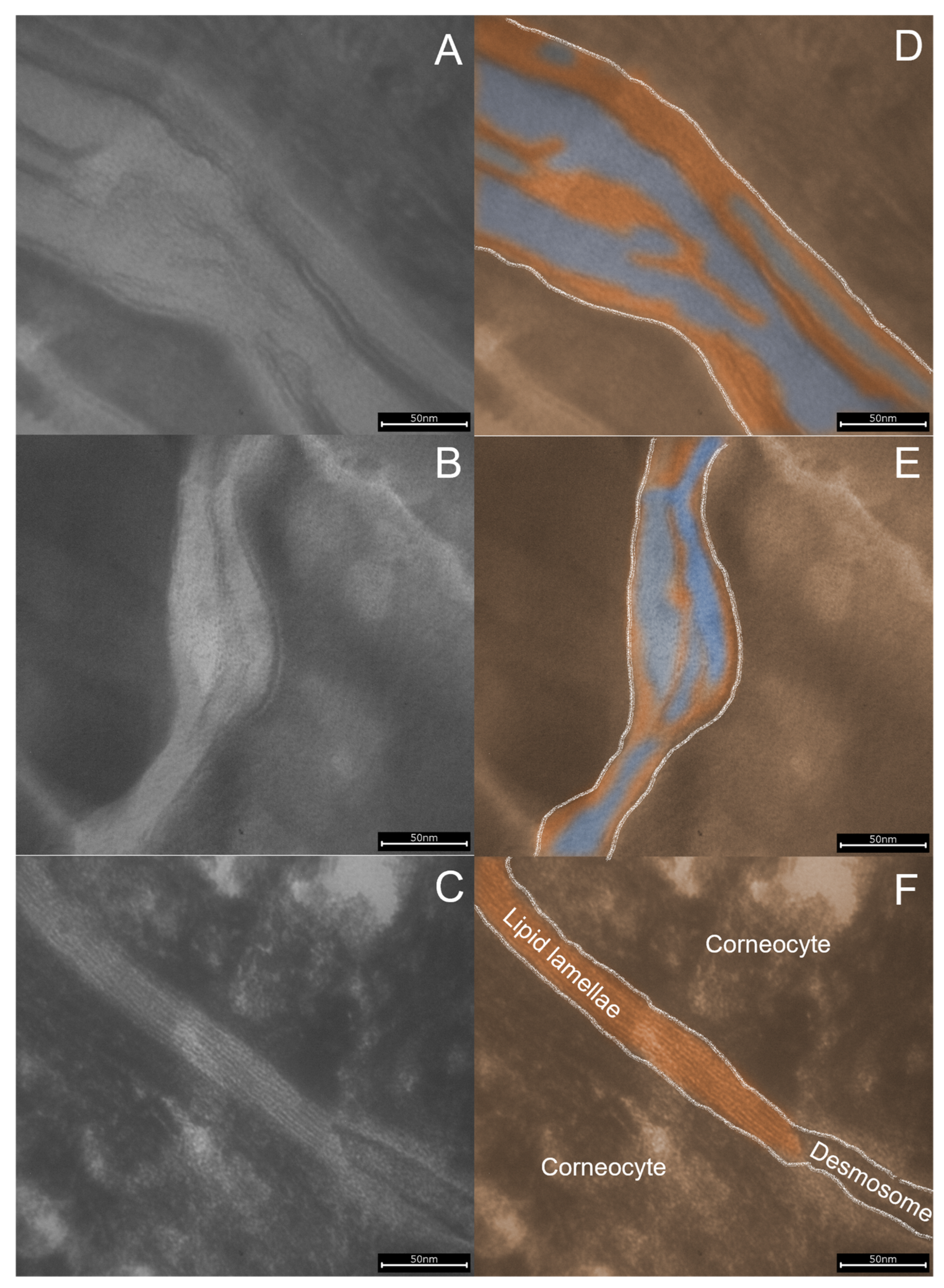
Figure 1D shows the semi-quantitative analysis of Lipbarvis® values (nICLL) in SC samples of treated and untreated area determined at baseline and on day 22. The area treated with the test product revealed a significant increase in nICLL on day 22 compared to the baseline (p < 0.001). A significantly higher amount of lipid lamellae in the intercellular space is therefore assumed for the treated area (p = 0.001), which is also visually displayable via TEM imaging (Figure 2). In a previous study [19], Lipbarvis® values between 40 and 80 nICCL were detected for very dry skin, whereas values of approximately 180 and higher have been described for healthy (normal) skin. No significant differences were observed between the baseline and the final visit for the untreated control.

3.4. SC Lipid Spectrum and Profile Analysis via HPTLC

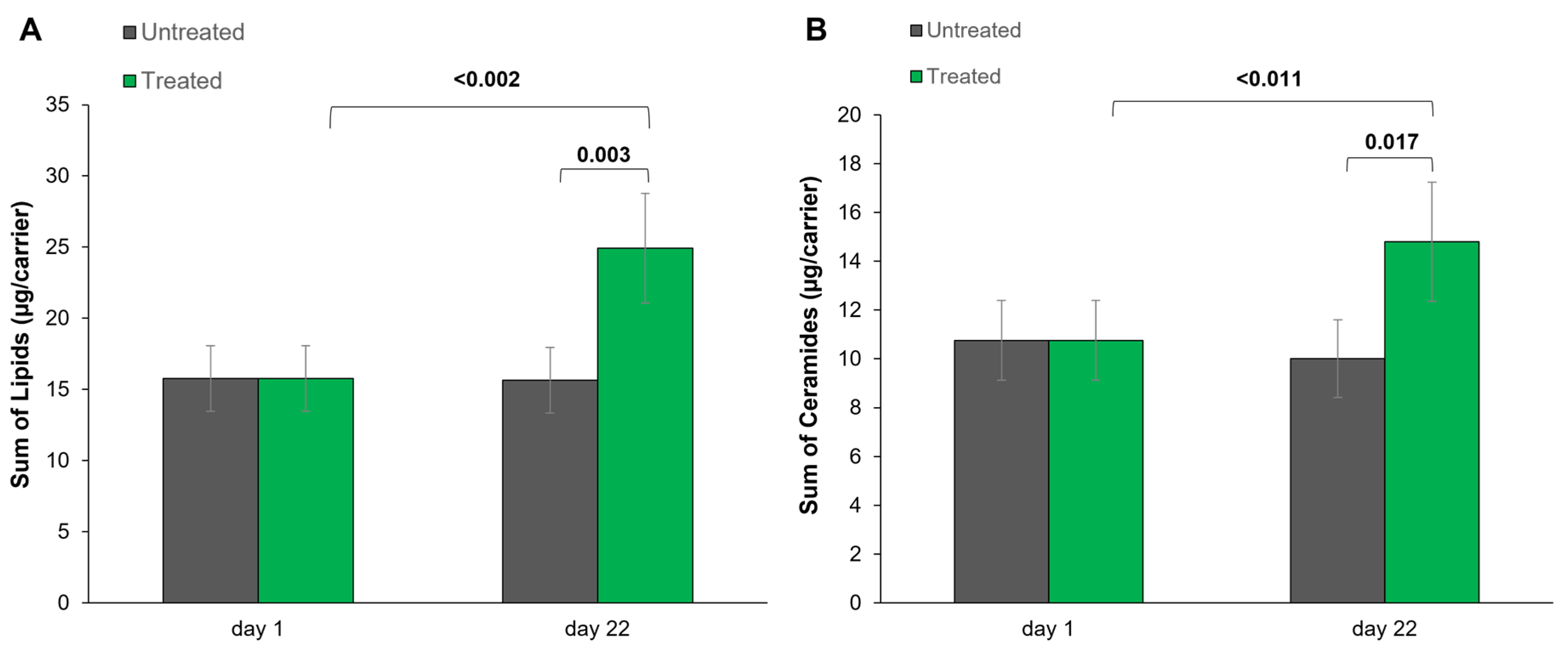
The sum of lipids (CHOL, FFA, CER EOS, CER NP and CER AP) was examined via HPTLC analysis for each subject in treated and untreated areas at both time points (day 1, day 22). As demonstrated in Figure 3A and Table 5, the sum of all analyzed lipids revealed significantly higher contents (µg per carrier) in the treated area than in the untreated area on day 22. The lipid content significantly increased in the treated area after three weeks of product application compared to the baseline values (mean 24.92 vs. mean 15.77), while no significant changes were observed for the untreated area between day 1 and day 22. As shown in Figure 3B, significantly higher values were detected on day 22 for the sum of ceramides in the treated area (CER EOS, CER NP and CER AP) compared to the baseline values (mean 14.80 vs. mean 10.75). No significant differences were found for the untreated area between day 1 and day 22.
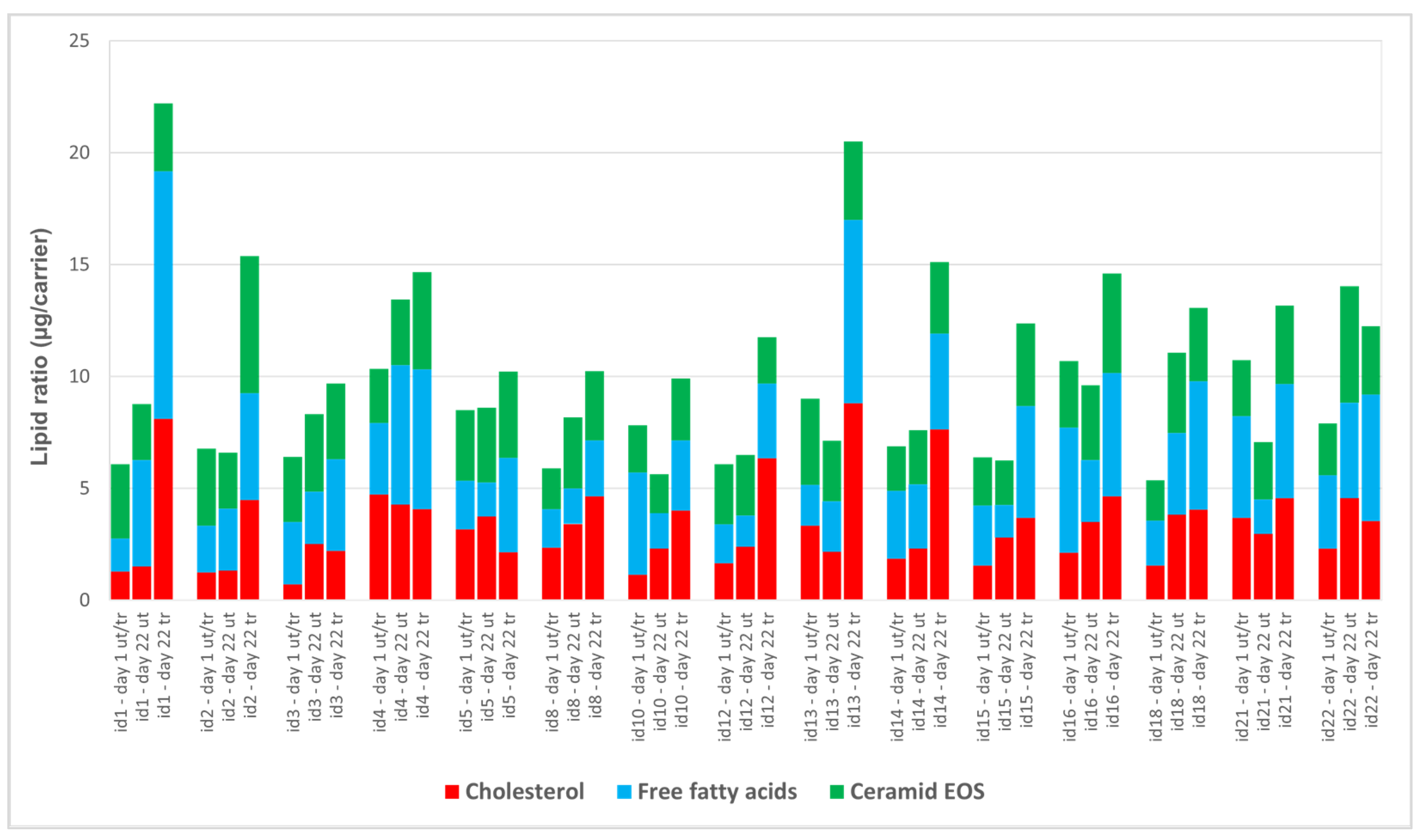
Using the densitometric data from HPTLC, lipid profile analysis was performed to determine the effect of the test product on the ratios of CHOL, FFA and CER EOS. Figure 4 shows the composition of CHOL, FFA and CER EOS at baseline (day 1) and after 21 days in which the test product was applied to one arm and the comparison arm was left untreated. Considerable interindividual differences between the subjects are demonstrated. For example, subject 1 shows a 5-fold increase in cholesterol levels after treatment, while free fatty acids doubled and ceramide EOS levels remained approximately the same. Subject 4 shows no change in cholesterol and free fatty acid levels after treatment, but a 1.5-fold increase in ceramide EOS between the untreated and treated skin. In addition to these very individual differences between subjects due to care, the data also show differences over time. While the lipid levels of subject 5 at the beginning of the study and after 21 days in the untreated arm are comparable, subject 18 has higher levels of cholesterol, free fatty acids and ceramide EOS in the untreated arm after 21 days than at the beginning of the study.
To better understand the effects of the product on the skin, the differences in the lipids considered between the treated and untreated skin were assessed (Figure 5). Of the 15 subjects studied, 14 subjects show higher levels of cholesterol, free fatty acids and ceramide EOS in the SC on the treated test patch than on the untreated comparative skin. Subject 22 also shows more free fatty acids in the SC on the treated skin than on the untreated comparison arm, but lower cholesterol and ceramide EOS values. A total of 10 test persons (1, 2, 8, 10, 12, 13, 14, 15, 16, 21) show higher cholesterol values in the SC than in the untreated comparison skin as result of care with the test product, while higher values of ceramide EOS were also detected in the SC of 10 test persons (1, 2, 4, 5, 10, 13, 14, 15, 16, 21). The free fatty acid content in the SC is increased in all of the treated test fields except for subject 4.

4. Discussion

A previous product evaluation has shown the positive impact of a plant-oil-containing skin care lotion with 10% urea and a product pH around 4.5 on the EPB function in subjects with dry skin and atopic diathesis [13]. The test product was shown to improve and stabilize the acidic nature of the skin surface, and to increase SC integrity, cohesion, and recovery in parallel. The present work, which is a direct continuation of the aforementioned study, has additionally shown (using further methods and end points) that the identical skin care product also improves the SC lipid matrix structure and lipid composition in dry and atopic-prone skin.
Dry skin and xerosis are based on an EPB dysfunction and characterized by an imbalanced epidermal differentiation, abnormal stratum corneum lipid levels and reduced water content in combination with decreased levels of natural moisturizing factors [4]. Moreover, xerosis is a typical symptom of several skin conditions and diseases, such as atopic skin. The EPB dysfunction in atopic skin is associated with reduced water-retaining compounds and decreased structural proteins, the deviated composition of SC lipids with a decrease in CER [5], and alterations of the intercellular lipid lamellae organization [19]. Furthermore, in several xerotic skin conditions [6,7,8], as well as in atopic skin [9,10,11], an increase in ss-pH is demonstrated. For instance, a shift in ss-pH between 0.1 and 0.9 units is described for atopic-prone skin compared to healthy skin [24]. Based on the abovementioned physiological abnormalities in dry and atopic skin, the present skin care product was developed to address and improve (i) skin acidification using a low product pH of 4.5 (and buffer capacity of 1.54), (ii) skin moisture balance via formulation with 10% urea, and (iii) SC lipid organization and composition using plant oils.
Recently, the relationship among skin acidification, EPB function and inflamed skin has been investigated in a few studies exploring atopic murine skin. It has been shown that stabilizing the physiological ss-pH via the topical application of lactobionic acid enhances the skin barrier function, reduces cytokine production, and regulates antimicrobial peptide expression in atopic mice [25]. In addition, EPB restoration after experimentally induced SC pH neutralization is reduced and inflammation enhanced in murine atopic skin [26]. Lee et al. [27] investigated the long-term effects of SC acidification using an atopic march animal model. It was demonstrated that application of a cream with a product pH of 2.8 minimizes atopic skin lesions and can additionally inhibit respiratory inflammation. More recently, another study by Jang et al. [28] also investigated the interaction between pH and patho-mechanisms in AD mice. It was shown that skin surface alkalization in AD-like dermatitis, impaired EPB function and, in turn, topical acidification of severe eczematous lesions decreases ss-pH, TEWL, and serine protease activity, i.e., the desquamation-processing enzyme kallikrein-5, and reduces dermatitis. The present work supports these data by showing that the product treatment in subgroup 1 (baseline ss-pH > 5.0) tends to show a higher decrease in ss-pH compared to the product treatment in subgroup 2 (baseline ss-pH ≤ 5.0). Moreover, in contrast to subgroup 2, the product treatment in subgroup 1 was accompanied by a slight decrease in TEWL, which could be interpreted as an improvement in the EPB physiology (Table 3).
The present study results on AD and atopic-prone skin are in line with numerous studies on aged skin, where the advantage of slightly topical skin acidification using pH 4.0 formulations was investigated. Normalization of the aged-induced elevated ss-pH to a physiological level via targeted topical skin acidification improves SC integrity, recovery and lipid organization and profile [21,29,30,31,32,33]. Additionally, effective skin acidification and maintenance of the ss-pH has recently been shown in 48 males and females over 50 of age with healthy skin [23]. Compared to the work of Blaak et al. [21] and Kilic et al. [33], the present study shows beneficial effects on the SC lipid matrix after product application in terms of lipid lamellae organization and lipid levels. In the present work, the SC lipid lamellae length was significantly improved over 22 days of product application (Figure 1), which could be interpreted as an increase in epidermal barrier integrity. Furthermore, the content of all of the investigated lipids was enhanced, with significant results for the sum of lipids, CER, CHOL, FFA and CER AP (Table 5). The lipid analysis thus shows a good correlation with the ultrastructural results of the lipid lamellae in the intercellular space. The content of lipids in the SC and the normalized length of the lipid lamellae in the intercellular space correspond to the values of healthy skin after three weeks of care [34] and are comparable to the results of the previous study [21].
Interestingly, the restoration of the epidermal skin barrier shown is a very individual process (Figure 5). While an increase in the analyzed lipids in the SC was measured in some subjects, other subjects showed a moderate increase in the analyzed skin lipids. Despite this individual compensation of the damaged epidermal skin barrier, the restoration of the lipid lamellae in the intercellular space after application of the care product is comparable.
The results show an increase in the CER EOS level for most of the test subjects as a result of the care treatment. The low CER EOS levels known for patients with atopic dermatitis could thus be improved and contribute to a healthy skin appearance [35]. Since the skin care product does not contain CER EOS, the increase in CER EOS levels could be due to a “de novo synthesis”.
The increase in CER EOS values differs between the test persons. A possible explanation for this could lie in the synthesis pathway of CER EOS. For the CER EOS, FFA are incorporated into the ceramide molecule [36,37]. If the chain length of the incorporated FFA differs, it is not recognized as CER EOS in the lipid analysis.
Similarly to the CER EOS, the proportion of FFA increases significantly in almost all subjects. The visible variations between the test persons could be explained by an individual metabolism or by the incorporation of the FFA into ceramides [38]. Cholesterol also shows a subject-dependent variation in the lipid content of the SC. In its function to increase the fluidity of lipid membranes, cholesterol could also positively influence the lipids in the SC not shown here.
In addition to topical skin acidification as a key element of the product’s performance, the present product efficacy is secondly based on significant skin hydration caused by 10% urea. Urea is part of the natural moisturizing factor of the epidermis and is well-known as an effective moisturizing and barrier-enhancing active compound for dermo-cosmetic formulations [39]. Today, it is broadly accepted and demonstrated that urea is significantly effective in skin care applications [40,41,42,43]. Regarding the impact on the EPB physiology, it was additionally shown that urea regulates genes, which are involved in keratinocyte differentiation and lipid synthesis [40]. As a result, SCH was significantly increased in the treated test site compared to the baseline values and compared to the untreated site on day 22, accompanied by reduced “skin dryness”, “skin roughness” and “feeling of tension” (Table 4).
To address the alterations in lipid organization and profile in atopic-prone skin, the present formula was additionally formulated with a specific lipid phase mainly containing two relevant plant oils: SAO and EPO. Natural oils have been commonly used in skin care for a very long time and are known to have various positive effects. Recently, the efficacy and the benefits of topically applied SAO and EPO were reviewed [44], and a special emphasis was placed on the positive impact on epidermal barrier integrity, recovery and lipid ratio. SAO is rich in mono- and poly-unsaturated fatty acids (oleic acid: 62.0–86.0%; linoleic acid: 20.0–30.0%) and EPO is mainly composed of oleic acid (74%) and linolenic acid (9%) [44]. Moreover, sweet almond oil and evening primrose oil can improve moisture balance, provide antioxidative and antipruritic properties, have (re-)lipidating effects and maintain the skin surface’s smoothness and softness [45,46,47,48,49,50]. Both plant oils are main ingredients of the test product and are characterized by an essential lipid profile and a high antioxidant content, e.g., containing mono- and poly-unsaturated fatty acids, several phytosterols and various tocopherols [44].
In total, the product’s efficacy seems to be mainly conditioned by a low product pH, a relevant urea content and two effective plant oils (i.e., SAO and EPO). The combination of these beneficial factors is shown to be highly effective in terms of supporting EPB function, and improving SC lipid organization and profile.

5. Conclusions

According to the present and previous data [13], adjusting the ss-pH in subjects with xerotic- and atopic-prone skin via the application of skin care formulation with a lowered pH of 4.5 in combination with a 10% urea concentration and a relevant amount of SAO and EPO seems to be recommendable. In addition to the active ingredients and given product’s pH, the product buffer capacity (Table 1) is also of dermato-cosmetic relevance [51].
Moreover, using such slightly acidic and adequate buffering formulations seems to be a strategy for targeted and direct skin acidification to overcome pathological pH changes and EPB malfunction in dry and atopic skin. However, the present data are limited to the marketed test product evaluated here. A transfer of the presented product’s effects to other comparable formulations with similar key product characteristics, like identical pH (buffer capacity), urea content or plant oil components, is not automatically possible. Furthermore, the effects shown are limited to the biophysical parameters used, especially concerning skin barrier function, and are not directly linked to the improvement of other dysfunctions in very dry and atopic skin, e.g., inflammatory processes. To overcome this study’s limitations, larger clinical long-term trials with additional control conditions could be helpful to obtain stronger evidence concerning the described skin care strategy for atopic dermatitis or subjects with dry skin and atopic diathesis.

Author Contributions

Conceptualization, J.B., D.D., S.D.-P. and P.S.; methodology, D.D., S.D.-P. and S.B.; investigation, D.D. and S.D.-P.; resources, K.-P.W., S.D.-P. and P.S.; writing—original draft preparation, J.B., I.S. and D.D.; writing—review and editing, S.D.-P., C.T., S.B., K.-P.W. and P.S.; visualization, J.B., I.S. and D.D.; supervision, P.S.; project administration, J.B., I.S. and D.D. All authors have read and agreed to the published version of the manuscript.

Funding

This research received no external funding.

Institutional Review Board Statement

The present human intervention study was conducted in accordance with the Revised Declaration of Helsinki, and in line with the European Community Good Clinical Practice (EC-GCP) standards. The study protocol was reviewed by an independent institutional review board for ethical approval (IRB by SGS proderm GmbH; IRB Code: 2018/006, Approval Date: 9 May 2018).

Informed Consent Statement

Informed consent was obtained from all subjects.

Data Availability Statement

Not applicable.

Acknowledgments

We are grateful to all our volunteers who have given up their time to take part in our study.

Conflicts of Interest

The authors J.B., C.T., I.S. and P.S. are employees of the study’s sponsor. The authors declare no conflict of interest.

References

  1. Elias, P.M. Stratum corneum defensive functions: An integrated view. J. Investig. Dermatol. 2005, 125, 183–200. [Google Scholar] [CrossRef] [Green Version]
  2. Menon, G.K.; Kligman, A.M. Barrier functions of human skin: A holistic view. Ski. Pharmacol. Physiol. 2009, 22, 178–189. [Google Scholar] [CrossRef]
  3. Bosko, C.A. Skin barrier insights: From bricks and mortar to molecules and microbes. J. Drugs Dermatol. 2019, 18, s63–s67. [Google Scholar]
  4. Barco, D.; Giménez-Arnau, A. Xerosis: A dysfunction of the epidermal barrier. Actas Dermosifiliogr. 2008, 99, 671–682. [Google Scholar] [CrossRef]
  5. Kim, B.E.; Leung, D.Y.M. Significance of skin barrier dysfunction in atopic dermatitis. Allergy Asthma Immunol. Res. 2018, 4, 12–16. [Google Scholar] [CrossRef] [Green Version]
  6. Öhman, H.; Vahlquist, A. The pH gradient over stratum corneum differs in x-linked recessive and autosomal dominant ichthyosis: A clue to the molecular origin of the “acid skin mantle”? J. Investig. Dermatol. 1998, 111, 674–677. [Google Scholar] [CrossRef] [PubMed] [Green Version]
  7. Eberlein-König, B.; Schäfer, T.; Huss-Marp, J.; Darsow, U.; Möhrenschläger, M.; Herbert, O.; Abeck, D.; Krämer, U.; Behrendt, H.; Ring, J. Skin surface pH, stratum corneum hydration, trans-epidermal water loss and skin roughness related to atopic eczema and skin dryness in a population of primary school children. Acta Derm. Venereol. 2000, 80, 188–191. [Google Scholar] [CrossRef] [Green Version]
  8. Man, M.Q.; Xin, S.J.; Song, S.P.; Cho, S.Y.; Zhang, X.J.; Tu, C.X.; Feingold, K.R.; Elias, P.M. Variation of skin surface pH, sebum content and stratum corneum hydration with age and gender in a large Chinese population. Ski. Pharmacol. Physiol. 2009, 22, 190–199. [Google Scholar] [CrossRef] [PubMed] [Green Version]
  9. Seidenari, S.; Giusti, G. Objective assessment of the skin of children affected by atopic dermatitis: A study of pH, capacitance and TEWL in eczematous and clinically uninvolved skin. Acta Derm. Venereol. 1995, 75, 429–433. [Google Scholar] [CrossRef] [PubMed]
  10. Sparavigna, A.; Setaro, M.; Gualandri, V. Cutaneous pH in children affected by atopic dermatitis and in healthy children: A multicenter study. Ski. Res. Technol. 2009, 5, 221–227. [Google Scholar] [CrossRef]
  11. Knor, T.; Meholjić-Fetahović, A.; Mehmedagić, A. Stratum corneum hydration and skin surface pH in patients with atopic dermatitis. Acta Dermatovenerol. Croat. 2011, 19, 242–247. [Google Scholar] [PubMed]
  12. Elias, P.M.; Wakefield, J.S.; Man, M.Q. Moisturizers versus current and next-generation barrier repair therapy for the management of atopic dermatitis. Ski. Pharmacol. Physiol. 2019, 32, 1–7. [Google Scholar] [CrossRef] [PubMed]
  13. Blaak, J.; Theiss, C.; Schleißinger, M.; Simon, I.; Schürer, N.Y.; Staib, P. A commercially available skin care lotion with a pH of 4.5 and 10% urea improves skin surface pH, stratum corneum hydration and epidermal barrier function in subjects with dry skin and atopic diathesis. J. Cosmet. Dermatol. Sci. Appl. 2020, 10, 116–133. [Google Scholar] [CrossRef]
  14. Diepgen, T.L.; Fartasch, M.; Hornstein, O.P. Criteria of atopic skin diathesis. Dermatosen 1991, 39, 79–83. [Google Scholar]
  15. Oman, S.V.; Ritonja, A. Die Pufferkapazität kosmetischer und pharmazeutischer Emulsionen. Parfümerie Und Kosmet. 1984, 65, 186–189. [Google Scholar]
  16. Berardesca, E.; Loden, M.; Serup, J.; Masson, P.; Rodrigues, L.M. The revised EEMCO guidance for the in vivo measurement of water in the skin. Ski. Res. Technol. 2018, 24, 351–358. [Google Scholar] [CrossRef]
  17. Rogiers, V. EEMCO Guidance for the assessment of transepidermal water loss in cosmetic science. Ski. Pharmacol. Appl. Ski. Physiol. 2001, 14, 117–128. [Google Scholar] [CrossRef]
  18. Parra, J.L.; Paye, M. EEMCO Guidance for the in vivo assessment of skin surface pH. Ski. Pharmacol. Appl. Ski. Physiol. 2003, 16, 188–202. [Google Scholar] [CrossRef]
  19. Dähnhardt-Pfeiffer, S.; Surber, C.; Wilhelm, K.P.; Dähnhardt, D.; Springmann, G.; Böttcher, M.; Förster-Holst, R. Noninvasive stratum corneum sampling and electron microscopical examination of skin barrier integrity: Pilot study with a topical glycerin formulation for atopic dermatitis. Ski. Pharmacol. Physiol. 2012, 25, 155–161. [Google Scholar] [CrossRef] [PubMed]
  20. Imokawa, G.; Akihito, A.; Jin, K.; Higaki, Y.; Kawashima, M.; Hidano, A. Decreased level of ceramides in stratum corneum of atopic dermatitis: An etiologic factor of atopic dry skin. J. Investig. Dermatol. 1991, 96, 523–526. [Google Scholar] [CrossRef] [PubMed] [Green Version]
  21. Blaak, J.; Dähnhardt, D.; Dähnhardt-Pfeiffer, S.; Bielfeldt, S.; Wilhelm, K.P.; Wohlfart, R.; Staib, P. A plant oil-containing pH 4 emulsion improves epidermal barrier structure and enhances ceramide levels in aged skin. Int. J. Cosmet. Sci. 2017, 39, 284–291. [Google Scholar] [CrossRef]
  22. Elias, P.M. The how, why and clinical importance of stratum corneum acidification. Exp. Dermatol. 2017, 26, 999–1003. [Google Scholar] [CrossRef] [PubMed] [Green Version]
  23. Schulte to Brinke, A.; Mehlich, A.; Doberenz, C.; Janssens-Böcker, C. Acidification of the skin and maintenance of the physiological skin pH value by buffered skin care products formulated around pH 4. J. Cosmet. Dermatol. Sci. Appl. 2021, 11, 44–57. [Google Scholar] [CrossRef]
  24. Danby, S.G.; Cork, M.J. pH in atopic dermatitis. Curr. Probl. Dermatol. 2018, 54, 95–107. [Google Scholar]
  25. Hatano, Y.; Man, M.Q.; Uchida, Y.; Crumrine, D.; Scharschmidt, T.C.; Kim, E.G.; Mauro, T.M.; Feingold, K.R.; Elias, P.M.; Holleran, W.M. Maintenance of an acidic stratum corneum prevents emergence of murine atopic dermatitis. J. Investig. Dermatol. 2009, 129, 1824–1835. [Google Scholar] [CrossRef] [PubMed] [Green Version]
  26. Sakai, T.; Hatano, Y.; Zhang, W.; Fujiwara, S. Defective maintenance of pH of stratum corneum is correlated with preferential emergence and exacerbation of atopic-dermatitis-like dermatitis in flaky-tail mice. J. Dermatol. Sci. 2014, 74, 222–228. [Google Scholar] [CrossRef] [PubMed]
  27. Lee, H.J.; Yoon, N.Y.; Lee, N.R.; Jung, M.; Kim, D.H.; Choi, E.H. Topical acidic cream prevents the development of atopic dermatitis- and asthma-like lesions in murine model. Exp. Dermatol. 2014, 23, 736–741. [Google Scholar] [CrossRef]
  28. Jang, H.; Matsuda, A.; Jung, K.; Karasawa, K.; Matsuda, K.; Oida, K.; Ishizaka, S.; Ahn, G.; Amagai, Y.; Moon, C.; et al. Skin pH is the master switch of kallikrein 5-mediated skin barrier destruction in a murine atopic dermatitis model. J. Investig. Dermatol. 2016, 136, 127–135. [Google Scholar] [CrossRef]
  29. Blaak, J.; Wohlfart, R.; Schürer, N.Y. Treatment of aged skin with a pH 4 skin care product normalizes increased skin surface pH and improves barrier function: Results of a pilot study. J. Cosmet. Dermatol. Sci. Appl. 2011, 1, 50–58. [Google Scholar] [CrossRef] [Green Version]
  30. Blaak, J.; Kaup, O.; Hoppe, W.; Baron-Ruppert, W.; Langheim, H.; Staib, P.; Wohlfart, R.; Lütje, D.; Schürer, N.Y. A long-term study to evaluate acidic skin care treatment in nursing home residents: Impact on epidermal barrier function and microflora in aged skin. Ski. Pharmacol. Physiol. 2015, 28, 269–279. [Google Scholar] [CrossRef]
  31. Behm, B.; Kemper, M.; Babilas, P.; Abels, C.; Schreml, S. Impact of a glycolic acid-containing pH 4 water-in-oil emulsion on skin pH. Ski. Pharmacol. Physiol. 2015, 28, 290–295. [Google Scholar] [CrossRef]
  32. Angelova-Fischer, I.; Fischer, T.W.; Abels, C.; Zillikens, D. Accelerated barrier recovery and enhancement of the barrier integrity and properties by topical application of a pH 4 vs. a pH 5.8 water-in-oil emulsion in aged skin. Br. J. Dermatol. 2018, 179, 471–477. [Google Scholar] [CrossRef]
  33. Kilic, A.; Masur, C.; Reich, H.; Knie, U.; Dähnhardt, D.; Dähnhardt-Pfeiffer, S.; Abels, C. Skin acidification with a water-in-oil emulsion (pH 4) restores disrupted epidermal barrier and improves structure of lipid lamellae in the elderly. J. Dermatol. 2019, 46, 457–465. [Google Scholar] [CrossRef] [PubMed] [Green Version]
  34. Dähnhardt, D.; Surber, C.; Dähnhardt-Pfeiffer, S. Influence of topical formulations: Lipid lamella organization and lipid composition of stratum corneum as a surrogate marker for barrier integrity. Curr. Probl. Dermatol. 2018, 54, 166–172. [Google Scholar]
  35. Di Nardo, A.; Wertz, P.; Giannetti, A.; Seidenari, S. Ceramide and cholesterol composition of the skin of patients with atopic dermatitis. Acta Derm. Venereol. 1998, 78, 27–30. [Google Scholar] [CrossRef] [PubMed] [Green Version]
  36. Rabionet, M.; Gorgas, K.; Sandhoff, R. Ceramide synthesis in the epidermis. Biochim. Biophys. Acta 2014, 1841, 422–434. [Google Scholar] [CrossRef] [PubMed]
  37. Feingold, K. Lamellar Bodies: The key to cutaneous barrier function. J. Investig. Dermatol. 2012, 132, 1951–1953. [Google Scholar] [CrossRef] [PubMed] [Green Version]
  38. Elias, P.M. Epilogue: Fixing the barrier—Theory and rational deployment. In Skin Barrier, 1st ed.; Elias, P.M., Feingold, K.R., Eds.; Taylor & Francis: New York, NY, USA, 2006; pp. 591–600. [Google Scholar]
  39. Loden, M. Urea as a moisturizing and barrier-enhancing ingredient. In Skin Moisturization, 2nd ed.; Rawlings, A.V., Leyden, J.J., Eds.; Informa Healthcare: London, UK, 2009; pp. 335–346. [Google Scholar]
  40. Grether-Beck, S.; Felsner, I.; Brenden, H.; Kohne, Z.; Majora, M.; Marini, A.; Jaenicke, T.; Rodriguez-Martin, M.; Trullas, C.; Hupe, M.; et al. Urea uptake enhances barrier function and antimicrobial defense in humans by regulating epidermal gene expression. J. Investig. Dermatol. 2012, 132, 1561–1572. [Google Scholar] [CrossRef] [PubMed] [Green Version]
  41. Celleno, L. Topical urea in skincare: A review. Dermatol. Ther. 2018, 31, e12690. [Google Scholar] [CrossRef]
  42. Scheinfeld, N.S. Urea: A review of scientific and clinical data. Skinmed 2010, 8, 102–106. [Google Scholar]
  43. Pan, M.; Heinicke, G.; Bernado, S.; Tsui, C.; Levitt, J. Urea: A comprehensive review of the clinical literature. Dermatol. Online J. 2013, 19, 20392. [Google Scholar] [CrossRef] [PubMed]
  44. Blaak, J.; Staib, P. An updated review on efficacy and benefits of sweet almond, evening primrose and jojoba oils in skin care applications. Int. J. Cosmet. Sci. 2022, 44, 1–9. [Google Scholar] [CrossRef] [PubMed]
  45. Gehring, W.; Bopp, R.; Rippke, F.; Gloor, M. Effect of topically applied evening primrose oil on epidermal barrier function in atopic dermatitis as a function of vehicle. Arzneimittelforsch 1999, 49, 635–642. [Google Scholar] [CrossRef]
  46. Zeichner, J.; Berson, D.; Donald, A. The use of an over-the-counter hand cream with sweet almond oil for the treatment of hand dermatitis. J. Drugs Dermatol. 2018, 17, 78–82. [Google Scholar] [PubMed]
  47. Mehri, Z.; Afrasiabifar, A.; Hosseini, N. Improved itchy quality of life following topical application of sweet almond oil in patients with uremic pruritus: A randomized, controlled trial. Jundishapur J. Chronic Dis. Care 2018, 7, e68164. [Google Scholar] [CrossRef] [Green Version]
  48. Bordoni, A.; Biagi, P.L.; Masi, M.; Ricci, G.; Fanelli, C.; Patrizi, A.; Ceccolini, E. Evening primrose oil (Efamol) in the treatment of children with atopic eczema. Drugs Exp. Clin. Res. 1998, 14, 291–297. [Google Scholar]
  49. Chung, B.Y.; Kim, J.H.; Cho, S.I.; Ahn, I.S.; Kim, H.O.; Park, C.W.; Lee, C.H. Dose-dependent effects of evening primrose oil in children and adolescents with atopic dermatitis. Ann. Dermatol. 2013, 25, 285–291. [Google Scholar] [CrossRef] [Green Version]
  50. Lio, P. Rapid improvement and protective effects of an almond oil-based ointment for diaper dermatitis. J. Drugs Dermatol. 2016, 15, s86–s90. [Google Scholar]
  51. Wohlrab, J.; Gebert, A. pH and buffer capacity of topical formulations. Curr. Probl. Dermatol. 2018, 54, 123–131. [Google Scholar]
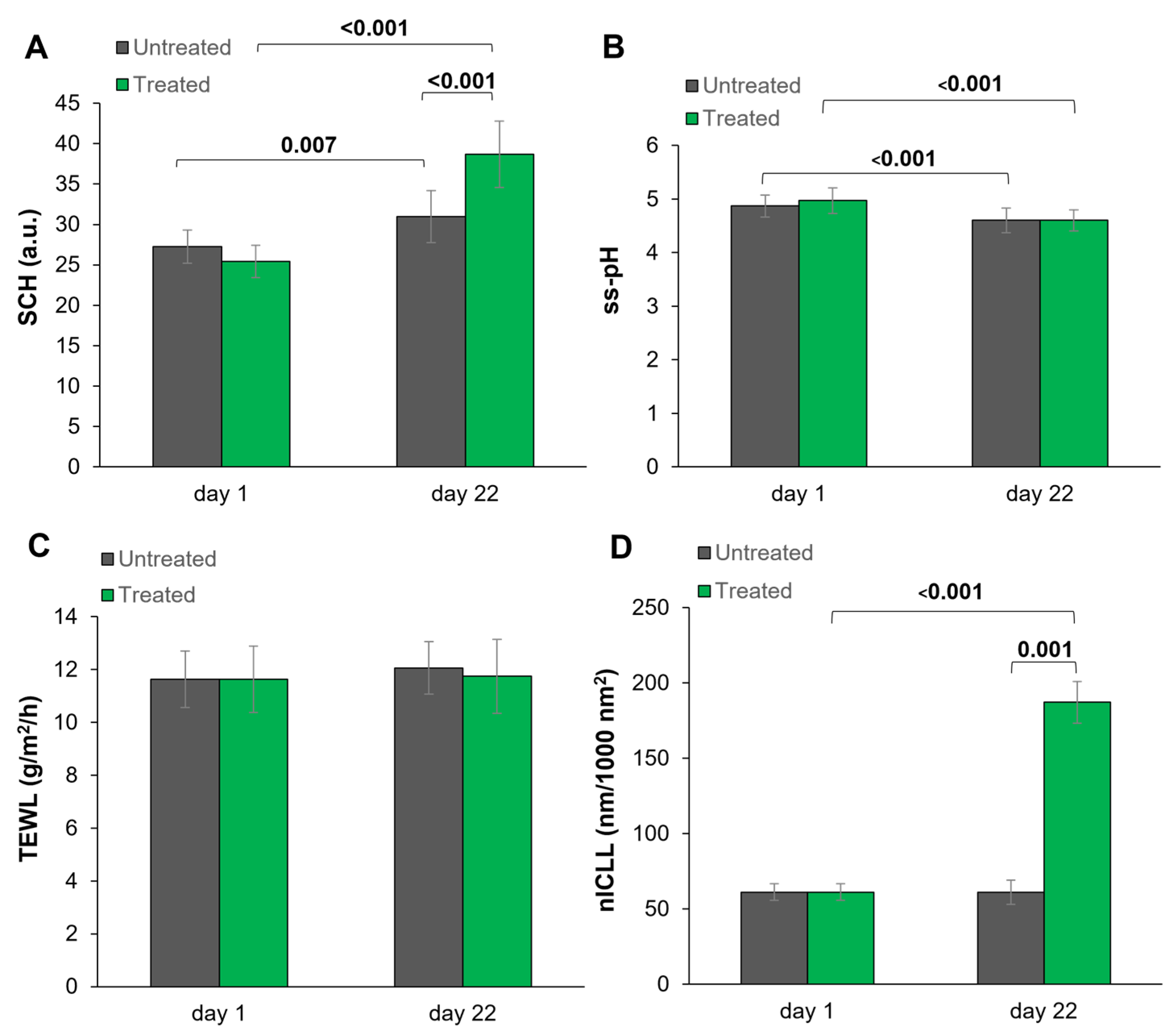
Figure 1. Arithmetic mean values and standard deviations for (A) SCH, (B) ss-pH, (C) TEWL, and (D) nICLL before (day 1) and after 3 weeks of application (day 22) are presented. Only significant p-values of ≤0.05 are illustrated. SCH: stratum corneum hydration (a.u.); ss-pH: skin surface pH; TEWL: transepidermal water loss (g/m2/h); nICLL: normalized intercellular lipid lamellae (nm/1000 nm2).
Cosmetics 10 00095 g001
Figure 2. TEM images of the intercellular lipid lamellae in the intercellular space of the stratum corneum at different time points and in different treatment areas. The baseline (day 1, t0) is represented in ((A) (uncolored)) and ((D) (colored)); the treatment areas (day 22, t1) are shown in ((B,E) (untreated)) and ((C,F) (treated)). Physiologically structured lipid lamellae are colored in orange, whereas intercellular space with a diminished lipid lamellae structure is colored in blue. TEM: transmission electron microscopy.
Cosmetics 10 00095 g002
Figure 3. Arithmetic mean values and standard deviations for (A) sum of lipids (CHOL, FFA, CER EOS, CER NP and CER AP) and (B) sum of ceramides (CER EOS, CER NP and CER AP) in SC samples before (day 1) and after three weeks of product application (day 22). Only significant p-values of ≤0.05 are illustrated. CHOL: cholesterol; FFA: free fatty acid; CER: ceramide.
Cosmetics 10 00095 g003
Figure 4. Stratum corneum lipid composition per subject (id). Before treatment (day 1 ut/tr) and at day 22 on the treated (day 22 tr) and untreated sites (day 22 ut). Sum of lipids: cholesterol, free fatty acids (palmitic acid, stearic acid, linolenic acid, linoleic acid, oleic acid) and ceramide EOS. tr: treated; ut: untreated.
Cosmetics 10 00095 g004
Figure 5. Difference in lipid composition between the treated and the untreated arms for each individual subject on day 22.
Cosmetics 10 00095 g005
Table 1. Composition (INCI) and specification of the test formulation (Internal No. NHH081).
Aqua, Urea, Glycerin, Prunus Amygdalus Dulcis Oil, Oenothera Biennis Oil, Tocopheryl Acetate, Cetearyl Alcohol, Panthenol, Glyceryl Stearate SE, Sodium Lactate, Phytosterols, Bisabolol, Rosmarinus Officinalis Leaf Extract, Citronellol, Benzyl Salicylate, Limonene, Geraniol, Linalool, Benzyl Alcohol, p-Anisic Acid, Parfum, Caprylyl Glycol, Lactic Acid, Sorbitan Oleate, Sodium Stearoyl Glutamate, Glycine Soja Oil, Ascorbyl Palmitate, Xanthan Gum, Triacetin, Tocopherol
Type of emulsion: o/w
pH value: 4.5 ± 0.1 (adjusted by Lactic Acid/Sodium Lactate)
Buffer Capacity (Lab. Sample No. 210819587): 1.54 (according to Oman et al. [15])
Content of urea [%]: 10.0
Plant Oils (sweet almond oil, SAO; evening primrose oil, EPO) are indicated in italics
Table 2. Schedule of study procedure.
Day 1 (t0)Day 2 to 21Day 22 (t1)
Preparation
Informed consent
In-/Exclusion criteria
Visual atopic scoring
(modified according to EAS)
Acclimatization (30 min)
Parameters
Biophysical measurements
(TEWL, SCH, ss-pH)
Visual objective and subjective scoring
(dryness, roughness, feeling of tension)
SC lipid matrix analysis using Lipbarvis®
(nICCL, Lipid composition and profile)
Application
Test product usage
(twice daily)
✕ *
* The first application of test materials was performed after the instrumental measurements and visual evaluation at the study site, under the supervision of a technician. EAS: Erlanger atopy score [14]; TEWL: transepidermal water loss; SCH: stratum corneum hydration; ss-pH: skin surface pH; nICCL: normalized intercellular lipid lamellae.
Table 3. Subgroup analysis for the biophysical parameters ss-pH, SCH and TEWL of the treated skin site. Subgroup definition based on baseline values: ss-pH > 5.0 versus ss-pH ≤ 5.0.
Day 1 (t0)
(Treated)
Day 22 (t1)
(Treated)
Diff
(Absolute val.)
Diff
(%)
Subgroup 1
Basis ss-pH > 5.0
(n = 10)
ss-pH5.43 ± 0.314.79 ± 0.48−0.64 ± 0.28−11.91 ± 5.36
SCH25.51 ± 8.7838.24 ± 14.7212.73 ± 8.9850.56 ± 31.63
TEWL12.01 ± 3.2111.01 ± 1.22−1.00 ± 2.82−4.30 ±19.13
Subgroup 2
Basis ss-pH ≤ 5.0
(n = 15)
ss-pH4.65 ± 0.284.47 ± 0.25−0.19 ± 0.29−3.81 ± 6.16
SCH25.39 ± 7.7438.99 ± 12.7313.61 ± 8.1455.82 ± 37.07
TEWL11.39 ± 2.0212.24 ± 3.460.85 ± 3.448.97 ± 30.27
Data presented as arithmetic mean values and standard deviation (because of the small subgroup sample size, no statistical analysis was conducted). Diff: difference day 1 (treated) versus day 22 (treated); ss-pH: skin surface pH; SCH: stratum corneum hydration (a.u.), TEWL: transepidermal water loss (g/m2/h).
Table 4. Objective visual scoring for dryness and roughness and subjective evaluation for the parameter feeling of tension. Scoring data presented as arithmetic mean and standard deviation.
ParameterTime PointCodeMeanSignificance (p-Value)
Comparison
(Untreated t1 vs. Treated t1)
Comparison
(t0 vs. t1)
Skin
dryness
Day 1 (t0)untreated0.9 (±0.4)------
treated0.9 (±0.6)---
Day 22 (t1)untreated0.9 (±0.6)<0.0010.745 ns
treated0.5 (±0.3)<0.001
Skin
roughness
Day 1 (t0)untreated0.7 (±0.4)------
treated0.8 (±0.4)---
Day 22 (t1)untreated0.7 (±0.5)<0.0010.998 ns
treated0.3 (±0.3)<0.001
Feeling
of tension
Day 1 (t0)untreated0.8 (±0.7)------
treated0.8 (±0.7)---
Day 22 (t1)untreated0.6 (±0.7)0.0080.144 ns
treated0.2 (±0.6)<0.001
Score: 0: none; 0.5: very slight; 1: slight; 2: moderate; 3: strong. ns: not significant; in bold = significant: p ≤ 0.05 (using Wilcoxon signed-rank test).
Table 5. Lipbarvis® and lipid analysis data related to time point and test site.
Time PointDay 1 (t0)Day 22 (t1)Significance
Test SiteUntreated Treated UntreatedTreatedp-Value *
(Untreated t1 vs. Treated t1)
nICCL (nm/1000 nm2)61.1361.1361.05187.070.001 sig
Sum of Lipids (µg/carrier)15.7715.7715.6424.920.003 sig
CHOL (µg/carrier)2.182.182.904.860.012 sig
FFA (µg/carrier)2.842.842.735.260.001 sig
Sum of CER (µg/carrier)10.7510.7510.0114.800.017 sig
CER EOS (µg/carrier)2.632.632.943.560.081 ns
CER NP (µg/carrier)3.463.462.845.190.078 ns
CER AP (µg/carrier)4.664.664.236.050.025 sig
Data presented as arithmetic mean values (n = 15). * Comparison and statistics on treatments of differences at day 22 (t1). identical sampling area on day 1 (t0). nICCL: normalized intercellular lipid lamellae; CHOL: cholesterol; FFA: free fatty acid; CER: ceramide; ns: not significant; sig: significant.
Disclaimer/Publisher’s Note: The statements, opinions and data contained in all publications are solely those of the individual author(s) and contributor(s) and not of MDPI and/or the editor(s). MDPI and/or the editor(s) disclaim responsibility for any injury to people or property resulting from any ideas, methods, instructions or products referred to in the content.