Article

Valorization of the Invasive Red Lionfish (Pterois volitans L.) as a Natural and Promising Source of Bioactive Hydrolysates with Antioxidant and Metal-Chelating Properties

by
Luis Chel-Guerrero
1,
David Betancur-Ancona
1,
Yasser Alejandro Chim-Chi
2,
Valentino Mukthar Sandoval-Peraza
3 and
Santiago Gallegos Tintoré
1,*
1
Facultad de Ingeniería Química, Universidad Autónoma de Yucatán, Campus de Ciencias Exactas e Ingenierías, Periférico Norte, Km 33.5, Tablaje Catastral 13615, Chuburná de Hidalgo Inn, Mérida C.P. 97203, Yucatán, Mexico
2
Instituto Tecnológico Superior de Calkiní, Tecnológico Nacional de México, Av. Ah-Canul, Calkiní C.P. 24900, Campeche, Mexico
3
Escuela de Ciencias de la Salud, Universidad del Valle de México, Calle 79 No. 500 Col. Dzityá, Altura km 9.5 de la Carretera Progreso-Mérida, Mérida C.P. 97302, Yucatán, Mexico
*
Author to whom correspondence should be addressed.
Resources 2025, 14(6), 94; https://doi.org/10.3390/resources14060094
Submission received: 3 April 2025 / Revised: 21 May 2025 / Accepted: 28 May 2025 / Published: 3 June 2025

Abstract

:
Lionfish is a predatory invasive species that endangers native species in the areas it colonizes. Hunting it is necessary to prevent this natural devastation while taking advantage of this unconventional natural source. The objective of this research was to utilize lionfish muscle to obtain hydrolysates with biological activities (antioxidant and chelating properties). The methodology of this study involved the obtention of hydrolysates with Alcalase® at 30 (H30), 60 (H60), and 90 (H90) min. Degree of hydrolysis (DH), amino acid, electrophoretic profile, and antioxidant and chelating activities were determined for the hydrolysates obtained. The amino acid composition showed a high nutritional value since all the hydrolysates fulfilled the requirements proposed by the FAO (except tryptophan) for children, adolescents, and adults. The DH was >30% at 60 and 90 min. In the electrophoretic analysis, protein and polypeptides were identified. DPPH radical scavenging was 27.78% at 30 min. Iron-chelating activity was 64.23% at 90 min, and copper-chelating capacity remained at >90% in all hydrolysates. Lionfish are an invasive and unexploited source of hydrolysates with potential applications in the feed and food industries.

Graphical Abstract

1. Introduction

The Indo-Pacific red lionfish (Pterois volitans) has emerged as a highly successful invasive species across the Western Atlantic, Caribbean Sea, and Gulf of Mexico, exerting considerable negative impacts on native fish populations and marine biodiversity [1]. Its voracious predatory nature, rapid reproduction, and broad habitat tolerance contribute to its detrimental ecological footprint, causing significant reductions in the populations of commercially and ecologically important reef fish [2]. Consequently, implementing effective control and management strategies is essential. Beyond direct removal efforts, exploring avenues to valorize the biomass of captured lionfish offers a dual benefit: mitigating ecological harm and transforming an otherwise problematic resource into value. Lionfish meat is characterized by a high protein content (approx. 20% wet weight, corresponding to over 90% on a dry basis) and a palatable flavor, making it suitable for human consumption and as a raw material for protein-derived products [3].
The enzymatic hydrolysis of fish proteins and other marine sources is a well-established method to produce bioactive peptides with various functional properties, including antioxidants, antihypertensive, and immunomodulatory effects [4]. These peptides, often more readily absorbed than intact proteins, can counteract oxidative stress by scavenging free radicals or chelating pro-oxidant metal ions [5]. Oxidative stress, resulting from an imbalance between reactive oxygen species (ROS) production and antioxidant defenses, can lead to cellular damage related to various chronic diseases. Metal ions like iron and copper, while essential, can catalyze oxidative reactions; thus, peptides with metal-chelating abilities can offer protective effects [6].
Chelating peptides, exhibiting antioxidant properties, are superior to metal salts for enhancing mineral bioavailability. These peptides are typically derived from the enzymatic hydrolysis of proteins from sources such as fish [7]. Iron, vital for oxygen transportation, energy metabolism, and cellular growth, is a common deficiency treated with salts, chelating agents, or peptides [8]. Copper, which can catalyze ROS-mediated DNA strand breaks and nucleotide oxidation, is effectively chelated by peptides that inhibit its oxidative activity, reducing disease risk [9]. Amino acids like histidine, methionine, and cysteine, along with small peptides, form complexes with copper, facilitating its intestinal absorption via amino acid transport systems [10]. While marine sources have been studied for bioactive peptides [9], P. volitans remains largely unexplored. Previous work by Chel et al. [11,12] used simulated digestion focusing on peptide fractions, but this study uses Alcalase® to evaluate how the hydrolysis time affects peptide antioxidant and metal-chelating activities. As an invasive species, lionfish is a promising, underutilized raw material for producing bioactive protein hydrolysates.
Building on this, the current study seeks to valorize the invasive red lionfish (P. volitans) by producing protein hydrolysates using Alcalase®. The study aimed to determine whether these enzymatically produced hydrolysates indeed possess the anticipated bioactive properties. Specifically, it aims to assess how hydrolysis duration influences their biochemical characteristics and functional activities, thereby highlighting their potential applications in functional foods or nutraceuticals.

2. Materials and Methods

The materials and methods used for producing and evaluating red lionfish hydrolysates, which contribute to scientific knowledge and enable result reproducibility, were the following:

2.1. Reagents

Alcalase® (2.4 AU/g) (P4860), butylhydroxyanisole (BHA), ethylenediaminetetraacetic acid (EDTA), o-phthalaldehyde, Tris-HCl, mercaptoethanol, 2,2-difenil-1-picrilhidrazil (DPPH) (D9132), diethyl ethoxymethylenemalonate (D94208), acid L-α-aminobutyric (162663), amino acid standard (AAS-18), ferrozine acid, and pyrocatechol violet, purchased from Sigma-Aldrich., St. Louis, MO, USA.

2.2. Sample

Lionfish specimens were collected by divers in Cozumel, Mexico (2017). The fish were gutted and filleted, and the fillets were frozen for transport to the laboratory. Fillet proximal composition was analyzed using AOAC methods [13] for moisture 925.09 (dry in oven at 105 °C for 4 h), fat 920.39 (Soxhlet extraction with hexane for 4 h), ash 920.39 (incineration at 550 °C for 4 h), and crude fiber 962.09 (acid (H2SO4 1.25%) and basic (NaOH 1.25%) digestions). Protein 954.01 was determined by Kjeldhal using 6.25 as the converting protein factor. Nitrogen-free extract was calculated as follows: 100%—fat—ash—crude fiber—protein.

2.3. Protein Hydrolysate Preparation

Fillets were freeze-dried and used to prepare protein hydrolysates in a one-liter hydrolysis reactor vessel; this was performed in duplicate. The fillets, freeze-dried at −47 °C and 13 × 10−3 mBar (Labconco model 6-Plus-85, Labconco Corporation, Kansas, MO, USA), were reconstituted to a 5% protein (w/v) solution and digested with Alcalase® (0.3 AU/g protein) for 90 min at 50 °C and pH 8. Aliquots were taken at 0, 30, 60, and 90 min (non-hydrolyzed protein, NHP, refers to the aliquot obtained at time zero of the hydrolysis process). Hydrolysis was halted by heating at 80 °C for 20 min. The hydrolysates were centrifuged at 11,227× g for 30 min. Part of these supernatants was used for bioactivity determinations, except for amino acid analysis, for which it was freeze-dried as described above. The samples were stored at −20 °C in polyethylene bottles to carry out subsequent tests [14].

2.4. Degree of Hydrolysis (DH)

The methodology proposed by Nielsen et al. [15] was used to quantify the free amino groups with o-phthalaldehyde (OPA) (Sigma P0657) in the presence of dithiothreitol (Sigma 3483123). HCl 6N was used at 100 °C for 24 h in a circulating air oven, with a ratio of 6 mL of acid per 4 mg of protein in the samples. After the time elapsed, the residual acid was evaporated using a vacuum oven at 90 °C and 600 mbar of pressure for 24 h. The resulting products were resuspended in a 1% SDS solution, producing a yellow coloration. From this, 200 µL was taken, and 1.5 mL of OPA was added. All spectrophotometer (Thermo Spectronic, Genesys 10UV, Waltham, MA, USA) readings were performed at 340 nm using deionized water as the control. The cleavage of peptide bonds was quantified using a calibration curve with L-serine (Sigma S4500) as a standard (0.1 mg/mL deionized water), according to Equation (1):
DH =   h h t o t × 100
where htot is the total number of peptide bonds per protein equivalent and h is the number of hydrolyzed bonds.

2.5. Sodium Dodecyl Sulfate–Polyacrylamide Gel Electrophoresis (SDS-PAGE)

This analysis was performed with 13% acrylamide gel and a 4% stacking gel. An amount of 5 mg of non-hydrolyzed muscle protein and hydrolysates was dissolved in a buffer (50 mM Tris-HCl, (pH 6.8), 10% glycerol, 1% SDS, and 0.01% bromophenol blue) and heated at 100 °C for 5 min. Runs were performed at 40 mA for 1.5 h in a Mini-protean electrophoresis chamber. The resulting gels were stained with 0.05% Coomassie Brilliant Blue G-250 and cleaned with an acetic acid/methanol/distilled water (1:4:5) solution. Wells were loaded with 10 µg of the protein or the hydrolysates [16]. A low-range molecular-weight standard (BIORAD-1610305) containing phosphorylase B (105.2 kDa), bovine serum albumin (84.2 kDa), ovalbumin (50.4 kDa), carbonic anhydrase (36.8 kDa), soybean trypsin inhibitor (29.0 kDa), and lysozyme (20.5 kDa) was used in muscle proteins.

2.6. Amino Acid Analysis

Amino acid profiles were obtained following the methodology proposed by Alaiz et al. [17]. For the preparation of the sample, 1 mg of protein for the hydrolysates and 2 mg for the fillet was dissolved in 1 mL of HCl (6 M), sealed with nitrogen in hydrolysis tubes, and then incubated in an oven at 110 °C for 24 h. Finally, the sample was dried in a vacuum oven at 80 °C. Dried samples were resuspended in 1 mL of sodium borate buffer (1 M: pH 9; with 0.02% of sodium azide), and 0.8 µL of diethyl ethoxymethylenemalonate was added. The derivatization reaction was performed at 50 °C with vigorous shaking for 50 min. The mixture was cooled to room temperature, and 15 µL was injected into the chromatograph.
The HPLC system consisted of a model AGILENT (Series 1100), Santa Clara, CA, USA with an automatic injector and UV–Vis detector. Separations were attained using a Nova Pack C-18, 4 µm inverse-phase column of 300 × 3.9 mm. Resolution of the amino acid derivates (sample and standards AAS-18) was run using a binary gradient system. The solvents used were (A) sodium acetate (25 mM; pH 6 with 0.02% of sodium azide) and (B) acetonitrile. The solvents were delivered to the column at a flow rate of 0.9 mL/min as follows: time 0–3 min linear gradient from A:B (91:9) to A:B (86:14); 3–13 min, elution with A:B (86:14); 13–30 min, linear gradient from A:B (86:14) to A:B (69:31); 30–35 min, elution with A:B (69:31).
For tryptophan, the methodology proposed by Yust et al. [18] was followed, using the same HPLC and column described above. Samples (2–10 mg) were dissolved into 3 mL of NaOH (4 M), sealed in hydrolysis tubes under nitrogen, and incubated in an oven at 100 °C for 4 h. Hydrolysates were cooled down on ice, neutralized to pH 7 using HCl (12 M), and diluted to 25 mL with sodium borate buffer (1 M; pH 9). Aliquots of these solutions were filtered through 0.45 µm Millex filters (Millipore) prior to injection. An amount of 15 µL of sample/standard was injected into the column. An isocratic elution system consisting of sodium acetate (2 mM; pH 6 with 0.02% of sodium azide) and acetonitrile (91:9) was used at a delivered 0.9 mL/min ratio.

2.7. Antioxidant Activity of Protein Hydrolysates

2.7.1. Free Radical Scavenging Activity

Protein hydrolysates (equivalent to 2 mg of protein) were added to DPPH in methanol (100 mL, 100 mM) in 96-well plates. The plate was shaken, and absorbance was measured at 517 nm after 30 min. An amount of 10 mg butylhydroxyanisole (BHA) was used as positive control [19]. The percentage of radical scavenging activity was calculated using Equation (2):
Radical   scavenging   activity   % =   Abs   control Abs   sample Abs   control × 100

2.7.2. Copper- and Iron-Chelating Activity

For copper-chelating activity, hydrolysates (equivalent to 4 mg of protein) were added to microtubes containing 1 mL 50 mM Na acetate (pH 6.0), 25 µL 4 mM pyrocatechol violet, and 10 mg CuSO4. An amount of 50 µg EDTA was used as positive control. Absorbance was measured at 632 nm after incubation for 1 min at room temperature [20]. A calibration curve was made with different concentrations of copper (2, 4, 6, 8, and 10 μg/μL). The copper concentration was determined using Equation (3).
Chelating   activity   % = Cu i Cu f Cu i × 100
where [Cu]i = initial concentration of Cu2+ and [Cu]f = final concentration of Cu2+.
Iron-chelating activity was measured based on the formation of Fe2+–ferrozine complexes [21]. Hydrolysates (equivalent to 4 mg of protein) were added to microtubes containing 1 mL 100 mM Na acetate buffer (pH 4.9) and 100 mL FeCl2∙4 H2O solution (0.01 mg Fe/mL water). An amount of 50 µg EDTA was used as positive control. Absorbance was measured at 562 nm after adding the ferrozine solution (50 µL, 40 mM) and incubating it for 30 min at room temperature. A calibration curve was made with different concentrations of iron (0.2, 0.4, 1, 1.5, and 2 µg/µL). The iron concentration was determined using Equation (4).
Chelating   activity   ( % ) = Fe i - Fe f Fe i ×   100
where [Fe]i = initial concentration of Fe2+ and [Fe]f = final concentration of Fe2+.

2.8. Statistical Design and Analysis

A one-way fixed-effects design was used, where hydrolysis time (NHP, H30, H60, and H90) was the experimental factor that generated the treatments. ANOVA was conducted using the degree of hydrolysis, antioxidant activity, and chelating activity as response variables. The significance level was set at 5%. When significant differences were detected, means were compared using a Duncan test [22]. All statistical analyses were performed with Statgraphics Plus 5.1. All treatments were conducted in triplicate.

3. Results

3.1. Proximal Composition

The moisture in the lionfish fillets was 81.3 ± 0.73. The fat, ash, crude fiber, protein, and NFE contents on dry basis were 3.11 ± 0.03; 3.26 ± 0.00; 0.97 ± 0.2; 91.5 ± 0.22; and 1.16 ± 0.01%, respectively, highlighting that the main component in the lionfish fillets was the protein.

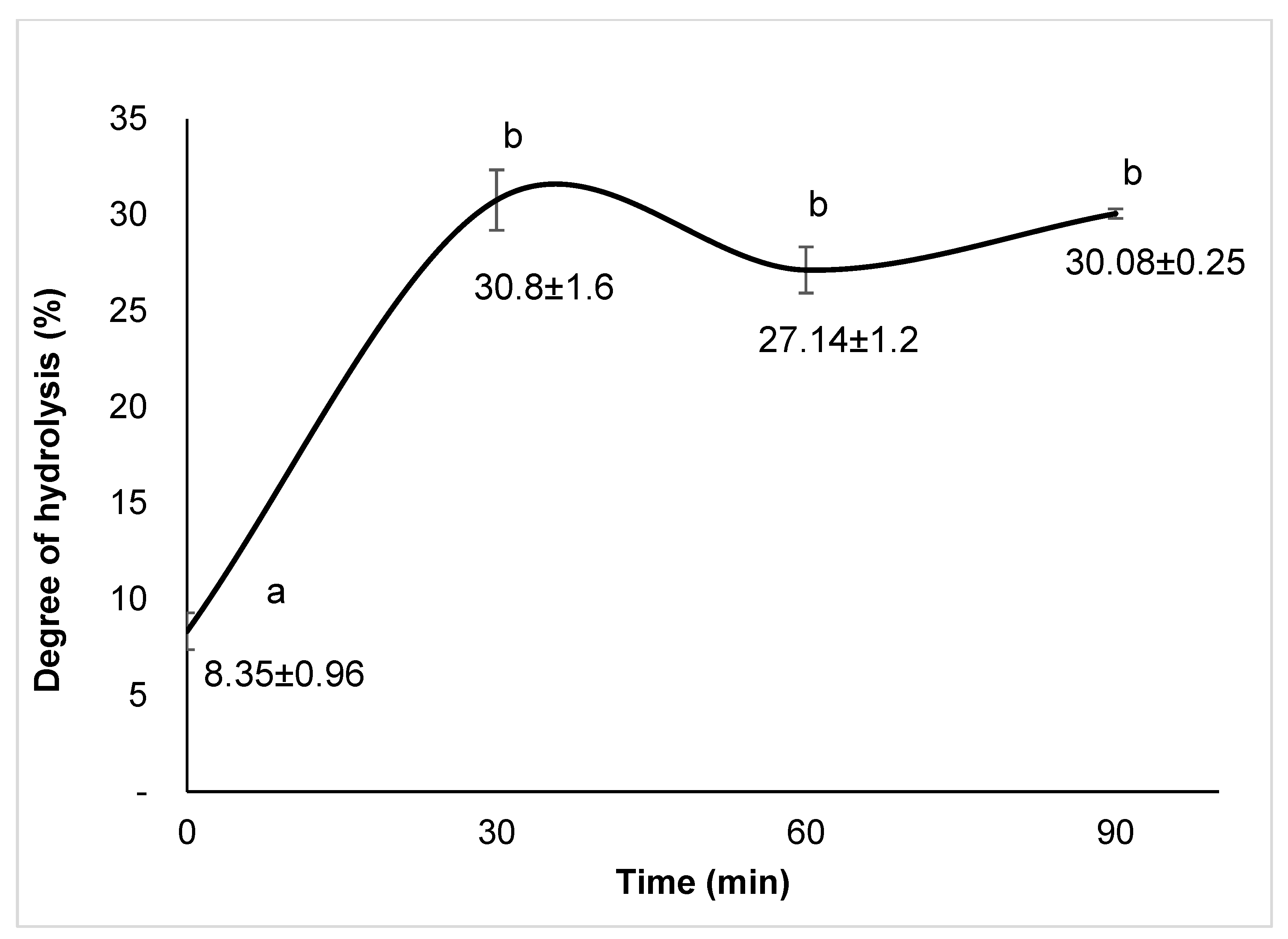
3.2. Degree of Hydrolysis (DH)

Hydrolysis was performed with Alcalase and the degree of hydrolysis (DH) was measured, noticing that the fillet of lionfish showed a previous hydrolysis process (8.53%) at time 0 and increased its DH exponentially around 30 min (30.8%), declined (27.1%) from 30 to 60 min, and recovered its previous level at 90 min, but without statistical differences (p > 0.05) in comparison with the non-hydrolyzed protein (Figure 1).

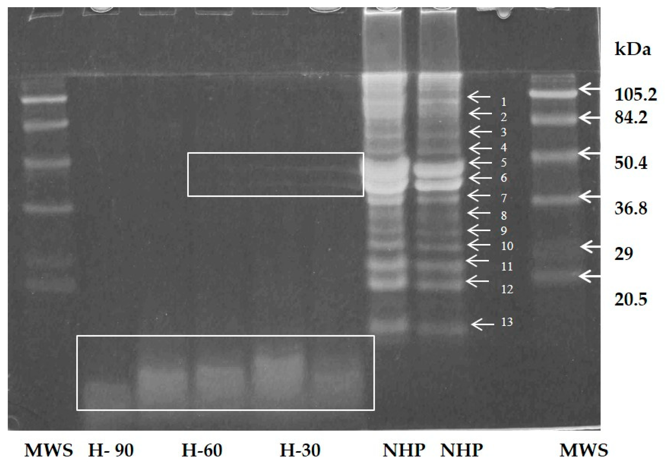
3.3. Electrophoretic Profile

The electrophoretic profile was measured in the hydrolysates. SDS-PAGE showed a clear degradation of the proteins into polypeptides of different sizes (Figure 2). The non-hydrolyzed fillets exhibited a series of proteins between 13.8 and 105 kDa. Two of these proteins, corresponding to two polypeptides (52.7 and 46.2 kDa), were found in high concentrations. H30 and H60 exhibited molecular weights (MW) ranging from <10 and 46.2 kDa. However, H90 exhibited < 10 kDa (Figure 2).

3.4. Amino Acid Profiles

The amino acid profiles for the non-hydrolyzed protein and hydrolysates at 0 (NHP), 30 (H30), 60 (H60), and 90 (H90) min are shown in Table 1. These results showed a high protein quality in the NHP, H30, H60, and H90, with values above those recommended by the FAO [23] for the essential amino acids requirements for infants, children and older children, adolescents, and adults for the following: Ile, Leu, Lys, Met + Cys (except H30), Phe + Tyr, Thr, Val, and His (except NHP). Only the tryptophan amount is not covered by any of the samples in this study. The NHP and hydrolysates also contained significant amounts of Tyr, Met, Trp, Leu, His, Pro, and Lys, all related to antioxidant potential.

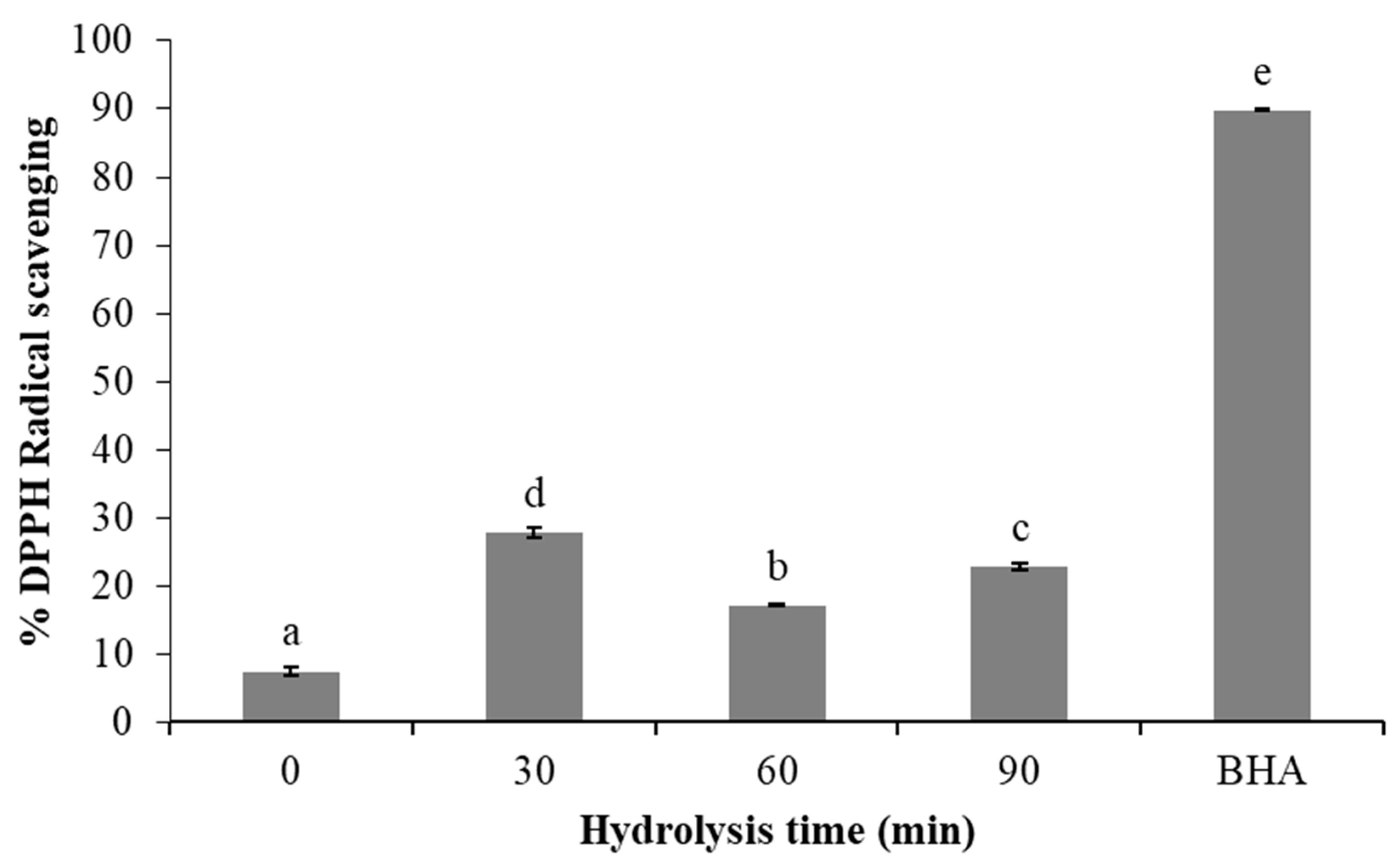
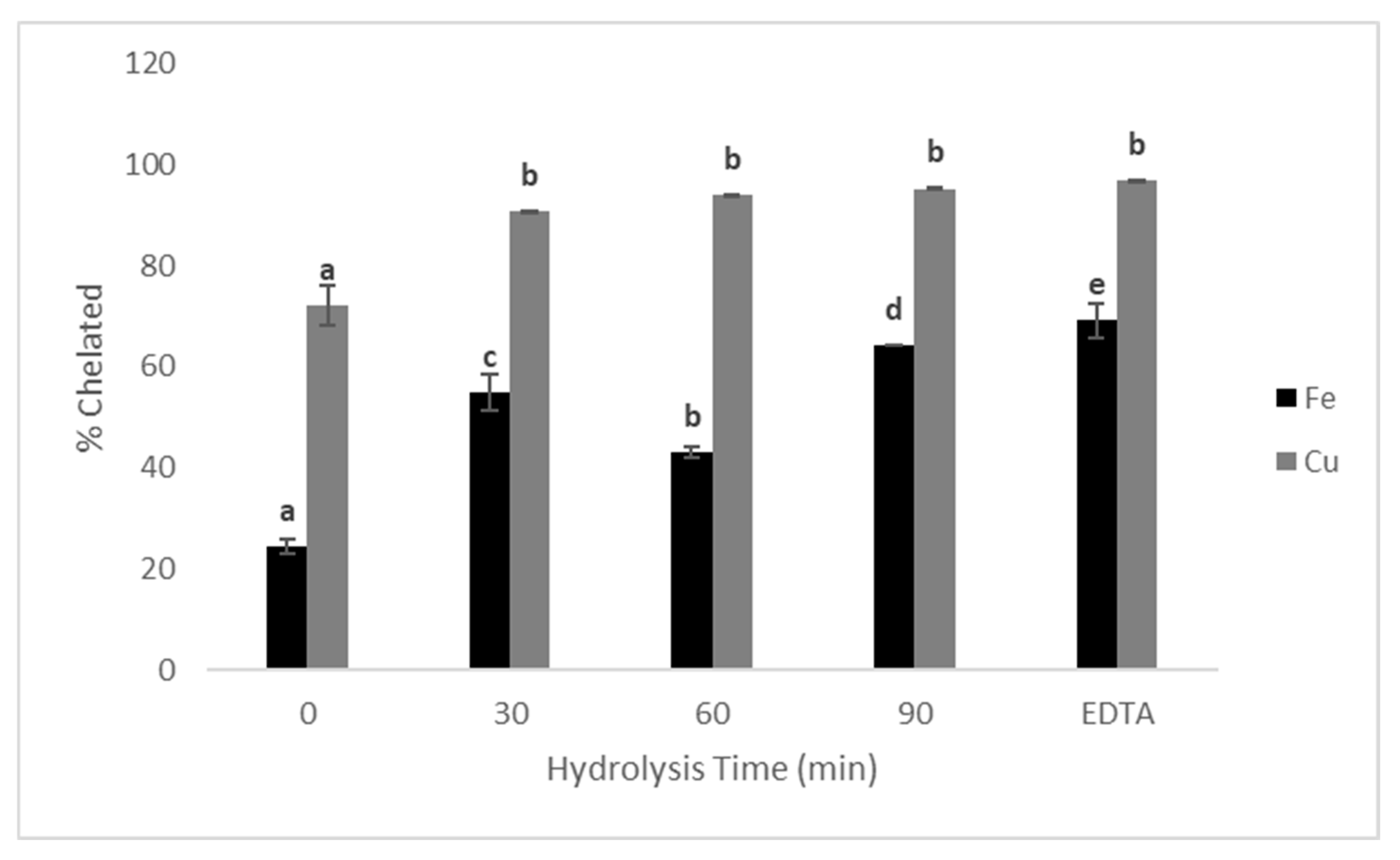
3.5. Antioxidant and Chelating Activity of Protein Hydrolysates

The hydrolysates and NHP of lionfish muscle exhibited antioxidant and metal-chelating activities as well, making them promising sources of peptides. The lionfish muscle protein hydrolysates showed DPPH scavenging activities, with the highest value (27.78%) found in the H30 hydrolysate (Figure 3). This was substantially lower than the 60.17% (5 μg) for the BHA standard.
Iron (Fe+2)- and copper (Cu+2)-chelating activities from the lionfish muscle protein hydrolysates ranged from 24 to 64% (Figure 4). For iron, the chelating capacity depended on the hydrolysis time (p < 0.05), with the highest activity at 90 min. The use of Alcalase® resulted in hydrolysates with chelating activity (69% for Fe+2 and 96.8% for Cu+2) comparable to that of 50 µg EDTA. Copper-chelating activity did not differ between the lionfish muscle proteins (90.6% in H30, 94.1% in H60, and 95.2% in H90); these values were similar to the 96.8% produced with 50 µg EDTA. Even the non-hydrolyzed protein showed 24% chelating activity due to endogamous hydrolysis of muscle proteins. Copper-chelating activity was higher than iron-chelating activity in the non-hydrolyzed muscle protein and all the hydrolysates (Figure 4). For example, in H30, copper-chelating activity was 30% higher than iron-chelating activity, whereas in H60, it was 48% higher than iron-chelating activity.

4. Discussion

Arias-González et al. [24] mention that lionfish have shown a reduction in fish recruitment by 79% in Bahamian coral reefs, particularly affecting teleost (78% volume) and crustacean (14%) species, so hunting lionfish for their protein could have a beneficial effect on native species in the area. This study highlights the potential for the valorization of the invasive red lionfish (P. volitans) through the conversion of its muscle protein into bioactive hydrolysates using Alcalase®. The high protein content of the lionfish muscle makes it a highly suitable raw material, addressing the pressing need to develop practical applications for this ecologically destructive species.

4.1. Proximate Composition

The protein content in lionfish is comparable and even higher than that of beef, but it is easier to digest. Other fish species have lower protein contents, such as cod (89%), tuna (86%), and blue whiting (76%). Lionfish is lean with a fat content no higher than 3.11%. This coincides with other lean fish species, which have fat contents of approximately 2.8% (dry base, db), as well as 90.1% protein and 7.1% ash. By contrast, fatty species have about 31.8% fat, 63.7% protein, and 4.5% ash (FAO, 2015), slightly higher than lionfish fillets (3.26%). This content of protein is adequate for its use as a substrate in enzymatic hydrolysis with Alcalase®. Eating lionfish is generally considered safe for humans. Scientific studies have confirmed that it is safe to consume, as the toxin that could affect humans is only present in the fish’s spines, not in the edible flesh. Furthermore, the toxin becomes deactivated 30 min after the fish dies [3].

4.2. Degree of Hydrolysis

According to Figure 1, the NHP appears to exhibit some degree of hydrolysis, likely due to the activity of endogenous proteases. In the muscle cells of fish and some invertebrates, proteins can degrade through autolysis caused by enzymes such as trypsin, chymotrypsin, and pepsin, as well as other enzymes from the viscera and digestive tract, including lysosomal and catheptic proteases [4]. This could explain the observed DH results. However, the increase in the DH (from 60 to 90 min of hydrolysis) was probably due to a kinetically driven reversal of the normal hydrolysis process, which occurs when peptide concentrations are high. In proteinase-catalyzed reactions, a condensation process may take place, leading to the formation of new high-molecular-weight proteins [25]. Transpeptidation may also be the prime mechanism, but few new covalent bonds are formed in the process, and physical forces, such as hydrophobic and ionic bonds, are the most important in this transition [26]. The highest DH for lionfish fillets (30.8 ± 1.6%) was higher than the 13.2–21% (250 min reaction time; Alcalase®, Subtilisin®, trypsin, and pancreatin) reported for round sardinella (Sardinella aurita), horse mackerel (Trachurus mediterraneus), axillary seabream (Pagellus acarne), bogue (Boops boops), and small-spotted catshark (Scyliorhinus canicula) [27]. It was also higher than the 10, 15, and 20% DH reported for Merluccius productus muscle hydrolyzed with Alcalase® [28]. Protein hydrolysates with DH values > 10% were reported to contain antioxidant and metal-chelating peptides, suggesting that hydrolysis of P. volitans muscle with Alcalase® could produce biologically active peptides [29,30,31].

4.3. Electrophoretic Profile

The electrophoretic analysis identified that the low MW (13.8 to 23.5 kDa) may correspond to the three myosin light chains, and those with MWs from 19.9 to 39.9 kDa to troponins. In hydrolysates, the identification of low-weight polypeptides confirms the initial DH values observed in the fillets. In electrophoretic analysis, proteins and peptides with electrophoretic patterns like these have included myosin (14–23 kDa), troponins (19–30 kDa), and parvalbumins (12 kDa). These kinds of proteins (MW < 30 kDa) have been identified in Gadus morhua, Merlangius merlangius, Melanogrammus aeglefinus, and Pollachius virens [32]. The similarity between these MW values suggests the presence of actin, a myofibrillar protein with an MW of 42 kDa, which corresponds to its monomer [33]. Actin is the second most abundant protein, representing 15–30% of total myofibrillar proteins and being the main constituent of fine filaments. In its monomeric form, G-actin has an MW of 42 kDa, but in muscle, it occurs as a polymer, F-actin. Some bands that showed in the gel may also correspond to sarcoplasmic proteins (SAR) since these also have a low MW (40–60 kDa). Found in concentrations as high as 20% in muscle cells, SAR can represent 25–30% of total muscle protein, depending on the species. The main component in this muscle fraction is myoglobin, a protein responsible for oxygen storage and transport that can be found in traces in white muscle only. Sarcoplasmic proteins are easily extracted in water and therefore are electrically charged and ideal for separation by electrophoresis [33], making them of great utility for species identification [34]. In addition, these values are higher than those found by Chel et al. [11] who found proteins with an estimated molecular weight of 45 and 42 kDa, which could be associated with myofibrillar proteins.

4.4. Amino Acid Profiles

A high level of antioxidant activity has been reported for the His-His dipeptide, as well as for Leu and Pro when combined with His-His [35]. Tripeptides such as Gly-Tyr-Tyr, Ala-Asn-Phe, and Ser-Asn-Phe also exhibit antioxidant properties [36]. These amino acids are present in the muscle protein hydrolysates analyzed here, supporting the bioactivity observed. The presence of these amino acids aligns with the findings reported by Chel et al. [12] in hydrolyzed lionfish treatments, where higher concentrations of Val, Ile, Leu, Thr, and Lys were observed, exceeding FAO requirements. In general, after hydrolysis, the amino acid composition remains relatively unchanged regarding the hydrolysis time, except for Trp, which increased significantly from H60, reaching a level similar (p > 0.05) to H90; Arg, Gly, and Ser also increased significantly from H30 (p < 0.05) and remained similar at H60 and H90 (p > 0.05). All these showed an increase compared to the non-hydrolyzed protein. Conversely, proline exhibited a significant decrease (p < 0.05) in content relative to the control treatment (NHP). Mohammad et al. [37] reported that the amino acid composition of commercial gelatine from tilapia scales changed to be increased after hydrolysis with alcalase. The results showed that both, gelatine and gelatine hydrolysate, contained glycine (18.3/100 and 18.9/100 g, respectively) as a main component, followed by proline, alanine, and hydroxyproline.
The amino acid profiles of the NHP and its hydrolysates indicate high nutritional quality, meeting most essential amino acid requirements (except tryptophan) as recommended by the FAO/WHO.

4.5. Antioxidant and Chelating Activities

Lionfish protein hydrolysates showed varying DPPH scavenging activities, likely due to endogenous proteases such as trypsin, chymotrypsin, pepsin, and lysosomal enzymes [4], which also influenced the degree of hydrolysis (Figure 1). Similar antioxidant hydrolysates from marine species—such as catfish, Pacific hake, microalgae, Alaska pollock, capelin, tuna backbone, oyster, and Belanger’s croaker—were reported using Alcalase® [4]. However, the DPPH activity observed here is notably higher than the 1.98% and 4.19% reported for peptides with eight and eleven amino acids (150 µM) from Pacific hake fillet [4]. Enzymes can significantly affect hydrolysate activity. For example, a study using subtilisin and trypsin on various marine fish showed that horse mackerel treated sequentially with both enzymes exhibited about 45% DPPH scavenging activity [27], while simultaneous addition yielded 35%. In the same study, mackerel and sardine hydrolysates had the highest activities (40–45%), compared to around 15% in other species. Additionally, the concentration and peptide size influence the scavenging capacity; in North Atlantic cod hydrolysates, DPPH activity increased with higher peptide concentrations, demonstrating a dose-dependent effect [38]. For example, 74.1% of the activity of the <3 kDa fraction was like that of BHT (79.8%) at a 0.2 mg/mL concentration. No difference (p > 0.05) in radical scavenging activity was observed between the raw hydrolysates and 3–5 kDa ultrafiltered fractions, and both of these exhibited higher activities than the >5 kDa fraction at the highest tested concentration (1.7 mg of protein). These activities are lower than those observed in the present study for the lionfish hydrolysates (2 mg of protein). As cited above, the decrease in bioactivity at 60 min followed by an increase at 90 min can be explained by other factors such as kinetic reversal, formation of new bonds, or complex enzymatic reactions, which, in the activity of Alcalase®, may produce peptides with varying bioactive properties over time [25,26].
Hydrolysates with antioxidant activity could donate hydrogen to ROS and stabilize them; in other words, they convert free radicals into stable compounds. A hydrolysate’s free radical scavenging activity depends on the quantity of antioxidant peptides released from it. This in turn can be affected by several factors, including the substrate properties, enzyme(s), processing conditions (pH, temperature, and enzyme/substrate ratio), and the extent of the hydrolysis reaction. Moreover, a hydrolysate’s degree of hydrolysis influences its DPPH scavenging capacity [27]. Antioxidant capacity measured via DPPH assay will also depend on the peptide size and solubility, amino acid composition and sequence, and, in the case of protein hydrolysates, the abundance of free amino acids [39]. As a result, different substrates, treated differently, will exhibit varying scavenging capacities, as evidenced by the research reported by Sharma et al. [40] and Shekoohi et al. [41]. For example, mackerel muscle (Scomber scombrus) has a DPPH scavenging capacity approximately 76 times higher than spotless smooth-hound cartilaginous (Mustelus griseus) and about 138 times higher than stonefish (Actinopyga lecanora). This difference may be attributed to the size and composition of the peptides present in the hydrolysates, which could have synergistic effects due to the different hydrolysis conditions with Alcalase®. Size may play an important role in the chelating capacity of peptides. The chelating activity of peptides depends on the molecular weight, peptide structure, amino acid composition, and steric effects [8]. The presence of acidic and basic amino acids is known to play a vital role in Fe2+ and Cu2+ chelation by peptides. Carboxyl groups in acidic amino acid side chains and amino groups in basic amino acid side chains are likely involved in the metal chelation of proteins and peptides [42]. More specifically, the peptide metal-chelating capacity depends on three primary factors: the electron density of the aromatic rings of Phe, Tyr, and Trp [43], the presence of acidic carboxyl groups that provide protein chains with an anionic character [44,45], and greater His exposure due to post-hydrolysis protein bending. The latter is the factor most likely affecting the chelating activity in the present study, since the levels of other amino acids were nearly the same (Table 1).
The iron-chelating values were higher than the 20% reported for Alaska pollock skin hydrolyzed with 0.3% Flavourzyme® at 50° C and pH 7.0, from 15 to 360 min [8]. In the same study, hydrolysis with 0.6% trypsin at 50° C and pH 8 produced three hydrolysates: one with 17% iron-chelating activity at 60 min, one with 28% activity at 120 min, and one with 27.5–34% activity between 120 and 369 min. Seemingly, smaller peptides have greater chelating capacity since hydrolysates produced at longer hydrolysis times exhibited greater chelating activity. However, the chelating activity also depends on the enzyme used and the enzyme/substrate ratio. For example, hydrolysis of anchovy muscle with trypsin for 100 min produced a hydrolysate with 50% chelating activity (like lionfish), but at 200 min, the resulting hydrolysate’s activity rose to about 80% [45]. When North Atlantic cod was hydrolyzed and ultrafractionated, all the peptide fractions of the hydrolysate exhibited an iron-chelating activity greater than 80%. None of the fractions attained the 93.7% activity of EDTA (0.2 mg/mL concentration). Of note is that, at protein concentrations below 1 mg/mL, the <3 kDa peptide fraction exhibited higher (p < 0.05) iron-chelating activity than the other fractions [46]. In the same hydrolysates obtained by Sharma et al. [40], it was observed that the mackerel hydrolysate with the highest DPPH scavenging capacity exhibited an activity approximately 1450 times more potent in chelating Cu than Fe. This high activity is attributed to the elevated content of His, similar to what was observed in our hydrolysates (Figure 4). Lionfish muscle protein hydrolysates exhibited antioxidant and metal-chelating activities, making them promising sources of antioxidant and chelating peptides. Recent studies state that chelating peptides can be used as nutritional iron supplements due to their high bioavailability, absorption, and stability [8]. Copper-chelating peptides are generally rich in histidine and prevent copper oxidative activity by chelating the metallic ion, being useful in preventing copper oxidative activity, which can damage cells in the stomach luminal space, as well as preventing copper-induced oxidation of LDL in the bloodstream. This discrepancy is probably due to how iron requires a higher number of chelating agents than copper and therefore has a higher coordination number [47]. The selectivity has been experimentally validated in vivo, where these chelators alleviate Cu2+-induced oxidative stress but not Fe2+-induced oxidative stress [48].
For humanity, it is important to meet the needs of the entire population. The UN has set a goal of eradicating hunger in 2030 (objective 2: zero hunger). To achieve this, it is essential to optimize the use of the available resources and explore new and unconventional food sources. In this sense, lionfish can be a source of high-value protein, with the hydrolysates obtained from it having a high nutritional and biological value (antioxidant and chelating properties). Based on these results, the hypothesis that lionfish is a novel source for obtaining antioxidant and chelating peptides that could be used in the food industry can be accepted.

5. Conclusions

The hydrolysis time clearly affected the qualities of hydrolysates produced from red lionfish protein obtained by enzymatic digestion with Alcalase®. The hydrolysate obtained at 30 min exhibited the highest antioxidant capacity in terms of DPPH radical scavenging, while the one produced at 90 min had the highest iron-chelating capacity. Copper-chelating capacity was high and mostly similar among the three hydrolysates (30, 60, and 90 min). It was also higher than their iron-chelating capacities. Electrophoretic evaluation of the hydrolysates helped to identify and quantify the molecular weights of the Pterois volitans proteins and polypeptides. Among these, there are sarcoplasmic proteins such as myoglobin and myofibrillar proteins such as actin. Amino acid analysis showed significant levels of Val, Ile, Leu, His, and Lys, all of which may account for the hydrolysates’ antioxidant activity; their chelating activity can be attributed to Asp and Glu. The amino acid profile also indicated that in the muscle protein and its hydrolysates, the protein was high-quality, having levels higher than recommended by the FAO (except for tryptophan). This work’s key innovation is transforming the invasive red lionfish into a valuable source of bioactive ingredients. Rather than merely an ecological threat, lionfish can provide protein-rich hydrolysates with potential applications as natural antioxidants, chelators, or nutraceuticals. Future studies should focus on isolating and characterizing specific bioactive peptides and assessing their health effects in vivo. Overall, utilizing lionfish for bioactive compounds offers a sustainable strategy to control its populations while generating economic and nutritional benefits.

Author Contributions

L.C.-G.: conceptualization, methodology, formal analysis, investigation; D.B.-A.: methodology, data curation, writing—original draft, writing—review and editing; Y.A.C.-C.: methodology, data curation, writing—original draft, writing—review and editing; V.M.S.-P.: methodology, writing—review and editing; S.G.T.: conceptualization, methodology, project administration, funding acquisition. All authors have read and agreed to the published version of the manuscript.

Funding

The research was supported by the “Programa para el Mejoramiento del Profesorado” project PROMEP/103.5/13/6979.

Institutional Review Board Statement

This protocol was approved by the Ethics Committee of SISTPROY (Project identification code: FIQI-2018-003) on 9 January 2018.

Data Availability Statement

The data presented in this study are available on request from the corresponding author due to privacy/ethical restrictions.

Acknowledgments

To the Autonomous University of Yucatan for providing its laboratories and facilities.

Conflicts of Interest

The authors declare no conflicts of interest.

References

  1. Del Río, L.; Navarro-Martínez, Z.M.; Cobián-Rojas, D.; Chevalier-Monteagudo, P.P.; Angulo-Valdes, J.A.; Rodriguez-Viera, L. Biology and ecology of the lionfish Pterois volitans/Pterois miles as invasive alien species: A review. PeerJ 2023, 11, e15728. [Google Scholar] [CrossRef] [PubMed]
  2. Bumbeer, J.; da Rocha, R.M.; Bornatowski, H.; Robert, M.d.C.; Ainsworth, C. Predicting impacts of lionfish (Pterois volitans) invasion in a coastal ecosystem of southern Brazil. Biol. Invasions 2017, 20, 1257–1274. [Google Scholar] [CrossRef]
  3. Castro-González, M.I.; Caballero-Vázquez, J.A.; Guerra-Infante, F.M.; López-Hurtado, M. Analysis of the chemical composition of the lionfish Pterois volitans as a food strategy for its control. Lat. Am. J. Aquat. Res. 2019, 47, 841–844. [Google Scholar] [CrossRef]
  4. Samaranayaka, A.G.; Li, E.C. Food-derived peptidic antioxidants: A review of their production, assessment, and potential applications. J. Funct. Foods 2011, 3, 229–254. [Google Scholar] [CrossRef]
  5. Zou, T.B.; He, T.P.; Li, H.B.; Tang, H.W.; Xia, E.Q. The structure-activity relationship of the antioxidant peptides from natural proteins. Molecules 2016, 21, 72. [Google Scholar] [CrossRef]
  6. Tak, Y.; Kaur, M.; Amarowicz, R.; Bhatia, S.; Gautam, C. Pulse Derived Bioactive Peptides as Novel Nutraceuticals: A Review. Int. J. Pept. Res. Ther. 2021, 27, 2057–2068. [Google Scholar] [CrossRef]
  7. Jiang, L.; Wang, B.; Li, B.; Wang, C.; Luo, Y. Preparation and identification of peptides and their zinc complexes with antimicrobial activities from silver carp (Hypophthalmichthys molitrix) protein hydrolysates. Food Res. Int. 2014, 64, 91–98. [Google Scholar] [CrossRef]
  8. Guo, L.; Hou, H.; Li, B.; Zhang, Z.; Wang, S.; Zhao, X. Preparation, isolation and identification of iron-chelating peptides derived from Alaska pollock skin. Process Biochem. 2013, 48, 988–993. [Google Scholar] [CrossRef]
  9. Guo, L.; Harnedy, P.A.; Li, B.; Hou, H.; Zhang, Z.; Zhao, X.; FitzGerald, R.J. Food protein-derived chelating peptides: Biofunctional ingredients for dietary mineral bioavailability enhancement. Trends Food Sci. Technol. 2014, 37, 92–105. [Google Scholar] [CrossRef]
  10. Gaetke, L.M.; Chow, C.K. Copper toxicity, oxidative stress, and antioxidant nutrients. Toxicology 2003, 189, 147–163. [Google Scholar] [CrossRef]
  11. Chel, L.; Cua, D.; Betancur, D.; Chuc, A.; Aranda, I.; Gallegos, S. Antioxidant and chelating activities from Lion fish (Pterois volitans L.) muscle protein hydrolysates produced by in vitro digestion using pepsin and pancreatin. Emir. J. Food Agric. 2020, 32, 62–72. [Google Scholar] [CrossRef]
  12. Chel, L.; Estrella, Y.; Betancur, D.; Aranda, I.; Castellanos, A.; Gallegos, S. Antioxidant, chelating, and angiotensin-converting enzyme inhibitory activities of peptide fractions from red lionfish (Pterois volitans L.) muscle protein hydrolysates. Int. Food Res. J. 2020, 27, 224–233. [Google Scholar]
  13. AOAC. International. Official Methods of Analysis—AOAC, 22nd ed.; Association of Official Analytical Chemists (AOAC): Rockville, MD, USA, 2023. [Google Scholar]
  14. Megías, C.; Pedroche, J.; Yust, M.M.; Girón, J.; Alaiz, M.; Millán, F.; Vioque, J. Affinity purification of copper chelating peptides from chickpea protein hydrolysates. J. Agric. Food Chem. 2007, 55, 3949–3954. [Google Scholar] [CrossRef] [PubMed]
  15. Nielsen, P.M.; Petersen, D.; Dambmann, C. Improved method for determining food protein degree of hydrolysis. J. Food Sci. 2001, 66, 642–646. [Google Scholar] [CrossRef]
  16. Schägger, H.; Von Jagow, G. Tricine-sodium dodecyl sulfate-polyacrylamide gel electrophoresis for the separation of proteins in the range from 1 to 100 kDa. Anal. Biochem. 1987, 166, 368–379. [Google Scholar] [CrossRef]
  17. Alaiz, M.; Navarro, J.L.; Girón, J.; Vioque, E. Amino acid analysis by high-performance liquid chromatography after derivatization with diethyl ethoxymethylenemalonate. J. Chromatogr. 1992, 591, 181–186. [Google Scholar] [CrossRef]
  18. Yust, M.M.; Pedroche, J.; Girón-Calle, J.; Vioque, J.; Millán, F.; Alaiz, M. Determination of tryptophan by high-performance liquid chromatography of alkaline hydrolysates with spectrophotometric detection. Food Chem. 2004, 85, 317–320. [Google Scholar] [CrossRef]
  19. Shimada, K.; Fujikawa, K.; Yahara, K.; Nakamura, T. Antioxidative properties of xanthan on the autoxidation of soybean oil in cyclodextrin emulsion. J. Agric. Food Chem. 1992, 40, 945–948. [Google Scholar] [CrossRef]
  20. Saiga, A.I.; Tanabe, S.; Nishimura, T. Antioxidant activity of peptides obtained from porcine myofibrillar proteins by protease treatment. J. Agric. Food Chem. 2003, 51, 3661–3667. [Google Scholar] [CrossRef]
  21. Carter, P. Spectrophotometric determination of serum iron at the submicrogram level with a new reagent (ferrozine). Anal. Biochem. 1971, 40, 450–458. [Google Scholar] [CrossRef]
  22. Montgomery, D.C. Design and Analysis of Experiments, 10th ed.; Wiley & Sons: Hoboken, NJ, USA, 2020. [Google Scholar]
  23. Food and Agriculture Organization. Dietary Protein Quality Evaluation in Human; Report of an FAO Expert Consultation 92; Food and Agriculture Organization: Auckland, New Zealand, 2011; pp. 27–30. ISSN 0254-4725. Available online: https://www.fao.org/ag/humannutrition/35978-02317b979a686a57aa4593304ffc17f06.pdf (accessed on 10 December 2024).
  24. Arias-González, J.; González-Gándara, C.; Cabrera, J.L.; Christensen, V. Predicted impact of the invasive lionfish Pterois volitans on the food web of a Caribbean coral reef. Environ. Res. 2011, 111, 917–925. [Google Scholar] [CrossRef] [PubMed]
  25. Benjakul, S.; Yarnpakdee, S.; Senphan, T.; Halldórsdóttir, S.M.; Kristinsson, H.G. Fish Protein Hydrolysates: Production, Bioactivities and Applications. In Antioxidants and Functional Components in Aquatic Foods; Kristinsson, H.G., Ed.; Wiley & Sons: Hoboken, NJ, USA, 2014. [Google Scholar] [CrossRef]
  26. Fodor, S.; Zhang, Z. Rearrangement of terminal amino acid residues in peptides by protease-catalyzed intramolecular transpeptidation. Anal. Biochem. 2006, 356, 282–290. [Google Scholar] [CrossRef] [PubMed]
  27. García, P.J.; Batista, I.; Pires, C.; Bandarra, N.M.; Espejo, F.G.; Guadix, A.; Guadix, E.M. Antioxidant activity of protein hydrolysates obtained from discarded Mediterranean fish species. Food Res. Int. 2014, 65, 469–476. [Google Scholar] [CrossRef]
  28. Pacheco, R.; Mazorra, M.A.; Ramírez, J.C. Functional properties of fish protein hydrolysates from Pacific whiting (Merluccius productus) muscle produced by a commercial protease. Food Chem. 2008, 109, 782–789. [Google Scholar] [CrossRef]
  29. Pratama, I.S.; Putra, Y.; Pangestuti, R.; Kim, S.-K.; Siahaan, E.A. Bioactive peptides-derived from marine by-products: Development, health benefits and potential application in biomedicine. Fish. Aquat. Sci. 2022, 25, 357–379. [Google Scholar] [CrossRef]
  30. Pérez, J.A.; Olivera, L.; Gómez, J.A.; Hernández, B. Release of multifunctional peptides by gastrointestinal digestion of sea cucumber (Isostichopus badionotus). J. Funct. Foods 2013, 5, 869–877. [Google Scholar] [CrossRef]
  31. Galla, N.R.; Pamidighantam, P.R.; Akula, S.; Karakala, B. Functional properties and in vitro antioxidant activity of roe protein hydrolysates of Channa striatus and Labeo rohita. Food Chem. 2012, 135, 1479–1484. [Google Scholar] [CrossRef]
  32. Rehbein, H.; Kündiger, R.; Yman, I.M.; Ferm, M.; Etienne, M.; Jerome, M.; Craig, A.; Mackie, I.; Jessen, F.; Martínez, I.; et al. Species identification of cooked fish by urea isoelectric focusing and sodium dodecylsulfate polyacrylamide gel electrophoresis: A collaborative study. Food Chem. 1999, 67, 33–339. [Google Scholar] [CrossRef]
  33. Mackie, I.M. Métodos de Identificación de Especies de Pescado Fresco o Procesado. In Tecnología del Procesado del Pescado, 2nd ed.; Hall, G.M., Ed.; Acribia: Zaragoza, Spain, 2001; pp. 175–181. [Google Scholar]
  34. Larraín, M.A.; Abugoch, L.; Quitral, V.; Vinagre, J.; Segovia, C. Capillary zone electrophoresis as a method for identification of golden kinglip (Genypterus blacodes) species during frozen storage. Food Chem. 2002, 76, 377–384. [Google Scholar] [CrossRef]
  35. Chai, T.T.; Law, Y.C.; Wong, F.C.; Kim, S.K. Enzyme-Assisted Discovery of Antioxidant Peptides from Edible Marine Invertebrates: A Review. Mar. Drugs 2017, 15, 42. [Google Scholar] [CrossRef]
  36. Jiménez, A.; Alaiz, M.; Vioque, J.; Rupérez, P. Health-promoting activities of ultra-filtered okara protein hydrolysates released by in vitro gastrointestinal digestion: Identification of active peptide from soybean lipoxygenase. Eur. Food Res. Technol. 2010, 230, 655–663. [Google Scholar] [CrossRef]
  37. Mohammad, A.W.; Kumar, A.G.; Basha, R.K. Optimization of enzymatic hydrolysis of tilapia (Oreochromis Spp.) scale gelatine. Int. Aquat. Res. 2015, 7, 27–39. [Google Scholar] [CrossRef]
  38. Jafarpour, A.; Gregersen, S.; Gomes, M.R.; Marcatili, P.; Olsen, T.H.; Jacobsen, C.; Overgaard, M.T.; Sørensen, A.D.M. Biofunctionality of enzymatically derived peptides from codfish (Gadus morhua) frame: Bulk in vitro properties, quantitative proteomics, and bioinformatic prediction. Mar. Drugs 2020, 18, 599. [Google Scholar] [CrossRef] [PubMed]
  39. Kou, X.; Gao, J.; Xue, Z.; Zhang, Z.; Wang, H.; Wang, X. Purification and identification of antioxidant peptides from chickpea (Cicer arietinum L.) albumin hydrolysates. LWT Food Sci. Technol. 2013, 50, 591–598. [Google Scholar] [CrossRef]
  40. Sharma, D.; Gite, S.; Tuohy, M.G. Exploring the Physicochemical Characteristics of Marine Protein Hydrolysates and the Impact of In Vitro Gastrointestinal Digestion on Their Bioactivity. Mar. Drugs 2024, 22, 452. [Google Scholar] [CrossRef]
  41. Shekoohi, N.; Carson, B.P.; Fitzgerald, R.J. Antioxidative, Glucose Management, and Muscle Protein Synthesis Properties of Fish Protein Hydrolysates and Peptides. J. Agric. Food Chem. 2024, 72, 21301–21317. [Google Scholar] [CrossRef]
  42. Sabeena, K.; Lystbæk, L.; Hauch, H.; Jacobsen, C.; Jakobsen, G.; Johansson, I.; Flemming, J. Antioxidant activity of Cod (Gadus morhua) protein hydrolysates: In vitro assays and evaluation in 5% fish oil-in-water emulsion. Food Chem. 2014, 149, 326–334. [Google Scholar] [CrossRef]
  43. Peng, X.; Kong, B.; Xia, X.; Liu, Q. Reducing and radical-scavenging activities of whey protein hydrolysates prepared with Alcalase. Int. Dairy J. 2010, 20, 360–365. [Google Scholar] [CrossRef]
  44. Pihlanto, A. Antioxidative peptides derived from milk proteins. Int. Dairy J. 2006, 16, 1306–1314. [Google Scholar] [CrossRef]
  45. Wu, H.; Liu, Z.; Zhao, Y.; Zeng, M. Enzymatic preparation and characterization of iron-chelating peptides from anchovy (Engraulis japonicus) muscle protein. Food Res. Int. 2012, 48, 435–441. [Google Scholar] [CrossRef]
  46. Mazorra, M.A.; Ramírez, J.C.; Yada, R.Y. Plant proteases for bioactive peptides release: A review. Crit. Rev. Food Sci. Nutr. 2018, 58, 2147–2163. [Google Scholar] [CrossRef] [PubMed]
  47. Kong, B.; Xiong, Y.L. Antioxidant activity of zein hydrolysates in a liposome system and the possible mode of action. J. Agric. Food Chem. 2006, 54, 6059–6068. [Google Scholar] [CrossRef] [PubMed]
  48. Rakshit, A.; Khatua, K.; Shanbhag, V.; Comba, P.; Datta, A. Cu2+ selective chelators relieve copper-induced oxidative stress in vivo. Chem. Sci. 2018, 9, 7916–7930. [Google Scholar] [CrossRef] [PubMed]
Figure 1. Degree of hydrolysis of lionfish proteins with Alcalase®. Different letters indicate statistical differences.
Resources 14 00094 g001
Figure 2. Electrophoretic profile of non-hydrolyzed and hydrolyzed proteins of lionfish: MWS, molecular-weight standard; H30, H60, and H90, hydrolysates at 30, 60, and 90 min with Alcalase®; NHP, non-hydrolyzed protein.
Resources 14 00094 g002
Figure 3. Free radical scavenging activity of non-hydrolyzed and hydrolyzed lionfish (2 mg of protein). Standard: 10 µg BHA. Different lowercase letters indicate significant differences (p < 0.05) between treatments.
Resources 14 00094 g003
Figure 4. Iron- and copper-chelating activities of hydrolyzed lionfish (4 mg of protein). Standard: 50 µg EDTA. Different lowercase letters in each mineral chelated activity bar indicate significant differences (p < 0.05).
Resources 14 00094 g004
Table 1. Amino acid content (g 100−1 protein) of non-hydrolyzed protein (NHP) and protein hydrolyzed with Alcalase® for 30, 60, and 90 min.
Amino AcidsHydrolysis Time (min)FAO [23]
EssentialsNHPH30H60H90
Ile4.54 ± 0.094.54 ± 0.034.38 ± 0.014.59 ± 0.033.0
Leu8.53 ± 0.189.02 ± 0.068.36 ± 0.048.62 ± 0.156.1
Lys9.88 ± 0.3410.69 ± 0.0110.31 ± 0.0210.24 ± 0.104.8
Met2.24 ± 0.221.36 ± 0.042.55 ± 0.042.25 ± 0.572.3 *
Cys0.046 ± 0.020.15 ± 0.000.18 ± 0.050.08 ± 0.01
Phe3.95 ± 0.083.61 ± 0.013.52 ± 0.003.55 ± 0.024.1 **
Tyr2.80 ± 0.192.71 ± 0.012.83 ± 0.032.89 ± 0.04
Thr4.31 ± 0.024.68 ± 0.014.56 ± 0.034.66 ± 0.002.5
Val4.80 ± 0.125.11 ± 0.034.91 ± 0.005.08 ± 0.024.0
His1.55 ± 0.011.74 ± 0.071.85 ± 0.001.76 ± 0.081.6
Trp0.34 ± 0.030.29 ± 0.050.58 ± 0.070.53 ± 0.030.66
Non-essentials
Ala3.37 ± 0.023.52 ± 0.123.53 ± 0.033.53 ± 0.00
Arg11.76 ± 0.0813.70 ± 0.0712.88 ± 0.0813.22 ± 0.11
Asp a10.64 ± 0.4511.28 ± 0.1911.13 ± 0.1811.44 ± 0.05
Glu b16.02 ± 0.3215.28 ± 0.0614.64 ± 0.3014.71 ± 0.12
Gly4.63 ± 0.015.42 ± 0.015.03 ± 0.025.14 ± 0.09
Ser3.38 ± 0.113.93 ± 0.044.01 ± 0.024.18 ± 0.11
Pro7.21 ± 0.173.17 ± 0.475.24 ± 0.743.981 ± 0.21
a Asp + Asn. b Glu + Gln. * Sulfur amino acids Met + Cys. ** Aromatic amino acids Phe + Tyr.
Disclaimer/Publisher’s Note: The statements, opinions and data contained in all publications are solely those of the individual author(s) and contributor(s) and not of MDPI and/or the editor(s). MDPI and/or the editor(s) disclaim responsibility for any injury to people or property resulting from any ideas, methods, instructions or products referred to in the content.

