DNMT/TET Imbalance and Network-Level DNA Methylation Remodeling in Ovarian Aging: Mechanistic Perspectives

Simple Summary
Abstract
1. Introduction
2. DNA Methylation Regulation of Gene Expression in Ovarian Cell Types
2.1. Genome-Wide Methylation Drift and Transcriptional Remodeling in Oocytes and Granulosa Cells
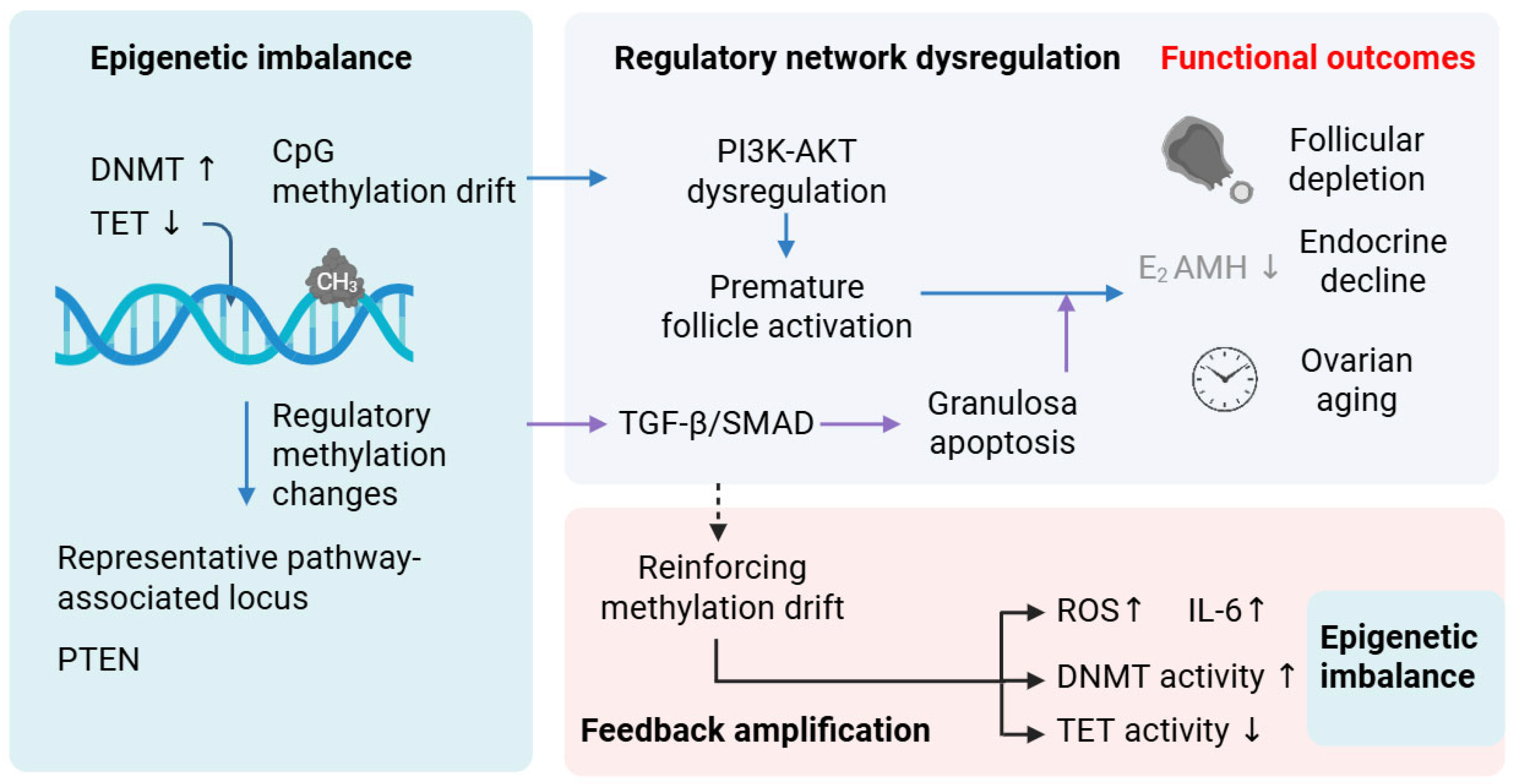
2.2. Convergence of Methylation Remodeling on PI3K–AKT and TGF-β/SMAD Signaling Networks
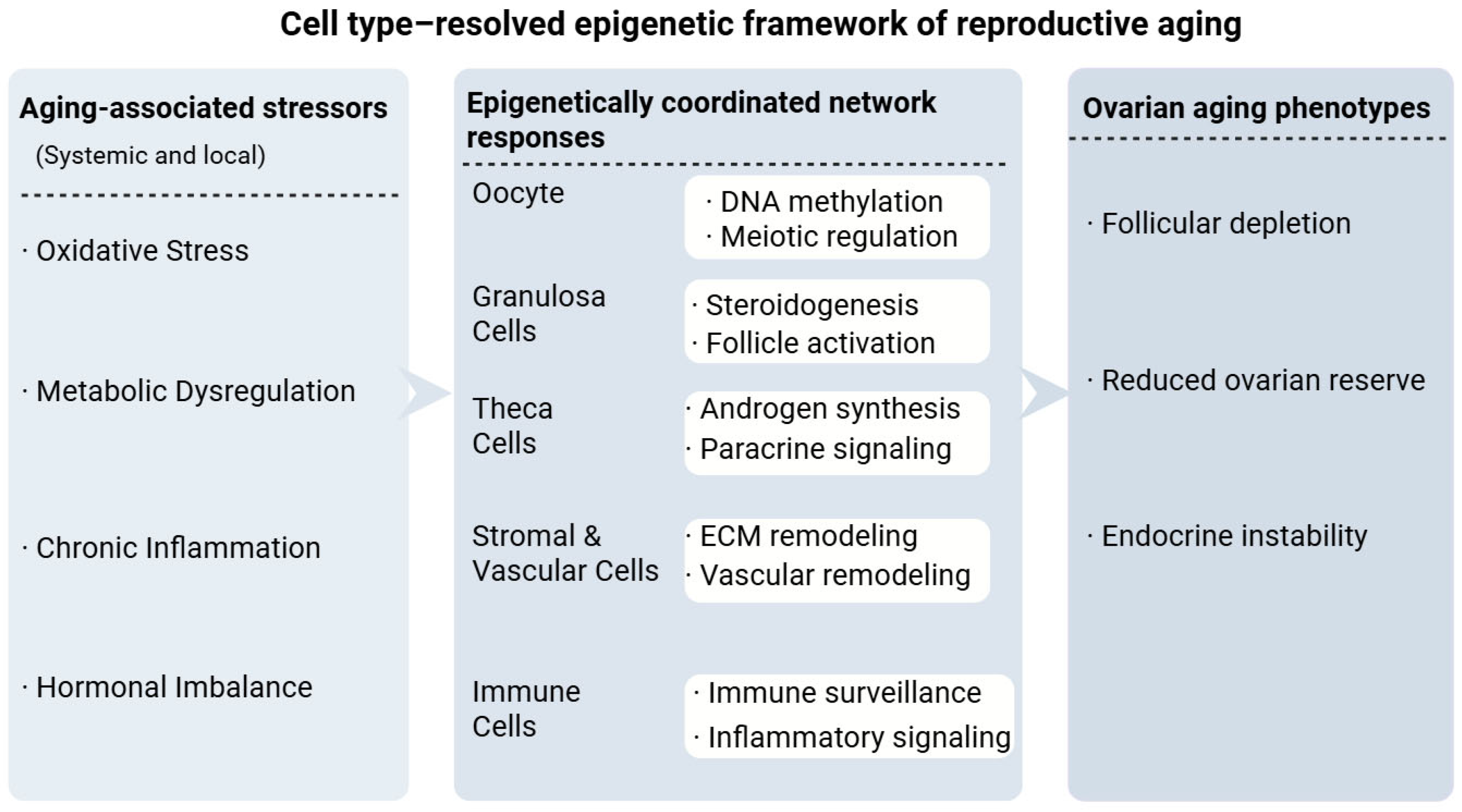
2.3. Methylation Remodeling Beyond the Follicle
3. Stress-Responsive Drivers of DNMT/TET Imbalance in Ovarian Aging
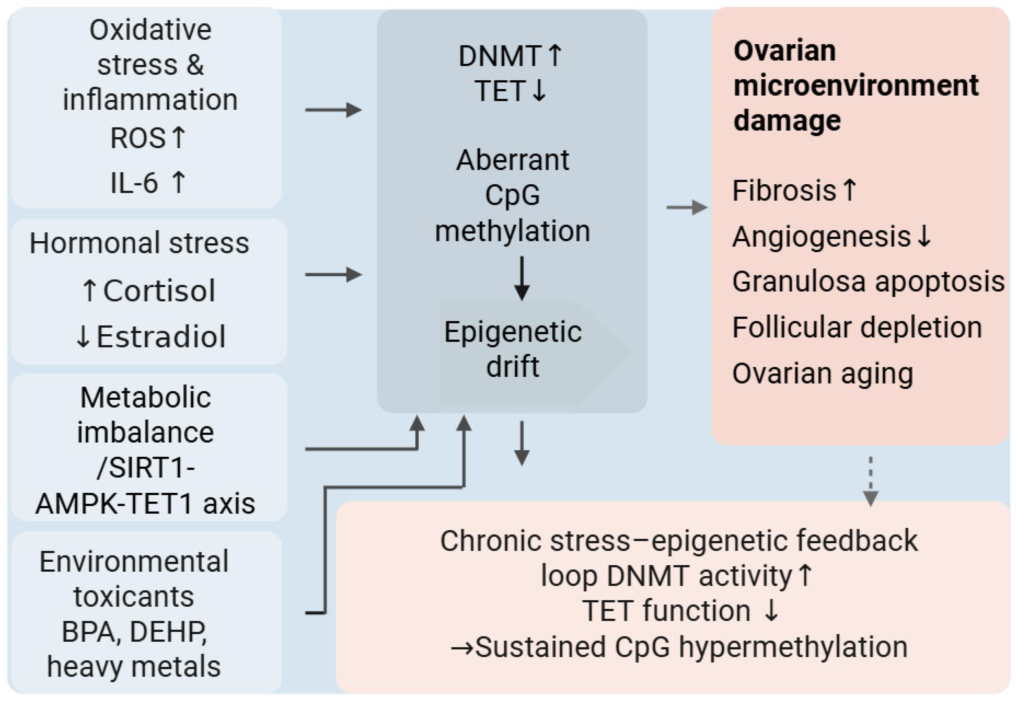
3.1. Oxidative Stress as a Primary Driver of DNMT/TET Imbalance and PI3K–AKT–FOXO3 Destabilization
3.2. Metabolic Dysfunction, One-Carbon Flux, and Energetic Imbalance as Central Determinants of Methylation Homeostasis
3.3. Environmental, Endocrine, Nutritional, and Circadian Influences Requiring Further Investigation
4. Methylation-Driven Pathway Rewiring in Ovarian Aging
Coupling of Genome Maintenance Networks to Methylation Drift
5. Epigenetic Biomarkers and Translational Perspectives
5.1. Circulating cfDNA Methylation
5.2. Epigenetic Clocks and Reproductive Aging
5.3. Artificial Intelligence and Multi-Modal Integration
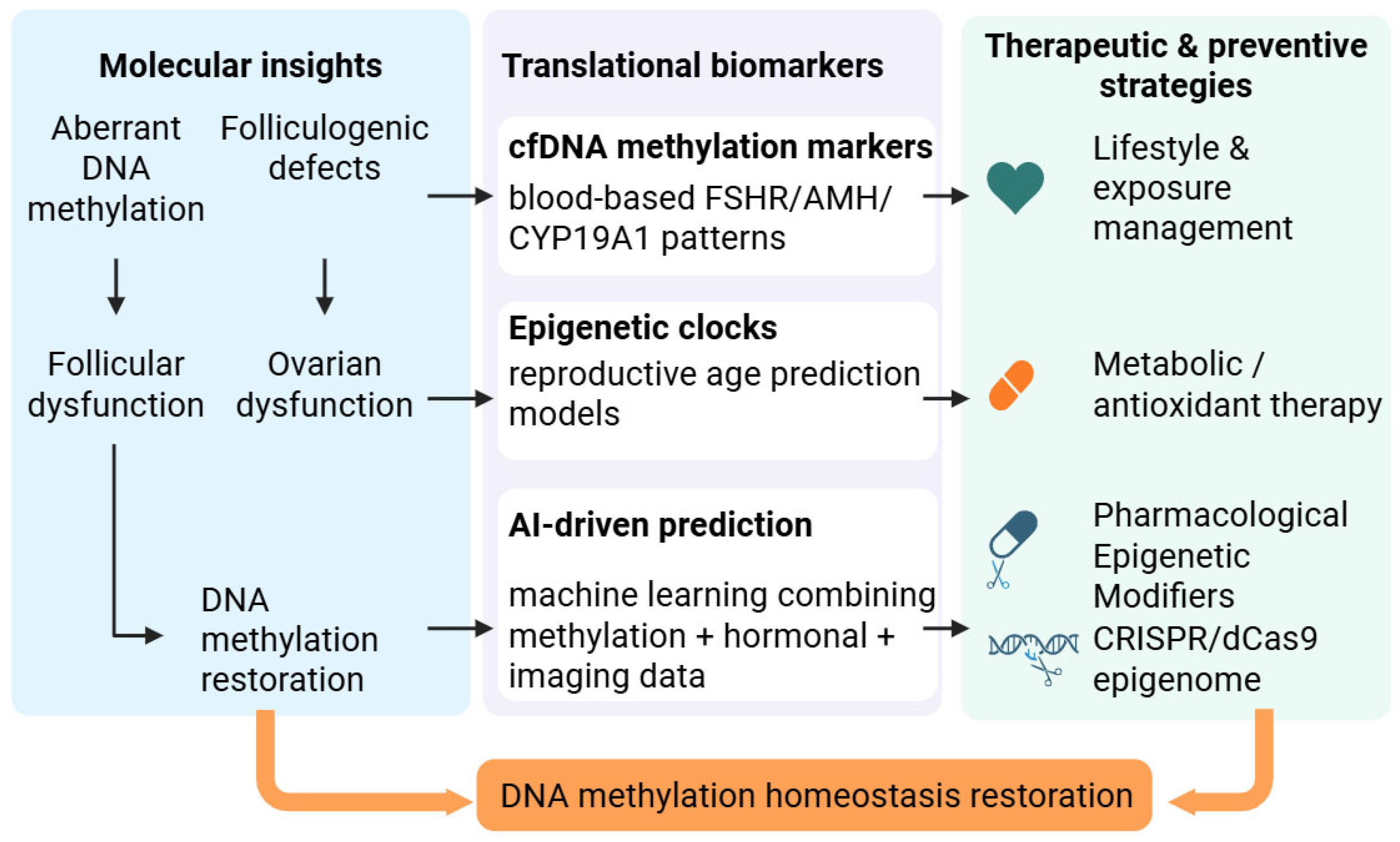
5.4. Clinical Translation and Therapeutic Considerations
6. Conclusions
Author Contributions
Funding
Institutional Review Board Statement
Informed Consent Statement
Data Availability Statement
Conflicts of Interest
Correction Statement
Abbreviations
| CpG | Cytosine–phosphate–guanine dinucleotide |
| DNMT | DNA methyltransferase |
| TET | Ten–eleven translocation dioxygenase |
| ROS | Reactive oxygen species |
| POI | Premature ovarian insufficiency |
| DOR | Diminished ovarian reserve |
| AMH | Anti-Müllerian hormone |
| FSH | Follicle-stimulating hormone |
| AFC | Antral follicle count |
| cfDNA | Cell-free DNA |
| PI3K | Phosphoinositide 3-kinase |
| AKT | Protein kinase B |
| mTOR | Mechanistic target of rapamycin |
| TGF-β | Transforming growth factor beta |
| SMAD | Suppressor of mothers against decapentaplegic |
References
- Wang, G.; Yang, R.; Zhang, H. Ovarian vascular aging: A hidden driver of mid-age female fertility decline. npj Aging 2025, 11, 24. [Google Scholar] [CrossRef] [Scilit] [PubMed]
- Smits, M.A.J.; Schomakers, B.V.; van Weeghel, M.; Wever, E.J.M.; Wüst, R.C.I.; Dijk, F.; Janssens, G.E.; Goddijn, M.T.; Mastenbroek, S.; Houtkooper, R.H.; et al. Human ovarian aging is characterized by oxidative damage and mitochondrial dysfunction. Hum. Reprod. 2023, 38, 2208–2220. [Google Scholar] [CrossRef] [Scilit]
- Isola, J.V.V.; Oca As, S.R.; Hubbart, C.R.; Ko, S.; Mondal, S.A.; Hense, J.D.; Carter, H.N.C.; Schneider, A.; Kovats, S.; Alberola-Ila, J.; et al. A single-cell atlas of the aging mouse ovary. Nat. Aging 2024, 4, 145–162. [Google Scholar] [CrossRef] [Scilit]
- Umehara, T.; Winstanley, Y.E.; Andreas, E.; Morimoto, A.; Williams, E.J.; Smith, K.M.; Carroll, J.; Febbraio, M.A.; Shimada, M.; Russell, D.L.; et al. Female reproductive life span is extended by targeted removal of fibrotic collagen from the mouse ovary. Sci. Adv. 2022, 8, eabn4564. [Google Scholar] [CrossRef] [Scilit]
- Zhao, S.; Cui, H.; Fang, X.; Xia, W.; Tao, C.; Li, J. Increased DNMT1 acetylation leads to global DNA methylation suppression in follicular granulosa cells during reproductive aging in mammals. BMC Genom. 2024, 25, 1030. [Google Scholar] [CrossRef] [Scilit]
- Bilmez, Y.; Talibova, G.; Ozturk, S. Expression of the histone lysine methyltransferases SETD1B, SETDB1, SETD2, and CFP1 exhibits significant changes in the oocytes and granulosa cells of aged mouse ovaries. Histochem. Cell Biol. 2022, 158, 79–95. [Google Scholar] [CrossRef] [Scilit]
- Dell’Aversana, C.; Cuomo, F.; Longobardi, S.; D’Hooghe, T.; Caprio, F.; Franci, G.; Santonastaso, M.; Colacurci, N.; Barone, S.; Pisaturo, V.; et al. Age-related miRNome landscape of cumulus oophorus cells during controlled ovarian stimulation protocols in IVF cycles. Hum. Reprod. 2021, 36, 1310–1325. [Google Scholar] [CrossRef] [Scilit]
- Lin, N.; Lin, J.; Plosch, T.; Sun, P.; Zhou, X. An Oxidative Stress-Related Gene Signature in Granulosa Cells Is Associated with Ovarian Aging. Oxidative Med. Cell. Longev. 2022, 2022, 1070968. [Google Scholar] [CrossRef] [Scilit] [PubMed]
- Lu, C.; Qin, C.; Fu, Z.; Wang, L.; Yi, Y.; Xin, M.; Zhen, X.; Han, C.; Lu, C. Metabolic dysregulation in patients with premature ovarian insufficiency revealed by integrated transcriptomic, methylomic and metabolomic analyses. Clin. Transl. Med. 2022, 12, e1006. [Google Scholar] [CrossRef] [Scilit]
- Zhu, Y.; Sun, H.; Gao, T.; Hou, S.; Li, Y.; Xu, Y.; Zhang, Q.; Feng, D. Ovarian remodeling and aging-related chronic inflammation and fibrosis in the mammalian ovary. J. Ovarian Res. 2025, 18, 133. [Google Scholar] [CrossRef] [Scilit] [PubMed]
- Jones, A.S.K.; Hannum, D.F.; Machlin, J.H.; Tan, A.; Ma, Q.; Ulrich, N.D.; Shen, Y.; Ciarelli, M.; Padmanabhan, V.; Marsh, E.E.; et al. Cellular atlas of the human ovary using morphologically guided spatial transcriptomics and single-cell sequencing. Sci. Adv. 2024, 10, eadm7506. [Google Scholar] [CrossRef] [Scilit]
- Wagner, M.; Yoshihara, M.; Douagi, I.; Damdimopoulos, A.; Panula, S.; Petropoulos, S.; Lu, H.; Pettersson, K.; Palm, K.; Katayama, S.; et al. Single-cell analysis of human ovarian cortex identifies distinct cell populations but no oogonial stem cells. Nat. Commun. 2020, 11, 1147. [Google Scholar] [CrossRef] [Scilit]
- Olsen, K.W.; Castillo-Fernandez, J.; Zedeler, A.; Freiesleben, N.C.; Bungum, M.; Chan, A.C.; Cardona, A.; Perry, J.R.B.; Skouby, S.O.; Borup, R.; et al. A distinctive epigenetic ageing profile in human granulosa cells. Hum. Reprod. 2020, 35, 1332–1345. [Google Scholar] [CrossRef] [Scilit]
- Kawai, T.; Richards, J.S.; Shimada, M. Large-scale DNA demethylation occurs in proliferating ovarian granulosa cells during mouse follicular development. Commun. Biol. 2021, 4, 1334. [Google Scholar] [CrossRef] [Scilit] [PubMed]
- Palla, G.; Pollner, P.; Börcsök, J.; Major, A.; Molnár, B.; Csabai, I. Hierarchy and control of ageing-related methylation networks. PLoS Comput. Biol. 2021, 17, e1009327. [Google Scholar] [CrossRef] [Scilit] [PubMed]
- Luan, Y.; So, W.; Dong, R.; Abazarikia, A.; Kim, S. KIT in oocytes: A key factor for oocyte survival and reproductive lifespan. eBioMedicine 2024, 106, 105263. [Google Scholar] [CrossRef] [Scilit]
- Ju, W.; Zhao, S.; Wu, H.; Yu, Y.; Li, Y.; Liu, D.; Lian, F.; Xiang, S. miR-6881-3p contributes to diminished ovarian reserve by regulating granulosa cell apoptosis by targeting SMAD4. Reprod. Biol. Endocrinol. 2024, 22, 17. [Google Scholar] [CrossRef] [Scilit]
- Hu, L.; Liao, M.; Liu, Y.; Xing, C.; Nong, L.; Yang, F.; Sun, S. Loss of AMPK activity induces organelle dysfunction and oxidative stress during oocyte aging. Biol. Direct 2024, 19, 29. [Google Scholar] [CrossRef] [Scilit]
- Sharma, N.; Coticchio, G.; Borini, A.; Tachibana, K.; Nasmyth, K.A.; Schuh, M. Changes in DNA repair compartments and cohesin loss promote DNA damage accumulation in aged oocytes. Curr. Biol. 2024, 34, 5131–5148. [Google Scholar] [CrossRef] [Scilit] [PubMed]
- Jin, C.; Wang, X.; Yang, J.; Kim, S.; Hudgins, A.D.; Gamliel, A.; Pei, M.; Contreras, D.; Devos, M.; Guo, Q.; et al. Molecular and genetic insights into human ovarian aging from single-nuclei multi-omics analyses. Nat. Aging 2025, 5, 275–290. [Google Scholar] [CrossRef] [Scilit] [PubMed]
- Wu, M.; Tang, W.; Chen, Y.; Xue, L.; Dai, J.; Li, Y.; Zhu, X.; Wu, C.; Xiong, J.; Zhang, J.; et al. Spatiotemporal transcriptomic changes of human ovarian aging and the regulatory role of FOXP1. Nat. Aging 2024, 4, 527–545. [Google Scholar] [CrossRef] [Scilit]
- An, Y.; Wang, Q.; Gao, K.; Zhang, C.; Ouyang, Y.; Li, R.; Ma, Z.; Wu, T.; Zhou, L.; Xie, Z.; et al. Epigenetic Regulation of Aging and its Rejuvenation. MedComm 2025, 6, e70369. [Google Scholar] [CrossRef] [Scilit] [PubMed]
- Pastor, W.A.; Aravind, L.; Rao, A. TETonic shift: Biological roles of TET proteins in DNA demethylation and transcription. Nat. Rev. Mol. Cell Biol. 2013, 14, 341–356. [Google Scholar] [CrossRef] [Scilit]
- Knight, A.K.; Spencer, J.B.; Smith, A.K. DNA methylation as a window into female reproductive aging. Epigenomics 2024, 16, 175–188. [Google Scholar] [CrossRef] [Scilit] [PubMed]
- Chamani, I.J.; Keefe, D.L. Epigenetics and Female Reproductive Aging. Front. Endocrinol. 2019, 10, 473. [Google Scholar] [CrossRef] [Scilit]
- Huang, X.; Jiang, Y.; Bao, H.; Wang, J.; Lin, R.; Yuan, J.; Xian, J.; Zhao, Y.; Chen, S. Role and Mechanism of Epigenetic Regulation in the Aging of Germ Cells: Prospects for Targeted Interventions. Aging Dis. 2024, 16, 146–167. [Google Scholar] [CrossRef] [Scilit]
- Klutstein, M.; Gonen, N. Epigenetic aging of mammalian gametes. Mol. Reprod. Dev. 2023, 90, 785–803. [Google Scholar] [CrossRef] [Scilit]
- Cani Ais, C.; Vasconcelos, S.; Santos, F.; Dória, S.; Marques, C.J. DNA methylation mechanisms in the maturing and ageing oocyte. Epigenet. Chromatin 2025, 18, 34. [Google Scholar] [CrossRef] [Scilit]
- Yu, B.; Russanova, V.R.; Gravina, S.; Hartley, S.; Mullikin, J.C.; Ignezweski, A.; Graham, J.; Segars, J.H.; Decherney, A.H.; Howard, B.H. DNA methylome and transcriptome sequencing in human ovarian granulosa cells links age-related changes in gene expression to gene body methylation and 3′-end GC density. Oncotarget 2015, 6, 3627–3643. [Google Scholar] [CrossRef] [Scilit] [PubMed]
- Yuan, T.; Jiao, Y.; de Jong, S.; Ophoff, R.A.; Beck, S.; Teschendorff, A.E. An integrative multi-scale analysis of the dynamic DNA methylation landscape in aging. PLoS Genet. 2015, 11, e1004996. [Google Scholar] [CrossRef] [Scilit]
- Zhou, X.; He, Y.; Li, N.; Bai, G.; Pan, X.; Zhang, Z.; Zhang, H.; Li, J.; Yuan, X. DNA methylation mediated RSPO2 to promote follicular development in mammals. Cell Death Dis. 2021, 12, 653. [Google Scholar] [CrossRef] [Scilit]
- Horvath, S. DNA methylation age of human tissues and cell types. Genome Biol. 2013, 14, R115. [Google Scholar] [CrossRef] [Scilit]
- Field, A.E.; Robertson, N.A.; Wang, T.; Havas, A.; Ideker, T.; Adams, P.D. DNA Methylation Clocks in Aging: Categories, Causes, and Consequences. Mol. Cell 2018, 71, 882–895. [Google Scholar] [CrossRef] [Scilit]
- Olsen, K.W.; Castillo-Fernandez, J.; Chan, A.C.; la Cour Freiesleben, N.; Zedeler, A.; Bungum, M.; Cardona, A.; Perry, J.R.B.; Skouby, S.O.; Hoffmann, E.R.; et al. Identification of a unique epigenetic profile in women with diminished ovarian reserve. Fertil. Steril. 2021, 115, 732–741. [Google Scholar] [CrossRef] [Scilit]
- Ferraretti, A.P.; La Marca, A.; Fauser, B.C.J.M.; Tarlatzis, B.; Nargund, G.; Gianaroli, L.; Eshre, W.G.O.P. ESHRE consensus on the definition of ‘poor response’ to ovarian stimulation for in vitro fertilization: The Bologna criteria. Hum. Reprod. 2011, 26, 1616–1624. [Google Scholar] [CrossRef] [Scilit] [PubMed]
- Hanson, B.M.; Tao, X.; Zhan, Y.; Jenkins, T.G.; Morin, S.J.; Scott, R.T.; Seli, E.U. Young women with poor ovarian response exhibit epigenetic age acceleration based on evaluation of white blood cells using a DNA methylation-derived age prediction model. Hum. Reprod. 2020, 35, 2579–2588. [Google Scholar] [CrossRef] [Scilit] [PubMed]
- Liu, L.; Cai, B.; Zhang, X.; Tan, J.; Huang, J.; Zhou, C. Differential transcriptional profiles of human cumulus granulosa cells in patients with diminished ovarian reserve. Arch. Gynecol. Obstet. 2022, 305, 1605–1614. [Google Scholar] [CrossRef] [Scilit]
- Wang, J.; Sun, X.; Yang, Z.; Li, S.; Wang, Y.; Ren, R.; Liu, Z.; Yu, D. Epigenetic regulation in premature ovarian failure: A literature review. Front. Physiol. 2023, 13, 998424. [Google Scholar] [CrossRef] [Scilit] [PubMed]
- Greene, A.D.; Patounakis, G.; Segars, J.H. Genetic associations with diminished ovarian reserve: A systematic review of the literature. J. Assist. Reprod. Genet. 2014, 31, 935–946. [Google Scholar] [CrossRef] [Scilit]
- Knight, A.K.; Hipp, H.S.; Abhari, S.; Gerkowicz, S.A.; Katler, Q.S.; Mckenzie, L.J.; Shang, W.; Smith, A.K.; Spencer, J.B. Markers of ovarian reserve are associated with reproductive age acceleration in granulosa cells from IVF patients. Hum. Reprod. 2022, 37, 2438–2445. [Google Scholar] [CrossRef] [Scilit]
- Morin, S.J.; Tao, X.; Marin, D.; Zhan, Y.; Landis, J.; Bedard, J.; Scott, R.T.; Seli, E. DNA methylation-based age prediction and telomere length in white blood cells and cumulus cells of infertile women with normal or poor response to ovarian stimulation. Aging 2018, 10, 3761–3773. [Google Scholar] [CrossRef] [Scilit]
- Sagvekar, P.; Kumar, P.; Mangoli, V.; Desai, S.; Mukherjee, S. DNA methylome profiling of granulosa cells reveals altered methylation in genes regulating vital ovarian functions in polycystic ovary syndrome. Clin. Epigenet. 2019, 11, 61. [Google Scholar] [CrossRef] [Scilit]
- Makrinou, E.; Drong, A.W.; Christopoulos, G.; Lerner, A.; Chapa-Chorda, I.; Karaderi, T.; Lavery, S.; Hardy, K.; Lindgren, C.M.; Franks, S. Genome-wide methylation profiling in granulosa lutein cells of women with polycystic ovary syndrome (PCOS). Mol. Cell. Endocrinol. 2020, 500, 110611. [Google Scholar] [CrossRef] [Scilit]
- Castillo-Fernandez, J.; Herrera-Puerta, E.; Demond, H.; Clark, S.J.; Hanna, C.W.; Hemberger, M.; Kelsey, G. Increased transcriptome variation and localised DNA methylation changes in oocytes from aged mice revealed by parallel single-cell analysis. Aging Cell 2020, 19, e13278. [Google Scholar] [CrossRef] [Scilit] [PubMed]
- Zhao, R.; Jiang, Y.; Zhao, S.; Zhao, H. Multiomics Analysis Reveals Molecular Abnormalities in Granulosa Cells of Women with Polycystic Ovary Syndrome. Front. Genet. 2021, 12, 648701. [Google Scholar] [CrossRef] [Scilit] [PubMed]
- Turan, V.; Oktay, K. BRCA-related ATM-mediated DNA double-strand break repair and ovarian aging. Hum. Reprod. Update 2020, 26, 43–57. [Google Scholar] [CrossRef] [Scilit] [PubMed]
- Titus, S.; Li, F.; Stobezki, R.; Akula, K.; Unsal, E.; Jeong, K.; Dickler, M.; Robson, M.; Moy, F.; Goswami, S.; et al. Impairment of BRCA1-related DNA double-strand break repair leads to ovarian aging in mice and humans. Sci. Transl. Med. 2013, 5, 172ra21. [Google Scholar] [CrossRef] [Scilit]
- Subramanian, G.N.; Greaney, J.; Wei, Z.; Olivier Becherel, M.L.H.A. Oocytes mount a noncanonical DNA damage response involving APC-Cdh1–mediated proteolysis. J. Cell. Biol. 2020, 4, e201907213. [Google Scholar] [CrossRef] [Scilit]
- Ge, Z.; Schatten, H.; Zhang, C.; Sun, Q. Oocyte ageing and epigenetics. Reproduction 2015, 149, R103–R114. [Google Scholar] [CrossRef] [Scilit]
- Gao, M.; Zhang, T.; Chen, T.; Chen, Z.; Zhu, Z.; Wen, Y.; Qin, S.; Bao, Y.; Zhao, T.; Li, H.; et al. Polycomb repressive complex 1 modulates granulosa cell proliferation in early folliculogenesis to support female reproduction. Theranostics 2024, 14, 1371–1389. [Google Scholar] [CrossRef] [Scilit] [PubMed]
- Wang, F.; Chen, X.; Sun, B.; Ma, Y.; Niu, W.; Zhai, J.; Sun, Y. Hypermethylation-mediated downregulation of lncRNA PVT1 promotes granulosa cell apoptosis in premature ovarian insufficiency via interacting with Foxo3a. J. Cell. Physiol. 2021, 236, 5162–5175. [Google Scholar] [CrossRef] [Scilit]
- Pan, Z.; Wang, X.; Di, R.; Liu, Q.; Hu, W.; Cao, X.; Guo, X.; He, X.; Lv, S.; Li, F.; et al. A 5-Methylcytosine Site of Growth Differentiation Factor 9 (GDF9) Gene Affects Its Tissue-Specific Expression in Sheep. Animals 2018, 8, 200. [Google Scholar] [CrossRef] [Scilit]
- Bassi, C.; Ho, J.; Srikumar, T.; Dowling, R.J.O.; Gorrini, C.; Miller, S.J.; Mak, T.W.; Neel, B.G.; Raught, B.; Stambolic, V. Nuclear PTEN controls DNA repair and sensitivity to genotoxic stress. Science 2013, 341, 395–399. [Google Scholar] [CrossRef] [Scilit] [PubMed]
- Ming, M.; He, Y. PTEN in DNA damage repair. Cancer Lett. 2012, 319, 125–129. [Google Scholar] [CrossRef] [Scilit]
- Reddy, P.; Liu, L.; Adhikari, D.; Jagarlamudi, K.; Rajareddy, S.; Shen, Y.; Du, C.; Tang, W.; Hamalainen, T.; Peng, S.L.; et al. Oocyte-specific deletion of Pten causes premature activation of the primordial follicle pool. Science 2008, 319, 611–613. [Google Scholar] [CrossRef] [Scilit]
- Lin, W.; Titus, S.; Moy, F.; Ginsburg, E.S.; Oktay, K. Ovarian Aging in Women with BRCA Germline Mutations. J. Clin. Endocrinol. Metab. 2017, 102, 3839–3847. [Google Scholar] [CrossRef] [Scilit]
- Winship, A.L.; Alesi, L.R.; Stringer, J.M.; Cao, Y.; Lewis, Y.M.; Tu, L.; Swindells, E.O.K.; Giridharan, S.; Cai, X.; Griffiths, M.J.; et al. Conditional loss of Brca1 in oocytes causes reduced litter size, ovarian reserve depletion and impaired oocyte in vitro maturation with advanced reproductive age in mice. eBioMedicine 2024, 106, 105262. [Google Scholar] [CrossRef] [Scilit]
- Oktay, K.; Turan, V.; Titus, S.; Stobezki, R.; Liu, L. BRCA Mutations, DNA Repair Deficiency, and Ovarian Aging. Biol. Reprod. 2015, 93, 67. [Google Scholar] [CrossRef] [Scilit]
- Yang, H.; Xie, Y.; Yang, D.; Ren, D. Oxidative stress-induced apoptosis in granulosa cells involves JNK, p53 and Puma. Oncotarget 2017, 8, 25310–25322. [Google Scholar] [CrossRef] [Scilit] [PubMed]
- Zhang, T.; Guan, Y.; Wang, S.; Wang, L.; Cheng, M.; Yuan, C.; Liu, Y.; Wang, Z. Bisphenol A induced abnormal DNA methylation of ovarian steroidogenic genes in rare minnow Gobiocypris rarus. Gen. Comp. Endocrinol. 2018, 269, 156–165. [Google Scholar] [CrossRef] [Scilit] [PubMed]
- Rattan, S.; Beers, H.K.; Kannan, A.; Ramakrishnan, A.; Brehm, E.; Bagchi, I.; Irudayaraj, J.M.K.; Flaws, J.A. Prenatal and ancestral exposure to di(2-ethylhexyl) phthalate alters gene expression and DNA methylation in mouse ovaries. Toxicol. Appl. Pharmacol. 2019, 379, 114629. [Google Scholar] [CrossRef] [Scilit]
- Shi, W.; Wang, D.; Xue, X.; Qiao, S.; Zhang, W.; Shi, J.; Huang, C. Epigenomic Landscape of Human Cumulus Cells in Premature Ovarian Insufficiency Using Single-Base Resolution Methylome and Hydroxymethylome. J. Cell. Mol. Med. 2024, 28, e70284. [Google Scholar] [CrossRef] [Scilit]
- Yao, J.; Huang, R.; Li, M.; Jiang, Y.; Wu, P.; Li, Y.; Peng, W.; Hua, C.; Huang, Y.; You, H.; et al. PTEN Expression in Human Granulosa Cells Is Associated with Ovarian Responses and Clinical Outcomes in IVF. Reprod. Sci. 2021, 28, 1910–1921. [Google Scholar] [CrossRef] [Scilit]
- Li, Q.; Pangas, S.A.; Jorgez, C.J.; Graff, J.M.; Weinstein, M.; Matzuk, M.M. Redundant roles of SMAD2 and SMAD3 in ovarian granulosa cells in vivo. Mol. Cell. Biol. 2008, 28, 7001–7011. [Google Scholar] [CrossRef] [Scilit] [PubMed]
- Gong, X.; Mcgee, E.A. Smad3 is required for normal follicular follicle-stimulating hormone responsiveness in the mouse. Biol. Reprod. 2009, 81, 730–738. [Google Scholar] [CrossRef] [Scilit] [PubMed]
- Yang, J.; Zhang, Y.; Xu, X.; Li, J.; Yuan, F.; Bo, S.; Qiao, J.; Xia, G.; Su, Y.; Zhang, M. Transforming growth factor-β is involved in maintaining oocyte meiotic arrest by promoting natriuretic peptide type C expression in mouse granulosa cells. Cell Death Dis. 2019, 10, 558. [Google Scholar] [CrossRef] [Scilit]
- Liu, W.; Chen, C.; Gao, Y.; Cui, X.; Zhang, Y.; Gu, L.; He, Y.; Li, J.; Gao, S.; Gao, R.; et al. Transcriptome Dynamics and Cell Dialogs Between Oocytes and Granulosa Cells in Mouse Follicle Development. Genom. Proteom. Bioinform. 2024, 22, qzad001. [Google Scholar] [CrossRef] [Scilit]
- Wigglesworth, K.; Lee, K.; Emori, C.; Sugiura, K.; Eppig, J.J. Transcriptomic diversification of developing cumulus and mural granulosa cells in mouse ovarian follicles. Biol. Reprod. 2015, 92, 23. [Google Scholar] [CrossRef] [Scilit] [PubMed]
- Zhou, C.; Guo, Q.; Lin, J.; Wang, M.; Zeng, Z.; Li, Y.; Li, X.; Xiang, Y.; Liang, Q.; Liu, J.; et al. Single-Cell Atlas of Human Ovaries Reveals the Role of the Pyroptotic Macrophage in Ovarian Aging. Adv. Sci. 2024, 11, e2305175. [Google Scholar] [CrossRef] [Scilit]
- Amargant, F.; Magalhaes, C.; Pritchard, M.T.; Duncan, F.E. Systemic low-dose anti-fibrotic treatment attenuates ovarian aging in the mouse. GeroScience 2025, 47, 3475–3495. [Google Scholar] [CrossRef] [Scilit]
- Sarkar, P.; Moore, M.; Ozmen, A.; Cetinkaya-Un, B.; Julie, V.; Imudia, A.N.; Lockwood, C.J.; Kayisli, U.A.; Guzeloglu-Kayisli, O. Enhanced ovarian FKBP51 expression is associated with ovarian aging: A molecular insight for age-related fertility in women. F&S Sci. 2025, 6, 152–163. [Google Scholar] [CrossRef] [Scilit] [PubMed]
- Wang, C.; Sun, Y. Induction of Collagen I by CXCL10 in Ovarian Theca-Stroma Cells via the JNK Pathway. Front. Endocrinol. 2022, 13, 823740. [Google Scholar] [CrossRef] [Scilit] [PubMed]
- Sun, W.; Li, M.; Yang, Y.; Dong, Q.; Tao, H.; Lu, C.; Yang, J. Novel insights on post-translational modifications of nucleic acid methylation proteins in hepatic fibrosis. Pharmacol. Res. 2025, 218, 107825. [Google Scholar] [CrossRef] [Scilit]
- Gong, Y.; Liu, M.; Zhang, Q.; Li, J.; Cai, H.; Ran, J.; Ma, L.; Ma, Y.; Quan, S. Lysine acetyltransferase 14 mediates TGF-β-induced fibrosis in ovarian endometrioma via co-operation with serum response factor. J. Transl. Med. 2024, 22, 561. [Google Scholar] [CrossRef] [Scilit] [PubMed]
- Voros, C.; Varthaliti, A.; Mavrogianni, D.; Athanasiou, D.; Athanasiou, A.; Athanasiou, A.; Papahliou, A.; Zografos, C.G.; Topalis, V.; Kondili, P.; et al. Epigenetic Alterations in Ovarian Function and Their Impact on Assisted Reproductive Technologies: A Systematic Review. Biomedicines 2025, 13, 730. [Google Scholar] [CrossRef] [Scilit]
- Dutta, S.; Sengupta, P.; Mottola, F.; Das, S.; Hussain, A.; Ashour, A.; Rocco, L.; Govindasamy, K.; Rosas, I.M.; Roychoudhury, S. Crosstalk Between Oxidative Stress and Epigenetics: Unveiling New Biomarkers in Human Infertility. Cells 2024, 13, 1846. [Google Scholar] [CrossRef] [Scilit]
- Guillaumet-Adkins, A.; Ya Ez, Y.; Peris-Diaz, M.D.; Calabria, I.; Palanca-Ballester, C.; Sandoval, J. Epigenetics and Oxidative Stress in Aging. Oxidative Med. Cell. Longev. 2017, 2017, 9175806. [Google Scholar] [CrossRef] [Scilit] [PubMed]
- Niu, Y.; Desmarais, T.L.; Tong, Z.; Yao, Y.; Costa, M. Oxidative stress alters global histone modification and DNA methylation. Free. Radic. Biol. Med. 2015, 82, 22–28. [Google Scholar] [CrossRef] [Scilit]
- Devine, P.J.; Perreault, S.D.; Luderer, U. Roles of reactive oxygen species and antioxidants in ovarian toxicity. Biol. Reprod. 2012, 86, 27. [Google Scholar] [CrossRef] [Scilit]
- O’Hagan, H.M.; Wang, W.; Sen, S.; Destefano Shields, C.; Lee, S.S.; Zhang, Y.W.; Clements, E.G.; Cai, Y.; Van Neste, L.; Easwaran, H.; et al. Oxidative damage targets complexes containing DNA methyltransferases, SIRT1, and polycomb members to promoter CpG Islands. Cancer Cell 2011, 20, 606–619. [Google Scholar] [CrossRef] [Scilit]
- Zhou, S.; Xi, Y.; Chen, Y.; Zhang, Z.; Wu, C.; Yan, W.; Luo, A.; Wu, T.; Zhang, J.; Wu, M.; et al. Ovarian Dysfunction Induced by Chronic Whole-Body PM2.5 Exposure. Small 2020, 16, e2000845. [Google Scholar] [CrossRef] [Scilit] [PubMed]
- García-Giménez, J.L.; Iba Ez-Cabellos, J.S.; Seco-Cervera, M.; Pallardó, F.V. Glutathione and cellular redox control in epigenetic regulation. Free. Radic. Biol. Med. 2014, 75, S3. [Google Scholar] [CrossRef] [Scilit]
- Belle, R.; Sara, H.; Salah, E.; Bhushan, B.; Szykowska, A.; Roper, G.; Tumber, A.; Kriaucionis, S.; Burgess-Brown, N.; Schofield, C.J.; et al. Focused Screening Identifies Different Sensitivities of Human TET Oxygenases to the Oncometabolite 2-Hydroxyglutarate. J. Med. Chem. 2024, 67, 4525–4540. [Google Scholar] [CrossRef] [Scilit]
- Ravichandran, M.; Rafalski, D.; Davies, C.I.; Ortega-Recalde, O.; Nan, X.; Glanfield, C.R.; Kotter, A.; Misztal, K.; Wang, A.H.; Wojciechowski, M.; et al. Pronounced sequence specificity of the TET enzyme catalytic domain guides its cellular function. Sci. Adv. 2022, 8, eabm2427. [Google Scholar] [CrossRef] [Scilit]
- Joshi, K.; Liu, S.; Breslin, P.S.J.; Zhang, J. Mechanisms that regulate the activities of TET proteins. Cell. Mol. Life Sci. 2022, 79, 363. [Google Scholar] [CrossRef] [Scilit]
- Jiang, W.; Block, M.E.; Boosani, C.S. Short communication: TNF-α and IGF-1 regulates epigenetic mechanisms of HDAC2 and HDAC10. PLoS ONE 2022, 17, e0263190. [Google Scholar] [CrossRef] [Scilit]
- Shi, Q.; Liu, R.; Chen, L. Ferroptosis inhibitor ferrostatin-1 alleviates homocysteine induced ovarian granulosa cell injury by regulating TET activity and DNA methylation. Mol. Med. Rep. 2022, 25, 130. [Google Scholar] [CrossRef] [Scilit]
- Liu, L.; Wang, H.; Xu, G.L.; Liu, L. Tet1 Deficiency Leads to Premature Ovarian Failure. Front. Cell. Dev. Biol. 2021, 9, 644135. [Google Scholar] [CrossRef] [Scilit]
- Vandaele, L.; Thys, M.; Bijttebier, J.; Van Langendonckt, A.; Donnay, I.; Maes, D.; Meyer, E.; Van Soom, A. Short-term exposure to hydrogen peroxide during oocyte maturation improves bovine embryo development. Reproduction 2010, 139, 505–511. [Google Scholar] [CrossRef] [Scilit] [PubMed]
- Khaghani, A.J.; Farrokh, P.; Zavareh, S. Epigenetic effects of Bisphenol A on granulosa cells of mouse follicles during in vitro culture: An experimental study. Int. J. Reprod. Biomed. 2021, 19, 129–136. [Google Scholar] [CrossRef] [Scilit] [PubMed]
- Arcanjo, R.B.; Vieira, M.C.; Sivaguru, M.; Nowak, R.A. Impact of mono(2-ethylhexyl) phthalate (MEHP) on the development of mouse embryo in vitro. Reprod. Toxicol. 2023, 115, 111–123. [Google Scholar] [CrossRef] [Scilit]
- John, G.B.; Gallardo, T.D.; Shirley, L.J.; Castrillon, D.H. Foxo3 is a PI3K-dependent molecular switch controlling the initiation of oocyte growth. Dev. Biol. 2008, 321, 197–204. [Google Scholar] [CrossRef] [Scilit]
- Ducreux, B.; Patrat, C.; Trasler, J.; Fauque, P. Transcriptomic integrity of human oocytes used in ARTs: Technical and intrinsic factor effects. Hum. Reprod. Update 2024, 30, 26–47. [Google Scholar] [CrossRef] [Scilit] [PubMed]
- Choi, S.; Friso, S. Modulation of DNA methylation by one-carbon metabolism: A milestone for healthy aging. Nutr. Res. Pract. 2023, 17, 597–615. [Google Scholar] [CrossRef] [Scilit] [PubMed]
- Inoue-Choi, M.; Nelson, H.H.; Robien, K.; Arning, E.; Bottiglieri, T.; Koh, W.; Yuan, J. Plasma S-adenosylmethionine, DNMT polymorphisms, and peripheral blood LINE-1 methylation among healthy Chinese adults in Singapore. BMC Cancer 2013, 13, 389. [Google Scholar] [CrossRef] [Scilit]
- Pauwels, S.; Ghosh, M.; Duca, R.C.; Bekaert, B.; Freson, K.; Huybrechts, I.; Langie, S.A.S.; Koppen, G.; Devlieger, R.; Godderis, L. Maternal intake of methyl-group donors affects DNA methylation of metabolic genes in infants. Clin. Epigenet. 2017, 9, 16. [Google Scholar] [CrossRef] [Scilit] [PubMed]
- Wang, Y.; Gao, X.; Yang, Z.; Yan, X.; He, X.; Guo, T.; Zhao, S.; Zhao, H.; Chen, Z. Deciphering the DNA methylome in women with PCOS diagnosed using the new international evidence-based guidelines. Hum. Reprod. 2023, 38, ii69–ii79. [Google Scholar] [CrossRef] [Scilit]
- Mentch, S.J.; Locasale, J.W. One-carbon metabolism and epigenetics: Understanding the specificity. Ann. NY Acad. Sci. 2016, 1363, 91–98. [Google Scholar] [CrossRef] [Scilit]
- Benayoun, B.A.; Kochersberger, A.; Garrison, J.L. Studying ovarian aging and its health impacts: Modern tools and approaches. Genes Dev. 2025, 39, 975–990. [Google Scholar] [CrossRef] [Scilit]
- Wei, S.; Tang, W.; Chen, D.; Xiong, J.; Xue, L.; Dai, Y.; Guo, Y.; Wu, C.; Dai, J.; Wu, M.; et al. Multiomics insights into the female reproductive aging. Ageing Res. Rev. 2024, 95, 102245. [Google Scholar] [CrossRef] [Scilit]
- Crujeiras, A.B.; Diaz-Lagares, A.; Moreno-Navarrete, J.M.; Sandoval, J.; Hervas, D.; Gomez, A.; Ricart, W.; Casanueva, F.F.; Esteller, M.; Fernandez-Real, J.M. Genome-wide DNA methylation pattern in visceral adipose tissue differentiates insulin-resistant from insulin-sensitive obese subjects. Transl. Res. 2016, 178, 13–24. [Google Scholar] [CrossRef] [Scilit] [PubMed]
- Takeshima, H.; Niwa, T.; Yamashita, S.; Takamura-Enya, T.; Iida, N.; Wakabayashi, M.; Nanjo, S.; Abe, M.; Sugiyama, T.; Kim, Y.; et al. TET repression and increased DNMT activity synergistically induce aberrant DNA methylation. J. Clin. Investig. 2020, 130, 5370–5379. [Google Scholar] [CrossRef] [Scilit]
- Wang, X.; Wang, L.; Xiang, W. Mechanisms of ovarian aging in women: A review. J. Ovarian Res. 2023, 16, 67. [Google Scholar] [CrossRef] [Scilit]
- Liu, X.; Zhao, Y.; Feng, Y.; Wang, S.; Zhang, J. Ovarian Aging: Mechanisms, Age-Related Disorders, and Therapeutic Interventions. MedComm 2025, 6, e70481. [Google Scholar] [CrossRef] [Scilit]
- Gou, M.; Li, J.; Yi, L.; Li, H.; Ye, X.; Wang, H.; Liu, L.; Sun, B.; Zhang, S.; Zhu, Z.; et al. Reprogramming of ovarian aging epigenome by resveratrol. PNAS Nexus 2022, 2, pgac310. [Google Scholar] [CrossRef] [Scilit]
- Zhang, H.; Yan, K.; Sui, L.; Nie, J.; Cui, K.; Liu, J.; Zhang, H.; Yang, X.; Lu, K.; Liang, X. Constant light exposure causes oocyte meiotic defects and quality deterioration in mice. Environ. Pollut. 2020, 267, 115467. [Google Scholar] [CrossRef] [Scilit]
- Lengyel, E.; Li, Y.; Weigert, M.; Zhu, L.; Eckart, H.; Javellana, M.; Ackroyd, S.; Xiao, J.; Olalekan, S.; Glass, D.; et al. A molecular atlas of the human postmenopausal fallopian tube and ovary from single-cell RNA and ATAC sequencing. Cell Rep. 2022, 41, 111838. [Google Scholar] [CrossRef] [Scilit] [PubMed]
- Hood, R.B.; Everson, T.M.; Ford, J.B.; Hauser, R.; Knight, A.; Smith, A.K.; Gaskins, A.J. Epigenetic age acceleration in follicular fluid and markers of ovarian response among women undergoing IVF. Hum. Reprod. 2024, 39, 2003–2009. [Google Scholar] [CrossRef] [Scilit] [PubMed]
- Masciangelo, R.; Hossay, C.; Chiti, M.C.; Manavella, D.D.; Amorim, C.A.; Donnez, J.; Dolmans, M. Role of the PI3K and Hippo pathways in follicle activation after grafting of human ovarian tissue. J. Assist. Reprod. Genet. 2020, 37, 101–108. [Google Scholar] [CrossRef] [Scilit]
- Ha, K.; Lee, G.E.; Palii, S.S.; Brown, K.D.; Takeda, Y.; Liu, K.; Bhalla, K.N.; Robertson, K.D. Rapid and transient recruitment of DNMT1 to DNA double-strand breaks is mediated by its interaction with multiple components of the DNA damage response machinery. Hum. Mol. Genet. 2011, 20, 126–140. [Google Scholar] [CrossRef] [Scilit]
- Kantidze, O.L.; Razin, S.V. 5-hydroxymethylcytosine in DNA repair: A new player or a red herring? Cell Cycle 2017, 16, 1499–1501. [Google Scholar] [CrossRef] [Scilit] [PubMed]
- Porcu, E.; Cillo, G.M.; Cipriani, L.; Sacilotto, F.; Notarangelo, L.; Damiano, G.; Dirodi, M.; Roncarati, I. Impact of BRCA1 and BRCA2 mutations on ovarian reserve and fertility preservation outcomes in young women with breast cancer. J. Assist. Reprod. Genet. 2020, 37, 709–715. [Google Scholar] [CrossRef] [Scilit]
- O’Hagan, H.M.; Mohammad, H.P.; Baylin, S.B. Double strand breaks can initiate gene silencing and SIRT1-dependent onset of DNA methylation in an exogenous promoter CpG island. PLoS Genet. 2008, 4, e1000155. [Google Scholar] [CrossRef] [Scilit]
- Bolcun-Filas, E.; Rinaldi, V.D.; White, M.E.; Schimenti, J.C. Reversal of female infertility by Chk2 ablation reveals the oocyte DNA damage checkpoint pathway. Science 2014, 343, 533–536. [Google Scholar] [CrossRef] [Scilit]
- Rinaldi, V.D.; Bloom, J.C.; Schimenti, J.C. Oocyte Elimination Through DNA Damage Signaling from CHK1/CHK2 to p53 and p63. Genetics 2020, 215, 373–378. [Google Scholar] [CrossRef] [Scilit] [PubMed]
- de Kat, A.C.; Broekmans, F.J.M.; Lambalk, C.B. Role of AMH in Prediction of Menopause. Front. Endocrinol. 2021, 12, 733731. [Google Scholar] [CrossRef] [Scilit] [PubMed]
- Aftabsavad, S.; Noormohammadi, Z.; Moini, A.; Karimipoor, M. Effect of bisphenol A on alterations of ICAM-1 and HLA-G genes expression and DNA methylation profiles in cumulus cells of infertile women with poor response to ovarian stimulation. Sci. Rep. 2021, 11, 9595. [Google Scholar] [CrossRef] [Scilit]
- Moss, J.; Magenheim, J.; Neiman, D.; Zemmour, H.; Loyfer, N.; Korach, A.; Samet, Y.; Maoz, M.; Druid, H.; Arner, P.; et al. Comprehensive human cell-type methylation atlas reveals origins of circulating cell-free DNA in health and disease. Nat. Commun. 2018, 9, 5068. [Google Scholar] [CrossRef] [Scilit]
- Liu, L.; Toung, J.M.; Jassowicz, A.F.; Vijayaraghavan, R.; Kang, H.; Zhang, R.; Kruglyak, K.M.; Huang, H.J.; Hinoue, T.; Shen, H.; et al. Targeted methylation sequencing of plasma cell-free DNA for cancer detection and classification. Ann. Oncol. 2018, 29, 1445–1453. [Google Scholar] [CrossRef] [Scilit]
- Conway, A.; Pearce, S.P.; Clipson, A.; Hill, S.M.; Chemi, F.; Slane-Tan, D.; Ferdous, S.; Hossain, A.S.M.M.; Kamieniecka, K.; White, D.J.; et al. A cfDNA methylation-based tissue-of-origin classifier for cancers of unknown primary. Nat. Commun. 2024, 15, 3292. [Google Scholar] [CrossRef] [Scilit]
- Liu, M.C.; Oxnard, G.R.; Klein, E.A.; Swanton, C.; Seiden, M.V.; CCGA Consortium. Sensitive and specific multi-cancer detection and localization using methylation signatures in cell-free DNA. Ann. Oncol. 2020, 31, 745–759. [Google Scholar] [CrossRef] [Scilit] [PubMed]
- Sun, K.; Jiang, P.; Chan, K.C.A.; Wong, J.; Cheng, Y.K.Y.; Liang, R.H.S.; Chan, W.; Ma, E.S.K.; Chan, S.L.; Cheng, S.H.; et al. Plasma DNA tissue mapping by genome-wide methylation sequencing for noninvasive prenatal, cancer, and transplantation assessments. Proc. Natl. Acad. Sci. USA 2015, 112, E5503–E5512. [Google Scholar] [CrossRef] [Scilit]
- Gai, W.; Zhou, Z.; Agbor-Enoh, S.; Fan, X.; Lian, S.; Jiang, P.; Cheng, S.H.; Wong, J.; Chan, S.L.; Jang, M.K.; et al. Applications of genetic-epigenetic tissue mapping for plasma DNA in prenatal testing, transplantation and oncology. eLife 2021, 10, e64356. [Google Scholar] [CrossRef] [Scilit]
- Klein, E.A.; Richards, D.; Cohn, A.; Tummala, M.; Lapham, R.; Cosgrove, D.; Chung, G.; Clement, J.; Gao, J.; Hunkapiller, N.; et al. Clinical validation of a targeted methylation-based multi-cancer early detection test using an independent validation set. Ann. Oncol. 2021, 32, 1167–1177. [Google Scholar] [CrossRef] [Scilit]
- Levine, M.E.; Lu, A.T.; Chen, B.H.; Hernandez, D.G.; Singleton, A.B.; Ferrucci, L.; Bandinelli, S.; Salfati, E.; Manson, J.E.; Quach, A.; et al. Menopause accelerates biological aging. Proc. Natl. Acad. Sci. USA 2016, 113, 9327–9332. [Google Scholar] [CrossRef] [Scilit]
- Mattox, A.K.; Douville, C.; Wang, Y.; Popoli, M.; Ptak, J.; Silliman, N.; Dobbyn, L.; Schaefer, J.; Lu, S.; Pearlman, A.H.; et al. The Origin of Highly Elevated Cell-Free DNA in Healthy Individuals and Patients with Pancreatic, Colorectal, Lung, or Ovarian Cancer. Cancer Discov. 2023, 13, 2166–2179. [Google Scholar] [CrossRef] [Scilit] [PubMed]
- Cheng, A.P.; Cheng, M.P.; Gu, W.; Sesing Lenz, J.; Hsu, E.; Schurr, E.; Bourque, G.; Bourgey, M.; Ritz, J.; Marty, F.M.; et al. Cell-free DNA tissues of origin by methylation profiling reveals significant cell, tissue, and organ-specific injury related to COVID-19 severity. Med 2021, 2, 411–422. [Google Scholar] [CrossRef] [Scilit] [PubMed]
- Mcnamara, M.E.; Loyfer, N.; Kiliti, A.J.; Schmidt, M.O.; Shabi-Porat, S.; Jain, S.S.; Martinez Roth, S.; Mcdeed, A.P.T.; Shahrour, N.; Ballew, E.; et al. Circulating cell-free methylated DNA reveals tissue-specific, cellular damage from radiation treatment. JCI Insight 2023, 8, e156529. [Google Scholar] [CrossRef] [Scilit]
- Prosz, A.; Pipek, O.; Börcsök, J.; Palla, G.; Szallasi, Z.; Spisak, S.; Csabai, I. Biologically informed deep learning for explainable epigenetic clocks. Sci. Rep. 2024, 14, 1306. [Google Scholar] [CrossRef] [Scilit]
- Bernabeu, E.; Mccartney, D.L.; Gadd, D.A.; Hillary, R.F.; Lu, A.T.; Murphy, L.; Wrobel, N.; Campbell, A.; Harris, S.E.; Liewald, D.; et al. Refining epigenetic prediction of chronological and biological age. Genome Med. 2023, 15, 12. [Google Scholar] [CrossRef] [Scilit]
- Chen, B.H.; Marioni, R.E.; Colicino, E.; Peters, M.J.; Ward-Caviness, C.K.; Tsai, P.; Roetker, N.S.; Just, A.C.; Demerath, E.W.; Guan, W.; et al. DNA methylation-based measures of biological age: Meta-analysis predicting time to death. Aging 2016, 8, 1844–1865. [Google Scholar] [CrossRef] [Scilit]
- Marinello, D.; Reschini, M.; Di Stefano, G.; Carullo, G.; Casalechi, M.; Tarantini, L.; Albetti, B.; Bollati, V.; Vigano, P.; Somigliana, E.; et al. Epigenetic age and fertility timeline: Testing an epigenetic clock to forecast in vitro fertilization success rate. Reprod. Biol. Endocrinol. 2025, 23, 99. [Google Scholar] [CrossRef] [Scilit]
- Monseur, B.; Murugappan, G.; Bentley, J.; Teng, N.; Westphal, L. Epigenetic clock measuring age acceleration via DNA methylation levels in blood is associated with decreased oocyte yield. J. Assist. Reprod. Genet. 2020, 37, 1097–1103. [Google Scholar] [CrossRef] [Scilit] [PubMed]
- Daredia, S.; Khodasevich, D.; Gladish, N.; Shen, H.; Nwanaji-Enwerem, J.C.; Bozack, A.K.; Needham, B.L.; Rehkopf, D.H.; Deardorff, J.; Cardenas, A. Timing of menarche and menopause and epigenetic aging among U.S. adults: Results from the National Health and Nutrition Examination Survey 1999–2002. Clin. Epigenet. 2025, 17, 31. [Google Scholar] [CrossRef] [Scilit]
- Wang, L.; Xu, S.; Chen, R.; Ding, Y.; Liu, M.; Hou, C.; Wu, Z.; Men, X.; Bao, M.; He, B.; et al. Exploring the causal association between epigenetic clocks and menopause age: Insights from a bidirectional Mendelian randomization study. Front. Endocrinol. 2024, 15, 1429514. [Google Scholar] [CrossRef] [Scilit]
- Wang, S.; Zheng, Y.; Li, J.; Yu, Y.; Zhang, W.; Song, M.; Liu, Z.; Min, Z.; Hu, H.; Jing, Y.; et al. Single-Cell Transcriptomic Atlas of Primate Ovarian Aging. Cell 2020, 180, 585–600. [Google Scholar] [CrossRef] [Scilit]
- Ng, S.; Masarone, S.; Watson, D.; Barnes, M.R. The benefits and pitfalls of machine learning for biomarker discovery. Cell Tissue Res. 2023, 394, 17–31. [Google Scholar] [CrossRef] [Scilit] [PubMed]
- Chen, F.; Chen, Y.; Mai, Q. Multi-Omics Analysis and Machine Learning Prediction Model for Pregnancy Outcomes After Intracytoplasmic Sperm Injection—In vitro Fertilization. Front. Public Health 2022, 10, 924539. [Google Scholar] [CrossRef] [Scilit] [PubMed]
- Kim, M.; Park, J.; Seonghee, O.; Jeong, B.; Byun, Y.; Shin, S.H.; Im, Y.; Cho, J.H.; Cho, E. Deep learning model integrating cfDNA methylation and fragment size profiles for lung cancer diagnosis. Sci. Rep. 2024, 14, 14797. [Google Scholar] [CrossRef] [Scilit]
- Zhou, Q.; Kang, G.; Jiang, P.; Qiao, R.; Lam, W.K.J.; Yu, S.C.Y.; Ma, M.L.; Ji, L.; Cheng, S.H.; Gai, W.; et al. Epigenetic analysis of cell-free DNA by fragmentomic profiling. Proc. Natl. Acad. Sci. USA 2022, 119, e2085115177. [Google Scholar] [CrossRef] [Scilit]
- Li, W.; Cheng, Y.; Cui, A.; Huang, M.; Huang, Q.; Wang, Q.; Xia, M.; Qiu, J.; Peng, Q.; Li, J.; et al. Multiomics Integration of Epigenetics, Proteomics, and Metabolomics Identifies Putative Drug Targets and Improves Early Prediction for Diabetes. Diabetes 2025, 74, 2418–2431. [Google Scholar] [CrossRef] [Scilit]
- Zhang, J.; Jia, S.; Zheng, Z.; Cao, L.; Zhou, J.; Fu, X. A multi-omic single-cell landscape of the aging mouse ovary. GeroScience 2025, 47, 4485–4498. [Google Scholar] [CrossRef] [Scilit] [PubMed]
- Olova, N.; Krueger, F.; Andrews, S.; Oxley, D.; Berrens, R.V.; Branco, M.R.; Reik, W. Comparison of whole-genome bisulfite sequencing library preparation strategies identifies sources of biases affecting DNA methylation data. Genome Biol. 2018, 19, 33. [Google Scholar] [CrossRef] [Scilit] [PubMed]
- Zhang, Y.; Yin, T.; Zhou, L. CRISPR/Cas9 technology: Applications in oocytes and early embryos. J. Transl. Med. 2023, 21, 746. [Google Scholar] [CrossRef] [Scilit]
- Tejedor, J.R.; Penarroya, A.; Gancedo-Verdejo, J.; Santamarina-Ojeda, P.; Perez, R.F.; Lopez-Tamargo, S.; Diez-Borge, A.; Alba-Linares, J.J.; Gonzalez-Del-Rey, N.; Urdinguio, R.G.; et al. CRISPR/dCAS9-mediated DNA demethylation screen identifies functional epigenetic determinants of colorectal cancer. Clin. Epigenet. 2023, 15, 133. [Google Scholar] [CrossRef] [Scilit]
- Liang, J.; Huang, F.; Hao, X.; Zhang, P.; Chen, R. Nicotinamide mononucleotide supplementation rescues mitochondrial and energy metabolism functions and ameliorates inflammatory states in the ovaries of aging mice. MedComm 2024, 5, e727. [Google Scholar] [CrossRef] [Scilit]
- Takeuchi, A.; Koga, K.; Satake, E.; Makabe, T.; Taguchi, A.; Miyashita, M.; Takamura, M.; Harada, M.; Hirata, T.; Hirota, Y.; et al. Endometriosis Triggers Excessive Activation of Primordial Follicles via PI3K-PTEN-Akt-Foxo3 Pathway. J. Clin. Endocrinol. Metab. 2019, 104, 5547–5554. [Google Scholar] [CrossRef] [Scilit]
- Zhang, Z.; Huang, L.; Brayboy, L.; Birrer, M. Single-cell analysis of ovarian myeloid cells identifies age-associated changes in macrophages and signaling dynamics? Biol. Reprod. 2025, 113, 373–386. [Google Scholar] [CrossRef] [Scilit]
- Fan, H.; Liu, Z.; Cahill, N.; Richards, J.S. Targeted disruption of Pten in ovarian granulosa cells enhances ovulation and extends the life span of luteal cells. Mol. Endocrinol. 2008, 22, 2128–2140. [Google Scholar] [CrossRef] [Scilit]
- Klann, T.S.; Black, J.B.; Chellappan, M.; Safi, A.; Song, L.; Hilton, I.B.; Crawford, G.E.; Reddy, T.E.; Gersbach, C.A. CRISPR-Cas9 epigenome editing enables high-throughput screening for functional regulatory elements in the human genome. Nat. Biotechnol. 2017, 35, 561–568. [Google Scholar] [CrossRef] [Scilit] [PubMed]



| Study | Sample Source and Size/Scope | Key Finding and Limitation |
|---|---|---|
| Yu et al., 2015 [29] | Human ovarian granulosa cells from young oocyte donors and older poor responders; 20 vs. 20, with RNA-seq in 6 vs. 6 and methylation analyses in pooled sets of 10 vs. 10 | Reported coordinated transcriptomic and methylomic differences in granulosa cells and highlighted AMH as an illustrative locus. Limited by cross-sectional design and partial reliance on pooled DNA. |
| Liu et al., 2022 [37] | Human cumulus granulosa cells from women with diminished ovarian reserve and normal ovarian reserve; 25 vs. 25 | Identified broad transcriptomic alterations related to follicular function and signaling pathways. Informative for pathway disruption, but not direct evidence of DNA methylation change. |
| Wang et al., 2023 [38] | Literature-based review; no single cohort applicable | Summarized epigenetic mechanisms in premature ovarian failure/insufficiency, but does not provide primary cohort-level evidence. |
| Greene et al., 2014 [39] | Systematic review of published studies; 21 studies plus 2 case reports | Summarized genetic associations with diminished ovarian reserve, but provides indirect rather than direct evidence for methylation remodeling. |
| Evidence Context | Representative Loci | Biological Relevance | Cell Type(s)/ Material | Key Implication |
|---|---|---|---|---|
| Human POI integrative [50,51] | FSHR, NOBOX, GDF9, PTEN; DNA repair loci | Folliculogenesis, ovarian reserve, genome integrity | Ovarian tissue, granulosa/cumulus cells, clinical samples | Supports distributed methylation-associated dysregulation in POI without consistent promoter-specific convergence on a single hub gene. |
| GDF9 methylation (mechanistic) [52] | GDF9 promoter CpG site | Oocyte signaling | Reporter and mechanistic models | Supports functional relevance of locus-specific methylation in oocyte-associated regulation. |
| PTEN–PI3K–AKT cross-talk with genome integrity [53,54,55] | PTEN, PI3K, AKT | Follicle activation, growth signaling, genome surveillance | Oocytes, ovarian tissue, animal models | Supports pathway-level vulnerability, while consistent PTEN promoter hypermethylation remains unconfirmed. |
| BRCA–oocyte genomic instability [56,57,58] | BRCA1, BRCA2 | DNA repair, oocyte genome stability, ovarian reserve | Human carrier studies, mouse ovarian models | Indicates convergence on genome stability networks rather than isolated loci alone. |
| Oxidative stress → granulosa apoptosis [59] | ROS–JNK–p53 axis | Granulosa apoptosis, follicular atresia | Granulosa cells/models | Suggests interaction between stress signaling and broader epigenetic remodeling. |
| Environmental toxicants → methylation changes [60,61] | Steroidogenic genes, imprinted loci, global 5-mC signals | Steroidogenesis, endocrine disruption | Ovarian tissue, granulosa/cumulus cells, exposure models | Supports distributed regulatory remodeling across functionally related loci. |
| Biomarker Category | Key Loci/Signature | Sample Context | Intended Clinical Use | Evidence Status and Representative References |
|---|---|---|---|---|
| Follicular development-associated methylation markers [34] | Ovarian function-related loci and methylation profiles | Granulosa cells (DOR cohorts) | Stratify DOR risk, ART response | Cross-sectional human data |
| Genome stability-associated epigenetic alterations [47] | ATM-, BRCA1-related loci | Oocytes; aging ovarian tissue | Mechanistic risk layering | Observational evidence |
| Follicular activation pathway-linked markers [147] | PTEN/FOXO3 regulatory regions | Animal and limited human datasets | Identify low activation threshold | Functional model evidence |
| Inflammation-related methylation signatures [148] | Inflammation-related signatures | Aged ovarian microenvironment | Context annotation, subtype stratification | Descriptive profiling |
| Epigenetic age acceleration metrics [40] | DNAm age acceleration | Peripheral blood | Early risk prediction, aging trajectory | Cohort-based studies |
| Putative ovarian-derived cfDNA methylation markers [120] | Tissue-derived methylation fragments | Plasma | Non-invasive monitoring concept | Conceptual evidence |
Disclaimer/Publisher’s Note: The statements, opinions and data contained in all publications are solely those of the individual author(s) and contributor(s) and not of MDPI and/or the editor(s). MDPI and/or the editor(s) disclaim responsibility for any injury to people or property resulting from any ideas, methods, instructions or products referred to in the content. |
© 2026 by the authors. Licensee MDPI, Basel, Switzerland. This article is an open access article distributed under the terms and conditions of the Creative Commons Attribution (CC BY) license.
Share and Cite
Lin, M.; Yang, S.; Huang, F.; Deng, X.; Shen, C.; Zhen, X.; Reheman, A. DNMT/TET Imbalance and Network-Level DNA Methylation Remodeling in Ovarian Aging: Mechanistic Perspectives. Biology 2026, 15, 577. https://doi.org/10.3390/biology15070577
Lin M, Yang S, Huang F, Deng X, Shen C, Zhen X, Reheman A. DNMT/TET Imbalance and Network-Level DNA Methylation Remodeling in Ovarian Aging: Mechanistic Perspectives. Biology. 2026; 15(7):577. https://doi.org/10.3390/biology15070577
Chicago/Turabian StyleLin, Miaofang, Sheng Yang, Fengwen Huang, Xiaoyifan Deng, Chengwan Shen, Xiangkai Zhen, and Aikebaier Reheman. 2026. "DNMT/TET Imbalance and Network-Level DNA Methylation Remodeling in Ovarian Aging: Mechanistic Perspectives" Biology 15, no. 7: 577. https://doi.org/10.3390/biology15070577
APA StyleLin, M., Yang, S., Huang, F., Deng, X., Shen, C., Zhen, X., & Reheman, A. (2026). DNMT/TET Imbalance and Network-Level DNA Methylation Remodeling in Ovarian Aging: Mechanistic Perspectives. Biology, 15(7), 577. https://doi.org/10.3390/biology15070577