Hidden Xyloglucan Architecture of the Pollen Intine in Gagea lutea Revealed by Sequential Enzymatic Unmasking


Simple Summary
Abstract
1. Introduction
2. Materials and Methods
2.1. Plant Material
2.2. Immunofluorescence
2.3. Immunogold
2.4. Enzymatic Pretreatments
2.5. Statistical Analysis
3. Results
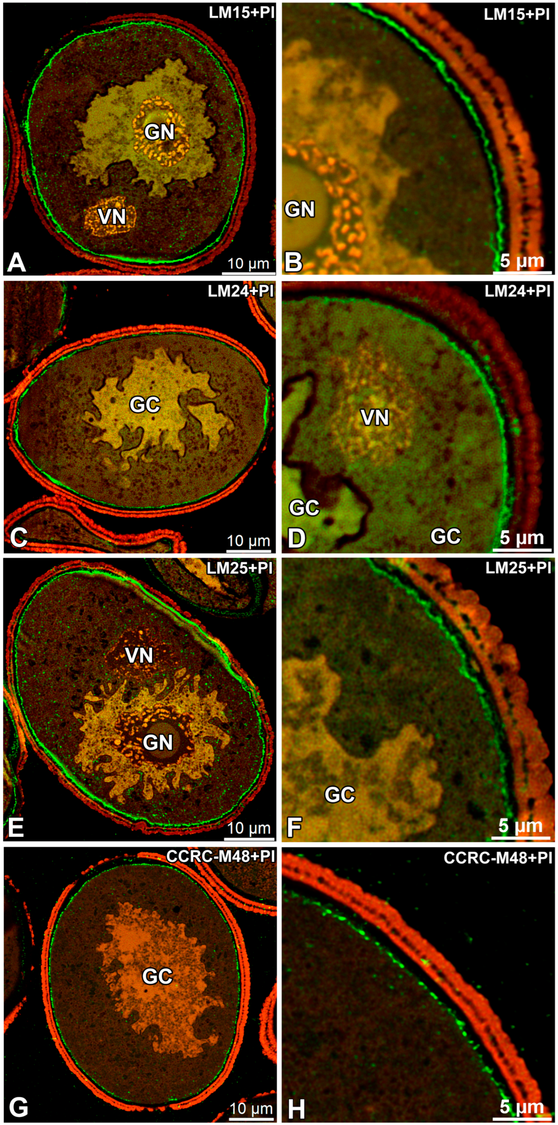
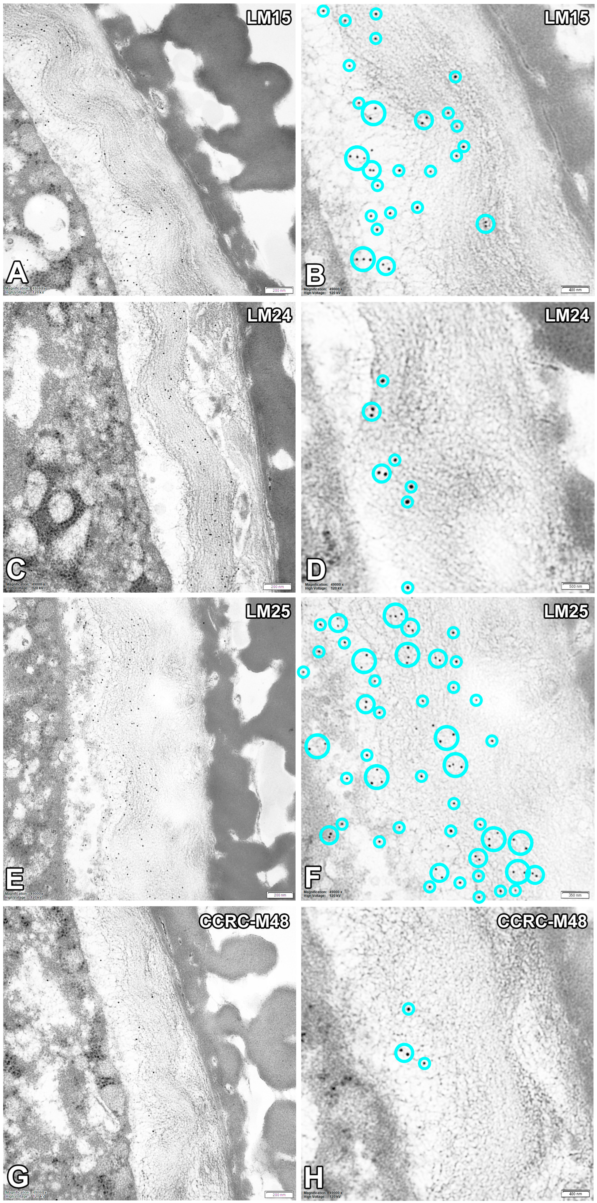
3.1. Untreated Mature Pollen Grains
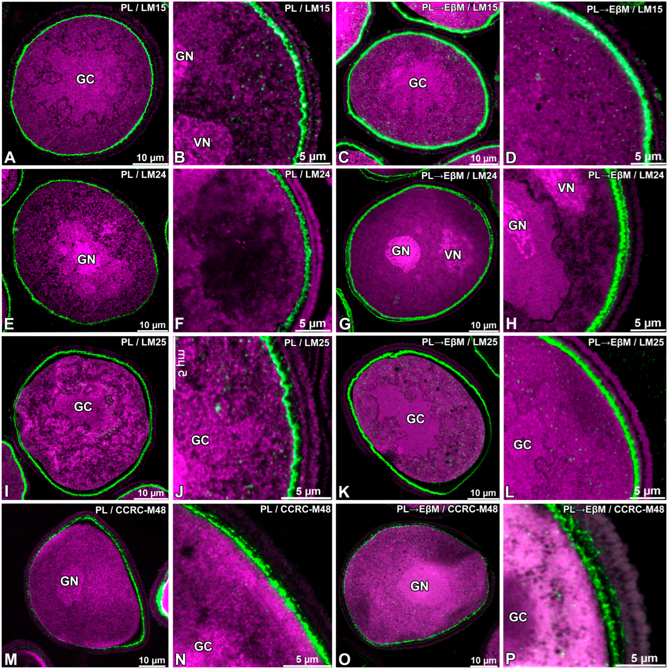
3.2. Enzymatic Digestion
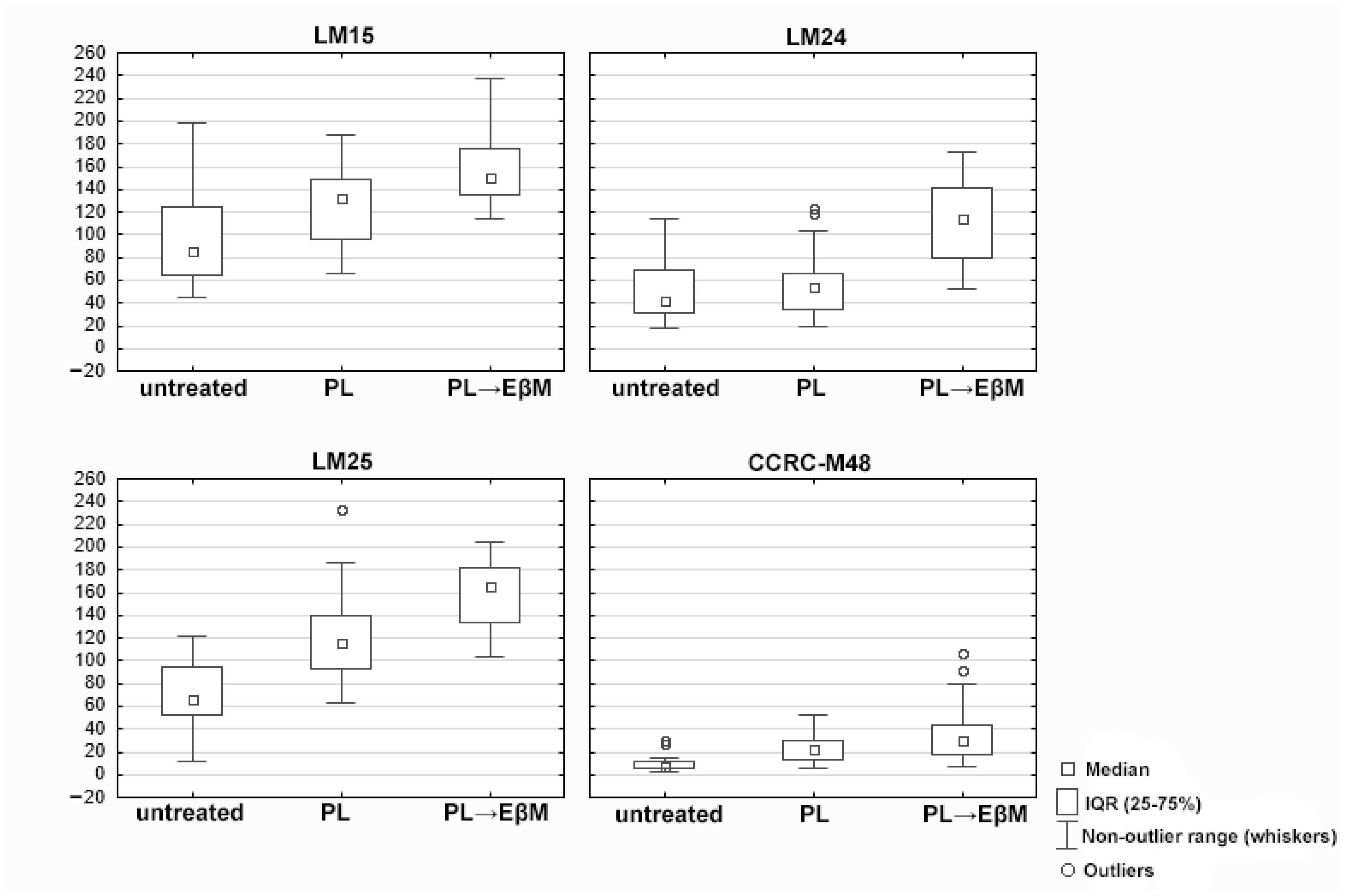
3.3. Quantification of Intine Fluorescence (MFI)
3.4. Control Reactions
4. Discussion
5. Conclusions
- Sequential pretreatments and enzymatic digestion (alkaline de-esterification followed by pectate lyase and endo-β-mannanase) increased the detectability and continuity of wall-associated signals for several probes. The most straightforward interpretation is that these treatment-dependent changes primarily reflect altered epitope accessibility (unmasking) within the intine matrix, rather than an increase in polymer abundance.
- Quantitative fluorescence measurements support a treatment-dependent change in epitope accessibility, with the combined digestion producing significant increases in LM15, LM24 and LM25, whereas pectate lyase alone did not.
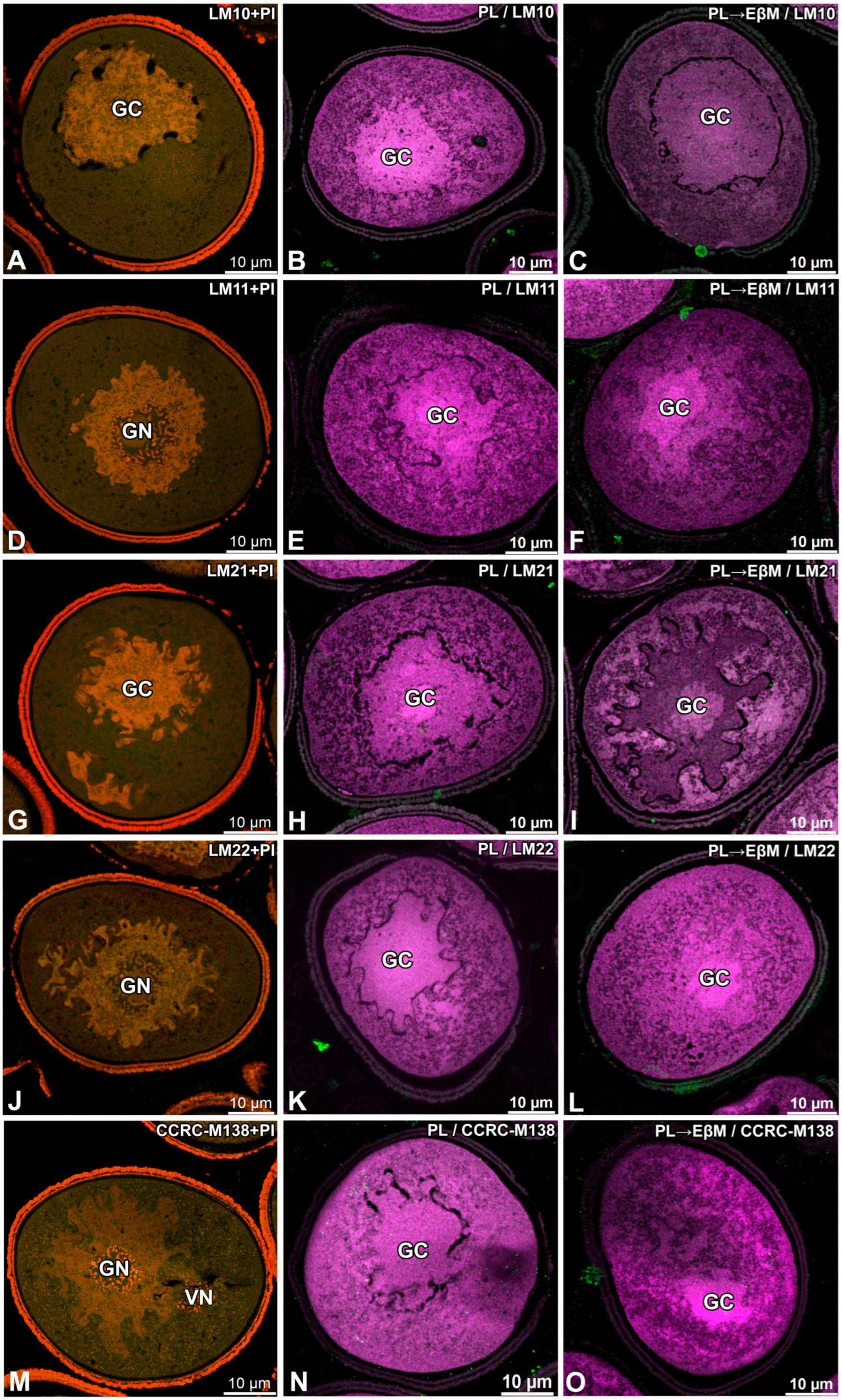
- Under the conditions used here, LM10, LM11, LM21, LM22 and CCRC-M138 remained below the detection threshold. Because immunolabelling reports epitope presence and accessibility, negative results cannot be interpreted as definitive absence of the corresponding polymers.
- CCRC-M48 did not show statistically significant intensity changes after digestion, indicating that different xyloglucan-associated epitopes can respond differently to the same unmasking workflow.
Supplementary Materials
Author Contributions
Funding
Institutional Review Board Statement
Informed Consent Statement
Data Availability Statement
Conflicts of Interest
References
- Scheller, H.V.; Ulvskov, P. Hemicelluloses. Annu. Rev. Plant Biol. 2010, 61, 263–289. [Google Scholar] [CrossRef] [Scilit]
- Carpita, N.C.; Gibeaut, D.M. Structural Models of Primary Cell Walls in Flowering Plants: Consistency of Molecular Structure with the Physical Properties of the Walls during Growth. Plant J. 1993, 3, 1–30. [Google Scholar] [CrossRef] [Scilit]
- O’Neill, M.A.; York, W.S. The Composition and Structure of Plant Primary Cell Walls. In The Plant Cell Wall; Annual Plant Reviews; Rose, J.K.C., Ed.; Blackwell Publishing: Oxford, UK, 2003; Volume 8, pp. 1–54. [Google Scholar]
- Amin, E.S.; Kandeel, K.M. The Hemicellulose of Pollen Grains of Phoenix dactylifera. Carbohydr. Res. 1972, 25, 261–263. [Google Scholar] [CrossRef] [Scilit] [PubMed]
- Rodriguez-Rosales, M.P.; Donaire, J.P. Cell Wall Acyl-Lipids, Proteins and Polysaccharides of Mature and Germinated Olive Pollen. Rev. Esp. Fisiol. 1990, 46, 371–377. [Google Scholar]
- Nakamura, N.; Yoshida, K.; Suzuki, H. Hemicellulose of the Pollen Tube Wall of Camellia japonica. Plant Cell Physiol. 1980, 21, 1383–1390. [Google Scholar] [CrossRef] [Scilit]
- Foubert-Mendes, S.; Silva, J.; Ferreira, M.J.; Pereira, L.G.; Coimbra, S. A Review on the Function of Arabinogalactan-Proteins during Pollen Grain Development and Pollen Tube Growth. Plant Reprod. 2025, 38, 8. [Google Scholar] [CrossRef] [Scilit]
- Bosch, M.; Hepler, P.K. Pectin Methylesterases and Pectin Dynamics in Pollen Tubes. Plant Cell 2005, 17, 3219–3226. [Google Scholar] [CrossRef] [Scilit]
- Hepler, P.K.; Rounds, C.M.; Winship, L.J. Control of Cell Wall Extensibility during Pollen Tube Growth. Mol. Plant 2013, 6, 998–1017. [Google Scholar] [CrossRef] [Scilit]
- Mollet, J.-C.; Leroux, C.; Dardelle, F.; Lehner, A. Cell Wall Composition, Biosynthesis and Remodeling during Pollen Tube Growth. Plants 2013, 2, 107–147. [Google Scholar] [CrossRef] [Scilit]
- Xu, R.; Liu, Z.; Wang, X.; Zhou, Y.; Zhang, B. Xylan clustering on the pollen surface is required for exine patterning. Plant Physiol. 2023, 193, 433–451. [Google Scholar] [CrossRef] [Scilit] [PubMed]
- Zhang, Y.; He, R.; Lian, J.; Zhou, Y.; Zhang, F.; Li, Q.; Yu, Y.; Feng, Y.; Yang, M.; Lei, H.; et al. OsmiR528 Regulates Rice-Pollen Intine Formation by Targeting a Uclacyanin to Influence Flavonoid Metabolism. Proc. Natl. Acad. Sci. USA 2020, 117, 727–732. [Google Scholar] [CrossRef] [Scilit]
- Jaffri, S.R.F.; MacAlister, C.A. Sequential Deposition and Remodeling of Cell Wall Polymers during Tomato Pollen Development. Front. Plant Sci. 2021, 12, 703713. [Google Scholar] [CrossRef] [Scilit]
- Ackerman, J.D. Convergence of Filiform Pollen Morphologies in Seagrasses: Functional Mechanisms. Evol. Ecol. 1995, 9, 139–153. [Google Scholar] [CrossRef] [Scilit]
- Lopez-Sanchez, P.; Cersosimo, J.; Wang, D.; Flanagan, B.; Stokes, J.R.; Gidley, M.J. Poroelastic Mechanical Effects of Hemicelluloses on Cellulosic Hydrogels under Compression. PLoS ONE 2015, 10, e0122132. [Google Scholar] [CrossRef] [Scilit] [PubMed]
- Dehors, J.; Mareck, A.; Kiefer-Meyer, M.-C.; Menu-Bouaouiche, L.; Lehner, A.; Mollet, J.-C. Evolution of Cell Wall Polymers in Tip-Growing Land Plant Gametophytes: Composition, Distribution, Functional Aspects and Their Remodeling. Front. Plant Sci. 2019, 10, 441. [Google Scholar] [CrossRef] [Scilit]
- Delmer, D.; Dixon, R.A.; Keegstra, K.; Mohnen, D. The Plant Cell Wall—Dynamic, Strong, and Adaptable—Is a Natural Shapeshifter. Plant Cell 2024, 36, 1257–1311. [Google Scholar] [CrossRef] [Scilit]
- Ziska, L.H.; Beggs, P.J. Anthropogenic Climate Change and Allergen Exposure: The Role of Pollen. J. Allergy Clin. Immunol. 2012, 129, 27–32. [Google Scholar] [CrossRef] [Scilit]
- D’Amato, G.; Cecchi, L. Effects of Climate Change on Environmental Factors in Respiratory Allergic Diseases. Clin. Exp. Allergy 2008, 38, 1264–1274. [Google Scholar] [CrossRef] [Scilit] [PubMed]
- Marcus, S.E.; Verhertbruggen, Y.; Herve, C.; Ordaz-Ortiz, J.J.; Farkas, V.; Pedersen, H.L.; Willats, W.G.T.; Knox, J.P. Pectic Homogalacturonan Masks Abundant Sets of Xyloglucan Epitopes in Plant Cell Walls. BMC Plant Biol. 2008, 8, 60. [Google Scholar] [CrossRef] [Scilit]
- Pedersen, H.L.; Fangel, J.U.; McCleary, B.; Ruzanski, C.; Rydahl, M.G.; Ralet, M.-C.; Farkas, V.; von Schantz, L.; Marcus, S.E.; Andersen, M.C.F.; et al. Versatile High Resolution Oligosaccharide Microarrays for Plant Glycobiology and Cell Wall Research. J. Biol. Chem. 2012, 287, 39429–39438. [Google Scholar] [CrossRef] [Scilit]
- Marcus, S.E.; Blake, A.W.; Benians, T.A.S.; Lee, K.J.D.; Poyser, C.; Donaldson, L.; Leroux, O.; Rogowski, A.; Petersen, H.L.; Boraston, A.; et al. Restricted Access of Proteins to Mannan Polysaccharides in Intact Plant Cell Walls. Plant J. 2010, 64, 191–203. [Google Scholar] [CrossRef] [Scilit]
- McCartney, L.; Marcus, S.E.; Knox, J.P. Monoclonal Antibodies to Plant Cell Wall Xylans and Arabinoxylans. J. Histochem. Cytochem. 2005, 53, 543–546. [Google Scholar] [CrossRef] [Scilit]
- Ruprecht, C.; Bartetzko, M.P.; Senf, D.; Dallabernadina, P.; Boos, I.; Andersen, M.C.F.; Kotake, T.; Knox, J.P.; Hahn, M.G.; Clausen, M.H.; et al. A Synthetic Glycan Microarray Enables Epitope Mapping of Plant Cell Wall Glycan-Directed Antibodies. Plant Physiol. 2017, 175, 1094–1104. [Google Scholar] [CrossRef] [Scilit] [PubMed]
- Pattathil, S.; Avci, U.; Baldwin, D.; Swennes, A.G.; McGill, J.A.; Popper, Z.; Bootten, T.; Albert, A.; Davis, R.H.; Chennareddy, C.; et al. A Comprehensive Toolkit of Plant Cell Wall Glycan-Directed Monoclonal Antibodies. Plant Physiol. 2010, 153, 514–525. [Google Scholar] [CrossRef] [Scilit] [PubMed]
- Liu, L.; Paulitz, J.; Pauly, M. The Presence of Fucogalactoxyloglucan and Its Synthesis in Rice Indicates Conserved Functional Importance in Plants. Plant Physiol. 2015, 168, 549–560. [Google Scholar] [CrossRef] [Scilit]
- Meents, M.J.; Motani, S.; Mansfield, S.D.; Samuels, A.L. Organization of Xylan Production in the Golgi During Secondary Cell Wall Biosynthesis. Plant Physiol. 2019, 181, 527–546. [Google Scholar] [CrossRef] [Scilit]
- Dumais, J. Mechanics and Hydraulics of Pollen Tube Growth. New Phytol. 2021, 232, 1549–1565. [Google Scholar] [CrossRef] [Scilit]
- Cameron, C.; Geitmann, A. Cell Mechanics of Pollen Tube Growth. Curr. Opin. Genet. Dev. 2018, 51, 11–17. [Google Scholar] [CrossRef] [Scilit]
- Rojas, E.R.; Hotton, S.; Dumais, J. Chemically Mediated Mechanical Expansion of the Pollen Tube Cell Wall. Biophys. J. 2011, 101, 1844–1853. [Google Scholar] [CrossRef] [Scilit] [PubMed]
- Chebli, Y.; Kaneda, M.; Zerzour, R.; Geitmann, A. The Cell Wall of the Arabidopsis Pollen Tube—Spatial Distribution, Recycling, and Network Formation of Polysaccharides. Plant Physiol. 2012, 160, 1940–1955. [Google Scholar] [CrossRef] [Scilit]
- Rydahl, M.G.; Hansen, A.R.; Kračun, S.K.; Mravec, J. Report on the Current Inventory of the Toolbox for Plant Cell Wall Analysis: Proteinaceous and Small Molecular Probes. Front. Plant Sci. 2018, 9, 581. [Google Scholar] [CrossRef] [Scilit] [PubMed]
- Kapusta, M.; Narajczyk, M.; Plachno, B.J. Arabinogalactan Proteins Mark the Generative Cell-Vegetative Cell Interface in Monocotyledonous Pollen Grains. Cells 2025, 14, 1549. [Google Scholar] [CrossRef] [Scilit]
- Beggs, P.J. Impacts of Climate Change on Aeroallergens: Past and Future. Clin. Exp. Allergy 2004, 34, 1507–1513. [Google Scholar] [CrossRef] [Scilit]
- Singer, B.D.; Ziska, L.H.; Frenz, D.A.; Gebhard, D.E.; Straka, J.G. Research Note: Increasing Amb a 1 Content in Common Ragweed (Ambrosia artemisiifolia) Pollen as a Function of Rising Atmospheric CO2 Concentration. Funct. Plant Biol. 2005, 32, 667–670. [Google Scholar] [CrossRef] [Scilit]
- Jiang, Y.; Lahlali, R.; Karunakaran, C.; Kumar, S.; Davis, A.R.; Bueckert, R.A. Seed Set, Pollen Morphology and Pollen Surface Composition Response to Heat Stress in Field Pea. Plant Cell Environ. 2015, 38, 2387–2397. [Google Scholar] [CrossRef] [Scilit] [PubMed]
- Read, S.M.; Clarke, A.E.; Bacic, A. Stimulation of Growth of Cultured Nicotiana tabacum W38 Pollen Tubes by Poly(ethylene glycol) and Cu(II) Salts. Protoplasma 1993, 177, 1–14. [Google Scholar] [CrossRef] [Scilit]
- Reynolds, E.S. The use of lead citrate at high pH as an electron-opaque stain in electron microscopy. J. Cell Biol. 1963, 17, 208–212. [Google Scholar] [CrossRef] [Scilit]
- Płachno, B.J.; Kapusta, M.; Stolarczyk, P.; Feldo, M.; Świątek, P. Do Arabinogalactan Proteins Occur in the Transfer Cells of Utricularia dichotoma? Int. J. Mol. Sci. 2024, 25, 6623. [Google Scholar] [CrossRef] [Scilit]
- Peña, M.J.; Kong, Y.; York, W.S.; O’Neill, M.A. A Galacturonic Acid-Containing Xyloglucan Is Involved in Arabidopsis Root Hair Tip Growth. Plant Cell 2012, 24, 4511–4524. [Google Scholar] [CrossRef] [Scilit]
- Plachno, B.J.; Kapusta, M.; Feldo, M.; Swiatek, P. Cell Wall Microdomains Analysis in the Quadrifids of Utricularia dichotoma. Int. J. Mol. Sci. 2025, 26, 832. [Google Scholar] [CrossRef] [Scilit]
- Vogel, J. Unique Aspects of the Grass Cell Wall. Curr. Opin. Plant Biol. 2008, 11, 301–307. [Google Scholar] [CrossRef] [Scilit]
- Burton, R.A.; Fincher, G.B. Evolution and Development of Cell Walls in Cereal Grains. Front. Plant Sci. 2014, 5, 456. [Google Scholar] [CrossRef] [Scilit]
- Kress, W.J.; Stone, D.E. Nature of the Sporoderm in Monocotyledons, with Special Reference to the Pollen Grains of Canna and Heliconia. Grana 1982, 21, 129–148. [Google Scholar] [CrossRef] [Scilit]
- Harris, P.J. Primary and Secondary Plant Cell Walls: A Comparative Overview. N. Z. J. For. Sci. 2006, 36, 36–53. [Google Scholar]
- Leroux, C.; Bouton, S.; Kiefer-Meyer, M.-C.; Fabrice, T.N.; Mareck, A.; Guénin, S.; Fournet, F.; Ringli, C.; Pelloux, J.; Driouich, A.; et al. PECTIN METHYLESTERASE48 Is Involved in Arabidopsis Pollen Grain Germination. Plant Physiol. 2015, 167, 367–380. [Google Scholar] [CrossRef] [Scilit]
- Hoedemaekers, K.; Derksen, J.; Hoogstrate, S.W.; Wolters-Arts, M.; Oh, S.A.; Twell, D.; Mariani, C.; Rieu, I. BURSTING POLLEN Is Required to Organize the Pollen Germination Plaque and Pollen Tube Tip in Arabidopsis thaliana. New Phytol. 2015, 206, 255–267. [Google Scholar] [CrossRef] [Scilit]
- Cascallares, M.; Setzes, N.; Marchetti, F.; López, G.A.; Distéfano, A.M.; Cainzos, M.; Zabaleta, E.; Pagnussat, G.C. A Complex Journey: Cell Wall Remodeling, Interactions, and Integrity During Pollen Tube Growth. Front. Plant Sci. 2020, 11, 599247. [Google Scholar] [CrossRef] [Scilit]
- Ladinig, U.; Hacker, J.; Neuner, G.; Wagner, J. How Endangered Is Sexual Reproduction of High-Mountain Plants by Summer Frosts? Frost Resistance, Frequency of Frost Events and Risk Assessment. Oecologia 2013, 171, 743–760. [Google Scholar] [CrossRef] [Scilit] [PubMed]
- Sunmonu, N.; Kudo, G. Warm Temperature Conditions Restrict the Sexual Reproduction and Vegetative Growth of the Spring Ephemeral Gagea lutea (Liliaceae). Plant Ecol. 2015, 216, 1419–1431. [Google Scholar] [CrossRef] [Scilit]
- Vieira, A.M.; Feijó, J.A. Hydrogel control of water uptake by pectins during in vitro pollen hydration of Eucalyptus globulus. Am. J. Bot. 2016, 103, 437–451. [Google Scholar] [CrossRef] [Scilit] [PubMed]
- Wang, R.; Dobritsa, A.A. Exine and Aperture Patterns on the Pollen Surface: Their Formation and Roles in Plant Reproduction. In Annual Plant Reviews Online; Roberts, J.A., Ed.; John Wiley & Sons, Ltd.: Chichester, UK, 2018; Volume 1, pp. 1–40. [Google Scholar]



| Antibody | PL vs. Untreated | PL → EβM vs. Untreated | Short Interpretation |
|---|---|---|---|
| LM15 | ↑ (ns; p = 0.293848) | ↑ (****; p = 0.000018) | Strongest increase after PL → EβM; PL not significant |
| LM24 | – (ns; p = 0.999113) | ↑ (****; p = 0.000018) | Increase only after PL → EβM; PL ~ no effect |
| LM25 | – (ns; p = 0.999998) | ↑ (****; p = 0.000018) | Increase mainly after PL → EβM; PL vs. untreated not significant (but PL → EβM > PL: **; p = 0.002655) |
| CCRC-M48 | ↑ (ns; p = 0.967402) | ↑ (ns; p = 0.144472) | Upward trend, but no significant differences after Tukey’s adjustment |
Disclaimer/Publisher’s Note: The statements, opinions and data contained in all publications are solely those of the individual author(s) and contributor(s) and not of MDPI and/or the editor(s). MDPI and/or the editor(s) disclaim responsibility for any injury to people or property resulting from any ideas, methods, instructions or products referred to in the content. |
© 2026 by the authors. Licensee MDPI, Basel, Switzerland. This article is an open access article distributed under the terms and conditions of the Creative Commons Attribution (CC BY) license.
Share and Cite
Kapusta, M.; Narajczyk, M.; Płachno, B.J. Hidden Xyloglucan Architecture of the Pollen Intine in Gagea lutea Revealed by Sequential Enzymatic Unmasking. Biology 2026, 15, 243. https://doi.org/10.3390/biology15030243
Kapusta M, Narajczyk M, Płachno BJ. Hidden Xyloglucan Architecture of the Pollen Intine in Gagea lutea Revealed by Sequential Enzymatic Unmasking. Biology. 2026; 15(3):243. https://doi.org/10.3390/biology15030243
Chicago/Turabian StyleKapusta, Małgorzata, Magdalena Narajczyk, and Bartosz J. Płachno. 2026. "Hidden Xyloglucan Architecture of the Pollen Intine in Gagea lutea Revealed by Sequential Enzymatic Unmasking" Biology 15, no. 3: 243. https://doi.org/10.3390/biology15030243
APA StyleKapusta, M., Narajczyk, M., & Płachno, B. J. (2026). Hidden Xyloglucan Architecture of the Pollen Intine in Gagea lutea Revealed by Sequential Enzymatic Unmasking. Biology, 15(3), 243. https://doi.org/10.3390/biology15030243