Tinea capitis in the Americas: Epidemiological Trends and Etiological Distribution from a Systematic Review
 , , ,
, , ,  ,
,  and
and 
Simple Summary
Abstract
1. Introduction
2. Materials and Methods
2.1. Study Selection and Eligibility Criteria
2.1.1. Data Acquisition
2.1.2. Quality Assessment
- a.
- Risk of bias in individual studies
- b.
- Risk of bias between studies
- c.
- Synthesis methods
- d.
- Sensitivity analyses
- e.
- Certainty of Evidence Assessment
2.2. Protocol Registration
3. Results
3.1. General Characteristics of the Included Studies
3.2. North America
3.3. Central America and the Caribbean
3.4. South America
3.5. Certainty of Evidence
4. Discussion
5. Conclusions
6. Limitations
Supplementary Materials
Author Contributions
Funding
Institutional Review Board Statement
Informed Consent Statement
Data Availability Statement
Acknowledgments
Conflicts of Interest
References
- Krize-Morun, I.; Matamoros-Montoya, K.; Segnini-Zamora, A.; Molina-Brays, C.; Bogantes-Barrantes, C. Querión Celso, dermatofitosis del cuero cabelludo infantil. Acta Med. Costarric. 2023, 65, 32–36. [Google Scholar] [CrossRef] [Scilit]
- Lemus-Espinoza, D.; Maniscalchi, M.T.; Villarroel, O.; Bónoli, S.; Wahab, F.; García, O. Micosis superficiales en pacientes del estado Anzoátegui, Venezuela, periodo 2002–2012. Invest. Clin. 2014, 55, 311–320. [Google Scholar] [PubMed]
- Mazur, L.; Tian, Z.; Shedlock, K.; Zurca, A.D.; Zhou, S.; Thomas, N.J.; Krawiec, C. A multicenter retrospective database evaluation of pediatric patients diagnosed with tinea capitis. Perm. J. 2023, 27, 82–89. [Google Scholar] [CrossRef] [Scilit] [PubMed]
- Cruz, R.; Ponce, E.; Calderón, L.; Delgado, N.; Vieille, P.; Piontelli, E. Micosis superficiales en la ciudad de Valparaíso, Chile. Período 2007–2009. Rev. Chil. Infectol. 2011, 28, 404–409. [Google Scholar] [CrossRef] [Scilit] [PubMed]
- Davel, G.; Perrotta, D.; Canteros, C.; Córdoba, S.; Rodero, L.; Brudny, M.; Abrantes, R. Estudio multicéntrico de micosis su-perficiales en Argentina. Rev. Argent. Microbiol. 1999, 31, 173–181. [Google Scholar] [PubMed]
- Page, M.J.; McKenzie, J.E.; Bossuyt, P.M.; Boutron, I.; Hoffmann, T.C.; Mulrow, C.D.; Shamseer, L.; Tetzlaff, J.M.; Akl, E.A.; Brennan, S.E.; et al. The PRISMA 2020 statement: An updated guideline for reporting systematic reviews. BMJ 2021, 372, n71. [Google Scholar] [CrossRef] [Scilit] [PubMed]
- World Health Organization. Adolescent Health [Internet]. Geneva: World Health Organization; WHO Adolescent Health. Adolescence Is Defined by WHO as 10–19 Years of Age. Available online: https://www.who.int/health-topics/adolescent-health?utm_source=chatgpt.com (accessed on 2 June 2026).
- World Health Organization. Adolescent Health [Internet]. Geneva: World Health Organization; WHO SEARO Adolescent Health Definitions. WHO Defines Adolescents as 10–19 Years, Youth as 15–24 Years, and Young People as 10–24 Years. Available online: https://www.who.int/southeastasia/health-topics/adolescent-health?utm_source=chatgpt.com (accessed on 2 June 2026).
- Marcoux, D.; Dang, J.; Auguste, H.; McCuaig, C.; Powell, J.; Hatami, A.; Maari, C.; Le Meur, J. Emergence of African species of dermatophytes in tinea capitis: A 17-year experience in a Montreal pediatric hospital. Pediatr. Dermatol. 2018, 35, 323–328. [Google Scholar] [CrossRef] [Scilit] [PubMed]
- Gupta, A.K.; Ginter, G. Itraconazole is effective in the treatment of tinea capitis caused by Microsporum canis. Pediatr. Dermatol. 2001, 18, 519–522. [Google Scholar] [CrossRef] [Scilit] [PubMed]
- Gupta, K.; Hooton, T.M.; Stamm, W.E. Increasing antimicrobial resistance and the management of uncomplicated community-acquired urinary tract infections. Ann. Intern. Med. 2001, 135, 41–50. [Google Scholar] [CrossRef] [Scilit] [PubMed]
- Valdez-Martinez, A.; Santoyo-Alejandre, M.I.; Arenas, R.; Isa-Pimentel, M.A.; Castillo-Cruz, J.; Huerta-Domínguez, K.D.; Soto-Torres, E.F.; Martínez-Herrera, E.; Pinto-Almazán, R. Tinea capitis in older adults: A neglected and misdiagnosed scalp infection—A systematic review of 503 reported cases. Antibiotics 2025, 14, 1211. [Google Scholar] [CrossRef] [Scilit] [PubMed]
- Tarr, C.L.; Patel, J.S.; Puhr, N.D.; Sowers, E.G.; Bopp, C.A.; Strockbine, N.A. Isolation of Trichophyton violaceum and Trichophyton soudanense in Baltimore, Maryland. J. Clin. Microbiol. 2007, 45, 461–465. [Google Scholar] [CrossRef] [Scilit] [PubMed]
- Cantrell, W.C.; Jacobs, M.K.; Sobera, J.O.; Parrish, C.A.; Warner, J.; Elewski, B.E. Tinea capitis in Birmingham: Survey of elementary school students. Pediatr. Dermatol. 2011, 28, 476–477. [Google Scholar] [CrossRef] [Scilit] [PubMed]
- Mirmirani, P.; Tucker, L.Y. Epidemiologic trends in pediatric tinea capitis: A population-based study from Kaiser Permanente Northern California. J. Am. Acad. Dermatol. 2013, 69, 916–921. [Google Scholar] [CrossRef] [Scilit] [PubMed]
- Leung, A.K.; Hon, K.L.; Leong, K.F.; Barankin, B.; Lam, J.M. Tinea capitis: An updated review. Recent. Pat. Inflamm. Allergy Drug Discov. 2020, 14, 58–68. [Google Scholar] [CrossRef] [Scilit] [PubMed]
- Cetner, A.; Krunic, A.; Tesic, V.; Janda, W. Tinea capitis due to Trichophyton soudanense in Chicago, Illinois: Report of a case. Pediatr. Dermatol. 2009, 26, 226–228. [Google Scholar] [CrossRef] [Scilit] [PubMed]
- Ghannoum, M.; Isham, N.; Hajjeh, R.; Cano, M.; Al-Hasawi, F.; Yearicka, D.; Warner, J.; Longa, L.; Jessup, C.; Elewski, B. Tinea capitis in Cleveland: Survey of elementary school students. J. Am. Acad. Dermatol. 2003, 48, 189–193. [Google Scholar] [CrossRef] [Scilit] [PubMed]
- Coloe, J.R.; Diab, M.; Moennich, J.; Diab, D.; Pawaskar, M.; Balkrishnan, R.; Bechtel, M.A. Tinea capitis among children in the Columbus area, Ohio, USA. Mycoses 2010, 53, 158–162. [Google Scholar] [CrossRef] [Scilit] [PubMed]
- Abdel-Rahman, S.M.; Talib, N.; Solidar, A.; Nopper, A.J.; Wyckoff, G.J. Examining Trichophyton tonsurans genotype and biochemical phenotype as determinants of disease severity in tinea capitis. Med. Mycol. 2008, 46, 217–223. [Google Scholar] [CrossRef] [Scilit] [PubMed]
- Abdel-Rahman, S.M.; Farrand, N.; Schuenemann, E.; Stering, T.K.; Preuett, B.; Magie, R.; Campbell, A. The Prevalence of Infections With Trichophyton tonsurans in Schoolchildren: The CAPITIS Study. Pediatrics 2010, 125, 966–973. [Google Scholar] [CrossRef] [Scilit] [PubMed]
- Möhrenschlager, M.; Seidl, H.P.; Ring, J.; Abeck, D. Pediatric tinea capitis: Recognition and management. Am. J. Clin. Dermatol. 2005, 6, 203–213. [Google Scholar] [CrossRef] [Scilit] [PubMed]
- Markey, R.J.; Staat, M.A.; Gerrety, M.J.T.; Lucky, A.W. Tinea capitis due to Trichophyton soudanense in Cincinnati, Ohio, in internationally adopted children from Liberia. Pediatr. Dermatol. 2003, 20, 408–410. [Google Scholar] [CrossRef] [Scilit] [PubMed]
- Sandoval, A.B.; Ortiz, J.A.; Rodríguez, J.M.; Vargas, A.G.; Quintero, D.G. Dermoscopic pattern in tinea capitis. Rev. Iberoam. Micol. 2010, 27, 151–152. [Google Scholar] [PubMed]
- Mayorga, J.; Esquivel-González, P.L.; Prado-Trillo, A.; Barba-Gómez, J.F. Clinical and epidemiologicalcharacteristics of patients with infection by Microsporum canis. Dermatol. Rev. Mex. 2016, 60, 18–23. [Google Scholar]
- Morán Maese, D.; Tarango-Martínez, V.M.; González Treviño, L.A.; Mayorga, J. Tiña de la cabeza en un adulto. Apropósito de un caso. Rev. Iberoam. Micol. 2005, 22, 54–56. [Google Scholar] [CrossRef] [Scilit] [PubMed]
- Orozco-Yee, E.A.; Rojas-Castañeda, R.G.; Guevara-Gutiérrez, E.; Mayorga-Rodríguez, J.; Tlacuilo-Parra, A. Dermato-fitosis causadas por Nannizzia gypsea: Informe de 155 casos en el oeste de México. Enferm. Infecc. Microbiol. Clin. 2025, 43, 205–209. [Google Scholar] [CrossRef] [Scilit]
- Segundo, C.; Martínez, A.; Arenas, R.; Fernández, R.; Cervantes, R.A. Superficial infections caused by Microsporum canis in humans and animals. Rev. Iberoam. Micol. 2004, 21, 39–41. [Google Scholar] [PubMed]
- Basurto, C.I.F.; Cukiert, M.S.; Durán, E.R.J.; Vega-Memije, M.E.; Terán, A.L.R. Understanding kerion Celsi in children: Diagnosis and therapeutic guidelines through an algorithm. Cureus 2024, 16, e58475. [Google Scholar] [CrossRef] [Scilit] [PubMed]
- Bonifaz, A.; Córdoba-García, B.; Simancas-Llanos, T.; Hernández, M.A.; Martínez-Herrera, E.; Tirado-Sánchez, A. Dermatophytosis caused by Nannizzia nana in two siblings. Rev. Iberoam. Micol. 2019, 36, 30–33. [Google Scholar] [CrossRef] [Scilit] [PubMed]
- Welsh, O.; Welsh, E.; Ocampo-Candiani, J.; Gomez, M.; Vera-Cabrera, L. Dermatophytoses in monterrey, méxico. Mycoses 2006, 49, 119–123. [Google Scholar] [CrossRef] [Scilit] [PubMed]
- Galván-Martínez, I.L.; Fernández-Martínez, R.; Narro-Llorente, R.; Moreno Coutiño, G.; Arenas, R. Frecuencia de tiña del cuerpo en un hospital del estado de Quintana Roo. Med. Interna Mex. 2017, 33, 5–11. [Google Scholar]
- Vargas-Navia, N.; Ayala Monroy, G.A.; Franco Rua, C.; Malagon Caicedo, J.P.; Rojas Hernandez, J.P. Tiña capitis en niños. Rev. Chil. Pediatr. 2020, 91, 789–798. [Google Scholar] [CrossRef] [Scilit]
- Sotelo, J.P.A.; Martinez, V.M.T.; Cabrera, L.V. Kerion celsi caused by Trichophyton tonsurans in an adult. An. Bras. Dermatol. 2022, 97, 516–519. [Google Scholar] [CrossRef] [Scilit] [PubMed]
- Frías-De-León, M.G.; Porras-López, C.; Martínez-Villarreal, J.D.; Villagómez-Figueroa, O.; Acosta-Altamirano, G.; Arenas, R.; Martínez Herrera, E.O. Tinea capitis. Current epidemiological data in a subtropical region of Mesoamerica (Guatemala). Dermatol. Rev. Mex. 2017, 61, 371–378. [Google Scholar]
- Martínez, E.; De León, S.; Pérez, E.; Pacheco, A.; Rivas, E.; Borjas, C.; Arenas, E. Tinea capitis. A report of 60 cases with hair parasitation and/or positive culture. Dermatol. CMQ 2009, 7, 98–101. [Google Scholar]
- Martinez, E.; Matta, V.; Porras, C.; Logeman, H.; Arenas, R. Dermatophytes and dermatophytosis Frequency in Guatemala during the period May 2008 to June 2009. Rev. Cient. 2012, 22, 19–23. [Google Scholar]
- Salas-Ocampo, O.; Gómez-Sáenz, A.; Álvarez-Cabalceta, H. Tinea capitis por Trichophyton mentagrophytes: Apropósito de dos casos en Guanacaste y Puntarenas, Costa Rica. Dermatol. Rev. Mex. 2020, 64, 75–79. [Google Scholar]
- Herrera Castro, M.Y.; de la Rosa Garibay, E.; Arenas, R.; Moreno Coutiño, G. Tiña del cuerpo. Datos micológicos yepidemiológicos de 10 años en un hospital de segundo nivel. Dermatol. Rev. Mex. 2025, 23, 15–19. [Google Scholar]
- Martínez-Suárez, H.; Guevara-Cabrera, N.; Mena, C.; Valencia, A.; Araiza, J.; Bonifaz, A. Tiña de la cabeza. Reporte de 122 casos. Dermatol. Cosm. Med. Quir. 2007, 5, 9–14. [Google Scholar]
- García-Agudo, L.; Galán, F.; García-Martos, P.; Carranza, R.; Rodríguez-Iglesias, M. Tiña de la cabeza: Descripción de los hallazgos dermatoscópicos en 37 pacientes. Rev. Iberoam. Micol. 2015, 32, 242–246. [Google Scholar] [CrossRef] [Scilit] [PubMed]
- Arenas, R. Dermatofitosis en México. Rev. Iberoam. Micol. 2002, 19, 63–67. [Google Scholar] [PubMed]
- Raccurt, C.P.; Dorsainvil, D.; Boncy, M.; Boncy, J.; Auguste, G. The emergence of Trichophyton tonsurans in Port-au-Prince, Haiti. Med. Mycol. 2009, 47, 197–200. [Google Scholar] [CrossRef] [Scilit] [PubMed]
- East-Innis, A.; Rainford, L.; Dunwell, P.; Barrett-Robinson, D.; Nicholson, A. The changing pattern of tinea capitis in Jamaica. West Indian Med. J. 2006, 55, 85–88. [Google Scholar] [CrossRef] [Scilit] [PubMed]
- Alvarez, C.; Runco, R.; Salim, R. Dermatophytoses in children: Retrospective revisión of 487 cases in Tecumán-Argentina. Bol. Micol. 2009, 24, 83–87. [Google Scholar]
- Santos, P.E.; Córdoba, S.; Rodero, L.L.; Carrillo-Muñoz, A.J.; Lopardo, H.A. Tinea capitis: Two years experience in a paediatric hospital of Buenos Aires, Argentina. Rev. Iberoam. Micol. 2010, 27, 104–106. [Google Scholar] [PubMed]
- Ballesté, R.; Fernández, N.; Mousqués, N.; Xavier, B.; Arteta, Z.; Mernes, M.; Gezuela, E. Dermatophytosis in assisted population at the Institute of Hygiene. Rev. Med. Urug. 2000, 16, 232–242. [Google Scholar]
- Walker, L.; Bianchi, M.; Maiolo, E.; Arechavala, A.; Santiso, G.; Messina, F.; Lehmann, E.; Schtirbu, R.; Negroni, R. Problemas clínicos en micología médica: Problema número 44. Rev. Iberoam. Micol. 2012, 29, 57–59. [Google Scholar] [CrossRef] [Scilit] [PubMed]
- Vides De la Hoz, P.; Piccolomini, M.; Almassio, A.; Abad, E.; Larralde, M. Tiña capitis por Trichophyton tonsurans en un paciente pediátrico. Arch. Argent. Pediatr. 2022, 120, e192–e196. [Google Scholar] [CrossRef] [Scilit] [PubMed]
- Negroni, R.; Wallach, J.; Arechavala, A.; Francos, J.L.; Messina, F.; Maiolo, E.; Walker, L. Problemas clínicos en micología médica: Problema número 42. Rev. Iberoam. Micol. 2011, 28, 56–59. [Google Scholar] [CrossRef] [Scilit] [PubMed]
- Jarrín Ortiz, L.P.; Guangasig Toapanta, V.H. Dermatofitosis: Innovación diagnóstica mediante técnicas moleculares. Rev. Científica Arbitr. Multidiscip. Pentaciencias. 2025, 7, 235–258. [Google Scholar] [CrossRef] [Scilit]
- Nardin, M.E.; Pelegri, D.G.; Manias, V.G.; Méndez, E.A. Agentes etiológicos de micosis superficiales aislados en un hospital de Santa Fe, Argentina. Rev. Argent. Microbiol. 2006, 38, 25–27. [Google Scholar] [PubMed]
- Monteagudo Sánchez, B.; Pereiro Ferreirós, M., Jr.; Fernández-Redondo, V.; Toribio Pérez, J. Tinea pedis causada por Trichophyton violaceum. Actas Dermo-Sifiliogr. 2002, 93, 59–61. [Google Scholar] [CrossRef] [Scilit]
- Schechtman, R.C.; Silva, N.D.V.; Quaresma, M.V.; Filho, F.B.; Buçard, A.M.; Sodré, C.T. Dermatoscopic findings as a complementary tool in the differential diagnosis of the etiological agent of tinea capitis. An. Bras. Dermatol. 2015, 90, S13–S15. [Google Scholar] [CrossRef] [Scilit] [PubMed]
- Brilhante, R.S.N.; Cordeiro, R.A.; Rocha, M.F.G.; Monteiro, A.J.; Meireles, T.E.F.; Sidrim, J.J.C. Tinea capitis in a dermatology center in the city of Fortaleza, Brazil: The role of Trichophyton tonsurans. Int. J. Dermatol. 2004, 43, 575–579. [Google Scholar] [CrossRef] [Scilit] [PubMed]
- Leung, A.K.; Lam, J.M.; Leong, K.F.; Hon, K.L. Tinea corporis: An updated review. Drugs Context. 2020, 9, 1–12. [Google Scholar] [CrossRef] [Scilit] [PubMed]
- Pires, C.A.A.; da Cruz, N.F.S.; Lobato, A.M.; de Sousa, P.O.; Carneiro, F.R.O.; Mendes, A.M.D. Perfil clínico, epidemiológico yterapéutico de la dermatofitosis. An. Bras. Dermatol. 2014, 89, 259–265. [Google Scholar] [CrossRef] [Scilit] [PubMed]
- Pinheiro, A.M.C.; Lobato, L.A.; Varella, T.C.N. Dermoscopy findings in tinea capitis: Case report and literature review. An. Bras. Dermatol. 2012, 87, 313–314. [Google Scholar] [CrossRef] [Scilit] [PubMed]
- Gürtler, T.G.R.; Diniz, L.M.; Nicchio, L. Tinea capitis micro-epidemic by Microsporum canis in a day care center of Vitória—Espírito Santo (Brazil). An. Bras. Dermatol. 2005, 80, 267–272. [Google Scholar]
- Martínez Campayo, N.; Rego Campuzano, I.; González de Aledo, M.; Arévalo Bermúdez, M.P.; Fernández Torres, R.M.; Fonseca, E. New Epidemiological Outcomes in Anthropophilic tinea capitis, a Case Series Study in Northwestern Spain. Actas Dermo-Sifiliogr. 2022, 113, T74–T77. [Google Scholar] [CrossRef] [Scilit] [PubMed]
- Veasey, J.V.; Muzy, G.d.S.C. Tinea capitis: Correlation of clinical presentations to agents identified in mycological culture. An. Bras. Dermatol. 2018, 93, 47–50. [Google Scholar] [CrossRef] [Scilit] [PubMed]
- Romero, M.; de la Rosa-Garibay, E.; Atoche, C.; González Estudillo, I.; Bonifaz, A.; Araiza, J.; Arenas, R. Consenso acional de Micosis Superficiales: Dermatofitosis. Dermatol. Cosm. Med. Quir. 2024, 22, 284–285. [Google Scholar]
- de Freitas, R.S.; Hernández-Arriagada, G.L.; Criado, P.R.; Martins, J.E.C.; Cardoso, J.L. Influence of the human activity in the Atlantic Forest and in the prevalence of etiological agents of dermatophytosis in a coastal city of Southern Brazil [Ubatuba (São Paulo)]. Int. J. Dermatol. 2008, 47, 865–867. [Google Scholar] [CrossRef] [Scilit] [PubMed]
- Di Chiacchio, N.; Madeira, C.L.; Humaire, C.R.; Silva, C.S.; Fernandes, L.H.G.; Reis, A.L.D. Superficial mycoses at the hospital do servidor público municipal de São Paulo between 2005 and 2011. An. Bras. Dermatol. 2014, 89, 67–71. [Google Scholar] [CrossRef] [Scilit] [PubMed]
- Silveira-Gomes, F.; de Oliveira, E.F.; Nepomuceno, L.B.; Pimentel, R.F.; Marques-Da-Silva, S.H.; Mesquita-Da-Costa, M. Dermatophytosis diagnosed at the Evandro Chagas Institute, Pará, Brazil. Braz. J. Microbiol. 2013, 44, 443–446. [Google Scholar] [CrossRef] [Scilit] [PubMed]
- Cukierman, E.; Camargo, T.Z.S.; Millan, L.P.B.; Freire, M.R.d.M.; Carneiro, L.F.M.; Waksman, R.D. Alopecia and pet: A case report. Einstein 2022, 20, eRC6881. [Google Scholar] [CrossRef] [Scilit] [PubMed]
- Van Gelderen, A.A. Tinea capitis inflamatoria por Trichophyton tonsurans var. sulfureum. Bol. Micol. 2012, 27, 39–45. [Google Scholar] [CrossRef]
- Moraes, M.S.; Godoy-Martínez, P.; Alchorne, M.M.; Boatto, H.F.; Fischman, O. Incidence of Tinea capitis in São Paulo, Brazil. Mycopathologia 2006, 162, 91–95. [Google Scholar] [CrossRef] [Scilit] [PubMed]
- Peixoto, R.R.G.B.; Meneses, O.M.S.; da Silva, F.O.; Donati, A.; Veasey, J.V. Tinea capitis: Correlation of clinical aspects, findings on direct mycological examination, and agents isolated from fungal culture. Int. J. Trichology 2020, 11, 232–238. [Google Scholar] [CrossRef] [Scilit] [PubMed]
- Cortez, A.C.A.; de Souza, J.V.B.; Sadahiro, A.; de Oliveira, J.A.A. Frequency and aetiology of dermatophytosis in children aged 12 years and under in the state of Amazonas, Brazil. Rev. Iberoam. Micol. 2012, 29, 223–226. [Google Scholar] [CrossRef] [Scilit] [PubMed]
- Grimalt, R. Management of tinea capitis in childhood. Clin. Cosmet. Investig. Dermatol. 2010, 3, 89–98. [Google Scholar] [CrossRef] [Scilit] [PubMed]
- de Aquino, P.M.L.P.; Lima, E.d.O.; de Farias, N.M.P. Tinea capitis em João Pessoa: Visão socioeconômica. An. Bras. Dermatol. 2003, 78, 713–720. [Google Scholar] [CrossRef] [Scilit]
- Mattei, A.S.; Beber, M.A.; Madrid, I.M. Dermatophytosis in small animals. SOJ Microbiol. Infect. Dis. 2014, 2, 1–6. [Google Scholar] [CrossRef] [Scilit]
- Callisaya, H.J.; Conde, A.D.; Choque, C.H. Frequency of germs causing superficial mycosis. BIOFARBO 2007, 15, 21–28. [Google Scholar]
- Manríquez, J.; Niklitschek, S. Nódulos fluctuantes en el cuero cabelludo de un niño. Enferm. Infecc. Microbiol. Clin. 2015, 33, 688–689. [Google Scholar] [CrossRef] [Scilit] [PubMed]
- Cortés, G.A.; Mardones, V.F.; Emelman, D.V. Etiología de la alopecia infantil. Rev. Chil. Pediatr. 2015, 86, 264–269. [Google Scholar] [CrossRef] [Scilit] [PubMed]
- Aranibar Duran, L.; Stevens González, J.; Palma Ducommun, C.; Zapata Manque, S.; Pizarro Olave, J. Estudio epidemiológico de la tinea capitis en una población de Santiago de Chile. La aparición de Trichophyton tonsurans como agente etiológico. Piel 2017, 32, 604–609. [Google Scholar] [CrossRef] [Scilit]
- González, F.E.; Rodríguez, J.A.; Muñoz, L.M.; Apráez, G.; Vásquez, L.R. An outbreak of trichophytic tinea capitis in a group of schoolchildren in a rural area of the department of Cauca, Colombia. Biomédica 2023, 43, 57–68. [Google Scholar] [CrossRef] [Scilit] [PubMed]
- Zuluaga, A.; Cáceres, D.H.; Arango, K.; de Bedout, C.; Cano, L.E. Epidemiology of tinea capitis: A 19 years’ experience in a Colombia, specialized clinical laboratory. Infect 2016, 20, 225–230. [Google Scholar]
- Sterling, N.A.; Rincón, D.A.; Barrera, S.; Sánchez, E.A.; Molina, D.Y.; Urán, M.E.; Jiménez, M.d.P. Brote de tinea capitis y otras micosis superficiales en una comunidad urbana de Medellín. Biomédica 2023, 43, 69–80. [Google Scholar] [CrossRef] [Scilit] [PubMed]
- López-Cisneros, C.L.; Morillo Argudo, D.A.; Plaza Trujillo, P.L. Croos-Sectional study: Superficial Mycosis in school children of a rural Parish of Cuenca, Ecuador. Rev. Med. HJCA 2018, 9, 249–254. [Google Scholar]
- Simonnet, C.; Berger, F.; Gantier, J.C. Epidemiology of superficial fungal diseases in French Guiana: A three-year retrospective analysis. Med. Mycol. 2011, 49, 608–611. [Google Scholar] [CrossRef] [Scilit] [PubMed]
- de Da Silva, D.O.; de Lacarrubba, F.; Guzmán, A.L. Dermatofitosis in children in Asunción, Paraguay. Dermatol. Cosmética Médica Quirúrgica 2005, 3, 22–26. [Google Scholar]
- Álvarez Montiel, I.; Montoya Ramírez, S. Tiña amiantacea: Diagnóstico diferencial de tiña capitis. Rev. Med. Hondur. 2024, 92, 84–85. [Google Scholar] [CrossRef] [Scilit]
- Aguilar-Fernández, G.; López, P.A.; Arce, N.; Martínez Mora, M. Dermatophytes: Casuistry in the Mycology Section of the Central Public Health Laboratory, Asunción—Paraguay (2000–2016). Del. Nac. 2018, 9, 4–11. [Google Scholar] [CrossRef] [Scilit]
- Caballero, A.; Riveli, V.; Correa, J.; Mendoza, G. Tinea capitis. Communication of 54 cases. Rev. Pediatr. Py. 2002, 29, 10–15. [Google Scholar]
- Cárdenas, A.; Tincopa Montoya, L.O.; Fernández, W.; Valverde López, J.C.; Agip Díaz, H.J. Tinea capitis, frequency of etiologic agents. Dermatol. Peru 2001, 11, 1–3. [Google Scholar]
- Cáceres-Ríos, H.; Rueda, M.; Ballona, R.; Bustamante, B. Comparison of terbinafine and griseofulvin in the treatment of tinea capitis. J. Am. Acad. Dermatol. 2000, 42, 80–84. [Google Scholar] [CrossRef] [Scilit] [PubMed]
- Perelli, A. Presence of Dermatophytes in children of an Educational Unit of Naguanagua, Carabobo State, Venezuela. Vitae 2010, 43, 8–13. [Google Scholar]
- Capote, A.; Ferrara, G.; Panizo, M.M.; García, N.; Alarcón, V.; Reviakina, V.; Dolande, M. Micosis superficiales: Casuística del Departamento de Micología del Instituto Nacional de Higiene “Rafael Rangel”, Caracas, Venezuela (2001–2014). Invest. Clin. 2016, 57, 47–58. [Google Scholar] [PubMed]
- Mata-Essayag, S.; Carrillo, Y.; Canache, O.; Larrea, F.; Colella, M.T.; Pineda, V.; Landaeta, M.E.; Pardi, G.; Olaizola, C.; Perez, C. Epidemiology of tinea capitis in Venezuela. Dermatol. Venez. 2013, 51, 15–19. [Google Scholar]



| Region | Predominant Dermatophyte Species | Frequently Reported Secondary Species | Predominant Transmission Pattern | Observed Epidemiological Trend |
|---|---|---|---|---|
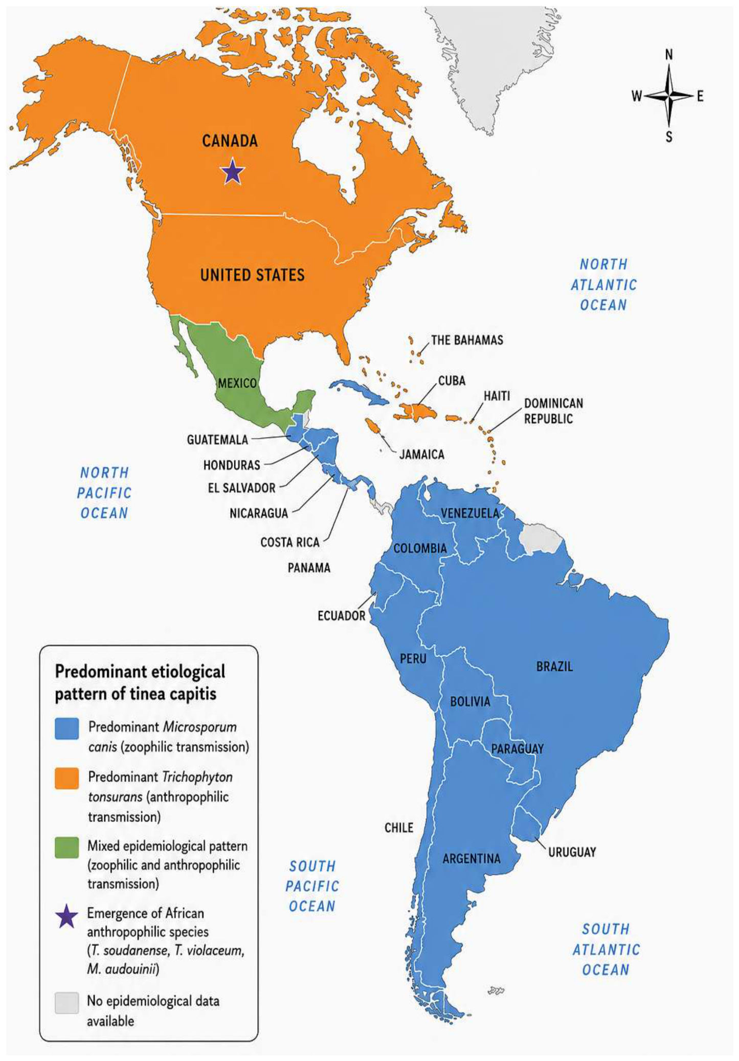
| North America | Trichophyton tonsurans | Microsporum canis, T. soudanense, T. violaceum, M. audouinii | Anthropophilic | Predominance of anthropophilic transmission, with increasing reports of traditionally African species, particularly in Canada, likely associated with migration and demographic changes. |
| Mexico | Microsporum canis | T. tonsurans, T. rubrum, T. mentagrophytes | Mixed (predominantly zoophilic) | Regional variability was observed, with M. canis remaining the predominant species in most studies, while T. tonsurans has become increasingly reported in urban populations. |
| Central America | Microsporum canis | M. gypseum, T. tonsurans, T. mentagrophytes, T. rubrum | Predominantly zoophilic | A relatively stable epidemiological pattern characterized by zoonotic transmission, with M. canis remaining the principal etiological agent. |
| Caribbean | Trichophyton tonsurans | M. canis, M. audouinii, T. mentagrophytes | Anthropophilic | Persistent predominance of anthropophilic transmission, particularly among school-aged children, with sustained circulation of T. tonsurans across multiple islands. |
| South America | Microsporum canis | T. tonsurans, T. mentagrophytes, T. rubrum, M. gypseum | Predominantly zoophilic | M. canis remains the predominant dermatophyte throughout most countries, although localized increases in anthropophilic species, particularly T. tonsurans, have been reported in some urban centers. |
| Region | Country | City/Region | Number of Cases | Sex (Predominant) | Age (Years) | Age Group | Etiological Agents (%) | Diagnostic Method | References |
|---|---|---|---|---|---|---|---|---|---|
| North America | Canada | Montreal | 315 | Male | 0 to 18 | Infant to Adolescent | M. audouinii (47.0); T. soudanense (38.4); T. violaceum (14.3) T. schoenleinii (0.3) | Direct microscopy | [9] |
| Toronto | 107 | Female | 1 to 12 | Child and Adolescent | M. canis (100%) | Direct microscopy + Culture | [10] | ||
| Toronto | 161 | NA | 6 to 12 | Child and Adolescent | T. tonsurans and T. violaceum (% NA) | Direct microscopy + Culture | [11] | ||
| Toronto | 1 | Female | 85 | Senior elderly | T. rubrum (100) | Direct microscopy + Culture | [12] | ||
| USA | Baltimore | 24 | Male | 1 to 47 | Adult | T. violaceum (66.7); T. soudanense (33.3) | [13] | ||
| Birmingham | 36 | Both | 9 to 10 | Child and Adolescent | T. tonsurans (100) | Direct microscopy + Culture | [14] | ||
| California | 495,539 | Male | 0 to 15 | Infant to Adolescent | T. tonsurans (91.8); M. canis (3.0); NA (5.2) | Direct microscopy + Culture | [15] | ||
| California | 1 | Male | 19 | Adolescent | T. tonsurans (100) | Direct microscopy + Culture | [16] | ||
| Chicago | 1 | Male | 6 | Child | T. soudanense (100) | Direct microscopy + Culture | [17] | ||
| Cleveland | 122 | Male | 3 to 12 | Child and Adolescent | T. tonsurans (99.2); M. canis (0.8) | Direct microscopy + Culture | [18] | ||
| Columbus | NA | Female | 6 to 13 | Child and Adolescent | T. tonsurans (88.9); T. violaceum (4.2); M. canis (2.1); T. rubrum (2.1); T. soudanense (1.6); T.verrucosum (0.5); T. mentagrophytes (0.5) | Direct microscopy + Culture | [19] | ||
| Kansas | 40 | Male | 2 to 7 | Child | T. tonsurans (97.6) T. rubrum (2.4) | Direct microscopy + Culture | [20] | ||
| Kansas | 689 | Male | 6 to 10 | Child | T. tonsurans (100.0) | NA | [21] | ||
| Kansas | 1 | Male | 5 | Child | T. rubrum | Direct microscopy + Culture | [22] | ||
| Ohio | 2 | Female | 5 and 6 | Child | T. soudanense | Direct microscopy + Culture | [23] | ||
| Mexico | Guadalajara | 7 | Male | 0 to 11 | Infant to Adolescent | M. canis (28.6); T. tonsurans (28.6); NA (42.8) | Direct microscopy + Culture | [24] | |
| Guadalajara | 913 | Male | 0 to 9 | Infant and Child | M. canis (100.0) | Direct microscopy + Culture | [25] | ||
| Jalisco | 1 | Female | 67 | Elderly | T. tonsurans | Direct microscopy + Culture | [26] | ||
| Jalisco | 23 | Female | 6 to 9 | Child | N. gypsea (100.0) | Direct microscopy + Culture | [27] | ||
| Mexico City | 21 | Female | 0 to 60 | Infant to Adult | M. canis (100.0) | Direct microscopy | [28] | ||
| Mexico City | 1 | Male | 6 | Child | M. canis (100) | Direct microscopy + Culture | [29] | ||
| Michoacan | 1 | Male | 8 | Child | N. nana (100) | Direct microscopy + Culture | [30] | ||
| Monterrey | 441 | Both | NA | NA | T. rubrum (45), T. tonsurans (21), M. canis (7.1) (global) | Direct microscopy + Culture | [31] | ||
| Monterrey | 1 | Male | 17 | Adolescent | M. canis (100) | Direct microscopy + Culture | [32] | ||
| Yucatán | 31 | Female | 1 to 22 | Child and young adult | T. mentagrophytes (100) | Direct microscopy + Culture | [33] | ||
| Zapopan | 1 | Female | 54 | Adult | T. tonsurans (100) | Direct microscopy + Culture | [34] | ||
| Guatemala | Guatemala | 325 | Male | 0 to 72 | Elderly | M. canis (82.0); M. gypseum (6.0) T. rubrum (5.0); T. tonsurans (2.5); Unidentified (4.5) | Direct microscopy + Culture | [35] | |
| Guatemala | 60 | Female | 1 to 14 | Child and Adolescent | M. canis (69.8); T. rubrum (13.2); M. gypseum (13.2); T. mentagrophytes (1.9); M. canis and T. rubrum (1.9) | Direct microscopy + Culture | [36] | ||
| Guatemala | 100 | Female | 1 to 14 | Child and Adolescent | T. rubrum; M. canis; T. mentagrophytes; M. gypseum; M. Audouinii (% NA) | Direct microscopy + Culture | [37] | ||
| Guanacaste and Puntarenas | 2 | Both | 4 and 5 | Child | T. mentagrophytes (100) | Direct microscopy + Culture | [38] | ||
| La Carpio-Leon XIII | 1 | Male | 9 | Child | M. gypseum (100) | Direct microscopy + Culture | [1] | ||
| La Habana | 1 | Male | 4 | Child | M. canis (100) | Direct microscopy + Culture | [39] | ||
| Santo Domingo and Mexico City | 135 | Male | 5 to 9 | Child | M. canis (68), T. tonsurans (20), M. audouinii (9), T. mentagrophytes (3) | Direct microscopy + Culture | [40] | ||
| Santo Domingo | 37 | Male | 1 to 9 | Child | T. tonsurans (41), M. canis (16), M. gypseum (5); NA (38) | Direct microscopy + Culture | [41] | ||
| Santo Domingo | 118 | Male | 6 to 8 | Child | T. tonsurans (61), M. audouinii (24), M. canis (11) | Direct microscopy + Culture | [42] | ||
| Haiti | Puerto Principe | 64 | Male | 1 to 16 | Child and Adolescent | T. tonsurans (63.6); T. mentagrophytes (14.5); M. audouinii (12.7); T. rubrum (7.3); M. gypseum (1.8) | Direct microscopy + Culture | [43] | |
| Jamaica | Kingston | 79 | Male | 2 to 49 | Child to adult | T. tonsurans (43.9); M. audouinii (37.8); T. mentagrophytes (8.5); T. rubrum (2.4); M. canis (1.2); M. gypseum (1.2); NS (4.9) | Direct microscopy + Culture | [44] | |
| Tucuman | 373 | Male | 0 to 14 | Infant to Adolescent | M. canis (93.8); T. tonsurans (3.5); M. gypseum (1.9); T. mentagrophytes (0.8) | Direct microscopy + Culture | [45] | ||
| Buenos Aires | 114 | Male | 5 | Child | M. canis (61.3); T. mentagrophytes (26.1); T. tonsurans (12.6) | Direct microscopy + Culture | [46] | ||
| Buenos Aires | 269 | Male | 0 to 49 | Infant to Adult | M. canis (81.4); T. mentagrophytes (8.9); M. gypseum (4.5); T. tonsurans (2.2); T. rubrum (2.2); NA (0.7) | Culture | [47] | ||
| Buenos Aires | 1 | Female | 19 | Adolescent | M. canis | Direct microscopy + Culture | [48] | ||
| Buenos Aires | 1 | Female | 8 | Child | T. tonsurans | Direct microscopy + Culture | [49] | ||
| Buenos Aires | 1 | Female | 43 | Adult | M. canis | Direct microscopy + Culture | [50] | ||
| Buenos Aires | 29 | Male | 3 to 14 | Child and Adolescent | T. tonsurans | Direct microscopy + Culture | [51] | ||
| Multicenter | 439 | Female | 1 to 11 | Child and Adolescent | M. canis (66.67), T. tonsurans (8.34), M. gypseum (8.34) | Direct microscopy + Culture | [5] | ||
| Río de Janeiro | 78 | Male | 2 to 12 | Child and Adolescent | M. canis (59), T. tonsurans (24), Other (<5%) | Direct microscopy + Culture | [52] | ||
| Río de Janeiro | 6 | Male | 1 to 9 | Child | M. canis (66.7), T. tonsurans (33.3) | Direct microscopy + Culture | [53] | ||
| Fortaleza | 438 | Female | 0 to 75 | Infant to Senior | T. tonsurans (54.4); M. canis (39.0); T. rubrum (4.4); T. mentagrophytes (1.5); M. gypseum (0.7) | Direct microscopy + Culture | [54] | ||
| Mato Grosso | 24 | NA | 0 to 12 | Infant to Adolescent | M. canis (53.6), T. mentagrophytes (24.3), NA (22.1) | Direct microscopy + Culture | [55] | ||
| Amazonas | 153 | Male | 1 to 9 | Child | T. tonsurans (~79.0), NA (21.0) | Direct microscopy + Culture | [56] | ||
| Brasilia | 1 | Female | 5 | Child | T. tonsurans (100.0) | Direct microscopy + Culture | [57] | ||
| Espiritu Santo | 11 | Male | 2 to 6 | Child | M. canis (100.0) | Direct microscopy + Culture | [58] | ||
| Sao Paulo | 30 | Male | 1 to 9 | Child | M. canis (56.6), T. tonsurans (36.6), Other (<5.0) | Direct microscopy + Culture | [59] | ||
| Sao Paulo | 1 | Male | 5 | Child | M. canis (100.0) | Direct microscopy + Culture | [60] | ||
| Sao Paulo | 99 | Male | 1 to 15 | Child to adolescent | Trichophyton spp. (43.43); M. canis (37.37) N. gypseum (37.37); NA (19.19) | Direct microscopy + Culture | [61] | ||
| Sao Paulo | 11 | Male | 1 to 50 | Child to Adult | M canis (33.3); T. mentagrophytes (33.3); T. tonsurans (16.7); M. gypseum (16.7) | Direct microscopy + Culture | [62] | ||
| Sao Paulo | 23 | Female | 0 to 20 | Infant to young adult | M. gypseum (63.6); M. canis (9.1); T. tonsurans (27.3) | Direct microscopy + Culture | [63] | ||
| Sao Paulo | 180 | Both | NS | NS | M. canis (65); T. tonsurans (28.3); Other (<5) | Direct microscopy + Culture | [64] | ||
| Sao Paulo | 1 | Female | 7 | Child | T. mentagrophytes (100) | Direct microscopy + Culture + biopsy | [65] | ||
| Sao Paulo | 112 | Male | 0 to 15 | Infant to Adolescent | M. canis (70.53); T. tonsurans (23.21); T. mentagrophytes (3.57); M. gypseum (1.78); T. rubrum (0.89) | Direct microscopy + Culture | [66] | ||
| Sao Paulo | 1 | Male | 24 | Young adult | M. canis (100) | Direct microscopy + Culture | [67] | ||
| Sao Paulo | 25 | Male | 0 to 12 | Infant to Adolescent | T. tonsurans (60); M. canis (40) | Direct microscopy + Culture | [68] | ||
| Sao Paulo | 596 | Male | 0 to 20 | Infant to young adult | M. canis (82.2); T. tonsurans (4.7); T. rubrum (3.3); N. gypsea (1.9); T. mentagrophytes (1.6); Trichophyton spp. (0.8) | Direct microscopy + Culture | [69] | ||
| Goiânia | 164 | Male | 0 to 3 | Infant and Child | M. canis (71.3); T. tonsurans (11) T. mentagrophytes (7.9) | Direct micros- copy + Culture | [70] | ||
| Paraíba | 53 | Female | 0 to 60 | Infant to elderly | T. rubrum (37.7); T. tonsurans (28.3); M. canis (24.5); T.mentagrophyes (9.5) | Direct microscopy + Culture | [71] | ||
| Santa Catarina | 1 | Female | 65 | Elderly | T. violaceum (100) | Direct microscopy + Culture | [72] | ||
| La Paz | 7 | Female | 1 to 8 | Child | T. mentagrophytes (33.3); M. gypseum (33.3); T. rubrum (16.7); M. canis (16.7) | Direct microscopy + Culture | [73] | ||
| Valparaiso | 24 | Female | 1 to 8 | Child | M. canis (92.9); M. audouinii (7.1) | Direct microscopy + Culture | [4] | ||
| Santiago | 1 | Male | 11 | Adolescent | M. canis (100) | Direct microscopy + Culture | [74] | ||
| Santiago | 73 | Male | 1 to 9 | Child | M. canis (86.6); T. tonsurans (3.3), M. gypseum (1.6), T. rubrum (1.6); NA (6.9) | Direct microscopy + Culture | [75] | ||
| Santiago | 155 | Male | 1 to 9 | Child | M. canis (89.8), T. tonsurans (7.1) NA (3.1) | Direct micros- copy + Culture | [76] | ||
| Valparaíso | 166 | NS | 1 to 8 | Child | M. canis (100) | Direct microscopy + Culture | [77] | ||
| Santiago | 155 | Male | 1 to 14 | Child and Adolescent | M. canis (89.8); T. tonsurans (7.1); T. rubrum (1.9); M. gypseum (0.6); NA (0.6) | Direct microscopy | [77] | ||
| Medellín | 133 | Male | 1 to 10 | Child and Adolescent | M. canis (86.0); M. gypseum (4.0); T. tonsurans (3.0); M. adouinii (3.0); T. mentagrophytes (3.0); NS (1.0) | Direct microscopy + Culture | [78] | ||
| Cauca | 32 | Male | 1 to 9 | Child | T. tonsurans (63); NA (37) | Direct microscopy + Culture | [79] | ||
| Medellín | 15 | NS | 0 to 58 | Infant to Adult | M. canis (86); Other (<5) | Direct microscopy + Culture | [80] | ||
| Ecuador | Azuay and Cuenca | 17 | Male | 3 to 14 | Child and Adolescent | T. mentagrophytes (27.7) T. tonsurans (22.2); M. audouinii (16.7); T. concentricum (5.6); T. violaceum (5.6); NA (22.2) | Direct microscopy + Culture | [81] | |
| French Guiana | Cayena | 202 | Female | 1 to 87 | Child to Senior | T. tonsurans (73.9); T. mentagrophytes (8.4); M. canis (7.5); M. audouinii (5.0); M. gypseum (1.7); M. langeronii (0.8); T. rubrum (0.8); T. verrucosum (0.8); NA (1.1) | Direct microscopy | [82] | |
| Paraguay | Asunción | 132 | Male | 0 to 14 | Child to adolescent | M. canis (45.4); T. mentagrophytes (47.0); T. rubrum (3.0); NA (4.6) | Direct microscopy + Culture | [83] | |
| Asunción | 1 | Female | 6 | Child | M. canis (100) | Direct microscopy + Culture | [84] | ||
| Asunción | 436 | Female | 0 to 57 | Infant to Adult | M. canis (52.1); T. mentagrophytes (17.0); T. tonsurans (16.5); T. rubrum (8.7); M. gypseum (4.3); T. verrucosum (1.1); M. nanum (0.2) | Direct microscopy + Culture | [85] | ||
| Itauguá | 54 | Female | 0 to 12 | Infant to Adolescent | T. mentagrophytes (32.3); T. rubrum (29.0); M. canis (25.8); T. tonsurans (3.2); M. gypseum (3.2); NA (6.4) | Direct microscopy | [86] | ||
| Peru | La Libertad | 78 | Female | 0 to 13 | Infant to Adolescent | M. canis (87.5); T. mentagrophytes (3.1); T. rubrum (3.1); NA (6.3) | Direct microscopy + Culture | [88] | |
| Lima | 50 | Female | 1 to 14 | Child and Adolescent | T. tonsurans 74%, M. canis 26% | Direct microscopy + Culture | [88] | ||
| Uruguay | Montevideo | 22 | Female | 0 to 11 | Infant to Adolescent | M. canis (77.8); M. gypseum (11.1); T. verrucosum (11.1) | Direct microscopy | [47] | |
| Venezuela | Anzoátegui | 1317 | Female | 0 to 15 | Infant to Adolescent | M. canis (<100) | Direct microscopy + Culture | [2] | |
| Carabobo | 12 | Male | 3 to 8 | Child | M. canis (100) | Direct microscopy | [89] | ||
| Caracas | 71 | NA | 0 to 9 | Infant and Child | M. canis (87.7%), T. tonsurans complex (9.1%); T. mentagrophytes Complex (1.8%); T. rubrum (1.4%) | Direct microscopy + Culture | [90] | ||
| Caracas | 296 | Male | 0 to 15 | Infant to Adolescent | M. canis (82.1); T. tonsurans (17.9) | Direct microscopy + Culture | [91] |
Disclaimer/Publisher’s Note: The statements, opinions and data contained in all publications are solely those of the individual author(s) and contributor(s) and not of MDPI and/or the editor(s). MDPI and/or the editor(s) disclaim responsibility for any injury to people or property resulting from any ideas, methods, instructions or products referred to in the content. |
© 2026 by the authors. Licensee MDPI, Basel, Switzerland. This article is an open access article distributed under the terms and conditions of the Creative Commons Attribution (CC BY) license.
Share and Cite
Guzmán Pérez, J.; Vite-Garín, T.; Hernández-Castro, R.; Zárate-Segura, P.B.; Bastida-González, F.; Arenas, R.; Pereira-Brunelli, J.; Suárez-Valencia, V.d.J.; Pinto-Almazán, R.; Martínez-Herrera, E. Tinea capitis in the Americas: Epidemiological Trends and Etiological Distribution from a Systematic Review. Biology 2026, 15, 1413. https://doi.org/10.3390/biology15161413
Guzmán Pérez J, Vite-Garín T, Hernández-Castro R, Zárate-Segura PB, Bastida-González F, Arenas R, Pereira-Brunelli J, Suárez-Valencia VdJ, Pinto-Almazán R, Martínez-Herrera E. Tinea capitis in the Americas: Epidemiological Trends and Etiological Distribution from a Systematic Review. Biology. 2026; 15(16):1413. https://doi.org/10.3390/biology15161413
Chicago/Turabian StyleGuzmán Pérez, Jessica, Tania Vite-Garín, Rigoberto Hernández-Castro, Paola Berenice Zárate-Segura, Fernando Bastida-González, Roberto Arenas, José Pereira-Brunelli, Víctor de Jesús Suárez-Valencia, Rodolfo Pinto-Almazán, and Erick Martínez-Herrera. 2026. "Tinea capitis in the Americas: Epidemiological Trends and Etiological Distribution from a Systematic Review" Biology 15, no. 16: 1413. https://doi.org/10.3390/biology15161413
APA StyleGuzmán Pérez, J., Vite-Garín, T., Hernández-Castro, R., Zárate-Segura, P. B., Bastida-González, F., Arenas, R., Pereira-Brunelli, J., Suárez-Valencia, V. d. J., Pinto-Almazán, R., & Martínez-Herrera, E. (2026). Tinea capitis in the Americas: Epidemiological Trends and Etiological Distribution from a Systematic Review. Biology, 15(16), 1413. https://doi.org/10.3390/biology15161413