Article

Genome-Wide Identification, Expression and Tissue-Specific Epigenetic Modification Analysis of the Su(var)3-9 SET Gene Family in Soybean

1
College of Agriculture and Forestry Science and Technology, Hebei North University, Zhangjiakou 075000, China
2
College of Pharmacy, Hebei North University, Zhangjiakou 075000, China
3
College of Life Science, Northeast Forestry University, Harbin 150040, China
4
National Key Laboratory of Crop Genetic Improvement, National Engineering Research Center of Rapeseed, Hubei Hongshan Laboratory, Huazhong Agricultural University, Wuhan 430070, China
5
Institute of Carbon Neutrality, Maoershan National Station for Forest Ecosystem Research, Northeast Forestry University, Harbin 150040, China
6
Key Laboratory of Sustainable Forest Ecosystem Management-Ministry of Education, School of Ecology, Northeast Forestry University, Harbin 150040, China
*
Author to whom correspondence should be addressed.
These authors contributed equally to this work.
Biology 2026, 15(13), 1085; https://doi.org/10.3390/biology15131085
Submission received: 26 May 2026 / Revised: 16 June 2026 / Accepted: 1 July 2026 / Published: 6 July 2026
(This article belongs to the Special Issue The Potential of Genetics and Plant Breeding in Crop Improvement)

Simple Summary

Su(var)3-9 SET genes encode pivotal histone methyltransferases responsible for H3K9 methylation, which modulates chromatin configuration and gene transcription. Nevertheless, systematic characterization of this gene family remains lacking in soybean. Here, we identified 23 GmSu(var)3-9 SET genes and analyzed their phylogeny, gene structure, conserved domains, cis-regulatory elements, expression patterns, and tissue-specific epigenetic modifications. The GmSu(var)3-9 SET gene family clustered into seven groups. Most of them exhibited tissue-specific expression. Among them, GmSUVR5, GmSUVH12 and GmSUVH13 had relatively high expression levels in meristems, roots and leaves, respectively. Further analysis indicated that the tissue-specific expression of GmSUVH12 might be related to histone modification. The gene expression profiling revealed that these three genes all played an important role in salt stress responses. These findings clarified the evolutionary and functional roles of GmSu(var)3-9 SET genes and provided an epigenetic foundation for molecular breeding aimed at improving soybean yield and environmental adaptability.

Abstract

Background: Su(var)3-9 SET genes encode key histone methyltransferases that catalyze H3K9 methylation, a modification generally associated with heterochromatin formation and transcriptional repression. Methods: We identified 23 GmSu(var)3-9 SETs and systematically characterized their tissue-specific epigenetic modifications as well as their phylogenetic relationships, chromosomal distributions, conserved domains, gene structures, GO annotations, collinearity, cis-regulatory elements, and expression profiles across diverse tissues and under salt stress. Results: These genes were divided into seven groups, exhibiting diverse structures and uneven distribution on chromosomes. Gene structure and conserved motif analyses revealed high structural diversity among family members, with variations in intron–exon distribution, conserved motifs, and functional domains. Promoter analysis detected multiple cis elements responsive to light, hormones, and abiotic stresses. Most genes showed preferential expression in meristems, roots, and leaves, and responded to salt stress. Co-expression network analysis revealed that these genes were co-expressed with stress- and development-related genes. GmSUVH12 histone modifications exhibited obvious tissue specificity. Conclusions: Overall, these results provided insights into the evolutionary and functional roles of GmSu(var)3-9 SETs in soybean.

1. Introduction

Histone methylation is catalyzed by histone lysine (K) methyltransferases (HKMTases), a highly conserved family of proteins in eukaryotes that possess histone methylation activity [1]. The catalytic core of HKMTases consists of a conserved ~130 amino acid residue region harboring lysine methyltransferase activity, namely the SET (SUPPRESSOR OF VARIATION, ENHANCER OF ZESTE AND TRITHORAX) domain. The SET domain was first discovered in Drosophila at the C-terminal of three regulatory proteins (Su(var)3-9, E(z), and Trithorax) [2,3,4,5]. In Aradiopsis thaliana and maize, SET genes are classified into five categories based on phylogenetic and structural organization, while the Su(var) homologous and related genes were clustered into clade 5, all of which possess the PreSET domain (PF05033) [6,7]. More recently, two additional classes (Class VI and Class VII) have been recognized [7,8,9]. In plants, the Su(var)3-9 SET gene family can be divided into two major subfamilies, SUVH and SUVR, based on the presence or absence of the SRA domain (PF02182). The SRA domain directly binds methylated DNA, thereby directing histone methylation in the adjacent chromatin [10], and may contribute to H3K9 methylation-mediated heterochromatin formation [11]. The Su(var)3-9 SET gene family in Arabidopsis thaliana consists of 15 members, of which 10 are SUVHs and five are SUVRs [12]. Four AtSUVH proteins have been proven to regulate heterochromatin silencing through their HMTase activity and the regulation of DNA methylation [13,14,15], while SUVR1, 2, and 4 are related to rRNA expression. Experimental verification shows that H3K9me is the preferred substrate of SUVR4, implying that SUVHs and SUVRs may cooperate to achieve different functional H3K9 methylation states [12].
The core of histone modification is in the distinct methylation levels of lysine residues at distinct sites on histone tails. Various lysine methylation marks possess unique functions, and their regulatory effects on gene expression rely on methylation levels (me1, me2, me3) and genomic context. H3K9me2 and H3K9me3 are the markers of constitutive heterochromatin in eukaryotes. H3K27me3 is a repressive histone mark mainly in euchromatic regions, while H3K4me3 and H3K36me3 are activating histone modifications [16]. SUVH4/5/6 proteins recognize methylated cytosines via the SRA domain and catalyze H3K9 dimethylation at adjacent genomic regions, with distinct preferential affinities for different types of DNA methylation (particularly non-CG methylation types) [17,18]. SUVH5 incorporates all the sequences and methylation states, while SUVH4 strongly tends towards CpHpG methylation and SUVH6, on the contrary, strongly prefers both CpHpG and CpHpH methylation [13,19,20]. The N-terminal conserved peptide of SUVH6 is recognized by the BAH (bromo-adjacent homology) domain of ASI1 (ANTI-SILENCING 1), an RNA- and chromatin-binding protein. The interaction in ASI1-BAH and SUVH6, mediated by specific recognition of the arginine residues at the N-terminal of SUVH6, promotes the deposition of H3K9me2 at the target site and influences gene expression in a position-dependent manner [21]. The suvh4suvh5suvh6 triple mutants exhibited a similar non-CG methylation deficiency phenomenon as the cmt3 mutant, indicating that SUVH4, SUVH5, and SUVH6 jointly regulate CMT3 activity [22]. SUVH1/3 interacts with SDJ1, SDJ2 and SDJ3 (DNAJ domain-containing homologs) to form the SUVH-SDJ complex, which localizes numerous methylated promoters and possesses transcriptional activation activity. This complex protects methylated promoter genes from transcriptional silencing and plays a crucial role in plant growth and development [23]. The recognition and occupancy of methylated DNA sites by SUVH2 and SUVH9, and the subsequent recruitment of Pol V, are essential for the RdDM pathway. Non-coding RNAs interact with 24-nucleotide small interfering RNAs (siRNAs) bound by ARGONAUTE4 (AGO4) to recruit DRM2, which catalyzes DNA methylation. The resulting methylated DNA further attracts SUVH2 and SUVH9, creating a self-reinforcing loop that promotes the maintenance of DNA methylation at RdDM target loci [24]. SUVH2 has a higher affinity for CG methylation, whereas SUVH9 mainly recognizes SHH methylation [25]. In Medicago, MtSUVR2 possesses histone methyltransferase activity and catalyzes the conversion of H3K9me1 to H3K9me2/3 in vitro. Under DNA damage conditions, the proportion of heterochromatin decreases in the suvr2 mutant, and the level of DSB damage marker γ-H2AX increases, indicating that MtSUVR2 protects DNA from damage through H3K9 methylation [26]. The N-terminal WIYLD domain of SUVR4 binds to ubiquitin; in the presence of free ubiquitin, the product specificity of SUVR4 shifts from dimethylation to trimethylation, converting H3K9me1 into H3K9me3 in vitro [27].
Evidence indicates that AHL10 (AT-Hook Motif Nuclear Localized 10) binds AT-rich DNA sequences located in the matrix attachment regions (MARs) of salt stress-responsive gene promoters and recruits the SUVH2/9 complex. The enrichment of the AHL10–SUVH2/9 complex at these promoter regions promotes H3K9me2 deposition, consequently repressing the transcription of salt stress-responsive genes. However, AHL10 phosphorylation triggers its protein degradation, thereby alleviating the repression of stress-responsive genes and enhancing plant salt tolerance [28]. During Arabidopsis embryogenesis, LEC2 (LEAFY COTYLEDON 2) activates the RdDM pathway, leading to the accumulation of high CHH methylation levels. The SUVH-SDJ complex recognizes these hypermethylated regions and recruits AHLs, increasing chromatin accessibility and activating the transcription of totipotent regulatory genes. Concurrently, the complex interacts with LEC2 to form a positive feedback regulatory pathway that further activates totipotent genes and promotes somatic embryogenesis formation [29]. The Arabidopsis KYP and SUVH5/6 directly interact with AS1-AS2 (ASYMMETRIC LEAVES1, AS1) and, by altering the histone H3 acetylation and H3K9 dimethylation levels, inhibit KNAT1 and KNAT2 to regulate leaf development [30]. SUVH2 contributes to the full DNA methylation at the AtSN1 (SINE-like retroelement) locus, an endogenous RdDM target site during the early seed development stage, whereas SUVH9 affects RdDM during the nutrient development stage. In the suvh2 and suvh9 double mutant, the reduction in RdDM at the AtSN1 site is pronounced [31]. SUVR2 interacts with its paralog SUVR1 to form a protein complex and associates with SNF2-related chromatin remodeling proteins CHR19, CHR27 and CHR28. This complex consequently mediates nucleosome positioning and drives transcriptional silencing [32].
Soybean (Glycine max L. Merr) is the primary leguminous crop and a vital global protein source, contributing substantially to human survival and sustainable development [33]. However, the functional characteristics of the Su(var)3-9 SET gene family in soybean remain largely uncharacterized. In this study, we identified 23 GmSu(var)3-9 SET genes from the soybean genome and analyzed their phylogenetic relationships, chromosomal distributions, gene structures, conserved protein domains, Gene Ontology annotations, tissue-specific epigenetic modification and expression patterns. Collectively, our results lay a foundation for elucidating the biological functions of the GmSu(var)3-9 SET gene family in soybean.

2. Materials and Methods

2.1. Identification of the GmSu(var)3-9 SET Gene Family

The amino acid sequences of the Su(var)3-9 SET gene in Arabidopsis thaliana and rice were used to query the protein sequences database of the soybean genome with the blastp v2.12.0 program (p-value < 1 × 10⁻¹⁰) [34,35]. The PreSET (PF05033) and SET domains (PF00856) were used to filter the candidates by HMMER 3.4 (http://www.hmmer.org, accessed on 2 January 2026) [36,37].
The online ExPASy program (https://www.expasy.org/, accessed on 2 January 2026), was used to determine the biochemical characteristics of GmSu(var)3-9 SET proteins, including the number of amino acids, the molecular weight (MW) and predicted isoelectric point (pI) parameters [38].

2.2. Phylogenetic Analysis

Phylogenetic relationships among the GmSu(var)3-9 SET genes were inferred using the neighbor-joining method with 1000 bootstrap corrections [39]. Full-length amino acid sequences from Arabidopsis thaliana, Glycine max, and Oryza sativa were chosen to construct the phylogenetic tree using MEGA12 software [40].

2.3. Gene Structure Analysis

We used the MEME online tool (http://meme-suite.org/, accessed on 5 January 2026) to identify 10 conserved motifs within the GmSu(var)3-9 SET gene family in soybean, with an E-value threshold of 10−5 [41]. The resulting files were visualized and processed using TBtools-II v2.476 software [42]. Gene structure analysis of the GmSu(var)3-9 SET genes was also performed using TBtools-II v2.476 [42], and gene structures were illustrated based on CDS and genomic DNA sequences. Furthermore, the reliability of the predicted protein sequences was verified using the SMART online server (http://smart.embl-heidelberg.de/, accessed on 5 January 2026) [43].

2.4. Chromosome Location Analysis, Collinearity Analysis and GO Annotation Analysis

Chromosomal localization data for the GmSu(var)3-9 SET genes were retrieved from the JGI Phytozome Ensemble Plants database. Chromosomal distribution maps were constructed using TBtools-II v2.476 software [42]. Full-length amino acid sequences were selected for collinearity analysis with soybean. Collinear relationships were identified using MCScanX and TBtools-II v2.476 software [42,44]. Gene Ontology (GO) analysis of the GmSu(var)3-9 SET genes was performed using the SoyBase database (https://www.soybase.org, accessed on 10 January 2026).

2.5. Cis-Acting Elements Analysis

We retrieved 2000 bp genomic sequences covering the promoter regions of the GmSu(var)3-9 SET gene family in Glycine max from the NCBI database. Cis-acting elements were predicted and analyzed using the PlantCARE website (http://bioinformatics.psb.ugent.be/webtools/plantcare/html/, accessed on 10 January 2026) and TBtools-II v2.476 software [42,45].

2.6. Expression Pattern Analysis and Co-Expression Network

Transcription data were acquired from the NCBI database (https://www.ncbi.nlm.nih.gov, accessed on 10 January 2026), under accession numbers SRP038111 and PRJNA810576. The expression heatmap was constructed in R, and gene expression levels were quantified based on fragments-per-kilobase-per-million (FPKM) values. The heatmap was generated according to the detected expression profiles. The co-expression network was derived from SoyBase (https://www.soybase.org/, accessed on 10 January 2026), and the method was referred to in Movahedi et al. [46].

2.7. Epigenetic Modification Analysis

The soybean reference genome (Glycine max Wm82.a2.v1) was used for BSMAP (https://github.com/genome-vendor/bsmap, accessed on 1 March 2026). The genomic DNA was extracted using a CTAB-based method. After Illumina second-generation sequencing was completed, the raw data was obtained. Raw data was subjected to quality control with FastQC 0.12.0 (https://github.com/s-andrews/FastQC, accessed on 1 March 2026); reads were filtered to retain clean reads. The resulting clean reads were then aligned to the soybean reference genome Glycine max Wm82.a2.v1 via Bsmap. Bonferroni correction was applied to adjust p values and determine the false discovery rate (FDR). Statistical significance was defined as FDR < 0.05. Perl scripts were employed to calculate methylation levels across CG, CHG and CHH contexts. The analysis methods are based on Zhang et al. [47]. The ChIP procedure is based on Saleh et al. [48]. Chromatin was sheared using a sonicator and incubated together with H3K4me3 (Abcam, ab8580) and H3K27me3 (Abcam, ab6002) to construct a library and then sequenced. The adapters were removed from the raw reads using fastp. Quality control was performed by FastQC. Peak calling was performed using SICER 1.1 (with W = 200, g = 1 window, FDR of H3K4me3 < 0.05; with W = 200, g = 3 windows, H3K27me3, <0.05). The methylation was visualized and analyzed using IGB software 10.2.0 with a wig file. The histone modification was visualized and analyzed using IGV software 2.19.8 with a bam file.

2.8. Plant Materials and Salt Stress Treatment

Soybean cultivars Williams 82 (W82) and Jack were used in this study. The experimental method was referenced from Zhang et al. [49]. The plant material was at a temperature of 25 °C under a 13 h light/11 h dark condition. When the soybean had grown to the VE stage, the seedlings were transferred to a hydroponic culture condition with a modified one-half-strength Hoagland nutrient solution. The meristem, leaves, epicotyl, hypocotyl and roots were collected separately in the VC stage to verify the expression of GmSu(var)3-9 SET genes in different tissues. When the unifoliate leaves were fully expanded (VC stage), we conducted the salt treatment with concentrations of 160 mM NaCl. When entering the the V2 period under normal conditions, we collected leaves and roots for salt tolerance analysis.

2.9. Quantitative RT-PCR (qRT-PCR) for GmSu(var)3-9 SET Genes

Total RNA was extracted from soybean tissues. The SYBR Green I Master mixture (Roche, Basel, Switzerland) was used as the RT-qPCR reagent. All RT-qPCR analyses were carried out using LightCycler 480 SYBR Green I Master (Roche, Basel, Switzerland). The expression levels of specific genes were calculated using the cycle threshold (Ct) 2−ΔΔCT method [50]. The RT-qPCR primers are shown in Table S1. We used the GmTubulin A gene as a reference gene [49].

3. Results

3.1. Identification and Phylogenetic Analysis of the GmSu(var)3-9 SET Gene Family

In order to identify all possible homologs of the GmSu(var)3-9 SET gene family in soybean, full-length amino acid sequences of the GmSu(var)3-9 SET proteins determined in Arabidopsis thaliana and Oryza Sativa were used to query the protein sequence database of the Glycine max genome via the blastp program. Sequences harboring both PreSET (PF05033) and SET (PF00856) conserved domains were reserved for subsequent analysis. A total of 23 GmSu(var)3-9 SET genes, comprising 15 GmSUVHs and eight GmSUVRs, were identified and named GmSUVH1~GmSUVH15 and GmSUVR1~GmSUVR8. The filtered GmSu(var)3-9 SET amino acid sequences were analyzed in the Expasy website to determine isoelectric point (PI) and molecular mass (kDa). The results show that the amino acid length of GmSu(var)3-9 SET proteins ranges from 301 amino acids to 1496 amino acids, and the molecular mass ranges from 33,638.64 to 168,898.58 Da, indicating the substantial variation in the protein properties of GmSu(var)3-9 SET numbers in soybean (Table S2).
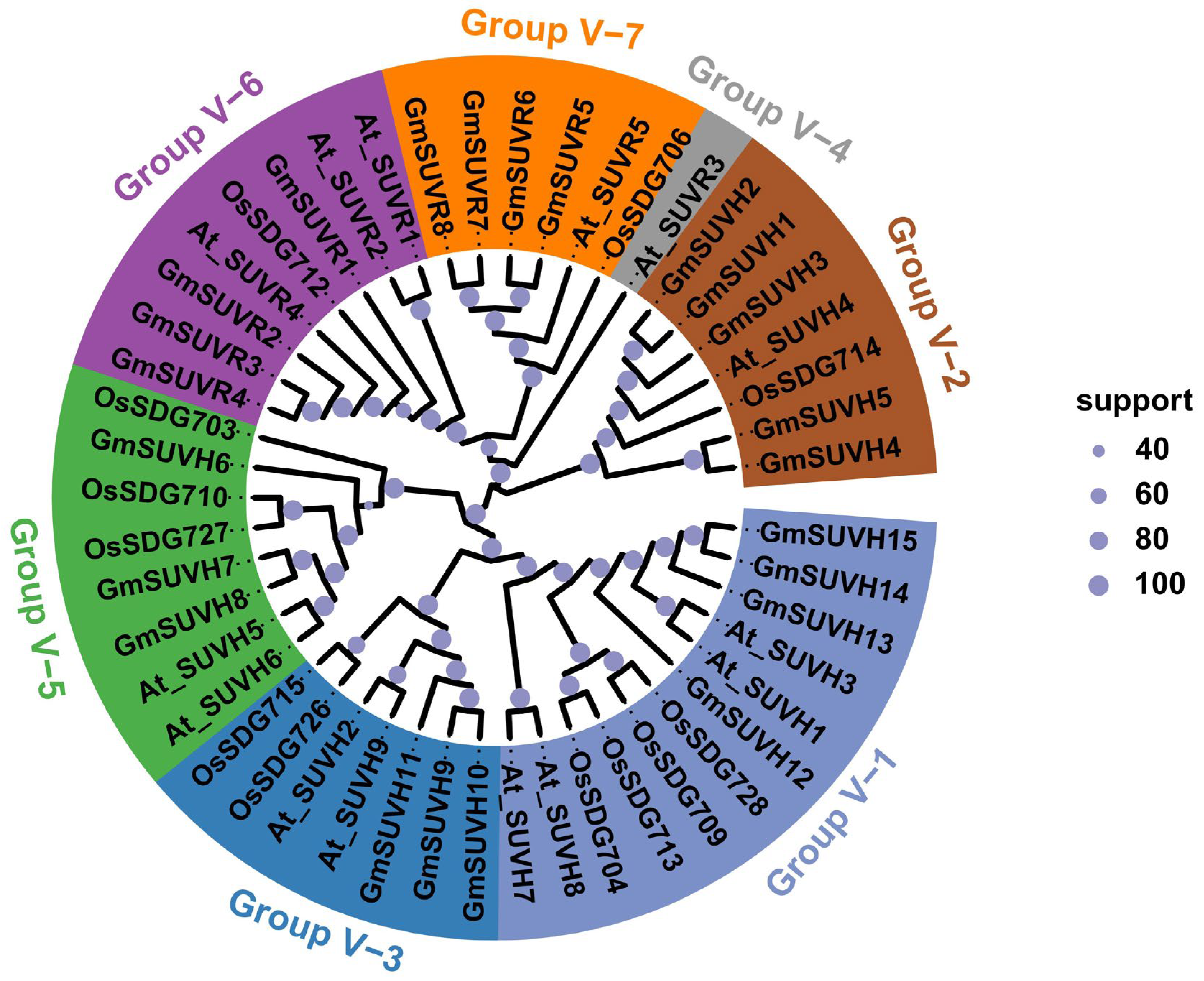
To dissect the evolutionary history of the Su(var)3-9 SET protein family and to establish phylogenetic relationships, a phylogenetic tree was built in MEGA12 using the neighbor-joining algorithm, with full-length Su(var)3-9 SET protein sequences from soybean, Arabidopsis and Oryza sativa (Figure 1, Table S3). Based on the branching characteristics and bootstrap values, the Su(var)3-9 SET genes are classified into seven major groups. GmSUVH12/13/14/15 belonged to Group V-1, GmSUVH1/2/3/4/5 belonged to Group V-2, GmSUVH9/10/11 belonged to Group V-3, GmSUVH6/7/8 belonged to Group V-5, GmSUVR1/2/3/4 belonged to Group V-6 and GmSUVR5/6/7/8 belonged to Group V-7. Arabidopsis SUVH10 was excluded because it might be a pseudogene [51]. Genes within the same group may tend to share similarities in their gene structures, protein architectures, and functional domains.

3.2. The Structures of Identified GmSu(var)3-9 SET Genes and Proteins in Soybean

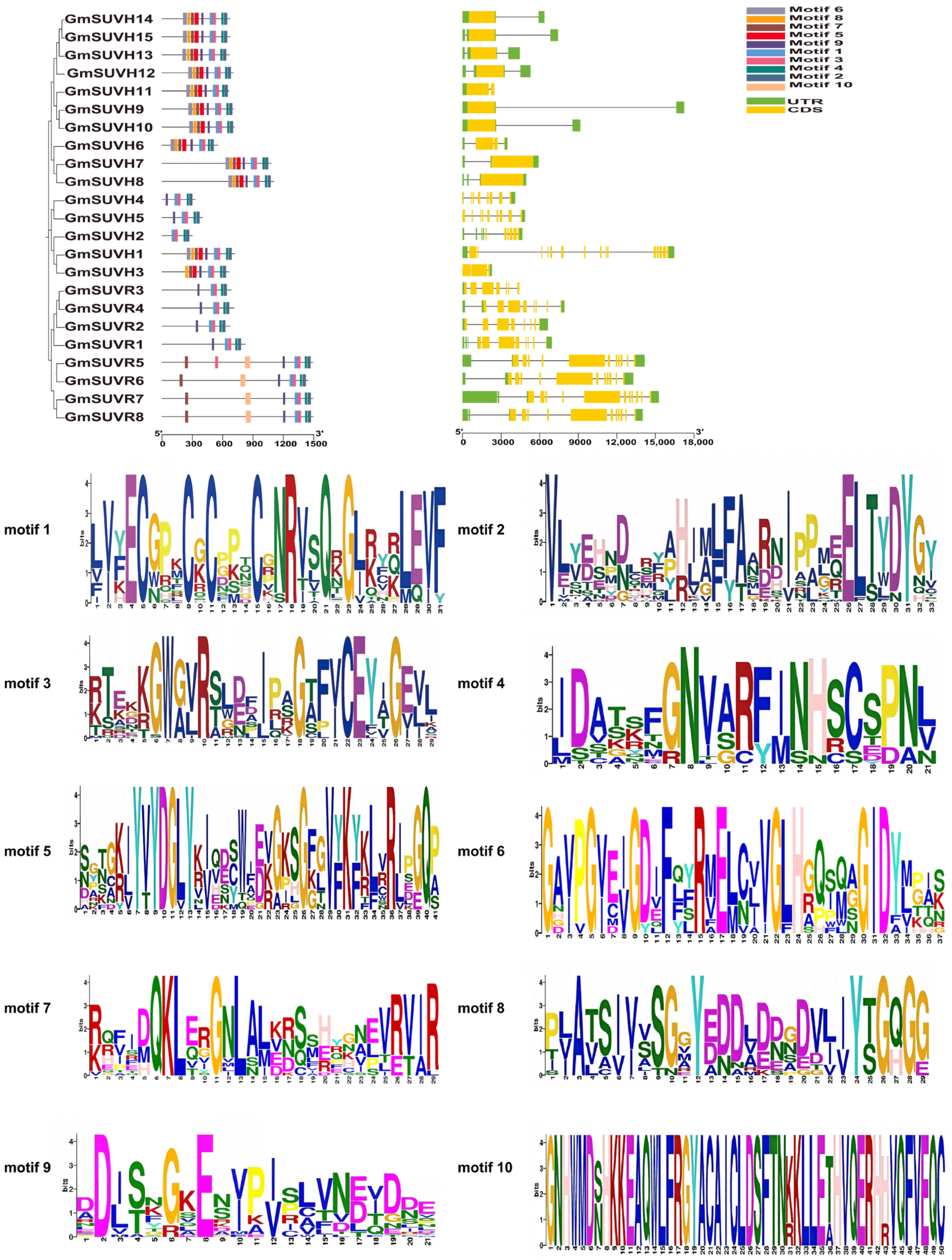
Gene structure analysis was performed to characterize the gene length and the exon–intron organization of GmSu(var)3-9 SET family members (Figure 2). Gene lengths ranged from 2282 bp to 16,692 bp. In eukaryotes, introns and exons alternate to form genes. Introns exhibit distinct regulatory roles, and their gain or loss may contribute to functional divergence among homologous genes [52]. In plants, up to 60% of genes undergo splicing, with the majority occurring in introns [53]. When intron-mediated enhancement (IME) was employed in Arabidopsis thaliana, mRNA levels were markedly increased, accompanied by a 40-fold enhancement of reporter enzyme activity [54]. Most GmSUVH members exhibited a simple structure with a few exons, while GmSUVR genes contained multiple exons separated by long introns. The GmSu(var)3-9 SET gene family in soybean exhibits marked differences in intron and exon numbers from those in Arabidopsis thaliana, Oryza sativa, and Populus trichocarpa, suggesting that the structure of GmSu(var)3-9 SET genes is diverse across different species [55].
MEME analysis identified ten conserved motifs (motif1~motif10) in GmSu(var)3-9 SET family proteins (Figure 2). The width, sites, and E-values are provided in Table S4. The conserved motif width ranged from approximately 21 to 50 amino acids, and the number of sites ranged from 4 to 23. Genes clustered closely within the phylogenetic tree shared similar motifs.
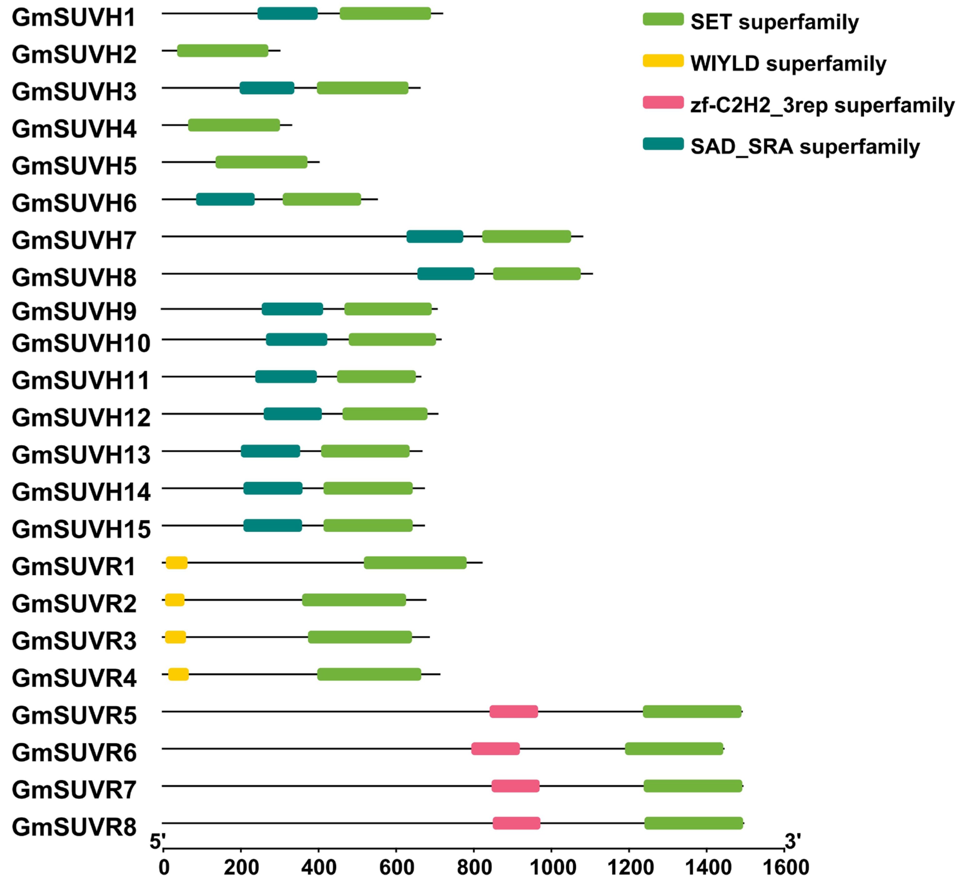
To further characterize the structural divergence of the GmSu(var)3-9 SET protein domain, all protein sequences were submitted to the SMART website (Figure 3). All the proteins contained a SET domain. The N-terminal WIYLD domain, which in Arabidopsis SUVR4 binds ubiquitin and converts H3K9me1 into H3K9me3 in vitro [27], was present in GmSUVR1/2/3/4. GmSUVR5/6/7/8 have a zf-C2H2 domain at their N-termini. The ZF-C2H2 domain belongs to a type of C2H2 zinc finger (Znf), which facilitates stable binding to target DNA and plays a crucial role in transcriptional regulation [56,57].

3.3. Chromosomal Location and Collinearity Analysis of the GmSu(var)3-9 SET Gene Family in Soybean

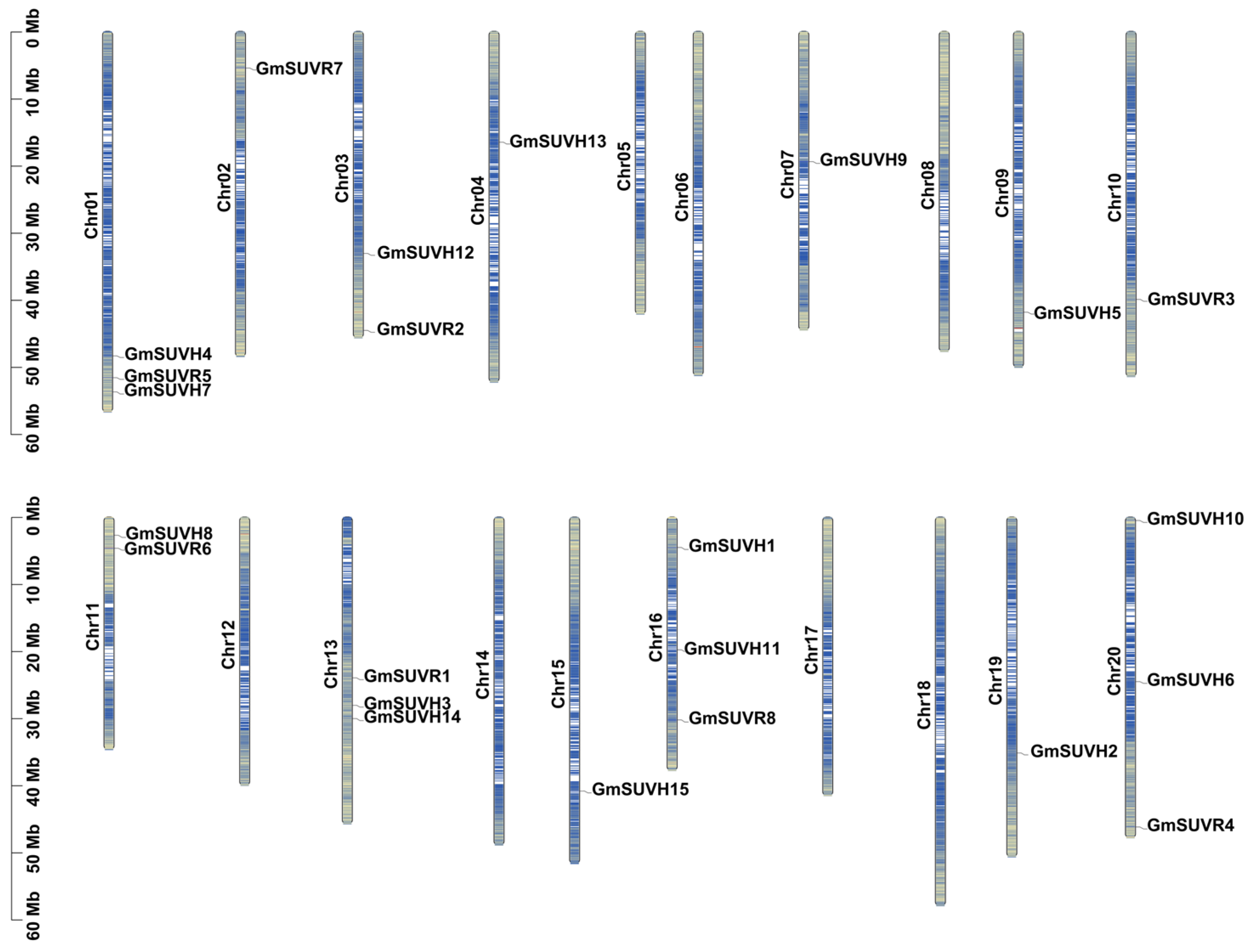
To investigate the arrangement of 23 GmSu(var)3-9 SET genes on chromosomes in the soybean genome, we revealed the chromosomal distribution pattern of this gene family (Figure 4). The gene location information is provided in Table S2. Chromosomes 1, 13, 16 and 20 each contained three GmSu(var)3-9 SET genes; chromosomes 3 and 11 each contained two; and chromosomes 2, 4, 7, 9, 10, 15 and 19 each contained one. Gene distribution across chromosomes was independent of chromosomal length. To explore the potential evolution relationship and further compare the GmSu(var)3-9 SET gene family collinearity among different species, a comparative analysis was performed between the GmSu(var)3-9 SET protein and homologues from the representative plants, including Arabidopsis thaliana, Oryza sativa, Populus trichocarpa, and Zea mays (Figure S1). The results show that there are seven (Arabidopsis thaliana), two (Oryza sativa), 11 (Populus trichocarpa), and three (Zea mays) GmSu(var)3-9 SET proteins showing high homology to members from the other four species, respectively. Clear collinear relationships were identified between GmSu(var)3-9 SET genes and their orthologs in all examined species, with the most extensive syntenic blocks observed between soybean and Populus trichocarpa.

3.4. GO Annotations of the GmSu(var)3-9 SET Gene Family

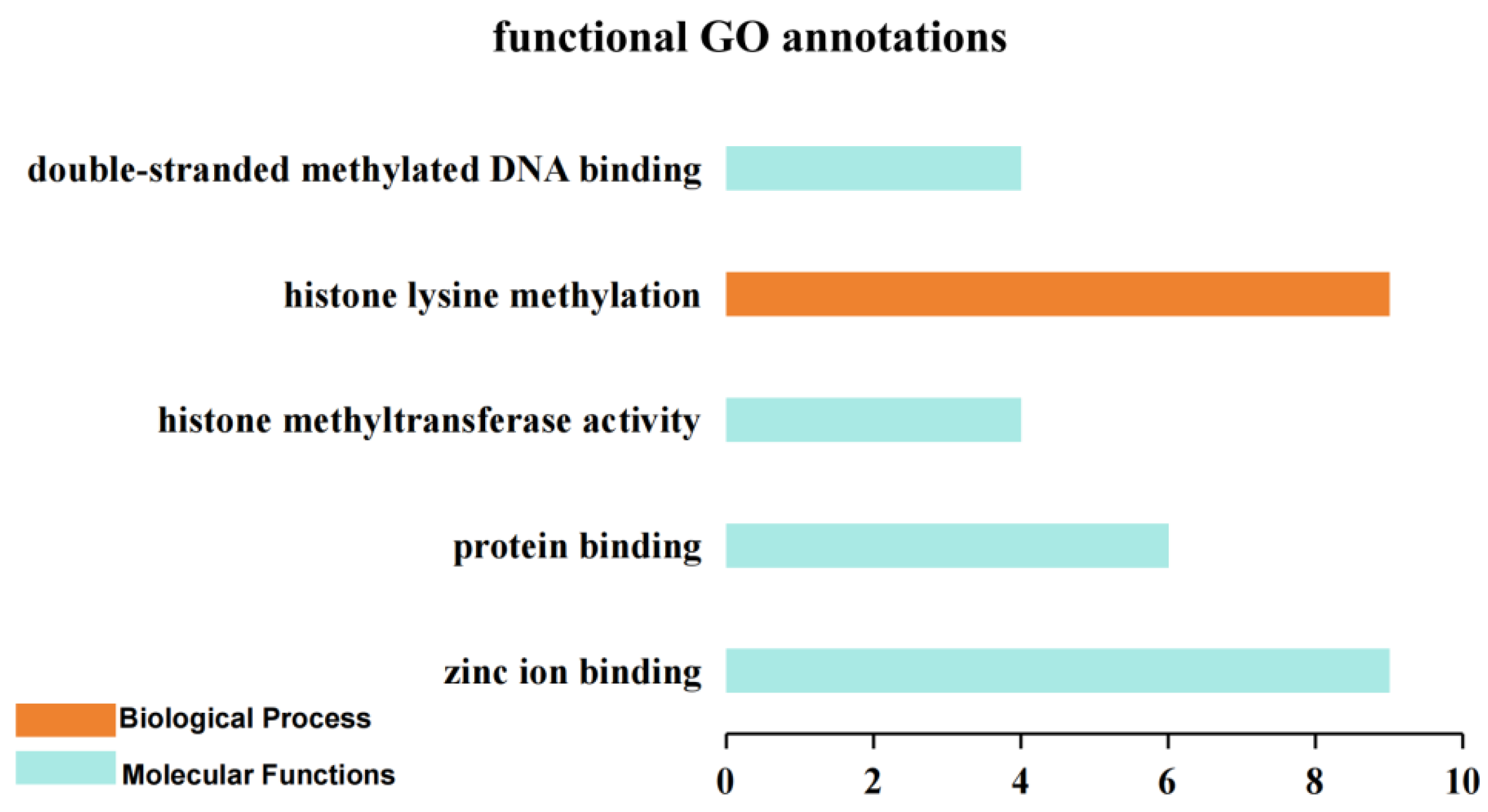
GO enrichment analysis was implemented to predict the potential biological functions of GmSu(var)3-9 SET genes (Figure 5, Table S5). GmSu(var)3-9 SETs were involved in different biological processes (BP) and molecular functions (MF). All the biological processes were related to histone lysine methylation (GO:0034968). Molecular functions included double-stranded methylated DNA binding (GO:0010385), histone methyltransferase activity (GO:0042054), protein binding (GO:0005515) and zinc ion binding (GO:0008270). The GmSu(var)3-9 SET gene family was functionally enriched in epigenetic regulation (histone lysine methylation) and multiple molecular binding/catalytic activities, indicating that GmSu(var)3-9 SET proteins mainly participate in epigenetic regulation that rely on binding and catalytic functions.

3.5. The Cis-Acting Regulatory Elements in the Promoter of the GmSu(var)3-9 SET Gene Family in Soybean

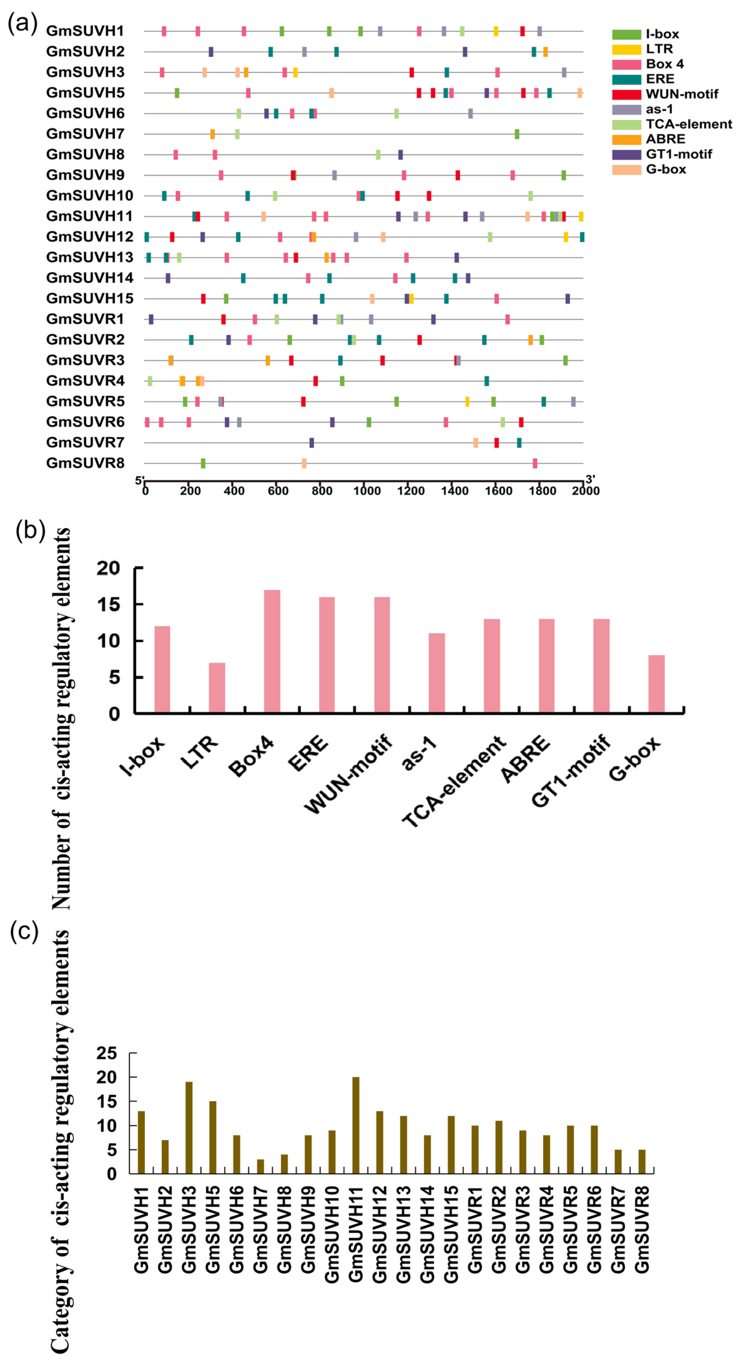
Cis-acting elements in organisms are responsible for binding transcription factors, playing a crucial role in regulating gene expression [58]. To elucidate the regulatory mechanism of the GmSu(var)3-9 SET gene family in response to biotic and abiotic stresses, the 2000 bp genomic sequence upstream of GmSu(var)3-9 SET promoters was queried using the Plant Care database to identify cis-regulatory elements (CREs). Multiple types of cis-regulatory elements, such as ABRE, ERE, TCA element, I-box, G-box, Box 4, WUN-motif, GT1-motif, as-1, and LTR, were detected within the 2000 bp promoter regions of these genes (Figure 6a). Among family members, GmSUVH11 carried the greatest abundance of cis-regulatory elements, while GmSUVH7 harbored far fewer, highlighting heterogeneous transcriptional regulatory complexity across the gene family (Figure 6b,c).
Light-responsive cis-elements, including Box-4, G-box, I-box and GT1-Motif, were detected in the promoter regions of GmSu(var)3-9 SET genes. Approximately 74% of these genes contained Box4. Nearly all GmSu(var)3-9 SET genes contained at least one light-responsive element in their promoter regions. As a core regulatory component involved in light signaling, the Box4 motif exerts a crucial function in modulating the growth duration of soybean. Loss of this motif leads to a marked upregulation of GmZTL4 expression and a shortened growth period [59]. Studies have indicated that light-responsive elements (e.g., Box-4 and G-box) are present in the promoter regions of genes involved in drought and salt stress responses, and these elements play critical roles in transcriptional regulation [60,61]. The MsLEA4-4 promoter contains G-box and abscisic acid response element (ABREs). Overexpression of MsLEA4-4 in Arabidopsis results in more lateral roots, higher chlorophyll content, and significantly higher survival rate under salt stress and abscisic acid treatment conditions relative to the wild type [62]. Under salt treatment, the transcription factors PpWRKY44 and ABRE-BINDING FACTOR3 (PpABF3) promote malic acid accumulation in pear. Specifically, PpABF3 enhances salt-induced malic acid accumulation by targeting the G-box cis-element located in the promoter of PpWRKY44 [63]. The GT1-motif in the promoter of rice OsRAV2 directly regulates its salt response [64]. As-1 was found to be linked with oxidative stress responses mediated by salicylic acid [65]. The WUN-motif (wound responsive element) was found in the promoters of 16 GmSu(var)3-9 SET genes, implying their potential functional involvement in plant biotic stress responses. LTR was identified as being related to drought induction and the regulation of drought response genes [66]. ABRE, TCA-element and ERE were found to participate in phytohormone regulation. A previous study revealed that the ABRE motif participated in dehydration and salinity stress [67]. ERE and salicylic acid-responsive cis-elements (TCA element) were present in 16 and 13 GmSu(var)3-9 SET promoters, respectively. In Arabidopsis, RAP2.11 regulates the expression of AtHAK5 by binding to the ERE Motif and GCC-box in the AtHAK5 promoter under low K+ conditions and participates in the response to low-potassium conditions [68]. Our results indicate that the GmSu(var)3-9 SET gene family may play crucial roles in soybean growth and development, as well as in responses to both biotic and abiotic stresses.

3.6. Expression Profiles of the GmSu(var)3-9 SET Gene Family in Soybean

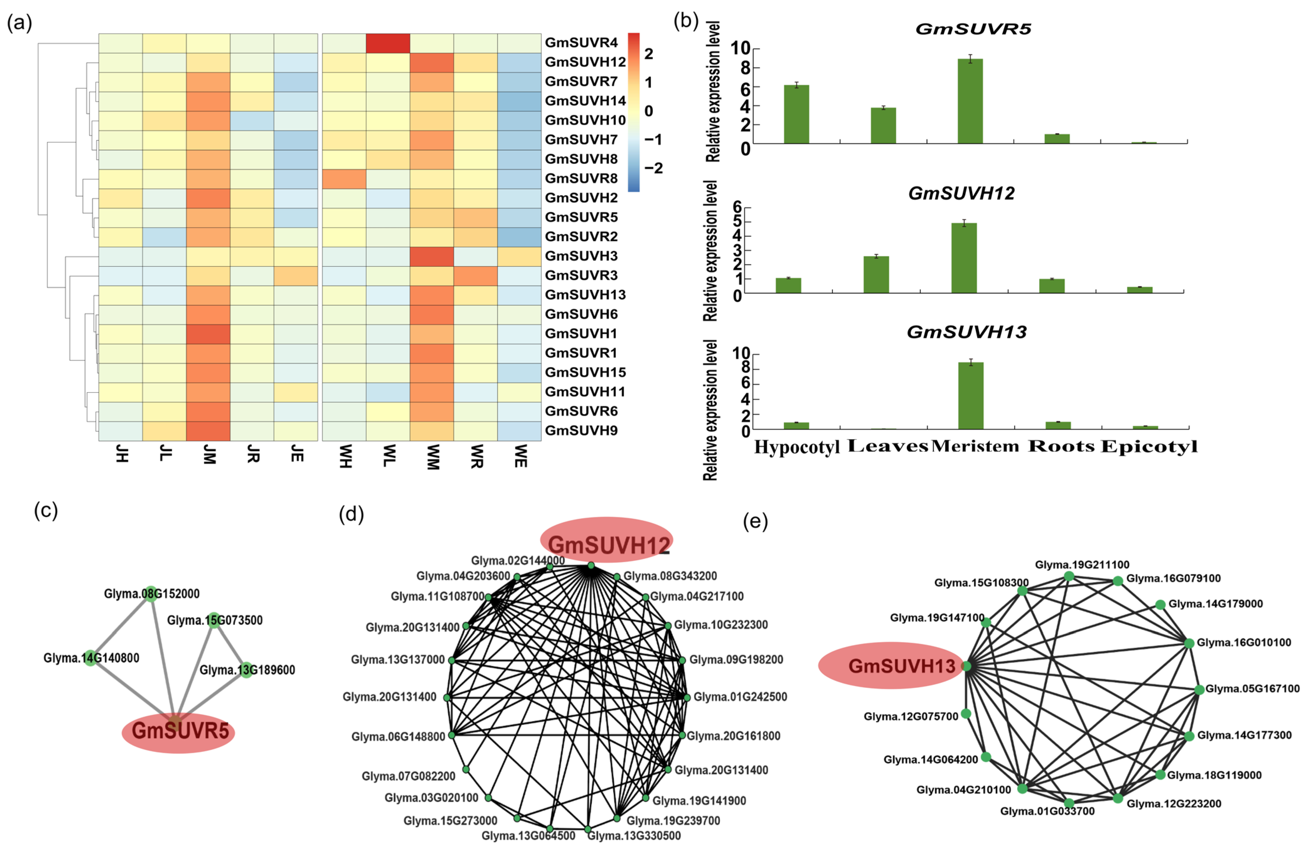
To better understand the potential roles of the GmSu(var)3-9 SET gene family in plant growth and development, expression profiles were examined in different tissues of two representative soybean cultivars, Jack and Williams 82, at the VC stage (Figure 7a, Table S6). The two cultivars were used to assess whether expression profiles differed between varieties. The heatmap showed that GmSu(var)3-9 SET gene expression varied substantially across tissues. The gene family was predominantly highly expressed in meristems, with relatively high expression also observed in roots and leaves; these patterns were similar in W82 and Jack. Sixteen and twelve genes showed high expression in Jack and W82 meristems, respectively. GmSu(var)3-9 SET genes with high expression levels in meristematic tissues possess promoters enriched in abundant light-responsive cis-acting elements. In meristems, GmSUVH1 and GmSUVH11 are both highly expressed, with consistent patterns across varieties. However, the expression of GmSUVR4 in leaves was notably higher in W82. Although W82 and Jack showed broadly similar GmSu(var)3-9 SET gene expression patterns, there are still significant differences between the two varieties. For verification of the RNA-seq data, RT-qPCR was performed on three genes to evaluate the expression pattern of three genes in the roots, leaves, meristem, epicotyl and hypocotyl of W82 (Figure 7b).
Co-expression network analysis based on the global dataset of SoyBase CoNekT was used to screen potential functionally associated upstream and downstream genes that co-expressed with GmSUVR5, GmSUVH12 and GmSUVH13 (Figure 7c–e, Table S7). The result showed that GmSUVR5 was co-expressed with Glyma.08G152000, which encodes a calcium-dependent lipid-binding (CALB domain) protein (Figure 7c). A previous study has shown that the knockdown of HvCaLB1 reduces its salt tolerance [69]. GmSUVH12 was co-expressed with Glyma.04G203600, which encodes an XS zinc finger domain protein (Figure 7d). The AtSGS3 (Suppressor Of Gene Silencing 3) protein belongs to the XS protein family and plays a crucial role in juvenile plant development and trans-acting siRNA production [70]. GmSUVH13 was co-expressed with Glyma.19G147100, a CCCH Zinc-Finger Protein (Figure 7e). CCCH-type zinc finger proteins play an important role in plant development and tolerance to various abiotic stresses, including salt, drought, flooding, low temperature, and oxidative stress [71]. These results demonstrate that the GmSu(var)3-9 SET family genes may function in development and stress adaptation through these regulatory partners.

3.7. Tissue-Specific Epigenetic Modifications of the Soybean GmSu(var)3-9 SET Gene Family

Plant epigenetics refers to a complex molecular regulatory network that controls gene expression without altering the DNA nucleotide sequence [72]. Key epigenetic regulatory mechanisms in plants include DNA methylation, histone modifications, and non-coding RNAs. Epigenetic modifications play a pivotal role in tissue differentiation and morphogenesis. In Arabidopsis thaliana, tissues with different levels of CLSY expression exhibited distinct DNA methylation patterns in an RdDM pathway-dependent manner [73]. Nitrogen-induced differentially expressed genes were predominantly enriched in pathways related to nutrient absorption, transport and metabolism in roots, whereas those in shoots were mainly involved in photosynthesis, carbon metabolism and growth and development. This differentiation is determined by organ-specific histone modifications [74].
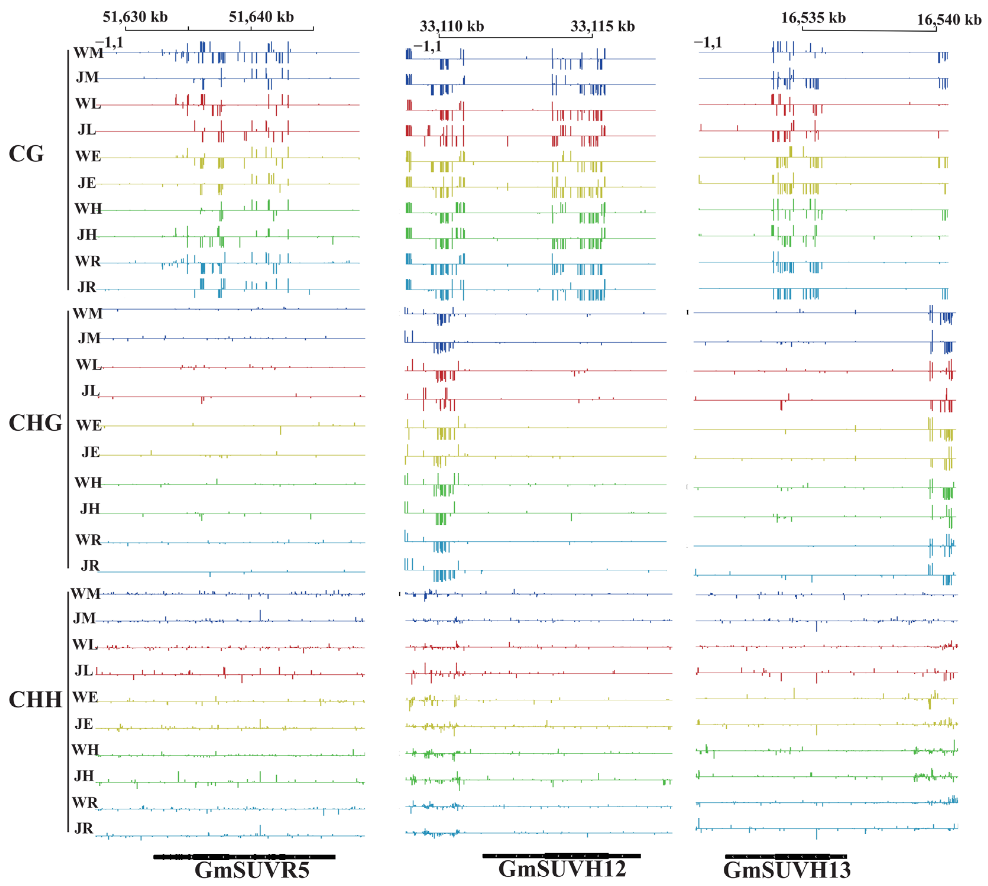
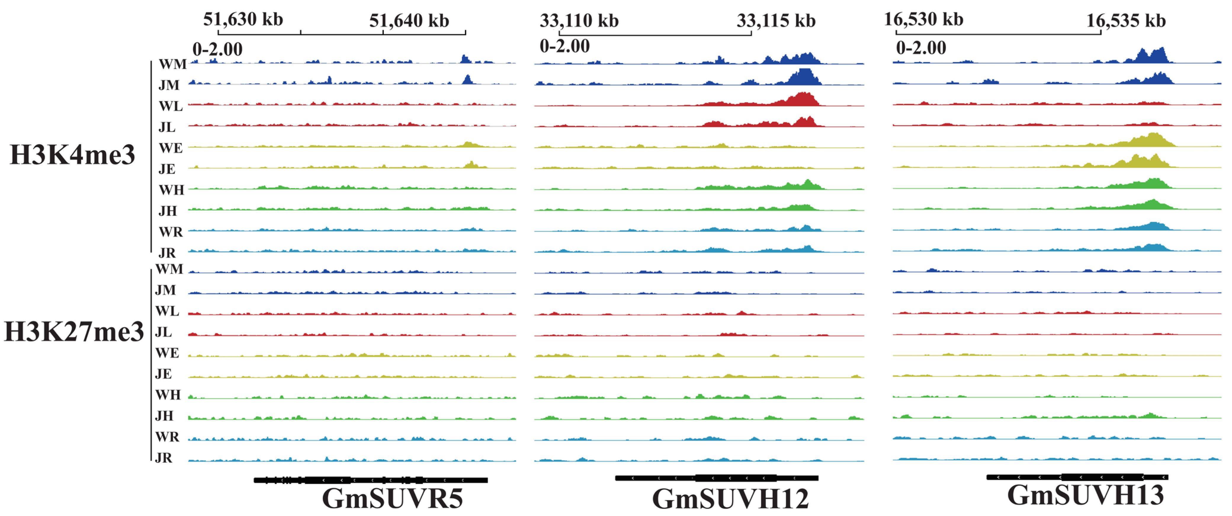
In order to better understand whether the expression of the GmSu(var)3-9 SET genes was affected by epigenetic modifications, we analyzed the DNA methylation and histone modifications of GmSUVR5, GmSUVH12 and GmSUVH13 across different tissues and soybean varieties. No significant differences in CG, CHG, or CHH methylation levels were detected among tissues and varieties for GmSUVR5 (Figure 8, Figure S2). It was plausible that additional regulatory mechanism(s), including repressive TF binding, were also involved in GmSUVR5 expression. GmSUVH12 displays elevated CG methylation levels mainly concentrated in the gene body, with no remarkable variation between W82 and Jack (Figure 8, Figure S2), and histone modification analysis further indicated higher H3K4me3 modification levels in leaves and meristem (Figure 9, Figure S3). DNA methylation modification levels of GmSUVH13 showed no remarkable difference between W82 and Jack (Figure 8, Figure S2). Histone modification analysis indicated higher H3K4me3 modification levels in W82 and Jack (Figure 9, Figure S3). However, GmSUVH13 had a relatively high expression level only at the meristem, suggesting maybe other mechanisms, including repressive TF binding, are also involved in its low transcript levels.

3.8. The Expression of the GmSu(var)3-9 SET Gene Family Under Salt Stress

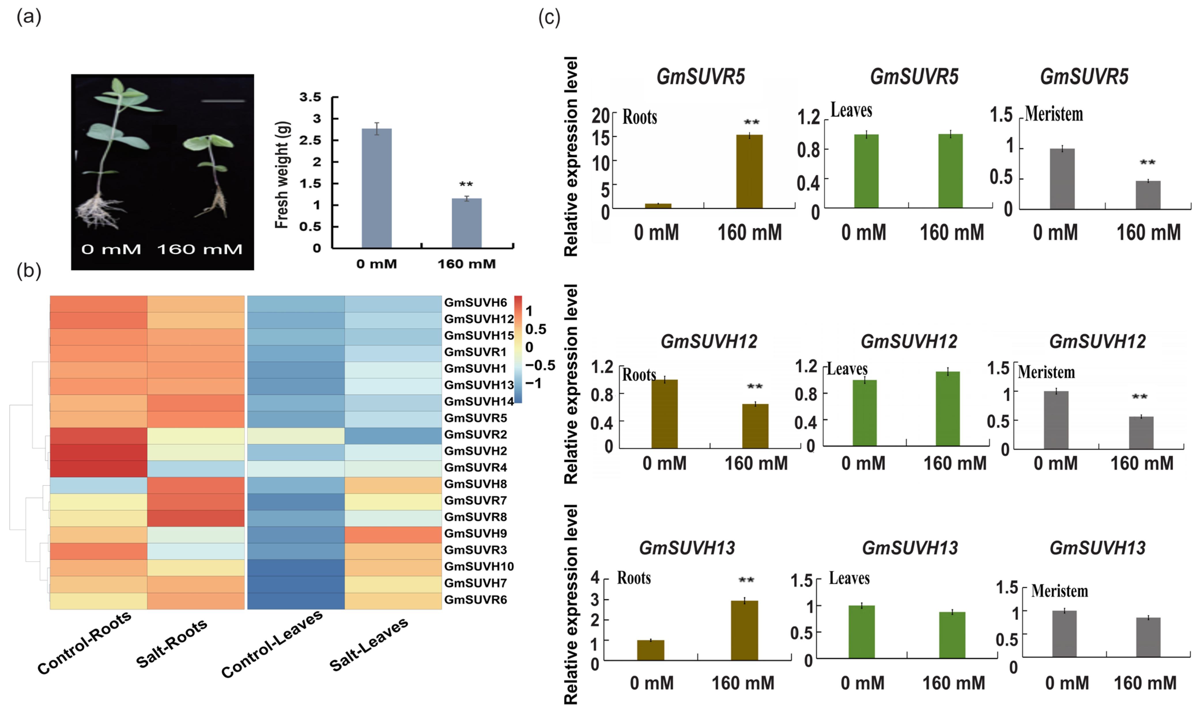
Cis-element analysis indicates that the GmSu(var)3-9 SET family contains multiple cis-acting elements for abiotic stress responsiveness. To further explore the expression level of this gene family under abiotic stress, we conducted gene expression level analysis and examined the phenotype of soybean under salt stress. A previous study has shown that the protein complex composed of OsBAG4, OsMYB106 and OsSUVH7 regulated the expression of OsHKT1;5 under salt stress conditions [75]. We first assessed soybean growth under salt stress. Compared with the control group, salt-treated soybeans showed relatively short and visible wilting (Figure 10a). Fresh weight under salt treatment decreased notably (Figure 10a). Transcriptome analysis revealed distinct expression patterns of GmSu(var)3-9 SET genes in roots and leaves. Both in control and treatment conditions, a higher number of GmSu(var)3-9 SET genes were expressed in roots than in leaves (Figure 10b, Table S6). The relative expression levels of three soybean GmSu(var)3-9 SET genes (GmSUVH12, GmSUVH13, and GmSUVR5) were analyzed in roots and leaves under control (0 mM NaCl) and salt stress (160 mM NaCl) conditions (Figure 10c). In roots, salt treatment significantly induced the expression of GmSUVR5 and GmSUVH13. In contrast, the expression of GmSUVH12 in roots was significantly downregulated under salt stress. In leaves, none of the three genes showed significant changes in response to salt treatment. We also examined the expression of these three genes in meristem. Compared with the control, the expression levels of GmSUVR5 and GmSUVH12 were distinctly downregulated under salt treatment. In contrast, the expression of GmSUVH13 showed only a slight, non-significant reduction under the salt stress condition. These results suggest that the GmSu(var)3-9 SET family genes exhibit divergent responses to salt stress.

4. Discussion

Histone lysine methylation, as a core component of the epigenetic regulatory network in eukaryotes, plays crucial roles in gene expression regulation, chromatin structure stability, and plant growth, development, and stress adaptation. Its dynamic modification is precisely regulated by histone lysine methyltransferases (HKMTs). The Su(var)3-9 SET gene family, an important branch of HKMTs, participates in biological processes such as heterochromatin silencing and gene transcription regulation through SET domain-mediated histone methylation activity [76]. The soybean genome contained 23 GmSu(var)3-9 SET family members, including 15 GmSUVH and eight GmSUVR genes — a number notably higher than that in Arabidopsis (15) and rice (12). The collinearity analysis results showed that the genes of the GmSu(var)3-9 SET gene family in soybean have a relatively higher degree of collinearity with the Su(var)3-9 SET gene family genes in Populus trichocarpa. These differences may be closely related to the two whole-genome duplication events that soybean has experienced during its evolution. Zhu et al. [55] analyzed the homologous sequences of Su(var)3-9 SET proteins in representative terrestrial plants and classified Su(var)3-9 SET into seven subtypes. Except for Group V-4, the remaining subtypes are referred to as cSUVH (core Su(var)3-9 homologues and related genes). Phylogenetic analysis in this study indicated that the soybean Su(var)3-9 SET gene family had a relatively close genetic relationship with the homologous genes of Arabidopsis thaliana. Apart from the absence of Group V-4, all subtypes were represented. GO enrichment analysis revealed that GmSu(var)3-9 SET genes mainly participate in biological processes such as histone lysine methylation (GO:0034968), methylated DNA binding (GO:0010385), etc., and had histone methyltransferase activity (GO:0042054) and zinc ion binding activity (GO:0008270). The functional annotations of this gene family were highly consistent with those of the homologous genes in Arabidopsis thaliana and rice, indicating that the core functions of the Su(var)3-9 SET gene family were evolutionarily conserved. In summary, the GmSu(var)3-9 SET gene family had preferentially retained its core functions during the genome duplication event of soybeans to ensure regulatory pathway stability, while also undergoing diversification through genomic rearrangement and gene replication differentiation.
Tissue expression profiling indicated that the GmSu(var)3-9 SET genes were highly expressed in the meristems, roots, and leaves, with similar expression patterns across different varieties (Jack and Williams 82), suggesting conserved regulatory function during the growth and development. Meristematic tissue, as a crucial site for cell division and differentiation, exhibits more active chromatin dynamics and gene expression regulation. The high expression of the GmSu(var)3-9 SET genes in this tissue may affect cell differentiation and organ formation by regulating the histone methylation status. This observation was consistent with the findings in Arabidopsis, in which Su(var)3-9 SET genes were involved in somatic embryogenesis and leaf development [29,30]. In Arabidopsis thaliana, AHL10 promotes the modification of H3K9me2 in the promoter region of salt-responsive genes by recruiting the SUVH2/9 complex, thereby inhibiting gene expression. Under salt stress, AHL10 is degraded, relieving this repression and enhancing salt tolerance [28]. The GmSu(var)3-9 SET proteins in soybean may regulate the expression of salt-responsive genes through a similar mechanism: under salt stress, some members of GmSUVH show decreased expression, resulting in a lower level of H3K9me2 modification, activating the transcription of downstream salt-tolerant genes, thereby enhancing the adaptability of soybeans to salt stress. Additionally, the N-terminal WIYLD domain of Arabidopsis SUVR4 may regulate H3K9 methylation levels through ubiquitin binding [27]. Furthermore, the promoter regions of highly expressed genes were enriched in light-responsive cis-elements (such as Box4 and G-box), and Box4 had been proven to be involved in the regulation of soybean growth stages [59], suggesting that GmSu(var)3-9 SET genes might regulate soybean growth and development in response to light signals. GmSUVR5, GmSUVH12, and GmSUVH13 exhibited potential functional associations with calcium-binding, XS-domain, and CCCH zinc-finger proteins via co-expression, implying that the GmSu(var)3-9 SET family might integrate epigenetic regulation with calcium signaling, small RNA biogenesis, and abiotic stress responses.
Plant epigenetics, which includes DNA methylation, histone modifications and non-coding RNAs, serves as a critical regulator of gene expression. In Arabidopsis root meristem cells, the columella root cap exhibited substantial hypermethylation accompanied by small RNA enrichment, alongside distinct tissue-specific cell identities [77]. Our analysis of three representative GmSu(var)3-9 SET genes revealed minimal variation in DNA methylation levels across tissues and soybean varieties, whereas histone modifications displayed distinct tissue-specific patterns. These results implied that histone modification, rather than DNA methylation, might act as the primary epigenetic determinant underlying the tissue-specific regulation of GmSu(var)3-9 SET genes in soybean.
In conclusion, this study provided an important reference for a deeper understanding of the functions and evolution of the GmSu(var)3-9 SET gene family in soybean. The tissue-specific expression patterns and epigenetic modification characteristics of these genes offered candidate genes and a theoretical basis for subsequent functional verification and molecular breeding. Further analysis of the biological functions of the members of this family, new ideas and technical support for soybean stress-resistant breeding can be expected.

5. Conclusions

Twenty-three Su(var)3-9 SET genes were identified in the soybean genome, including 15 SUVH genes and eight SUVR genes. Analyses of gene structure, conserved motifs, and domains have shown that members of the GmSu(var)3-9 SET gene family were relatively evolutionarily conserved. All identified GmSu(var)3-9 SET genes contained a SET domain, with some members also possessing either a WIYLD domain or a zf-C2H2 domain. Expression patterns of these genes exhibited tissue specificity. Tissue-specific epigenetic modifications, particularly histone modifications, displayed distinct patterns across soybean tissues. In summary, these findings indicated the potential roles of the GmSu(var)3-9 SET gene family in soybean growth, development, and stress responses.

Supplementary Materials

The following supporting information can be downloaded at: https://www.mdpi.com/article/10.3390/biology15131085/s1, Table S1: The primers of qRT-PCR; Table S2: GmSu(var)3-9 SET gene family screening, naming and location; Table S3: Su(var)3-9 SET GeneBank numbers of Arabidopsis thaliana and Oryza sativa; Table S4: E-values, sites and width of conserved motifs in GmSu(var)3-9 SET genes; Table S5: The functional annotations of the GmSu(var)3-9 SET genes in soybean; Table S6: FPKM of the GmSu(var)3-9 SET genes in soybean; Table S7: Genes present in co-expression network; Figure S1: Collinearity analysis of the GmSu(var)3-9 SET gene family between Glycine max and four representative plant species (Arabidopsis thaliana, Oryza sativa, Populus trichocarpa, and Zea mays); Figure S2: Statistics of CG, CHG and CHH methylation levels of GmSUVR5, GmSUVH12 and GmSUVH13 in Jack (J) and Williams82 (W); Figure S3: Statistics of H3K4me3 methylation levels of GmSUVH12 and GmSUVH13 in Jack (J) and Williams82 (W).

Author Contributions

Conceptualization, M.W. and N.J.; methodology, W.Z., L.C., L.W. and Z.Z.; validation, L.C., L.W., Z.Z., J.W. and H.X.; resources, L.X.; writing—original draft preparation, M.W.; writing—review and editing, M.W., W.Z., L.X. and N.J.; Data curation, Z.Z., J.W., L.X. and H.X.; supervision, M.W., L.X. and N.J.; funding acquisition, M.W. and N.J. All authors have read and agreed to the published version of the manuscript.

Funding

This research was Funded by Science Research Project of Hebei Education Department (ZC2025042); the Doctoral Start-up Fund of Hebei North University (BSJJ202430); and the Doctoral Start-up Fund of Hebei North University (BSJJ202427).

Institutional Review Board Statement

Not applicable.

Informed Consent Statement

Not applicable.

Data Availability Statement

The original contributions presented in this study are included in the article and Supplementary Materials. Further inquiries can be directed to the corresponding author.

Conflicts of Interest

The authors declare no conflicts of interest.

References

  1. Rea, S.; Eisenhaber, F.; O’Carroll, D.; Strahl, B.D.; Sun, Z.-W.; Schmid, M.; Opravil, S.; Mechtler, K.; Ponting, C.P.; Allis, C.D.; et al. Regulation of chromatin structure by site-specific histone H3 methyltransferases. Nature 2000, 406, 593–599. [Google Scholar] [CrossRef] [PubMed]
  2. Dorn, R.; Krauss, V.; Reuter, G.; Saumweber, H. The enhancer of position-effect variegation of Drosophila, E(var)3-93D, codes for a chromatin protein containing a conserved domain common to several transcriptional regulators. Proc. Natl. Acad. Sci. USA 1993, 90, 11376–11380. [Google Scholar] [CrossRef] [PubMed]
  3. Jones, R.S.; Gelbart, W.M. The Drosophila Polycomb-group gene Enhancer of zeste contains a region with sequence similarity to trithorax. Mol. Cell. Biol. 1993, 13, 6357–6366. [Google Scholar] [CrossRef] [PubMed]
  4. Tschiersch, B.; Hofmann, A.; Krauss, V.; Dorn, R.; Korge, G.; Reuter, G. The protein encoded by the Drosophila position-effect variegation suppressor gene Su(var)3-9 combines domains of antagonistic regulators of homeotic gene complexes. EMBO J. 1994, 13, 3822–3831. [Google Scholar] [CrossRef] [PubMed]
  5. Stassen, M.J.; Bailey, D.; Nelson, S.; Chinwalla, V.; Harte, P.J. The Drosophila trithorax proteins contain a novel variant of the nuclear receptor type DNA binding domain and an ancient conserved motif found in other chromosomal proteins. Mech. Dev. 1995, 52, 209–223. [Google Scholar] [CrossRef] [PubMed]
  6. Springer, N.M.; Napoli, C.A.; Selinger, D.A.; Pandey, R.; Cone, K.C.; Chandler, V.L.; Kaeppler, H.F.; Kaeppler, S.M. Comparative analysis of SET domain proteins in maize and Arabidopsis reveals multiple duplications preceding the divergence of monocots and dicots. Plant Physiol. 2003, 132, 907–925. [Google Scholar] [CrossRef] [PubMed]
  7. Ng, D.W.; Wang, T.; Chandrasekharan, M.B.; Aramayo, R.; Kertbundit, S.; Hall, T.C. Plant SET domain-containing proteins: Structure, function and regulation. Biochim. ET Biophys. Acta 2007, 1769, 316–329. [Google Scholar] [CrossRef] [PubMed]
  8. Jenuwein, T.; Allis, C.D. Translating the histone code. Science 2001, 293, 1074–1080. [Google Scholar] [CrossRef] [PubMed]
  9. Ying, Z.; Mulligan, R.M.; Janney, N.; Houtz, R.L. Rubisco small and large subunit N-methyltransferases. Bi- and mono-functional methyltransferases that methylate the small and large subunits of Rubisco. J. Biol. Chem. 1999, 274, 36750–36756. [Google Scholar] [CrossRef] [PubMed]
  10. Du, J.; Jonson, L.M.; Groth, M.; Feng, S.; Hale, C.J.; Li, S.; Vashisht, A.A.; Wohlschlegel, J.A.; Patel, D.J.; Jacobsen, S.E. Mechanism of DNA methylation-directed histone methylation by KRYPTONITE. Mol. Cell 2014, 55, 495–504. [Google Scholar] [CrossRef] [PubMed]
  11. Naumann, K.; Fischer, A.; Hofmann, I.; Krauss, V.; Phalke, S.; Irmler, K.; Hause, G.; Aurich, A.C.; Dorn, R.; Jenuwein, T.; et al. Pivotal role of AtSUVH2 in heterochromatic histone methylation and gene silencing in Arabidopsis. EMBO J. 2005, 24, 1418–1429. [Google Scholar] [CrossRef] [PubMed]
  12. Thorstensen, T.; Fischer, A.; Sandvik, S.V.; Johnsen, S.S.; Grini, P.E.; Reuter, G.; Aalen, R.B. The Arabidopsis SUVR4 protein is a nucleolar histone methyltransferase with preference for monomethylated H3K9. Nucleic Acids Res. 2006, 34, 5461–5470. [Google Scholar] [CrossRef] [PubMed]
  13. Jackson, J.P.; Lindroth, A.M.; Cao, X.; Jacobsen, S.E. Control of CpNpG DNA methylation by the KRYPTONITE histone H3 methyltransferase. Nature 2002, 416, 556–560. [Google Scholar] [CrossRef] [PubMed]
  14. Jackson, J.P.; Johnson, L.; Jasencakova, Z.; Zhang, X.; PerezBurgos, L.; Singh, P.B.; Cheng, X.; Schubert, I.; Jenuwein, T.; Jacobsen, S.E. Dimethylation of histone H3 lysine 9 is a critical mark for DNA methylation and gene silencing in Arabidopsis thaliana. Chromosoma 2004, 112, 308–315. [Google Scholar] [CrossRef] [PubMed]
  15. Jasencakova, Z.; Soppe, W.J.; Meister, A.; Gernand, D.; Turner, B.M.; Schubert, I. Histone modifications in Arabidopsis- high methylation of H3 lysine 9 is dispensable for constitutive heterochromatin. Plant J. Cell Mol. Biol. 2003, 33, 471–480. [Google Scholar] [CrossRef] [PubMed]
  16. Xue, Y.; Cao, X.; Chen, X.; Deng, X.; Deng, X.W.; Ding, Y.; Dong, A.; Duan, C.G.; Fang, X.; Gong, L.; et al. Epigenetics in the modern era of crop improvements. Sci. China Life Sci. 2025, 68, 1570–1609. [Google Scholar] [CrossRef] [PubMed]
  17. Li, X.; Harris, C.J.; Zhong, Z.; Chen, W.; Liu, R.; Jia, B.; Wang, Z.; Li, S.; Jacobsen, S.E.; Du, J. Mechanistic insights into plant SUVH family H3K9 methyltransferases and their binding to context-biased non-CG DNA methylation. Proc. Natl. Acad. Sci. USA 2018, 115, E8793–E8802. [Google Scholar] [CrossRef] [PubMed]
  18. Xu, L.; Jiang, H. Writing and Reading Histone H3 Lysine 9 Methylation in Arabidopsis. Front. Plant Sci. 2020, 11, 452. [Google Scholar] [CrossRef] [PubMed]
  19. Johnson, L.M.; Bostick, M.; Zhang, X.; Kraft, E.; Henderson, I.; Callis, J.; Jacobsen, S.E. The SRA methyl-cytosine-binding domain links DNA and histone methylation. Curr. Biol. 2007, 17, 379–384. [Google Scholar] [CrossRef] [PubMed]
  20. Rajakumara, E.; Law, J.A.; Simanshu, D.K.; Voigt, P.; Johnson, L.M.; Reinberg, D.; Patel, D.J.; Jacobsen, S.E. A dual flip-out mechanism for 5mC recognition by the Arabidopsis SUVH5 SRA domain and its impact on DNA methylation and H3K9 dimethylation in vivo. Genes Dev. 2011, 25, 137–152. [Google Scholar] [CrossRef] [PubMed]
  21. Zhang, J.; Yuan, J.; Lin, J.; Chen, L.; You, L.Y.; Chen, S.; Peng, L.; Wang, C.H.; Du, J.; Duan, C.G. Molecular basis of locus-specific H3K9 methylation catalyzed by SUVH6 in plants. Proc. Natl. Acad. Sci. USA 2023, 120, e2208525120. [Google Scholar] [CrossRef] [PubMed]
  22. Ebbs, M.L.; Bender, J. Locus-Specific Control of DNA Methylation by the Arabidopsis SUVH5 Histone Methyltransferase. Plant Cell 2006, 18, 1166–1176. [Google Scholar] [CrossRef] [PubMed]
  23. Zhao, Q.Q.; Lin, R.N.; Li, L.; Chen, S.; He, X.J. A methylated-DNA-binding complex required for plant development mediates transcriptional activation of promoter methylated genes. J. Integr. Plant Biol. 2019, 61, 120–139. [Google Scholar] [CrossRef] [PubMed]
  24. Liu, Z.W.; Shao, C.R.; Zhang, C.J.; Zhou, J.X.; Zhang, S.W.; Li, L.; Chen, S.; Huang, H.W.; Cai, T.; He, X.J. The SET domain proteins SUVH2 and SUVH9 are required for Pol V occupancy at RNA-directed DNA methylation loci. PLoS Genet. 2014, 10, e1003948. [Google Scholar] [CrossRef] [PubMed]
  25. Johnson, L.M.; Law, J.A.; Khattar, A.; Henderson, I.R.; Jacobsen, S.E. SRA-domain proteins required for DRM2-mediated de novo DNA methylation. PLoS Genet. 2008, 4, e1000280. [Google Scholar] [CrossRef] [PubMed]
  26. Liu, Q.; Liu, P.; Ji, T.; Zheng, L.; Shen, C.; Ran, S.; Liu, J.; Zhao, Y.; Niu, Y.; Wang, T.; et al. The histone methyltransferase SUVR2 promotes DSB repair via chromatin remodeling and liquid-liquid phase separation. Mol. Plant 2022, 15, 1157–1175. [Google Scholar] [CrossRef] [PubMed]
  27. Veiseth, S.V.; Rahman, M.A.; Yap, K.L.; Fischer, A.; Egge-Jacobsen, W.; Reuter, G.; Zhou, M.M.; Aalen, R.B.; Thorstensen, T. The SUVR4 histone lysine methyltransferase binds ubiquitin and converts H3K9me1 to H3K9me3 on transposon chromatin in Arabidopsis. PLoS Genet. 2011, 7, e1001325. [Google Scholar] [CrossRef] [PubMed]
  28. Guo, P.; Chong, L.; Jiao, Z.; Xu, R.; Niu, Q.; Zhu, Y. Salt stress activates the CDK8-AHL10-SUVH2/9 module to dynamically regulate salt tolerance in Arabidopsis. Nat. Commun. 2025, 16, 2454. [Google Scholar] [CrossRef] [PubMed]
  29. Peng, J.; Zhang, Q.; Tang, L.P.; Xu, B.J.; Laux, T.; Zhang, X.S.; Su, Y.H. LEC2 induces somatic cell reprogramming through epigenetic activation of plant cell totipotency regulators. Nat. Commun. 2025, 16, 4185. [Google Scholar] [CrossRef] [PubMed]
  30. Hung, F.-Y.; Feng, Y.-R.; Hsin, K.-T.; Shih, Y.-H.; Chang, C.-H.; Zhong, W.; Lai, Y.-C.; Xu, Y.; Yang, S.; Sugimoto, K.; et al. Arabidopsis histone H3 lysine 9 methyltransferases KYP/SUVH5/6 are involved in leaf development by interacting with AS1-AS2 to repress KNAT1 and KNAT2. Commun. Biol. 2023, 6, 219. [Google Scholar] [CrossRef]
  31. Kuhlmann, M.; Mette, M.F. Developmentally non-redundant SET domain proteins SUVH2 and SUVH9 are required for transcriptional gene silencing in Arabidopsis thaliana. Plant Mol. Biol. 2012, 79, 623–633. [Google Scholar] [CrossRef] [PubMed]
  32. Han, Y.-F.; Dou, K.; Ma, Z.-Y.; Zhang, S.-W.; Huang, H.-W.; Li, L.; Cai, T.; Chen, S.; Zhu, J.-K.; He, X.-J. SUVR2 is involved in transcriptional gene silencing by associating with SNF2-related chromatin-remodeling proteins in Arabidopsis. Cell Res. 2014, 24, 1445–1465. [Google Scholar] [CrossRef] [PubMed]
  33. Koberg, M.; Abu-Much, R.; Gedanken, A. Optimization of bio-diesel production from soybean and wastes of cooked oil: Combining dielectric microwave irradiation and a SrO catalyst. Bioresour. Technol. 2011, 102, 1073–1078. [Google Scholar] [CrossRef] [PubMed]
  34. Huala, E.; Dickerman, A.W.; Garcia-Hernandez, M.; Weems, D.; Reiser, L.; LaFond, F.; Hanley, D.; Kiphart, D.; Zhuang, M.; Huang, W.; et al. The Arabidopsis Information Resource (TAIR): A comprehensive database and web-based information retrieval, analysis, and visualization system for a model plant. Nucleic Acids Res. 2001, 29, 102–105. [Google Scholar] [CrossRef] [PubMed]
  35. Goodstein, D.M.; Shu, S.; Howson, R.; Neupane, R.; Hayes, R.D.; Fazo, J.; Mitros, T.; Dirks, W.; Hellsten, U.; Putnam, N.; et al. Phytozome: A comparative platform for green plant genomics. Nucleic Acids Res. 2012, 40, D1178–D1186. [Google Scholar] [CrossRef] [PubMed]
  36. El-Gebali, S.; Mistry, J.; Bateman, A.; Eddy, S.R.; Luciani, A.; Potter, S.C.; Qureshi, M.; Richardson, L.J.; Salazar, G.A.; Smart, A.; et al. The Pfam protein families database in 2019. Nucleic Acids Res. 2019, 47, D427–D432. [Google Scholar] [CrossRef] [PubMed]
  37. Finn, R.D.; Clements, J.; Eddy, S.R. HMMER web server: Interactive sequence similarity searching. Nucleic Acids Res. 2011, 39, W29–W37. [Google Scholar] [CrossRef] [PubMed]
  38. Artimo, P.; Jonnalagedda, M.; Arnold, K.; Baratin, D.; Csardi, G.; de Castro, E.; Duvaud, S.; Flegel, V.; Fortier, A.; Gasteiger, E.; et al. ExPASy: SIB bioinformatics resource portal. Nucleic Acids Res. 2012, 40, W597–W603. [Google Scholar] [CrossRef] [PubMed]
  39. Saitou, N.; Nei, M. The neighbor-joining method: A new method for reconstructing phylogenetic trees. Mol. Biol. Evol. 1987, 4, 406–425. [Google Scholar] [CrossRef] [PubMed]
  40. Kumar, S.; Stecher, G.; Suleski, M.; Sanderford, M.; Sharma, S.; Tamura, K. MEGA12: Molecular Evolutionary Genetic Analysis Version 12 for Adaptive and Green Computing. Mol. Biol. Evol. 2024, 41, msae263. [Google Scholar] [CrossRef] [PubMed]
  41. Bailey, T.L.; Boden, M.; Buske, F.A.; Frith, M.; Grant, C.E.; Clementi, L.; Ren, J.; Li, W.W.; Noble, W.S. MEME SUITE: Tools for motif discovery and searching. Nucleic Acids Res. 2009, 37, W202–W208. [Google Scholar] [CrossRef] [PubMed]
  42. Chen, C.; Wu, Y.; Li, J.; Wang, X.; Zeng, Z.; Xu, J.; Liu, Y.; Feng, J.; Chen, H.; He, Y.; et al. TBtools-II: A “one for all, all for one” bioinformatics platform for biological big-data mining. Mol. Plant. 2023, 16, 1733–1742. [Google Scholar] [CrossRef] [PubMed]
  43. Schultz, J.; Milpetz, F.; Bork, P.; Ponting, C.P. SMART, a simple modular architecture research tool: Identification of signaling domains. Proc. Natl. Acad. Sci. USA 1998, 95, 5857–5864. [Google Scholar] [CrossRef] [PubMed]
  44. Wang, Y.; Tang, H.; Debarry, J.D.; Tan, X.; Li, J.; Wang, X.; Lee, T.H.; Jin, H.; Marler, B.; Guo, H.; et al. MCScanX: A toolkit for detection and evolutionary analysis of gene synteny and collinearity. Nucleic Acids Res. 2012, 40, e49. [Google Scholar] [CrossRef] [PubMed]
  45. Rombauts, S.; Déhais, P.; Van Montagu, M.; Rouzé, P. PlantCARE, a plant cis-acting regulatory element database. Nucleic Acids Res. 1999, 27, 295–296. [Google Scholar] [CrossRef] [PubMed]
  46. Movahedi, S.; Van de Peer, Y.; Vandepoele, K. Comparative network analysis reveals that tissue specificity and gene function are important factors influencing the mode of expression evolution in Arabidopsis and rice. Plant Physiol. 2011, 156, 1316–1330. [Google Scholar] [CrossRef] [PubMed]
  47. Zhang, Q.; Wang, D.; Lang, Z.; He, L.; Yang, L.; Zeng, L.; Li, Y.; Zhao, C.; Huang, H.; Zhang, H.; et al. Methylation interactions in Arabidopsis hybrids require RNA-directed DNA methylation and are influenced by genetic variation. Proc. Natl. Acad. Sci. USA 2016, 113, E4248–E4256. [Google Scholar] [CrossRef] [PubMed]
  48. Saleh, A.; Alvarez-Venegas, R.; Avramova, Z. An efficient chromatin immunoprecipitation (ChIP) protocol for studying histone modifications in Arabidopsis plants. Nat. Protoc. 2008, 3, 1018–1025. [Google Scholar] [CrossRef] [PubMed]
  49. Zhang, M.; Cao, J.; Zhang, T.; Xu, T.; Yang, L.; Li, X.; Ji, F.; Gao, Y.; Ali, S.; Zhang, Q.; et al. A Putative Plasma Membrane Na+/H+ Antiporter GmSOS1 Is Critical for Salt Stress Tolerance in Glycine max. Front. Plant Sci. 2022, 13, 870695. [Google Scholar] [CrossRef] [PubMed]
  50. Schmittgen, T.D.; Livak, K.J. Analyzing real-time PCR data by the comparative C(T) method. Nat. Protoc. 2008, 3, 1101–1108. [Google Scholar] [CrossRef] [PubMed]
  51. Baumbusch, L.O.; Thorstensen, T.; Krauss, V.; Fischer, A.; Naumann, K.; Assalkhou, R.; Schulz, I.; Reuter, G.; Aalen, R.B. The Arabidopsis thaliana genome contains at least 29 active genes encoding SET domain proteins that can be assigned to four evolutionarily conserved classes. Nucleic Acids Res. 2001, 29, 4319–4333. [Google Scholar] [CrossRef] [PubMed]
  52. Lynch, M.; Conery, J.S. The evolutionary fate and consequences of duplicate genes. Science 2000, 290, 1151–1155. [Google Scholar] [CrossRef] [PubMed]
  53. Cui, Y.; Zhang, C.; Cai, M. Prediction and feature analysis of intron retention events in plant genome. Comput. Biol. Chem. 2017, 68, 219–223. [Google Scholar] [CrossRef] [PubMed]
  54. Rose, A.B.; Carter, A.; Korf, I.; Kojima, N. Intron sequences that stimulate gene expression in Arabidopsis. Plant Mol. Biol. 2016, 92, 337–346. [Google Scholar] [CrossRef] [PubMed]
  55. Zhu, X.; Ma, H.; Chen, Z. Phylogenetics and evolution of Su(var)3-9 SET genes in land plants: Rapid diversification in structure and function. BMC Evol. Biol. 2011, 11, 63. [Google Scholar] [CrossRef] [PubMed]
  56. Pabo, C.O.; Peisach, E.; Grant, R.A. Design and selection of novel Cys2His2 zinc finger proteins. Annu. Rev. Biochem. 2001, 70, 313–340. [Google Scholar] [CrossRef] [PubMed]
  57. Wolfe, S.A.; Nekludova, L.; Pabo, C.O. DNA recognition by Cys2His2 zinc finger proteins. Annu. Rev. Biophys. Biomol. Struct. 2000, 29, 183–212. [Google Scholar] [CrossRef] [PubMed]
  58. Wittkopp, P.J.; Kalay, G. Cis-regulatory elements: Molecular mechanisms and evolutionary processes underlying divergence. Nat. Rev. Genet. 2012, 13, 59–69. [Google Scholar] [CrossRef] [PubMed]
  59. Sun, J.; Liu, Y.; Zheng, Y.; Xue, Y.; Fan, Y.; Ma, X.; Ji, Y.; Liu, G.; Zhang, X.; Li, Y.; et al. The MADS-box transcription factor GmFULc promotes GmZTL4 gene transcription to modulate maturity in soybean. J. Integr. Plant Biol. 2024, 66, 1603–1619. [Google Scholar] [CrossRef] [PubMed]
  60. Shariatipour, N.; Heidari, B. Meta-Analysis of Expression of the Stress Tolerance Associated Genes and Uncover their Cis-Regulatory Elements in Rice (Oryza sativa L.). Open Bioinform. J. 2020, 13, 39–49. [Google Scholar] [CrossRef]
  61. Gilmartin, P.M.; Sarokin, L.; Memelink, J.; Chua, N.H. Molecular light switches for plant genes. Plant Cell 1990, 2, 369–378. [Google Scholar] [CrossRef] [PubMed]
  62. Jia, H.; Wang, X.; Shi, Y.; Wu, X.; Wang, Y.; Liu, J.; Fang, Z.; Li, C.; Dong, K. Overexpression of Medicago sativa LEA4-4 can improve the salt, drought, and oxidation resistance of transgenic Arabidopsis. PLoS ONE 2020, 15, e0234085. [Google Scholar] [CrossRef] [PubMed]
  63. Alabd, A.; Cheng, H.; Ahmad, M.; Wu, X.; Peng, L.; Wang, L.; Yang, S.; Bai, S.; Ni, J.; Teng, Y. ABRE-BINDING FACTOR3-WRKY DNA-BINDING PROTEIN44 module promotes salinity-induced malate accumulation in pear. Plant Physiol. 2023, 192, 1982–1996. [Google Scholar] [CrossRef] [PubMed]
  64. Duan, Y.-B.; Li, J.; Qin, R.-Y.; Xu, R.-F.; Li, H.; Yang, Y.-C.; Ma, H.; Li, L.; Wei, P.-C.; Yang, J.-B. Identification of a regulatory element responsible for salt induction of rice OsRAV2 through ex situ and in situ promoter analysis. Plant Mol. Biol. 2016, 90, 49–62. [Google Scholar] [CrossRef] [PubMed]
  65. Garretón, V.; Carpinelli, J.; Jordana, X.; Holuigue, L. The as-1 promoter element is an oxidative stress-responsive element and salicylic acid activates it via oxidative species. Plant Physiol. 2002, 130, 1516–1526. [Google Scholar] [CrossRef] [PubMed]
  66. Niu, L.; Chu, H.D.; Tran, C.D.; Nguyen, K.H.; Pham, H.X.; Le, D.T.; Li, W.; Wang, W.; Le, T.D.; Tran, L.-S.P. The GATA Gene Family in Chickpea: Structure Analysis and Transcriptional Responses to Abscisic Acid and Dehydration Treatments Revealed Potential Genes Involved in Drought Adaptation. J. Plant Growth Regul. 2020, 39, 1647–1660. [Google Scholar] [CrossRef]
  67. Ain-Ali, Q.U.; Mushtaq, N.; Amir, R.; Gul, A.; Tahir, M.; Munir, F. Genome-wide promoter analysis, homology modeling and protein interaction network of Dehydration Responsive Element Binding (DREB) gene family in Solanum tuberosum. PLoS ONE 2021, 16, e0261215. [Google Scholar] [CrossRef] [PubMed]
  68. Kim, M.J.; Ruzicka, D.; Shin, R.; Schachtman, D.P. The Arabidopsis AP2/ERF Transcription Factor RAP2.11 Modulates Plant Response to Low-Potassium Conditions. Mol. Plant 2012, 5, 1042–1057. [Google Scholar] [CrossRef] [PubMed]
  69. Tong, T.; Zhang, Y.; Zhou, X.; Jiang, W.; Chen, G.; Zeng, F.; Xue, D.; Chen, Z.-H.; Qin, Y.; Deng, F. Molecular and physiological evidence of HvCaLB1 regulates calcium homeostasis and salt tolerance in Barley. Plant Growth Regul. 2024, 104, 455–473. [Google Scholar] [CrossRef]
  70. Peragine, A.; Yoshikawa, M.; Wu, G.; Albrecht, H.L.; Poethig, R.S. SGS3 and SGS2/SDE1/RDR6 are required for juvenile development and the production of trans-acting siRNAs in Arabidopsis. Genes Dev. 2004, 18, 2368–2379. [Google Scholar] [CrossRef] [PubMed]
  71. Han, G.; Qiao, Z.; Li, Y.; Wang, C.; Wang, B. The Roles of CCCH Zinc-Finger Proteins in Plant Abiotic Stress Tolerance. Int. J. Mol. Sci. 2021, 22, 8327. [Google Scholar] [CrossRef] [PubMed]
  72. Meyer, P. Epigenetic variation and environmental change. J. Exp. Bot. 2015, 66, 3541–3548. [Google Scholar] [CrossRef] [PubMed]
  73. Zhou, M.; Coruh, C.; Xu, G.; Martins, L.M.; Bourbousse, C.; Lambolez, A.; Law, J.A. The CLASSY family controls tissue-specific DNA methylation patterns in Arabidopsis. Nat. Commun. 2022, 13, 244. [Google Scholar] [CrossRef] [PubMed]
  74. Julian, R.; Patrick, R.M.; Li, Y. Organ-specific characteristics govern the relationship between histone code dynamics and transcriptional reprogramming during nitrogen response in tomato. Commun. Biol. 2023, 6, 1225. [Google Scholar] [CrossRef] [PubMed]
  75. Wang, J.; Nan, N.; Li, N.; Liu, Y.; Wang, T.J.; Hwang, I.; Liu, B.; Xu, Z.Y. A DNA Methylation Reader-Chaperone Regulator-Transcription Factor Complex Activates OsHKT1;5 Expression during Salinity Stress. Plant Cell 2020, 32, 3535–3558. [Google Scholar] [CrossRef] [PubMed]
  76. Malagnac, F.; Bartee, L.; Bender, J. An Arabidopsis SET domain protein required for maintenance but not establishment of DNA methylation. EMBO J. 2002, 21, 6842–6852. [Google Scholar] [CrossRef] [PubMed]
  77. Kawakatsu, T.; Stuart, T.; Valdes, M.; Breakfield, N.; Schmitz, R.J.; Nery, J.R.; Urich, M.A.; Han, X.; Lister, R.; Benfey, P.N.; et al. Unique cell-type-specific patterns of DNA methylation in the root meristem. Nat. Plants 2016, 2, 16058. [Google Scholar] [CrossRef] [PubMed]
Figure 1. Phylogenetic analysis of Su(var)3-9 SET proteins in Arabidopsis, Oryza sativa and soybean. The rooted neighbor-joining (NJ) phylogenetic tree of the Su(var)3-9 SET family was clustered with bootstrap values shown for each clade in different sizes. Different groups are marked in different colors.
Biology 15 01085 g001
Figure 2. The gene structure and conserved motifs of GmSu(var)3-9 SET genes. A total of 10 motifs were identified using the MEME website. The amino acid length was estimated using the scale bar at the bottom. Different colors indicate distinct types of amino acid residues, and the font size reflects the frequency of each amino acid.
Biology 15 01085 g002
Figure 3. Analysis of the conserved domains in the GmSu(var)3-9 SET gene family. All GmSu(var)3-9 SET genes contain a SET domain.
Biology 15 01085 g003
Figure 4. Chromosome location of the GmSu(var)3-9 SET gene family in soybean. A total of 23 GmSu(var)3-9 SET genes were distributed on chromosomes 1–20. The chromosome numbers are indicated on the left side. The scale of chromosomal length is shown in Mb.
Biology 15 01085 g004
Figure 5. Functional GO annotations of the GmSu(var)3-9 SET gene family in soybean. Different biological processes were represented by different colors. The horizontal axis represents the biological process number.
Biology 15 01085 g005
Figure 6. The cis-acting regulatory elements in the promoter of the GmSu(var)3-9 SET gene family. Different colors represent different elements. (a) Genomic DNA sequences of the 2 kb upstream promoters of the GmSu(var)3-9 SET genes to identify cis-acting regulatory elements. (b) Number of cis-acting regulatory elements. (c) Number of cis-acting regulatory elements present in each GmSu(var)3-9 SET gene.
Biology 15 01085 g006
Figure 7. (a) The expression levels of GmSu(var)3-9 SET genes in Jack (J) and Williams82 (W). The color gradient from blue to red indicates increasing expression levels. The clustering tree on the left was constructed based on gene expression profiles. The horizontal axis shows the expression of the same gene across various tissues. The column represents the expression of different genes within the same tissue. All the FPKM values were normalized using the Z-score. (b) RT-qPCR verification of the expression of GmSUVR5, GmSUVH12 and GmSUVH13 in leaves, meristem, roots, epicotyl and hypocotyl at the VC stage. M: meristem; U: unifoliate leaves; R: roots; E: epicotyl; H: hypocotyl. Values are means ± SD (n = 3). (c) Co-expression network for GmSUVR5. (d) Co-expression network for GmSUVH12. (e) Co-expression network for GmSUVH13.
Biology 15 01085 g007
Figure 8. Integrated Genome Browser (IGB) screenshots depicting the CG, CHG, and CHH methylation profiles of the GmSUVR5, GmSUVH12, and GmSUVH13 in Jack (J) and Williams82 (W). Different colors represent different tissues. Methylation levels at cytosine sites are shown by vertical bars.
Biology 15 01085 g008
Figure 9. IGV snapshots illustrating the H3K4me3 and H3K27me3 enrichment profiles across the GmSUVR5, GmSUVH12, and GmSUVH13 gene loci in Jack (J) and Williams82 (W). Different colors represent different tissues. Histone methylation levels are shown by vertical bars.
Biology 15 01085 g009
Figure 10. Expression patterns of the GmSu(var)3-9 SET genes under salt conditions in Williams82. (a) Growth of the Williams82 wild-type plants in liquid medium containing 0 mM NaCl and 160 mM NaCl. (b) The expression levels of GmSu(var)3-9 SET genes under salt conditions. All the FPKM values were normalized using the Z-score. (c) RT-qPCR verification of expression levels of GmSUVR5, GmSUVH12 and GmSUVH13 in leaves, roots and meristem. Values are means ± SD (n = 3). Significant differences are indicated by Student’s t-tests (** p < 0.01).
Biology 15 01085 g010
Disclaimer/Publisher’s Note: The statements, opinions and data contained in all publications are solely those of the individual author(s) and contributor(s) and not of MDPI and/or the editor(s). MDPI and/or the editor(s) disclaim responsibility for any injury to people or property resulting from any ideas, methods, instructions or products referred to in the content.