Article

Phenotypic and Genotypic Characterization of Enterococcus spp. Isolated from Freshwater Lakes and Rivers: Antimicrobial Resistance, Virulence Determinants and Biofilm Formation


by
Katarzyna Grudlewska-Buda
,
Natalia Wiktorczyk-Kapischke
*,
Anna Sędzicka
,
Szymon Soboń
,
Anna Budzyńska
,
Julia Czuba
and
Krzysztof Skowron
Department of Microbiology, Ludwik Rydygier Collegium Medicum in Bydgoszcz, Nicolaus Copernicus University in Torun, 85-094 Bydgoszcz, Poland
*
Author to whom correspondence should be addressed.
Biology 2026, 15(13), 1056; https://doi.org/10.3390/biology15131056
Submission received: 3 June 2026 / Revised: 26 June 2026 / Accepted: 30 June 2026 / Published: 2 July 2026
(This article belongs to the Section Microbiology)

Simple Summary

Enterococcus spp. are present in the environment and contribute to hospital-acquired infections. In recent years, an increase in antibiotic resistance among enterococci has been observed. Surface waters may be a source of enterococci. Therefore, the aim of this study was to conduct phenotypic and genotypic characterization of Enterococcus spp. isolated from freshwater (lakes and rivers) in north–central Poland, with particular emphasis on antimicrobial susceptibility (disk diffusion method), virulence genes (multiplex PCR), and biofilm formation ability. Surface water samples were collected during the 2022 and 2023 growing seasons. A total of 96 Enterococcus spp. were isolated from 328 freshwater samples. Twelve Enterococcus species were confirmed, with E. faecalis (24.0%) and E. hirae (21.9%) being the most frequently isolated. Thirty-one isolates (32.3%) were resistant to at least one antibiotic, and two isolates were classified as multidrug-resistant. The most common virulence genes were gelE, srtA, and hyl. Most isolates did not form biofilms, whereas biofilm formation was confirmed in eight strains (six weak and two moderate producers), with no strong biofilm-forming strains detected. These findings indicate that freshwater environments can serve as reservoirs for Enterococcus spp. with antimicrobial resistance and virulence-associated traits, supporting the need for ongoing monitoring within the One Health framework.

Abstract

Enterococci are environmentally persistent bacteria that are relevant to both water quality and the spread of antimicrobial resistance. This study aimed to phenotypically and genotypically characterize Enterococcus spp. isolated from freshwater (lakes and rivers) in north–central Poland, with a focus on antimicrobial susceptibility, virulence genes, and biofilm-forming capacity. Surface water samples were collected during the vegetation seasons of 2022 and 2023. Enterococci were isolated by culture-based methods and confirmed by MALDI-TOF MS. Antimicrobial susceptibility testing (AST) was performed using the disk diffusion method, virulence-associated genes were detected by multiplex PCR, and biofilm formation was evaluated using a crystal violet assay. In total, 96 Enterococcus spp. isolates representing 12 species were identified from 328 freshwater samples, with E. faecalis (24.0%) and E. hirae (21.9%) being the most frequently isolated. Thirty-one isolates (32.3%) were resistant to at least one antibiotic, and two isolates were classified as multidrug-resistant. The most prevalent virulence genes were gelE, srtA, and hyl. The ace gene was detected exclusively in E. faecalis. Most isolates were non-biofilm producers, while biofilm formation was confirmed in 8 strains (6 weak and 2 moderate producers), with no strong biofilm-forming strains detected. These findings suggest that freshwater ecosystems may constitute important reservoirs of antimicrobial-resistant and virulence-associated Enterococcus spp., underscoring the importance of continuous surveillance within the One Health framework.

1. Introduction

Members of the genus Enterococcus are Gram-positive, facultatively anaerobic cocci that naturally inhabit the gastrointestinal tracts of humans and animals but have also been widely documented in diverse environmental matrices such as soil, sediments, and freshwater systems. Their extraordinary phenotypic plasticity enables them to withstand a broad range of abiotic stressors, including temperature variation, starvation, osmotic stress, and desiccation, facilitating survival outside host environments and colonization of aquatic ecosystems [1,2].
Although enterococci are commensal in the gastrointestinal microbiota, they have emerged as significant opportunistic pathogens in clinical settings, contributing to urinary tract infections, bacteremia, infective endocarditis, and wound infections, particularly in immunocompromised individuals [3,4,5]. The pathogenic potential of Enterococcus faecalis and Enterococcus faecium is underpinned by an array of virulence determinants, such as surface adhesins (e.g., Ace, Acm), endocarditis-associated antigens (EfaAfs/EfaAfm), extracellular enzymes (e.g., gelatinase, hyaluronidase), and proteins involved in biofilm formation (e.g., Esp, Ebp, Pil) [2,6,7]. These factors facilitate adhesion to host tissues, invasion, biofilm formation, immune evasion, and persistence within both host and other environments [8].
A defining feature of enterococci is their capacity for antimicrobial resistance (AMR), including both intrinsic resistance and the acquisition of resistance determinants through horizontal gene transfer. Enterococcus spp. often exhibit resistance to β-lactams, aminoglycosides, macrolides, and fluoroquinolones. A particular threat is posed by vancomycin-resistant enterococci (VRE), which are increasingly causing infections in humans. The occurrence of AMR in enterococci poses a significant public health challenge, as infections caused by multidrug-resistant (MDR) strains are associated with limited therapeutic options, treatment failures, and increased healthcare burden [5,9,10,11].
Surface waters such as lakes and rivers are increasingly recognized as critical components of the global AMR landscape. Enterococci from animal and human sources are introduced to aquatic environments via wastewater discharges, agricultural runoff, stormwater, and other anthropogenic inputs, where they may persist and disseminate resistance and virulence genes [2,12,13]. For example, rivers and treated wastewater effluents have been shown to carry enterococci with similar multidrug resistance and virulence profiles, suggesting environmental waters can act as conduits for resistant strains originally associated with human activity. Enterococci also serve as practical fecal pollution indicators in water quality assessments, with ecological models demonstrating that sewage intrusion is a primary driver of elevated enterococcal counts in recreational waters (climate- and flow-based predictive modeling) [14,15,16,17].
Biofilm formation further contributes to the ecological success of enterococci in aquatic environments. Biofilms confer protection against environmental stressors and antimicrobials, and act as hotspots for horizontal gene transfer of AMR and virulence determinants, enhancing the persistence of resistant and potentially pathogenic strains [1,14,18]. E. faecalis has frequently been found to form more robust biofilms than E. faecium, and the presence of biofilm-associated genes correlates with resistance profiles and survival under environmental stress [6].
Despite the recognized importance of enterococci in environmental and clinical contexts, detailed characterization of phenotypic and genotypic traits of Enterococcus strains specifically isolated from natural freshwater bodies (lakes and rivers) remains comparatively limited. Many studies focus on wastewater, reclaimed water, or anthropogenically influenced river systems, but less attention has been given to open lake ecosystems and their potential role as reservoirs for resistant and virulent enterococci. These findings are in line with our previous studies on the characterization of Enterococcus strains isolated from wild animals, further supporting the role of natural ecosystems as reservoirs of phenotypically and genotypically diverse enterococci [19].
The aim of this study was pheno- and genotypic characterization of Enterococcus spp. isolated from lakes and rivers, focusing on antimicrobial susceptibility, occurrence of virulence genes, and biofilm-forming capacity. This study integrates antimicrobial susceptibility testing, molecular detection of virulence determinants, and biofilm quantification to comprehensively characterize Enterococcus spp. isolated from freshwater environments. Our approach aligns with the One Health framework, a concept that emerged in the early 2000s and gained international recognition through the One World, One Health initiative and the establishment of the One Health Initiative Task Force by the American Veterinary Medical Association in 2007. The framework was further refined by the One Health High-Level Expert Panel (OHHLEP), which in 2021 developed the currently accepted operational definition of One Health as an integrated approach recognizing the interdependence of human, animal, plant, and ecosystem health [20]. Accordingly, the health of a population is closely linked to the quality of its environment, including the surrounding flora and fauna. Effective assessment of microorganism transmission and factors influencing their pathogenicity, including virulence determinants and antimicrobial resistance genes, therefore requires close collaboration among specialists in biological, medical, veterinary, and genetic sciences [21,22,23]. These findings contribute to a better understanding of the dynamics of antimicrobial resistance in freshwater ecosystems, which provide suitable conditions for the spread and exchange of genetic determinants among microorganisms.

2. Materials and Methods

2.1. Study Design and Sample Collection

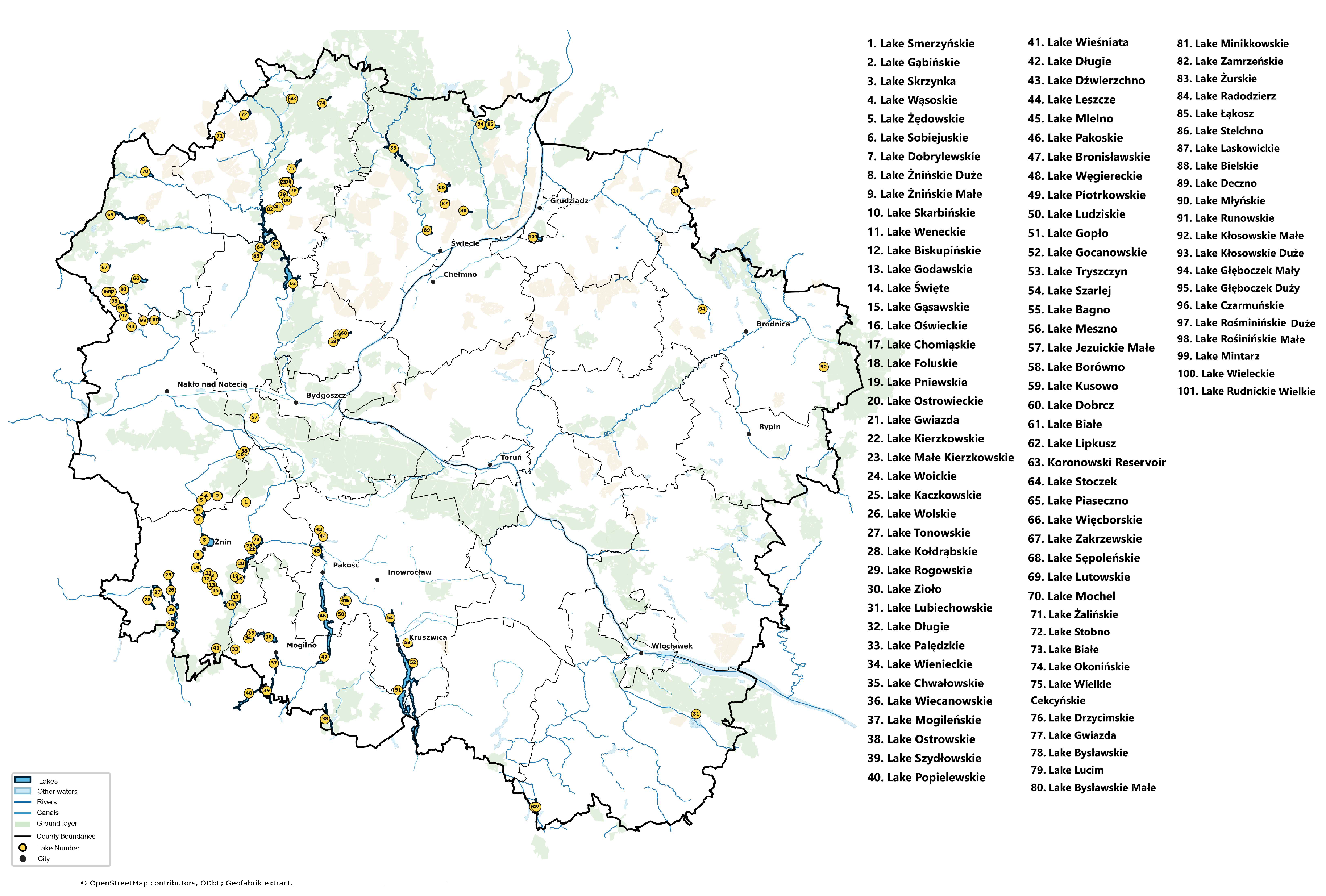
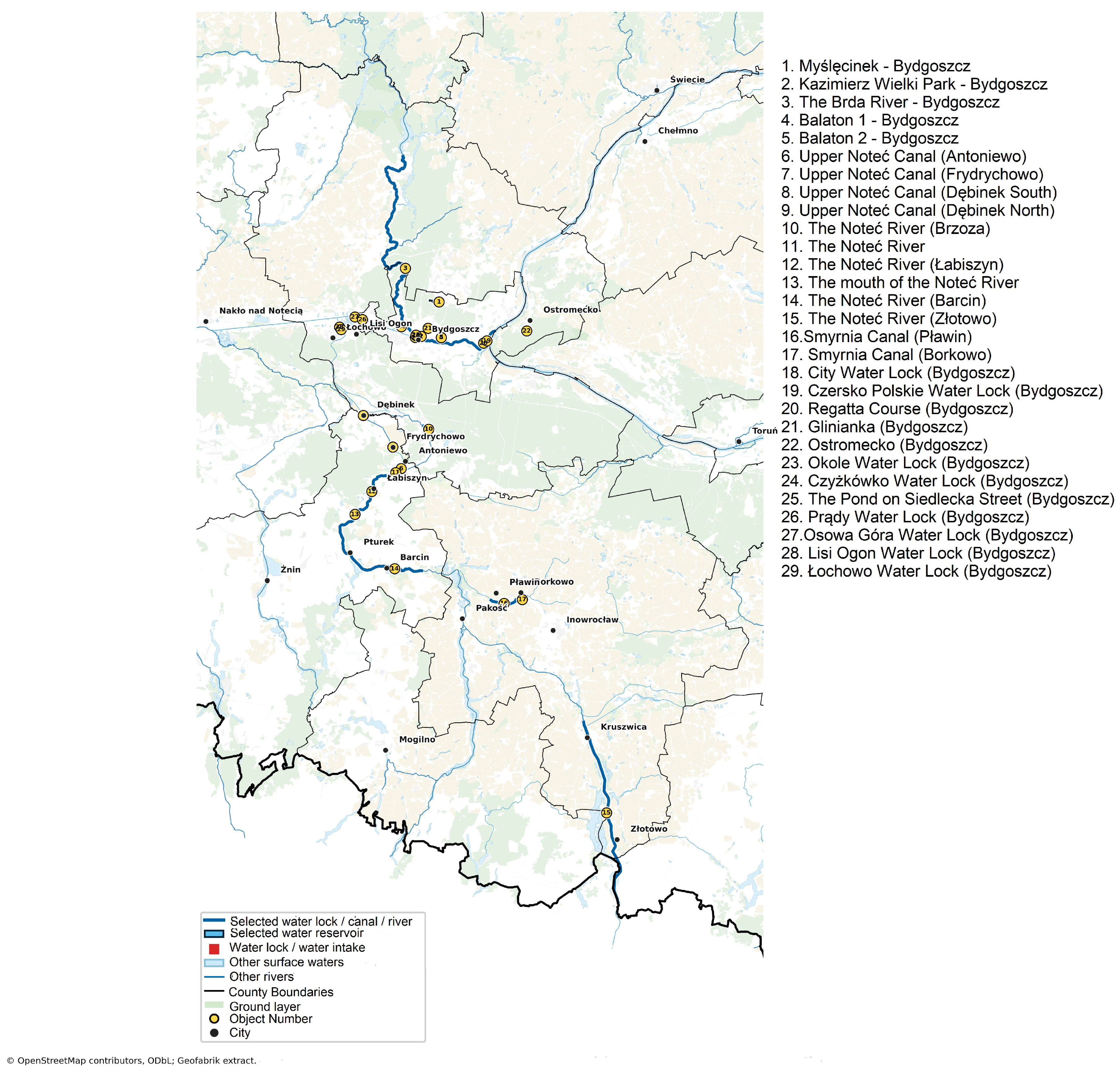
This study was conducted on freshwater samples collected from lakes and rivers located in the Kuyavian–Pomeranian Voivodeship (north–central Poland) (Figure 1 and Figure 2). Water sampling was performed during the vegetation seasons of 2022 and 2023. Sampling sites were selected to represent different types of freshwater ecosystems and varying degrees of anthropogenic impact (Figure 1 and Figure 2). The sites included both lakes and watercourses located in different environmental settings. Samples were collected from wild bathing sites as well as organized and protected recreational areas. In addition, some water bodies were surrounded by agricultural land, whereas others were located in the vicinity of livestock farms, representing different types and intensities of anthropogenic pressure. Surface water samples were collected aseptically into sterile containers and transported to the laboratory under refrigerated conditions for immediate microbiological analysis.

2.2. Isolation and Identification of Enterococcus spp.

Isolation of enterococci was performed using standard culture-based methods recommended for the detection of fecal indicator bacteria. Water samples were filtered through sterile membrane filters with a pore size of 0.45 µm (Merck, Darmstadt, Germany). Following filtration, the membrane filters were placed on a selective medium, bile esculin azide agar (Merck). After incubation under aerobic conditions at 35–37 °C for 24–48 h, colonies with morphology typical for Enterococcus spp. were selected for further analysis.
Presumptive isolates were confirmed as members of the genus Enterococcus based on matrix-assisted laser desorption ionization time-of-flight mass spectrometry (MALDI-TOF MS) (Bruker, Billerica, MA, USA) according to the manufacturer’s instructions. To preserve the research material, single colonies of identified microorganisms were transferred to Eppendorf tubes with Brain Heart Infusion broth (BHI, Becton-Dickinson, Franklin Lakes, NJ, USA) and 15.0% glycerol (Avantor, Gliwice, Poland) and frozen at −80 °C.

2.3. Preparation of Strains

The tested strains were inoculated onto Columbia agar containing 5% sheep blood (CAB, Graso, Starogard Gdański, Poland) and incubated for 24 h at 37 °C. A single colony was subcultured onto CAB, incubated under the same conditions, and used for further testing.

2.4. Antimicrobial Susceptibility Testing (AST)

Antimicrobial susceptibility of the confirmed Enterococcus isolates was determined using the disk diffusion method on Mueller–Hinton agar (MHA) (Becton Dickinson), in accordance with the recommendations of the European Committee on Antimicrobial Susceptibility Testing (EUCAST, version 15.0). Bacterial suspensions were adjusted to the 0.5 McFarland standard and inoculated uniformly onto the agar surface.
The following antimicrobial agents were tested: ampicillin (AMP, 10 µg), imipenem (IMP, 10 µg), ciprofloxacin (CIP, 5 µg), levofloxacin (LEV, 5 µg), norfloxacin (NOR, 10 µg), gentamicin (CN, 120 µg; high-level resistance), streptomycin (S, 300 µg; high-level resistance), teicoplanin (TEC, 30 µg), vancomycin (VA, 5 µg), quinupristin–dalfopristin (QDA, 15 µg; tested only for E. faecium), eravacycline (ERV, 20 µg), tigecycline (TGC, 15 µg), linezolid (LZD, 10 µg), and nitrofurantoin (F, 100 µg; tested only for E. faecalis).
Plates were incubated at 35 °C for 18 ± 2 h; for glycopeptides, incubation was extended to 24 h. Zones of inhibition were measured in millimeters and interpreted according to EUCAST breakpoints (v. 15) [24]. Enterococcus faecalis ATCC 29212 was used as a quality control strain.

2.5. Detection of Virulence Genes by Multiplex PCR

Genotypic characterization of Enterococcus isolates was performed by detection of selected virulence genes using multiplex polymerase chain reaction (PCR). Genomic DNA was extracted from overnight cultures using thermal method [25].
Multiplex PCRs were designed to detect a total of ten virulence and biofilm formation-associated genes—namely agg (aggregation substance); ace (adhesion to collagen); efaAfs (endocarditis-associated antigen); gelE (gelatinase); hyl (hyaluronidase); srtA (sortase A); ebpA, ebpB, and ebpC (endocarditis and biofilm-associated pili); and pil (pili)—according to Stępień-Pyśniak et al. [26] with own modification (Table 1). The E. faecalis ATCC 29212 strain was used as positive control. A negative control containing nuclease-free water instead of template DNA was included in each PCR run.

2.6. Microtiter Plate Biofilm Production Assay

The ability of Enterococcus isolates to form biofilms was evaluated using a microtiter plate assay. Briefly, overnight bacterial cultures were diluted in Tryptic Soy Broth (TSB) (bioMérieux, Marcy-l’Étoile, France) and inoculated into sterile 96-well polystyrene microplates (Profilab, Warsaw, Poland). After incubation under static conditions at 37 °C for 24 h, non-adherent cells were removed by washing with phosphate-buffered saline (PBS) (Avantor). Adherent biofilms were fixed and stained with 0,1% crystal violet solution (Avantor). Then, crystal violet was removed and wells were washed with water until colorless washings. The plates were dried and then 200 μL of ethanol (Stanlab, Lublin, Poland) was added. After 5 min of shaking (400 RPM, at room temperature) absorbance at 570 nm (Abs570) was read in a Hidex Sense multidetection reader (HIDEX, Turku, Finland). The average Abs570 value, obtained from triplicates for each strain, was determined. The classification of strains as strong, moderate, or weak biofilm producers was based on a comparison of their absorbance values (A) with those of the negative control (K−), following the method described by Stepanović et al. [27]. Cut-off values were established: [K−] < [A]  ≤  2 × [K−]—weak biofilm producer; 2 × [K−] < [A]  ≤  4 × [K−]—moderate biofilm producer, 4 × [K−] < [A]—strong biofilm producer.

2.7. Statistical Analysis

Statistical analysis of the biofilm-related results was performed using Excel and Statistica 13.3 (TIBCO Software Inc., San Ramon, CA, USA). To assess the presence of significant differences between weak and moderate biofilm-forming strain groups, one-way analysis of variance (ANOVA) followed by Tukey’s multiple comparison test was applied. A p-value ≤ 0.05 was considered statistically significant.

3. Results

3.1. Occurrence of Enterococcus spp. in Freshwater Samples

During two consecutive sampling campaigns conducted in 2022 and 2023, a total of 328 freshwater samples were collected from lake and river ecosystems. Identification of the 96 Enterococcus isolates revealed the presence of 12 species (Table 2). In some cases, more than one strain was isolated from a single sample. The most frequently isolated species was E. faecalis (23; 24.0%), while the second species was E. hirae (21; 21.9%). One strain was isolated from each of the following species: E. avium, E. columbae, E. moraviensis, E. canis species (Table 2).

3.2. Antibiotic Susceptibility of Enterococcus spp. Strains

The AST results showed different resistance profiles; indeed, 31/96 (32.3%) of the isolated enterococci were resistant to at least one antimicrobial tested in the present study. Of these, 18/31 (58.1%) isolates were resistant to one antibiotic, 4/31 (12.9%) to two antibiotics, 3/96 (9.7%) to three antibiotics, and 1/31 (3.2%) E. aquimarinus isolate to eight antibiotics. One E. faecalis isolate and one E. aquimarinus isolate were classified as multidrug-resistant (MDR), as they were resistant to at least one antibiotic from at least three classes of antimicrobial agents active against the respective species.
The strains tested exhibited resistance to the majority of the antibiotics included in the study (Table 3). Resistance to fluoroquinolones was confirmed in one isolate each of E. faecalis (4.3%) and E. aquimarinus (50%). The remaining species were susceptible to ciprofloxacin. One (50%) E. aquimarinus isolate, one (4.8%) E. hirae isolate, and five (21.7%) E. faecalis isolates exhibited the high-level gentamicin resistance phenotype (HLGR). High-level streptomycin resistance (HLSR) was observed only in E. faecalis (10; 43.5%) and E. hirae (4; 19.0%). Notably, three (13.0%) isolates of E. faecalis described above showed the HLAR phenotype, indicating resistance to high concentrations of aminoglycosides.
For vancomycin, resistance was detected in all 19 (100.0%) E. casseliflavus isolates, which is associated with their intrinsic resistance to this antibiotic (VanC phenotype). Additionally, non-susceptibility was observed in two (9.5%) E. hirae isolates, one (50%) E. aquimarinus isolate, and one (4.3%) E. faecalis isolate. Five (31.3%) E. faecium isolates were resistant to quinupristin–dalfopristin. No resistance was detected to teicoplanin, linezolid, or nitrofurantoin.

3.3. Prevalence of Virulence-Associated Genes

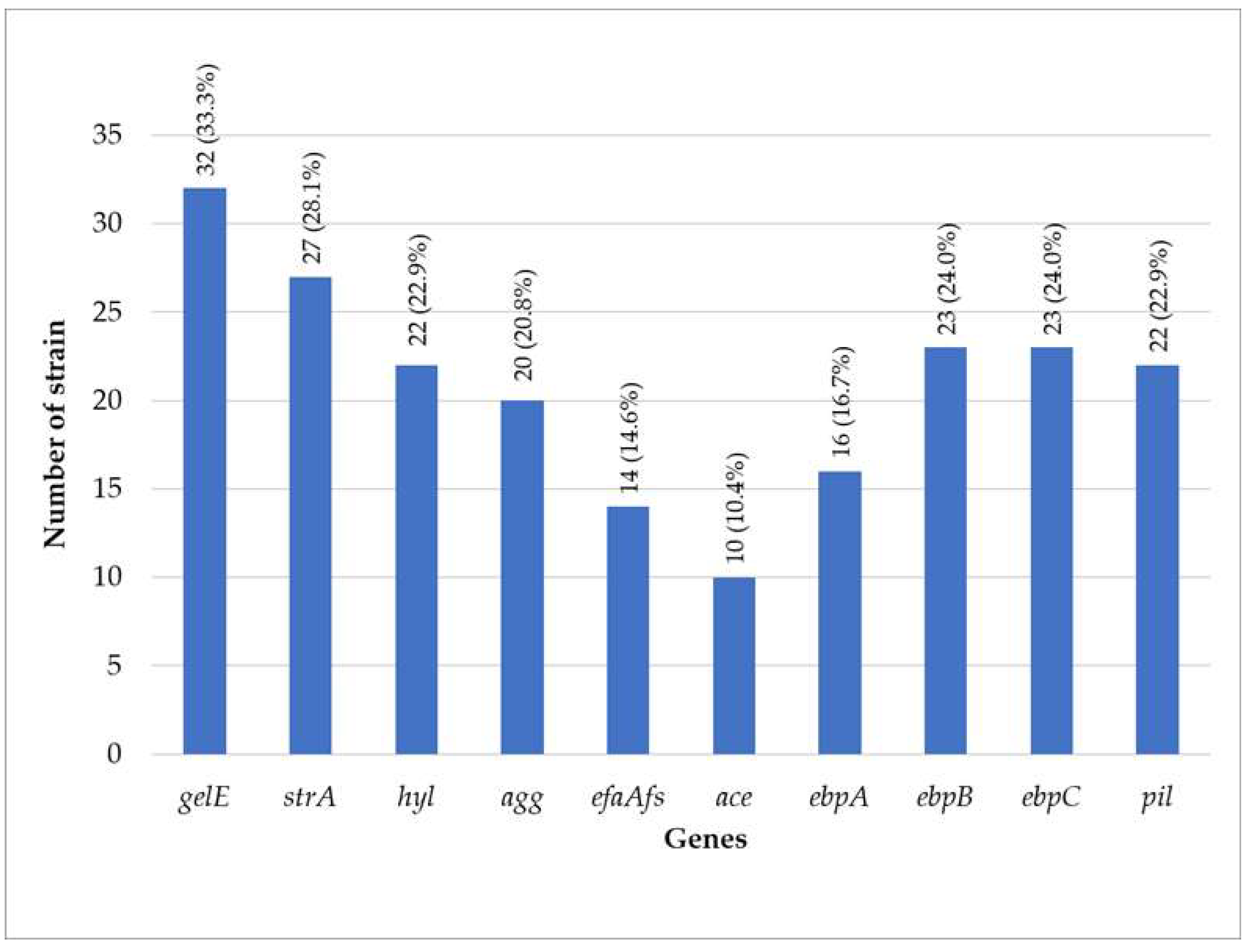
Among the 96 isolates, the most prevalent virulence gene was gelE, detected in 32 (33.3%) strains, followed by srtA in 27 (28.1%) and hyl in 22 (22.9%). The genes agg, efaAfs, and ace were identified in 20 (20.8%), 14 (14.6%), and 10 (10.4%) isolates, respectively (Figure 3).
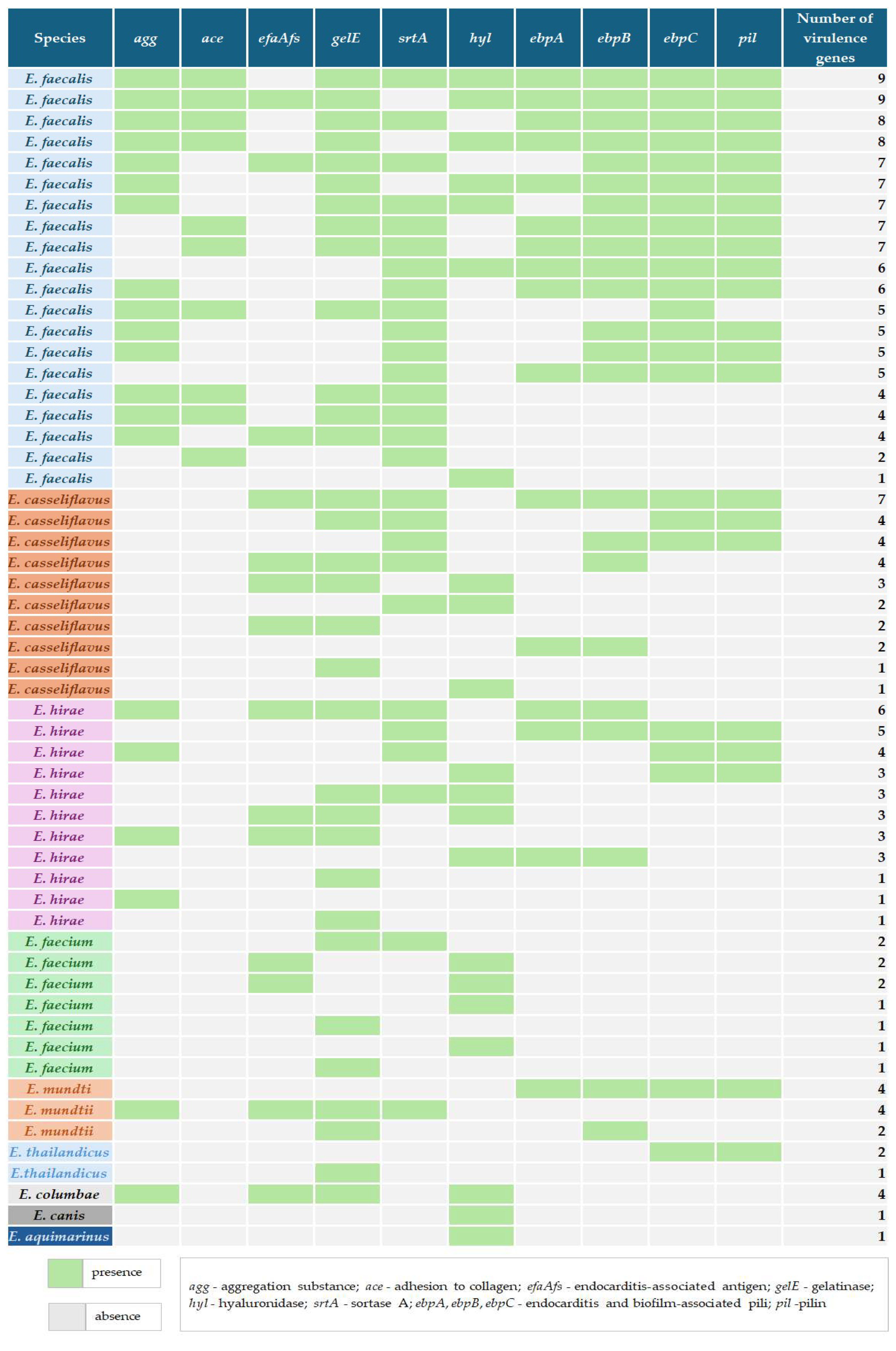
The detailed distribution of virulence genes is presented in Table 4 and Figure 4. At the species level, E. faecalis showed the highest prevalence of several determinants, including gelE (13; 56.5%), srtA (16; 69.6%), agg (14; 60.9%), and hyl (7; 30.4%), while ace was detected exclusively in this species (10; 43.5%). The gelE gene was also relatively frequent in E. hirae (6; 28.6%) and E. casseliflavus (6; 21.6%), whereas hyl was present in E. faecium (5; 31.3%) and E. hirae (4; 19.0%). The efaAfs gene occurred at lower frequencies across several species, including E. casseliflavus (4; 21%) and E. faecalis (3; 13%). Overall, the distribution of virulence genes varied across species, with some genes (e.g., gelE and srtA) widely distributed, while others, such as ace, showed a more restricted, species-specific occurrence. The least prevalent virulence determinant was ace, detected exclusively in E. faecalis isolates, where it occurred in 10/96 strains (10.4%). Analysis of the cumulative number of virulence determinants revealed that isolates carrying one virulence gene were the most common profile (13/96 isolates, 13.5%). Profiles containing seven virulence genes were identified in 6/96 isolates (6.3%). The highest virulence burden was observed exclusively in E. faecalis, in which isolates carrying 8–9 virulence genes were detected (4/23 isolates, 17.4%). In contrast, most non-faecalis Enterococcus species carried only one to four virulence determinants (Table 4 and Figure 4).

3.4. Biofilm-Forming Ability of Enterococcus Isolates

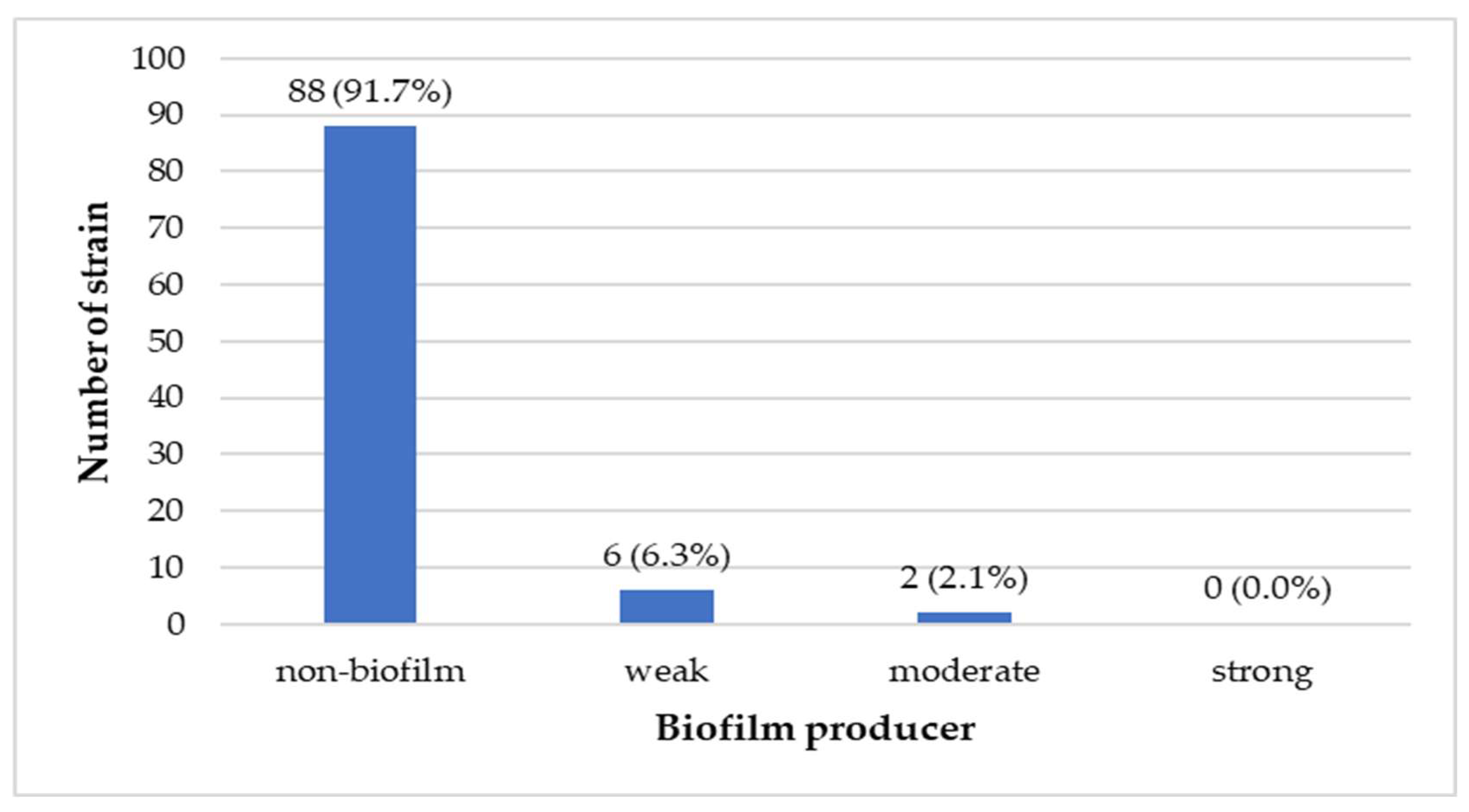
Assessment of biofilm formation using the microtiter plate assay showed that 88 isolates (91.7%) were not capable of biofilm formation (Figure 5). Biofilm-forming ability was confirmed in 8 (8.3%) strains representing the following species: E. faecalis, E. casseliflavus, E. hirae, E. faecium, and E. columbae. Among them, six (6.3%) strains were classified as weak biofilm producers, whereas two (2.1%) strains were identified as moderate biofilm producers. Within the studied strains, weak biofilm production was observed in two E. faecalis strains (8.7%), two E. casseliflavus strains (10.5%), one E. faecium strain (6.3%), and one E. columbae strain (100%). In contrast, moderate biofilm-forming ability was detected only in two E. hirae strains (9.5%) (Figure 5).
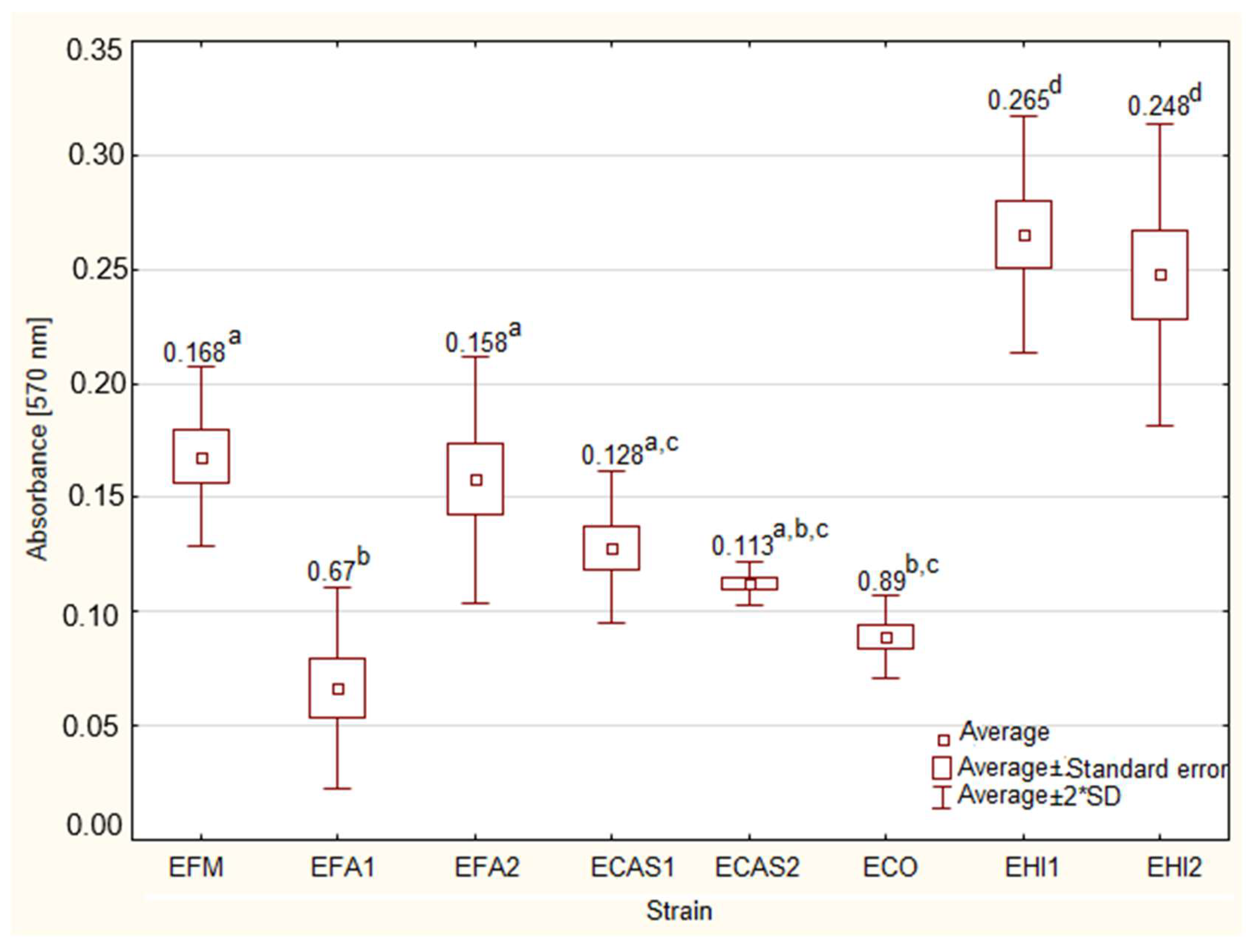
Statistically significant differences in biofilm-forming intensity were observed among the Enterococcus spp. strains with weak and moderate biofilm formation (p ≤ 0.05) (Figure 6). The mean absorbance recorded for the E. faecium strain was 0.168, whereas the E. columbae strain exhibited a mean absorbance value of 0.89. Among the E. faecalis strains, the mean absorbance values were 0.67 and 0.158. For E. casseliflavus strains, the mean absorbance values were 0.128 and 0.113. For E. hirae strains with moderate biofilm formation the mean absorbance value was 0.248 and 0.265.

4. Discussion

Bacteria belonging to the genus Enterococcus are commonly used as indicators of water quality and fecal contamination in aquatic environments. Maintaining high water quality, both for drinking and recreational purposes, is crucial for the protection of public health. The presence of Enterococcus spp. in samples collected from water reservoirs may indicate the occurrence of pathogens associated with gastrointestinal and other infections [28,29]. Various Enterococcus species may be isolated from surface waters depending on multiple environmental factors, including water temperature, precipitation intensity, proximity to industrial areas, and urbanization level [30]. The increasing prevalence of antibiotic-resistant bacteria constitutes a serious threat to both human and animal health, as antimicrobial therapy remains one of the primary approaches for treatment of bacterial infections. Moreover, natural environments, including surface waters, are currently considered both reservoirs and transmission routes of antimicrobial-resistant bacteria and resistance genes [31]. This observation is consistent with the One Health concept, which emphasizes the close interconnection between environmental, animal, and human health, as well as the role of environmental ecosystems in the dissemination of antimicrobial resistance.

4.1. Prevalence of Enterococcus spp. in Freshwater

The results obtained in the present study demonstrated considerable diversity among Enterococcus isolates recovered from freshwater lakes. Among the 12 identified species, E. hirae, E. faecalis, and E. casseliflavus were the most prevalent. Similar observations have been reported in environmental studies investigating aquatic ecosystems, where Enterococcus populations were shown to vary depending on anthropogenic pressure, wastewater contamination, and climatic conditions [32,33,34]. Although E. faecalis and E. faecium are traditionally regarded as the species most frequently associated with human infections, non-faecalis enterococci, including E. hirae and E. casseliflavus, are increasingly recognized as important environmental species capable of harboring virulence and antimicrobial resistance determinants [3].

4.2. Antibiotic Resistance Among Enterococcus spp. Strains

In our study 32.3% isolates exhibited resistance to at least one antimicrobial agent, confirming that freshwater ecosystems may serve as reservoirs of resistant enterococci. Particularly noteworthy was the identification of MDR isolates of E. faecalis and E. aquimarinus. Similar findings were described by Mbanga et al. [32], who demonstrated the presence of antimicrobial resistance genes and mobile genetic elements in environmental Enterococcus isolates recovered from wastewater-associated aquatic environments.
In a study conducted by Lazăr et al. [31], antimicrobial resistance profiles were evaluated among Enterococcus spp., including E. faecalis, E. faecium, E. mundtii, and E. casseliflavus isolates recovered from four lakes in Romania. The authors demonstrated that all 20 analyzed isolates were susceptible to penicillin, ampicillin, and levofloxacin. Similar findings were obtained in the present study. Among the 96 analyzed isolates, only one strain exhibited resistance to ampicillin, whereas resistance to levofloxacin was identified in two isolates. These observations may suggest that β-lactams and fluoroquinolones remain relatively effective therapeutic options against environmental Enterococcus strains.
High-level aminoglycoside resistance phenotypes, including HLGR and HLSR, were predominantly associated with E. faecalis isolates. This observation is clinically important because high-level resistance to aminoglycosides abolishes the synergistic effect between aminoglycosides and cell wall-active agents commonly used in enterococcal therapy. Previous studies also demonstrated that environmental enterococci may carry resistance determinants similar to those detected in clinical isolates [35,36].
Resistance to vancomycin was observed primarily among E. casseliflavus isolates, which is consistent with the intrinsic VanC phenotype characteristic of this species. However, resistance to vancomycin detected in several isolates of E. hirae, E. faecalis, and E. aquimarinus may indicate environmental dissemination of glycopeptide-resistant enterococci. Environmental transmission of vancomycin-resistant enterococci has been increasingly documented in aquatic ecosystems impacted by wastewater discharge and anthropogenic activity [37]. Importantly, none of the isolates analyzed in the present study exhibited resistance to linezolid or teicoplanin, suggesting that resistance to last-resort antimicrobials remains relatively uncommon among enterococci isolated from the investigated lakes.
In the present study, only two (2.1%) Enterococcus spp. isolates were classified as MDR. However, studies conducted by other authors suggest that the occurrence of MDR enterococci in aquatic environments may be substantially higher and strongly dependent on the source of isolation and geographical location. For example, Gotkowska-Płachta [14], in a study performed on Enterococcus spp. isolated from wastewater and the Łyna River in Olsztyn, demonstrated a very high prevalence of MDR strains. Among 283 analyzed isolates, 94.0% of wastewater-derived strains and 69.7% of river isolates were classified as MDR. Similarly, Iweriebor et al. [38], investigating Enterococcus spp. isolated from hospital wastewater in South Africa, reported high-level resistance to different antimicrobial agents, with several isolates exhibiting resistance to 8–10 antibiotics simultaneously. In the present study, only one isolate, identified as E. aquimarinus, demonstrated resistance to a comparably high number of antimicrobials, being non-susceptible to 8 out of 14 tested antibiotics. These findings indicate that the prevalence of MDR enterococci in aquatic environments may vary considerably worldwide and is likely influenced by environmental conditions, anthropogenic pressure, wastewater management practices, and the degree of exposure to antimicrobial agents. In particular, wastewater environments, especially hospital-associated effluents, appear to constitute important reservoirs of highly resistant Enterococcus strains [39,40].

4.3. Occurrence of Virulence-Associated Genes Among Enterococcus spp.

The analysis of virulence-associated genes revealed that gelE, srtA, and hyl were the most prevalent determinants among the investigated isolates. The predominance of gelE observed in the present study is in agreement with previous reports demonstrating that gelatinase is one of the most frequently detected virulence factors among environmental enterococci [35]. Gelatinase contributes to degradation of host tissues, biofilm maturation, and persistence under adverse environmental conditions, thereby increasing bacterial survival and adaptability [41]. The highest accumulation of virulence determinants was identified in E. faecalis isolates, which frequently carried multiple genes simultaneously, including agg, gelE, srtA, ebpA, ebpB, ebpC, and pil. Moreover, 8–9 virulence factors were detected in four strains of this species. These findings are consistent with previous studies demonstrating that E. faecalis possesses a broader repertoire of virulence-associated genes compared with other Enterococcus species [35,41,42]. The ace gene, encoding a collagen-binding protein involved in adhesion, was detected exclusively in E. faecalis isolates, confirming its species-associated distribution reported previously in clinical and environmental strains.

4.4. Biofilm Formation by Enterococcus spp. Strains

Despite the relatively frequent occurrence of virulence-associated genes related to adhesion and colonization, only a small proportion of isolates demonstrated phenotypic biofilm-forming ability. Most strains were classified as non-biofilm producers, whereas weak and moderate biofilm production was detected only in several isolates representing E. faecalis, E. casseliflavus, E. hirae, E. faecium, and E. columbae. None of the strains demonstrated strong biofilm-forming ability. Similar discrepancies between the presence of virulence genes and actual biofilm formation have been previously reported, suggesting that environmental conditions strongly influence gene expression and biofilm development [41].
Although the biofilm-forming ability of the investigated isolates was relatively limited, environmental biofilms remain highly important from an ecological and epidemiological perspective. Biofilm structures facilitate bacterial persistence in aquatic ecosystems and may promote horizontal transfer of antimicrobial resistance genes between environmental microorganisms [43]. Consequently, freshwater biofilms may act as reservoirs of opportunistic pathogens and antimicrobial resistance determinants, representing a potential threat to both environmental and public health.
Overall, the findings of the present study highlight the ecological significance of Enterococcus spp. isolated from freshwater lakes and confirm that surface waters may serve as reservoirs of potentially pathogenic and antimicrobial-resistant enterococci. The coexistence of antimicrobial resistance, virulence-associated determinants, and biofilm-related traits supports the One Health concept, emphasizing the interconnectedness of environmental, animal, and human health [37,44]. Continuous monitoring of freshwater ecosystems is therefore essential for assessing microbiological water quality and limiting the environmental dissemination of resistant bacteria.

5. Conclusions

The present study demonstrated that freshwater environments can harbor diverse Enterococcus species, including isolates exhibiting antimicrobial resistance and carrying multiple virulence-associated genes. Among the analyzed isolates, E. faecalis showed the highest prevalence of virulence determinants, particularly those associated with adhesion and biofilm formation. Although most isolates did not demonstrate phenotypic biofilm-forming ability, weak and moderate biofilm producers were identified, indicating that some environmental enterococci possess traits that may facilitate persistence under environmental conditions.
The coexistence of antimicrobial resistance, virulence-associated determinants, and biofilm-related traits among the analyzed isolates highlights the genetic diversity of environmental enterococci and their potential relevance as indicators of antimicrobial resistance dissemination in aquatic ecosystems. These findings contribute to a better understanding of the occurrence and distribution of resistance- and virulence-related traits in freshwater environments and support further investigation of environmental enterococci within the One Health framework. Incorporating phenotypic and genotypic characterization into environmental monitoring programs may provide additional insights into the ecology of antimicrobial resistance in surface waters.

Author Contributions

Conceptualization, K.G.-B., N.W.-K. and K.S.; methodology, K.G.-B., N.W.-K. and K.S.; validation, A.B. and J.C.; formal analysis, K.G.-B. and K.S.; investigation, A.S. and S.S.; resources, K.S. and K.G.-B.; data curation, A.S. and S.S.; writing—original draft preparation, N.W.-K., A.B. and J.C.; writing—review and editing, K.G.-B., N.W.-K. and K.S.; visualization, N.W.-K. and A.B.; supervision, K.S.; project administration, K.S. and N.W.-K.; funding acquisition, K.S. and K.G.-B. All authors have read and agreed to the published version of the manuscript.

Funding

This research received no external funding.

Institutional Review Board Statement

Not applicable.

Informed Consent Statement

Not applicable.

Data Availability Statement

The data presented in this study are available on request from the corresponding author.

Conflicts of Interest

The authors declare no conflicts of interest.

References

  1. Byappanahalli, M.N.; Nevers, M.B.; Korajkic, A.; Staley, Z.R.; Harwood, V.J. Enterococci in the environment. Microbiol. Mol. Biol. Rev. 2012, 76, 685–706. [Google Scholar] [CrossRef] [PubMed]
  2. Solaiman, S.; Patterson, R.; Davey, K.; Katz, Y.; Payne-Sturges, D.; Sapkota, A.R.; Micallef, S.A. Effects of season and water type on the distribution and antimicrobial resistance of Enterococcus faecalis and Ent. faecium from surface and reclaimed water. J. Appl. Microbiol. 2022, 133, 477–487. [Google Scholar] [CrossRef] [PubMed]
  3. García-Solache, M.; Rice, L.B. The Enterococcus: A model of adaptability to its environment. Clin. Microbiol. Rev. 2019, 32, e00058-18. [Google Scholar] [CrossRef] [PubMed]
  4. Willett, J.L.E.; Dunny, G.M. Insights into ecology, pathogenesis, and biofilm formation of Enterococcus faecalis from functional genomics. Microbiol. Mol. Biol. Rev. 2025, 89, e00081-23. [Google Scholar] [CrossRef] [PubMed]
  5. Davis, B.C.; Keenum, I.; Calarco, J.; Liguori, K.; Milligan, E.; Pruden, A.; Harwood, V.J. Towards the standardization of Enterococcus culture methods for waterborne antibiotic resistance monitoring: A critical review of trends across studies. Water Res. X 2022, 17, 100161. [Google Scholar] [CrossRef] [PubMed]
  6. Adegoke, A.A.; Madu, C.E.; Reddy, P.; Fatunla, O.K.; Stenström, T.A.; Okoh, A.I. Virulence and antibiotic resistance genes in Enterococcus from wastewater for reuse and their health impact. Microorganisms 2025, 13, 1045. [Google Scholar] [CrossRef] [PubMed]
  7. Șchiopu, P.; Toc, D.A.; Colosi, I.A.; Costache, C.; Ruospo, G.; Berar, G.; Gălbău, Ș.-G.; Ghilea, A.C.; Botan, A.; Pană, A.-G.; et al. An Overview of the Factors Involved in Biofilm Production by the Enterococcus Genus. Int. J. Mol. Sci. 2023, 24, 11577. [Google Scholar] [CrossRef] [PubMed]
  8. Barnes, A.M.T.; Frank, K.L.; Dunny, G.M. Enterococcal Endocarditis: Hiding in Plain Sight. Front. Cell. Infect. Microbiol. 2021, 11, 722482. [Google Scholar] [CrossRef] [PubMed]
  9. Berendonk, T.U.; Manaia, C.M.; Merlin, C.; Fatta-Kassinos, D.; Cytryn, E.; Walsh, F.; Buergmann, H.; Sørum, H.; Norström, M.; Pons, M.-N.; et al. Tackling antibiotic resistance: The environmental framework. Nat. Rev. Microbiol. 2015, 13, 310–317. [Google Scholar] [CrossRef] [PubMed]
  10. Karkman, A.; Do, T.T.; Walsh, F.; Virta, M.P.J. Antibiotic-resistance genes in waste water. Trends Microbiol. 2018, 26, 220–228. [Google Scholar] [CrossRef] [PubMed]
  11. Silva, V.; Freitas, C.; Ribeiro, J.; Igrejas, G.; Poeta, P. Comparative analysis of antibiotic resistance and biofilm formation in Enterococcus spp. across One Health domains. FEMS Microbes 2025, 6, xtaf005. [Google Scholar] [CrossRef] [PubMed]
  12. Ibekwe, A.M.; Obayiuwana, A.C.; Murinda, S.E. Enterococcus Species and Their Antimicrobial Resistance in an Urban Watershed Affected by Different Anthropogenic Sources. Water 2024, 16, 116. [Google Scholar] [CrossRef]
  13. Amos, G.C.A.; Ploumakis, S.; Zhang, L.; Hawkey, P.M.; Gaze, W.H.; Wellington, E.M.H. The widespread dissemination of integrons throughout bacterial communities in a riverine system. ISME J. 2018, 12, 681–691. [Google Scholar] [CrossRef] [PubMed]
  14. Gotkowska-Płachta, A. The prevalence of virulent and multidrug-resistant enterococci in river water and in treated and untreated municipal and hospital wastewater. Int. J. Environ. Res. Public Health 2021, 18, 563. [Google Scholar] [CrossRef] [PubMed]
  15. Manaia, C.M.; Rocha, J.; Scaccia, N.; Marano, R.; Radu, E.; Biancullo, F.; Cerqueira, F.; Fortunato, G.; Iakovides, I.C.; Zammit, I.; et al. Antibiotic resistance in wastewater treatment plants: Tackling the black box. Environ. Int. 2018, 115, 312–324. [Google Scholar] [CrossRef] [PubMed]
  16. Harwood, V.J.; Staley, C.; Badgley, B.D.; Borges, K.; Korajkic, A. Microbial source tracking markers for detection of fecal contamination in environmental waters: Relationships between pathogens and human health outcomes. FEMS Microbiol. Rev. 2014, 38, 1–40. [Google Scholar] [CrossRef] [PubMed]
  17. Karkman, A.; Pärnänen, K.; Larsson, D.G.J. Fecal pollution can explain antibiotic resistance gene abundances in anthropogenically impacted environments. Nat. Commun. 2019, 10, 80. [Google Scholar] [CrossRef] [PubMed]
  18. Abe, K.; Nomura, N.; Suzuki, S. Biofilms: Hot spots of horizontal gene transfer (HGT) in aquatic environments, with a focus on a new HGT mechanism. FEMS Microbiol. Ecol. 2020, 96, fiaa031. [Google Scholar] [CrossRef] [PubMed]
  19. Skowron, K.; Borkowski, W.; Wiktorczyk-Kapischke, N.; Budzyńska, A.; Wilk, M.; Czuba, J.; Fiderewicz, J.; Skonieczna-Kurpiel, J.; Grudlewska-Buda, K. Occurrence and assessment of antibiotic resistance and virulence of Enterococcus spp. strains isolated from fecal samples of wild animals. Sci. Rep. 2025, 15, 16957. [Google Scholar] [CrossRef] [PubMed]
  20. One Health High-Level Expert Panel (OHHLEP); Adisasmito, W.B.; Almuhairi, S.; Behravesh, C.B.; Bilivogui, P.; Bukachi, S.A.; Casas, N.; Becerra, N.C.; Charron, D.F.; Chaudhary, A.; et al. One Health: A new definition for a sustainable and healthy future. PLoS Pathog. 2022, 18, e1010537. [Google Scholar] [CrossRef] [PubMed]
  21. Aslam, B.; Khurshid, M.; Arshad, M.I.; Muzammil, S.; Rasool, M.; Yasmeen, N.; Shah, T.; Chaudhry, T.H.; Rasool, M.H.; Shahid, A.; et al. Antibiotic Resistance: One Health One World Outlook. Front. Cell. Infect. Microbiol. 2021, 11, 771510, Erratum in Front. Cell. Infect. Microbiol. 2024, 14, 1488430. https://doi.org/10.3389/fcimb.2024.1488430.. [Google Scholar] [CrossRef] [PubMed]
  22. Humboldt-Dachroeden, S.; Mantovani, A. Assessing Environmental Factors within the One Health Approach. Medicina 2021, 57, 240. [Google Scholar] [CrossRef] [PubMed]
  23. Pitt, S.J.; Gunn, A. The One Health Concept. Br. J. Biomed. Sci. 2024, 81, 12366. [Google Scholar] [CrossRef] [PubMed]
  24. European Committee on Antimicrobial Susceptibility Testing (EUCAST). Breakpoints Tables for Interpretation of MICs and Zones Diameters. Version 15.0. 2025. Available online: https://www.eucast.org/fileadmin/src/media/PDFs/EUCAST_files/Breakpoint_tables/v_15.0_Breakpoint_Tables.pdf (accessed on 10 December 2025).
  25. Kovačević, J.; Mesak, L.R.; Allen, K.J. Occurrence and characterization of Listeria spp. in ready-to-eat retail foods from Vancouver, British Columbia. Food Microbiol. 2012, 30, 372–378. [Google Scholar] [PubMed]
  26. Stępień-Pyśniak, D.; Hauschild, T.; Kosikowska, U.; Dec, M.; Urban-Chmiel, R. Biofilm formation capacity and presence of virulence factors among commensal Enterococcus spp. from wild birds. Sci. Rep. 2019, 9, 11204. [Google Scholar] [CrossRef] [PubMed]
  27. Stepanovic, S.; Vukovic, D.; Dakic, I.; Savić, B.; Švabić-Vlahović, M. A modified microtiter-plate test for quantification of staphylococcal biofilm formation. J. Microbiol. Methods 2000, 40, 175–179. [Google Scholar] [CrossRef] [PubMed]
  28. Martzy, R.; Kolm, C.; Brunner, K.; Mach, R.L.; Krska, R.; Šinkovec, H.; Sommer, R.; Farnleitner, A.H.; Reischer, G.H. A loop-mediated isothermal amplification (LAMP) assay for the rapid detection of Enterococcus spp. in water. Water Res. 2017, 122, 62–69. [Google Scholar] [CrossRef] [PubMed]
  29. Holcomb, D.A.; Stewart, J.R. Microbial indicators of fecal pollution: Recent progress and challenges in assessing water quality. Curr. Environ. Health Rep. 2020, 7, 311–324. [Google Scholar] [CrossRef] [PubMed]
  30. Cho, S.; Jackson, C.R.; Frye, J.G. The prevalence and antimicrobial resistance phenotypes of Salmonella, Escherichia coli and Enterococcus sp. in surface water. Lett. Appl. Microbiol. 2020, 71, 3–25. [Google Scholar] [CrossRef] [PubMed]
  31. Lazăr, V.; Gheorghe, I.; Curutiu, C.; Savin, I.; Marinescu, F.; Cristea, V.C.; Dobre, D.; Popa, G.L.; Chifiriuc, M.C.; Popa, M.I. Antibiotic resistance profiles in cultivable microbiota isolated from some romanian natural fishery lakes included in Natura 2000 network. BMC Vet. Res. 2021, 17, 52. [Google Scholar] [CrossRef] [PubMed]
  32. Mbanga, J.; Amoako, D.G.; Abia, A.L.K.; Allam, M.; Ismail, A.; Essack, S.Y. Genomic Analysis of Enterococcus spp. Isolated from a Wastewater Treatment Plant and Its Associated Waters in Umgungundlovu District, South Africa. Front. Microbiol. 2021, 12, 648454. [Google Scholar] [CrossRef] [PubMed]
  33. Korajkic, A.; McMinn, B.R.; Staley, Z.R.; Ahmed, W.; Harwood, V.J. Antibiotic-Resistant Enterococcus Species in Marine Habitats: A Review. Curr. Opin. Environ. Sci. Health 2020, 19, 92–100. [Google Scholar] [CrossRef] [PubMed]
  34. Zaheer, R.; Cook, S.R.; Barbieri, R.; Goji, N.; Cameron, A.; Petkau, A.; Polo, R.O.; Tymensen, L.; Stamm, C.; Song, J.; et al. Surveillance of Enterococcus spp. reveals distinct species and antimicrobial resistance diversity across a One-Health continuum. Sci. Rep. 2020, 10, 3937, Erratum in Sci. Rep. 2020, 10, 13401. https://doi.org/10.1038/s41598-020-69044-5.. [Google Scholar] [CrossRef] [PubMed]
  35. Geraldes, C.; Tavares, L.; Gil, S.; Oliveira, M. Enterococcus Virulence and Resistant Traits Associated with Its Permanence in the Hospital Environment. Antibiotics 2022, 11, 857. [Google Scholar] [CrossRef] [PubMed]
  36. Dželalija, M.; Kvesić, M.; Novak, A.; Fredotović, Ž.; Kalinić, H.; Šamanić, I.; Ordulj, M.; Jozić, S.; Goić Barišić, I.; Tonkić, M.; et al. Microbiome profiling and characterization of virulent and vancomycin-resistant Enterococcus faecium from treated and untreated wastewater, beach water and clinical sources. Sci. Total Environ. 2023, 858, 159720. [Google Scholar] [CrossRef] [PubMed]
  37. Larsson, D.G.J.; Flach, C.F. Antibiotic resistance in the environment. Nat. Rev. Microbiol. 2022, 20, 257–269. [Google Scholar] [CrossRef] [PubMed]
  38. Iweriebor, B.C.; Gaqavu, S.; Obi, L.C.; Nwodo, U.U.; Okoh, A.I. Antibiotic susceptibilities of Enterococcus species isolated from hospital and domestic wastewater effluents in Alice, Eastern Cape Province of South Africa. Int. J. Environ. Res. Public Health 2015, 12, 4231–4246. [Google Scholar] [CrossRef] [PubMed]
  39. Gotkowska-Płachta, A.; Gołaś, I. The importance of enterococci in the monitoring of fecal pollution in river water in forests and urban areas. Water 2023, 15, 3708. [Google Scholar] [CrossRef]
  40. Ghazvinian, M.; Asgharzadeh Marghmalek, S.; Gholami, M.; Amir Gholami, S.; Amiri, E.; Goli, H.R. Antimicrobial resistance patterns, virulence genes, and biofilm formation in enterococci strains collected from different sources. BMC Infect. Dis. 2024, 24, 274. [Google Scholar] [CrossRef] [PubMed]
  41. Ch’ng, J.H.; Chong, K.K.L.; Lam, L.N.; Wong, J.J.; Kline, K.A. Biofilm-associated infection by enterococci. Nat. Rev. Microbiol. 2019, 17, 82–94, Erratum in Nat. Rev. Microbiol. 2019, 17, 124. https://doi.org/10.1038/s41579-018-0128-7.. [Google Scholar] [CrossRef] [PubMed]
  42. Padilla, C.; Lobos, O. Virulence, bacterocin genes and antibacterial susceptibility in Enterococcus faecalis strains isolated from water wells for human consumption. SpringerPlus 2013, 2, 43. [Google Scholar] [CrossRef] [PubMed]
  43. Flores-Vargas, G.; Bergsveinson, J.; Lawrence, J.R.; Korber, D.R. Environmental biofilms as reservoirs for antimicrobial resistance. Front. Microbiol. 2021, 12, 766242. [Google Scholar] [CrossRef] [PubMed]
  44. Franklin, A.M.; Weller, D.L.; Durso, L.M.; Bagley, M.; Davis, B.C.; Frye, J.G.; Grim, C.J.; Ibekwe, A.M.; Jahne, M.A.; Keely, S.P.; et al. A one health approach for monitoring antimicrobial resistance: Developing a national freshwater pilot effort. Front. Water 2024, 6, 1359109. [Google Scholar] [CrossRef] [PubMed]
Figure 1. Surface water sampling sites in the Kuyavian–Pomeranian Voivodeship (lakes). Cartographic data: OpenStreetMap, ODbL 1.0 license.
Biology 15 01056 g001
Figure 2. Surface water sampling sites in the Kuyavian–Pomeranian Voivodeship (other sampling sites in the Kuyavian–Pomeranian Voivodeship). Cartographic data: OpenStreetMap, ODbL 1.0 license.
Biology 15 01056 g002
Figure 3. The number of Enterococcus spp. strains in which selected genes encoding virulence factors were confirmed.
Biology 15 01056 g003
Figure 4. Heatmap of VG profiles from Enterococcus isolates investigated in the present study. Isolates are grouped according to Enterococcus species.
Biology 15 01056 g004
Figure 5. Evaluation of biofilm formation of Enterococcus spp. strains using crystal violet assay.
Biology 15 01056 g005
Figure 6. Results of the assessment of the biofilm formation ability by biofilm-forming strains (EFM—E. faecium, EFA—E. faecalis, ECAS—E. casseliflavus, ECO—E. columbae, EHI—E. hirae, SD—Standard deviation); a,b,c,d—values marked with different letters differ statistically significantly.
Biology 15 01056 g006
Table 1. PCR amplification conditions used for detection of virulence genes in Enterococcus spp.
GenesProduct Size [bp]PCR Program
agg510Initial denaturation—94 °C/2 min
16 cycles: denaturation—94 °C/30 s
annealing—58 °C/45 s
extension—72 °C/45 s
19 cycles: denaturation—94 °C/30 s
annealing—50 °C/45 s
extension—72 °C/45 s
Final extension—72 °C/7 min
ace320
efaAfs705Initial denaturation—95 °C/15 min
35 cycles: denaturation—94 °C/1 min
annealing—49 °C/1 min
extension—72 °C/1 min
Final extension—72 °C/7 min
gelE419
ebpC487Initial denaturation—95 °C/15 min
35 cycles: denaturation—94 °C/1 min
annealing—53 °C/1 min
extension—72 °C/1 min
Final extension—72 °C/7 min
pil620
ebpA613Initial denaturation—95 °C/15 min
35 cycles: denaturation—94 °C/1 min
annealing—55 °C/1 min
extension—72 °C/1 min
Final extension—72 °C/7 min
ebpB504
asa1375Initial denaturation—94 °C/2 min
16 cycles: denaturation—94 °C/30 s
annealing—55 °C/45 s
extension—72 °C/45 s
19 cycles: denaturation—94 °C/30 s
annealing—48 °C/45 s
extension—72 °C/45 s
Final extension—72 °C/7 min
hyl276
srtA612
Table 2. Distribution of Enterococcus species isolated from freshwater samples.
Species2022 [n (%)]2023 [n (%)]Total [n (%)]
E. faecalis9 (23.1)14 (25.0)23 (24.0)
E. hirae6 (15.4)15 (26.8)21 (21.9)
E. casseliflavus8 (20.5)11 (19.6)19 (19.8)
E. faecium11 (28.2)5 (8.9)16 (16.7)
E. mundtii4 (10.3)2 (3.6)6 (6.3)
E. thailandicus0 (0.0)3 (5.4)3 (3.1)
E. durans1 (2.6)1 (1.8)2 (2.1)
E. aquimarinus0 (0.0)2 (3.6)2 (2.1)
E. avium0 (0.0)1 (1.8)1 (1.0)
E. columbae0 (0.0)1 (1.8)1 (1.0)
E. moraviensis0 (0.0)1 (1.8)1 (1.0)
E. canis0 (0.0)1 (1.8)1 (1.0)
TOTAL395796
Table 3. Antimicrobial resistance among Enterococcus isolates.
Antimicrobial AgentE. faecium
n = 16 (%)
E. faecalis
n = 23 (%)
E. casseliflavus
n = 19 (%)
E. hirae
n = 21 (%)
E. moraviensis
n = 1 (%)
E. aquimarinus
n = 2 (%)
Total n = 96 (%) **
Ampicillin0 (0.0)0 (0.0)1 (5.3)0 (0.0)0 (0.0)0 (0.0)1 (1.0)
Imipenem0 (0.0)0 (0.0)1 (5.3)0 (0.0)0 (0.0)0 (0.0)1 (1.0)
Ciprofloxacin0 (0.0)1 (4.3)0 (0.0)0 (0.0)0 (0.0)1 (50.0)2 (2.1)
Levofloxacin0 (0.0)1 (4.3)0 (0.0)0 (0.0)0 (0.0)1 (50.0)2 (2.1)
Norfloxacin0 (0.0)1 (4.3)0 (0.0)0 (0.0)0 (0.0)1 (50.0)2 (2.1)
Gentamicin 0 (0.0)5 (21.7)0 (0.0)1 (4.8)0 (0.0)1 (50.0)7 (7.3)
Streptomycin 0 (0.0)10 (43.5)0 (0.0)4 (19.0)0 (0.0)0 (0.0)14 (14.6)
Vancomycin0 (0.0)1 (4.3)19 (100.0)2 (9.5)0 (0.0)1 (50.0)25 (26.0)
Eravacycline0 (0.0)1 (4.3)0 (0.0)2 (9.5)0 (0.0)0 (0.0)2 (2.1)
Tigecycline0 (0.0)1 (4.3)0 (0.0)0 (0.0)0 (0.0)0 (0.0)1 (1.0)
Quinupristin–dalfopristin *5 (31.3)NANANANANA5 (5.2)
* Quinupristin–dalfopristin tested only for E. faecium. ** Only species with at least one resistant isolate are presented.
Table 4. Distribution of virulence-associated genes among Enterococcus species.
Virulence GeneE. faecalis
n = 23 (%)
E. hirae
n = 21 (%)
E. casseliflavus
n = 19 (%)
E. faecium
n = 16 (%)
E. mundtii
n = 6 (%)
E. canis
n = 1 (%)
E. columbae
n = 1 (%)
E. thailandicus
n = 3 (%)
E. aquimarinus
n = 2 (%)
Total
n = 96 (%)
srtA16 (69.6)4 (19.0)5 (26.3)1 (6.25)1 (16.7)0 (0.0)0 (0.0)0 (0.0)0 (0.0)27 (28.1)
efaAfs3 (13.0)3 (14.3)4 (21.0)2 (12.5)1 (16.7)0 (0.0)1 (100.0)0 (0.0)0 (0.0)14 (14.6)
ace10 (43.5)0 (0.0)0 (0.0)0 (0.0)0 (0.0)0 (0.0)0 (0.0)0 (0.0)0 (0.0)10 (10.4)
agg14 (60.9)4 (19.0)0 (0.0)0 (0.0)1 (16.7)0 (0.0)1 (100.0)0 (0.0)0 (0.0)20 (20.8)
gelE13 (56.5)6 (28.6)6 (21.6)3 (18.8)2 (33.3)0 (0.0)1 (100.0)1 (33.3)0 (0.0)32 (33.3)
hyl7 (30.4)4 (19.0)3 (15.8)5 (31.3)0 (0.0)1 (100.)1 (100.0)0 (0.0)1 (50.0)22 (22.9)
ebpA10 (43.5)3 (14.3)2 (10.5)0 (0.0)1 (16.7)0 (0.0)0 (0.0)0 (0.0)0 (0.0)16 (16.7)
ebpB13 (56.5)3 (14.3)5 (26.3)0 (0.0)2 (33.3)0 (0.0)0 (0.0)0 (0.0)0 (0.0)23 (24.0)
ebpC15 (65.2)3 (14.3)3 (15.8)0 (0.0)1 (16.7)0 (0.0)0 (0.0)1 (33.3)0 (0.0)23 (24.0)
pil14 (60.9)3 (14.3)3 (15.8)0 (0.0)1 (16.7)0 (0.0)0 (0.0)1 (33.3)0 (0.0)22 (22.9)
1 VG1 (4.3)3 (14.3)2 (10.5)4 (25.0)0 (0.0)1 (100.0)0 (0.0)1 (33.3)1 (50.0)13 (13.5)
2 VGs1 (4.3)0 (0.0)3 (15.8)3 (18.8)1 (16.7)0 (0.0)0 (0.0)1 (33.3)0 (0.0)9 (9.4)
3 VGs0 (0.0)5 (23.8)1 (5.3)0 (0.0)0 (0.0)0 (0.0)0 (0.0)0 (0.0)0 (0.0)6 (6.2)
4 VGs3 (13.0)1 (4.8)3 (15.8)0 (0.0)2 (33.3)0 (0.0)1 (100.0)0 (0.0)0 (0.0)10 (10.4)
5 VGs4 (17.4)1 (4.8)0 (0.0)0 (0.0)0 (0.0)0 (0.0)0 (0.0)0 (0.0)0 (0.0)5 (5.2)
6 VGs2 (8.7)1 (4.8)0 (0.0)0 (0.0)0 (0.0)0 (0.0)0 (0.0)0 (0.0)0 (0.0)3 (3.1)
7 VGs5 (21.7)0 (0.0)1 (5.3)0 (0.0)0 (0.0)0 (0.0)0 (0.0)0 (0.0)0 (0.0)6 (6.2)
8–9 VGs4 (17.4)0 (0.0)0 (0.0)0 (0.0)0 (0.0)0 (0.0)0 (0.0)0 (0.0)0 (0.0)4 (4.2)
VG—virulence gene.
Disclaimer/Publisher’s Note: The statements, opinions and data contained in all publications are solely those of the individual author(s) and contributor(s) and not of MDPI and/or the editor(s). MDPI and/or the editor(s) disclaim responsibility for any injury to people or property resulting from any ideas, methods, instructions or products referred to in the content.