Article

Characterization and Immune Function of NOD1 in Snakehead (Channa argus)

1
School of Marine Science and Engineering, Qingdao Agricultural University, Qingdao 266109, China
2
Shandong Qidu Pharmaceutical Co., Ltd., Zibo 255400, China
3
Jining Fishery Development and Resource Conservation Center, Jining 272000, China
*
Authors to whom correspondence should be addressed.
These authors contributed equally to this work.
Biology 2026, 15(12), 942; https://doi.org/10.3390/biology15120942
Submission received: 14 April 2026 / Revised: 7 June 2026 / Accepted: 9 June 2026 / Published: 16 June 2026
(This article belongs to the Section Immunology)

Simple Summary

As the most representative member of the NLR family of pattern recognition receptors, NOD1 plays a crucial role in various biological processes, including innate and adaptive immunity in vertebrates. This study primarily aimed to characterize the role and response mechanisms of NOD1 in the innate immune response of snakehead. The NOD1 transcript was highly expressed in the mucosal immune tissues of snakehead, and its overexpression significantly induces NF-κB activity. Further studies revealed that NOD1 specifically recognizes iE-DAP and acts synergistically with the RIPK2 gene to enhance NF-κB promoter activity. Together, these findings reveal that NOD1 plays a vital role in the fish’s natural defense system. Understanding this process can help scientists develop better strategies to prevent disease outbreaks in farmed fish, reducing the need for antibiotics and supporting sustainable aquaculture.

Abstract

The innate immune response is a critical defense mechanism by which vertebrates recognize and eliminate invading pathogens. Pattern recognition receptors (PRRs) detect pathogen-associated molecular patterns and activate downstream signaling pathways. NOD1, a classic PRR of the NLR family, recruits the adaptor protein RIPK2 to initiate antibacterial signaling. In this study, we cloned and characterized the NOD1 gene from snakehead (Channa argus). Briefly, the full-length NOD1 cDNA is 2829 bp encoding 943 amino acids, showing high homology with Perciformes. The qPCR analysis revealed widespread NOD1 gene expression in various tissues, with significant upregulation in the gill (p < 0.05) and spleen (p < 0.05) following bacterial infection. Overexpression of the NOD1 gene activated the NF-κB signaling pathway in a dose- and time-dependent manner, and specifically responded to the bacterial ligand iE-DAP but not to other tested ligands. Furthermore, NOD1 synergized with the downstream adaptor RIPK2 to enhance NF-κB activity, and direct protein interaction between NOD1 and RIPK2 was confirmed by co-immunoprecipitation. Taken together, these findings demonstrate that snakehead NOD1 plays a critical role in the host antimicrobial immune response.

1. Introduction

Innate immunity serves as the first line of defense against invading pathogens, initiating with the recognition of pathogen-associated molecular patterns (PAMPs) by pattern recognition receptor (PRR) [1]. Among various PRR families, the NOD-like receptor (NLR) family is one of the largest in terms of the number of genes encoded in vertebrate genomes [2,3]. NLR proteins can sense a variety of pathogenic and non-pathogenic stimuli, thereby inducing distinct signal transduction mechanisms [4]. For instance, members of the NLR family are capable of activating multiple innate immune pathways, including nuclear factor-κB (NF-κB) signaling and cytokine production, as well as inducing innate immune cell death [5]. NLRs are involved in two main processes: firstly, they act as regulators of inflammation, and secondly, they form multi-protein inflammasome complexes. The formation of these complexes triggers the proteolytic cleavage of caspase-1 and induces pyroptosis [6,7].
The structural characteristics of NLRs are pivotal in determining their specificity and the extent of their activity, consisting of three core domains: an N-terminal variable domain for downstream signal transduction, a central nucleotide-binding domain (NBD) (also known as the NACHT domain) for oligomerization, and a C-terminal leucine-rich repeat (LRR) domain [8,9]. The LRR domain can sense PAMPs and damage-associated molecular patterns (DAMPs) in host cells to initiate innate immune responses [5]. Based on the characteristics of additional unique domains, NLRs are classified into five categories: NLRA, NLRB, NLRC, NLRP, and NLRX. Among them, NLRC1 contains a caspase recruitment domain (CARD) at the N-terminal region and is also known as NOD1 [10]. Specifically, as an intracellular pattern recognition receptor, NOD1 plays a critical role in antibacterial immunity by recognizing the bacterial peptidoglycan motif iE-DAP within the host cytosol after pathogens or their components gain access to the intracellular compartment, and binds to RIP2 through its CARD, thereby activating the downstream NF-κB and MAPK pathways [11].
The NOD1-RIP2 axis and its domains are conserved in teleosts [12,13], such as zebrafish (Danio rerio) [14,15], goldfish (Carassius auratus L.) [16,17], rainbow trout (Oncorhynchus mykiss) [18], sturgeon (Acipenser ruthenus) [19] and Chinese perch (Siniperca chuatsi) [20]. Snakehead (Channa argus) is an economically important freshwater fish species widely cultured in East Asia, especially in China. However, intensive farming has made it highly susceptible to bacterial diseases, among which Nocardia seriolae infection is the most prevalent [21]. N. seriolae is the primary pathogen causing nocardiosis in various fish species, leading to significant economic losses in the aquaculture industry [22,23]. Naturally infected snakehead exhibit typical clinical signs, including slow swimming and reduced food intake, skin wounds, anal swelling, ascites, and white granulomatous lesions in the liver, spleen, and kidney [24]. Gross pathological examination reveals numerous ivory-white nodules (1–5 mm in diameter) scattered on multiple internal organs, accompanied by visceral tissue swelling and hemorrhage. Histopathological examination shows typical systemic granuloma formation with well-defined granulomatous structures and extensive cellular infiltration [25]. Pathogenicity testing has demonstrated that N. seriolae infection in snakehead results in mortality rates ranging from 70% to 100% [22]. N. seriolae was selected as the representative pathogen in this study for several reasons. First, this bacterium is the primary etiological agent of nocardiosis in a wide range of economically important marine and freshwater fish species. It has been reported to infect approximately 42 species of marine and freshwater fish worldwide, including largemouth bass (Micropterus salmoides), spotted sea bass (Lateolabrax maculatus), large yellow croaker (Larimichthys crocea), Chinese rice-field eel (Monopterus albus), and snakehead [22,26], thereby causing substantial economic losses to the aquaculture industry [26]. Second, in snakehead, nocardiosis is one of the most prevalent and devastating bacterial diseases under intensive farming conditions, and N. seriolae infection in this species has been shown to induce mortality rates [22]. Third, N. seriolae has a long incubation period, a slow but progressive infection course, and a strong propensity to establish chronic granulomatous infections in host tissues [23], making it an excellent model for investigating host antibacterial immune mechanisms, particularly those involving intracellular pattern recognition receptors. Importantly, N. seriolae is a facultative intracellular parasite that can survive within macrophages, and NOD1 is also intracellular. Therefore, N. seriolae infection is particularly suitable for studying NOD1-mediated recognition and signaling. Furthermore, N. seriolae possesses the molecular basis for NOD1 recognition. As a Gram-positive actinobacterium belonging to the order Mycobacteriales, N. seriolae has a complex, multi-layered cell wall that contains abundant peptidoglycan as a structural component [27]. The peptidoglycan of Nocardia species, including N. seriolae, contains meso-diaminopimelic acid (meso-DAP) as a key constituent of the tetrapeptide side chain [28,29]. Meso-DAP is an essential component of bacterial peptidoglycan and serves as the direct structural precursor of γ-D-glutamyl-meso-diaminopimelic acid (iE-DAP), the minimal bioactive motif specifically recognized by NOD1 [30,31]. Furthermore, genomic analysis of N. seriolae has identified the complete set of genes required for peptidoglycan biosynthesis, including murE-2 (UDP-N-acetylmuramoyl-L-alanyl-D-glutamate-2,6-diaminopimelate ligase), responsible for adding meso-DAP to the peptidoglycan precursor, and lysA (diaminopimelate decarboxylase), involved in meso-DAP metabolism [32]. Thus, N. seriolae naturally produces the NOD1 ligand iE-DAP as an integral component of its cell wall peptidoglycan. This provides a direct molecular link between the pathogen and the NOD1 signaling pathway, justifying the use of N. seriolae infection as a relevant in vivo model to study NOD1-mediated antibacterial immune responses in snakehead. Given that NOD1 is a key intracellular pattern recognition receptor involved in antibacterial immunity, we selected N. seriolae as the representative pathogen to investigate the immune function of NOD1 in snakehead. Despite its commercial importance, the innate immune mechanisms, especially the NOD-like receptor family, remain poorly characterized in this species. Therefore, understanding the function of key immune genes such as the NOD1 gene in snakehead is essential for developing effective disease control strategies and supporting sustainable aquaculture. In the present study, the full-length NOD1 gene in snakehead was characterized, which possesses conserved CARD, NACHT, and LRR domains and exhibits close homology and conserved genomic synteny with Perciformes. The expression analysis revealed that NOD1 was highly expressed in the intestine and spleen, while N. seriolae challenge induced significant upregulation in the gill and spleen. Subcellular localization confirmed that NOD1 was mainly distributed in the cytoplasm. Functional verification demonstrated that NOD1 activated NF-κB in a dose- and time-dependent manner, and specifically responded to iE-DAP rather than other ligands. Furthermore, the complete structural domains of NOD1 were indispensable for maintaining its NF-κB-activating capacity, and only the full-length NOD1 exerted effective activation under iE-DAP stimulation. In addition, NOD1 collaborated with the downstream adaptor RIPK2 to synergistically enhance NF-κB activity, and direct protein interaction existed between NOD1 and RIPK2, though their binding affinity was not enhanced by iE-DAP or PGN stimulation.

2. Materials and Methods

2.1. Fish and Cell Lines

The snakeheads were purchased from Shandong Province, China, and weighed approximately 20 g each. Initially, the fish were acclimated in a recirculating freshwater system for 7 days prior to the bacterial infection experiments. During the acclimation period, the water quality parameters were maintained as follows: pH 7.8 ± 0.5, temperature 22 ± 0.5 °C, salinity 0 ppt, and dissolved oxygen 7.0 ± 0.5 mg/L. The snakeheads used in this study were purchased from a certified breeding farm, where routine pathogen and disease surveillance is conducted to ensure the health status of the stock. No obvious signs of disease were observed in the snakeheads during the acclimatization period; furthermore, no signs of disease were observed in the control group during the experiment. Nine organs/tissues including blood, brain, skin, liver, muscle, head kidney, gill, spleen and intestine were collected from four healthy snakeheads for constitutive expression analysis. Each tissue from each fish was divided into three equal portions, respectively. Thus, three independent pooled samples were obtained per tissue, each containing tissue portions from all four fish. For the in vivo stimulation experiments, liver, spleen, head kidney, skin, gill, and intestine samples were collected at 6, 12, 24, 48 and 72 h post-infection. N. seriolae was administered via intraperitoneal (IP) injection at a dose of 100 μL per fish (1 × 10⁸ CFU/mL, i.e., 1 × 10⁷ CFU per fish). The control fish received an equal volume of PBS. For the in vivo challenge experiments, 15 fish were sampled at each time point, for a total of 90 fish. During the 72 h post-infection period, all the fish were starved to avoid any effect of feeding on immune parameters.
Epithelioma papulosum cyprini (EPC) cells were maintained in M199 medium (Hyclone, Logan, UT, USA) containing 10% fetal bovine serum (FBS, HyClone, Logan, UT, USA) at 28 °C with 5% CO2. HEK 293T cells (human embryonic kidney 293T) were grown in DMEM (HyClone, Logan, UT, USA) with 10% FBS at 37 °C in a 5% CO2 environment. N. seriolae was inoculated in Ogawa medium at 28 °C with 180 rpm.
All the experiments were performed in accordance with local government regulations, and all procedures involving fish were conducted following the ethical guidelines established by the Animal Ethics Committee of Qingdao Agricultural University (QAU-IACUC-2023-021).

2.2. RNA Extraction, cDNA Synthesis and Quantitative Real-Time PCR

Total RNA was isolated using RNAiso reagent (Takara, Shiga-ken, Japan), and the first-strand cDNA template was synthesized using a PrimeScript™ 1st strand cDNA Synthesis Kit (Takara, Shiga-ken, Japan) according to the manufacturer’s instructions. Quantitative real-time PCR (qRT-PCR) was performed on a QuantStudiO™ 5 real-time PCR instrument (Thermo Fisher Scientific, Asheville, NC, USA) using the following cycling conditions: initial incubation at 95 °C for 2 min, followed by 40 cycles of 95 °C for 5 s, 60 °C for 30 s, and 65 °C for 5 s. The qPCR reaction mixture consisted of 5 μL of 2 × SYBR green pro taq HS premix (Accurate, Changsha, China), 1 μL of cDNA template, 0.4 μL of forward primer (10 μM), 0.4 μL of reverse primer (10 μM), and 3.2 μL of nuclease-free water. The specific primers are listed in Table 1. The relative expression levels were calculated using the 2−ΔΔCt method and normalized to β-actin gene.

2.3. Bioinformatic Analyses and Gene Cloning

The gene and protein sequences used in bioinformatics were compared with homologous sequences through the Basic Local Alignment Search Tool (BLAST) version 2.17.0 on the website of the National Center for Biotechnology Information (NCBI, http://blast.ncbi.nlm.nih.gov/Blast.cgi (accessed on 5 December 2024)). Multiple alignments of proteins were performed using Clustal W (version 2.1). A phylogenetic tree was constructed using the neighbor-joining (NJ) method in MEGA X software (version 10.2.4), with 1000 times bootstrap.
Each PCR mixture contained 12.5 μL of PrimerSTAR (Takara, Shiga-ken, Japan), 1.0 μL of cDNA, 1.0 μL of forward primer, 1.0 μL of reverse primer, and nuclease-free water to bring the final volume to 25 μL. Following amplification, the products were visualized via agarose gel electrophoresis, then purified with the FastPure® Gel DNA Extraction Mini Kit (Vazyme, Nanjing, China). Subsequently, they were ligated into the pMD18-T vector (Takara, Shiga-ken, Japan) and introduced into competent DH5α. Positive transformants were sequenced by Beijing Tsingke Biotech Co., Ltd. (Beijing, China), and the primer sequences are provided in Table 1.

2.4. Subcellular Localization

The ORF of NOD1 and RIPK2 genes were subcloned into the pEGFP-N2 vector to generate NOD1-GFP and RIPK2-GFP fluorescent fusion expression vectors. HEK 293T cells were plated into 24-well plates and incubated overnight. NOD1-GFP and RIPK2-GFP expression plasmids (1 μg/well) were transfected into the HEK 293T cells using Lipofectamine 2000 (Invitrogen, Carlsbad, CA, USA) according to the manufacturer’s protocol. At 24 h post-transfection (hpt), the cells were fixed with 4% paraformaldehyde for 10 min, and permeabilized with 1% Triton X-100 solution at room temperature for 10 min. DAPI was added to the coverslip for observation under a confocal laser scanning microscope (ZEISS, Jena, Germany).

2.5. Co-Immunoprecipitation (Co-IP) and Western Blotting

The ORFs of NOD1 and RIPK2 were inserted into the pcDNA3.1-myc-His(A) and p3×FLAG-CMV14 vectors with Myc and Flag tags, respectively. NOD1-Myc and RIPK2-Flag were transfected into HEK 293T cells by using Lipofectamine 2000. The cells were collected at 48 hpt and lysed 15 min at 4 °C in RIPA lysis buffer (Beyotime, Shanghai, China) supplemented with PMSF (Beyotime, Shanghai, China). After centrifugation, 40 μL mixed liquid was taken as the input sample. Anti-Myc or anti-Flag immunomagnetic beads (Beyotime, Shanghai, China) were added to the remaining samples and incubated overnight at 4 °C for Co-IP. Upon completion, the samples received 1× SDS loading buffer and were then boiled for 10 min. Subsequent separation of protein samples occurred via 8% or 12% SDS-PAGE gels, followed by transfer onto a 0.22 μm polyvinylidene fluoride membrane (Millipore, Jaffrey, NH, USA). The membrane, after being rinsed with PBST buffer, was blocked for 1 h at room temperature using 5% non-fat milk, then incubated overnight with primary antibody at a 1:5000 dilution. Following another PBST wash, the membrane was exposed to secondary antibody for 1 h. Lastly, visualization and detection were carried out with the ChemiDoc™ MP system (Bio-Rad, Irvine, CA, USA).

2.6. Luciferase Activity Assays

The luciferase reporter vectors of NF-κB, pRL-TK, and eukaryotic expression mutant plasmids of NOD, including NOD-ΔCARD, NOD-ΔLRRs, NOD-CARD, NOD-ΔNACHT, and NOD-LRRs, were constructed. A series of plasmid mixtures were transfected into EPC cells seeded in 24-well plates via Lipofectamine 2000 transfection reagent. The empty vector was employed as the control group, and the cells were stimulated with LPS (10 μg/mL), LTA (10 μg/mL), poly(I:C) (10 μg/mL), PGN (10 μg/mL), and iE-DAP (10 μg/mL) (stock concentration: 5 mg/mL for all ligands). LPS, LTA and PGN were purchased from Sigma-Aldrich (St. Louis, MO, USA), while poly(I:C) and iE-DAP were purchased from Invitrogen (Carlsbad, CA, USA). The cells in each well were collected at 24 hpt and washed once with PBS. Subsequently, 200 μL of Lysis Buffer (Beyotime, Shanghai, China) was added to lyse the cells. The luciferase activity was measured using the dual-luciferase reporter gene assay kit (Beyotime, Shanghai, China) with a microplate reader.

2.7. Statistical Analyses

Statistical analysis of the experimental data was performed with SPSS 20.0 software. Prior to statistical analysis, all data were checked for normality using the Shapiro–Wilk test and for homogeneity of variances using Levene’s test. The results met the assumptions for parametric tests. Student’s t-test was then employed for comparisons between two groups, and one-way ANOVA followed by Duncan’s multiple range test was used for multiple comparisons. Each experiment was conducted a minimum of three times, and the results are expressed as means ± standard error (SE). Differences reaching statistical significance are denoted by * p < 0.05.

3. Results

3.1. Identification of NOD1 in Snakehead

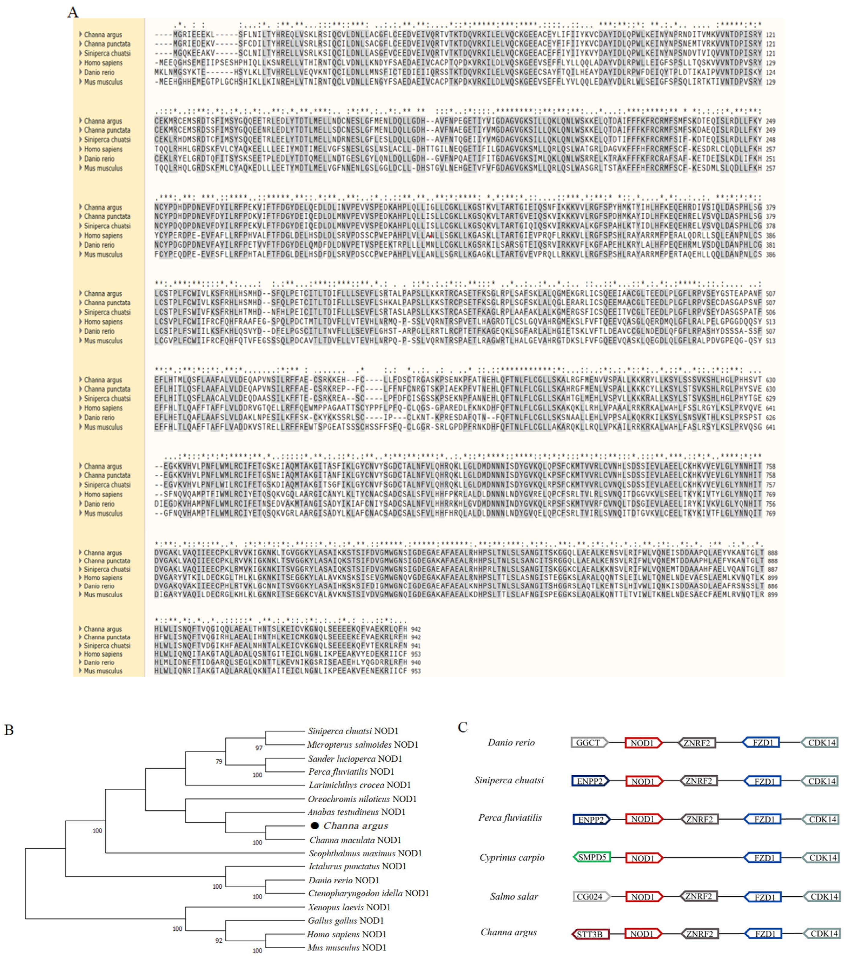
The NOD1 gene identified in snakehead contains an open reading frame of 2829 bp, encoding 943 amino acids. Multiple sequence alignment results showed that the snakehead NOD1 protein was highly similar to that of other teleosts. (Figure 1A). Phylogenetic analysis revealed that snakehead NOD1 protein shares high sequence similarity with NOD1 from other vertebrates, exhibiting particularly close homology with orthologs from Perciformes (Figure 1B). Moreover, the NOD1 locus is evolutionarily conserved and arranged in tandem with the ZNRF2 gene (Figure 1C).

3.2. Expression of the NOD1 Gene

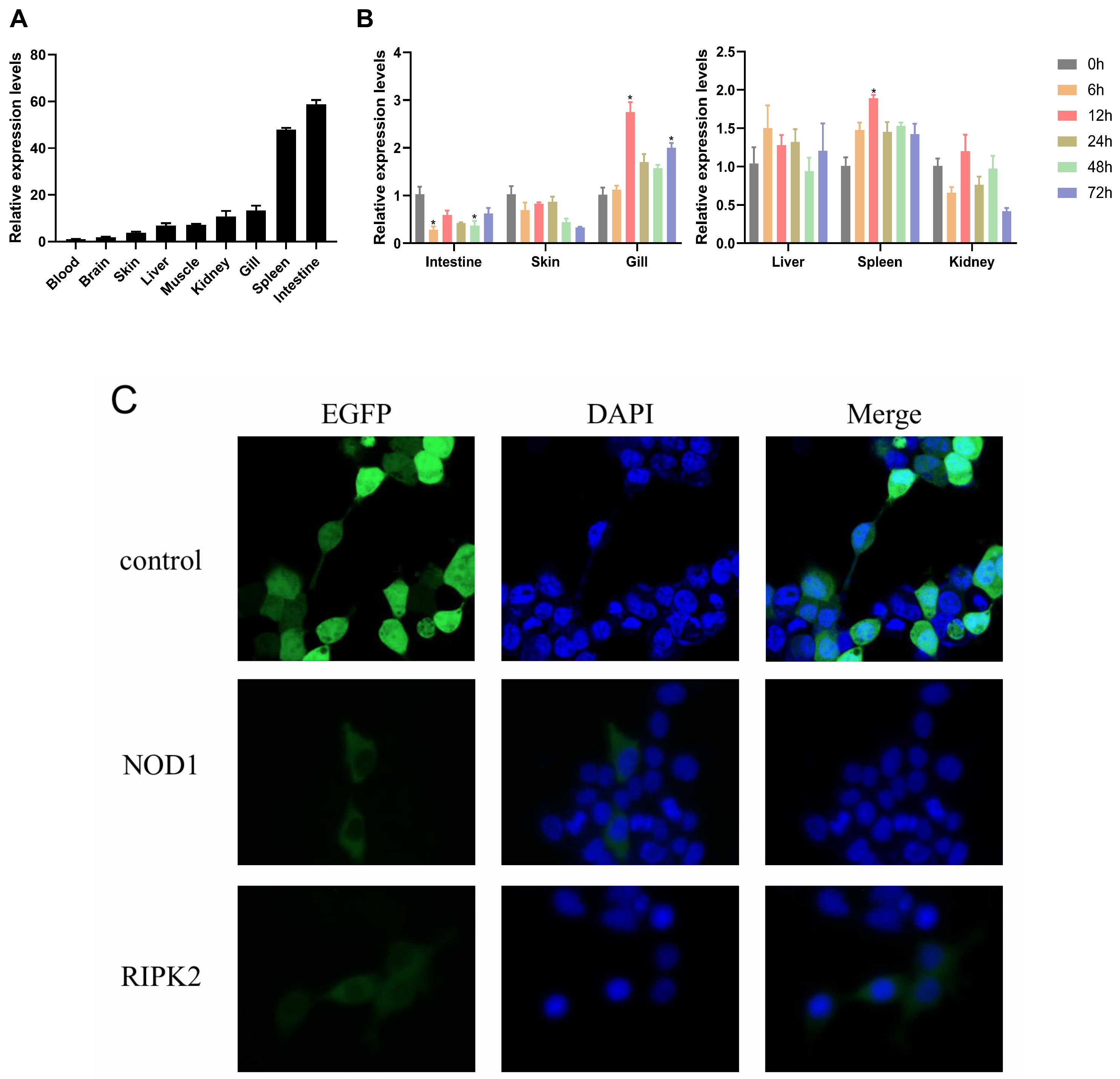
To explore the expression pattern of NOD1 in snakehead, qRT-PCR was performed to detect the transcription levels of NOD1. The results showed that NOD1 was expressed in the liver, spleen, head kidney, intestine, skin, gill, muscle, blood, and brain with variable expression levels. Notably, low expression levels were detected in the blood and brain, while high expression was observed in the intestine (58.8-fold), followed by the spleen (47.9-fold) (Figure 2A).
Following intraperitoneal injection of N. seriolae, liver, spleen, head kidney, intestine, skin, and gill samples were collected and the expression of NOD1 was analyzed by qRT-PCR. Upon N. seriolae infection, the transcription of NOD1 was significantly upregulated in the gill and spleen, whereas its expression was downregulated in the intestine, skin, and head kidney, but remained unchanged in the liver (Figure 2B).
To determine the subcellular localization of snakehead NOD1 protein, the ORF of NOD1 was cloned into the pEGFP-N2 vector with GFP tag. The recombinant plasmid NOD1-N2 and the empty pEGFP-N2 control plasmid were separately transfected into HEK 293T cells. At 24 hpt, fluorescence microscopic observation revealed that the GFP signal of the empty vector was uniformly distributed throughout the cytoplasm and nucleus. In contrast, the NOD1-GFP fusion protein was predominantly localized in the cytosolic (Figure 2C).

3.3. The Activation of NF-κB by NOD1

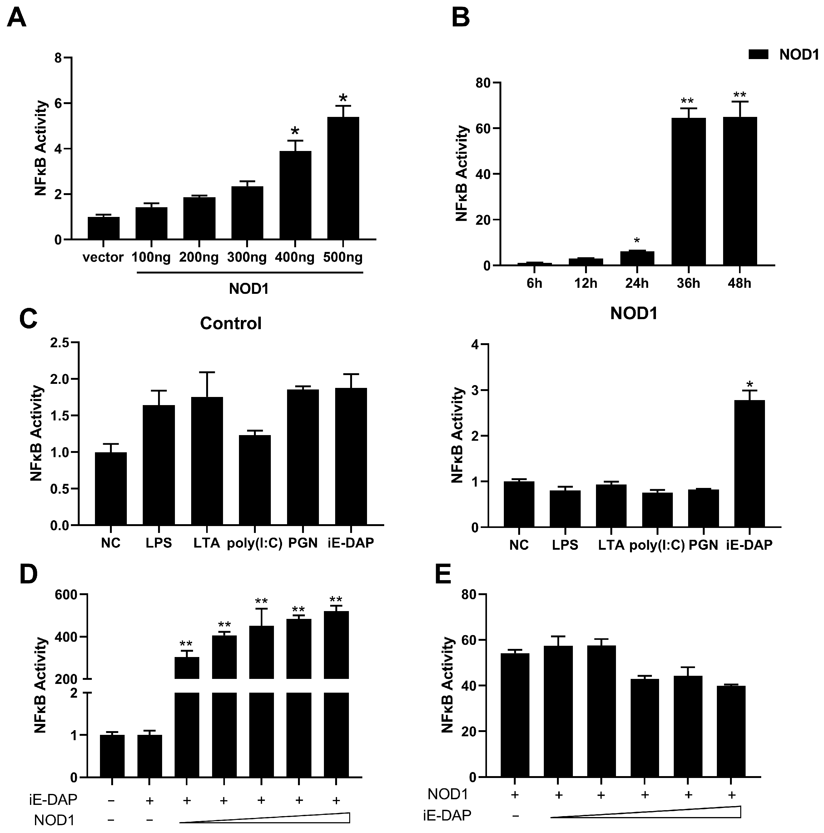
To investigate the PAMPs specifically recognized by snakehead NOD1 during antimicrobial immune responses, the NOD1 expression plasmid or empty control plasmid was co-transfected with the NF-κB luciferase reporter and the internal control plasmid pRL-TK into the EPC cells. At 24 hpt, NF-κB activity was examined following treatment with different stimuli, including poly(I:C), LPS, iE-DAP, PGN, and LTA, at various concentrations and time points. The results showed that NF-κB activity increased in a dose-dependent manner with the escalating dosage of the NOD1 expression plasmid (Figure 3A), and significant activation of NF-κB was observed at 48 h after transfection (Figure 3B). Compared with the control groups, only iE-DAP was capable of triggering NOD1-mediated NF-κB activation (Figure 3C), and this activating effect was further enhanced with the increase in NOD1 plasmid dosage (Figure 3D); the other ligands failed to induce notable NF-κB activation (Figure 3C). Furthermore, variations in iE-DAP concentration exerted no significant influence on NOD1-dependent NF-κB activation (Figure 3E).
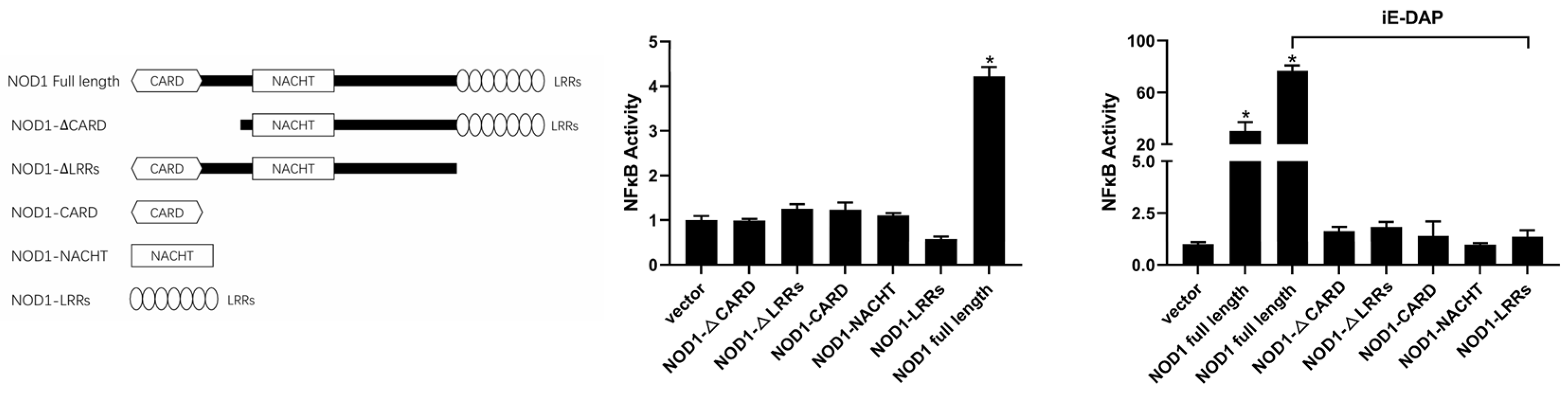
To further investigate the functional roles of distinct domains of snakehead NOD1 protein in regulating NF-κB activation, a series of eukaryotic expression plasmids carrying truncated NOD1 mutants were constructed (Figure 4). Dual-luciferase reporter assays demonstrated that any domain deletion completely abolished the ability of NOD1 to activate NF-κB. Moreover, individual overexpression of the NACHT domain or LRR domains failed to efficiently induce NF-κB activation (Figure 4). Upon stimulation with iE-DAP, significant NF-κB activation was only detected in the cells transfected with the full-length NOD1 (Figure 4).

3.4. Interaction of NOD1 and RIPK2

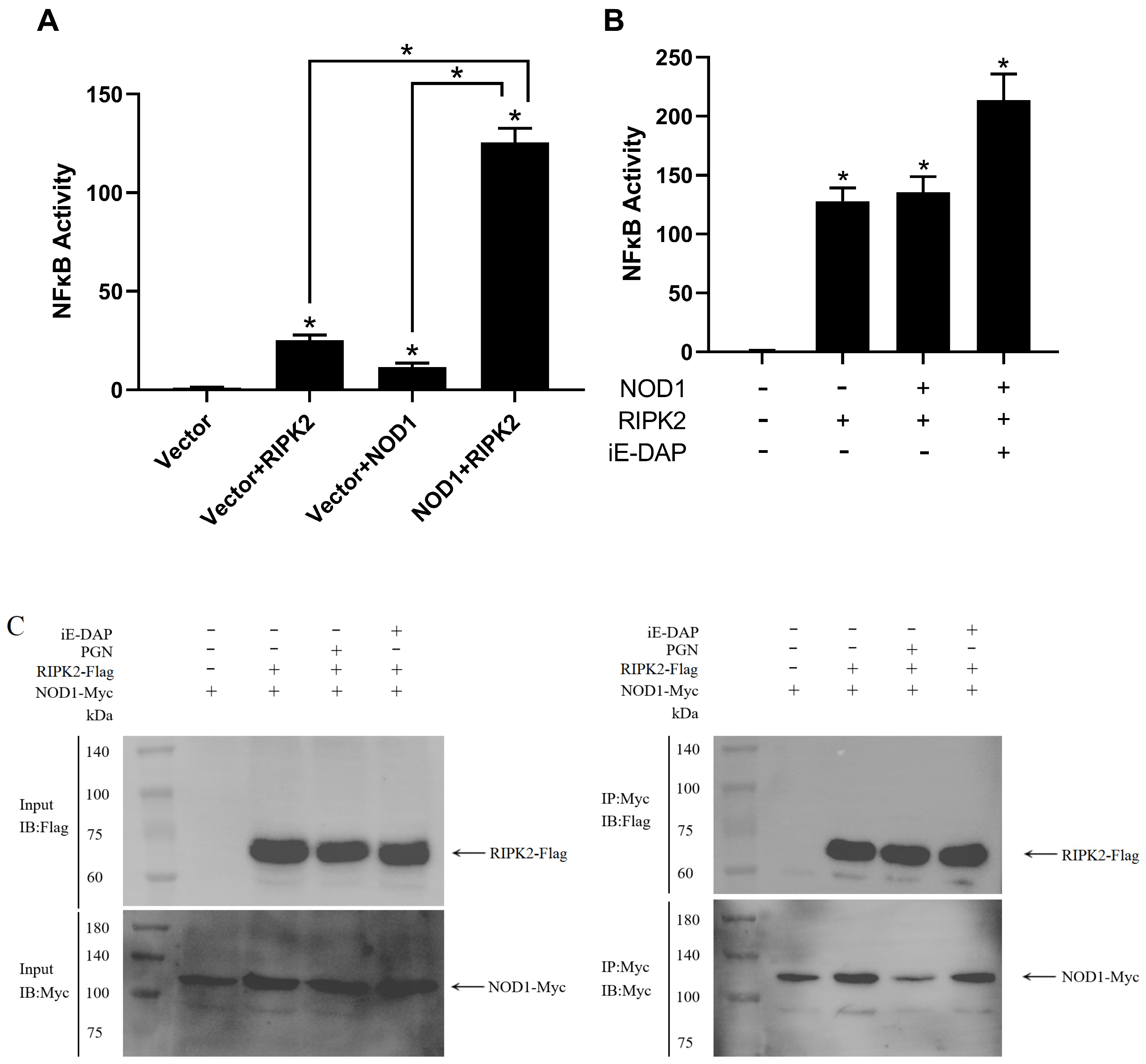
In mammals, RIPK2 has been well characterized as the key adaptor protein downstream of the NOD1-mediated signaling pathway. Accordingly, the correlation between snakehead RIPK2 and NOD1 signaling cascade was investigated in the present study. Dual-luciferase reporter assays showed that individual overexpression of either RIPK2 or NOD1 triggered mild activation of NF-κB. Notably, co-transfection of RIPK2 and NOD1 expression plasmids resulted in a significant enhancement of NF-κB activation and NF-κB activity increased dramatically following iE-DAP stimulation (Figure 5A,B). Subsequently, RIPK2 and NOD1 were co-transfected into HEK293T cells, and their protein interaction was determined via co-immunoprecipitation (Co-IP). The results verified a direct binding interaction between NOD1 and RIPK2. Moreover, this interaction affinity was not further strengthened upon stimulation with iE-DAP or PGN (Figure 5C). The original, unmodified scans of the Western blot membranes are provided in Supplementary Materials.

4. Discussion

As mentioned earlier, the innate immune system serves as the body’s first line of defense against invading pathogenic microorganisms and plays a pivotal role in the recognition of these microorganisms and the immune response during the early stages of infection [33,34]. NLRs are a class of PRRs that are primarily localized in the cytoplasm of host cells and are relatively conservative in evolutionary terms; homologous genes have been identified in both fish and mammals [35,36,37]. In mammals, NOD1 effectively recognizes iE-DAP—a PGN motif found in Gram-negative bacteria and certain Gram-positive bacteria—within the PGN structure of pathogens that have entered host cells, thereby activating downstream NF-κB signaling pathways [10,38,39]. However, there is currently limited research on the role of NOD1 in fish, and we do not yet fully understand its specific functions in ligand recognition specificity, adaptor protein usage, and tissue expression profiles, as well as its downstream signaling pathways. Hence, the main goal of the present research is to systematically detect and characterize the expression profiles of snakehead NOD1, and to investigate its role in inflammatory responses. Synteny analysis suggests that NOD1 is evolutionarily conserved and may play a critical role in host defense throughout evolution. The preservation of genetic data implies that snakehead NOD1 could exhibit comparable functionality to its mammalian counterpart, with the ability to detect conserved bacterial peptidoglycan motifs and initiate inflammatory pathways.
Sequence analysis revealed that NOD1 lacks a signal peptide, indicating that it is an intracellular protein; this finding was further validated by subcellular localization experiments. This is consistent with findings from studies in higher vertebrates indicating that NOD1 is an intracellular pattern recognition receptor [40]. In mammals, the NOD1 protein primarily consists of three distinct functional domains: CARD, NACHT, and LRR. Among these, the CARD is responsible for binding downstream adaptor proteins, while the LRR domain is responsible for recognizing and binding antigens [41]. Similar to the mammalian NOD1 protein, the NOD1 protein in snakehead consists of the typical CARD-NACHT-LRR structure, and phylogenetic analysis also indicates a close evolutionary relationship with mammalian NOD1 proteins.
Studies in other teleosts have shown that the NOD1 gene was expressed in all examined tissues, and the NOD1 protein was highly enriched in immune-related tissues, suggesting that NOD1 played an important role in the host immune response [17,42,43,44,45,46]. Consistent with previous studies, NOD1 was expressed in all nine tissues of snakehead, with the highest expression levels in the intestine, followed by the spleen, gill, and head kidney. Similar to this, zebrafish NOD1 was highly expressed in the intestine, spleen, and liver [37]; in Chinese perch, NOD1 enrichment was detected in the head kidney, gill, and spleen [47], and NOD1 was most highly expressed in the liver, gill, and skin of miiuy croaker (Miichthys miiuy) [48]. These are the primary mucosal immune tissues and classical immune organs of fish, suggesting that NOD1 may play an indispensable role in the innate immune system of the snakehead [49]. To further understand the cellular basis of this tissue distribution pattern, we compared our findings with known NOD1-expressing cell types in higher vertebrates. Our qPCR analysis revealed high NOD1 transcript abundance in the intestine, spleen, and gill of snakehead. While single-cell resolution data are not yet available for snakehead, comparative insights can be drawn from single-cell RNA sequencing and well-established expression studies in higher vertebrates. In humans, NOD1 is expressed in a broad range of cell types, including intestinal epithelial cells [50], as well as immune cells such as alveolar macrophages, monocyte-derived macrophages, and peripheral blood monocytes [51]. NOD1 expression has also been detected in CD14+ monocytes, CD1a+ immature dendritic cells [52], and B lymphocytes [53]. Accordingly, the abundant NOD1 transcripts we observed in the snakehead spleen and gill likely reflect expression in analogous cell populations, particularly macrophages and lymphocytes, which are central to antibacterial immune surveillance. The highest expression in the intestine is likely attributed to its role as a major mucosal immune barrier, continuously exposed to commensal microbiota and dietary antigens, thus requiring heightened immune surveillance [50]. Although single-cell transcriptomic studies in teleost fish (e.g., turbot [54], largemouth bass [55], and large yellow croaker [56]) have successfully resolved immune cell landscapes, none of these studies have yet directly assigned NOD1 expression to specific cell types at single-cell resolution. Therefore, while the overall tissue expression pattern of snakehead NOD1 is broadly consistent with the cellular distribution profile known for mammalian NOD1, future studies employing in situ hybridization, single-cell RNA sequencing, or immunohistochemistry will be necessary to identify the precise NOD1-expressing cell populations in snakehead tissues.
Earlier investigations demonstrated that NOD1 serves a critical function in host innate defense against bacterial pathogens. For example, inhibiting NOD1 expression in zebrafish impairs the ability of juvenile fish to resist bacterial proliferation [57]; in orange-spotted grouper (Epinephelus coioides), NOD1 expression in the spleen significantly increased 72 h after infection with Vibrio alginolyticus as measured by qRT-PCR [46]. After infection with Vibrio anguillarum, NOD1 expression in the spleen of miiuy croaker exhibited a trend of initially decreasing followed by an increase, also determined by qRT-PCR [48]. In this study, we further explored the expression changes of NOD1 after bacterial infection and found that the expression of NOD1 was upregulated at certain time points in the spleen, liver, and gill tissues after N. seriolae infection, indicating that NOD1 is actively involved in antibacterial immune responses. In contrast, NOD1 expression was significantly reduced in the intestine. Several factors may account for this downregulation: first, N. seriolae infection can cause intestinal epithelial cell necrosis and tissue damage, directly reducing the number of NOD1-expressing cells [58]; second, as an intracellular pathogen, N. seriolae may induce apoptosis of macrophages, which are major NOD1-expressing cells, thereby diminishing the expression level [59]; third, the pathogen possesses immunosuppressive and immune evasion capabilities, potentially leading to transient suppression of immune gene expression in mucosal tissues [60]. Pathogen infection may induce apoptosis or trigger the early stages of programmed cell death, leading to a decline in overall protein synthesis capacity, which in turn affects the expression of NOD1 as a specific protein. Since the present study largely relies on in vitro overexpression models, further in vivo validation using gene knockdown or knockout approaches is needed to confirm the physiological role of NOD1.
NOD1 primarily recognizes antigens through its C-terminal LRR domain and further interacts with the adaptor protein RIPK2 via its N-terminal CARD to activate downstream signaling pathways [61]. Our study revealed that snakehead NOD1 significantly activates the downstream NF-κB signaling pathway, and that the induced NF-κB activity increases in a concentration-dependent manner with increasing NOD1 levels. This finding is consistent with observations reported in other teleost fish [62,63,64]. Furthermore, iE-DAP stimulation resulted in significantly higher NF-κB activity compared to the other four ligands or the control group, and a marked enhancement of NF-κB signaling pathway activation was observed upon co-exposure to NOD1 and iE-DAP. Similar results were also observed in golden pompano (Trachinotus ovatus) and Nile tilapia (Oreochromis niloticus) [62,63].

5. Conclusions

Taken together, the snakehead NOD1 gene was obtained by cloning in this study. Through sequence analysis, it was found that NOD1 encodes 943 amino acids. Phylogenetic analysis showed that the snakehead NOD1 protein was closely related to the Perciformes. At the same time, the qPCR results indicated that NOD1 was expressed in all tissues. In the case of bacterial infection, NOD1 expression was upregulated in the spleen and gill tissues. The results from the dual-luciferase reporter assays showed that overexpression of NOD1 significantly induced NF-κB activity and activated NF-κB in a dose- and time-dependent manner. Moreover, NOD1 synergistically enhanced NF-κB activity with the downstream adaptor protein RIPK2. These findings suggest that snakehead NOD1 contributes to antibacterial innate immune signaling through NF-κB activation.

Supplementary Materials

The following supporting information can be downloaded at: https://www.mdpi.com/article/10.3390/biology15120942/s1.

Author Contributions

Formal analysis, B.W. and Y.L. (Yiying Liu); methodology, B.W. and Y.L. (Yiying Liu); investigation, B.W. and Q.F.; validation, X.Z. (Xiaochen Zhu), Q.F. and M.C.; data curation, X.Z. (Xiaochen Zhu), B.W. and Y.L. (Yang Li); writing—original draft, B.W. and Y.L. (Yiying Liu); writing—review and editing, X.Z. (Xiaoyan Zhang), G.W. and C.L.; conceptualization, C.L. and N.Y.; visualization, C.L. and N.Y.; supervision, C.L. and G.W.; project administration, C.L. and N.Y.; funding acquisition, C.L. and G.W. All authors have read and agreed to the published version of the manuscript.

Funding

This study was supported by the Shandong Provincial Natural Science Foundation (Grant 436 No. ZR2024QC027), Natural Science Foundation of China (NO. 32073005), Shandong Technical System of Fish Industry (SDAIT-12-02).

Institutional Review Board Statement

The experimental protocols were approved by the Committee on the Ethics of Animal Experiments of Qingdao Agricultural University IACUC (Institutional Animal Care and Use Committee) on 10 March 2023.

Data Availability Statement

The original contributions presented in this study are included in the article. Further enquiries can be directed to the corresponding author.

Conflicts of Interest

Author Xiaochen Zhu was employed by the company Shandong Qidu Pharmaceutical Co., Ltd. The remaining authors declare that the research was conducted in the absence of any commercial or financial relationships that could be construed as a potential conflict of interest.

References

  1. Chen, R.; Zou, J.; Chen, J.; Zhong, X.; Kang, R.; Tang, D. Pattern Recognition Receptors: Function, Regulation and Therapeutic Potential. Signal Transduct. Target. Ther. 2025, 10, 216. [Google Scholar] [CrossRef] [PubMed]
  2. Philpott, D.J.; Sorbara, M.T.; Robertson, S.J.; Croitoru, K.; Girardin, S.E. Nod Proteins: Regulators of Inflammation in Health and Disease. Nat. Rev. Immunol. 2014, 14, 9–23. [Google Scholar] [PubMed]
  3. Tsankov, B.K.; Luchak, A.; Carr, C.; Philpott, D.J. The Effects of Nod-Like Receptors on Adaptive Immune Responses. Biomed. J. 2024, 47, 100637. [Google Scholar] [CrossRef] [PubMed]
  4. Chou, W.-C.; Jha, S.; Linhoff, M.W.; Ting, J.P.-Y. The Nlr Gene Family: From Discovery to Present Day. Nat. Rev. Immunol. 2023, 23, 635–654. [Google Scholar] [CrossRef] [PubMed]
  5. Gong, T.; Liu, L.; Jiang, W.; Zhou, R. Damp-Sensing Receptors in Sterile Inflammation and Inflammatory Diseases. Nat. Rev. Immunol. 2020, 20, 95–112. [Google Scholar] [PubMed]
  6. Kanneganti, T.-D.; Özören, N.; Body-Malapel, M.; Amer, A.; Park, J.-H.; Franchi, L.; Whitfield, J.; Barchet, W.; Colonna, M.; Vandenabeele, P.; et al. Bacterial Rna and Small Antiviral Compounds Activate Caspase-1 through Cryopyrin/Nalp3. Nature 2006, 440, 233–236. [Google Scholar] [CrossRef] [PubMed]
  7. Mariathasan, S.; Newton, K.; Monack, D.M.; Vucic, D.; French, D.M.; Lee, W.P.; Roose-Girma, M.; Erickson, S.; Dixit, V.M. Differential Activation of the Inflammasome by Caspase-1 Adaptors Asc and Ipaf. Nature 2004, 430, 213–218. [Google Scholar] [CrossRef] [PubMed]
  8. Meunier, E.; Broz, P. Evolutionary Convergence and Divergence in Nlr Function and Structure. Trends Immunol. 2017, 38, 744–757. [Google Scholar] [CrossRef] [PubMed]
  9. Sundaram, B.; Tweedell, R.E.; Kumar, S.P.; Kanneganti, T.-D. The Nlr Family of Innate Immune and Cell Death Sensors. Immunity 2024, 57, 674–699. [Google Scholar] [CrossRef] [PubMed]
  10. Caruso, R.; Warner, N.; Inohara, N.; Núñez, G. NOD1 and Nod2: Signaling, Host Defense, and Inflammatory Disease. Immunity 2014, 41, 898–908. [Google Scholar] [CrossRef] [PubMed]
  11. Inohara, N.; Koseki, T.; Lin, J.; del Peso, L.; Lucas, P.C.; Chen, F.F.; Ogura, Y.; Núñez, G. An Induced Proximity Model for Nf-Kappa B Activation in the NOD1/Rick and Rip Signaling Pathways. J. Biol. Chem. 2000, 275, 27823–27831. [Google Scholar] [CrossRef] [PubMed]
  12. Sahoo, B.R. Structure of Fish Toll-Like Receptors (Tlr) and Nod-Like Receptors (Nlr). Int. J. Biol. Macromol. 2020, 161, 1602–1617. [Google Scholar] [CrossRef] [PubMed]
  13. Swain, B.; Miryala, K.R. NOD-like Receptors in Fish: Evolution, Structure, Immune signaling, and Targeting for Aquaculture Vaccine Adjuvants. Front. Immunol. 2025, 16, 1665071. [Google Scholar] [CrossRef] [PubMed]
  14. Wu, X.M.; Chen, W.Q.; Hu, Y.W.; Cao, L.; Nie, P.; Chang, M.X. Rip2 Is a Critical Regulator for Nlrs Signaling and Mhc Antigen Presentation but Not for Mapk and Pi3k/Akt Pathways. Front. Immunol. 2018, 9, 726. [Google Scholar] [CrossRef] [PubMed]
  15. Wu, X.M.; Zhang, J.; Li, P.W.; Hu, Y.W.; Cao, L.; Ouyang, S.; Bi, Y.H.; Nie, P.; Chang, M.X. NOD1 Promotes Antiviral Signaling by Binding Viral Rna and Regulating the Interaction of Mda5 and Mavs. J. Immunol. 2020, 204, 2216–2231. [Google Scholar] [CrossRef] [PubMed]
  16. Xie, J.; Belosevic, M. Functional Characterization of Receptor-Interacting Serine/Threonine Kinase 2 (Rip2) of the Goldfish (Carassius auratus L.). Dev. Comp. Immunol. 2015, 48, 76–85. [Google Scholar] [CrossRef] [PubMed]
  17. Xie, J.; Hodgkinson, J.W.; Katzenback, B.A.; Kovacevic, N.; Belosevic, M. Characterization of Three Nod-Like Receptors and Their Role in Antimicrobial Responses of Goldfish (Carassius auratus L.) Macrophages to Aeromonas Salmonicida and Mycobacterium Marinum. Dev. Comp. Immunol. 2013, 39, 180–187. [Google Scholar] [CrossRef] [PubMed]
  18. Jang, J.H.; Kim, H.; Kim, Y.J.; Cho, J.H. Molecular Cloning and Functional Analysis of Nucleotide-Binding Oligomerization Domain-Containing Protein 1 in Rainbow Trout, Oncorhynchus mykiss. Fish Shellfish Immunol. 2016, 51, 53–63. [Google Scholar] [CrossRef] [PubMed]
  19. Chen, D.; Zhu, H.; Lu, L.; Chen, Y.; Zhang, X.; Huang, X.; Ouyang, P.; Geng, Y.; Li, Z. Identification, Characterization and the Inflammatory Regulating Effect of NOD1/2 in Sturgeon. Fish Shellfish Immunol. 2024, 146, 109407. [Google Scholar] [CrossRef] [PubMed]
  20. Peng, X.Y.; Wang, K.L.; Li, L.; Li, B.; Wu, X.Y.; Zhang, Z.W.; Li, N.; Liu, L.H.; Nie, P.; Chen, S.N. Transcription of NOD1 and Nod2 and Their Interaction with Card9 and RIPK2 in Ifn Signaling in a Perciform Fish, the Chinese Perch, Siniperca chuatsi. Front. Immunol. 2024, 15, 1374368. [Google Scholar] [CrossRef] [PubMed]
  21. Teng, J.; Zhao, Y.; Meng, Q.L.; Zhu, S.R.; Chen, H.J.; Xue, L.Y.; Ji, X.S. Transcriptome analysis in the spleen of Northern Snakehead (Channa argus) challenged with Nocardia seriolae. Genomics 2022, 114, 110357. [Google Scholar] [CrossRef] [PubMed]
  22. Zhang, W.; Zhou, K.; Huang, L.; Yang, N.; Lin, L.; Chen, L.; Yao, J.; Dong, M.; Shen, J.; Pan, X. Biological characteristics and pathogenicity comparison of Nocardia seriolae isolated from Micropterus salmoides and Channa argus. Front. Vet. Sci. 2024, 11, 1367066. [Google Scholar] [CrossRef] [PubMed]
  23. Wang, G.L.; Xu, Y.J.; Jin, S.; Zhu, J.L.; Zhu, W.Y. Research on the Nocardiosis and Pathogen in Reared Snakehead, Ophiocephalus argus Cantor. Acta Hydrobiol. Sin. 2009, 33, 277–283. [Google Scholar] [CrossRef]
  24. Zhang, N.; Zhang, H.; Dong, Z.; Wang, W. Molecular Identification of Nocardia seriolae and Comparative Analysis of Spleen Transcriptomes of Hybrid Snakehead (Channa maculata Female × Channa argus Male) with Nocardiosis Disease. Front. Immunol. 2022, 13, 778915. [Google Scholar] [CrossRef] [PubMed]
  25. Zhou, T.; Cai, P.; Li, J.; Dan, X.; Li, Z. Pathological Variations and Immune Response in Channa argus Infected with Pathogenic Nocardia seriolae strain. Fish Shellfish. Immunol. 2024, 150, 109554. [Google Scholar] [CrossRef] [PubMed]
  26. Liu, Y.; Chen, G.; Xia, L.; Lu, Y. A Review on the Pathogenic Bacterium Nocardia seriolae: Aetiology, Pathogenesis, Diagnosis And Vaccine Development. Rev. Aquac. 2023, 15, 14–34. [Google Scholar]
  27. Lechevalier, M.P.; Horan, A.C.; Lechevalier, H. Lipid Composition in the Classification of Nocardiae and Mycobacteria. J. Bacteriol. 1971, 105, 313–318. [Google Scholar] [CrossRef] [PubMed]
  28. Lechevalier, M.P.; Lechevalier, H. Chemical composition as a criterion in the classification of aerobic actinomycetes. Int. J. Syst. Evol. Microbiol. 1970, 20, 435. [Google Scholar] [CrossRef]
  29. Zhou, Z.-Y.; Bai, S.-J.; He, J.; Xiong, Q.-X.; Zhong, Z.-D.; Lu, C.-W.; Kuang, L.-F.; Jian, Z.-R.; Gu, J.-L.; Liu, M.-Z.; et al. Pathogenicity, ultrastructure and genomics analysis of Nocardia seriolae isolated from largemouth bass (Micropterus salmoides). Microb. Pathog. 2025, 205, 107715. [Google Scholar] [CrossRef] [PubMed]
  30. Chamaillard, M.; Hashimoto, M.; Horie, Y.; Masumoto, J.; Qiu, S.; Saab, L.; Ogura, Y.; Kawasaki, A.; Fukase, K.; Kusumoto, S.; et al. An Essential Role for NOD1 in Host Recognition of Bacterial Peptidoglycan Containing Diaminopimelic Acid. Nat. Immunol. 2003, 4, 702–707. [Google Scholar] [CrossRef] [PubMed]
  31. Girardin, S.E.; Boneca, I.G.; Carneiro, L.A.M.; Antignac, A.; Jéhanno, M.; Viala, J.; Tedin, K.; Taha, M.-K.; Labigne, A.; Zäthringer, U.; et al. NOD1 Detects a Unique Muropeptide From Gram-Negative Bacterial Peptidoglycan. Science 2003, 300, 1584–1587. [Google Scholar] [CrossRef] [PubMed]
  32. Han, H.; Kwak, M.; Ha, S.; Yang, S.; Kim, J.D.; Cho, K.; Kim, T.; Cho, M.Y.; Kim, B.; Jung, S.; et al. Genomic Characterization of Nocardia seriolae Strains Isolated from Diseased Fish. Microbiologyopen 2019, 8, e00656. [Google Scholar] [PubMed]
  33. Magnadóttir, B. Innate Immunity of Fish (Overview). Fish Shellfish Immunol. 2006, 20, 137–151. [Google Scholar] [CrossRef] [PubMed]
  34. Dezfuli, B.S.; Lorenzoni, M.; Carosi, A.; Giari, L.; Bosi, G. Teleost Innate Immunity, an Intricate Game between Immune Cells and Parasites of Fish Organs: Who Wins, Who Loses. Front. Immunol. 2023, 14, 1250835. [Google Scholar] [CrossRef] [PubMed]
  35. Meylan, E.; Tschopp, J.; Karin, M. Intracellular Pattern Recognition Receptors in the Host Response. Nature 2006, 442, 39–44. [Google Scholar] [CrossRef] [PubMed]
  36. Benko, S.; Philpott, D.J.; Girardin, S.E. The Microbial and Danger Signals That Activate Nod-Like Receptors. Cytokine 2008, 43, 368–373. [Google Scholar] [CrossRef] [PubMed]
  37. Laing, K.J.; Purcell, M.K.; Winton, J.R.; Hansen, J.D. A Genomic View of the Nod-Like Receptor Family in Teleost Fish: Identification of a Novel Nlr Subfamily in Zebrafish. BMC Evol. Biol. 2008, 8, 42. [Google Scholar] [CrossRef] [PubMed]
  38. Strober, W.; Murray, P.J.; Kitani, A.; Watanabe, T. Signalling Pathways and Molecular Interactions of NOD1 and Nod2. Nat. Rev. Immunol. 2006, 6, 9–20. [Google Scholar] [PubMed]
  39. Correa, R.G.; Milutinovic, S.; Reed, J.C. Roles of NOD1 (Nlrc1) and Nod2 (Nlrc2) in Innate Immunity and Inflammatory Diseases. Biosci. Rep. 2012, 32, 597–608. [Google Scholar] [CrossRef] [PubMed]
  40. Le Bourhis, L.; Benko, S.; Girardin, S. NOD1 and Nod2 in Innate Immunity and Human Inflammatory Disorders. Biochem. Soc. Trans. 2007, 35, 1479–1484. [Google Scholar] [CrossRef] [PubMed]
  41. Inohara, N.; Nuñez, G. Nods: Intracellular Proteins Involved in Inflammation and Apoptosis. Nat. Rev. Immunol. 2003, 3, 371–382. [Google Scholar] [CrossRef] [PubMed]
  42. Sha, Z.; Abernathy, J.W.; Wang, S.; Li, P.; Kucuktas, H.; Liu, H.; Peatman, E.; Liu, Z. Nod-Like Subfamily of the Nucleotide-Binding Domain and Leucine-Rich Repeat Containing Family Receptors and Their Expression in Channel Catfish. Dev. Comp. Immunol. 2009, 33, 991–999. [Google Scholar] [CrossRef] [PubMed]
  43. Li, J.; Kong, L.; Gao, Y.; Wu, C.; Xu, T. Characterization of Nlr-a Subfamily Members in Miiuy Croaker and Comparative Genomics Revealed Nlrx1 Underwent Duplication and Lose in Actinopterygii. Fish Shellfish Immunol. 2015, 47, 397–406. [Google Scholar] [CrossRef] [PubMed]
  44. Paria, A.; Makesh, M.; Chaudhari, A.; Purushothaman, C.; Rajendran, K. Nucleotide-Binding Oligomerization Domain-Containing Protein 1 (NOD1) in Asian Seabass, Lates calcarifer: Cloning, Ontogeny and Expression Analysis Following Bacterial Infection or Ligand Stimulation. Fish Shellfish Immunol. 2018, 79, 153–162. [Google Scholar] [CrossRef] [PubMed]
  45. Chen, W.; Xu, Q.; Chang, M.; Nie, P.; Peng, K. Molecular Characterization and Expression Analysis of Nuclear Oligomerization Domain Proteins NOD1 and Nod2 in Grass Carp Ctenopharyngodon idella. Fish Shellfish Immunol. 2010, 28, 18–29. [Google Scholar] [CrossRef] [PubMed]
  46. Hou, Q.-H.; Yi, S.-B.; Ding, X.; Zhang, H.-X.; Sun, Y.; Zhang, Y.; Liu, X.-C.; Lu, D.-Q.; Lin, H.-R. Differential Expression Analysis of Nuclear Oligomerization Domain Proteins NOD1 and Nod2 in Orange-Spotted Grouper (Epinephelus coioides). Fish Shellfish Immunol. 2012, 33, 1102–1111. [Google Scholar] [CrossRef] [PubMed]
  47. Gu, T.; Lu, L.; Wang, J.; Tian, L.; Wei, W.; Wu, X.; Xu, Q.; Chen, G. The NOD1 and Nod2 in Mandarinfish (Siniperca chuatsi): Molecular Characterization, Tissue Distribution, and Expression Analysis. BMC Genet. 2018, 19, 61. [Google Scholar] [CrossRef] [PubMed]
  48. Li, J.; Gao, Y.; Xu, T. Comparative Genomic and Evolution of Vertebrate NOD1 and Nod2 Genes and Their Immune Response in Miiuy Croaker. Fish Shellfish Immunol. 2015, 46, 387–397. [Google Scholar] [CrossRef] [PubMed]
  49. Li, H.; Liu, H.; Bi, L.; Liu, Y.; Jin, L.; Peng, R. Immunotoxicity of Microplastics in Fish. Fish Shellfish Immunol. 2024, 150, 109619. [Google Scholar] [CrossRef] [PubMed]
  50. Kim, J.G.; Lee, S.J.; Kagnoff, M.F. NOD1 is an Essential Signal Transducer in Intestinal Epithelial Cells Infected With Bacteria That Avoid Recognition by Toll-Like Receptors. Infect Immun. 2004, 72, 1487–1495. [Google Scholar] [CrossRef] [PubMed]
  51. Juárez, E.; Carranza, C.; Hernández-Sánchez, F.; Loyola, E.; Escobedo, D.; León-Contreras, J.C.; Hernández-Pando, R.; Torres, M.; Sada, E. Nucleotide-Oligomerizing Domain-1 (NOD1) Receptor Activation Induces Pro-Inflammatory Responses and Autophagy in Human Alveolar Macrophages. BMC Pulm. Med. 2014, 14, 152. [Google Scholar] [CrossRef] [PubMed][Green Version]
  52. Fritz, J.H.; Girardin, S.E.; Fitting, C.; Werts, C.; Mengin-Lecreulx, D.; Caroff, M.; Cavaillon, J.-M.; Philpott, D.J.; Adib-Conquy, M. Synergistic Stimulation Of Human Monocytes And Dendritic Cells By Toll-Like Receptor 4 And NOD1- and NOD2-activating Agonists. Eur. J. Immunol. 2005, 35, 2459–2470. [Google Scholar] [CrossRef] [PubMed]
  53. Petterson, T.; Jendholm, J.; Månsson, A.; Bjartell, A.; Riesbeck, K.; Cardell, L.-O. Effects of NOD-like Receptors In Human B Lymphocytes and Crosstalk Between NOD1/NOD2 And Toll-Like Receptors. J. Leukoc. Biol. 2011, 89, 177–187. [Google Scholar] [PubMed]
  54. Mu, D.; Yang, J.; Jiang, Y.; Wang, Z.; Chen, W.; Huang, J.; Zhang, Y.; Liu, Q.; Yang, D. Single-Cell Transcriptomic Analysis Reveals Neutrophil as Orchestrator during β-Glucan-Induced Trained Immunity in a Teleost Fish. J. Immunol. 2022, 209, 783–795. [Google Scholar] [CrossRef] [PubMed]
  55. Zhou, Z.-Y.; Jian, Z.-R.; Gu, J.-L.; Jiang, X.-Y.; He, J.; Gao, Y.-C.; Lian, S.; Bai, S.-J.; Lu, C.-W.; Kuang, L.-F.; et al. Single-Cell Sequencing Reveals Metabolic Reprogramming and Immune Regulation Underlie The Maintenance Of Teleost Granulomas. Fish Shellfish Immunol. 2026, 173, 111287. [Google Scholar] [CrossRef] [PubMed]
  56. Hu, C.; Cui, J.; He, R.; Han, M.; Liu, Y.; Zhou, J.; Fan, D.; Ding, X.; Xiang, L.; Lou, B.; et al. Single-Cell Profiling Reveals Immune Cell Diversity And Infection-Driven Remodeling In The Spleen Of Marine Teleost Larimichthys crocea. Fundam. Res. 2025, 9, 22. [Google Scholar]
  57. Oehlers, S.H.; Flores, M.V.; Hall, C.J.; Swift, S.; Crosier, K.E.; Crosier, P.S. The Inflammatory Bowel Disease (Ibd) Susceptibility Genes NOD1 and Nod2 Have Conserved Anti-Bacterial Roles in Zebrafish. Dis. Model. Mech. 2011, 4, 832–841. [Google Scholar] [CrossRef] [PubMed]
  58. Liu, Z.; Dong, J.; Ke, X.; Yi, M.; Cao, J.; Gao, F.; Wang, M.; Ye, X.; Lu, M. Isolation, identification, and pathogenic characteristics of Nocardia seriolae in largemouth bass Micropterus salmoides. Dis. Aquat. Organ. 2022, 149, 33–45. [Google Scholar] [CrossRef] [PubMed]
  59. Liu, W.; Deng, Y.; Tan, A.; Zhao, F.; Chang, O.; Wang, F.; Lai, Y.; Huang, Z. Intracellular behavior of Nocardia seriolae and its apoptotic effect on RAW264.7 macrophages. Front. Cell. Infect. Microbiol. 2023, 13, 1138422. [Google Scholar] [CrossRef] [PubMed]
  60. Teng, J.; Li, Y.; Zhao, Y.; Zhang, Y.; Chen, D.; Liu, J.; Cui, M.; Ji, X. Integrated analysis of proteome and transcriptome revealed changes in multiple signaling pathways involved in immunity in the northern snakehead (Channa argus) during Nocardia seriolae infection. Front. Cell. Infect. Microbiol. 2024, 14, 1482901. [Google Scholar] [CrossRef] [PubMed]
  61. Inohara, N.; Ogura, Y.; Nuñez, G. Nods: A Family of Cytosolic Proteins That Regulate the Host Response to Pathogens. Curr. Opin. Microbiol. 2002, 5, 76–80. [Google Scholar] [CrossRef] [PubMed]
  62. Wang, Y.; Yang, S.; Cai, X.; Huang, Z.; Tan, K.; Xu, P. Functional Characterization of NOD1 from Golden Pompano Trachinotus Ovatus. Fish Shellfish Immunol. 2024, 149, 109566. [Google Scholar] [CrossRef] [PubMed]
  63. He, J.; Meng, Z.; Lu, D.; Liu, X.; Lin, H. Recognition of Dap and Activation of NF-κB by Cytosolic Sensor NOD1 in Oreochromis niloticus. Fish Shellfish Immunol. 2021, 110, 75–85. [Google Scholar] [CrossRef] [PubMed]
  64. Li, M.; Wang, Q.-L.; Lu, Y.; Chen, S.-L.; Li, Q.; Sha, Z.-X. Expression Profiles of Nods in Channel Catfish (Ictalurus punctatus) after Infection with Edwardsiella tarda, Aeromonas hydrophila, Streptococcus iniae and Channel Catfish Hemorrhage Reovirus. Fish Shellfish Immunol. 2012, 33, 1033–1141. [Google Scholar] [CrossRef] [PubMed]
Figure 1. Identification of NOD1 in snakehead. (A) A multi-sequence comparison of snakehead NOD1 and other species. Asterisks (*) indicate identical residues; dots (.) indicate conservative substitutions. (B) Phylogenetic analysis of NOD1 in snakehead and other vertebrates. Phylogenetic analysis was done using MEGA X by performing the neighbor-joining method with 1000 bootstrap replicates. GenBank accession numbers of selected NOD1 amino acid sequences: Homo sapiens NOD1 (AAD28350.1), Mus musculus NOD1 (AAN52479.1), Gallus gallus NOD1 (AFS49704.1), Xenopus laevis NOD1 (XP_018124808.1), Danio rerio NOD1 (XP_002665106.3), Siniperca chuatsi NOD1 (XP_044063118.1), Micropterus salmoides NOD1 (XP_038568219.1), Sander lucioperca NOD1 (XP_035862690.1), Perca fluviatilis NOD1 (XP_039661787.1), Larimichthys crocea NOD1 (XP_010737413.3), Oreochromis niloticus NOD1 (XP_003446247.1), Anabas testudineus NOD1 (XP_026229064.1), Channa punctata NOD1 (QDH76353.1), Scophthalmus maximus NOD1 (XP_035485565.2), Ictalurus punctatus NOD1 (NP_001186996.1), Ctenopharyngodon idella NOD1 (XP_051720971.1). (C) Synteny analysis of NOD1 in snakehead.
Biology 15 00942 g001
Figure 2. The expression level of NOD1 in different tissues. (A) Total RNA was extracted from nine tissues (brain, gill, muscle, skin, intestine, blood, liver, spleen and kidney) and analyzed by qPCR to examine the tissue expression of NOD1. The relative mRNA level in each sample was normalized to the level of β-actin and the expression levels were calibrated against tissue that had the lowest expression level. (B) The expression level of NOD1 after N. seriolae infection. The results were expressed as the means ± standard error from three independent triplicated experiments and significant difference was indicated with * p < 0.05. (C) The subcellular localization of NOD1 and RIPK2 in the HEK293 cells. The cells were transfected with pEGFP-N2, NOD1-GFP and RIPK2-GFP. After 24 h, the cells were fixed and the nucleus stained with DAPI. Green represents the target gene; blue represents the nucleus.
Biology 15 00942 g002
Figure 3. The activation of NF-κB by NOD1. EPC cells were co-transfected with 100 ng, 200 ng, 300 ng, 400 ng and 500 ng NOD1 expression plasmids respectively along with NF-κB reporter gene plasmids to perform the concentration gradient experiment (A), and co-transfected with NOD1 expression plasmids and NF-κB reporter gene plasmids into cells to perform the time gradient experiment (B); the luciferase activity of NF-κB was detected at 24 h post-transfection. (C) EPC cells were transfected with NOD1 expression plasmids or empty control vectors together with NF-κB and pRL-TK, and then transfected with LTA, poly(I:C), iE-DAP and PGN to stimulate the cells; at 24 h post-transfection, the luciferase activity of NF-κB was detected. (D) After co-transfecting cells with NOD1 plasmids of varying concentrations, NF-κB, and pRL-TK for 24 h, the cells were stimulated with iE-DAP, and luciferase activity was measured 6 h after stimulation. (E) Cells were co-transfected with NOD1 expression plasmids, NF-κB and pRL-TK internal reference plasmid 24 h later, and then different concentrations of iE-DAP stimulated the cells intracellularly; the luciferase activity of NF-κB was detected at 24 h post-stimulation. The data are expressed as mean ± SE from three independent experiments, with each sample measured in triplicate and significant difference indicated with * p < 0.05 and ** p < 0.01.
Biology 15 00942 g003
Figure 4. A schematic representation of full-length NOD1 and the variants used in this study. NF-κB promoter activation by NOD1 and its variants. The data are expressed as mean ± SE from three independent experiments, with each sample measured in triplicate, and * indicating p < 0.05.
Biology 15 00942 g004
Figure 5. The association between NOD1 and RIPK2. (A) The association between NOD1 and RIPK2 in NF-κB activation. (B) RIPK2 and NOD1 plasmids were transfected along with the NF-κB plasmid into the cells, then the cells were stimulated with iE-DAP, and 6 h later, the luciferase activity of NF-κB was detected. pRL-TK as the internal control, and the data are expressed as mean ± SE of three independent experiments with each sample measured in triplicate. * indicates p < 0.05. (C) The interaction of RIPK2 with NOD1 through co-immunoprecipitation (Co-IP) analysis.
Biology 15 00942 g005
Table 1. The primers used in this study.
PrimerSequence (5′ to 3′)
NOD1-FATGGGTCGAATAGAAGAGGAG
NOD1-RTCAGTGGAACTGCAGTCTC
Myc-NOD1-FTCCAGTGTGGTGGAATTCATGGGTCGAATAGAAGAGGAG
Myc-NOD1-RGGGCCCTCTAGACTCGAGGTGGAACTGCAGTCTC
Myc-NOD1-ΔCARD-FTCCAGTGTGGTGGAATTCATGTGGCTGAAAGAAATCAACTACA
Myc-NOD1-△LRR-RGGGCCCTCTAGACTCGAGATGCTGTAGCACAAAGTTCAGA
Myc-NOD1-CARD-RGGGCCCTCTAGACTCGAGTTCTTTCAGCCATGGCTGGA
Myc-NOD1-NACHT-FTCCAGTGTGGTGGAATTCATGGAAGGTGAAACCATTTATGTG
Myc-NOD1-NACHT-RGGGCCCTCTAGACTCGAGGTGCTCCTGCTCCTTAAAGT
Myc-NOD1-LRRs-FTCCAGTGTGGTGGAATTCATGCGTCAGAAGCTCCTGGGGC
NOD1-GFP-FCGTCAGATCCGCTAGCATGGGTCGAATAGAAGAGGAG
NOD1-GFP-RACGGCCGGTGGATCCGGTGGAACTGCAGTCTC
RIPK2-FATGGAGCCTGCGGCTATGGGCTG
RIPK2-RCTACATATTCCTGGGGATATT
Flag-RIPK2-FACCGTCAGAATTAAGCTTATGGAGCCTGCGGCTATGGGCTG
Flag-RIPK2-RGTAGTCAGCCCGGGATCCCATATTCCTGGGGATATT
RIPK2-GFP-FCGTCAGATCCGCTAGCATGGAGCCTGCGGCTATGGGCTG
RIPK2-GFP-RACGGCCGGTGGATCCGCATATTCCTGGGGATATT
qNOD1-FGTCAGACAGCAGCATTGAGG
qNOD1-RCCAATCTTGACGACTCGCAG
qRIPK2-FGAAGCTGACCGACCTGTACT
qRIPK2-RTGCAGAAGAACTCAGGCTCA
β-actin-FCACTGTGCCCATCTACGAG
β-actin-RCCATCTCCTGCTCGAAGTC
Disclaimer/Publisher’s Note: The statements, opinions and data contained in all publications are solely those of the individual author(s) and contributor(s) and not of MDPI and/or the editor(s). MDPI and/or the editor(s) disclaim responsibility for any injury to people or property resulting from any ideas, methods, instructions or products referred to in the content.