Tuning Secretomes for Regenerative Medicine


Simple Summary
Abstract
1. Introduction
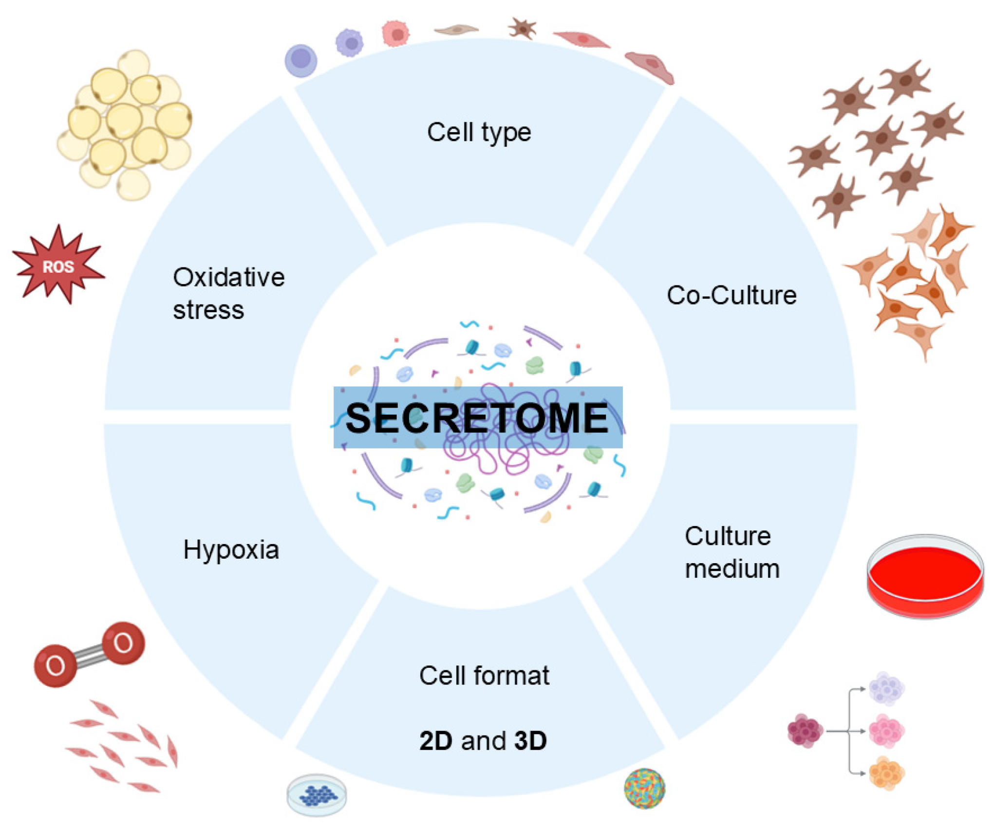
- Different cell types;
- Co-cultures;
- Culture medium;
- Dynamics in secretome composition as a function of culture time;
- Cell format; 2D versus 3D cultures, spheroids, organoids;
- Hypoxia;
- Oxidative stress.
2. Materials and Methods
3. Different Cell Types
3.1. Platelets
3.2. Endothelial Cells
3.3. Osteoblasts
3.4. Chondrocytes
3.5. Adipocytes
3.6. Tenocytes
3.7. Neural Cells
3.8. Immune Cells
3.9. Mesenchymal Stem Cells (MSCs)
3.10. Induced Pluripotent Stem Cells (iPSCs)
3.11. Comparison of Secretomes Harvested from Different Cell Types
4. Co-Culture-Derived Secretome
5. Culture Medium
5.1. Modulation of Secretome by Culture Supplementation of Specific Factors
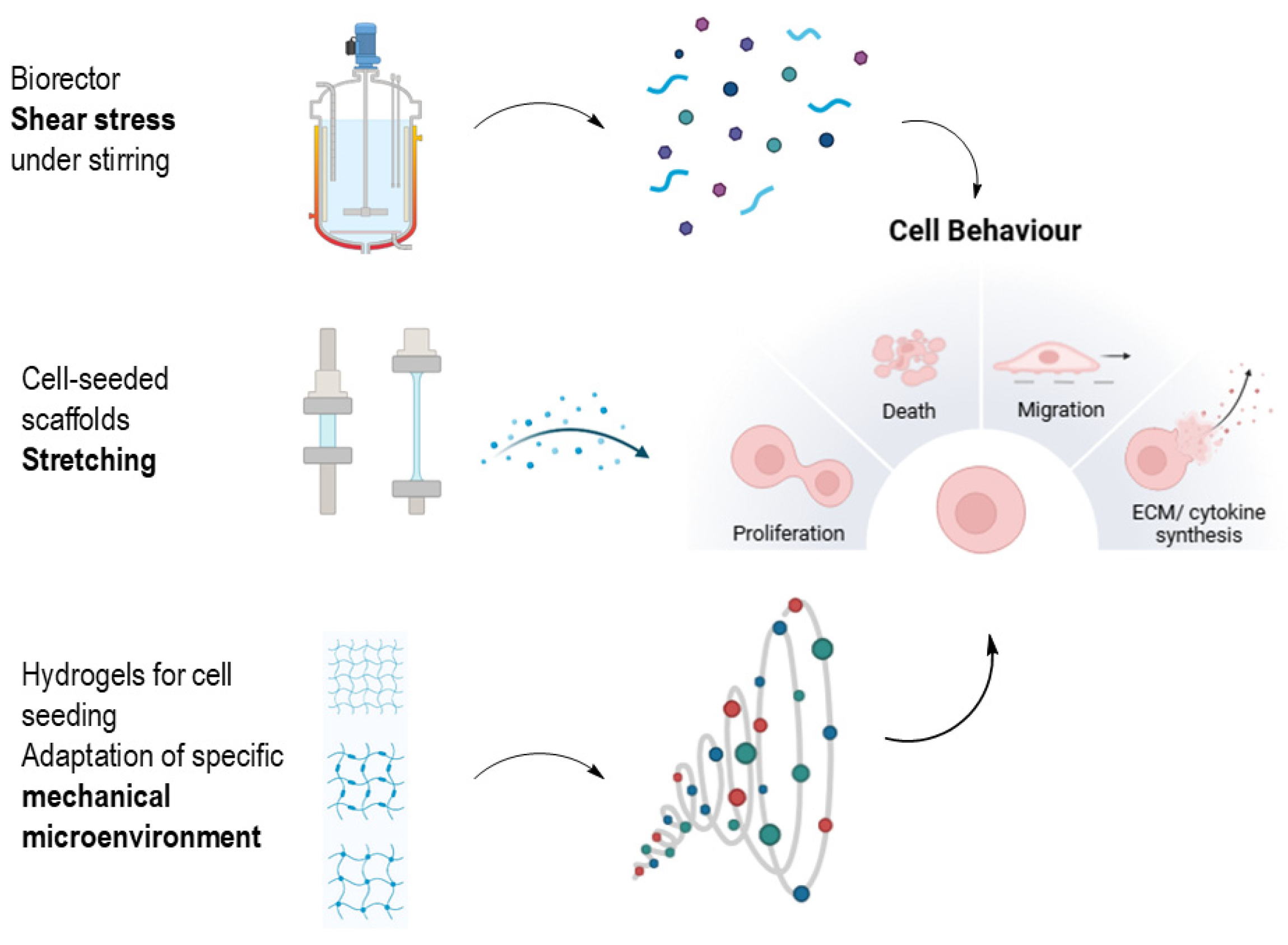
5.2. Static Versus Dynamic Cultivation
6. Dynamics in Secretome Composition as a Function of Culture Time
7. Cell Format
8. Hypoxia
9. Oxidative Stress
10. Characterization of Secretomes
10.1. Analysis of All Secretome Proteins: Proteomics
10.2. Analysis of Specific Proteins in the Secretome
10.3. Analysis of Extracellular Vesicles
10.4. EV Delivery Approaches and Clinical Applications
11. Major Preclinical and Clinical Applications of Secretomes
12. Challenges and Future Directions
13. Conclusions
Funding
Institutional Review Board Statement
Informed Consent Statement
Data Availability Statement
Acknowledgments
Conflicts of Interest
Abbreviations
| 2D | Two-dimensional |
| 3D | Three-dimensional |
| AAM | Alternatively activated macrophages |
| ADP | Adenosine diphosphate |
| ATP | Adenosine triphosphate |
| ASCs | Adipose-derived stem cells |
| ANG | Angiopoietin |
| α-SMA | Alpha smooth muscle actin |
| B6 | Black 6 mice (C57BL/6) |
| BMPs | Bone morphogenetic proteins |
| BDNF | Brain-derived neurotrophic factor |
| BM-MSCs | Bone marrow-derived mesenchymal stem cells |
| CXCL12 | CXC motif chemokine 12 or stromal cell-derived factor 1 |
| CCL21 | CC motif chemokine ligand 21 |
| CLAs | Complex lymphatic anomalies |
| CSF | Colony stimulating factor |
| CCL-5 | CC motif chemokine ligand 5 |
| COMP | Cartilage oligomeric protein |
| C-EVs | Cartilage extracellular vesicles |
| CDNF | Cerebral dopamine neurotrophic factor |
| ECs | Endothelial cells |
| ECM | Extracellular matrix |
| eNOS | Endothelial nitric oxide synthase |
| EVs | Extracellular vesicles |
| HPLN | Proteoglycan link protein 1 |
| IGFs | Insulin like growth factors |
| IGFBPs | Insulin like growth factor binding proteins |
| IL-3; IL-6; IL-7; IL-8 | Interleukin-3; -6; -7; -8; |
| iPSCs | Induced pluripotent stem cells |
| IPF | Idiopathic pulmonary fibrosis |
| JAK/Stat3/MMP-9 | Januskinase/Signal Transducer and Activator of Transcription 3/Matrix-Metalloproteinase 9 pathway |
| LC-MS/MS | Liquid chromatography mass spectrometry/mass spectrometry |
| LECs | Lymphatic endothelial cells |
| LOX | Lysyl oxidase |
| MSCs | Mesenchymal stem cells |
| MMPs | Matrix metalloproteinases |
| M-CSF | Macrophage colony-stimulating factor |
| miRNA | Micro RNAs |
| MRL/Mpj | Murphy Roths Large mice |
| MCP-1 | Monocyte Chemoattractant Protein-1 |
| M2 | Macrophages polarized M2 (alternatively activated macrophages) |
| NGF | Nerve growth factor |
| NT-3 | Neurotrophin-3 |
| NETs | Neutrophil extracellular traps |
| NO | Nitric oxide |
| OPG | Osteoprotegerin |
| OHDA | Hydroxydopamine |
| OECs | Olfactory ensheathing cells |
| PRP | Platelet-rich plasma |
| PD-L1 | Programmed Cell Death 1 Ligand 1 |
| PAHSAs | Protecting lipokines |
| PAI-1 | Plasminogen activator inhibitor-1 |
| PC12 | Dopamine-producing cell line |
| RANKL | RANK ligand |
| ROS | Reactive oxygen species |
| SCF | Stem cell factor |
| sEGFR | Soluble epidermal growth factor receptor |
| TxA2 | Thromboxan A2 |
| TIMP-1 | Tissue inhibitor of matrix metalloproteinase-1 |
| TNF-α | Tumor necrosis factor alpha |
| TGF-β1; TGF-β2 | Transforming growth factor beta 1 and 2 |
| VEGF | Vascular endothelial growth factor |
| VEGF-C | Vascular endothelial growth factor-C |
| VEGF-D | Vascular endothelial growth factor-D |
| WNT5A | Wingless-related integration site protein 5A |
| WNT16 | Wingless-related integration site protein 16 |
References
- Bogatcheva, N.V.; Coleman, M.E. Conditioned Medium of Mesenchymal Stromal Cells: A New Class of Therapeutics. Biochemistry 2019, 84, 1375–1389. [Google Scholar] [CrossRef] [PubMed]
- Vizoso, F.J.; Eiro, N.; Cid, S.; Schneider, J.; Perez-Fernandez, R. Mesenchymal Stem Cell Secretome: Toward Cell-Free Therapeutic Strategies in Regenerative Medicine. Int. J. Mol. Sci. 2017, 18, 1852. [Google Scholar] [CrossRef] [PubMed]
- Bar, J.K.; Lis-Nawara, A.; Grelewski, P.G. Dental Pulp Stem Cell-Derived Secretome and Its Regenerative Potential. Int. J. Mol. Sci. 2021, 22, 12018. [Google Scholar] [CrossRef] [PubMed]
- Li, X.; Zhang, D.; Yu, Y.; Wang, L.; Zhao, M. Umbilical cord-derived mesenchymal stem cell secretome promotes skin regeneration and rejuvenation: From mechanism to therapeutics. Cell Prolif. 2024, 57, e13586. [Google Scholar] [CrossRef] [PubMed]
- Trigo, C.M.; Rodrigues, J.S.; Camões, S.P.; Solá, S.; Miranda, J.P. Mesenchymal stem cell secretome for regenerative medicine: Where do we stand? J. Adv. Res. 2025, 70, 103–124. [Google Scholar] [CrossRef] [PubMed]
- Terrovitis, J.V.; Smith, R.R.; Marbán, E. Assessment and optimization of cell engraftment after transplantation into the heart. Circ. Res. 2010, 106, 479–494. [Google Scholar] [CrossRef] [PubMed]
- Wolint, P.; Miescher, I.; Mechakra, A.; Jäger, P.; Rieber, J.; Calcagni, M.; Giovanoli, P.; Vogel, V.; Snedeker, J.G.; Buschmann, J. Therapeutic Potential of Mesenchymal Stem Cell and Tenocyte Secretomes for Tendon Repair: Proteomic Profiling and Functional Characterization In Vitro and In Ovo. Int. J. Mol. Sci. 2025, 26, 3622. [Google Scholar] [CrossRef] [PubMed]
- Műzes, G.; Sipos, F. Mesenchymal Stem Cell-Derived Secretome: A Potential Therapeutic Option for Autoimmune and Immune-Mediated Inflammatory Diseases. Cells 2022, 11, 2300. [Google Scholar] [CrossRef] [PubMed]
- Zriek, F.; Di Battista, J.A.; Alaaeddine, N. Mesenchymal Stromal Cell Secretome: Immunomodulation, Tissue Repair and Effects on Neurodegenerative Conditions. Curr. Stem Cell Res. Ther. 2021, 16, 656–669. [Google Scholar] [CrossRef] [PubMed]
- Tiedemann, K.; Tsao, S.; Komarova, S.V. Platelets and osteoblasts: Secretome connections. Am. J. Physiol. Cell Physiol. 2022, 323, C347–C353. [Google Scholar] [CrossRef] [PubMed]
- Loiola, R.A.; Hachani, J.; Duban-Deweer, S.; Sevin, E.; Bugno, P.; Kowalska, A.; Rizzi, E.; Shimizu, F.; Kanda, T.; Mysiorek, C.; et al. Secretome of brain microvascular endothelial cells promotes endothelial barrier tightness and protects against hypoxia-induced vascular leakage. Mol. Med. 2024, 30, 132. [Google Scholar] [CrossRef] [PubMed]
- Solorzano, E.; Alejo, A.L.; Ball, H.C.; Robinson, G.T.; Solorzano, A.L.; Safadi, R.; Douglas, J.; Kelly, M.; Safadi, F.F. The Lymphatic Endothelial Cell Secretome Inhibits Osteoblast Differentiation and Bone Formation. Cells 2023, 12, 2482. [Google Scholar] [CrossRef] [PubMed]
- Granata, V.; Possetti, V.; Parente, R.; Bottazzi, B.; Inforzato, A.; Sobacchi, C. The osteoblast secretome in Staphylococcus aureus osteomyelitis. Front. Immunol. 2022, 13, 1048505. [Google Scholar] [CrossRef] [PubMed]
- Sanchez, C.; Bay-Jensen, A.C.; Pap, T.; Dvir-Ginzberg, M.; Quasnichka, H.; Barrett-Jolley, R.; Mobasheri, A.; Henrotin, Y. Chondrocyte secretome: A source of novel insights and exploratory biomarkers of osteoarthritis. Osteoarthr. Cartil. 2017, 25, 1199–1209. [Google Scholar] [CrossRef] [PubMed]
- Almeida, J.; Costa, J.; Coelho, P.; Cea, V.; Galesio, M.; Noronha, J.P.; Diniz, M.S.; Prudêncio, C.; Soares, R.; Sala, C.; et al. Adipocyte proteome and secretome influence inflammatory and hormone pathways in glioma. Metab. Brain Dis. 2019, 34, 141–152. [Google Scholar] [CrossRef] [PubMed]
- Daskalaki, E.; Pillon, N.J.; Krook, A.; Wheelock, C.E.; Checa, A. The influence of culture media upon observed cell secretome metabolite profiles: The balance between cell viability and data interpretability. Anal. Chim. Acta 2018, 1037, 338–350. [Google Scholar] [CrossRef] [PubMed]
- Yi, Y.; Wang, X.R.; Chen, H.T.; Huang, W.Y.; Feng, L.X.; Fang, S.B.; Xiong, G.X. Development of a Serum-Free Culture Method for Endothelial Cells of the Stria Vascularis and Their Pro-Inflammatory Secretome Changes Induced by Oxidative Stress. Clin. Exp. Otorhinolaryngol. 2023, 16, 37–48. [Google Scholar] [CrossRef] [PubMed]
- Widjaja, S.L.; Salimo, H.; Yulianto, I.; Soetrisno. Proteomic analysis of hypoxia and non-hypoxia secretome mesenchymal stem-like cells from human breastmilk. Saudi J. Biol. Sci. 2021, 28, 4399–4407. [Google Scholar] [CrossRef] [PubMed]
- Jurk, K.; Kehrel, B.E. Platelets: Physiology and Biochemistry. Semin. Thromb. Hemost. 2024, 50, 794–803. [Google Scholar] [CrossRef] [PubMed]
- Harrison, P.; Cramer, E.M. Platelet alpha-granules. Blood Rev. 1993, 7, 52–62. [Google Scholar] [CrossRef] [PubMed]
- Blair, P.; Flaumenhaft, R. Platelet α-granules: Basic biology and clinical correlates. Blood Rev. 2009, 23, 177–189. [Google Scholar] [CrossRef] [PubMed]
- Jurk, K. Platelet granules—Secretory and secretive. Hamostaseologie 2017, 37, 208–210. [Google Scholar] [CrossRef] [PubMed]
- Yao, L.; Pang, L.; Zhang, C.; Yang, S.; Wang, J.; Li, Y.; Li, T.; Xiong, Y.; Li, J.; Tang, X. Platelet-Rich Plasma for Arthroscopic Rotator Cuff Repair: A 3-Arm Randomized Controlled Trial. Am. J. Sports Med. 2024, 52, 3495–3504. [Google Scholar] [CrossRef] [PubMed]
- Zhang, J.; Wang, J.H. Platelet-rich plasma releasate promotes differentiation of tendon stem cells into active tenocytes. Am. J. Sports Med. 2010, 38, 2477–2486. [Google Scholar] [CrossRef] [PubMed]
- Vetrano, M.; Castorina, A.; Vulpiani, M.C.; Baldini, R.; Pavan, A.; Ferretti, A. Platelet-Rich Plasma Versus Focused Shock Waves in the Treatment of Jumper’s Knee in Athletes. Am. J. Sports Med. 2013, 41, 795–803. [Google Scholar] [CrossRef] [PubMed]
- Anjana, J.; Kuttappan, S.; Keyan, K.S.; Nair, M.B. Evaluation of osteoinductive and endothelial differentiation potential of Platelet-Rich Plasma incorporated Gelatin-Nanohydroxyapatite Fibrous Matrix. J. Biomed. Mater. Res. Part B-Appl. Biomater. 2016, 104, 771–781. [Google Scholar] [CrossRef] [PubMed]
- Chalidis, B.; Givissis, P.; Papadopoulos, P.; Pitsilos, C. Molecular and Biologic Effects of Platelet-Rich Plasma (PRP) in Ligament and Tendon Healing and Regeneration: A Systematic Review. Int. J. Mol. Sci. 2023, 24, 2744. [Google Scholar] [CrossRef] [PubMed]
- Gupta, S.; Paliczak, A.; Delgado, D. Evidence-based indications of platelet-rich plasma therapy. Expert. Rev. Hematol. 2021, 14, 97–108. [Google Scholar] [CrossRef] [PubMed]
- Shimizu, Y.; Ntege, E.H.; Sunami, H.; Inoue, Y. Regenerative medicine strategies for hair growth and regeneration: A narrative review of literature. Regen. Ther. 2022, 21, 527–539. [Google Scholar] [CrossRef] [PubMed]
- Sebbagh, P.; Hirt-Burri, N.; Scaletta, C.; Abdel-Sayed, P.; Raffoul, W.; Gremeaux, V.; Laurent, A.; Applegate, L.A.; Gremion, G. Process Optimization and Efficacy Assessment of Standardized PRP for Tendinopathies in Sports Medicine: Retrospective Study of Clinical Files and GMP Manufacturing Records in a Swiss University Hospital. Bioengineering 2023, 10, 409. [Google Scholar] [CrossRef] [PubMed]
- Bielecki, T.; Dohan Ehrenfest, D.M. Platelet-rich plasma (PRP) and Platelet-Rich Fibrin (PRF): Surgical adjuvants, preparations for in situ regenerative medicine and tools for tissue engineering. Curr. Pharm. Biotechnol. 2012, 13, 1121–1130. [Google Scholar] [CrossRef] [PubMed]
- Silva, F.; Chauca-Bajaña, L.; Caponio, V.C.A.; Cueva, K.A.S.; Velasquez-Ron, B.; Padín-Iruegas, M.E.; Almeida, L.L.; Lorenzo-Pouso, A.I.; Suárez-Peñaranda, J.M.; Pérez-Sayáns, M. Regeneration of periodontal intrabony defects using platelet-rich fibrin (PRF): A systematic review and network meta-analysis. Odontology 2024, 112, 1047–1068. [Google Scholar] [CrossRef] [PubMed]
- Cieslik-Bielecka, A.; Dohan Ehrenfest, D.M.; Lubkowska, A.; Bielecki, T. Microbicidal properties of Leukocyte- and Platelet-Rich Plasma/Fibrin (L-PRP/L-PRF): New perspectives. J. Biol. Regul. Homeost. Agents 2012, 26, 43s–52s. [Google Scholar] [PubMed]
- Wang, J.; Li, Y.; Qi, L.; Mamtilahun, M.; Liu, C.; Liu, Z.; Shi, R.; Wu, S.; Yang, G.Y. Advanced rehabilitation in ischaemic stroke research. Stroke Vasc. Neurol. 2024, 9, 328–343. [Google Scholar] [CrossRef] [PubMed]
- Weyand, A.C.; Flood, V.H. Von Willebrand Disease: Current Status of Diagnosis and Management. Hematol. Oncol. Clin. N. Am. 2021, 35, 1085–1101. [Google Scholar] [CrossRef]
- Jin, H.; Zhu, X.; Liu, H.; Wang, L.; Liu, S.; Zhang, H. Type-I Collagen Polypeptide-Based Composite Nanofiber Membranes for Fast and Efficient Bone Regeneration. ACS Biomater. Sci. Eng. 2024, 10, 5632–5640. [Google Scholar] [CrossRef] [PubMed]
- Vieira, J.S.; Giovanini, A.; Görhinger, I.; Gonzaga, C.C.; Costa-Casagrande, T.A.; Deliberador, T.M. Use of Low-Dose Alendronate Improves Cranial Bone Repair and Is Associated with an Increase of Osteocalcin: An Experimental Study. J. Oral. Maxillofac. Surg. 2017, 75, 1873–1881. [Google Scholar] [CrossRef] [PubMed]
- Deininger, C.; Wagner, A.; Heimel, P.; Salzer, E.; Vila, X.M.; Weißenbacher, N.; Grillari, J.; Redl, H.; Wichlas, F.; Freude, T.; et al. Enhanced BMP-2-Mediated Bone Repair Using an Anisotropic Silk Fibroin Scaffold Coated with Bone-like Apatite. Int. J. Mol. Sci. 2021, 23, 283. [Google Scholar] [CrossRef] [PubMed]
- Deng, Z.H.; Li, Y.S.; Gao, X.; Lei, G.H.; Huard, J. Bone morphogenetic proteins for articular cartilage regeneration. Osteoarthr. Cartil. 2018, 26, 1153–1161. [Google Scholar] [CrossRef] [PubMed]
- Polacek, M.; Bruun, J.A.; Johansen, O.; Martinez, I. Comparative Analyses of the Secretome from Dedifferentiated and Redifferentiated Adult Articular Chondrocytes. Cartilage 2011, 2, 186–196. [Google Scholar] [CrossRef] [PubMed]
- Morawski, M.; Brückner, G.; Arendt, T.; Matthews, R.T. Aggrecan: Beyond cartilage and into the brain. Int. J. Biochem. Cell Biol. 2012, 44, 690–693. [Google Scholar] [CrossRef] [PubMed]
- Smith, M.M.; Melrose, J. COMP Is a Biomarker of Cartilage Destruction, Extracellular Matrix and Vascular Remodeling and Tissue Repair. Int. J. Mol. Sci. 2025, 26, 9182. [Google Scholar] [CrossRef] [PubMed]
- Nakamura, Y.; Kita, S.; Tanaka, Y.; Fukuda, S.; Obata, Y.; Okita, T.; Nishida, H.; Takahashi, Y.; Kawachi, Y.; Tsugawa-Shimizu, Y.; et al. Adiponectin Stimulates Exosome Release to Enhance Mesenchymal Stem-Cell-Driven Therapy of Heart Failure in Mice. Mol. Ther. 2020, 28, 2203–2219. [Google Scholar] [CrossRef] [PubMed]
- Rashid, A.; Wang, Q.; Song, Y. All hands on deck: Adipocytes lept-in to drive nerve regeneration. Cell Metab. 2023, 35, 2095–2096. [Google Scholar] [CrossRef] [PubMed]
- Muñoz-Cánoves, P.; Scheele, C.; Pedersen, B.K.; Serrano, A.L. Interleukin-6 myokine signaling in skeletal muscle: A double-edged sword? FEBS J. 2013, 280, 4131–4148. [Google Scholar] [CrossRef] [PubMed]
- Schulze-Tanzil, G.G.; Delgado-Calcares, M.; Stange, R.; Wildemann, B.; Docheva, D. Tendon healing: A concise review on cellular and molecular mechanisms with a particular focus on the Achilles tendon. Bone Jt. Res. 2022, 11, 561–574. [Google Scholar] [CrossRef] [PubMed]
- Wang, Y.; Wang, G.; Liu, H. Tenascin-C: A Key Regulator in Angiogenesis during Wound Healing. Biomolecules 2022, 12, 1689. [Google Scholar] [CrossRef] [PubMed]
- Kaar, J.L.; Li, Y.; Blair, H.C.; Asche, G.; Koepsel, R.R.; Huard, J.; Russell, A.J. Matrix metalloproteinase-1 treatment of muscle fibrosis. Acta Biomater. 2008, 4, 1411–1420. [Google Scholar] [CrossRef] [PubMed]
- Xia, M.; Zhao, T.; Wang, X.; Li, Y.; Li, Y.; Zheng, T.; Li, J.; Feng, Y.; Wei, Y.; Sun, P. Brain-derived Neurotrophic Factor and Its Applications through Nanosystem Delivery. Iran. J. Pharm. Res. 2021, 20, 137–151. [Google Scholar] [CrossRef] [PubMed]
- Yavuz Saricay, L.; Gonzalez Monroy, J.E.; Fulton, A.B. Can Nerve Growth Factor (NGF) Be a Treatment Option for Pediatric Eye Diseases? Semin. Ophthalmol. 2023, 38, 427–432. [Google Scholar] [CrossRef] [PubMed]
- Bilbao, D.; Luciani, L.; Johannesson, B.; Piszczek, A.; Rosenthal, N. Insulin-like growth factor-1 stimulates regulatory T cells and suppresses autoimmune disease. EMBO Mol. Med. 2014, 6, 1423–1435. [Google Scholar] [CrossRef] [PubMed]
- Ni, L.; Lu, J. Interferon gamma in cancer immunotherapy. Cancer Med. 2018, 7, 4509–4516. [Google Scholar] [CrossRef] [PubMed]
- Voskoboinik, I.; Whisstock, J.C.; Trapani, J.A. Perforin and granzymes: Function, dysfunction and human pathology. Nat. Rev. Immunol. 2015, 15, 388–400. [Google Scholar] [CrossRef] [PubMed]
- Luan, X.; Chen, P.; Li, Y.; Yuan, X.; Miao, L.; Zhang, P.; Cao, Q.; Song, X.; Di, G. TNF-α/IL-1β-licensed hADSCs alleviate cholestatic liver injury and fibrosis in mice via COX-2/PGE2 pathway. Stem Cell Res. Ther. 2023, 14, 100. [Google Scholar] [CrossRef] [PubMed]
- Dirja, B.T.; Putra, A.; Amalina, N.D. Mesenchymal stem cell-derived secretome accelerates third-degree burn wound healing: Effects on proliferation, angiogenesis, and fibrosis regulation. Narra J. 2025, 5, e1828. [Google Scholar] [CrossRef] [PubMed]
- Cao, Z.; Ye, T.; Sun, Y.; Ji, G.; Shido, K.; Chen, Y.; Luo, L.; Na, F.; Li, X.; Huang, Z.; et al. Targeting the vascular and perivascular niches as a regenerative therapy for lung and liver fibrosis. Sci. Transl. Med. 2017, 9, eaai8710. [Google Scholar] [CrossRef] [PubMed]
- Cheng, H.; Huang, H.; Guo, Z.; Chang, Y.; Li, Z. Role of prostaglandin E2 in tissue repair and regeneration. Theranostics 2021, 11, 8836–8854. [Google Scholar] [CrossRef] [PubMed]
- Deng, B.; Wehling-Henricks, M.; Villalta, S.A.; Wang, Y.; Tidball, J.G. IL-10 Triggers Changes in Macrophage Phenotype That Promote Muscle Growth and Regeneration. J. Immunol. 2012, 189, 3669–3680. [Google Scholar] [CrossRef] [PubMed]
- Lesizza, P.; Aleksova, A.; Ortis, B.; Beltrami, A.P.; Giacca, M.; Sinagra, G. Regenerative Medicine and Biomarkers for Dilated Cardiomyopathy. In Dilated Cardiomyopathy: From Genetics to Clinical Management; Sinagra, G., Merlo, M., Pinamonti, B., Eds.; Springer: Cham, Switzerland, 2019; pp. 173–185. [Google Scholar] [CrossRef] [PubMed]
- Chinnici, C.M.; Iannolo, G.; Cittadini, E.; Carreca, A.P.; Nascari, D.; Timoneri, F.; Bella, M.D.; Cuscino, N.; Amico, G.; Carcione, C.; et al. Extracellular Vesicle-Derived microRNAs of Human Wharton’s Jelly Mesenchymal Stromal Cells May Activate Endogenous VEGF-A to Promote Angiogenesis. Int. J. Mol. Sci. 2021, 22, 2045. [Google Scholar] [CrossRef] [PubMed]
- Harrell, C.R.; Volarevic, A.; Djonov, V.; Volarevic, V. Mesenchymal Stem Cell-Derived Exosomes as New Remedy for the Treatment of Neurocognitive Disorders. Int. J. Mol. Sci. 2021, 22, 1433. [Google Scholar] [CrossRef] [PubMed]
- Pellitteri-Hahn, M.C.; Warren, M.C.; Didier, D.N.; Winkler, E.L.; Mirza, S.P.; Greene, A.S.; Olivier, M. Improved mass spectrometric proteomic profiling of the secretome of rat vascular endothelial cells. J. Proteome Res. 2006, 5, 2861–2864. [Google Scholar] [CrossRef] [PubMed]
- Alwjwaj, M.; Kadir, R.R.A.; Bayraktutan, U. The secretome of endothelial progenitor cells: A potential therapeutic strategy for ischemic stroke. Neural Regen. Res. 2021, 16, 1483–1489. [Google Scholar] [CrossRef] [PubMed]
- Lucas, E.D.; Tamburini, B.A.J. Lymph Node Lymphatic Endothelial Cell Expansion and Contraction and the Programming of the Immune Response. Front. Immunol. 2019, 10, 36. [Google Scholar] [CrossRef] [PubMed]
- Wilting, J.; Schweigerer, L. Development and engineering of lymphatic endothelial cells: Clinical implications. Curr. Pharm. Des. 2004, 10, 75–80. [Google Scholar] [CrossRef] [PubMed]
- Thomas, S.; Jaganathan, B.G. Signaling network regulating osteogenesis in mesenchymal stem cells. J. Cell Commun. Signal 2022, 16, 47–61. [Google Scholar] [CrossRef] [PubMed]
- Sun, X.; Li, K.; Hase, M.; Zha, R.; Feng, Y.; Li, B.Y.; Yokota, H. Suppression of breast cancer-associated bone loss with osteoblast proteomes via Hsp90ab1/moesin-mediated inhibition of TGFβ/FN1/CD44 signaling. Theranostics 2022, 12, 929–943. [Google Scholar] [CrossRef] [PubMed]
- Polacek, M.; Bruun, J.A.; Johansen, O.; Martinez, I. Differences in the secretome of cartilage explants and cultured chondrocytes unveiled by SILAC technology. J. Orthop. Res. 2010, 28, 1040–1049. [Google Scholar] [CrossRef] [PubMed]
- Ma, K.; Zhu, B.; Wang, Z.; Cai, P.; He, M.; Ye, D.; Yan, G.; Zheng, L.; Yang, L.; Zhao, J. Articular chondrocyte-derived extracellular vesicles promote cartilage differentiation of human umbilical cord mesenchymal stem cells by activation of autophagy. J. Nanobiotechnol. 2020, 18, 163. [Google Scholar] [CrossRef] [PubMed]
- English, J.; Orofino, J.; Cederquist, C.T.; Paul, I.; Li, H.; Auwerx, J.; Emili, A.; Belkina, A.; Cardamone, D.; Perissi, V. GPS2-mediated regulation of the adipocyte secretome modulates adipose tissue remodeling at the onset of diet-induced obesity. Mol. Metab. 2023, 69, 101682. [Google Scholar] [CrossRef] [PubMed]
- Veilleux, C.; Roy, M.; Zgheib, A.; Desjarlais, M.; Annabi, B. Evidence for a JAK2/STAT3 proinflammatory and vasculogenic mimicry interrelated molecular signature in adipocyte-derived mesenchymal stromal/stem cells. Cell Commun. Signal 2025, 23, 291. [Google Scholar] [CrossRef] [PubMed]
- Koerner, A.; Kratzsch, J.; Kiess, W. Adipocytokines: Leptin—The classical, resistin—The controversical, adiponectin—The promising, and more to come. Best. Pract. Res. Clin. Endocrinol. Metab. 2005, 19, 525–546. [Google Scholar] [CrossRef] [PubMed]
- Le Lay, S.; Scherer, P.E. Exploring adipose tissue-derived extracellular vesicles in inter-organ crosstalk: Implications for metabolic regulation and adipose tissue function. Cell Rep. 2025, 44, 115732. [Google Scholar] [CrossRef] [PubMed]
- Pogodziński, D.; Ostrowska, L.; Smarkusz-Zarzecka, J.; Zyśk, B. Secretome of Adipose Tissue as the Key to Understanding the Endocrine Function of Adipose Tissue. Int. J. Mol. Sci. 2022, 23, 2309. [Google Scholar] [CrossRef] [PubMed]
- Marvin, J.C.; Brakewood, M.E.; Poon, M.L.S.; Andarawis-Puri, N. Regenerative MRL/MpJ tendon cells exhibit sex differences in morphology, proliferation, mechanosensitivity, and cell-ECM organization. J. Orthop. Res. 2023, 41, 2273–2286. [Google Scholar] [CrossRef] [PubMed]
- Marvin, J.C.; Liu, E.J.; Chen, H.H.; Shiovitz, D.A.; Andarawis-Puri, N. Proteins Derived From MRL/MpJ Tendon Provisional Extracellular Matrix and Secretome Promote Pro-Regenerative Tenocyte Behavior. bioRxiv 2024. [Google Scholar] [CrossRef] [PubMed]
- Maleki, E.; Karimizade, A.; Esfandiary, F.; Karimpour Malekshah, A.; Mirzaei, M.; Talebpour Amiri, F. Regenerative effects of secretome from tendon-derived stem cells and treadmill training on Achilles tendon healing in rats. Histochem. Cell Biol. 2026, 164, 11. [Google Scholar] [CrossRef] [PubMed]
- Laurent, A.; Porcello, A.; Jeannerat, A.; Peneveyre, C.; Coeur, A.; Abdel-Sayed, P.; Scaletta, C.; Michetti, M.; de Buys Roessingh, A.; Jordan, O.; et al. Lyophilized Progenitor Tenocyte Extracts: Sterilizable Cytotherapeutic Derivatives with Antioxidant Properties and Hyaluronan Hydrogel Functionalization Effects. Antioxidants 2023, 12, 163. [Google Scholar] [CrossRef] [PubMed]
- Mendes-Pinheiro, B.; Teixeira, F.G.; Anjo, S.I.; Manadas, B.; Behie, L.A.; Salgado, A.J. Secretome of Undifferentiated Neural Progenitor Cells Induces Histological and Motor Improvements in a Rat Model of Parkinson’s Disease. Stem Cells Transl. Med. 2018, 7, 829–838. [Google Scholar] [CrossRef] [PubMed]
- Polazzi, E.; Altamira, L.E.; Eleuteri, S.; Barbaro, R.; Casadio, C.; Contestabile, A.; Monti, B. Neuroprotection of microglial conditioned medium on 6-hydroxydopamine-induced neuronal death: Role of transforming growth factor beta-2. J. Neurochem. 2009, 110, 545–556. [Google Scholar] [CrossRef] [PubMed]
- Feng, L.; Meng, H.; Wu, F.; Cheng, B.; He, X.; Wang, X.; Li, Z.; Liu, S. Olfactory ensheathing cells conditioned medium prevented apoptosis induced by 6-OHDA in PC12 cells through modulation of intrinsic apoptotic pathways. Int. J. Dev. Neurosci. 2008, 26, 323–329. [Google Scholar] [CrossRef] [PubMed]
- Safi, R.; Gardaneh, M.; Panahi, Y.; Maghsoudi, N.; Zaefizadeh, M.; Gharib, E. Optimized quantities of GDNF overexpressed by engineered astrocytes are critical for protection of neuroblastoma cells against 6-OHDA toxicity. J. Mol. Neurosci. 2012, 46, 654–665. [Google Scholar] [CrossRef] [PubMed]
- Meissner, F.; Scheltema, R.A.; Mollenkopf, H.J.; Mann, M. Direct proteomic quantification of the secretome of activated immune cells. Science 2013, 340, 475–478. [Google Scholar] [CrossRef] [PubMed]
- Dipta, P.; Sarsenbayeva, A.; Shmuel, M.; Forno, F.; Eriksson, J.W.; Pereira, M.J.; Abalo, X.M.; Wabitsch, M.; Thaysen-Andersen, M.; Tirosh, B. Macrophage-derived secretome is sufficient to confer olanzapine-mediated insulin resistance in human adipocytes. Compr. Psychoneuroendocrinol. 2021, 7, 100073. [Google Scholar] [CrossRef] [PubMed]
- Fogg, K.C.; Olson, W.R.; Miller, J.N.; Khan, A.; Renner, C.; Hale, I.; Weisman, P.S.; Kreeger, P.K. Alternatively activated macrophage-derived secretome stimulates ovarian cancer spheroid spreading through a JAK2/STAT3 pathway. Cancer Lett. 2019, 458, 92–101. [Google Scholar] [CrossRef] [PubMed]
- Li, K.; Sun, X.; Li, H.; Ma, H.; Zhou, M.; Minami, K.; Tamari, K.; Ogawa, K.; Pandya, P.H.; Saadatzadeh, M.R.; et al. Suppression of osteosarcoma progression by engineered lymphocyte-derived proteomes. Genes. Dis. 2023, 10, 1641–1656. [Google Scholar] [CrossRef] [PubMed]
- Olinger, B.; Banarjee, R.; Dey, A.; Tsitsipatis, D.; Tanaka, T.; Ram, A.; Nyunt, T.; Daya, G.N.; Peng, Z.; Shrivastava, M.; et al. The secretome of senescent monocytes predicts age-related clinical outcomes in humans. Nat. Aging 2025, 5, 1266–1279. [Google Scholar] [CrossRef] [PubMed]
- Binet, F.; Cagnone, G.; Crespo-Garcia, S.; Hata, M.; Neault, M.; Dejda, A.; Wilson, A.M.; Buscarlet, M.; Mawambo, G.T.; Howard, J.P.; et al. Neutrophil extracellular traps target senescent vasculature for tissue remodeling in retinopathy. Science 2020, 369, eaay5356. [Google Scholar] [CrossRef] [PubMed]
- Blanch-Ruiz, M.A.; Ortega-Luna, R.; Martínez-Cuesta, M.; Álvarez, Á. The Neutrophil Secretome as a Crucial Link between Inflammation and Thrombosis. Int. J. Mol. Sci. 2021, 22, 4170. [Google Scholar] [CrossRef] [PubMed]
- Verma, S.; Upadhya, R.; Bhat, S.; Neelavar Seetharam, R. Mesenchymal stromal cells and their derivatives: A regenerative frontier in burn wound healing. Biochem. Biophys. Res. Commun. 2026, 816, 153653. [Google Scholar] [CrossRef] [PubMed]
- Plock, J.A.; Schnider, J.T.; Solari, M.G.; Zheng, X.X.; Gorantla, V.S. Perspectives on the use of mesenchymal stem cells in vascularized composite allotransplantation. Front. Immunol. 2013, 4, 175. [Google Scholar] [CrossRef] [PubMed]
- Plock, J.A.; Schnider, J.T.; Zhang, W.; Schweizer, R.; Tsuji, W.; Kostereva, N.; Fanzio, P.M.; Ravuri, S.; Solari, M.G.; Cheng, H.-Y.; et al. Adipose- and Bone Marrow-Derived Mesenchymal Stem Cells Prolong Graft Survival in Vascularized Composite Allotransplantation. Transplantation 2015, 99, 1765–1773. [Google Scholar] [CrossRef] [PubMed]
- Quertainmont, R.; Cantinieaux, D.; Botman, O.; Sid, S.; Schoenen, J.; Franzen, R. Mesenchymal Stem Cell Graft Improves Recovery after Spinal Cord Injury in Adult Rats through Neurotrophic and Pro-Angiogenic Actions. PLoS ONE 2012, 7, e39500. [Google Scholar] [CrossRef] [PubMed]
- Ren, X.; Zhuang, H.; Zhang, Y.; Zhou, P. Cerium oxide nanoparticles-carrying human umbilical cord mesenchymal stem cells counteract oxidative damage and facilitate tendon regeneration. J. Nanobiotechnol. 2023, 21, 359. [Google Scholar] [CrossRef] [PubMed]
- Senesi, L.; De Francesco, F.; Marchesini, A.; Pangrazi, P.P.; Bertolini, M.; Riccio, V.; Riccio, M. Efficacy of Adipose-Derived Mesenchymal Stem Cells and Stromal Vascular Fraction Alone and Combined to Biomaterials in Tendinopathy or Tendon Injury: Systematic Review of Current Concepts. Medicina 2023, 59, 273. [Google Scholar] [CrossRef] [PubMed]
- Safarova, Y.; Umbayev, B.; Hortelano, G.; Askarova, S. Mesenchymal stem cells modifications for enhanced bone targeting and bone regeneration. Regen. Med. 2020, 15, 1579–1594. [Google Scholar] [CrossRef] [PubMed]
- Savukinas, U.B.; Enes, S.R.; Sjoland, A.A.; Westergren-Thorsson, G. Concise Review: The Bystander Effect: Mesenchymal Stem Cell-Mediated Lung Repair. Stem Cells 2016, 34, 1437–1444. [Google Scholar] [CrossRef] [PubMed]
- Shin, T.-H.; Kim, H.-S.; Choi, S.W.; Kang, K.-S. Mesenchymal Stem Cell Therapy for Inflammatory Skin Diseases: Clinical Potential and Mode of Action. Int. J. Mol. Sci. 2017, 18, 244. [Google Scholar] [CrossRef] [PubMed]
- Shin, T.H.; Lee, B.C.; Choi, S.W.; Shin, J.H.; Kang, I.; Lee, J.Y.; Kim, J.J.; Lee, H.K.; Jung, J.E.; Choi, Y.W.; et al. Human adipose tissue-derived mesenchymal stem cells alleviate atopic dermatitis via regulation of B lymphocyte maturation. Oncotarget 2017, 8, 512–522. [Google Scholar] [CrossRef] [PubMed]
- Kern, S.; Eichler, H.; Stoeve, J.; Kluter, H.; Bieback, K. Comparative analysis of mesenchymal stem cells from bone marrow, umbilical cord blood, or adipose tissue. Stem Cells 2006, 24, 1294–1301. [Google Scholar] [CrossRef] [PubMed]
- Buschmann, J.; Gao, S.; Härter, L.; Hemmi, S.; Welti, M.; Werner, C.M.L.; Calcagni, M.; Cinelli, P.; Wanner, G.A. Yield and proliferation rate of adipose-derived stem cells as a function of age, BMI and harvest site: Increasing the yield by using adherent and supernatant fractions? Cytotherapy 2013, 15, 1098–1105. [Google Scholar] [CrossRef] [PubMed]
- Kamat, P.; Frueh, F.S.; McLuckie, M.; Sanchez-Macedo, N.; Wolint, P.; Lindenblatt, N.; Plock, J.A.; Calcagni, M.; Buschmann, J. Adipose tissue and the vascularization of biomaterials: Stem cells, microvascular fragments and nanofat-a review. Cytotherapy 2020, 22, 400–411. [Google Scholar] [CrossRef] [PubMed]
- Schneider, I.; Calcagni, M.; Buschmann, J. Adipose-derived stem cells applied in skin diseases, wound healing and skin defects: A review. Cytotherapy 2022, 25, 105–119. [Google Scholar] [CrossRef] [PubMed]
- Giannoni, P.; Mastrogiacomo, M.; Alini, M.; Pearce, S.G.; Corsi, A.; Santolini, F.; Muraglia, A.; Bianco, P.; Cancedda, R. Regeneration of large bone defects in sheep using bone marrow stromal cells. J. Tissue Eng. Regen. Med. 2008, 2, 253–262. [Google Scholar] [CrossRef] [PubMed]
- Hsieh, C.-F.; Yan, Z.; Schumann, R.G.; Milz, S.; Pfeifer, C.G.; Schieker, M.; Docheva, D. In Vitro Comparison of 2D-Cell Culture and 3D-Cell Sheets of Scleraxis-Programmed Bone Marrow Derived Mesenchymal Stem Cells to Primary Tendon Stem/Progenitor Cells for Tendon Repair. Int. J. Mol. Sci. 2018, 19, 2272. [Google Scholar] [CrossRef] [PubMed]
- Mastrogiacomo, M.; Papadimitropoulos, A.; Cedola, A.; Peyrin, F.; Giannoni, P.; Pearce, S.G.; Alini, M.; Giannini, C.; Guagliardi, A.; Cancedda, R. Engineering of bone using bone marrow stromal cells and a silicon-stabilized tricalcium phosphate bioceramic: Evidence for a coupling between bone formation and scaffold resorption. Biomaterials 2007, 28, 1376–1384. [Google Scholar] [CrossRef] [PubMed]
- Kiselevskii, M.V.; Vlasenko, R.Y.; Stepanyan, N.G.; Shubina, I.Z.; Sitdikova, S.M.; Kirgizov, K.I.; Varfolomeeva, S.R. Secretome of Mesenchymal Bone Marrow Stem Cells: Is It Immunosuppressive or Proinflammatory? Bull. Exp. Biol. Med. 2021, 172, 250–253. [Google Scholar] [CrossRef] [PubMed]
- Marques, C.R.; Marote, A.; Mendes-Pinheiro, B.; Teixeira, F.G.; Salgado, A.J. Cell secretome based approaches in Parkinson’s disease regenerative medicine. Expert. Opin. Biol. Ther. 2018, 18, 1235–1245. [Google Scholar] [CrossRef] [PubMed]
- Mitchell, R.; Mellows, B.; Sheard, J.; Antonioli, M.; Kretz, O.; Chambers, D.; Zeuner, M.T.; Tomkins, J.E.; Denecke, B.; Musante, L.; et al. Secretome of adipose-derived mesenchymal stem cells promotes skeletal muscle regeneration through synergistic action of extracellular vesicle cargo and soluble proteins. Stem Cell Res. Ther. 2019, 10, 116. [Google Scholar] [CrossRef] [PubMed]
- Simão, V.A.; Floriano, J.F.; Cesário, R.C.; Tonon, K.D.S.; de Oliveira, L.R.C.; Delella, F.K.; Almeida, F.; Santos, L.D.D.; Seiva, F.R.F.; de Campos Zuccari, D.A.P.; et al. Extracellular Signaling Molecules from Adipose-Derived Stem Cells and Ovarian Cancer Cells Induce a Hybrid Epithelial-Mesenchymal Phenotype in a Bidirectional Interaction. Cells 2025, 14, 374. [Google Scholar] [CrossRef] [PubMed]
- Yassine, S.; Alaaeddine, N. Mesenchymal Stem Cell Exosomes and Cancer: Controversies and Prospects. Adv. Biol. 2022, 6, e2101050. [Google Scholar] [CrossRef]
- Gupta, K.; Perkerson, R.B., 3rd; Parsons, T.M.; Angom, R.; Amerna, D.; Burgess, J.D.; Ren, Y.; McLean, P.J.; Mukhopadhyay, D.; Vibhute, P.; et al. Secretome from iPSC-derived MSCs exerts proangiogenic and immunosuppressive effects to alleviate radiation-induced vascular endothelial cell damage. Stem Cell Res. Ther. 2024, 15, 230. [Google Scholar] [CrossRef] [PubMed]
- Dane, D.M.; Cao, K.; Zhang, Y.A.; Kernstine, K.H.; Gazdhar, A.; Geiser, T.; Hsia, C.C.W. Inhalational delivery of induced pluripotent stem cell secretome improves postpneumonectomy lung structure and function. J. Appl. Physiol. 2020, 129, 1051–1061. [Google Scholar] [CrossRef] [PubMed]
- Manda, V.; Pavelka, J.; Lau, E. Proteomics applications in next generation induced pluripotent stem cell models. Expert. Rev. Proteom. 2024, 21, 217–228. [Google Scholar] [CrossRef] [PubMed]
- Berg Luecke, L.; Waas, M.; Littrell, J.; Wojtkiewicz, M.; Castro, C.; Burkovetskaya, M.; Schuette, E.N.; Buchberger, A.R.; Churko, J.M.; Chalise, U.; et al. Surfaceome mapping of primary human heart cells with CellSurfer uncovers cardiomyocyte surface protein LSMEM2 and proteome dynamics in failing hearts. Nat. Cardiovasc. Res. 2023, 2, 76–95. [Google Scholar] [CrossRef] [PubMed]
- Varderidou-Minasian, S.; Verheijen, B.M.; Schätzle, P.; Hoogenraad, C.C.; Pasterkamp, R.J.; Altelaar, M. Deciphering the Proteome Dynamics during Development of Neurons Derived from Induced Pluripotent Stem Cells. J. Proteome Res. 2020, 19, 2391–2403. [Google Scholar] [CrossRef] [PubMed]
- Reilly, L.; Lara, E.; Ramos, D.; Li, Z.; Pantazis, C.B.; Stadler, J.; Santiana, M.; Roberts, J.; Faghri, F.; Hao, Y.; et al. A fully automated FAIMS-DIA mass spectrometry-based proteomic pipeline. Cell Rep. Methods 2023, 3, 100593. [Google Scholar] [CrossRef] [PubMed]
- Vethe, H.; Bjørlykke, Y.; Ghila, L.M.; Paulo, J.A.; Scholz, H.; Gygi, S.P.; Chera, S.; Ræder, H. Probing the missing mature β-cell proteomic landscape in differentiating patient iPSC-derived cells. Sci. Rep. 2017, 7, 4780. [Google Scholar] [CrossRef] [PubMed]
- Canepa, D.D.; Casanova, E.A.; Arvaniti, E.; Tosevski, V.; Märsmann, S.; Eggerschwiler, B.; Halvachizadeh, S.; Buschmann, J.; Barth, A.A.; Plock, J.A.; et al. Identification of ALP+/CD73+ defining markers for enhanced osteogenic potential in human adipose-derived mesenchymal stromal cells by mass cytometry. Stem Cell Res. Ther. 2021, 12, 7. [Google Scholar] [CrossRef] [PubMed]
- Kim, J.M.; Kim, J.; Kim, Y.H.; Kim, K.T.; Ryu, S.H.; Lee, T.G.; Suh, P.G. Comparative secretome analysis of human bone marrow-derived mesenchymal stem cells during osteogenesis. J. Cell. Physiol. 2013, 228, 216–224. [Google Scholar] [CrossRef] [PubMed]
- Wang, Y.; Xu, F.; Zhong, J.Y.; Lin, X.; Shan, S.K.; Guo, B.; Zheng, M.H.; Yuan, L.Q. Exosomes as Mediators of Cell-to-Cell Communication in Thyroid Disease. Int. J. Endocrinol. 2020, 2020, 4378345. [Google Scholar] [CrossRef] [PubMed]
- Fyfe, J.; Casari, I.; Manfredi, M.; Falasca, M. Role of lipid signalling in extracellular vesicles-mediated cell-to-cell communication. Cytokine Growth Factor. Rev. 2023, 73, 20–26. [Google Scholar] [CrossRef] [PubMed]
- King, D.R.; Sedovy, M.W.; Eaton, X.; Dunaway, L.S.; Good, M.E.; Isakson, B.E.; Johnstone, S.R. Cell-To-Cell Communication in the Resistance Vasculature. Compr. Physiol. 2022, 12, 3833–3867. [Google Scholar] [CrossRef] [PubMed]
- Beal, J.R.; Ma, Q.; Bagchi, I.C.; Bagchi, M.K. Role of Endometrial Extracellular Vesicles in Mediating Cell-to-Cell Communication in the Uterus: A Review. Cells 2023, 12, 2584. [Google Scholar] [CrossRef] [PubMed]
- Wu, Y.; Wang, X.; Zhao, D.; Lin, R.; Zhang, X.; Lin, X. Tendon-Bone Healing: Synergistic Role of Platelets and Mesenchymal Stem Cells in Tissue Engineering. Tissue Eng. Part B Rev. 2025, 32, 191–201. [Google Scholar] [CrossRef] [PubMed]
- Thiam, F.; Phogat, S.; Abokor, F.A.; Osei, E.T. In vitro co-culture studies and the crucial role of fibroblast-immune cell crosstalk in IPF pathogenesis. Respir. Res. 2023, 24, 298. [Google Scholar] [CrossRef] [PubMed]
- Lin, S.-Y.; Wu, S.C.; Liu, Z.-M.; Chou, P.P.-H.; Zhao, C.; Ho, M.-L.; Lu, C.-C. Bone marrow stromal and anterior cruciate ligament remnant cell co-culture-derived extracellular vesicles promote cell activity in both cell types. J. Cell. Mol. Med. 2024, 28, e70049. [Google Scholar] [CrossRef] [PubMed]
- Liang, T.; Zhu, L.; Gao, W.; Gong, M.; Ren, J.; Yao, H.; Wang, K.; Shi, D. Coculture of endothelial progenitor cells and mesenchymal stem cells enhanced their proliferation and angiogenesis through PDGF and Notch signaling. FEBS Open Bio 2017, 7, 1722–1736. [Google Scholar] [CrossRef] [PubMed]
- Kastner, N.; Mester-Tonczar, J.; Winkler, J.; Traxler, D.; Spannbauer, A.; Rüger, B.M.; Goliasch, G.; Pavo, N.; Gyöngyösi, M.; Zlabinger, K. Comparative Effect of MSC Secretome to MSC Co-culture on Cardiomyocyte Gene Expression Under Hypoxic Conditions in vitro. Front. Bioeng. Biotechnol. 2020, 8, 502213. [Google Scholar] [CrossRef] [PubMed]
- Zuk, P.A.; Zhu, M.; Mizuno, H.; Huang, J.; Futrell, J.W.; Katz, A.J.; Benhaim, P.; Lorenz, H.P.; Hedrick, M.H. Multilineage cells from human adipose tissue: Implications for cell-based therapies. Tissue Eng. 2001, 7, 211–228. [Google Scholar] [CrossRef] [PubMed]
- Rogulska, O.; Vavrinova, E.; Vackova, I.; Havelkova, J.; Gotvaldova, K.; Abaffy, P.; Kubinova, S.; Sima, M.; Rossner, P.; Bacakova, L.; et al. The role of cytokine licensing in shaping the therapeutic potential of wharton’s jelly MSCs: Metabolic shift towards immunomodulation at the expense of differentiation. Stem Cell Res. Ther. 2025, 16, 199. [Google Scholar] [CrossRef] [PubMed]
- Cadelano, F.; Giannasi, C.; Rossi, N.; Della Morte, E.; Niada, S.; Talò, G.; Mistretta, D.A.; Moretti, M.; Peretti, G.M.; Mangiavini, L.; et al. Evaluation of MSC-Secretome Effects in an Ex Vivo Compartmentalized Osteochondral Interface Model. Stem Cells Int. 2026, 2026, 3275855. [Google Scholar] [CrossRef] [PubMed]
- Ramakrishnan, P.; Jalaludeen, A.M.; Vinayagam, S.; Gnanasekaran, L.; Durairaj, T.; Rajamohan, R.; Sundaram, T. Mesenchymal stromal cell secretome in scaffold-based drug delivery: Advances, applications, and future directions. Int. J. Biol. Macromol. 2025, 329, 147919. [Google Scholar] [CrossRef] [PubMed]
- Liu, F.D.; Tam, K.; Pishesha, N.; Poon, Z.; Van Vliet, K.J. Improving hematopoietic recovery through modeling and modulation of the mesenchymal stromal cell secretome. Stem Cell Res. Ther. 2018, 9, 268. [Google Scholar] [CrossRef] [PubMed]
- Lim, K.T.A.; Patil, T.V.; Patel, D.K.; Dutta, S.D.; Ganguly, K.; Randhawa, A. Mesenchymal stem cells, the secretome and biomaterials: Regenerative medicine application. Biocell 2022, 46, 2201–2208. [Google Scholar] [CrossRef]
- Burghoff, S.; Schrader, J. Secretome of human endothelial cells under shear stress. J. Proteome Res. 2011, 10, 1160–1169. [Google Scholar] [CrossRef] [PubMed]
- Jui, E.; Kingsley, G.; Jimenez, S.; Birla, R.K.; Keswani, S.G.; Grande-Allen, K.J. Shear-Induced Macrophage Secretome Promotes Endothelial Permeability. bioRxiv 2025. [Google Scholar] [CrossRef] [PubMed]
- Baumgartner, W.; Welti, M.; Hild, N.; Hess, S.C.; Stark, W.J.; Meier Buergisser, G.; Giovanoli, P.; Buschmann, J. Tissue mechanics of piled critical size biomimetic and biominerizable nanocomposites: Formation of bioreactor-induced stem cell gradients under perfusion and compression. J. Mech. Behav. Biomed. Mater. 2015, 47, 124–134. [Google Scholar] [CrossRef] [PubMed]
- Baumgartner, W.; Wolint, P.; Hofmann, S.; Nüesch, C.; Calcagni, M.; Brunelli, M.; Buschmann, J. Impact of Electrospun Piezoelectric Core-Shell PVDFhfp/PDMS Mesh on Tenogenic and Inflammatory Gene Expression in Human Adipose-Derived Stem Cells: Comparison of Static Cultivation with Uniaxial Cyclic Tensile Stretching. Bioengineering 2022, 9, 21. [Google Scholar] [CrossRef] [PubMed]
- Turinetto, V.; Vitale, E.; Giachino, C. Senescence in Human Mesenchymal Stem Cells: Functional Changes and Implications in Stem Cell-Based Therapy. Int. J. Mol. Sci. 2016, 17, 1164. [Google Scholar] [CrossRef] [PubMed]
- Foti, R.; Storti, G.; Palmesano, M.; Scioli, M.G.; Fiorelli, E.; Terriaca, S.; Cervelli, G.; Kim, B.S.; Orlandi, A.; Cervelli, V. Senescence in Adipose-Derived Stem Cells: Biological Mechanisms and Therapeutic Challenges. Int. J. Mol. Sci. 2024, 25, 8390. [Google Scholar] [CrossRef] [PubMed]
- Kračun, D.; Görlach, A.; Snedeker, J.G.; Buschmann, J. Reactive oxygen species in tendon injury and repair. Redox Biol. 2025, 81, 103568. [Google Scholar] [CrossRef] [PubMed]
- Wei, W.; Riley, N.M.; Yang, A.C.; Kim, J.T.; Terrell, S.M.; Li, V.L.; Garcia-Contreras, M.; Bertozzi, C.R.; Long, J.Z. Cell type-selective secretome profiling in vivo. Nat. Chem. Biol. 2021, 17, 326–334. [Google Scholar] [CrossRef]
- Ribeiro, C.A.; Salgado, A.J.; Fraga, J.S.; Silva, N.A.; Reis, R.L.; Sousa, N. The secretome of bone marrow mesenchymal stem cells-conditioned media varies with time and drives a distinct effect on mature neurons and glial cells (primary cultures). J. Tissue Eng. Regen. Med. 2011, 5, 668–672. [Google Scholar] [CrossRef] [PubMed]
- Kolliopoulos, V.; Vidana Gamage, H.E.; Polanek, M.; Ling, M.W.Y.; Lin, A.; Guldberg, R.; Nelson, E.; Spiller, K.; Harley, B.A.C. Mineralized collagen scaffold pore architecture and glycosaminoglycan content biases anti-inflammatory macrophage phenotype. bioRxiv 2026. [Google Scholar] [CrossRef] [PubMed]
- Carter, K.; Lee, H.J.; Na, K.S.; Fernandes-Cunha, G.M.; Blanco, I.J.; Djalilian, A.; Myung, D. Characterizing the impact of 2D and 3D culture conditions on the therapeutic effects of human mesenchymal stem cell secretome on corneal wound healing in vitro and ex vivo. Acta Biomater. 2019, 99, 247–257. [Google Scholar] [CrossRef] [PubMed]
- Gerini, G.; Mari, E.; Pontecorvi, P.; Camero, S.; Romano, E.; Ranieri, D.; Megiorni, F.; Fioramonti, P.; Angeloni, A.; Marchese, C.; et al. 3D culturing as a promising strategy to enhance the angiogenic potential of adipose stem cell-derived secretome: Insights into the role of miR-145-5p/ANGPT2 axis. Stem Cell Res. Ther. 2025, 16, 153. [Google Scholar] [CrossRef] [PubMed]
- Lee, J.S.; Chae, S.; Yoon, D.; Yoon, D.; Chun, W.; Kim, G.H. Angiogenic factors secreted from human ASC spheroids entrapped in an alginate-based hierarchical structure via combined 3D printing/electrospinning system. Biofabrication 2020, 12, 045028. [Google Scholar] [CrossRef] [PubMed]
- Dinh, P.C.; Paudel, D.; Brochu, H.; Popowski, K.D.; Gracieux, M.C.; Cores, J.; Huang, K.; Hensley, M.T.; Harrell, E.; Vandergriff, A.C.; et al. Inhalation of lung spheroid cell secretome and exosomes promotes lung repair in pulmonary fibrosis. Nat. Commun. 2020, 11, 1064. [Google Scholar] [CrossRef] [PubMed]
- Madrigal, M.; Fernández, P.L.; Lleonart, R.; Carreño, L.; Villalobos Gorday, K.A.; Rodríguez, E.; de Cupeiro, K.; Restrepo, C.M.; Rao, K.S.J.; Riordan, N.H. Comparison of Cost and Potency of Human Mesenchymal Stromal Cell Conditioned Medium Derived from 2- and 3-Dimensional Cultures. Bioengineering 2023, 10, 930. [Google Scholar] [CrossRef] [PubMed]
- Subramaniam, P.; Shafiee, M.N.; Fadilah, N.I.M.; Fauzi, M.B.; Maarof, M. Secretome as a novel regenerative strategy for atopic dermatitis: A comprehensive review. Stem Cell Res. Ther. 2026, 17, 98. [Google Scholar] [CrossRef] [PubMed]
- Hodge, J.G.; Robinson, J.L.; Mellott, A.J. Mesenchymal Stem Cell Extracellular Vesicles from Tissue-Mimetic System Enhance Epidermal Regeneration via Formation of Migratory Cell Sheets. Tissue Eng. Regen. Med. 2023, 20, 993–1013. [Google Scholar] [CrossRef] [PubMed]
- Kwon, J.W.; Savitri, C.; An, B.; Yang, S.W.; Park, K. Mesenchymal stem cell-derived secretomes-enriched alginate/extracellular matrix hydrogel patch accelerates skin wound healing. Biomater. Res. 2023, 27, 107. [Google Scholar] [CrossRef] [PubMed]
- Rajesh, A.; Ju, E.D.E.; Oxford, K.A.; Harman, R.M.; Van de Walle, G.R. The mesenchymal stromal cell secretome promotes tissue regeneration and increases macrophage infiltration in acute and methicillin-resistant Staphylococcus aureus-infected skin wounds in vivo. Cytotherapy 2024, 26, 1400–1410. [Google Scholar] [CrossRef] [PubMed]
- Jagannathan, L.; Cuddapah, S.; Costa, M. Oxidative stress under ambient and physiological oxygen tension in tissue culture. Curr. Pharmacol. Rep. 2016, 2, 64–72. [Google Scholar] [CrossRef] [PubMed]
- Liu, Y.; Ciotti, G.E.; Eisinger-Mathason, T.S.K. Hypoxia and the Tumor Secretome. Adv. Exp. Med. Biol. 2019, 1136, 57–69. [Google Scholar] [CrossRef] [PubMed]
- Amansyah, F.; Budu, B.; Achmad, M.H.; Daud, N.; Putra, A.; Massi, M.N.; Bukhari, A.; Hardjo, M.; Parewangi, L.; Patellongi, I. Secretome of Hypoxia-Preconditioned Mesenchymal Stem Cells Promotes Liver Regeneration and Anti-Fibrotic Effect in Liver Fibrosis Animal Model. Pak. J. Biol. Sci. 2024, 27, 18–26. [Google Scholar] [CrossRef] [PubMed]
- Huang, H.; Ruan, Y.; Li, C.; Zheng, H.; Tang, Y.; Chen, Y.; He, F.; Liu, Y.; Wu, G.; Li, Z.; et al. Hypoxia Microenvironment Preconditioning Attenuated Myocardial Ischemia-Reperfusion Injury via Stc1-Mediating Cardiomyocyte Self-Protection and Neutrophil Polarization. Adv. Sci. 2025, 12, e2411880. [Google Scholar] [CrossRef]
- Fredianto, M.; Herman, H.; Dias Ismiarto, Y.; Putra, A.; Alif, I.; Amalina, N.D.; Ariq Nazar, M. Secretome of hypoxia-preconditioned mesenchymal stem cells enhance the expression of HIF-1a and bFGF in a rotator cuff tear model. Med. Glas. 2023, 20, 242–248. [Google Scholar] [CrossRef]
- Ding, Z.; Yan, Z.; Yuan, X.; Tian, G.; Wu, J.; Fu, L.; Yin, H.; He, S.; Ning, C.; Zheng, Y.; et al. Apoptotic extracellular vesicles derived from hypoxia-preconditioned mesenchymal stem cells within a modified gelatine hydrogel promote osteochondral regeneration by enhancing stem cell activity and regulating immunity. J. Nanobiotechnol. 2024, 22, 74. [Google Scholar] [CrossRef] [PubMed]
- Wang, S.; Umrath, F.; Cen, W.; Salgado, A.J.; Reinert, S.; Alexander, D. Pre-Conditioning with IFN-γ and Hypoxia Enhances the Angiogenic Potential of iPSC-Derived MSC Secretome. Cells 2022, 11, 988. [Google Scholar] [CrossRef] [PubMed]
- Kukumberg, M.; Phermthai, T.; Wichitwiengrat, S.; Wang, X.; Arjunan, S.; Chong, S.Y.; Fong, C.Y.; Wang, J.W.; Rufaihah, A.J.; Mattar, C.N.Z. Hypoxia-induced amniotic fluid stem cell secretome augments cardiomyocyte proliferation and enhances cardioprotective effects under hypoxic-ischemic conditions. Sci. Rep. 2021, 11, 163. [Google Scholar] [CrossRef] [PubMed]
- Tu, C.; Van der Vreken, A.; Meeus, F.; van den Broecke, L.; Brons, J.; De Veirman, K.; Vanderkerken, K.; De Bruyne, E.; Breckpot, K.; Menu, E. Hypoxia promotes BCMA loss and a suppressive secretome thereby hindering CAR T cell therapy in multiple myeloma. Exp. Hematol. Oncol. 2026, 15, 1. [Google Scholar] [CrossRef] [PubMed]
- Nasution, I.; Putra, A.; Setiawan, E. Synergistic Effects of Hypoxia-Preconditioned Mesenchymal Stem Cells Secretome and Alkaline Water in Alleviating Oxidative Stress and Inflammation in Type 2 Diabetic Rats. Mater. Sociomed. 2025, 37, 4–10. [Google Scholar] [CrossRef]
- Stella, G.M.; Marchiò, C.; Bari, E.; Ferrarotti, I.; Bertuccio, F.R.; Di Gennaro, A.; Abbott, D.M.; Putignano, P.; Campo, I.; Torre, M.L.; et al. The Genes-Stemness-Secretome Interplay in Malignant Pleural Mesothelioma: Molecular Dynamics and Clinical Hints. Int. J. Mol. Sci. 2023, 24, 3496. [Google Scholar] [CrossRef] [PubMed]
- Huang, M.; Wu, Q.; Jiang, Z.H. Epigenetic Alterations under Oxidative Stress in Stem Cells. Oxid. Med. Cell. Longev. 2022, 2022, 6439097. [Google Scholar] [CrossRef] [PubMed]
- Rahimi, B.; Panahi, M.; Saraygord-Afshari, N.; Taheri, N.; Bilici, M.; Jafari, D.; Alizadeh, E. The secretome of mesenchymal stem cells and oxidative stress: Challenges and opportunities in cell-free regenerative medicine. Mol. Biol. Rep. 2021, 48, 5607–5619. [Google Scholar] [CrossRef] [PubMed]
- Alibrandi, S.; Mordà, D.; Scimone, C.; D’Ascola, A.; Aliquò, F.; Pozzato, A.; Scalinci, S.Z.; D’Angelo, R.; Sidoti, A.; Donato, L. QMR(®) and Patient Blood-Derived Secretome Modulate RPE microRNA Networks Under Oxidative Stress. Int. J. Mol. Sci. 2025, 26, 8614. [Google Scholar] [CrossRef] [PubMed]
- Liu, P.; Xie, X.R.; Wu, H.; Li, H.; Chi, J.S.; Liu, X.M.; Luo, J.; Tang, Y.; Xu, C.X. Conditioned medium of mesenchymal stem cells pretreated with H(2)O(2) promotes intestinal mucosal repair in acute experimental colitis. Sci. Rep. 2022, 12, 20772. [Google Scholar] [CrossRef] [PubMed]
- Beutgen, V.M.; Shinkevich, V.; Pörschke, J.; Meena, C.; Steitz, A.M.; Pogge von Strandmann, E.; Graumann, J.; Gómez-Serrano, M. Secretome Analysis Using Affinity Proteomics and Immunoassays: A Focus on Tumor Biology. Mol. Cell. Proteom. 2024, 23, 100830. [Google Scholar] [CrossRef] [PubMed]
- Pinheiro-Machado, E.; Getova, V.E.; Harmsen, M.C.; Burgess, J.K.; Smink, A.M. Towards standardization of human adipose-derived stromal cells secretomes. Stem Cell Rev. Rep. 2023, 19, 2131–2140. [Google Scholar] [CrossRef] [PubMed]
- Alvarez-Castelao, B.; Schanzenbächer, C.T.; Hanus, C.; Glock, C.; Tom Dieck, S.; Dörrbaum, A.R.; Bartnik, I.; Nassim-Assir, B.; Ciirdaeva, E.; Mueller, A.; et al. Cell-type-specific metabolic labeling of nascent proteomes in vivo. Nat. Biotechnol. 2017, 35, 1196–1201. [Google Scholar] [CrossRef] [PubMed]
- Häussler, R.S.; Bendes, A.; Iglesias, M.; Sanchez-Rivera, L.; Dodig-Crnković, T.; Byström, S.; Fredolini, C.; Birgersson, E.; Dale, M.; Edfors, F.; et al. Systematic Development of Sandwich Immunoassays for the Plasma Secretome. Proteomics 2019, 19, e1900008. [Google Scholar] [CrossRef] [PubMed]
- Diehl, J.N.; Ray, A.; Collins, L.B.; Peterson, A.; Alexander, K.C.; Boutros, J.G.; Ikonomidis, J.S.; Akerman, A.W. A standardized method for plasma extracellular vesicle isolation and size distribution analysis. PLoS ONE 2023, 18, e0284875. [Google Scholar] [CrossRef] [PubMed]
- Tian, Y.; Gong, M.; Hu, Y.; Liu, H.; Zhang, W.; Zhang, M.; Hu, X.; Aubert, D.; Zhu, S.; Wu, L.; et al. Quality and efficiency assessment of six extracellular vesicle isolation methods by nano-flow cytometry. J. Extracell. Vesicles 2020, 9, 1697028. [Google Scholar] [CrossRef] [PubMed]
- Manno, M.; Bongiovanni, A.; Margolis, L.; Bergese, P.; Arosio, P. The physico-chemical landscape of extracellular vesicles. Nat. Rev. Bioeng. 2025, 3, 68–82. [Google Scholar] [CrossRef]
- Guo, J.; Wu, C.; Lin, X.; Zhou, J.; Zhang, J.; Zheng, W.; Wang, T.; Cui, Y. Establishment of a simplified dichotomic size-exclusion chromatography for isolating extracellular vesicles toward clinical applications. J. Extracell. Vesicles 2021, 10, e12145. [Google Scholar] [CrossRef] [PubMed]
- Chen, A.; Tian, H.; Yang, N.; Zhang, Z.; Yang, G.Y.; Cui, W.; Tang, Y. Towards extracellular vesicle delivery systems for tissue regeneration: Material design at the molecular level. Extracell. Vesicles Circ. Nucl. Acids 2022, 3, 323–356. [Google Scholar] [CrossRef] [PubMed]
- Su, X.; Wang, H.; Li, Q.; Chen, Z. Extracellular Vesicles: A Review of Their Therapeutic Potentials, Sources, Biodistribution, and Administration Routes. Int. J. Nanomed. 2025, 20, 3175–3199. [Google Scholar] [CrossRef] [PubMed]
- Sharma, S.; Garg, N.; Dora, C.P. Chapter11—Emerging applications of extracellular vesicles in regenerative medicine and tissue engineering. In Extracellular Vesicle-Based Biovectors for Diagnostic and Biomedical Applications; Kumar, A., Singh, C., Kaushik, A.K., Eds.; Academic Press: Cambridge, MA, USA, 2026; pp. 289–312. [Google Scholar] [CrossRef]
- Kronstadt, S.M.; Patel, D.B.; Born, L.J.; Levy, D.; Lerman, M.J.; Mahadik, B.; McLoughlin, S.T.; Fasuyi, A.; Fowlkes, L.; Van Heyningen, L.H.; et al. Mesenchymal Stem Cell Culture within Perfusion Bioreactors Incorporating 3D-Printed Scaffolds Enables Improved Extracellular Vesicle Yield with Preserved Bioactivity. Adv. Healthc. Mater. 2023, 12, e2300584. [Google Scholar] [CrossRef] [PubMed]
- Smack, C.; Johnson, B.; Nyalwidhe, J.O.; Semmes, O.J.; Yang, L. Small extracellular vesicles: Roles and clinical application in prostate cancer. Adv. Cancer Res. 2024, 161, 119–190. [Google Scholar] [CrossRef] [PubMed]
- Limonta, P.; Marchesi, S.; Giannitti, G.; Casati, L.; Fontana, F. The biological function of extracellular vesicles in prostate cancer and their clinical application as diagnostic and prognostic biomarkers. Cancer Metastasis Rev. 2024, 43, 1611–1627. [Google Scholar] [CrossRef] [PubMed]
- Figueroa-Valdés, A.I.; Luz-Crawford, P.; Herrera-Luna, Y.; Georges-Calderón, N.; García, C.; Tobar, H.E.; Araya, M.J.; Matas, J.; Donoso-Meneses, D.; de la Fuente, C.; et al. Clinical-grade extracellular vesicles derived from umbilical cord mesenchymal stromal cells: Preclinical development and first-in-human intra-articular validation as therapeutics for knee osteoarthritis. J. Nanobiotechnol. 2025, 23, 13. [Google Scholar] [CrossRef] [PubMed]
- Li, Y.; Fu, T.; Yu, W.; Wen, H.; Wang, Z.; Lyu, Z.; Wen, X.; Ba, T.; Chen, Z.; Shan, K.; et al. Mesenchymal stem cells and extracellular vesicles for knee osteoarthritis: Clinical application, mechanism exploration and prospect. Stem Cell Res. Ther. 2025, 16, 688. [Google Scholar] [CrossRef] [PubMed]
- Zhou, X.; Xie, F.; Wang, L.; Zhang, L.; Zhang, S.; Fang, M.; Zhou, F. The function and clinical application of extracellular vesicles in innate immune regulation. Cell. Mol. Immunol. 2020, 17, 323–334. [Google Scholar] [CrossRef] [PubMed]
- Chen, L.; Zhu, S.; Guo, S.; Tian, W. Mechanisms and clinical application potential of mesenchymal stem cells-derived extracellular vesicles in periodontal regeneration. Stem Cell Res. Ther. 2023, 14, 26. [Google Scholar] [CrossRef] [PubMed]
- Liu, W.; Jin, M.; Chen, Q.; Li, Q.; Xing, X.; Luo, Y.; Sun, X. Insight into extracellular vesicles in vascular diseases: Intercellular communication role and clinical application potential. Cell Commun. Signal 2023, 21, 310. [Google Scholar] [CrossRef] [PubMed]
- Yu, S.; Wei, Y.; Xu, Y.; Zhang, Y.; Li, J.; Zhang, J. Extracellular vesicles in breast cancer drug resistance and their clinical application. Tumour Biol. 2016, 37, 2849–2861. [Google Scholar] [CrossRef] [PubMed]
- Zhou, Y.; Ding, D.; Liu, F.; Han, F. Small Extracellular Vesicles: Unraveling Their Roles in Ovarian Cancer Progression and Tapping Into Clinical Application Potential. Int. J. Nanomed. 2025, 20, 15997–16025. [Google Scholar] [CrossRef] [PubMed]
- Urabe, F.; Kosaka, N.; Kimura, T.; Egawa, S.; Ochiya, T. Extracellular vesicles: Toward a clinical application in urological cancer treatment. Int. J. Urol. 2018, 25, 533–543. [Google Scholar] [CrossRef] [PubMed]
- Bahmani, L.; Ullah, M. Different Sourced Extracellular Vesicles and Their Potential Applications in Clinical Treatments. Cells 2022, 11, 1989. [Google Scholar] [CrossRef] [PubMed]
- Nazari-Shafti, T.Z.; Neuber, S.; Duran, A.G.; Exarchos, V.; Beez, C.M.; Meyborg, H.; Krüger, K.; Wolint, P.; Buschmann, J.; Böni, R.; et al. MiRNA Profiles of Extracellular Vesicles Secreted by Mesenchymal Stromal Cells-Can They Predict Potential Off-Target Effects? Biomolecules 2020, 10, 1353. [Google Scholar] [CrossRef] [PubMed]
- Bone Marrow Mesenchymal Stem Cell Derived Extracellular Vesicles Infusion Treatment for COVID-19 Associated Acute Respiratory Distress Syndrome (ARDS): A Phase II Clinical Trial. 2020. Available online: https://clinicaltrials.gov/study/NCT04493242 (accessed on 21 May 2026).
- Administration of sEV Derived From UC-MSC in Patients with Osteoarthritis of the Knee: Safety Determination in a Pilot Dose-escalation Study. 2024. Available online: https://clinicaltrials.gov/study/NCT06431152 (accessed on 21 May 2026).
- Zare, S.; Jafarzadeh, A.; Zare, S.; Shamloo, A. Exploring the dermatological applications of human mesenchymal stem cell secretome: A comprehensive review. Stem Cell Res. Ther. 2025, 16, 177. [Google Scholar] [CrossRef] [PubMed]
- Hambarsari, Y.; Suroto, S.; Mirawati, D.K.; Soetrisno, S.; Wasita, B.; Widyaningsih, V.; Pamungkasari, E.P.; Subandi, S.; Danuaji, R.; Hamidi, B.L. A Systematic Review of Secretome-Based Therapies for Alzheimer’s Disease: Bridging the Preclinical and Clinical Gap. Preprints 2024. [Google Scholar] [CrossRef]
- Ali, L.; Shaaban, F.; Sokhn, E.S.; Saleh, F.A. A systematic review of preclinical studies on therapeutic potential of mesenchymal stem/stromal cells and their secretome in bacterial infections. Stem Cell Res. Ther. 2025, 16, 456. [Google Scholar] [CrossRef] [PubMed]
- Alrushaid, N.; AlQurashi, N.A.; Alobaidi, B.S.; Khan, F.A. Cell secretome as a potential anticancer therapeutic agent: Composition, mechanisms, preclinical evidence, and translational challenges. Front. Oncol. 2026, 16, 1729022. [Google Scholar] [CrossRef] [PubMed]
- Chouaib, B.; Haack-Sørensen, M.; Chaubron, F.; Cuisinier, F.; Collart-Dutilleul, P.Y. Towards the Standardization of Mesenchymal Stem Cell Secretome-Derived Product Manufacturing for Tissue Regeneration. Int. J. Mol. Sci. 2023, 24, 12594. [Google Scholar] [CrossRef] [PubMed]
- Beer, L.; Mildner, M.; Ankersmit, H.J. Cell secretome based drug substances in regenerative medicine: When regulatory affairs meet basic science. Ann. Transl. Med. 2017, 5, 170. [Google Scholar] [CrossRef]
- Jammes, M.; Contentin, R.; Audigié, F.; Cassé, F.; Galéra, P. Effect of pro-inflammatory cytokine priming and storage temperature of the mesenchymal stromal cell (MSC) secretome on equine articular chondrocytes. Front. Bioeng. Biotechnol. 2023, 11, 1204737. [Google Scholar] [CrossRef] [PubMed]
- Rogulska, O.; Vackova, I.; Prazak, S.; Turnovcova, K.; Kubinova, S.; Bacakova, L.; Jendelova, P.; Petrenko, Y. Storage conditions affect the composition of the lyophilized secretome of multipotent mesenchymal stromal cells. Sci. Rep. 2024, 14, 10243. [Google Scholar] [CrossRef] [PubMed]



| Cell Source | Major Secretome Components | Biological Function | Application in Regenerative Medicine |
|---|---|---|---|
| Thrombocytes | TGF-β | ECM regulation; collagen synthesis | Tendon repair [23,24] |
| VEGF | Promotes angiogenesis | Bone repair [32] | |
| Fibrinogen | Converted to fibrin: clotting | As antimicrobials [33] | |
| Endothelial cells | Nitric oxide (NO) | Vasodilation prevents clotting | Ischemic stroke [34] |
| vWF | Platelet adhesion, blood clotting | Bleeding disorder [35] | |
| Osteoblasts | Collagen I | ECM synthesis | Bone repair [36] |
| Osteocalcin | Binding Ca and hydroxyapatite | Cranial bone repair [37] | |
| BMP-2 | Osteogenesis and angiogenesis | Bone repair [38] Cartilage repair [39] | |
| Chondrocytes | Collagen II | ECM synthesis | Cartilage regeneration [40] |
| Aggrecan | Water uptake | Central nervous system [41] | |
| COMP | Stabilizing collagen network of ECM | Chondrogenesis [42] | |
| Adipocytes | Adiponectin | Anti-inflammatory and tissue repairing | Enhancing MSC effects [43] |
| Leptin | Regulation of energy | Nerve regeneration [44] | |
| IL-6 | Dual: Pro-inflammatory and regenerative | Muscle regeneration [45] | |
| Tenocytes | Collagen I | ECM synthesis | Tendon regeneration [46] |
| Tenascin C | Provisional matrix wound healing | Wound healing [47] | |
| MMPs | Tissue remodeling | Muscle fibrosis [48] | |
| Neural cells | BDNF | Neuroprotection | Neurological diseases [49] |
| NGF | Axonogenesis | Eye diseases [50] | |
| IGF-1 | Peripheral metabolism | Multiple sclerosis [51] | |
| Immune cells | INFs | Immune cell activation; antiviral and/or antibacterial | Immunotherapy, cancer treatment [52] |
| Perforin | Pore-forming; cytotoxic | Immunotherapy [53] | |
| TNF-α | Immune activation and inflammation | Pretreatment of hASCs for liver injury [54] | |
| Mesenchymal stem cells (MSCs) | VEGF | Pro-angiogenic | Burn wound healing [55] |
| HGF | Mitogen; cell survival | Lung and liver fibrosis [56] | |
| PGE2 | Anti-inflammatory; M1-M2 shift; activation of stem cells | Tissue regeneration [57] | |
| IL-10 | Anti-inflammatory; M1-M2 shift | Muscle regeneration [58] | |
| Galectins | Control autoimmune reactions; suppress T and NK cell proliferation | Cardiomyopathy [59] | |
| EVs or exosomes | Cargo may vary; protective miRNAs | Support angiogenesis in wound healing [60] Neurocognitive disorders [61] |
| Medical Field/Indication | Study Type | Cell Source for Secretome | Key Therapeutic Outcomes and Findings | Reference |
|---|---|---|---|---|
| Pulmonary Medicine | Clinical Trial (Phase II, RCT, 102 patients) | Bone marrow MSCs | Intravenous infusion showed improved survival rates and oxygenation; safely reduced severe respiratory inflammation | [194] |
| Cardiovascular Medicine | Clinical Trial (Phase I, Dose-escalation) | Umbilical cord matrix MSCs | Evaluated safety of targeted intra-articular injections using small extracellular vesicles manufactured under strict GMP constraints | [195] |
| Dermatology and Aesthetics (Skin Rejuvenation, Scarring, Hair Loss) | Clinical Systematic Review (17 clinical studies up to 2024) | ASCs and BMSCs | Confirmed favorable visual improvements in tissue remodeling, scar reduction, and hair follicle activation through topical/subcutaneous delivery | [196] |
| Neurology (Alzheimer’s Disease) | Preclinical Systematic Review (21 in vivo rodent studies) | Neural cells and MSCs | Significantly reduced amyloid plaque accumulation, suppressed reactive gliosis, and enhanced hippocampal neuronal density | [197] |
| Infectious Disease (Bacterial Infections & Sepsis) | Preclinical Systematic Review (37 in vivo rodent models) | BMSCs | Boosted host immune responses, decreased bacterial load in tissue, and improved overall survival rates in acute systemic infection | [198] |
| Oncology (Anticancer Therapeutics) | Preclinical In Vivo Models (Rodent tumor models) | Wharton’s jelly MSCs | Inflammatory-primed or engineered EV platforms resulted in 55–85% inhibition of tumor growth and cell migration across breast/lung model | [199] |
Disclaimer/Publisher’s Note: The statements, opinions and data contained in all publications are solely those of the individual author(s) and contributor(s) and not of MDPI and/or the editor(s). MDPI and/or the editor(s) disclaim responsibility for any injury to people or property resulting from any ideas, methods, instructions or products referred to in the content. |
© 2026 by the author. Licensee MDPI, Basel, Switzerland. This article is an open access article distributed under the terms and conditions of the Creative Commons Attribution (CC BY) license.
Share and Cite
Buschmann, J. Tuning Secretomes for Regenerative Medicine. Biology 2026, 15, 941. https://doi.org/10.3390/biology15120941
Buschmann J. Tuning Secretomes for Regenerative Medicine. Biology. 2026; 15(12):941. https://doi.org/10.3390/biology15120941
Chicago/Turabian StyleBuschmann, Johanna. 2026. "Tuning Secretomes for Regenerative Medicine" Biology 15, no. 12: 941. https://doi.org/10.3390/biology15120941
APA StyleBuschmann, J. (2026). Tuning Secretomes for Regenerative Medicine. Biology, 15(12), 941. https://doi.org/10.3390/biology15120941