


Simple Summary
Life is found almost everywhere on Earth, including places with extreme heat, salt, or pressure. These extreme environments are home to unique microbes, known as extremophiles, that are specially adapted to these harsh conditions. But does living in such a place still feel “stressful” to them? Evidence affirms that stress remains a continuous pressure, necessitating a high-maintenance homeostasis sustained by constant energy investment. This persistent stress acts as a powerful engine for change. First, it speeds up the rate at which the microbes’ genetic code changes and is shared between different organisms, leading to new abilities faster than normal. Second, this stress reshapes how these microbes interact with each other, effectively rebuilding the social and competitive networks within their community. Importantly, these two processes fuel each other. Genetic changes provide the tools for new types of interactions, and the new community structure, in turn, influences which genetic changes are most useful in the future. Understanding this cycle is crucial. It explains how life can thrive at its very limits and helps us predict how microscopic ecosystems might respond to our planet’s rapid environmental changes. The lessons learned from life on the edge could also inspire new innovations in biotechnology.
Abstract
Persistently high levels of abiotic stress define extreme environments. Even for adapted extremophiles, we argue this stress remains a continuous physiological challenge, necessitating energetically costly homeostasis. Crucially, this persistent pressure drives a self-reinforcing feedback loop across biological scales: it accelerates genomic evolution and concurrently reshapes ecological network architecture. Genomic innovations provide new traits for network reconfiguration, while the restructured network acts as a selective filter guiding subsequent evolution. This loop underpins extreme ecosystem resilience—the capacity for stress-induced adaptive restructuring. We synthesize mechanisms of this stress-adaptation interplay, propose testable hypotheses and outline experimental evolution approaches to validate this predictive framework for microbial responses to global change.
1. Introduction
Extreme environments are those in which one or more physicochemical conditions consistently approach or exceed the tolerance limits of most life forms [1]. Representative examples include deep-sea hydrothermal vents [2], saline lakes [3], and the hadal zones of the ocean [4]. The microorganisms that inhabit these niches, collectively known as extremophiles, have evolved unique survival strategies over vast evolutionary timescales, thereby continually redefining the limits of biological tolerance. These extreme environments have revealed numerous deeply branching and novel microbial taxa, thus profoundly expanding our understanding of life’s diversity [5,6,7].
For non-extremophiles, extreme conditions like high temperature or salinity are lethal. Extremophiles, by contrast, possess specialized adaptations [8,9,10,11,12,13,14], which might make it appear that the very conditions lethal to others have become largely negligible for extremophiles. However, the extreme pressures are not static filters but persistent, dynamic drivers of change. Even for these specialists, the stress remains a continuous physiological challenge. For instance, thermophiles must constitutively manage protein unfolding at high metabolic cost [15,16]; acidophiles continuously expel H+ at a significantly increased ATP demand [17]. Such persistent homeostatic demands require constant energy investment. Thus, adaptation in extremophiles is not the absence of stress but its perpetual, energy-intensive management.
At the population and community levels, this continuous physiological management transforms abiotic stress into a persistent driver of a dual process—accelerated microbial evolution and ecological network reconfiguration. These two levels are interconnected and mutually reinforcing, jointly shaping ecosystem stability, function, and evolutionary trajectory. Sustained stress elevates evolutionary rates by increasing genetic variation, modulating horizontal gene transfer (HGT), and imposing strong directional selection [18,19,20]; the resulting adaptive changes then provide the genetic basis for novel interactions, restructuring the ecological network and determining its resilience. This reciprocal dynamic constitutes a feedback cycle that operates across molecular and community scales.
Here, we systematically dissect this feedback cycle. First, we detail the specific molecular mechanisms—from error-prone polymerases to chaperone systems—through which stress directly accelerates genetic variation. Second, we link these genomic innovations to quantifiable changes in network topology. Finally, we integrate these scales to propose testable hypotheses regarding the resilience and evolutionary trajectory of life under extreme conditions.
2. The Dynamic and Compound Nature of Environmental Stressors
In extreme environments, the ecological and evolutionary significance of abiotic stressors lies not merely in the instantaneous challenge they pose to organisms (Table 1). More critically, it lies in their multidimensional complexity, which includes how stressors fluctuate over time and how multiple stressors interact with one another in many cases. Temporal dynamics, including periodic or stochastic fluctuations, are inherent even to environments dominated by a single stressor. Superimposed on the temporal dimension, the simultaneous or sequential occurrence of multiple stressors introduces additional coupling effects, such as synergy or antagonism, that cannot be deduced from any single stressor considered in isolation. Together, these attributes form a sophisticated “stress system” that transcends simple tolerance models. Adaptation to extreme environments, therefore, is fundamentally an evolutionary response to this dynamic and interactive network of abiotic pressures [21,22,23,24,25,26].
Table 1.
Primary impacts of extreme environmental stresses on microorganisms.
Understanding the evolutionary and ecological implications of this complexity requires distinguishing two qualitatively distinct types of extreme environments. Stable extreme environments—such as deep subsurface brines, abyssal hadal trenches, and highly acidic geothermal pools—maintain physicochemical parameters near-constant and beyond the biokinetic range of most life. In these settings, selection is unidirectional. Despite high absolute stress, the maintenance cost is predictable. Microbial communities accordingly tend to evolve into streamlined, low diversity, and highly specialized consortia [27]. Evolution is predominantly purifying, pruning non-essential functions. In contrast, fluctuating extreme environments exhibit temporal oscillations, as seen in intertidal salt marshes, alpine permafrost, and desert biological soil crusts. The relentless shifting of stress boundaries keeps DNA repair systems alert, sustains competence for horizontal gene transfer, and forces continuous network reorganization [28]. This distinction clarifies why the highest rates of evolutionary innovation often emerge not from the hottest or saltiest extremes, but from interfaces and transition zones where stress is both high and variable.
This dichotomy manifests clearly when comparing organisms that inhabit different positions along the stability spectrum. Thermoacidophilic archaea of the order Sulfolobales, which dominate stable, sulfur-rich geothermal pools, maintain a constitutively low membrane permeability by incorporating tetraether lipids with cyclopentane rings, a structural adaptation that is energetically expensive but permanently required [29,30]. Halophilic archaea inhabiting seasonally fluctuating salt lakes, by contrast, dynamically remodel their membrane lipid composition—shifting between glycerol diether and bilayer-forming lipids—in response to changing salinity—a flexible but fast-acting strategy that conserves energy during periods of moderate osmotic pressure [31]. These contrasting “hard-wired” versus “responsive” investments recur across the domains of life and across biological scales, from membrane architecture to DNA repair systems.
The contrasting strategies of constitutive versus inducible investment in proteostasis provide a molecular illustration of this dichotomy. In hyperthermophilic archaea inhabiting stable extreme environments, the group I chaperonin (Thermosome) is constitutively overexpressed to massive levels, representing a fixed metabolic cost suited to relentless, predictable stress [32]. In contrast, organisms in fluctuating extreme environments rely on the DnaK/DnaJ/GrpE (Hsp70) system, which is strongly inducible via transcriptional regulators such as HrcA [33]. This “hard-wired” versus “flexible” divide extends beyond chaperones to osmolytes, DNA repair systems, and membrane remodeling pathways, forming a consistent adaptive logic that distinguishes microbial strategies across the extremophilic spectrum [34]. Yet the adaptive logic itself is shaped fundamentally by the temporal structure of stress—a dimension that warrants closer examination.
That temporal dimension is most pronounced in fluctuating environments, where key physicochemical parameters exhibit pronounced periodic or pulsed fluctuations [35,36,37,38,39,40,41]. This variability presents a greater challenge than stable extremes, as it demands physiological plasticity and metabolic resilience to cope with continually shifting stress intensities [42,43,44]. For instance, in arid and semi-arid ecosystems, diurnal extremes combine high daytime temperature, low humidity, and intense radiation, causing severe water deficit and photo-oxidative damage; nighttime temperature drops, however, may permit transient rehydration via dew formation [45]. Consequently, biological activity is compressed into narrow temporal windows, necessitating precise synchronization of water-use strategies, metabolic rhythms, and repair mechanisms with these highly predictable stress-relaxation cycles [46,47,48,49,50]. In hypersaline habitats such as salt marshes and seasonal salt lakes, salinity oscillates widely due to evaporation, precipitation, and tidal influence, favoring the evolution of sensitive osmosensing pathways and rapid regulatory networks for the synthesis and degradation of compatible solutes to maintain turgor and ion homeostasis [14,51,52]. In polar and alpine environments, macro-seasonal fluctuations dominate life-history strategies: prolonged polar nights or winters bring persistent cold, freezing, and resource scarcity, whereas brief growing seasons bring ice melt, moderate temperatures, and continuous light [53]. Organisms must therefore employ strategies such as deep dormancy, freeze tolerance, and highly compressed life cycles to align growth and reproduction with these short favorable periods [54,55,56,57]. In summary, the persistence and evolution of life in extreme environments reflect a comprehensive adaptation to a multivariate stress field that is interactive in factor space and dynamic in time. Temporal fluctuations shape the rhythms of physiological elasticity and behavioral plasticity, while multidimensional complexity drives the evolution of systemic and integrated adaptive strategies.
The co-occurrence and interaction of multiple stressors create multidimensional complexity [58,59]. These combined effects are often nonlinear, manifesting as synergistic enhancement or, occasionally, antagonism—where the total impact exceeds or falls short of the sum of individual stresses [60,61,62,63,64,65,66]. Synergistic effects substantially increase the difficulty of adaptation. A classic example is the salt–drought combination: this co-stress not only jointly lowers environmental water potential, causing severe osmotic stress, but also intertwines ion-specific toxicity (such as Na+ competition with K+ uptake), ionic imbalance, and dehydration-induced metabolic disruption, simultaneously attacking multiple cellular functions [67,68,69,70,71,72,73]. Another example is deep sea environment, where the interaction between low temperature and high pressure shows complexity in this setting: both generally act synergistically to reduce membrane fluidity, challenging membrane protein function and transport; however, the physical effect of pressure on water structure can partly inhibit ice-crystal formation and growth, thereby offering some antagonism to freezing injury [74,75,76,77,78,79,80]. Thus, under combined stress, an organism’s overall resilience depends not only on its tolerance to specific primary stressors but also on its integrated antioxidant defenses, damage-repair capacity, and homeostatic networks that maintain energy metabolism and redox balance [81,82]. Crucially, oxidative stress frequently serves as a common secondary pathway converging from diverse primary stressors [83,84,85,86,87]. The thiol-disulfide redox buffering systems—thioredoxins, glutaredoxins, and peroxiredoxins—are central to managing this challenge and intersect directly with gene regulation through redox-sensitive transcription factors such as OxyR and SoxRS [88,89]. In anaerobic extreme environments, reductive stress—defined by an excess of reducing equivalents (elevated NADH/NAD+ and NADPH/NADP+ ratios)—poses a challenge as fundamental as oxidative stress, yet it has received far less attention. When electron acceptors are scarce, over-reduced electron carriers accumulate, inhibiting key catabolic dehydrogenases and disrupting the cellular redox poise required for biosynthetic reactions. To counter this, strict anaerobes deploy specialized ferredoxin-thioredoxin reductases (FTRs) that channel excess reducing power from ferredoxin to thioredoxin, thereby modulating the redox state of downstream target proteins [90,91]. This system has been biochemically characterized in methanogenic archaea such as Methanosarcina acetivorans, where a recently described group 4 FTR-like enzyme uses unique [4Fe–4S] clusters to regulate catalytic activity in response to cellular redox status, directly linking environmental electron availability to the transcriptional and post-translational reprogramming of metabolism [90]. In stable anaerobic extremes such as deep subsurface brines, where electron acceptor limitation is chronic and predictable, reductive stress responses are constitutively engaged; in fluctuating anaerobic interfaces such as intertidal sediments, where redox conditions oscillate with tidal cycles, these systems must be rapidly inducible to match the shifting availability of terminal electron acceptors.
3. Stress-Driven Acceleration of Genomic Innovation
3.1. Stress-Induced Elevation of Mutation Rates
Extreme stress accelerates microbial evolution by directly increasing genetic diversity through elevated mutation rates [92,93]. This occurs via a molecular cascade: stressors increase DNA damage load and complexity, overwhelm high-fidelity repair systems, and activate error-prone damage tolerance pathways (Figure 1a) [94,95,96].
Figure 1.
The impact of extreme environmental factors on microbial evolution. (a) Extreme environmental factors act as a driver for DNA mutations in microorganisms. Different colors on the parental strand represent the four canonical bases. Stress factor induces base misincorporation during replication, yielding mutated daughter strands with altered base pair composition.; (b) Extreme environmental factors influence horizontal gene transfer in microorganisms. Red DNA indicates genetic material originating from the donor cell; blue DNA indicates genetic material originating from the recipient cell. DNA release, transformation, and conjugation collectively introduce donor DNA into the recipient, producing a new cell that harbors both recipient (blue) and donor (red) genetic information.
Multiple stressors act collectively to raise the mutation rate and diversity of DNA lesions [95,97,98,99]. For instance, high temperature causes structural damage and interferes with DNA replication process [100,101]; ionizing radiation and ultraviolet light directly induce strand breaks and cyclobutane pyrimidine dimers [102,103]; oxidative stress—triggered by heat, radiation, high salinity, or heavy metals—leads to a surge in intracellular reactive oxygen species (ROS), resulting in widespread oxidative base modifications [104,105]. If unrepaired, these lesions can interfere with DNA polymerases during replication, introducing base mismatches. Moreover, extreme conditions impair the very systems that would normally correct such errors. Core repair pathways—including base-excision repair, nucleotide-excision repair, mismatch repair, and homologous recombination—rely on precisely structured enzyme complexes and coordinated cascade reactions [106,107,108,109,110,111]. Stressors interfere with these systems at multiple levels: temperature can denature repair enzymes, reduce their substrate-binding affinity, or slow the kinetics of enzymatic reactions [10,112]; and ATP depletion, cofactor scarcity, or redox imbalance induced by stress directly limit repair efficiency [113,114]. When damage outpaces repair, the resulting stall triggers a global stress response—most notably, the prokaryotic SOS system. The specific mutational signatures generated are a direct consequence of the particular DNA damage landscape and the error-prone polymerases recruited. In many bacteria, the SOS response is orchestrated by RecA-mediated cleavage of the LexA repressor. This derepresses Pol V, which generates transversion mutations, particularly AT → TA changes, while Pol IV primarily causes-1 frameshift mutations at homopolymeric sequence motifs. The predominance of one polymerase over another is stress-specific: UV radiation strongly induces Pol V, whereas nutrient starvation or certain alkylating agents may favor Pol IV [115]. This mechanistic specificity means the evolutionary potential is biased by the stressor itself. Whether stress-induced mutagenesis represents an evolved adaptive strategy or an unavoidable byproduct of overwhelmed repair systems remains debated. The bet-hedging interpretation posits that elevated mutation rates are selectively favored in fluctuating environments, where generating phenotypic diversity during rare favorable windows outweighs the cost of deleterious mutations. Alternative models, however, emphasize that the SOS response is fundamentally a damage-tolerance mechanism, with mutagenesis arising as a molecular side effect that selection has not eliminated simply because the benefit of lesion bypass outweighs the fidelity cost. The observation that specific polymerases are preferentially induced by distinct stressor types—Pol V by UV damage, DinB by alkylation stress—lends weight to the adaptive view, suggesting tailored mutagenic responses rather than generic error-prone repair [116,117].
Beyond the direct damage caused by ROS, the cellular redox environment itself modulates mutation rates. A shift toward a more oxidizing intracellular milieu—reflected in altered ratios of reduced to oxidized glutathione (GSH/GSSG) or thioredoxin (Trx(SH)2/Trx-S2)—can directly impact the activity of DNA repair enzymes, many of which contain redox-sensitive cysteine residues in their active sites [118]. For instance, the DNA glycosylase MutY, which excises misincorporated adenines opposite 8-oxoguanine, is inactivated under oxidizing conditions [119], allowing oxidative lesions to persist into subsequent rounds of replication [120]. Consequently, the elevated mutation rates observed under combined extreme stresses are not solely attributable to increased DNA damage load but also to a stress-imposed relaxation of repair fidelity mediated by redox imbalance [121]. This redox–mutagenesis axis is particularly pronounced in fluctuating extreme environments, where episodic oxidative bursts and reductive recovery cycles alternately impair and restore repair capacity, creating temporally defined windows of hypermutability.
The tempo and mode of this mutation-rate elevation, however, differ fundamentally between the stable and fluctuating extreme environments. In stable extremes, selection is continuous and unidirectional, favoring a sustained but controlled increase in mutagenesis. In fluctuating extremes, pulses of hypermutability during stress episodes are followed by purifying selection during benign intervals, creating a “stop-and-go” evolutionary dynamic. This pulsed pattern of mutagenesis is best understood not as a failure of repair but as an evolved bet-hedging strategy. By rapidly generating genetic variation precisely when survival is threatened, populations increase the probability that at least some individuals will carry adaptive mutations should conditions worsen further. However, to mitigate the inevitable cost of deleterious mutations, microorganisms tightly regulate these pathways, activating them primarily when the survival imperative outweighs the risk of genetic load [122,123,124]. Thus, stress controllably elevates mutation rates, providing a direct mechanistic link between environmental pressure and accelerated genomic evolution.
Beyond direct mutagenesis, stress can also accelerate phenotypic evolution through non-genetic mechanisms. The molecular chaperone Hsp90 normally buffers the phenotypic effects of cryptic mutations by ensuring proper folding of metastable proteins [125]. Under extreme conditions—whether thermal, osmotic, or oxidative—the demand for Hsp90 shifts dramatically toward managing acute protein damage, titrating its activity away from these otherwise buffered clients [126,127]. When this buffering capacity is released, the accumulated genetic variation is suddenly unmasked, revealing novel phenotypes upon which selection can act. This mechanism is particularly consequential in fluctuating extreme environments, where repeated cycles of Hsp90 titration during stress episodes and Hsp90 restoration during benign intervals can sequentially expose and then stabilize cryptic variation, effectively accelerating the phenotypic exploration of sequence space without requiring new mutations [126,127]. This provides a rapid, stress-induced pathway to phenotypic novelty that operates in parallel with elevated mutation rates, and one that directly links environmental fluctuation to the tempo of evolutionary innovation.
Direct evidence that extreme environments accelerate mutation accumulation has emerged from long-term evolution experiments with thermophilic bacteria. Long-term mutation accumulation experiments in thermophilic bacteria have demonstrated elevated rates of hydrolytic DNA damage at high temperatures, with cytosine deamination generating characteristic GC → AT transitions. Critically, mutations affecting DNA repair genes can create a positive-feedback loop in which thermally compromised repair further elevates the mutation rate [128].
3.2. Stress-Mediated Enhancement of Horizontal Gene Transfer
Extreme environmental stress enhances the intercellular exchange of genetic material by both weakening physical barriers and actively inducing the molecular machinery of uptake [129,130,131,132]. Stress conditions directly alter membrane lipid composition and fluidity, increasing permeability to extracellular DNA [133,134,135,136,137]. Simultaneously, stress-induced cellular damage or lysis releases substantial genomic DNA into the environment, providing a continuous source of transforming material [138,139,140].
However, increased membrane permeability alone does not fully account for the efficiency of HGT observed in extreme environments. In many bacteria, DNA uptake is not a passive leak but an actively regulated physiological state termed competence. In model organisms such as Bacillus subtilis, competence is governed by the ComABCDE quorum-sensing system and the master transcription factor ComK [141]. Stressors such as DNA damage or nutrient limitation can directly induce ComK expression, thereby linking environmental adversity to genetic receptivity [142]. Redox balance plays a similarly underappreciated role in modulating HGT frequency. In several bacterial pathogens, the activation of competence and the induction of prophages are both controlled by the redox-sensitive transcriptional regulator OxyR or by the SOS regulon, which itself responds to the accumulation of single-stranded DNA—a lesion exacerbated under oxidizing conditions that impair replication fidelity [143]. More directly, the formation of the relaxosome during conjugation requires the precise folding of TraI and TraY at the oriT sequence; oxidative modification of the cysteine residues coordinating the relaxase active site can either activate or inhibit cleavage activity depending on the specific redox chemistry of the enzyme [144]. In fluctuating extreme environments, the redox-mediated competence window is likely rhythmically gated, opening during the recovery phase when ROS levels subside and cellular energy charge is restored—precisely when DNA repair pathways are also most active and the integration of newly acquired sequences into the genome is most favorable [145]. This redox gating of HGT adds another layer of temporal structuring to the genetic exchange dynamics in extreme environments. The structure of the uptake machinery itself, specifically the Type IV pilus-like pseudopilus and the ComEC/DNA uptake channel, is evolutionarily tuned for function under specific conditions. For instance, homologous systems in thermophilic bacteria possess thermostable ComEC variants with altered transmembrane domains that maintain channel integrity under the very conditions that increase membrane fluidity [146]. Opinions diverge on whether the elevated HGT frequencies observed in extreme environments reflect active selection for competence or are merely passive consequences of membrane damage and lysis [147,148]. Evidence from these thermophilic competence systems, where ComEC channels and associated pseudopilins display clear thermoadaptive modifications in their transmembrane domains, argues for active maintenance of DNA uptake machinery under persistent thermal stress. However, the contribution of stress-induced membrane leakiness cannot be entirely dismissed, particularly in hyperosmotic environments where lipid remodeling may transiently increase permeability to extracellular DNA.
Beyond competence, stress promotes HGT through additional routes. Conjugation machinery encoded on plasmids can be directly activated by environmental stressors, promoting the lateral dissemination of mobile genetic elements [149,150,151,152]. For conjugation, the relaxosome, composed of the relaxase TraI, the accessory protein TraY, and the origin-of-transfer oriT sequence, is essential for initiating DNA transfer; stress-induced DNA damage can generate free linear DNA that provides a substrate for relaxosome activity, accelerating plasmid mobilization [153]. Transduction, mediated by bacteriophages, may further contribute to HGT, particularly under conditions that induce prophage activation in lysogenized host genomes, though its quantitative importance in extreme environments remains less explored relative to transformation and conjugation.
The population-genetic impact of HGT is magnified by intense selection. In stable extreme environments, low population density and stable niche boundaries may limit HGT frequency, favoring vertical inheritance. In contrast, fluctuating extreme environments, with their disturbance-driven DNA release, community mixing, and stress-induced competence, act as hotspots for HGT, where acquired adaptive gene clusters can rapidly sweep through populations.
3.3. Spatial Propagation of Stress-Induced Genetic Resources
Beyond driving in situ evolution, extreme environments function as engines of genetic innovation that can influence microbial evolution across much broader spatial scales [154]. Physical vectors such as water flow, aerosolization, or animal vectors can transport viable extremophiles or their free DNA into adjacent non-extreme environments [132,155,156,157]. Upon arrival in these relaxed selective settings, this influx of “foreign” genetic resources can fundamentally alter the evolutionary trajectory of resident mesophilic communities. For example, thermophiles washed out from hot springs into cold streams, while unable to outcompete native populations, may serve as vectors for stress-tolerance genes via HGT [132,158]. This “gene spillover” effect implies that the evolutionary impact of an extreme environment extends far beyond its physical boundaries, creating a “genetic subsidy” for surrounding ecosystems. Stable extreme environments may produce rare but impactful genetic exports. Fluctuating extreme environments, as dynamic engines of innovation, likely serve as more consistent and prolific sources of pre-assembled adaptive modules for the broader biosphere.
4. Stress-Driven Reorganization of Ecological Networks
4.1. Genetic Novelty Rewires Interaction Edges
Persistent stress in extreme environments, by altering the rules of species interactions, systematically drives the structural and functional reorganization of entire community ecological networks. Gene acquisitions, losses, or functional innovations at the micro-scale first alter the niche of a population, redefining the nature and strength of its interactions with other community members, leading to the creation, loss, or re-weighting of links—the rewiring of specific interaction edges—which ultimately drives fundamental changes in global network properties (Figure 2).
Figure 2.
Stress-driven adaptive reconfiguration of microbial ecological networks. Different colored circles represent distinct microbial species, with circle size proportional to population size. Red and blue DNA indicate genetic material of different origins. Different colored solid lines of varying weight denote different types and strengths of interspecies interactions. Stress factors alter network topology by shifting species abundance, connectivity, and centrality, producing a reconfigured network with new hubs (New Bridge) and altered interaction architecture.
The acquisition of novel functions is a key driver for creating new network hubs and expanding connectivity [159,160,161]. For instance, if a previously peripheral species acquires, via HGT, a key set of enzymes for degrading a toxic pollutant such as polycyclic aromatic hydrocarbons, it transforms from an ordinary heterotroph into the primary agent for breaking down that compound [162,163]. This functional evolution immediately expands its niche, attracting a range of follower species that depend on its degradation products, thereby forming a dense, cooperative module centered on this detoxifier [164,165]. Topologically, this manifests as a sharp increase in the connectedness and bridging role of this new central player. Conversely, the loss of critical cooperative functions can trigger module disintegration and network simplification [159,161,166]. Furthermore, the evolution of competitive and defensive mechanisms directly reshapes interaction patterns and network stability. For instance, acquiring aggressive traits like a type VI secretion system introduces strong antagonistic interactions into the network [167,168], often driving the network toward a sparser, more antagonistic, and segregated structure.
4.2. The Rise and Fall of Modules and Hubs
The changes in individual interaction edges accumulate to drive the reconfiguration of global network architecture—measurable shifts in topological properties such as modularity, centrality distribution, and connection density—which directly regulates functional output and stability. The shifts in modularity are directly linked to the community function and robustness [169,170]. A highly modular network implies the formation of several tightly connected, functionally semi-autonomous formations within the community [171,172]. This structure provides a buffering effect: disturbance affecting non-core species within one module may be contained locally without cascading into system-wide collapse. However, if the lost species is a keystone bridge-node connecting different modules, the metabolic coupling or signalling between modules will be disrupted [159,173]. Shifts in centrality serve as topological markers of the dynamic relocation of functional cores. The identity of these bridge-nodes—and thus the vulnerability of the entire network—depends heavily on which environmental parameter dominates community organization. Among the strongest organizers is redox potential.
A particularly powerful illustration of stress-driven network reorganization is observed along redox gradients. In deep-sea hydrothermal vents or stratified saline lakes, the spatial transition from oxidized to reduced fluids dictates a cascade of electron acceptors: O2 → NO3− → Mn(IV)/Fe(III) → SO42− → CO2. This abiotic gradient serves as a hard template for community assembly. The transition from sulfate-reducing to methanogenic zones represents a clear network boundary where bridge species—such as syntrophic bacteria capable of anaerobic methane oxidation (ANME)—emerge as the sole topological links [174]. The failure of these specific bridge nodes, which operate under extreme thermodynamic constraints, can lead to cascading module collapse far more abruptly than in oxic zones [175]. This underscores how the type of stress imposes specific, predictable architectural features on the interaction network. The molecular underpinnings of such topology-defining interactions are increasingly accessible. In the sulfate-methane transition zones of deep-sea sediments, the syntrophic partnership between anaerobic methanotrophic (ANME) archaea and sulfate-reducing bacteria is mediated by direct interspecies electron transfer (DIET) via multi-heme cytochromes and, in some consortia, conductive pili [176]. Disruption of the genes encoding these electron-transfer structures does not merely impair metabolism; it severs the sole topological link between the methane-oxidizing and sulfate-reducing modules, causing the network to fragment into two disconnected subgraphs. This experimentally confirms that the bridge-node status of these syntrophic partners is not merely a network inference artifact but is physically instantiated in specific, identifiable gene products.
This principle extends beyond redox chemistry to the broader contrast between stable and fluctuating extreme environments. Stable extreme environments tend to produce networks with high modularity but sparse inter-module connections and stable hubs, reflecting predictable selection. Fluctuating extreme environments, conversely, favor high modularity coupled with more numerous and dynamic bridge species, reflecting the need to buffer and adapt to shifting stress regimes. In this light, the redox-stratified network represents an extreme case of the stable-environment configuration: its bridge-nodes are chemically defined by thermodynamic thresholds, and their failure modes are correspondingly abrupt. The redox architecture of these networks, by defining which species serve as bridge-nodes, also indirectly channels the evolutionary trajectory of the community: the intense selective pressure concentrated on these thermodynamically constrained hubs favors the acquisition—via HGT—of metabolic modules that expand the electron acceptor repertoire, thereby feeding back into the network topology by potentially creating new interaction links.
4.3. The Network as an Evolutionary Filter
The newly formed network is not an evolutionary endpoint. It reflexively becomes a powerful and structured selective environment that guides subsequent micro-evolution, creating a feedback loop. This entire process—from stress-triggered rewiring through topological reconfiguration to the establishment of a new stable architecture—constitutes ecological network reorganization.
Specifically, when a tolerant species rises to become a central hub, the dense connections it forms with its syntrophic partners create a highly interdependent local module [177,178]. For this hub species, subsequent selection will strongly favor maintaining and optimizing its core service function; any mutation that weakens this function will be under strong negative selection [179]. Meanwhile, for its dependent species, selection will favor traits that enhance the efficiency of utilizing this specific resource [180,181,182,183]. This “interaction-mediated selection” constrains the evolutionary paths according to network roles. This completes the dynamic, layered feedback loop: initial stress triggers genetic innovation and network reorganization; the restructured network then defines new selective rules based on topology and ecological interactions; and subsequent evolution proceeds under these new rules, further consolidating the network architecture. This loop underscores that a community’s evolutionary trajectory is not dictated solely by the external environment but is profoundly shaped by its own evolving internal structure.
4.4. Resilience Through Adaptive Restructuring
The ultimate consequence of this feedback cycle is enhanced system resilience [184]. In extreme environments, system resilience refers to the capacity to absorb disturbances while maintaining core structure, functions, and key ecological processes essentially unchanged [185,186,187].
Mechanistically, system resilience is rooted in multi-layered buffering capacities. At the abiotic-stress level, communities can mitigate stress through collective responses, such as the cooperative synthesis of compatible solutes [188,189]. At the network topology level, a community with high modularity confines damage to specific modules, preventing cascading collapse [190], while functional redundancy allows the network to redistribute flux and preserve overall functional output [188,191,192].
The most profound manifestation of system resilience under extreme conditions, however, lies in its capacity for adaptive transformation through evolutionary innovation. When disturbance intensity exceeds resistance and recovery thresholds, the system may initiate a process of adaptive restructuring, including HGT, rapid evolution, and topological restructuring [193,194,195,196]—enabling it to convert a formerly harmful compound into a resource or establish a new stable state [197,198].
Therefore, resilience in extreme environments is not merely resistance to change, but the capacity for stress-induced adaptive reorganization. It is sustained by the continuous feedback between accelerated evolution and network reorganization.
5. Convergent Evidence from Natural Systems and Experimental Evolution
The stress-driven acceleration of microbial evolution and its ecological consequences are supported by a convergent, multi-tiered body of evidence. This evidence spans macro-temporal patterns in Earth’s history, genomic-level mechanisms observed in natural populations, and direct experimental validation under controlled laboratory conditions. Together, they construct a coherent chain linking intense environmental selection to rapid genomic innovation, and subsequently to the functional reorganization of ecological networks.
On geological timescales, the evolutionary trajectories of extremophilic lineages are tightly coupled with periods of major environmental upheaval, such as Korarchaeota and the Great Oxidation Event (~2.42 billion years ago) [199]. This indicates that global extremes acted as intense selective drivers, compressing the time required for major adaptive radiations. Such deep-time records are more likely to capture the stable, unidirectional evolutionary outputs of stable extreme environments. The rapid, oscillating dynamics of fluctuating extreme environments, in contrast, may leave subtler, higher-frequency signatures in the paleontological and genomic record.
At the genomic level, accelerated evolution is evidenced by the rapid acquisition and remodeling of genetic material. Pangenome analyses of poly-extremotolerant bacteria like Exiguobacterium show that distinct ecotypes arise through the gain or loss of specific metabolic gene clusters, while obligate symbionts demonstrate efficient, selection-driven genome streamlining [200,201].
While genomic signatures from natural populations document the end-products of stress-driven evolution, Adaptive Laboratory Evolution (ALE) provides direct, causal validation of the underlying dynamics [202]. By imposing defined, intensifying stressors, ALE forces microbial populations to accumulate adaptive mutations over hundreds to thousands of generations [203]. It is crucial to note that standard ALE experiments, which impose constant selective pressure, most closely model the evolutionary dynamics of stable extreme environments. To simulate fluctuating dynamics, future experiments should employ oscillating stress regimes, periodically switching stressor type or intensity [204,205]. This distinction is essential for using ALE to probe the full spectrum of stress-driven evolution.
6. Conclusions
This review has argued that persistent abiotic stress in extreme environments drives a self-reinforcing feedback loop. Accelerated genomic innovation—via elevated mutation rates, enhanced horizontal gene transfer, and chaperone-mediated release of cryptic variation—continuously supplies new traits that reorganize ecological network architecture. The restructured network, in turn, generates novel selective pressures that channel subsequent evolution. The outcome of this reciprocal dynamic is not passive resistance but a distinctive capacity for adaptive reorganization.
Grounding this framework more firmly requires recognizing two points. First, extremophile adaptation is not the elimination of stress but its energy-intensive, continuous management. Second, environments impose qualitatively different evolutionary regimes depending on whether their stressors are stable or fluctuating; the latter serve as particularly potent engines of genetic and topological turnover.
It must also be acknowledged that these extreme systems function not in isolation but as sources of genetic subsidy to the surrounding biosphere through gene spillover. Yet substantial gaps persist. The molecular regulation of mutagenic and horizontal transfer pathways has been dissected largely in model mesophiles rather than in phylogenetically deep-branching extremophiles. Network-level inferences remain predominantly correlative, and the temporal mismatch between evolutionary dynamics and experimental observation limits causal certainty [206,207].
Addressing these gaps calls for time-resolved meta-omics, adaptive laboratory evolution under oscillating stress regimes, and the construction of defined synthetic consortia for controlled network rewiring experiments. Within this landscape, three questions are now tractable: whether network modularity scales with the variance rather than the mean of abiotic stress, whether constitutive chaperone investment imposes a measurable trade-off against metabolic evolvability, and whether synthetic communities under sustained stress recapitulate the bridge-node turnover observed in natural systems. Beyond their fundamental interest, the principles emerging from this feedback loop offer a blueprint for evolutionary engineering, where the deliberate manipulation of stress regimes—pulsed versus continuous—can direct microbial consortia toward functional stability or novel catabolic pathways for applications in bioremediation, bioprocessing, and ecosystem management [208].
Author Contributions
Writing—original draft preparation, H.Z.; software, H.Z. and L.Z.; validation, H.Z., Z.H., E.C., Y.W. and H.J.; writing—review and editing, H.Z., L.Z., Z.H., E.C., Y.W., H.J. and Y.Z. All authors have read and agreed to the published version of the manuscript.
Funding
This work was financially supported by the Science and Technology Project of Xizang Autonomous Region, China (XZ202402ZD0005; XZ202501ZY0091), the Open Project of the State Key Laboratory of Microbial Technology (M2025-11), the Open Project of the Key Laboratory of Microbial Resources Development and Utilization in Special Habitats, Ningxia (NXTSL2502), the Project of Naqu Science and Technology Bureau (GZFCG2024-21707), and the Distinguished Professor Program of Xizang University (ZDTPJS202402).
Institutional Review Board Statement
Not applicable.
Informed Consent Statement
Not applicable.
Data Availability Statement
This review does not involve the creation of new data.
Conflicts of Interest
The authors declare no conflicts of interest.
References
- Gomez, F. Extreme Environment. In Encyclopedia of Astrobiology; Springer: Berlin/Heidelberg, Germany, 2014; pp. 1–3. [Google Scholar]
- Corliss, J.B.; Dymond, J.; Gordon, L.I.; Edmond, J.M.; von Herzen, R.P.; Ballard, R.D.; Green, K.; Williams, D.; Bainbridge, A.; Crane, K.J.S. Submarine thermal springs on the Galapagos Rift. Science 1979, 203, 1073–1083. [Google Scholar] [CrossRef] [Scilit]
- Eugster, H.P.; Hardie, L.A. Saline lakes. In Lakes: Chemistry, Geology, Physics; Springer: Berlin/Heidelberg, Germany, 1978; pp. 237–293. [Google Scholar]
- Jamieson, A. The Hadal Zone: Life in the Deepest Oceans; Cambridge University Press: Cambridge, UK, 2015. [Google Scholar]
- Marzban, G.; Tesei, D. The Extremophiles: Adaptation Mechanisms and Biotechnological Applications. Biology 2025, 14, 412. [Google Scholar] [CrossRef] [Scilit]
- Prokofeva Maria, I.; Karaseva Alina, I.; Tulenkov Adolf, S.; Klyukina Alexandra, A.; Suzina Natalia, E.; Bale Nicole, J.; Mets, A.; Schleper, C.; Elcheninov Alexander, G.; Kochetkova Tatiana, V. Polysaccharide-degrading archaea dominate acidic hot springs: Genomic and cultivation insights into a novel Thermoproteota lineage. mSystems 2025, 10, e00710-25. [Google Scholar] [CrossRef] [Scilit] [PubMed]
- Karnachuk, O.V.; Lukina, A.P.; Avakyan, M.R.; Kadnikov, V.V.; Begmatov, S.; Beletsky, A.V.; Vlasova, K.G.; Novikov, A.A.; Shcherbakova, V.A.; Mardanov, A.V.; et al. Novel thermophilic genera Geochorda gen. nov. and Carboxydochorda gen. nov. from the deep terrestrial subsurface reveal the ecophysiological diversity in the class Limnochordia. Front. Microbiol. 2024, 15, 1441865. [Google Scholar] [CrossRef] [Scilit]
- Bitonti, A.; Puglisi, R.; Meli, M.; Martin, S.R.; Colombo, G.; Temussi, P.A.; Pastore, A. Recipes for Inducing Cold Denaturation in an Otherwise Stable Protein. J. Am. Chem. Soc. 2022, 144, 7198–7207. [Google Scholar] [CrossRef] [Scilit]
- Zhu, M.; Song, Y.; Yuan, Q.; Yang, Y. Accurately predicting optimal conditions for microorganism proteins through geometric graph learning and language model. Commun. Biol. 2024, 7, 1709. [Google Scholar] [CrossRef] [Scilit]
- Leuenberger, P.; Ganscha, S.; Kahraman, A.; Cappelletti, V.; Boersema, P.J.; von Mering, C.; Claassen, M.; Picotti, P. Cell-wide analysis of protein thermal unfolding reveals determinants of thermostability. Science 2017, 355, eaai7825. [Google Scholar] [CrossRef] [Scilit] [PubMed]
- Amano, C.; Zhao, Z.; Sintes, E.; Reinthaler, T.; Stefanschitz, J.; Kisadur, M.; Utsumi, M.; Herndl, G.J. Limited carbon cycling due to high-pressure effects on the deep-sea microbiome. Nat. Geosci. 2022, 15, 1041–1047. [Google Scholar] [CrossRef] [Scilit]
- Deghelt, M.; Cho, S.H.; Sun, J.; Govers, S.K.; Janssens, A.; Dachsbeck, A.V.; Remaut, H.K.; Huang, K.C.; Collet, J.F. Peptidoglycan–outer membrane attachment generates periplasmic pressure to prevent lysis in Gram-negative bacteria. Nat. Microbiol. 2025, 10, 1963–1974. [Google Scholar] [CrossRef] [Scilit]
- Pillai, E.K.; Brunet, T. Archaea go multicellular under pressure. Science 2025, 388, 28–29. [Google Scholar] [CrossRef] [Scilit] [PubMed]
- Viver, T.; Conrad, R.E.; Orellana, L.H.; Urdiain, M.; Gonzalez-Pastor, J.E.; Hatt, J.K.; Amann, R.; Anton, J.; Konstantinidis, K.T.; Rossello-Mora, R. Distinct ecotypes within a natural haloarchaeal population enable adaptation to changing environmental conditions without causing population sweeps. ISME J. 2021, 15, 1178–1191. [Google Scholar] [CrossRef] [Scilit]
- Venev, S.V.; Zeldovich, K.B. Thermophilic Adaptation in Prokaryotes Is Constrained by Metabolic Costs of Proteostasis. Mol. Biol. Evol. 2018, 35, 211–224. [Google Scholar] [CrossRef] [Scilit] [PubMed]
- Mueller, F.M.; Müller, A.L.; Gu, W.; Abdollah-Nia, F.; Sun, J.; Ahn, J.K.; Huang, K.C.; Williamson, J.R.; Spormann, A.M. Non-canonical resource allocation in heterotrophically growing Thermoanaerobacter kivui. Nat. Commun. 2025, 16, 8489. [Google Scholar] [CrossRef] [Scilit]
- Baker-Austin, C.; Dopson, M. Life in acid: pH homeostasis in acidophiles. Trends Microbiol. 2007, 15, 165–171. [Google Scholar] [CrossRef] [Scilit]
- Good, B.H.; Bhatt, A.S.; McDonald, M.J. Unraveling the tempo and mode of horizontal gene transfer in bacteria. Trends Microbiol. 2025, 33, 853–865. [Google Scholar] [CrossRef] [Scilit]
- Mojica, E.A.; Kültz, D. Physiological mechanisms of stress-induced evolution. J. Exp. Biol. 2022, 225, jeb243264. [Google Scholar] [CrossRef] [Scilit]
- Somayaji, A.; Dhanjal, C.R.; Lingamsetty, R.; Vinayagam, R.; Selvaraj, R.; Varadavenkatesan, T.; Govarthanan, M. An insight into the mechanisms of homeostasis in extremophiles. Microbiol. Res. 2022, 263, 127115. [Google Scholar] [CrossRef] [Scilit]
- Slonczewski, J.L.; Coker, J.A.; DasSarma, S. Microbial growth with multiple stressors. Microbe 2010, 5, 110–116. [Google Scholar] [CrossRef] [Scilit]
- Niu, X.; Wang, H.; Wang, T.; Zhang, P.; Zhang, H.; Wang, H.; Kong, X.; Xie, S.; Xu, J. The combination of multiple environmental stressors strongly alters microbial community assembly in aquatic ecosystems. J. Environ. Manag. 2024, 350, 119594. [Google Scholar] [CrossRef] [Scilit]
- Abdelwahed, E.K.; Hussein, N.A.; Moustafa, A.; Moneib, N.A.; Aziz, R.K. Gene Networks and Pathways Involved in Escherichia coli Response to Multiple Stressors. Microorganisms 2022, 10, 1793. [Google Scholar] [CrossRef] [Scilit] [PubMed]
- Schimel, J.; Balser, T.C.; Wallenstein, M. Microbial stress-response physiology and its implications for ecosystem function. Ecology 2007, 88, 1386–1394. [Google Scholar] [CrossRef] [Scilit] [PubMed]
- Philippot, L.; Griffiths, B.S.; Langenheder, S. Microbial Community Resilience across Ecosystems and Multiple Disturbances. Microbiol. Mol. Biol. Rev. 2021, 85, 10–1128. [Google Scholar] [CrossRef] [Scilit]
- Mitchell, A.; Romano, G.H.; Groisman, B.; Yona, A.; Dekel, E.; Kupiec, M.; Dahan, O.; Pilpel, Y. Adaptive prediction of environmental changes by microorganisms. Nature 2009, 460, 220–224. [Google Scholar] [CrossRef] [Scilit] [PubMed]
- McCarver, A.C.; Lessner, F.H.; Soroeta, J.M.; Lessner, D.J. Methanosarcina acetivorans utilizes a single NADPH-dependent thioredoxin system and contains additional thioredoxin homologues with distinct functions. Microbiology 2017, 163, 62–74. [Google Scholar] [CrossRef] [Scilit] [PubMed]
- Behringer, M.G.; Ho, W.-C.; Miller, S.F.; Worthan, S.B.; Cen, Z.; Stikeleather, R.; Lynch, M. Trade-offs, trade-ups, and high mutational parallelism underlie microbial adaptation during extreme cycles of feast and famine. Curr. Biol. 2024, 34, 1403–1413.e1405. [Google Scholar] [CrossRef] [Scilit]
- Chong, P.L.-G. Archaea membranes in response to extreme acidic environments. Front. Biophys. 2024, 1, 1338019. [Google Scholar] [CrossRef] [Scilit]
- Rastadter, K.; Wurm, D.J.; Spadiut, O.; Quehenberger, J. The Cell Membrane of Sulfolobus spp.-Homeoviscous Adaption and Biotechnological Applications. Int. J. Mol. Sci. 2020, 21, 3935. [Google Scholar] [CrossRef] [Scilit]
- Kates, M. Biology of halophilic bacteria, Part II. Membrane lipids of extreme halophiles: Biosynthesis, function and evolutionary significance. Experientia 1993, 49, 1027–1036. [Google Scholar] [CrossRef] [Scilit]
- Shockley, K.R.; Ward, D.E.; Chhabra, S.R.; Conners, S.B.; Montero, C.I.; Kelly, R.M. Heat shock response by the hyperthermophilic archaeon Pyrococcus furiosus. Appl. Environ. Microbiol. 2003, 69, 2365–2371. [Google Scholar] [CrossRef] [Scilit]
- Schulz, A.; Schumann, W. hrcA, the first gene of the Bacillus subtilis dnaK operon encodes a negative regulator of class I heat shock genes. J. Bacteriol. 1996, 178, 1088–1093. [Google Scholar] [CrossRef] [Scilit]
- Yancey, P.H. Organic osmolytes as compatible, metabolic and counteracting cytoprotectants in high osmolarity and other stresses. J. Exp. Biol. 2005, 208, 2819–2830. [Google Scholar] [CrossRef] [Scilit]
- Spiridonov, A.; Lovejoy, S. Life rather than climate influences diversity at scales greater than 40 million years. Nature 2022, 607, 307–312. [Google Scholar] [CrossRef] [Scilit]
- Nguyen, J.; Lara-Gutierrez, J.; Stocker, R. Environmental fluctuations and their effects on microbial communities, populations and individuals. FEMS Microbiol. Rev. 2021, 45, fuaa068. [Google Scholar] [CrossRef] [Scilit]
- Franch-Gras, L.; Garcia-Roger, E.M.; Franch, B.; Carmona, M.J.; Serra, M. Quantifying unpredictability: A multiple-model approach based on satellite imagery data from Mediterranean ponds. PLoS ONE 2017, 12, e0187958. [Google Scholar] [CrossRef] [Scilit]
- Bieg, C.; Gellner, G.; McCann, K.S. Stability of consumer-resource interactions in periodic environments. Proc. Biol. Sci. 2023, 290, 20231636. [Google Scholar] [CrossRef] [Scilit]
- Woodward, G.; Bonada, N.; Brown, L.E.; Death, R.G.; Durance, I.; Gray, C.; Hladyz, S.; Ledger, M.E.; Milner, A.M.; Ormerod, S.J.; et al. The effects of climatic fluctuations and extreme events on running water ecosystems. Philos. Trans. R. Soc. B Biol. Sci. 2016, 371, 20150274. [Google Scholar] [CrossRef] [Scilit]
- Nielsen, U.N.; Wall, D.H.; Adams, B.J.; Virginia, R.A.; Ball, B.A.; Gooseff, M.N.; McKnight, D.M. The ecology of pulse events: Insights from an extreme climatic event in a polar desert ecosystem. Ecosphere 2012, 3, 1–15. [Google Scholar] [CrossRef] [Scilit]
- Sabo, J.L.; Post, D.M. Quantifying Periodic, Stochastic, and Catastrophic Environmental Variation. Ecol. Monogr. 2008, 78, 19–40. [Google Scholar] [CrossRef] [Scilit]
- Fu, F.X.; Tschitschko, B.; Hutchins, D.A.; Larsson, M.E.; Baker, K.G.; McInnes, A.; Kahlke, T.; Verma, A.; Murray, S.A.; Doblin, M.A. Temperature variability interacts with mean temperature to influence the predictability of microbial phenotypes. Glob. Change Biol. 2022, 28, 5741–5754. [Google Scholar] [CrossRef] [Scilit] [PubMed]
- Bernhardt, J.R.; O’Connor, M.I.; Sunday, J.M.; Gonzalez, A. Life in fluctuating environments. Philos. Trans. R. Soc. B Biol. Sci. 2020, 375, 20190454. [Google Scholar] [CrossRef] [Scilit] [PubMed]
- Rain-Franco, A.; Peter, H.; Pavan de Moraes, G.; Beier, S. The cost of adaptability: Resource availability constrains functional stability under pulsed disturbances. mSphere 2024, 9, e00727-23. [Google Scholar] [CrossRef] [Scilit]
- Pointing, S.B.; Belnap, J. Microbial colonization and controls in dryland systems. Nat. Rev. Microbiol. 2012, 10, 551–562. [Google Scholar] [CrossRef] [Scilit]
- Wankmuller, F.J.P.; Delval, L.; Lehmann, P.; Baur, M.J.; Cecere, A.; Wolf, S.; Or, D.; Javaux, M.; Carminati, A. Global influence of soil texture on ecosystem water limitation. Nature 2024, 635, 631–638. [Google Scholar] [CrossRef] [Scilit]
- Berdugo, M.; Delgado-Baquerizo, M.; Soliveres, S.; Hernández-Clemente, R.; Zhao, Y.; Gaitán, J.J.; Gross, N.; Saiz, H.; Maire, V.; Lehman, A.; et al. Global ecosystem thresholds driven by aridity. Science 2020, 367, 787–790. [Google Scholar] [CrossRef] [Scilit]
- Hui, R.; Zhao, R.; Liu, L.; Zhu, R.; Li, G.; Wei, Y. Effects of UV-B, water deficit and their combination on Bryum argenteum plants. Russ. J. Plant Physiol. 2016, 63, 216–223. [Google Scholar] [CrossRef] [Scilit]
- Hu, C.; Gao, K.; Whitton, B.A. Semi-arid Regions and Deserts. In Ecology of Cyanobacteria II: Their Diversity in Space and Time; Whitton, B.A., Ed.; Springer: Dordrecht, The Netherlands, 2012; pp. 345–369. [Google Scholar]
- Xie, Z.; Wang, Y.; Liu, Y.; Liu, Y. Ultraviolet-B exposure induces photo-oxidative damage and subsequent repair strategies in a desert cyanobacterium Microcoleus vaginatus Gom. Eur. J. Soil Biol. 2009, 45, 377–382. [Google Scholar] [CrossRef] [Scilit]
- Shen, C.; Zhang, C.; Xin, P.; Kong, J.; Li, L. Salt Dynamics in Coastal Marshes: Formation of Hypersaline Zones. Water Resour. Res. 2018, 54, 3259–3276. [Google Scholar] [CrossRef] [Scilit]
- Gunde-Cimerman, N.; Plemenitas, A.; Oren, A. Strategies of adaptation of microorganisms of the three domains of life to high salt concentrations. FEMS Microbiol. Rev. 2018, 42, 353–375. [Google Scholar] [CrossRef] [Scilit]
- Sugden, S.; Davis, C.L.; Quinn, M.W.; Whyte, L.G. Current and projected effects of climate change in cryosphere microbial ecosystems. Nat. Rev. Microbiol. 2025, 24, 239–254. [Google Scholar] [CrossRef] [Scilit]
- Chmura, H.E.; Duncan, C.; Burrell, G.; Barnes, B.M.; Buck, C.L.; Williams, C.T. Climate change is altering the physiology and phenology of an arctic hibernator. Science 2023, 380, 846–849. [Google Scholar] [CrossRef] [Scilit]
- Poppeliers, S.W.M.; Hefting, M.; Dorrepaal, E.; Weedon, J.T. Functional microbial ecology in arctic soils: The need for a year-round perspective. FEMS Microbiol. Ecol. 2022, 98, fiac134. [Google Scholar] [CrossRef] [Scilit]
- Lazzaro, A.; Hilfiker, D.; Zeyer, J. Structures of Microbial Communities in Alpine Soils: Seasonal and Elevational Effects. Front. Microbiol. 2015, 6, 1330. [Google Scholar] [CrossRef] [Scilit] [PubMed]
- Löffler, U.C.M.; Cypionka, H.; Löffler, J. Soil microbial activity along an arctic-alpine altitudinal gradient from a seasonal perspective. Eur. J. Soil Sci. 2008, 59, 842–854. [Google Scholar] [CrossRef] [Scilit]
- Knop, J.M.; Mukherjee, S.; Jaworek, M.W.; Kriegler, S.; Manisegaran, M.; Fetahaj, Z.; Ostermeier, L.; Oliva, R.; Gault, S.; Cockell, C.S.; et al. Life in Multi-Extreme Environments: Brines, Osmotic and Hydrostatic Pressure horizontal line A Physicochemical View. Chem. Rev. 2023, 123, 73–104. [Google Scholar] [CrossRef] [Scilit] [PubMed]
- Peters, J.; Oliva, R.; Calio, A.; Oger, P.; Winter, R. Effects of Crowding and Cosolutes on Biomolecular Function at Extreme Environmental Conditions. Chem. Rev. 2023, 123, 13441–13488. [Google Scholar] [CrossRef] [Scilit]
- Beauchesne, D.; Cazelles, K.; Archambault, P.; Dee, L.E.; Gravel, D. On the sensitivity of food webs to multiple stressors. Ecol. Lett. 2021, 24, 2219–2237. [Google Scholar] [CrossRef] [Scilit]
- Xie, J.; Wang, T.; Zhang, P.; Zhang, H.; Wang, H.; Wang, K.; Zhang, M.; Xu, J. Effects of multiple stressors on freshwater food webs: Evidence from a mesocosm experiment. Environ. Pollut. 2024, 348, 123819. [Google Scholar] [CrossRef] [Scilit]
- Grunst, M.L.; Grunst, A.S.; Grémillet, D.; Fort, J. Combined threats of climate change and contaminant exposure through the lens of bioenergetics. Glob. Change Biol. 2023, 29, 5139–5168. [Google Scholar] [CrossRef] [Scilit]
- Pirotta, E.; Thomas, L.; Costa, D.P.; Hall, A.J.; Harris, C.M.; Harwood, J.; Kraus, S.D.; Miller, P.J.O.; Moore, M.J.; Photopoulou, T.; et al. Understanding the combined effects of multiple stressors: A new perspective on a longstanding challenge. Sci. Total Environ. 2022, 821, 153322. [Google Scholar] [CrossRef] [Scilit]
- Carmichael, H.; Warfield, R.; Yvon-Durocher, G. Reconciling Variability in Multiple Stressor Effects Using Environmental Performance Curves. Ecol. Lett. 2025, 28, e70065. [Google Scholar] [CrossRef] [Scilit]
- Orr, J.A.; Luijckx, P.; Arnoldi, J.F.; Jackson, A.L.; Piggott, J.J. Rapid evolution generates synergism between multiple stressors: Linking theory and an evolution experiment. Glob. Change Biol. 2022, 28, 1740–1752. [Google Scholar] [CrossRef] [Scilit]
- Kefford, B.J.; Nichols, S.J.; Duncan, R.P. The cumulative impacts of anthropogenic stressors vary markedly along environmental gradients. Glob. Change Biol. 2023, 29, 590–602. [Google Scholar] [CrossRef] [Scilit]
- Ma, Y.; Dias, M.C.; Freitas, H. Drought and Salinity Stress Responses and Microbe-Induced Tolerance in Plants. Front. Plant Sci. 2020, 11, 591911. [Google Scholar] [CrossRef] [Scilit]
- Gupta, A.; Mishra, R.; Rai, S.; Bano, A.; Pathak, N.; Fujita, M.; Kumar, M.; Hasanuzzaman, M. Mechanistic Insights of Plant Growth Promoting Bacteria Mediated Drought and Salt Stress Tolerance in Plants for Sustainable Agriculture. Int. J. Mol. Sci. 2022, 23, 3741. [Google Scholar] [CrossRef] [Scilit]
- Gamalero, E.; Glick, B.R. Recent Advances in Bacterial Amelioration of Plant Drought and Salt Stress. Biology 2022, 11, 437. [Google Scholar] [CrossRef] [Scilit]
- Otlewska, A.; Migliore, M.; Dybka-Stepien, K.; Manfredini, A.; Struszczyk-Swita, K.; Napoli, R.; Bialkowska, A.; Canfora, L.; Pinzari, F. When Salt Meddles Between Plant, Soil, and Microorganisms. Front. Plant Sci. 2020, 11, 553087. [Google Scholar] [CrossRef] [Scilit] [PubMed]
- Marulanda, A.; Barea, J.-M.; Azcón, R. Stimulation of Plant Growth and Drought Tolerance by Native Microorganisms (AM Fungi and Bacteria) from Dry Environments: Mechanisms Related to Bacterial Effectiveness. J. Plant Growth Regul. 2009, 28, 115–124. [Google Scholar] [CrossRef] [Scilit]
- Bakhshandeh, E.; Gholamhosseini, M.; Yaghoubian, Y.; Pirdashti, H. Plant growth promoting microorganisms can improve germination, seedling growth and potassium uptake of soybean under drought and salt stress. Plant Growth Regul. 2019, 90, 123–136. [Google Scholar] [CrossRef] [Scilit]
- Bogati, K.; Walczak, M. The Impact of Drought Stress on Soil Microbial Community, Enzyme Activities and Plants. Agronomy 2022, 12, 189. [Google Scholar] [CrossRef] [Scilit]
- Hayakawa, K.; Ueno, Y.; Kawamura, S.; Kato, T.; Hayashi, R. Microorganism inactivation using high-pressure generation in sealed vessels under sub-zero temperature. Appl. Microbiol. Biotechnol. 1998, 50, 415–418. [Google Scholar] [CrossRef] [Scilit] [PubMed]
- Wirsen, C.O.; Jannasch, H.W. Activity of marine psychrophilic bacteria at elevated hydrostatic pressures and low temperatures. Mar. Biol. 1975, 31, 201–208. [Google Scholar] [CrossRef] [Scilit]
- Winnikoff, J.R.; Budin, I. Homeocurvature: A new dimension of membrane adaptation to extreme environments. Prog. Lipid Res. 2025, 100, 101355. [Google Scholar] [CrossRef] [Scilit] [PubMed]
- Zheng, R.; Wang, C.; Cai, R.; Shan, Y.; Sun, C. Mechanisms of nucleic acid degradation and high hydrostatic pressure tolerance of a novel deep-sea wall-less bacterium. mBio 2023, 14, e0095823. [Google Scholar] [CrossRef] [Scilit]
- Somero, G.N. Solutions: How adaptive changes in cellular fluids enable marine life to cope with abiotic stressors. Mar. Life Sci. Technol. 2022, 4, 389–413. [Google Scholar] [CrossRef] [Scilit]
- Baskaran, A.; Kaari, M.; Venugopal, G.; Manikkam, R.; Joseph, J.; Bhaskar, P.V. Anti freeze proteins (Afp): Properties, sources and applications—A review. Int. J. Biol. Macromol. 2021, 189, 292–305. [Google Scholar] [CrossRef] [Scilit] [PubMed]
- Jung, W.H.; Lee, S.Y.; Lee, Y.; Ahn, D.J. Freezing-driven ionic charge imbalance leads to pore formation and osmotic injury of lipid membranes. Comput. Biol. Med. 2025, 189, 109960. [Google Scholar] [CrossRef] [Scilit] [PubMed]
- Pan, Y.; Cheng, J.H.; Sun, D.W. Metabolomic analyses on microbial primary and secondary oxidative stress responses. Compr. Rev. Food Sci. Food Saf. 2021, 20, 5675–5697. [Google Scholar] [CrossRef] [Scilit]
- Sies, H.; Mailloux, R.J.; Jakob, U. Fundamentals of redox regulation in biology. Nat. Rev. Mol. Cell Biol. 2024, 25, 701–719. [Google Scholar] [CrossRef] [Scilit]
- Marcen, M.; Ruiz, V.; Serrano, M.J.; Condon, S.; Manas, P. Oxidative stress in E. coli cells upon exposure to heat treatments. Int. J. Food Microbiol. 2017, 241, 198–205. [Google Scholar] [CrossRef] [Scilit]
- Xie, Z.; Jian, H.; Jin, Z.; Xiao, X. Enhancing the Adaptability of the Deep-Sea Bacterium Shewanella piezotolerans WP3 to High Pressure and Low Temperature by Experimental Evolution under H2O2 Stress. Appl. Environ. Microbiol. 2018, 84, e02342-17. [Google Scholar] [CrossRef] [Scilit]
- Yovchevska, L.; Miteva-Staleva, J.; Dishliyska, V.; Stoyancheva, G.; Gocheva, Y.; Abrashev, R.; Spasova, B.; Angelova, M.; Krumova, E. Response to Salt Stress of the Halotolerant Filamentous Fungus Penicillium chrysogenum P13. Molecules 2025, 30, 1196. [Google Scholar] [CrossRef] [Scilit] [PubMed]
- Oskouee, S.; Feghhi, S.A.H.; Soleimani, N. Antibiotic susceptibility variations of Methicillin-resistant Staphylococcus aureus after gamma irradiation. Int. J. Radiat. Biol. 2020, 96, 390–393. [Google Scholar] [CrossRef] [Scilit] [PubMed]
- Shuryak, I.; Matrosova, V.Y.; Gaidamakova, E.K.; Tkavc, R.; Grichenko, O.; Klimenkova, P.; Volpe, R.P.; Daly, M.J. Microbial cells can cooperate to resist high-level chronic ionizing radiation. PLoS ONE 2017, 12, e0189261. [Google Scholar] [CrossRef] [Scilit] [PubMed]
- Anjou, C.; Lotoux, A.; Morvan, C.; Martin-Verstraete, I. From ubiquity to specificity: The diverse functions of bacterial thioredoxin systems. Environ. Microbiol. 2024, 26, e16668. [Google Scholar] [CrossRef] [Scilit]
- Pomposiello, P.J.; Demple, B. Redox-operated genetic switches: The SoxR and OxyR transcription factors. Trends Biotechnol. 2001, 19, 109–114. [Google Scholar] [CrossRef] [Scilit]
- Prakash, D.; Xiong, J.; Chauhan, S.S.; Walters, K.A.; Kruse, H.; Yennawar, N.; Golbeck, J.H.; Guo, Y.; Ferry, J.G. Catalytic Activity of the Archetype from Group 4 of the FTR-like Ferredoxin:Thioredoxin Reductase Family Is Regulated by Unique S = 7/2 and S = 1/2 [4Fe–4S] Clusters. Biochemistry 2024, 63, 1588–1598. [Google Scholar] [CrossRef] [Scilit]
- Jacquot, J.-P.; Gelhaye, E.; Rouhier, N.; Corbier, C.; Didierjean, C.; Aubry, A. Thioredoxins and related proteins in photosynthetic organisms: Molecular basis for thiol dependent regulation. Biochem. Pharmacol. 2002, 64, 1065–1069. [Google Scholar] [CrossRef] [Scilit]
- Nevo, E. Evolution of genome–phenome diversity under environmental stress. Proc. Natl. Acad. Sci. USA 2001, 98, 6233–6240. [Google Scholar] [CrossRef] [Scilit]
- Wani, A.K.; Akhtar, N.; Sher, F.; Navarrete, A.A.; Americo-Pinheiro, J.H.P. Microbial adaptation to different environmental conditions: Molecular perspective of evolved genetic and cellular systems. Arch. Microbiol. 2022, 204, 144. [Google Scholar] [CrossRef] [Scilit]
- Tan, Y.S.; Zhang, R.K.; Liu, Z.H.; Li, B.Z.; Yuan, Y.J. Microbial Adaptation to Enhance Stress Tolerance. Front. Microbiol. 2022, 13, 888746. [Google Scholar] [CrossRef] [Scilit]
- Jee, J.; Rasouly, A.; Shamovsky, I.; Akivis, Y.; Steinman, S.R.; Mishra, B.; Nudler, E. Rates and mechanisms of bacterial mutagenesis from maximum-depth sequencing. Nature 2016, 534, 693–696. [Google Scholar] [CrossRef] [Scilit] [PubMed]
- Kaplan, Y.; Reich, S.; Oster, E.; Maoz, S.; Levin-Reisman, I.; Ronin, I.; Gefen, O.; Agam, O.; Balaban, N.Q. Observation of universal ageing dynamics in antibiotic persistence. Nature 2021, 600, 290–294. [Google Scholar] [CrossRef] [Scilit]
- Ravikumar, A.; Arzumanyan, G.A.; Obadi, M.K.A.; Javanpour, A.A.; Liu, C.C. Scalable, Continuous Evolution of Genes at Mutation Rates above Genomic Error Thresholds. Cell 2018, 175, 1946–1957 e1913. [Google Scholar] [CrossRef] [Scilit]
- Gu, J.; Wang, X.; Ma, X.; Sun, Y.; Xiao, X.; Luo, H. Unexpectedly high mutation rate of a deep-sea hyperthermophilic anaerobic archaeon. ISME J. 2021, 15, 1862–1869. [Google Scholar] [CrossRef] [Scilit] [PubMed]
- Shen, L.; Liu, Y.; Chen, L.; Lei, T.; Ren, P.; Ji, M.; Song, W.; Lin, H.; Su, W.; Wang, S.; et al. Genomic basis of environmental adaptation in the widespread poly-extremophilic Exiguobacterium group. ISME J. 2024, 18, wrad020. [Google Scholar] [CrossRef] [Scilit] [PubMed]
- Becher, I.; Andres-Pons, A.; Romanov, N.; Stein, F.; Schramm, M.; Baudin, F.; Helm, D.; Kurzawa, N.; Mateus, A.; Mackmull, M.T.; et al. Pervasive Protein Thermal Stability Variation during the Cell Cycle. Cell 2018, 173, 1495–1507.E18. [Google Scholar] [CrossRef] [Scilit]
- Habibi, P.; Ostad, S.N.; Heydari, A.; Aliebrahimi, S.; Montazeri, V.; Foroushani, A.R.; Monazzam, M.R.; Ghazi-Khansari, M.; Golbabaei, F. Effect of heat stress on DNA damage: A systematic literature review. Int. J. Biometeorol. 2022, 66, 2147–2158. [Google Scholar] [CrossRef] [Scilit]
- Martinez-Fernandez, L.; Banyasz, A.; Esposito, L.; Markovitsi, D.; Improta, R. UV-induced damage to DNA: Effect of cytosine methylation on pyrimidine dimerization. Signal Transduct. Target. Ther. 2017, 2, 17021. [Google Scholar] [CrossRef] [Scilit]
- Xiang, Y.; Laurent, B.; Hsu, C.H.; Nachtergaele, S.; Lu, Z.; Sheng, W.; Xu, C.; Chen, H.; Ouyang, J.; Wang, S.; et al. RNA m6A methylation regulates the ultraviolet-induced DNA damage response. Nature 2017, 543, 573–576. [Google Scholar] [CrossRef] [Scilit]
- Fleming, A.M.; Burrows, C.J. On the irrelevancy of hydroxyl radical to DNA damage from oxidative stress and implications for epigenetics. Chem. Soc. Rev. 2020, 49, 6524–6528. [Google Scholar] [CrossRef] [Scilit]
- Cobley, J.N. 50 shades of oxidative stress: A state-specific cysteine redox pattern hypothesis. Redox Biol. 2023, 67, 102936. [Google Scholar] [CrossRef] [Scilit]
- Sancar, A.; Tang, M.-S. Nucleotide excision repair. Photochem. Photobiol. 1993, 57, 905–921. [Google Scholar] [CrossRef] [Scilit]
- Nakano, T.; Morishita, S.; Katafuchi, A.; Matsubara, M.; Horikawa, Y.; Terato, H.; Salem, A.M.H.; Izumi, S.; Pack, S.P.; Makino, K.; et al. Nucleotide Excision Repair and Homologous Recombination Systems Commit Differentially to the Repair of DNA-Protein Crosslinks. Mol. Cell 2007, 28, 147–158. [Google Scholar] [CrossRef] [Scilit]
- Reardon, J.T.; Sancar, A. Nucleotide excision repair. Prog. Nucleic Acid Res. Mol. Biol. 2005, 79, 183–235. [Google Scholar] [CrossRef] [Scilit]
- Krejci, L.; Altmannova, V.; Spirek, M.; Zhao, X. Homologous recombination and its regulation. Nucleic Acids Res. 2012, 40, 5795–5818. [Google Scholar] [CrossRef] [Scilit]
- Seeberg, E.; Eide, L.; Bjørås, M. The base excision repair pathway. Trends Biochem. Sci. 1995, 20, 391–397. [Google Scholar] [CrossRef] [Scilit]
- Fishel, R.; Lee, J.-B. Mismatch Repair. In DNA Replication, Recombination, and Repair: Molecular Mechanisms and Pathology; Hanaoka, F., Sugasawa, K., Eds.; Springer: Tokyo, Japan, 2016; pp. 305–339. [Google Scholar]
- Nedwell, D.B. Effect of low temperature on microbial growth: Lowered affinity for substrates limits growth at low temperature. FEMS Microbiol. Ecol. 1999, 30, 101–111. [Google Scholar] [CrossRef]
- Navas, L.E.; Carnero, A. NAD+ metabolism, stemness, the immune response, and cancer. Signal Transduct. Target. Ther. 2021, 6, 2. [Google Scholar] [CrossRef] [Scilit]
- Wright, R.H.G.; Lioutas, A.; Le Dily, F.; Soronellas, D.; Pohl, A.; Bonet, J.; Nacht, A.S.; Samino, S.; Font-Mateu, J.; Vicent, G.P.; et al. ADP-ribose–derived nuclear ATP synthesis by NUDIX5 is required for chromatin remodeling. Science 2016, 352, 1221–1225. [Google Scholar] [CrossRef] [Scilit]
- Tang, M.; Shen, X.; Frank, E.G.; O’Donnell, M.; Woodgate, R.; Goodman, M.F. UmuD′2C is an error-prone DNA polymerase, Escherichia coli pol V. Proc. Natl. Acad. Sci. USA 1999, 96, 8919–8924. [Google Scholar] [CrossRef] [Scilit]
- Wagner, J.; Gruz, P.; Kim, S.-R.; Yamada, M.; Matsui, K.; Fuchs, R.P.P.; Nohmi, T. The dinB Gene Encodes a Novel E. coli DNA Polymerase, DNA Pol IV, Involved in Mutagenesis. Mol. Cell 1999, 4, 281–286. [Google Scholar] [CrossRef] [Scilit]
- MacLean, R.C.; Torres-Barceló, C.; Moxon, R. Evaluating evolutionary models of stress-induced mutagenesis in bacteria. Nat. Rev. Genet. 2013, 14, 221–227. [Google Scholar] [CrossRef] [Scilit]
- Antelmann, H.; Helmann, J.D. Thiol-based redox switches and gene regulation. Antioxid. Redox Signal. 2011, 14, 1049–1063. [Google Scholar] [CrossRef] [Scilit]
- Olivieri, A.; Nardi, A.N.; D’Abramo, M. Theoretical Insights into MutY Glycosylase DNA Repair Mechanism. Biochemistry 2024, 63, 1991–1998. [Google Scholar] [CrossRef] [Scilit]
- Ayna, A.; Caglayan, C.; Taysi, S. Cellular and Molecular Mechanisms of Oxidative DNA Damage and Repair. Medicina 2025, 61, 2013. [Google Scholar] [CrossRef] [Scilit]
- Martínez, L.E.; Gómez, G.; Ramírez, N.; Franco, B.; Robleto, E.A.; Pedraza-Reyes, M. 8-OxoG-Dependent Regulation of Global Protein Responses Leads to Mutagenesis and Stress Survival in Bacillus subtilis. Antioxidants 2024, 13, 332. [Google Scholar] [CrossRef] [Scilit]
- Simsek, E.; Kim, M. The emergence of metabolic heterogeneity and diverse growth responses in isogenic bacterial cells. ISME J. 2018, 12, 1199–1209. [Google Scholar] [CrossRef] [Scilit]
- Yaakov, G.; Lerner, D.; Bentele, K.; Steinberger, J.; Barkai, N. Coupling phenotypic persistence to DNA damage increases genetic diversity in severe stress. Nat. Ecol. Evol. 2017, 1, 16. [Google Scholar] [CrossRef] [Scilit]
- Schreiber, F.; Littmann, S.; Lavik, G.; Escrig, S.; Meibom, A.; Kuypers, M.M.; Ackermann, M. Phenotypic heterogeneity driven by nutrient limitation promotes growth in fluctuating environments. Nat. Microbiol. 2016, 1, 16055. [Google Scholar] [CrossRef] [Scilit]
- Rutherford, S.L.; Lindquist, S. Hsp90 as a capacitor for morphological evolution. Nature 1998, 396, 336–342. [Google Scholar] [CrossRef] [Scilit]
- Jarosz, D.F.; Lindquist, S. Hsp90 and Environmental Stress Transform the Adaptive Value of Natural Genetic Variation. Science 2010, 330, 1820–1824. [Google Scholar] [CrossRef] [Scilit]
- Queitsch, C.; Sangster, T.A.; Lindquist, S. Hsp90 as a capacitor of phenotypic variation. Nature 2002, 417, 618–624. [Google Scholar] [CrossRef] [Scilit]
- Starkuviene, V.; Fritz, H.-J. A novel type of uracil-DNA glycosylase mediating repair of hydrolytic DNA damage in the extremely thermophilic eubacterium Thermus thermophilus. Nucleic Acids Res. 2002, 30, 2097–2102. [Google Scholar] [CrossRef] [Scilit]
- Johnsen, A.R.; Kroer, N. Effects of stress and other environmental factors on horizontal plasmid transfer assessed by direct quantification of discrete transfer events. FEMS Microbiol. Ecol. 2007, 59, 718–728. [Google Scholar] [CrossRef] [Scilit]
- Acar Kirit, H.; Bollback, J.P.; Lagator, M. The Role of the Environment in Horizontal Gene Transfer. Mol. Biol. Evol. 2022, 39, msac220. [Google Scholar] [CrossRef] [Scilit]
- Kamal, S.M.; Simpson, D.J.; Wang, Z.; Ganzle, M.; Romling, U. Horizontal Transmission of Stress Resistance Genes Shape the Ecology of Beta- and Gamma-Proteobacteria. Front. Microbiol. 2021, 12, 696522. [Google Scholar] [CrossRef] [Scilit]
- Fuchsman, C.A.; Collins, R.E.; Rocap, G.; Brazelton, W.J. Effect of the environment on horizontal gene transfer between bacteria and archaea. PeerJ 2017, 5, e3865. [Google Scholar] [CrossRef] [Scilit]
- Liu, Q.-H.; Yuan, L.; Li, Z.-H.; Leung, K.M.Y.; Sheng, G.-P. Natural Organic Matter Enhances Natural Transformation of Extracellular Antibiotic Resistance Genes in Sunlit Water. Environ. Sci. Technol. 2024, 58, 17990–17998. [Google Scholar] [CrossRef] [Scilit]
- Zuke Jason, D.; Burton Briana, M. From isotopically labeled DNA to fluorescently labeled dynamic pili: Building a mechanistic model of DNA transport to the cytoplasmic membrane. Microbiol. Mol. Biol. Rev. 2024, 88, e00125-23. [Google Scholar] [CrossRef] [Scilit]
- Wang, Y.; Sun, S.; Yu, L.; Hu, S.; Fan, W.; Leng, F.; Ma, J. Optimization and mechanism exploration for Escherichia coli transformed with plasmid pUC19 by the combination with ultrasound treatment and chemical method. Ultrason. Sonochem. 2021, 74, 105552. [Google Scholar] [CrossRef] [Scilit]
- Wu, J.; Zhou, J.-H.; Liu, D.-F.; Wu, J.; He, R.-L.; Cheng, Z.-H.; Li, H.-H.; Li, W.-W. Phthalates Promote Dissemination of Antibiotic Resistance Genes: An Overlooked Environmental Risk. Environ. Sci. Technol. 2023, 57, 6876–6887. [Google Scholar] [CrossRef] [Scilit] [PubMed]
- Zhao, N.; Xu, J.; Jiao, L.; Liu, M.; Zhang, T.; Li, J.; Wei, X.; Fan, M. Acid adaptive response of Alicyclobacillus acidoterrestris: A strategy to survive lethal heat and acid stresses. Food Res. Int. 2022, 157, 111364. [Google Scholar] [CrossRef] [Scilit]
- Heredia-Ponce, Z.; Secchi, E.; Toyofuku, M.; Marinova, G.; Savorana, G.; Eberl, L. Genotoxic stress stimulates eDNA release via explosive cell lysis and thereby promotes streamer formation of Burkholderia cenocepacia H111 cultured in a microfluidic device. npj Biofilms Microbiomes 2023, 9, 96. [Google Scholar] [CrossRef] [Scilit] [PubMed]
- Crippen, C.S.; Michael, J.R., Jr.; Sanchez, S.; Szymanski, C.M. Multidrug Resistant Acinetobacter Isolates Release Resistance Determinants Through Contact-Dependent Killing and Bacteriophage Lysis. Front. Microbiol. 2020, 11, 1918. [Google Scholar] [CrossRef] [Scilit]
- Feng, J.; Ma, L.; Nie, J.; Konkel, M.E.; Lu, X. Environmental Stress-Induced Bacterial Lysis and Extracellular DNA Release Contribute to Campylobacter jejuni Biofilm Formation. Appl. Environ. Microbiol. 2018, 84, e02068-17. [Google Scholar] [CrossRef] [Scilit] [PubMed]
- Magnuson, R.; Solomon, J.; Grossman, A.D. Biochemical and genetic characterization of a competence pheromone from B. subtilis. Cell 1994, 77, 207–216. [Google Scholar] [CrossRef] [Scilit]
- Auchtung, J.M.; Grossman, A.D. Extracellular Peptide Signaling and Quorum Responses in Development, Self-Recognition, and Horizontal Gene Transfer in Bacillus subtilis. In Chemical Communication Among Bacteria; American Society for Microbiology Press: Washington, DC, USA, 2008; pp. 13–30. [Google Scholar]
- Fernández-Otal, Á.; Guío, J.; Sarasa-Buisan, C.; Peleato, M.L.; Fillat, M.F.; Lanas, Á.; Bes, M.T. Functional characterization of Fur from the strict anaerobe Clostridioides difficile provides insight into its redox-driven regulatory capacity. FEBS J. 2024, 291, 3604–3627. [Google Scholar] [CrossRef] [Scilit]
- Koraimann, G.; Wagner, M.A. Social behavior and decision making in bacterial conjugation. Front. Cell. Infect. Microbiol. 2014, 4, 54. [Google Scholar] [CrossRef] [Scilit]
- Vesel, N.; Stare, E.; Štefanič, P.; Floccari, V.A.; Mandic-Mulec, I.; Dragoš, A. Naturally competent bacteria and their genetic parasites—A battle for control over horizontal gene transfer? FEMS Microbiol. Rev. 2025, 49, fuaf035. [Google Scholar] [CrossRef] [Scilit]
- Jaenicke, R.; Böhm, G. The stability of proteins in extreme environments. Curr. Opin. Struct. Biol. 1998, 8, 738–748. [Google Scholar] [CrossRef] [Scilit]
- Zhaxybayeva, O.; Nesbø, C.L. Impact of Horizontal Gene Transfer on Adaptations to Extreme Environments. J. Mol. Biol. 2026, 438, 169403. [Google Scholar] [CrossRef] [Scilit] [PubMed]
- Johnsborg, O.; Eldholm, V.; Håvarstein, L.S. Natural genetic transformation: Prevalence, mechanisms and function. Res. Microbiol. 2007, 158, 767–778. [Google Scholar] [CrossRef] [Scilit]
- Li, Y.-Q.; Zhang, C.-M.; Liu, Y. Antihistamine drug loratadine at environmentally relevant concentrations promotes conjugative transfer of antibiotic resistance genes: Coeffect of oxidative stress and ion transport. J. Environ. Manag. 2025, 376, 124430. [Google Scholar] [CrossRef] [Scilit]
- Xu, J.; Ding, D.; Fan, Y.; Chen, R.; Xia, Y.; Liang, Y.; Ding, Y.; Feng, H. The overlooked risk of horizontal transfer of plasmid-borne antibiotic resistance genes induced by synthetic phenolic antioxidants. J. Hazard. Mater. 2025, 488, 137459. [Google Scholar] [CrossRef] [Scilit]
- Gao, Y.; Guo, Y.; Wang, L.; Guo, L.; Shi, B.; Zhu, L.; Wang, J.; Kim, Y.M.; Wang, J. Tebuconazole exacerbates co-occurrence and horizontal transfer of antibiotic resistance genes. Pestic. Biochem. Physiol. 2024, 204, 106026. [Google Scholar] [CrossRef] [Scilit] [PubMed]
- Lee, C.; Choo, K.; Lee, S.J. Active Transposition of Insertion Sequences by Oxidative Stress in Deinococcus geothermalis. Front. Microbiol. 2020, 11, 558747. [Google Scholar] [CrossRef] [Scilit]
- Howard, M.T.; Nelson, W.C.; Matson, S.W. Stepwise Assembly of a Relaxosome at the F Plasmid Origin of Transfer. J. Biol. Chem. 1995, 270, 28381–28386. [Google Scholar] [CrossRef] [Scilit]
- Kothari, A.; Soneja, D.; Tang, A.; Carlson, H.K.; Deutschbauer, A.M.; Mukhopadhyay, A. Native Plasmid-Encoded Mercury Resistance Genes Are Functional and Demonstrate Natural Transformation in Environmental Bacterial Isolates. mSystems 2019, 4, e00588-19. [Google Scholar] [CrossRef] [Scilit]
- Rastelli, E.; Corinaldesi, C.; Dell’Anno, A.; Lo Martire, M.; Greco, S.; Cristina Facchini, M.; Rinaldi, M.; O’Dowd, C.; Ceburnis, D.; Danovaro, R. Transfer of labile organic matter and microbes from the ocean surface to the marine aerosol: An experimental approach. Sci. Rep. 2017, 7, 11475. [Google Scholar] [CrossRef] [Scilit]
- Xiao, P.; Wu, Y.; Zuo, J.; Grossart, H.P.; Sun, R.; Li, G.; Jiang, H.; Cheng, Y.; Wang, Z.; Geng, R.; et al. Differential microbiome features in lake-river systems of Taihu basin in response to water flow disturbance. Front. Microbiol. 2024, 15, 1479158. [Google Scholar] [CrossRef] [Scilit] [PubMed]
- Rahlff, J.; Esser, S.P.; Plewka, J.; Heinrichs, M.E.; Soares, A.; Scarchilli, C.; Grigioni, P.; Wex, H.; Giebel, H.A.; Probst, A.J. Marine viruses disperse bidirectionally along the natural water cycle. Nat. Commun. 2023, 14, 6354. [Google Scholar] [CrossRef] [Scilit] [PubMed]
- van Elsas, J.D.; Chiurazzi, M.; Mallon, C.A.; Elhottovā, D.; Krištůfek, V.; Salles, J.F. Microbial diversity determines the invasion of soil by a bacterial pathogen. Proc. Natl. Acad. Sci. USA 2012, 109, 1159–1164. [Google Scholar] [CrossRef] [Scilit]
- Garza, D.R.; Gonze, D.; Faust, K. Keystone concept revisited: Insights into microbial community dynamics and control. Nat. Rev. Microbiol. 2026, 24, 359–371. [Google Scholar] [CrossRef] [Scilit]
- Ferreiro, A.; Crook, N.; Gasparrini, A.J.; Dantas, G. Multiscale Evolutionary Dynamics of Host-Associated Microbiomes. Cell 2018, 172, 1216–1227. [Google Scholar] [CrossRef] [Scilit]
- Banerjee, S.; Schlaeppi, K.; van der Heijden, M.G.A. Keystone taxa as drivers of microbiome structure and functioning. Nat. Rev. Microbiol. 2018, 16, 567–576. [Google Scholar] [CrossRef] [Scilit]
- Dong, Y.; Wu, S.; Fan, H.; Li, X.; Li, Y.; Xu, S.; Bai, Z.; Zhuang, X. Ecological selection of bacterial taxa with larger genome sizes in response to polycyclic aromatic hydrocarbons stress. J. Environ. Sci. 2022, 112, 82–93. [Google Scholar] [CrossRef] [Scilit] [PubMed]
- Yuan, K.; Chen, B.; Qing, Q.; Zou, S.; Wang, X.; Luan, T. Polycyclic aromatic hydrocarbons (PAHs) enrich their degrading genera and genes in human-impacted aquatic environments. Environ. Pollut. 2017, 230, 936–944. [Google Scholar] [CrossRef] [Scilit]
- Du, H.; Pan, J.; Zou, D.; Huang, Y.; Liu, Y.; Li, M. Microbial active functional modules derived from network analysis and metabolic interactions decipher the complex microbiome assembly in mangrove sediments. Microbiome 2022, 10, 224. [Google Scholar] [CrossRef] [Scilit] [PubMed]
- Handley, K.M.; Piceno, Y.M.; Hu, P.; Tom, L.M.; Mason, O.U.; Andersen, G.L.; Jansson, J.K.; Gilbert, J.A. Metabolic and spatio-taxonomic response of uncultivated seafloor bacteria following the Deepwater Horizon oil spill. ISME J. 2017, 11, 2569–2583. [Google Scholar] [CrossRef] [Scilit]
- Melero-Jimenez, I.J.; Sorokin, Y.; Merlin, A.; Li, J.; Couce, A.; Friedman, J. Mutualism breakdown underpins evolutionary rescue in an obligate cross-feeding bacterial consortium. Nat. Commun. 2025, 16, 3482. [Google Scholar] [CrossRef] [Scilit]
- Song, L.; Xu, L.; Zhang, P.; Li, S.; Qu, Y.; Zhao, Y.; Shi, Z.; Ma, R.; Li, Y.; Chen, Y.; et al. A Dual-Targeting T6SS DNase Drives Bacterial Antagonism and Eukaryotic Apoptosis via the cGAS-STING-TNF Axis. Adv. Sci. 2025, 12, e2504086. [Google Scholar] [CrossRef] [Scilit]
- Unterweger, D.; Kostiuk, B.; Pukatzki, S. Adaptor Proteins of Type VI Secretion System Effectors. Trends Microbiol. 2017, 25, 8–10. [Google Scholar] [CrossRef] [Scilit] [PubMed]
- Yang, X.; Dai, Z.; Yuan, R.; Guo, Z.; Xi, H.; He, Z.; Wei, M. Effects of Salinity on Assembly Characteristics and Function of Microbial Communities in the Phyllosphere and Rhizosphere of Salt-Tolerant Avicennia marina Mangrove Species. Microbiol. Spectr. 2023, 11, e03000-22. [Google Scholar] [CrossRef] [Scilit] [PubMed]
- Li, C.; Jin, L.; Zhang, C.; Li, S.; Zhou, T.; Hua, Z.; Wang, L.; Ji, S.; Wang, Y.; Gan, Y.; et al. Destabilized microbial networks with distinct performances of abundant and rare biospheres in maintaining networks under increasing salinity stress. iMeta 2023, 2, e79. [Google Scholar] [CrossRef] [Scilit]
- Yang, X.; Feng, K.; Wang, S.; Yuan, M.M.; Peng, X.; He, Q.; Wang, D.; Shen, W.; Zhao, B.; Du, X.; et al. Unveiling the deterministic dynamics of microbial meta-metabolism: A multi-omics investigation of anaerobic biodegradation. Microbiome 2024, 12, 166. [Google Scholar] [CrossRef] [Scilit]
- Wu, L.; Yang, Y.; Chen, S.; Zhao, M.; Zhu, Z.; Yang, S.; Qu, Y.; Ma, Q.; He, Z.; Zhou, J.; et al. Long-term successional dynamics of microbial association networks in anaerobic digestion processes. Water Res. 2016, 104, 1–10. [Google Scholar] [CrossRef] [Scilit]
- Jiang, G.; Ruan, Z.; Yin, Y.; Hu, C.; Tian, L.; Lu, J.-n.; Wang, S.; Tang, Y.-T.; Qiu, R.; Chao, Y. Keystone species in microbial communities: From discovery to soil heavy metal-remediation. J. Hazard. Mater. 2025, 494, 138753. [Google Scholar] [CrossRef] [Scilit]
- Knittel, K.; Boetius, A. Anaerobic Oxidation of Methane: Progress with an Unknown Process. Annu. Rev. Microbiol. 2009, 63, 311–334. [Google Scholar] [CrossRef] [Scilit]
- Orcutt Beth, N.; Sylvan Jason, B.; Knab Nina, J.; Edwards Katrina, J. Microbial Ecology of the Dark Ocean above, at, and below the Seafloor. Microbiol. Mol. Biol. Rev. 2011, 75, 361–422. [Google Scholar] [CrossRef] [Scilit]
- Murali, R.; Yu, H.; Speth, D.R.; Wu, F.; Metcalfe, K.S.; Crémière, A.; Laso-Pèrez, R.; Malmstrom, R.R.; Goudeau, D.; Woyke, T.; et al. Physiological potential and evolutionary trajectories of syntrophic sulfate-reducing bacterial partners of anaerobic methanotrophic archaea. PLoS Biol. 2023, 21, e3002292. [Google Scholar] [CrossRef] [Scilit] [PubMed]
- Borin, J.M.; Lee, J.J.; Lucia-Sanz, A.; Gerbino, K.R.; Weitz, J.S.; Meyer, J.R. Rapid bacteria-phage coevolution drives the emergence of multiscale networks. Science 2023, 382, 674–678. [Google Scholar] [CrossRef] [Scilit]
- Arnold, B.J.; Huang, I.T.; Hanage, W.P. Horizontal gene transfer and adaptive evolution in bacteria. Nat. Rev. Microbiol. 2022, 20, 206–218. [Google Scholar] [CrossRef] [Scilit]
- Cosmo, L.G.; Assis, A.P.A.; de Aguiar, M.A.M.; Pires, M.M.; Valido, A.; Jordano, P.; Thompson, J.N.; Bascompte, J.; Guimaraes, P.R., Jr. Indirect effects shape species fitness in coevolved mutualistic networks. Nature 2023, 619, 788–792. [Google Scholar] [CrossRef] [Scilit]
- Chang, C.-Y.; Bajić, D.; Vila, J.C.C.; Estrela, S.; Sanchez, A. Emergent coexistence in multispecies microbial communities. Science 2023, 381, 343–348. [Google Scholar] [CrossRef] [Scilit] [PubMed]
- Wang, C.; Huang, Y.; Zhang, Z.; Hao, H.; Wang, H. Absence of the nahG-like gene caused the syntrophic interaction between Marinobacter and other microbes in PAH-degrading process. J. Hazard. Mater. 2020, 384, 121387. [Google Scholar] [CrossRef] [Scilit]
- Lee, S.-M.; Thapa Magar, R.; Jung, M.K.; Kong, H.G.; Song, J.Y.; Kwon, J.H.; Choi, M.; Lee, H.J.; Lee, S.Y.; Khan, R.; et al. Rhizobacterial syntrophy between a helper and a beneficiary promotes tomato plant health. ISME J. 2024, 18, wrae120. [Google Scholar] [CrossRef] [Scilit] [PubMed]
- Yin, Q.; Gu, M.; Hermanowicz, S.W.; Hu, H.; Wu, G. Potential interactions between syntrophic bacteria and methanogens via type IV pili and quorum-sensing systems. Environ. Int. 2020, 138, 105650. [Google Scholar] [CrossRef] [Scilit] [PubMed]
- Afkhami, M.E. Past microbial stress benefits tree resilience. Science 2023, 380, 798–799. [Google Scholar] [CrossRef] [Scilit]
- Huang, Y.; Wen, L.; Zhang, L.; Xu, J.; Wang, W.; Hu, H.; Xu, P.; Li, Z.; Tang, H. Community-integrated multi-omics facilitates the isolation of an organohalide dehalogenation microorganism. Innovation 2023, 4, 10035. [Google Scholar] [CrossRef] [Scilit]
- Ezzat, L.; Peter, H.; Bourquin, M.; Busi, S.B.; Michoud, G.; Fodelianakis, S.; Kohler, T.J.; Lamy, T.; Geers, A.; Pramateftaki, P.; et al. Diversity and biogeography of the bacterial microbiome in glacier-fed streams. Nature 2025, 637, 622–630. [Google Scholar] [CrossRef] [Scilit]
- Tian, S.; Dong, Y.; Pang, S.; Yuan, G.; Cai, S.; Zhang, P.; Chen, Y.; Zhang, M. Driving role of acid mine drainage on microbial community assembly and species coexistence in paddy soil profiles. J. Environ. Sci. 2025, 156, 771–783. [Google Scholar] [CrossRef] [Scilit]
- Schaerer, L.; Putman, L.; Bigcraft, I.; Byrne, E.; Kulas, D.; Zolghadr, A.; Aloba, S.; Ong, R.; Shonnard, D.; Techtmann, S. Coexistence of specialist and generalist species within mixed plastic derivative-utilizing microbial communities. Microbiome 2023, 11, 224. [Google Scholar] [CrossRef] [Scilit]
- Liao, H.; Wu, L.; Luo, Y.; Asif, H.; Huang, X.; Xia, X. Slower-growing species promote interspecific cooperation and coexistence under acid stress through cross-feeding. Nat. Commun. 2025, 17, 643. [Google Scholar] [CrossRef] [Scilit]
- Blake, C.; Barber, J.N.; Connallon, T.; McDonald, M.J. Evolutionary shift of a tipping point can precipitate, or forestall, collapse in a microbial community. Nat. Ecol. Evol. 2024, 8, 2325–2335. [Google Scholar] [CrossRef] [Scilit]
- Gralka, M.; Szabo, R.; Stocker, R.; Cordero, O.X. Trophic Interactions and the Drivers of Microbial Community Assembly. Curr. Biol. 2020, 30, R1176–R1188. [Google Scholar] [CrossRef] [Scilit]
- Vidal, M.C.; Wang, S.P.; Rivers, D.M.; Althoff, D.M.; Segraves, K.A. Species richness and redundancy promote persistence of exploited mutualisms in yeast. Science 2020, 370, 346–350. [Google Scholar] [CrossRef] [Scilit]
- Mercado, J.V.; Koyama, M.; Nakasaki, K. Co-occurrence network analysis reveals loss of microbial interactions in anaerobic digester subjected to repeated organic load shocks. Water Res. 2022, 221, 118754. [Google Scholar] [CrossRef] [Scilit]
- Zhang, R.; Zhuang, J.; Guo, X.; Dai, T.; Ye, Z.; Liu, R.; Li, G.; Yang, Y. Microbial functional heterogeneity induced in a petroleum-polluted soil profile. J. Hazard. Mater. 2024, 465, 133391. [Google Scholar] [CrossRef] [Scilit]
- Cheng, S.; Tang, X.; Huang, X.; Li, Y.; Huang, S.; He, D.; Moreno-Jiménez, E.; Xu, J.; Rillig, M.C.; Dai, Z.; et al. Stressor Combinations Shift Soil Microbial Communities From Rare to Unknown Taxa and Alter Genomic Strategies. Glob. Change Biol. 2026, 32, e70704. [Google Scholar] [CrossRef] [Scilit]
- Merz, E.; Hale, R.J.; Saberski, E.; Kenitz, K.M.; Carter, M.L.; Bowman, J.S.; Barton, A.D. Temperature alters interactions and keystone taxa in the marine microbiome. ISME J. 2026, 20, wraf287. [Google Scholar] [CrossRef] [Scilit]
- Lin, Z.; Ruan, C.; Xia, R.; Liao, J.; Zhu, L.; Wang, D.; Alvarez, P.J.J.; Yu, P. Bacterium–Phage Interactions Enhance Biofilm Resilience during Membrane Filtration Biofouling under Oxidative and Hydraulic Stresses. Environ. Sci. Technol. 2025, 59, 8614–8628. [Google Scholar] [CrossRef] [Scilit]
- Zhang, D.; Li, H.; Yang, Q.; Xu, Y. Microbial-mediated conversion of soil organic carbon co-regulates the evolution of antibiotic resistance. J. Hazard. Mater. 2024, 471, 134404. [Google Scholar] [CrossRef] [Scilit]
- Su, L.; Teske, A.P.; Marshall, I.P.G.; Zeng, Z.; Wang, Y.; Konhauser, K.O.; Li, Y.; Hou, S.; Li, J. Genomic adaptation strategies to habitat switching in Korarchaeota. Sci. Adv. 2026, 12, eaea1035. [Google Scholar] [CrossRef] [Scilit]
- Zhang, D.; Zhu, Z.; Li, Y.; Li, X.; Guan, Z.; Zheng, J. Comparative Genomics of Exiguobacterium Reveals What Makes a Cosmopolitan Bacterium. mSystems 2021, 6, e0038321. [Google Scholar] [CrossRef] [Scilit]
- Srivastava, A.K.; Srivastava, R.; Bharati, A.P.; Singh, A.K.; Sharma, A.; Das, S.; Tiwari, P.K.; Srivastava, A.K.; Chakdar, H.; Kashyap, P.L.; et al. Analysis of Biosynthetic Gene Clusters, Secretory, and Antimicrobial Peptides Reveals Environmental Suitability of Exiguobacterium profundum PHM11. Front. Microbiol. 2021, 12, 785458. [Google Scholar] [CrossRef] [Scilit]
- Pauli, B.; Ona, L.; Hermann, M.; Kost, C. Obligate mutualistic cooperation limits evolvability. Nat. Commun. 2022, 13, 337. [Google Scholar] [CrossRef] [Scilit]
- Zorraquino, V.; Kim, M.; Rai, N.; Tagkopoulos, I. The Genetic and Transcriptional Basis of Short and Long Term Adaptation across Multiple Stresses in Escherichia coli. Mol. Biol. Evol. 2017, 34, 707–717. [Google Scholar] [CrossRef] [Scilit]
- Trivellin, C.; Ekman, D.; Persson, K.; Gupta, M.; Olsson, L.; Desai, M.M. Impact of fluctuating environments on the fitness and robustness of evolving laboratory and industrial Saccharomyces cerevisiae strains. bioRxiv 2025. [Google Scholar] [CrossRef] [Scilit]
- Elena, S.F.; Lenski, R.E. Evolution experiments with microorganisms: The dynamics and genetic bases of adaptation. Nat. Rev. Genet. 2003, 4, 457–469. [Google Scholar] [CrossRef] [Scilit]
- Faust, K.; Raes, J. Microbial interactions: From networks to models. Nat. Rev. Microbiol. 2012, 10, 538–550. [Google Scholar] [CrossRef] [Scilit]
- Oña, L.; Shreekar, S.K.; Kost, C. Disentangling microbial interaction networks. Trends Microbiol. 2025, 33, 619–634. [Google Scholar] [CrossRef] [Scilit]
- Lindemann, S.R.; Bernstein, H.C.; Song, H.-S.; Fredrickson, J.K.; Fields, M.W.; Shou, W.; Johnson, D.R.; Beliaev, A.S. Engineering microbial consortia for controllable outputs. ISME J. 2016, 10, 2077–2084. [Google Scholar] [CrossRef] [Scilit]
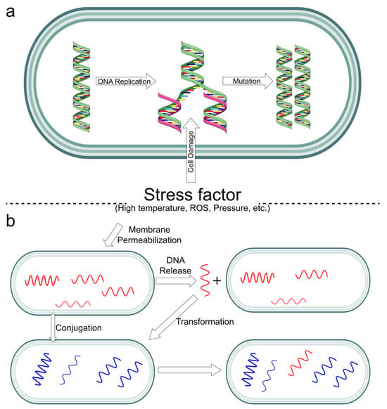
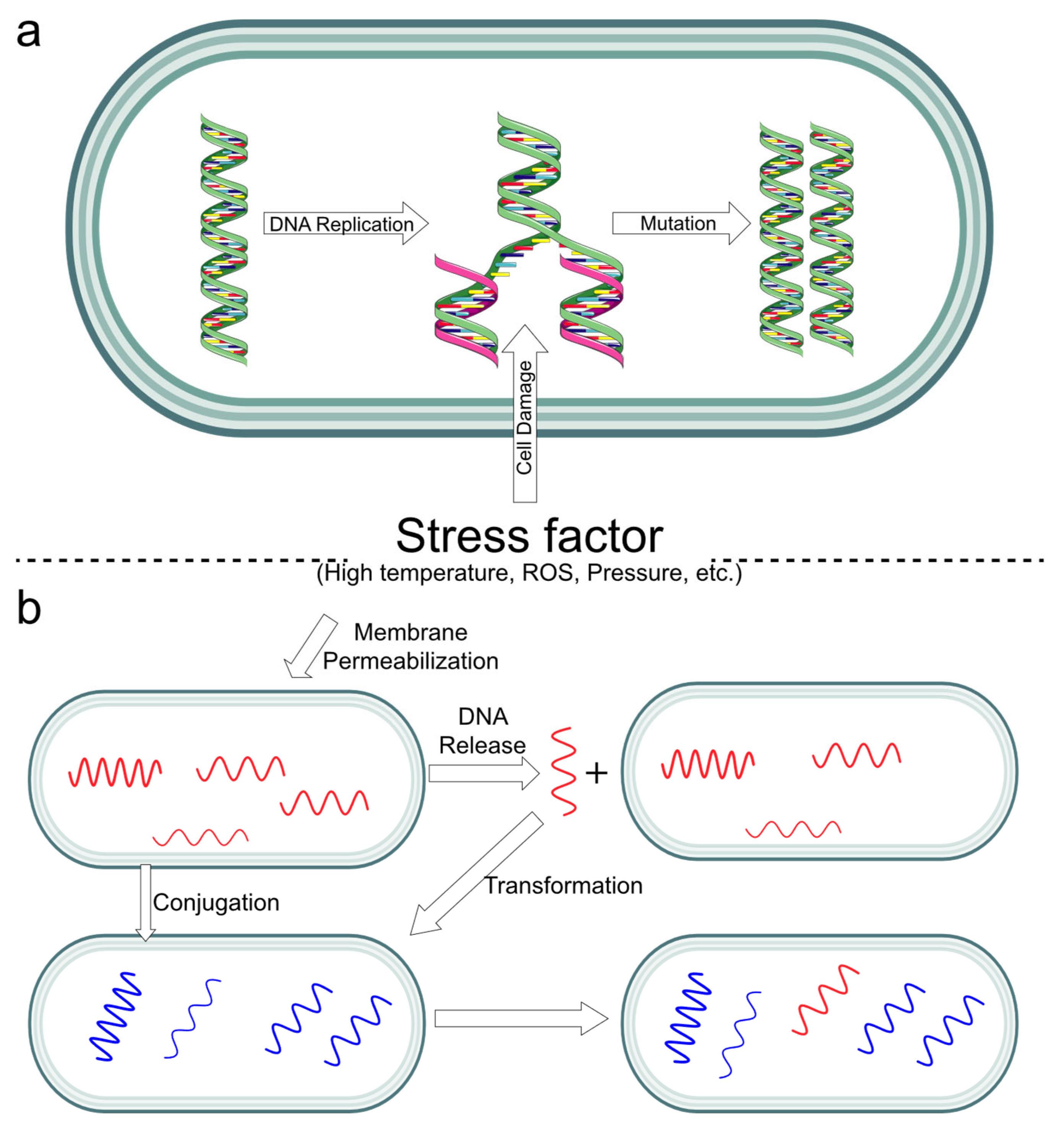
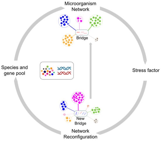
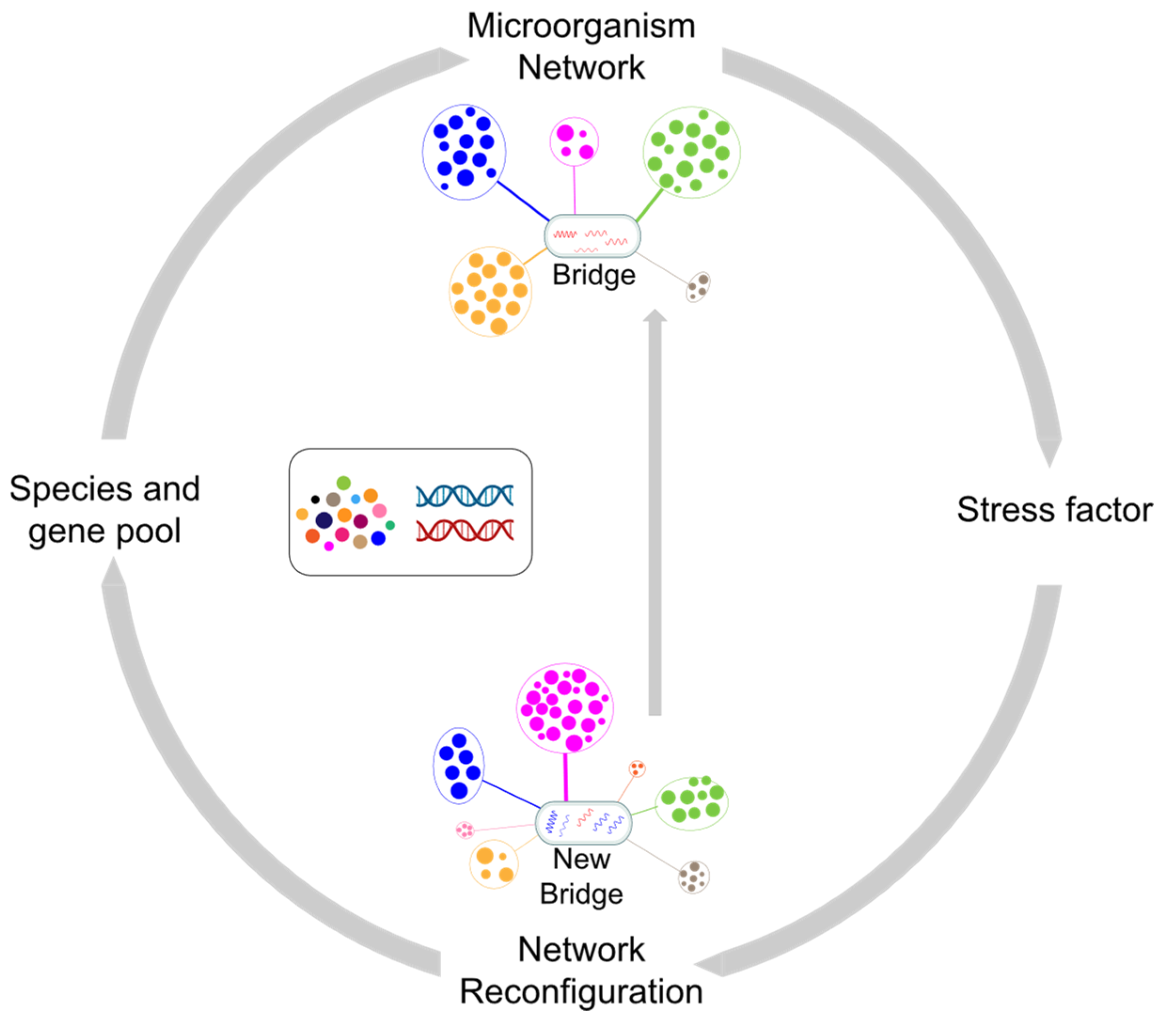

Disclaimer/Publisher’s Note: The statements, opinions and data contained in all publications are solely those of the individual author(s) and contributor(s) and not of MDPI and/or the editor(s). MDPI and/or the editor(s) disclaim responsibility for any injury to people or property resulting from any ideas, methods, instructions or products referred to in the content. |
© 2026 by the authors. Licensee MDPI, Basel, Switzerland. This article is an open access article distributed under the terms and conditions of the Creative Commons Attribution (CC BY) license.