1. Introduction
Plants possess a sophisticated, multi-layered innate immune system that primarily comprises two interconnected defense tiers: pattern-triggered immunity (PTI) and effector-triggered immunity (ETI) [
1,
2]. PTI is initiated when cell-surface pattern recognition receptors (PRRs) detect conserved microbe- or pathogen-associated molecular patterns (MAMPs or PAMPs), thereby establishing basal defense responses [
3,
4]. To counteract this, pathogens secrete effector proteins that suppress host immune signaling. In turn, these effectors can be recognized by plant intracellular resistance proteins, leading to activation of ETI, which is often associated with a hypersensitive response and programmed cell death that effectively restrict pathogen spread [
5,
6].
As the first line of defense in plant immunity, PRR-mediated recognition plays a central role in immune activation. All known PRRs are cell-surface localized receptor-like kinases (RLKs) or receptor-like proteins (RLPs), both of which contain an extracellular domain (ECD) and a single-pass transmembrane helix. The primary structural distinction is that RLKs possess an intracellular kinase domain, whereas RLPs lack this catalytic activity. These receptor families have undergone extensive expansion in plant genomes, and constitute a substantial proportion of protein-coding genes [
7]. For instance, the
Arabidopsis genome encodes more than 600 RLKs, representing approximately 2.5% of all protein-coding genes, underscoring their broad involvement in diverse biological processes, including immune signaling [
8,
9,
10].
Emerging evidence has extended the functional scope of plant cell-surface receptors from pathogen recognition to the perception of abiotic environmental cues, particularly temperature-related stress [
11]. For instance, heat stress compromises the accumulation of the prototypical PRR FLS2 and attenuates flg22-induced immune outputs, whereas cold stress transcriptionally upregulates several PRRs, including FLS2 and its co-receptor BAK1. Moreover, certain receptors, such as the leucine-rich repeat (LRR) RLK KOIN (KINASE ON THE INSIDE), directly participate in cold signaling [
12]. Together, these findings highlight the necessity of characterizing PRR functions under complex, real-world conditions where biotic and abiotic stresses frequently co-occur.
Most well-characterized PRRs contain leucine-rich repeat (LRR) ECDs, which represent the most prevalent class of plant RLKs and RLPs. One of the best-studied LRR-RLKs in
Arabidopsis is FLS2, which recognizes the bacterial peptide epitope flg22 derived from flagellin. Upon flg22 perception, the co-receptor BAK1 associates with FLS2 and phosphorylates
Botrytis-induced kinase 1 (BIK1), thereby initiating downstream signaling cascades that promote plant immune responses [
13].
Although most characterized PRRs possess an LRR-type ECD, several additional receptor families have also been identified in plants. The
Arabidopsis genome encodes approximately 75 lectin receptor-like kinases (LecRLKs), including 42 L-type, 32 G-type, and 1 C-type members [
14,
15]. L-type LecRLKs harbor a single legume-lectin domain in their extracellular region, and C-type LecRLKs contain a calcium-dependent lectin domain, whereas G-type LecRLKs contain an α-D-mannose-binding bulb lectin domain together with an S-locus glycoprotein (SLG) domain, and may additionally contain an epidermal growth factor (EGF) domain and/or a plasminogen-apple-nematode (PAN) domain. For example, RK3 contains a PAN domain. Notably, LecRLKs appear to be plant-specific, as no homologs have been identified in yeast or human genomes. Emerging evidence indicates that LecRLKs play important roles in plant innate immunity [
16]. Initially predicted to bind carbohydrate-associated ligands due to their lectin domains, several LecRLKs have since been shown to function as receptors for extracellular ATP (eATP), NAD
+ (eNAD
+), NADP
+ (eNADP
+), and the bacterial metabolite 3-OH-C10:0, mediating plant immune responses [
15]. Although many
LecRLKs are transcriptionally induced during pathogen invasion or tissue damage [
17,
18], the biological functions and molecular mechanisms of most LecRLKs in plant immunity remain poorly understood.
Using gene expression analysis from Expression Atlas (
https://www.ebi.ac.uk/gxa, accessed on 27 January 2026), we identified
RK3 (
AT4G21380) as one of the most strongly induced G-type LecRLK genes following treatment with flg22. A previous study also reported that
RK3 expression is upregulated by the intracellular coiled-coil nucleotide-binding leucine-rich repeat (CC-NB-LRR) receptor proteins RPP7 and RPP4 in response to the oomycete pathogen
Peronospora parasitica, suggesting a potential role in
Arabidopsis immunity [
19]. However, the molecular mechanisms of
RK3 in plant immunity remain unclear.
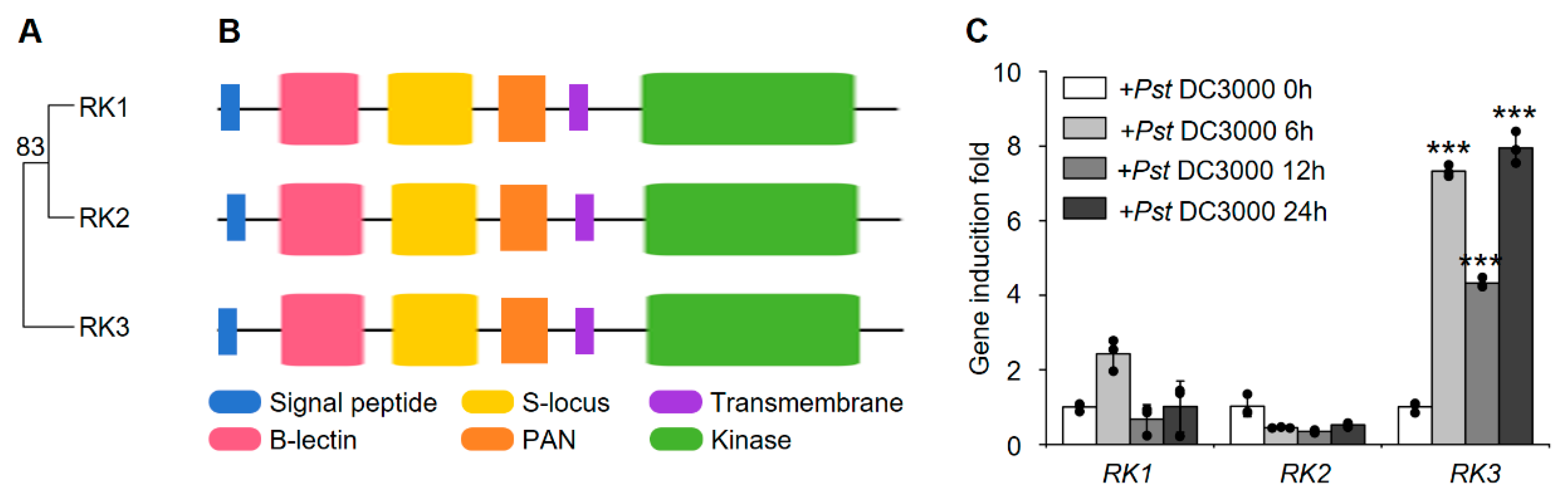
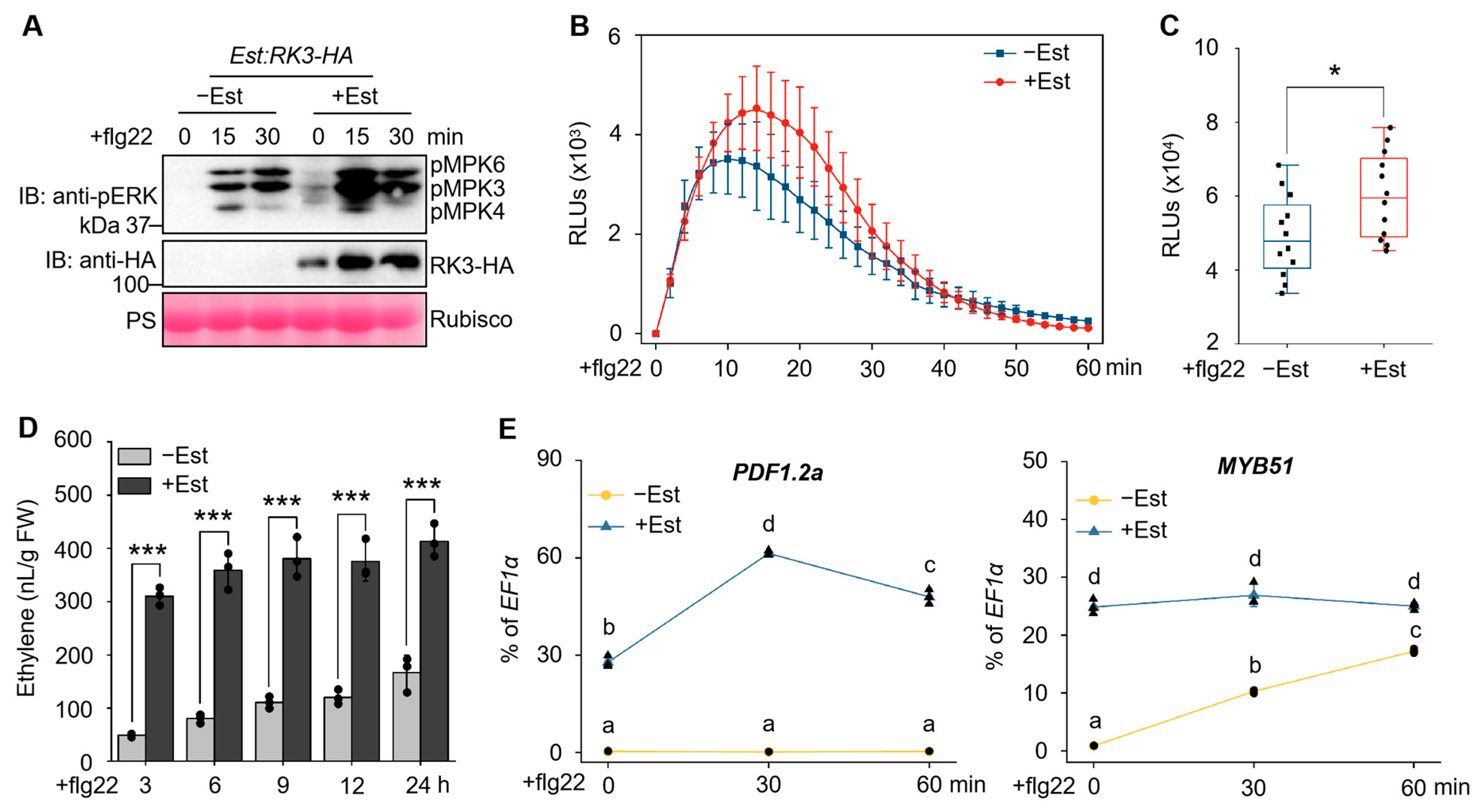
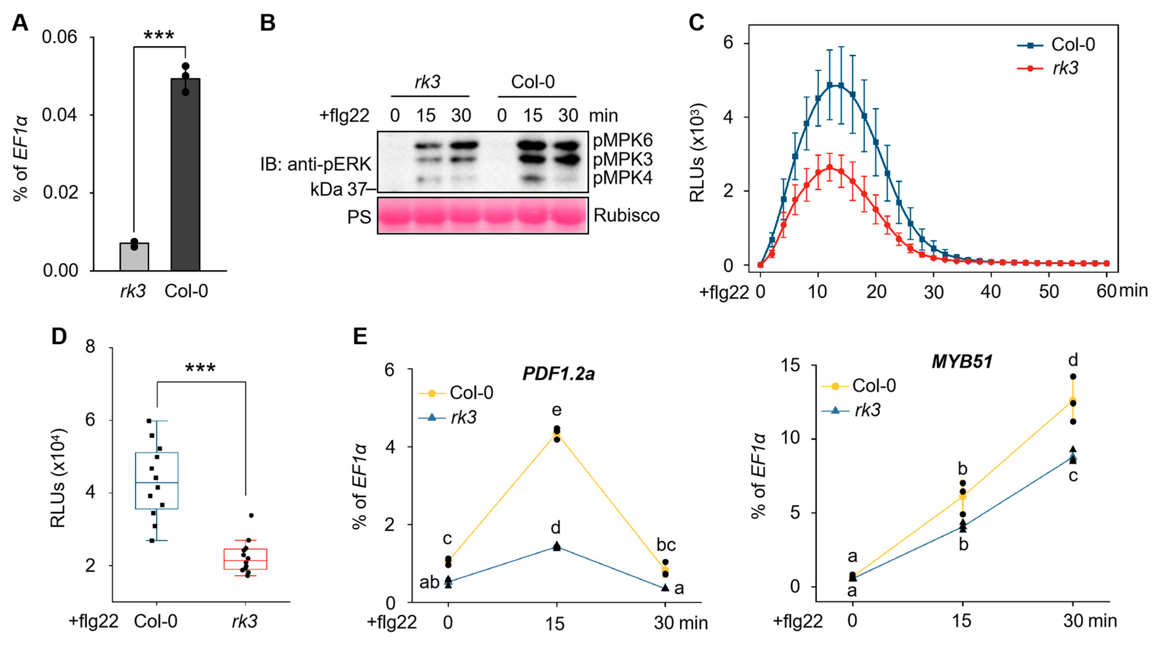
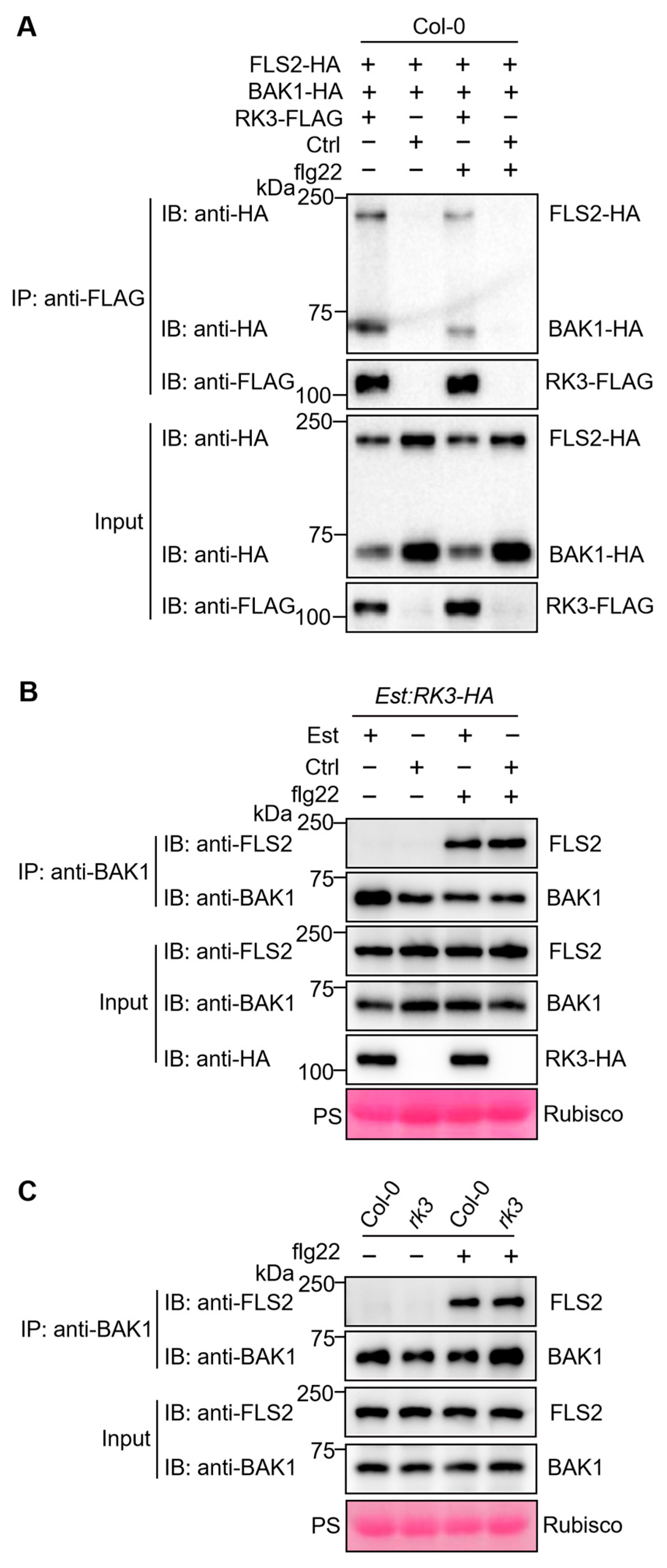
In this study, we demonstrated that RK3 expression is strongly induced following infection with Pseudomonas syringae pv. tomato DC3000 (Pst DC3000). Further genetic analyses using RK3 overexpressing plants and T-DNA insertion mutants demonstrated that RK3 positively regulates flg22-triggered immune responses, including MAPK activation, reactive oxygen species (ROS) burst, defense gene expression, and resistance to Pst DC3000. Co-immunoprecipitation assays revealed that RK3 constitutively interacts with the PRR complex components FLS2 and BAK1. Notably, a kinase domain-deleted version of RK3 (RK3-ΔK) maintained its interaction with FLS2 and BAK1, and retained immune-enhancing activity, suggesting that the extracellular domain of RK3 through its simultaneous association with FLS2 and BAK1, supports flg22 signaling without requiring kinase activity. Furthermore, interfamily transfer of RK3 into tomato significantly enhanced disease resistance, highlighting its potential utility for engineering disease resistance in crops. Collectively, our findings identified RK3 as a novel positive regulator of pattern-triggered immunity and revealed a previously unrecognized functional mode of action for G-type LecRLKs.
2. Materials and Methods
2.1. Biological Materials
Wild-type controls consisted of Arabidopsis thaliana (Col-0) and tomato (Solanum lycopersicum cv. Micro-Tom). The T-DNA insertional lines rk3 (SALK_109125) and rk3-2 (SALK_001986) were obtained from the Arabidopsis Biological Resource Center (ABRC).
For transcriptional profiling, MAPK activity assays, and ethylene production measurements, surface-sterilized seeds were plated onto half-strength Murashige and Skoog (MS) medium and kept at 22 °C under continuous illumination (60 μE m−2 s−1) for 7 days. They were then transferred into sealed gas chromatography (GC) vials containing liquid half-strength MS medium and incubated for an additional 3–10 days. Thereafter, the seedlings were treated with 10 μM estradiol, 100 nM flg22, 100 nM/300 nM/1 μM flgII-28, or the corresponding solvent controls. Ethylene accumulation was recorded at predetermined time points, and tissue samples were harvested for reverse transcription quantitative PCR (RT-qPCR) or immunoblotting.
For protoplast isolation, ROS burst measurements, and pathogen challenge experiments, plants were grown in soil under a 14 h light/10 h dark cycle at 22 °C for 4 weeks preceding leaf collection.
2.2. Molecular Cloning and Plant Transformation
The coding sequence (CDS) of
RK3 was retrieved from the TAIR database (
https://www.arabidopsis.org/, accessed on 6 October 2021). Primers were designed using SnapGene software (version 6.0, from GSL Biotech LLC, Chicago, IL, USA): the forward primer incorporated
SalI and
BamHI sites, and the reverse primer incorporated
SpeI and
StuI sites (all restriction enzymes were purchased from Thermo Fisher Scientific, Waltham, MA, USA). Primers were synthesized by Sangon Biotech (Shanghai, China). The CDS was amplified from Col-0 cDNA by PCR using FastPfu high-fidelity DNA polymerase (TransGen Biotech, Beijing, China) and first ligated into a
pMD19T-vector (Takara, Kusatsu, Japan). After Sanger sequencing verification (Sangon Biotech), the confirmed fragment was digested with
SalI and
SpeI and ligated into the estradiol-inducible
pER8-Est:HA vector to generate
Est:RK3-HA. For transient protoplast assays, the same verified CDS was digested with
BamHI and
StuI and inserted into
pHBT-35S:FLAG, yielding
35S:RK3-FLAG. All constructs were verified by colony PCR and restriction digestion before use.
The kinase-domain-deleted variant, RK3-ΔK, was generated by designing a reverse primer immediately upstream of the predicted kinase domain (deleting the kinase domain and all subsequent sequences), while retaining the same forward and reverse restriction sites (SalI/BamHI and SpeI/StuI, respectively) as for the full-length RK3. Using the sequence-verified pMD19T-RK3 as template, the truncated CDS was amplified by PCR and then cloned into pER8-Est:HA and pHBT-35S:Myc following the same procedure (pMD19T-vector cloning, Sanger sequencing, restriction digestion with SalI/SpeI for pER8 and BamHI/StuI for pHBT), resulting in Est:RK3-ΔK-HA and 35S:RK3-ΔK-Myc, respectively.
For co-immunoprecipitation experiments, the CDSs of FLS2 and BAK1 were individually amplified and cloned into
pHBT-35S:HA via
BamHI and
StuI sites, yielding
35S:FLS2-HA and
35S:BAK1-HA. The same
pMD19T-vector and sequencing steps were applied to ensure sequence fidelity. All primer sequences used for these constructs are listed in
Supplementary Table S2.
Arabidopsis transformants (Col-0 background) were generated via
Agrobacterium-mediated floral dip [
20]. For each construct, roughly 50 T1 lines were examined for transgene expression by anti-HA immunoblotting (anti-HA antibody from Thermo Fisher Scientific). Among these, T2 lines harboring a single T-DNA insertion (confirmed by segregation analysis on hygromycin-containing medium) and displaying robust, reproducible transgene expression were selected for all subsequent assays. To conserve space in the main figures, data from one representative line are presented; this line faithfully reflected the phenotypes observed across multiple independent transformants. All results from another independent line are shown in the
Supplementary Figures.
2.3. Bioinformatic Analysis
2.4. Transcript Level Analysis
RNA extraction and cDNA synthesis were performed according to the manufacturer’s instructions (TRIzol, Invitrogen, Carlsbad, CA, USA; PrimeScript RT, Takara). Relative transcript levels were determined by RT-qPCR using a CFX Connect Real-Time PCR System (Bio-Rad, Hercules, CA, USA) with SYBR Green detection (Toyobo, Osaka, Japan), as described previously [
21].
EF1α served as the endogenous control for
Arabidopsis, and
ACT for tomato. Three biological replicates were performed for each genotype and treatment. Relative expression was calculated using the 2−ΔΔCt method [
22] and presented either as fold change relative to untreated controls or as a percentage of the reference gene transcript level. All primer sequences are listed in
Supplementary Table S1.
2.5. Ethylene Quantification
Ethylene production was quantified by gas chromatography (PANNA A60, Beijing, China) as described previously [
21]. Two-week-old
Arabidopsis or 2.5-week-old tomato seedlings grown in GC vials with hydroponic medium were treated with 10 μM estradiol (Est), 100 nM flg22 (for
Arabidopsis), 100 nM flgII-28 (for tomato), or solvent controls (0.1% DMSO for estradiol). A standard curve was generated using ethylene standard gas (99.9% purity, National Institute of Metrology, Beijing, China). At specified times post-elicitation, headspace gas was collected, and ethylene concentrations—normalized to fresh weight—were derived from standard curves. Three independent biological replicates (10
Arabidopsis or 3 tomato seedlings per vial) were analyzed for each treatment and time point.
2.6. Co-Immunoprecipitation and Immunoblotting Assays
Protoplast isolation, transformation, and co-immunoprecipitation (co-IP) assays were carried out as described previously [
21] with the following buffer specifications. The washing buffer contained 100 mM NaCl, 10 mM HEPES (pH 7.5), 1 mM EDTA (pH 8.0), 10% (
v/
v) glycerol, and 0.5% (
v/
v) Triton X-100. The lysis buffer was prepared by adding the following inhibitors per 1 mL of washing buffer immediately before use: 2 μL of 1 M NaF (final 2 mM), 2 μL of 1 M Na
3VO
4 (final 2 mM), 2.5 μL of 0.4 M DTT (final 1 mM), and 20 μL of 50× protease inhibitor cocktail (Thermo Fisher Scientific; final 1×). After an overnight recovery period, the transformed protoplasts were exposed to 100 nM flg22 for 10 min prior to protein extraction. Immunoprecipitation was conducted using anti-FLAG (Thermo Fisher Scientific), anti-BAK1 (Agrisera, Vännäs, Sweden), or anti-Myc (Sigma-Aldrich, St. Louis, MO, USA) antibodies, and subsequent immunoblotting was performed with anti-HA, anti-FLAG, anti-Myc, anti-FLS2 (Agrisera), or anti-BAK1 antibodies, all following published procedures [
21].
For MAPK activation assays, total proteins extracted from 10-day-old
Arabidopsis or 14-day-old tomato seedlings treated with 100 nM flg22 or 300 nM flgII-28 (sampled at the indicated time points). The extraction buffer was the same as the lysis buffer described above. Protein extracts were subjected to immunoblot analysis using anti-phospho-ERK1/2 antibody (Sigma-Aldrich) as described previously [
21].
2.7. Measurement of Reactive Oxygen Species Burst
To measure ROS production, a luminol-based chemiluminescence assay was employed as described previously [
23]. Leaf discs (5 mm diameter) excised from 4-week-old plants were pre-incubated in either sterile water or 10 μM estradiol for 24 h. After this treatment, the discs were transferred into a reaction mixture containing 100 nM flg22, 100 μM luminol (Sigma-Aldrich), and 10 μg/mL horseradish peroxidase (HRP, Sigma-Aldrich). Luminescence was then recorded at 2 min intervals for a total of 60 min using a GloMax microplate luminometer (Promega, Madison, WI, USA). For each treatment, at least 20 leaf discs were used as biological replicates, and the data are presented either as kinetic curves of relative luminescence over time or as integrated relative light units (RLU).
2.8. Evaluation of Pathogen Resistance
For disease resistance assays, leaves of 4-week-old transgenic plants were first infiltrated with either 10 μM estradiol or the corresponding solvent control (0.1% DMSO). After 24 h, the same leaves were challenged with a bacterial suspension of
Pst DC3000 adjusted to an optical density at 600 nm (OD
600) of 0.0005 in 10 mM MgCl
2. Three days post-inoculation, bacterial multiplication was determined as described previously [
21]. In brief, leaf discs (0.2 cm
2 each) were collected from three individual plants per genotype and treatment (serving as three biological replicates), pooled, homogenized, serially diluted, and plated on Luria–Bertani (LB) agar containing 50 μg/mL rifampicin (Yeasen Biotech, Shanghai, China). Each dilution was plated in triplicate as technical replicates.
2.9. Generation of Transgenic Tomato Plants
Tomato (
Solanum lycopersicum cv. Micro-Tom) transformation was carried out as described previously, with modifications [
24]. Leaf explants (5 mm × 5 mm) excised from 10-day-old seedlings were preincubated for 1 day on T1 medium (MS salts containing 3% sucrose, 2 mg/L zeatin, and 0.1 mg/L IAA). The explants were then infected with an
Agrobacterium tumefaciens strain GV3101 suspension (OD
600 = 0.3 in T1 medium supplemented with 100 μM acetosyringone, Sigma-Aldrich) for 10 min, followed by co-cultivation on the same medium for 2 days in the dark. After co-cultivation, explants were moved to selection medium (T1 medium with 10 mg/L hygromycin and 200 mg/L timentin) to induce shoot regeneration. Regenerated shoots of 2–3 cm in length were excised and rooted on half-strength MS medium containing 3% sucrose, 2 mg/L IBA, and 100 mg/L timentin. Putative transgenic lines were initially screened by hygromycin resistance, and positive transformants were confirmed by immunoblotting using anti-HA antibody. For each construct, at least two independent T1 lines were selected and propagated to the T2 generation; all subsequent experiments were performed using T2 plants derived from these independent lines.
2.10. Data Analysis and Statistics
Results are shown as mean ± standard deviation (SD), with the number of independent biological replicates (n ≥ 3) indicated in each figure legend. Pairwise comparisons between two groups were evaluated using two-tailed Student’s t-test. For multiple comparisons, one-way or two-way analysis of variance (ANOVA) followed by Tukey’s honest significant difference (HSD) post hoc test was applied. Statistical analyses were performed using commercial software (IBM SPSS Statistics version 27, IBM, Armonk, NY, USA; GraphPad Prism version 9.5.1, GraphPad Software, San Diego, CA, USA). Significance levels are denoted as follows: * p < 0.05, ** p < 0.01, *** p < 0.001 for pairwise tests, and different lowercase letters (p < 0.05) for multiple comparisons.
4. Discussion
LecRLKs constitute a large subfamily of RLKs, and their extracellular lectin domains are thought to reversibly bind carbohydrate ligands. Among LecRLKs, G-type members possess the most structurally complex extracellular structure, featuring multiple additional domains beyond the lectin domain. Notably, the SLG domain is homologous to S-locus glycoproteins that govern self- and interspecific incompatibility in
Brassica species [
30,
31,
32]. In self-compatible species such as
Arabidopsis, S-locus-related genes have undergone functional divergence, enabling them to regulate vegetative growth and stress adaptation [
15].
Many G-type LecRLKs are transcriptionally induced upon pathogen invasion, and accumulating evidence highlights their involvement in plant immunity. However, side-by-side comparison reveals that RK3 exhibits a unique combination of features that distinguish it from all previously characterized members. For instance, the identification of LORE as the receptor for the bacterial metabolite 3-OH-C10:0 underscores the importance of G-type LecRLKs in immune activation [
18]. In contrast, RK3 does not directly bind a known ligand; instead, it constitutively associates with the FLS2-BAK1 complex to promote PTI.
RDA2 functions in [5-(3,4-dichlorophenyl)furan-2-yl]-piperidine-1-ylmethanethione (DFPM)-induced immune responses but does not participate in immune signaling mediated by peptide elicitors such as Pep1 and flg22 [
33]; RK3, by contrast, positively regulates flg22-triggered immunity. In
Nicotiana benthamiana, NbERK1 associates with BAK1 and SOBIR1 to regulate the perception of the expansin-like apoplastic elicitor protein, PcEXLX1 [
34], thereby enhancing resistance to
Phytophthora capsici. Although NbERK1 similarly interacts with BAK1, RK3 acts through a kinase-independent mechanism (as demonstrated by the functional RK3-ΔK variant), a feature not reported for NbERK1. In rice,
Pid2 confers resistance to
Magnaporthe oryzae strain ZB15; intriguingly, a single amino acid substitution in the transmembrane domain distinguishes resistant and susceptible alleles, yet the
Pid2-mediated signaling pathway remains largely unexplored [
35]. Unlike
Pid2, whose kinase domain is essential for resistance,
RK3 retains immune-enhancing activity even after deletion of its kinase domain. Conversely,
ERN1 negatively regulates immunity against root-knot nematodes, as evidenced by enhanced flg22-induced responses and disease resistance in
ern1 mutants, although the underlying mechanism remains unclear [
36]. Thus, while
ERN1 acts as a negative regulator,
RK3 serves as a positive regulator of PTI, further highlighting their functional divergence. Despite the apparent importance of G-type LecRLKs in disease resistance, only a small fraction has been functionally characterized. Taken together, these comparisons indicate that RK3 represents a previously unrecognized mode of action among G-type LecRLKs—one that is distinct from the conventional roles of direct ligand receptors (LORE), negative regulators (ERN1), or signaling adaptors that strictly require kinase activity (NbERK1, Pid2).
In this study, we found that
RK3 was highly induced by
Pst DC3000,
Pst DC3000 strains carrying AvrRpt2 or AvrRpm1, as well as the fungal pathogen
Botrytis cinerea. Biochemical analysis further revealed that RK3 constitutively interacted with FLS2 and BAK1 in a ligand-independent manner. Overexpression of
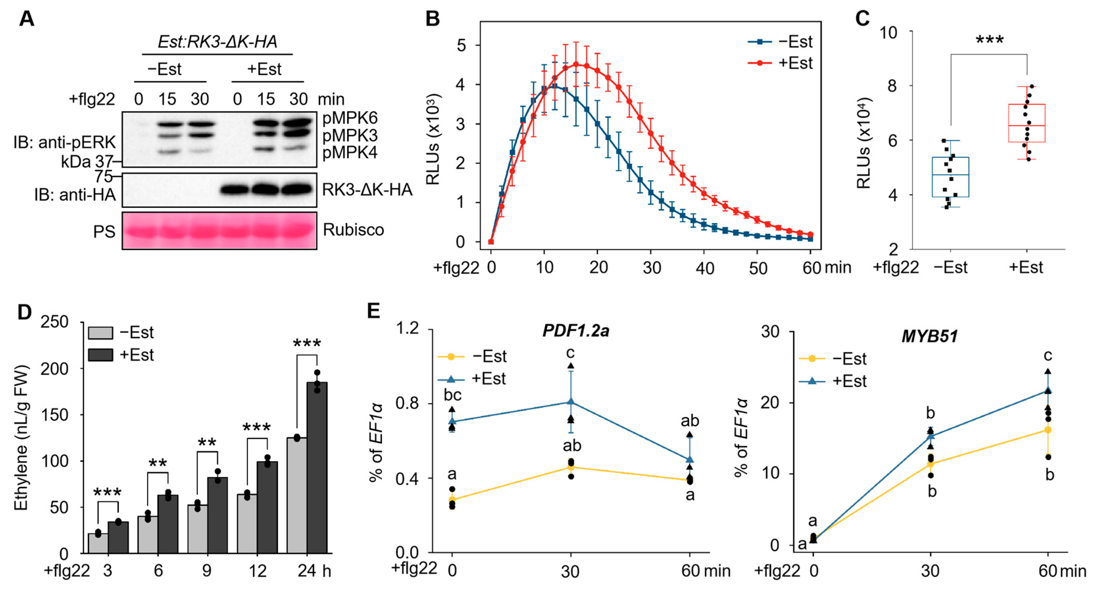
RK3 markedly enhanced flg22-induced hallmark responses including ROS burst, MAPK activation, ethylene production and increased expression of marker genes; conversely, the loss-of-function T-DNA insertion mutants attenuated these responses. These results suggest that RK3, through its constitutive association with the FLS2-BAK1 complex, promotes flg22-induced immune responses. Notably, deletion of the KD of RK3 did not affect the interaction of RK3 with FLS2 or BAK1, nor did it impair its ability to enhance flg22-induced immune responses, indicating that the ECD of RK3 is likely the functional region responsible for RK3-mediated immune potentiation.
RK3 was previously proposed to be involved in the switch from self-incompatibility to self-fertility in
A. thaliana [
31]. Our findings uncovered a distinct mode of function for
RK3 in self-compatible
A. thaliana, suggesting a functional evolutionary shift in S-locus genes.
RK3 and its two close homologs,
RK1 and
RK2, belong to clade VI of G-lectin LecRLKs (subclade A1b) [
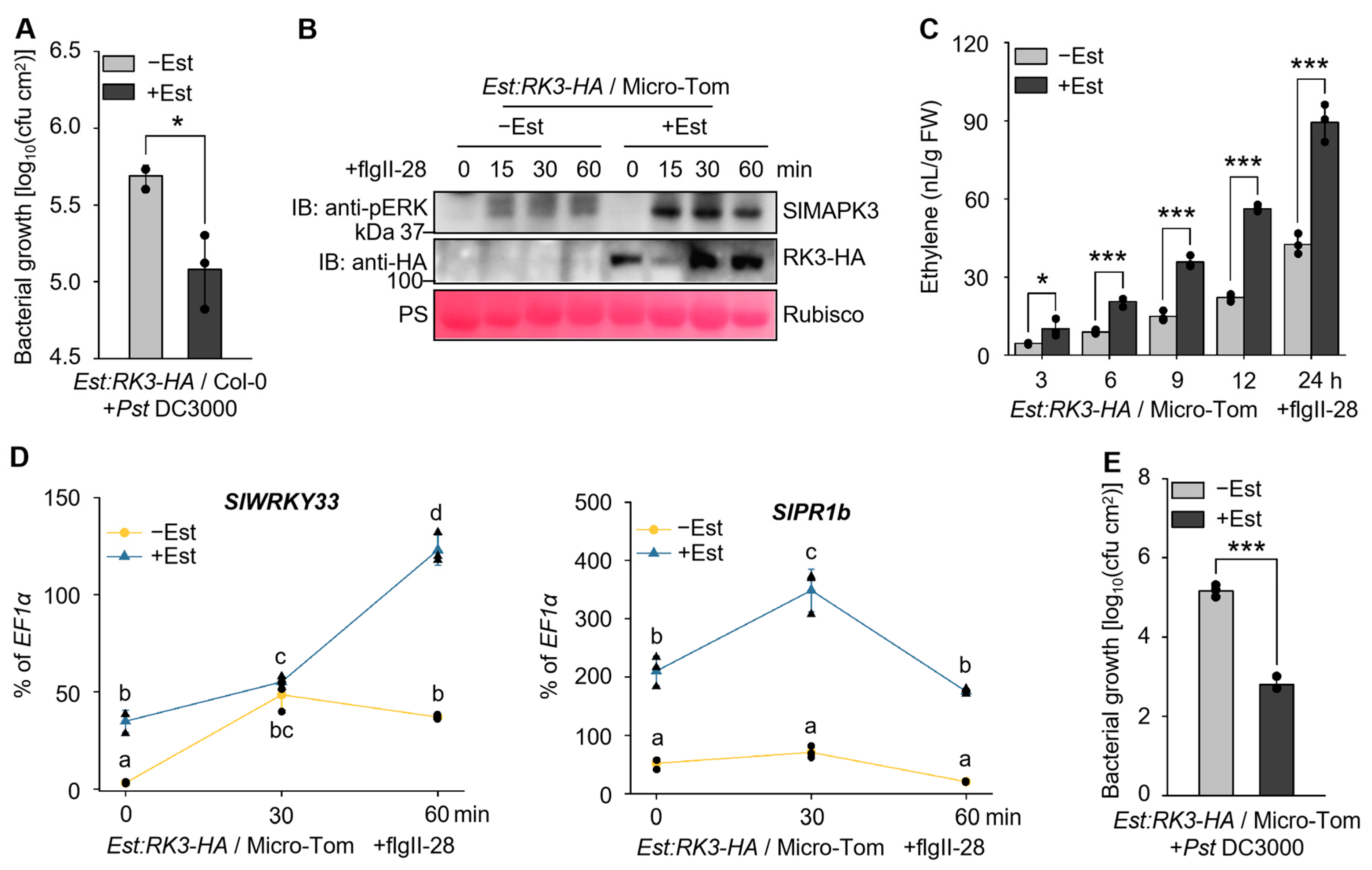
25]. Our study revealed that RK3 promotes PTI by constitutively associating with an LRR-RLK-type PRR complex, and this function extends beyond FLS2, as ectopic expression of
RK3 in tomato also enhances flgII-28-induced immune responses. Interestingly, the other two members of clade VI, SBP1 (At4g27300) and SBP2 (At4g27290) (subclade A1c), form LRR-RLP-type receptor complexes through similar ligand-independent interactions with multiple RLPs and the co-receptors BAK1 and SOBIR1. Although kinase activity is dispensable for SBP1/SBP2 function in promoting ligand-induced immunity, their interactions are mediated by the kinase domain rather than the extracellular domain. Moreover, the contribution of SBP1/SBP2 to RLP-mediated immunity is comparable to that of SOBIR1 [
37]. Taken together with our findings, these observations underscore the critical role of clade VI of G-lectin LecRLKs in sustaining full PTI output. Although pathogen- and flg22-induced expression of
RK1 and
RK2 is relatively low, we examined flg22-induced ethylene production in
rk2 T-DNA insertion lines and found no significant reduction compared with wild-type plants. For
RK1, the very low level of transcriptional induction precluded a detailed PTI analysis in this study. These results suggest that
RK3 is a major contributor to flg22-triggered immunity. Whether
RK1 and
RK2 contribute to LRR-RLK-mediated immunity or other biological processes warrants future investigation.
In
Arabidopsis, several RLKs belonging to distinct classes have been shown to interact with FLS2 and/or BAK1 to modulate flg22-induced immune signaling. For instance, the malectin-type RLK, FER, scaffolds the FLS2-BAK1 complex in a flg22-dependent manner and promotes its formation [
38]. This function is inhibited by RALF peptide perception and requires the KD of FER [
39]. Similarly, the Malectin/LRR-RLK, IOS1, constitutively associates with FLS2 and BAK1, and enhances MAMP-induced complex formation [
40]. In contrast, three LRR-RLKs—BIR2, NIK1 and QSK1—negatively regulate flg22-triggered immunity through constitutive or flg22-dependent interactions with BAK1 and/or FLS2 [
41,
42,
43]. Our study identified a G-type LecRLK, RK3, as a new type of positive regulator that interacts with FLS2 and BAK1 independently of flg22 stimulation. However, unlike canonical scaffolds that promote ligand-induced receptor heterodimerization or stabilize the components of the complex, RK3 does not appear to enhance flg22-triggered FLS2-BAK1 association or protein abundance. RK3 may help organize the signaling-competent nanodomain at the plasma membrane, facilitating the efficient recruitment or activation of downstream signaling components upon PAMP perception [
44]. Future studies are needed to elucidate the mechanistic basis of RK3-mediated immune potentiation. Moreover, understanding how diverse positive and negative regulators coordinately interact with FLS2/BAK1 in vivo across spatiotemporal dimensions will provide a more comprehensive view of plant PTI regulation.
5. Conclusions
In this study, we identified RK3, a G-type LecRLK, as a positive regulator of PTI in Arabidopsis. RK3 expression is rapidly and strongly induced upon bacterial and fungal pathogen challenge. Through gain- and loss-of-function analyses, we demonstrated that RK3 potentiates flg22-triggered immune outputs, including MAPK activation, ROS burst, ethylene production, defense gene expression, and enhanced resistance to Pst DC3000. Mechanistically, RK3 constitutively associates with the FLS2-BAK1 receptor complex in a ligand-independent manner, yet it does not promote flg22-induced FLS2-BAK1 heteromerization. Remarkably, the kinase domain of RK3 is dispensable for its immune-enhancing activity, as a kinase-deleted variant (RK3-ΔK) retains both the interaction with FLS2/BAK1 and the ability to boost flg22-triggered immunity. This reveals a non-canonical, kinase-independent mode of action for a G-type LecRLK in PTI signaling. Furthermore, ectopic expression of RK3 in tomato enhances flgII-28-induced immune responses and bacterial resistance, indicating that RK3 function is conserved between the crucifer and nightshade families, highlighting its potential as a tool for engineering broad-spectrum disease resistance in crops. Collectively, our findings uncover a previously unrecognized regulatory mechanism in plant immunity, expanding the functional repertoire of G-type LecRLKs and providing a valuable target for crop improvement.