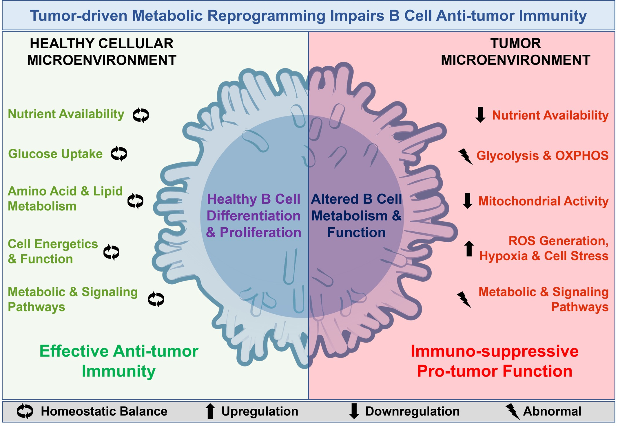
Metabolic Reprogramming of B Cells in Cancer: Effects of Altered Energetics

 , , and
Simple Summary
Abstract
1. Introduction
2. Metabolic Pathways in B Cells: A Functional Overview
3. TME and Its Role in Altered B Cell Metabolism
4. Metabolic Reprogramming of B Cells in Cancer
5. Functional Consequences of Altered Energetics in B Cells
6. Therapeutic Opportunities in Altered B Cell Metabolism
6.1. Therapeutic Approaches for Targeting Metabolic Alteration in Malignant B Cells
- (A)
- Targeting mTORC1/mTORC2 pathway
- (B)
- Targeting PI3K-AKT-mTOR pathway
- (C)
- Targeting amino acid and lipid metabolism
- (D)
- Targeting mitochondrial dynamics and inducing apoptosis
6.2. Therapeutic Strategy for Targeting Tumor-Infiltrating B Cells (TIL-B Cells) in Cancer
7. Challenges and Future Directions
8. Conclusions
Author Contributions
Funding
Institutional Review Board Statement
Informed Consent Statement
Data Availability Statement
Acknowledgments
Conflicts of Interest
References
- Wang, J.; He, Y.; Hu, F.; Hu, C.; Sun, Y.; Yang, K.; Yang, S. Metabolic Reprogramming of Immune Cells in the Tumor Microenvironment. Int. J. Mol. Sci. 2024, 25, 12223. [Google Scholar] [CrossRef]
- Phad, G.E.; Pinto, D.; Foglierini, M.; Akhmedov, M.; Rossi, R.L.; Malvicini, E.; Cassotta, A.; Fregni, C.S.; Bruno, L.; Sallusto, F.; et al. Clonal structure, stability and dynamics of human memory B cells and circulating plasmablasts. Nat. Immunol. 2022, 23, 1076–1085. [Google Scholar] [CrossRef]
- Bader, J.E.; Voss, K.; Rathmell, J.C. Targeting Metabolism to Improve the Tumor Microenvironment for Cancer Immunotherapy. Mol. Cell 2020, 78, 1019–1033. [Google Scholar] [CrossRef]
- Ammirante, M.; Luo, J.L.; Grivennikov, S.; Nedospasov, S.; Karin, M. B-cell-derived lymphotoxin promotes castration-resistant prostate cancer. Nature 2010, 464, 302–305. [Google Scholar] [CrossRef]
- Helmink, B.A.; Reddy, S.M.; Gao, J.; Zhang, S.; Basar, R.; Thakur, R.; Yizhak, K.; Sade-Feldman, M.; Blando, J.; Han, G.; et al. B cells and tertiary lymphoid structures promote immunotherapy response. Nature 2020, 577, 549–555. [Google Scholar] [CrossRef]
- Buck, M.D.; Sowell, R.T.; Kaech, S.M.; Pearce, E.L. Metabolic Instruction of Immunity. Cell 2017, 169, 570–586. [Google Scholar] [CrossRef]
- Caro-Maldonado, A.; Wang, R.; Nichols, A.G.; Kuraoka, M.; Milasta, S.; Sun, L.D.; Gavin, A.L.; Abel, E.D.; Kelsoe, G.; Green, D.R.; et al. Metabolic reprogramming is required for antibody production that is suppressed in anergic but exaggerated in chronically BAFF-exposed B cells. J. Immunol. 2014, 192, 3626–3636. [Google Scholar] [CrossRef]
- Jellusova, J.; Cato, M.H.; Apgar, J.R.; Ramezani-Rad, P.; Leung, C.R.; Chen, C.; Richardson, A.D.; Conner, E.M.; Benschop, R.J.; Woodgett, J.R.; et al. Gsk3 is a metabolic checkpoint regulator in B cells. Nat. Immunol. 2017, 18, 303–312. [Google Scholar] [CrossRef]
- Metur, S.P.; Klionsky, D.J. Adaptive immunity at the crossroads of autophagy and metabolism. Cell. Mol. Immunol. 2021, 18, 1096–1105. [Google Scholar] [CrossRef]
- Li, S.; Zhang, Y.; Tong, H.; Sun, H.; Liao, H.; Li, Q.; Ma, X. Metabolic regulation of immunity in the tumor microenvironment. Cell Rep. 2025, 44, 116463. [Google Scholar] [CrossRef]
- Wang, Z.; Lu, Z.; Lin, S.; Xia, J.; Zhong, Z.; Xie, Z.; Xing, Y.; Qie, J.; Jiao, M.; Li, Y.; et al. Leucine-tRNA-synthase-2-expressing B cells contribute to colorectal cancer immunoevasion. Immunity 2022, 55, 1748. [Google Scholar] [CrossRef]
- Hu, T.; Liu, C.H.; Lei, M.; Zeng, Q.; Li, L.; Tang, H.; Zhang, N. Metabolic regulation of the immune system in health and diseases: Mechanisms and interventions. Signal Transduct. Target. Ther. 2024, 9, 268. [Google Scholar] [CrossRef]
- Imahashi, N.; Basar, R.; Huang, Y.; Wang, F.; Baran, N.; Banerjee, P.P.; Lu, J.; Nunez Cortes, A.K.; Uprety, N.; Ensley, E.; et al. Activated B cells suppress T-cell function through metabolic competition. J. Immunother. Cancer 2022, 10, e005644. [Google Scholar] [CrossRef]
- Muraoka, S.; Baba, T.; Akazawa, T.; Katayama, K.I.; Kusumoto, H.; Yamashita, S.; Kohjimoto, Y.; Iwabuchi, S.; Hashimoto, S.; Hara, I.; et al. Tumor-derived lactic acid promotes acetylation of histone H3K27 and differentiation of IL-10-producing regulatory B cells through direct and indirect signaling pathways. Int. J. Cancer 2025, 156, 840–852. [Google Scholar] [CrossRef]
- Warburg, O. On the origin of cancer cells. Science 1956, 123, 309–314. [Google Scholar] [CrossRef] [PubMed]
- Vander Heiden, M.G.; Cantley, L.C.; Thompson, C.B. Understanding the Warburg effect: The metabolic requirements of cell proliferation. Science 2009, 324, 1029–1033. [Google Scholar] [CrossRef] [PubMed]
- Chen, C.; Zheng, H.; Horwitz, E.M.; Ando, S.; Araki, K.; Zhao, P.; Li, Z.; Ford, M.L.; Ahmed, R.; Qu, C.K. Mitochondrial metabolic flexibility is critical for CD8(+) T cell antitumor immunity. Sci. Adv. 2023, 9, eadf9522. [Google Scholar] [CrossRef] [PubMed]
- Schindlmaier, K.; Haitzmann, T.; Bubalo, V.; Konrad, B.; Jelwan, J.; Bluemel, G.; Rittchen, S.; Jager, V.; Dengler, M.A.; Brcic, L.; et al. Metabolic adaptation of glucose-deprived macrophages involves partial gluconeogenesis. Proc. Natl. Acad. Sci. USA 2025, 122, e2419568122. [Google Scholar] [CrossRef]
- Huang, K.; Han, Y.; Chen, Y.; Shen, H.; Zeng, S.; Cai, C. Tumor metabolic regulators: Key drivers of metabolic reprogramming and the promising targets in cancer therapy. Mol. Cancer 2025, 24, 7. [Google Scholar] [CrossRef]
- Johnstone, J.C.; Yazicioglu, Y.F.; Clarke, A.J. Fuelling B cells: Dynamic regulation of B cell metabolism. Curr. Opin. Immunol. 2024, 91, 102484. [Google Scholar] [CrossRef]
- Doughty, C.A.; Bleiman, B.F.; Wagner, D.J.; Dufort, F.J.; Mataraza, J.M.; Roberts, M.F.; Chiles, T.C. Antigen receptor-mediated changes in glucose metabolism in B lymphocytes: Role of phosphatidylinositol 3-kinase signaling in the glycolytic control of growth. Blood 2006, 107, 4458–4465. [Google Scholar] [CrossRef] [PubMed]
- Simon-Molas, H.; Del Prete, R.; Kabanova, A. Glucose metabolism in B cell malignancies: A focus on glycolysis branching pathways. Mol. Oncol. 2024, 18, 1777–1794. [Google Scholar] [CrossRef]
- Sharma, R.; Smolkin, R.M.; Chowdhury, P.; Fernandez, K.C.; Kim, Y.; Cols, M.; Alread, W.; Yen, W.F.; Hu, W.; Wang, Z.M.; et al. Distinct metabolic requirements regulate B cell activation and germinal center responses. Nat. Immunol. 2023, 24, 1358–1369. [Google Scholar] [CrossRef]
- Waters, L.R.; Ahsan, F.M.; Wolf, D.M.; Shirihai, O.; Teitell, M.A. Initial B Cell Activation Induces Metabolic Reprogramming and Mitochondrial Remodeling. iScience 2018, 5, 99–109. [Google Scholar] [CrossRef]
- Gracie, C.J.; Mitchell, R.; Johnstone, J.C.; Clarke, A.J. The unusual metabolism of germinal center B cells. Trends Immunol. 2025, 46, 416–428. [Google Scholar] [CrossRef]
- Lam, W.Y.; Bhattacharya, D. Metabolic Links Between Plasma Cell Survival, Secretion, and Stress. Trends Immunol. 2018, 39, 19–27. [Google Scholar] [CrossRef] [PubMed]
- Price, M.J.; Patterson, D.G.; Scharer, C.D.; Boss, J.M. Progressive Upregulation of Oxidative Metabolism Facilitates Plasmablast Differentiation to a T-Independent Antigen. Cell Rep. 2018, 23, 3152–3159. [Google Scholar] [CrossRef]
- Peeters, R.; Jellusova, J. Lipid metabolism in B cell biology. Mol. Oncol. 2024, 18, 1795–1813. [Google Scholar] [CrossRef] [PubMed]
- Ji, X.; Wu, L.; Marion, T.; Luo, Y. Lipid metabolism in regulation of B cell development and autoimmunity. Cytokine Growth Factor Rev. 2023, 73, 40–51. [Google Scholar] [CrossRef]
- Won, W.J.; Bachmann, M.F.; Kearney, J.F. CD36 is differentially expressed on B cell subsets during development and in responses to antigen. J. Immunol. 2008, 180, 230–237. [Google Scholar] [CrossRef]
- Gong, Y.; Xu, R.; Gao, G.; Li, S.; Liu, Y. The role of fatty acid metabolism on B cells and B cell-related autoimmune diseases. Inflamm. Res. 2025, 74, 75. [Google Scholar] [CrossRef]
- Dufort, F.J.; Gumina, M.R.; Ta, N.L.; Tao, Y.; Heyse, S.A.; Scott, D.A.; Richardson, A.D.; Seyfried, T.N.; Chiles, T.C. Glucose-dependent de novo lipogenesis in B lymphocytes: A requirement for atp-citrate lyase in lipopolysaccharide-induced differentiation. J. Biol. Chem. 2014, 289, 7011–7024. [Google Scholar] [CrossRef] [PubMed]
- Iborra-Pernichi, M.; Ruiz Garcia, J.; Velasco de la Esperanza, M.; Estrada, B.S.; Bovolenta, E.R.; Cifuentes, C.; Prieto Carro, C.; Gonzalez Martinez, T.; Garcia-Consuegra, J.; Rey-Stolle, M.F.; et al. Defective mitochondria remodelling in B cells leads to an aged immune response. Nat. Commun. 2024, 15, 2569. [Google Scholar] [CrossRef]
- Houten, S.M.; Violante, S.; Ventura, F.V.; Wanders, R.J. The Biochemistry and Physiology of Mitochondrial Fatty Acid beta-Oxidation and Its Genetic Disorders. Annu. Rev. Physiol. 2016, 78, 23–44. [Google Scholar] [CrossRef]
- Cheung, D.M.S.; Razsolkov, M.; Bonacina, F.; Andrews, S.; Sumoreeah, M.; Sinclair, L.V.; Howden, A.J.M.; Arthur, J.S.C. Lipopolysaccharide stimulates dynamic changes in B cell metabolism to promote proliferation. bioRxiv 2025. bioRxiv:2024.2012.2016.628649. [Google Scholar] [CrossRef]
- Boothby, M.R.; Brookens, S.K.; Raybuck, A.L.; Cho, S.H. Supplying the trip to antibody production-nutrients, signaling, and the programming of cellular metabolism in the mature B lineage. Cell. Mol. Immunol. 2022, 19, 352–369. [Google Scholar] [CrossRef] [PubMed]
- Yang, L.; Chu, Z.; Liu, M.; Zou, Q.; Li, J.; Liu, Q.; Wang, Y.; Wang, T.; Xiang, J.; Wang, B. Amino acid metabolism in immune cells: Essential regulators of the effector functions, and promising opportunities to enhance cancer immunotherapy. J. Hematol. Oncol. 2023, 16, 59. [Google Scholar] [CrossRef]
- Fu, Y.; Wang, L.; Yu, B.; Xu, D.; Chu, Y. Immunometabolism shapes B cell fate and functions. Immunology 2022, 166, 444–457. [Google Scholar] [CrossRef]
- Masle-Farquhar, E.; Broer, A.; Yabas, M.; Enders, A.; Broer, S. ASCT2 (SLC1A5)-Deficient Mice Have Normal B-Cell Development, Proliferation, and Antibody Production. Front. Immunol. 2017, 8, 549. [Google Scholar] [CrossRef]
- Diniz, V.L.S.; Alvares-Saraiva, A.M.; Serdan, T.D.A.; Dos Santos-Oliveira, L.C.; Cruzat, V.; Lobato, T.B.; Manoel, R.; Alecrim, A.L.; Machado, O.A.; Hirabara, S.M.; et al. Essential metabolism required for T and B lymphocyte functions: An update. Clin. Sci. 2023, 137, 807–821. [Google Scholar] [CrossRef]
- Raza, I.G.A.; Clarke, A.J. B Cell Metabolism and Autophagy in Autoimmunity. Front. Immunol. 2021, 12, 681105. [Google Scholar] [CrossRef]
- Guo, R.; Sun, Y.; Lim, M.Y.; Shah, H.; Paulo, J.A.; Ahmed, R.A.; Li, W.; Zhang, Y.; Yang, H.; Wang, L.W.; et al. Multiomic analysis reveals a key BCAT1 role in mTOR activation by B cell receptor and TLR9. J. Clin. Investig. 2025, 135, e186258. [Google Scholar] [CrossRef]
- Yazicioglu, Y.F.; Marin, E.; Andrew, H.F.; Bentkowska, K.; Johnstone, J.C.; Mitchell, R.; Wong, Z.Y.; Zec, K.; Fergusson, J.; Borsa, M.; et al. Asparagine availability controls germinal center B cell homeostasis. Sci. Immunol. 2024, 9, eadl4613. [Google Scholar] [CrossRef] [PubMed]
- Lam, W.Y.; Jash, A.; Yao, C.H.; D’Souza, L.; Wong, R.; Nunley, R.M.; Meares, G.P.; Patti, G.J.; Bhattacharya, D. Metabolic and Transcriptional Modules Independently Diversify Plasma Cell Lifespan and Function. Cell Rep. 2018, 24, 2479–2492.e6. [Google Scholar] [CrossRef]
- Suzuki, M.; Sujino, T.; Chiba, S.; Harada, Y.; Goto, M.; Takahashi, R.; Mita, M.; Hamase, K.; Kanai, T.; Ito, M.; et al. Host-microbe cross-talk governs amino acid chirality to regulate survival and differentiation of B cells. Sci. Adv. 2021, 7, eabd6480. [Google Scholar] [CrossRef] [PubMed]
- Egawa, T.; Bhattacharya, D. Regulation of metabolic supply and demand during B cell activation and subsequent differentiation. Curr. Opin. Immunol. 2019, 57, 8–14. [Google Scholar] [CrossRef]
- Vivas-Garcia, Y.; Efeyan, A. The metabolic plasticity of B cells. Front. Mol. Biosci. 2022, 9, 991188. [Google Scholar] [CrossRef]
- Khodadadi, L.; Cheng, Q.; Radbruch, A.; Hiepe, F. The Maintenance of Memory Plasma Cells. Front. Immunol. 2019, 10, 721. [Google Scholar] [CrossRef]
- Foh, B.; Buhre, J.S.; Sina, C.; Ehlers, M. Influence of nutrients and metabolites on the differentiation of plasma cells and implications for autoimmunity. Front. Immunol. 2022, 13, 1004644. [Google Scholar] [CrossRef]
- Messika, E.J.; Lu, P.S.; Sung, Y.J.; Yao, T.; Chi, J.T.; Chien, Y.H.; Davis, M.M. Differential effect of B lymphocyte-induced maturation protein (Blimp-1) expression on cell fate during B cell development. J. Exp. Med. 1998, 188, 515–525. [Google Scholar] [CrossRef] [PubMed]
- Shapiro-Shelef, M.; Lin, K.I.; McHeyzer-Williams, L.J.; Liao, J.; McHeyzer-Williams, M.G.; Calame, K. Blimp-1 is required for the formation of immunoglobulin secreting plasma cells and pre-plasma memory B cells. Immunity 2003, 19, 607–620. [Google Scholar] [CrossRef]
- Garaud, S.; Buisseret, L.; Solinas, C.; Gu-Trantien, C.; de Wind, A.; Van den Eynden, G.; Naveaux, C.; Lodewyckx, J.N.; Boisson, A.; Duvillier, H.; et al. Tumor infiltrating B-cells signal functional humoral immune responses in breast cancer. JCI Insight 2019, 5, e129641. [Google Scholar] [CrossRef] [PubMed]
- Kraja, F.P.; Jurisic, V.B.; Hromic-Jahjefendic, A.; Rossopoulou, N.; Katsila, T.; Mirjacic Martinovic, K.; De Las Rivas, J.; Diaconu, C.C.; Szoor, A. Tumor-infiltrating lymphocytes in cancer immunotherapy: From chemotactic recruitment to translational modeling. Front. Immunol. 2025, 16, 1601773. [Google Scholar] [CrossRef] [PubMed]
- Qin, M.; Jin, Y.; Pan, L.Y. Tertiary lymphoid structure and B-cell-related pathways: A potential target in tumor immunotherapy. Oncol. Lett. 2021, 22, 836. [Google Scholar] [CrossRef]
- Wang, S.S.; Liu, W.; Ly, D.; Xu, H.; Qu, L.; Zhang, L. Tumor-infiltrating B cells: Their role and application in anti-tumor immunity in lung cancer. Cell. Mol. Immunol. 2019, 16, 6–18. [Google Scholar] [CrossRef] [PubMed]
- Zhang, E.; Ding, C.; Li, S.; Zhou, X.; Aikemu, B.; Fan, X.; Sun, J.; Zheng, M.; Yang, X. Roles and mechanisms of tumour-infiltrating B cells in human cancer: A new force in immunotherapy. Biomark. Res. 2023, 11, 28. [Google Scholar] [CrossRef]
- Guillaume, S.M.; Foster, W.S.; San Martin Molina, I.; Watson, E.M.; Innocentin, S.; Kennedy, G.M.; Denton, A.E.; Linterman, M.A. Lung B cells in ectopic germinal centers undergo affinity maturation. Proc. Natl. Acad. Sci. USA 2025, 122, e2416855122. [Google Scholar] [CrossRef]
- Iperi, C.; Bordron, A.; Dueymes, M.; Pers, J.O.; Jamin, C. Metabolic Program of Regulatory B Lymphocytes and Influence in the Control of Malignant and Autoimmune Situations. Front. Immunol. 2021, 12, 735463. [Google Scholar] [CrossRef]
- Nahar, R.; Ramezani-Rad, P.; Mossner, M.; Duy, C.; Cerchietti, L.; Geng, H.; Dovat, S.; Jumaa, H.; Ye, B.H.; Melnick, A.; et al. Pre-B cell receptor-mediated activation of BCL6 induces pre-B cell quiescence through transcriptional repression of MYC. Blood 2011, 118, 4174–4178. [Google Scholar] [CrossRef]
- Tan, R.; Nie, M.; Long, W. The role of B cells in cancer development. Front. Oncol. 2022, 12, 958756. [Google Scholar] [CrossRef]
- Calderon, L.; Schindler, K.; Malin, S.G.; Schebesta, A.; Sun, Q.; Schwickert, T.; Alberti, C.; Fischer, M.; Jaritz, M.; Tagoh, H.; et al. Pax5 regulates B cell immunity by promoting PI3K signaling via PTEN down-regulation. Sci. Immunol. 2021, 6, eabg5003. [Google Scholar] [CrossRef]
- Jain, S.; Chodisetti, S.B.; Agrewala, J.N. CD40 signaling synergizes with TLR-2 in the BCR independent activation of resting B cells. PLoS ONE 2011, 6, e20651. [Google Scholar] [CrossRef]
- Ma, J.; Wu, Y.; Ma, L.; Yang, X.; Zhang, T.; Song, G.; Li, T.; Gao, K.; Shen, X.; Lin, J.; et al. A blueprint for tumor-infiltrating B cells across human cancers. Science 2024, 384, eadj4857. [Google Scholar] [CrossRef] [PubMed]
- Flores-Borja, F.; Blair, P. Mechanisms of induction of regulatory B cells in the tumour microenvironment and their contribution to immunosuppression and pro-tumour responses. Clin. Exp. Immunol. 2022, 209, 33–45. [Google Scholar] [CrossRef] [PubMed]
- Matsumura, Y.; Watanabe, R.; Fujimoto, M. Suppressive mechanisms of regulatory B cells in mice and humans. Int. Immunol. 2023, 35, 55–65. [Google Scholar] [CrossRef] [PubMed]
- Berthelot, J.M.; Jamin, C.; Amrouche, K.; Le Goff, B.; Maugars, Y.; Youinou, P. Regulatory B cells play a key role in immune system balance. Jt. Bone Spine 2013, 80, 18–22. [Google Scholar] [CrossRef]
- Meng, X.; Grotsch, B.; Luo, Y.; Knaup, K.X.; Wiesener, M.S.; Chen, X.X.; Jantsch, J.; Fillatreau, S.; Schett, G.; Bozec, A. Hypoxia-inducible factor-1alpha is a critical transcription factor for IL-10-producing B cells in autoimmune disease. Nat. Commun. 2018, 9, 251. [Google Scholar] [CrossRef]
- Wejksza, K.; Lee-Chang, C.; Bodogai, M.; Bonzo, J.; Gonzalez, F.J.; Lehrmann, E.; Becker, K.; Biragyn, A. Cancer-produced metabolites of 5-lipoxygenase induce tumor-evoked regulatory B cells via peroxisome proliferator-activated receptor alpha. J. Immunol. 2013, 190, 2575–2584. [Google Scholar] [CrossRef]
- Crawford, J.; Cohen, H.J. The essential role of L-glutamine in lymphocyte differentiation in vitro. J. Cell. Physiol. 1985, 124, 275–282. [Google Scholar] [CrossRef]
- Prasad, P.; Ghosh, S.; Roy, S.S. Glutamine deficiency promotes stemness and chemoresistance in tumor cells through DRP1-induced mitochondrial fragmentation. Cell. Mol. Life Sci. 2021, 78, 4821–4845. [Google Scholar] [CrossRef]
- Jin, J.; Byun, J.K.; Choi, Y.K.; Park, K.G. Targeting glutamine metabolism as a therapeutic strategy for cancer. Exp. Mol. Med. 2023, 55, 706–715. [Google Scholar] [CrossRef]
- Xue, W.; Wu, K.; Guo, X.; Chen, C.; Huang, T.; Li, L.; Liu, B.; Chang, H.; Zhao, J. The pan-cancer landscape of glutamate and glutamine metabolism: A comprehensive bioinformatic analysis across 32 solid cancer types. Biochim. Biophys. Acta Mol. Basis Dis. 2024, 1870, 166982. [Google Scholar] [CrossRef]
- Cluntun, A.A.; Lukey, M.J.; Cerione, R.A.; Locasale, J.W. Glutamine Metabolism in Cancer: Understanding the Heterogeneity. Trends Cancer 2017, 3, 169–180. [Google Scholar] [CrossRef] [PubMed]
- Piper, C.J.M.; Rosser, E.C.; Oleinika, K.; Nistala, K.; Krausgruber, T.; Rendeiro, A.F.; Banos, A.; Drozdov, I.; Villa, M.; Thomson, S.; et al. Aryl Hydrocarbon Receptor Contributes to the Transcriptional Program of IL-10-Producing Regulatory B Cells. Cell Rep. 2019, 29, 1878–1892.e7. [Google Scholar] [CrossRef] [PubMed]
- Basson, C.; Serem, J.C.; Hlophe, Y.N.; Bipath, P. The tryptophan-kynurenine pathway in immunomodulation and cancer metastasis. Cancer Med. 2023, 12, 18691–18701. [Google Scholar] [CrossRef]
- Gouasmi, R.; Ferraro-Peyret, C.; Nancey, S.; Coste, I.; Renno, T.; Chaveroux, C.; Aznar, N.; Ansieau, S. The Kynurenine Pathway and Cancer: Why Keep It Simple When You Can Make It Complicated. Cancers 2022, 14, 2793. [Google Scholar] [CrossRef]
- Zhou, L.; Wu, D.; Zhou, Y.; Wang, D.; Fu, H.; Huang, Q.; Qin, G.; Chen, J.; Lv, J.; Lai, S.; et al. Tumor cell-released kynurenine biases MEP differentiation into megakaryocytes in individuals with cancer by activating AhR-RUNX1. Nat. Immunol. 2023, 24, 2042–2052. [Google Scholar] [CrossRef]
- Zou, F.; Qiu, Y.; Huang, Y.; Zou, H.; Cheng, X.; Niu, Q.; Luo, A.; Sun, J. Effects of short-chain fatty acids in inhibiting HDAC and activating p38 MAPK are critical for promoting B10 cell generation and function. Cell Death Dis. 2021, 12, 582. [Google Scholar] [CrossRef]
- Dagenais-Lussier, X.; Loucif, H.; Beji, C.; Telittchenko, R.; Routy, J.P.; van Grevenynghe, J. Latest developments in tryptophan metabolism: Understanding its role in B cell immunity. Cytokine Growth Factor Rev. 2021, 59, 111–117. [Google Scholar] [CrossRef] [PubMed]
- Tousif, S.; Wang, Y.; Jackson, J.; Hough, K.P.; Strenkowski, J.G.; Athar, M.; Thannickal, V.J.; McCusker, R.H.; Ponnazhagan, S.; Deshane, J.S. Indoleamine 2, 3-Dioxygenase Promotes Aryl Hydrocarbon Receptor-Dependent Differentiation Of Regulatory B Cells in Lung Cancer. Front. Immunol. 2021, 12, 747780. [Google Scholar] [CrossRef]
- Rosser, E.C.; Piper, C.J.M.; Matei, D.E.; Blair, P.A.; Rendeiro, A.F.; Orford, M.; Alber, D.G.; Krausgruber, T.; Catalan, D.; Klein, N.; et al. Microbiota-Derived Metabolites Suppress Arthritis by Amplifying Aryl-Hydrocarbon Receptor Activation in Regulatory B Cells. Cell Metab. 2020, 31, 837–851 e810. [Google Scholar] [CrossRef]
- Guan, H.; Lan, Y.; Wan, Y.; Wang, Q.; Wang, C.; Xu, L.; Chen, Y.; Liu, W.; Zhang, X.; Li, Y.; et al. PD-L1 mediated the differentiation of tumor-infiltrating CD19(+) B lymphocytes and T cells in Invasive breast cancer. Oncoimmunology 2016, 5, e1075112. [Google Scholar] [CrossRef]
- Zhao, Y.; Shen, M.; Feng, Y.; He, R.; Xu, X.; Xie, Y.; Shi, X.; Zhou, M.; Pan, S.; Wang, M.; et al. Regulatory B cells induced by pancreatic cancer cell-derived interleukin-18 promote immune tolerance via the PD-1/PD-L1 pathway. Oncotarget 2018, 9, 14803–14814. [Google Scholar] [CrossRef]
- Mao, Y.; Wang, Y.; Dong, L.; Zhang, Q.; Wang, C.; Zhang, Y.; Li, X.; Fu, Z. Circulating exosomes from esophageal squamous cell carcinoma mediate the generation of B10 and PD-1(high) Breg cells. Cancer Sci. 2019, 110, 2700–2710. [Google Scholar] [CrossRef]
- Ye, L.; Zhang, Q.; Cheng, Y.; Chen, X.; Wang, G.; Shi, M.; Zhang, T.; Cao, Y.; Pan, H.; Zhang, L.; et al. Tumor-derived exosomal HMGB1 fosters hepatocellular carcinoma immune evasion by promoting TIM-1(+) regulatory B cell expansion. J. Immunother. Cancer 2018, 6, 145. [Google Scholar] [CrossRef]
- Zhang, J.; Lu, Q.; Liu, W.; Zhou, N. The metabolic role of lactate dehydrogenase in the growth of diffuse large B cell lymphoma. Ann. Hematol. 2025, 104, 545–558. [Google Scholar] [CrossRef] [PubMed]
- Chan, L.N.; Muschen, M. B-cell identity as a metabolic barrier against malignant transformation. Exp. Hematol. 2017, 53, 1–6. [Google Scholar] [CrossRef]
- Chan, L.N.; Chen, Z.; Braas, D.; Lee, J.W.; Xiao, G.; Geng, H.; Cosgun, K.N.; Hurtz, C.; Shojaee, S.; Cazzaniga, V.; et al. Metabolic gatekeeper function of B-lymphoid transcription factors. Nature 2017, 542, 479–483. [Google Scholar] [CrossRef]
- Liu, B.; Wu, Y.; Xiang, A.; Yu, Y.; Song, S.; Zhang, Y.; Gu, J.; Lu, H.; Xu, P.; Liu, F.; et al. The hexosamine biosynthetic pathway drives tumor immune evasion via translational control of PD-L1 at the elongation level. Cell Rep. 2025, 44, 116249. [Google Scholar] [CrossRef] [PubMed]
- Tang, W.; Gao, Y.; Hong, S.; Wang, S. GFPT1 accelerates immune escape in breast cancer by modifying PD-L1 via O-glycosylation. BMC Cancer 2024, 24, 1071. [Google Scholar] [CrossRef] [PubMed]
- Huang, D.; Cai, H.; Huang, H. Serine metabolism in tumor progression and immunotherapy. Discov. Oncol. 2025, 16, 628. [Google Scholar] [CrossRef]
- Wang, S.; Yuan, X.; Song, G.; Yang, L.; Jin, J.; Liu, W. Serine Metabolic Reprogramming in Tumorigenesis, Tumor Immunity, and Clinical Treatment. Adv. Nutr. 2023, 14, 1050–1066. [Google Scholar] [CrossRef]
- Ashton, T.M.; McKenna, W.G.; Kunz-Schughart, L.A.; Higgins, G.S. Oxidative Phosphorylation as an Emerging Target in Cancer Therapy. Clin. Cancer Res. 2018, 24, 2482–2490. [Google Scholar] [CrossRef] [PubMed]
- Noble, R.A.; Thomas, H.; Zhao, Y.; Herendi, L.; Howarth, R.; Dragoni, I.; Keun, H.C.; Vellano, C.P.; Marszalek, J.R.; Wedge, S.R. Simultaneous targeting of glycolysis and oxidative phosphorylation as a therapeutic strategy to treat diffuse large B-cell lymphoma. Br. J. Cancer 2022, 127, 937–947. [Google Scholar] [CrossRef]
- Norberg, E.; Lako, A.; Chen, P.H.; Stanley, I.A.; Zhou, F.; Ficarro, S.B.; Chapuy, B.; Chen, L.; Rodig, S.; Shin, D.; et al. Differential contribution of the mitochondrial translation pathway to the survival of diffuse large B-cell lymphoma subsets. Cell Death Differ. 2017, 24, 251–262. [Google Scholar] [CrossRef]
- Ricci, J.E.; Chiche, J. Metabolic Reprogramming of Non-Hodgkin’s B-Cell Lymphomas and Potential Therapeutic Strategies. Front. Oncol. 2018, 8, 556. [Google Scholar] [CrossRef]
- Wei, P.; Bott, A.J.; Cluntun, A.A.; Morgan, J.T.; Cunningham, C.N.; Schell, J.C.; Ouyang, Y.; Ficarro, S.B.; Marto, J.A.; Danial, N.N.; et al. Mitochondrial pyruvate supports lymphoma proliferation by fueling a glutamate pyruvate transaminase 2-dependent glutaminolysis pathway. Sci. Adv. 2022, 8, eabq0117. [Google Scholar] [CrossRef] [PubMed]
- Davids, M.S. Targeting BCL-2 in B-cell lymphomas. Blood 2017, 130, 1081–1088. [Google Scholar] [CrossRef]
- Klanova, M.; Klener, P. BCL-2 Proteins in Pathogenesis and Therapy of B-Cell Non-Hodgkin Lymphomas. Cancers 2020, 12, 938. [Google Scholar] [CrossRef]
- Wu, J.; Meng, F.; Ran, D.; Song, Y.; Dang, Y.; Lai, F.; Yang, L.; Deng, M.; Song, Y.; Zhu, J. The Metabolism and Immune Environment in Diffuse Large B-Cell Lymphoma. Metabolites 2023, 13, 734. [Google Scholar] [CrossRef]
- Zhong, X.; Zhang, W.; Zhang, W.; Yu, N.; Li, W.; Song, X. FASN contributes to ADM resistance of diffuse large B-cell lymphoma by inhibiting ferroptosis via nf-kappaB/STAT3/GPX4 axis. Cancer Biol. Ther. 2024, 25, 2403197. [Google Scholar] [CrossRef]
- Rozovski, U.; Harris, D.M.; Li, P.; Liu, Z.; Jain, P.; Ferrajoli, A.; Burger, J.; Thompson, P.; Jain, N.; Wierda, W.; et al. STAT3-activated CD36 facilitates fatty acid uptake in chronic lymphocytic leukemia cells. Oncotarget 2018, 9, 21268–21280. [Google Scholar] [CrossRef] [PubMed]
- Montagne, A.; Kotta, K.; Kielbassa-Elkadi, K.; Martins, I.; Martinez-Climent, J.A.; Kroemer, G.; Thieblemont, C.; Baud, V. Fatty Acid Metabolism Provides an Essential Survival Signal in OxPhos and BCR DLBCL Cells. Biomedicines 2025, 13, 707. [Google Scholar] [CrossRef]
- Peeters, R.; Cuenca-Escalona, J.; Zaal, E.A.; Hoekstra, A.T.; Balvert, A.C.G.; Vidal-Manrique, M.; Blomberg, N.; van Deventer, S.J.; Stienstra, R.; Jellusova, J.; et al. Fatty acid metabolism in aggressive B-cell lymphoma is inhibited by tetraspanin CD37. Nat. Commun. 2022, 13, 5371. [Google Scholar] [CrossRef]
- Liu, X.; Wang, L.; Jiang, W.; Lu, W.; Yang, J.; Yang, W. B cell lymphoma with different metabolic characteristics show distinct sensitivities to metabolic inhibitors. J. Cancer 2018, 9, 1582–1591. [Google Scholar] [CrossRef]
- Cai, Y.; Lv, L.; Lu, T.; Ding, M.; Yu, Z.; Chen, X.; Zhou, X.; Wang, X. alpha-KG inhibits tumor growth of diffuse large B-cell lymphoma by inducing ROS and TP53-mediated ferroptosis. Cell Death Discov. 2023, 9, 182. [Google Scholar] [CrossRef] [PubMed]
- Wei, Z.; Liu, X.; Cheng, C.; Yu, W.; Yi, P. Metabolism of Amino Acids in Cancer. Front. Cell Dev. Biol. 2020, 8, 603837. [Google Scholar] [CrossRef] [PubMed]
- Chen, J.; Cui, L.; Lu, S.; Xu, S. Amino acid metabolism in tumor biology and therapy. Cell Death Dis. 2024, 15, 42. [Google Scholar] [CrossRef]
- Fei, F.; Zheng, M.; Xu, Z.; Sun, R.; Chen, X.; Cao, B.; Li, J. Plasma Metabolites Forecast Occurrence and Prognosis for Patients with Diffuse Large B-Cell Lymphoma. Front. Oncol. 2022, 12, 894891. [Google Scholar] [CrossRef]
- Cheong, J.E.; Sun, L. Targeting the IDO1/TDO2-KYN-AhR Pathway for Cancer Immunotherapy—Challenges and Opportunities. Trends Pharmacol. Sci. 2018, 39, 307–325. [Google Scholar] [CrossRef]
- Wang, W.; Yu, L.; Li, Z.; Xiao, Y.; Jiang, H.; Tang, Y.L.; Chen, Y.; Xue, H. Dysregulated arginine metabolism in precursor B-cell acute lymphoblastic leukemia in children: A metabolomic study. BMC Pediatr. 2024, 24, 540. [Google Scholar] [CrossRef] [PubMed]
- Metayer, L.E.; Brown, R.D.; Carlebur, S.; Burke, G.A.A.; Brown, G.C. Mechanisms of cell death induced by arginase and asparaginase in precursor B-cell lymphoblasts. Apoptosis 2019, 24, 145–156. [Google Scholar] [CrossRef]
- Sosnowska, A.; Chlebowska-Tuz, J.; Matryba, P.; Pilch, Z.; Greig, A.; Wolny, A.; Grzywa, T.M.; Rydzynska, Z.; Sokolowska, O.; Rygiel, T.P.; et al. Inhibition of arginase modulates T-cell response in the tumor microenvironment of lung carcinoma. Oncoimmunology 2021, 10, 1956143. [Google Scholar] [CrossRef] [PubMed]
- Sharma, V.; Fernando, V.; Zheng, X.; Choi, E.S.; Sweef, O.; Thomas, V.; Szpendyk, J.; Furuta, S. Immunogenic shift of arginine metabolism triggers systemic metabolic and immunological reprogramming to suppress HER2 + breast cancer. Cancer Metab. 2025, 13, 15. [Google Scholar] [CrossRef]
- Wu, H.; Deng, Y.; Feng, Y.; Long, D.; Ma, K.; Wang, X.; Zhao, M.; Lu, L.; Lu, Q. Epigenetic regulation in B-cell maturation and its dysregulation in autoimmunity. Cell. Mol. Immunol. 2018, 15, 676–684. [Google Scholar] [CrossRef]
- Gan, H.; Shen, T.; Chupp, D.P.; Taylor, J.R.; Sanchez, H.N.; Li, X.; Xu, Z.; Zan, H.; Casali, P. B cell Sirt1 deacetylates histone and non-histone proteins for epigenetic modulation of AID expression and the antibody response. Sci. Adv. 2020, 6, eaay2793. [Google Scholar] [CrossRef]
- Calciolari, B.; Scarpinello, G.; Tubi, L.Q.; Piazza, F.; Carrer, A. Metabolic control of epigenetic rearrangements in B cell pathophysiology. Open Biol. 2022, 12, 220038. [Google Scholar] [CrossRef]
- Chien, W.W.; Le Beux, C.; Rachinel, N.; Julien, M.; Lacroix, C.E.; Allas, S.; Sahakian, P.; Cornut-Thibaut, A.; Lionnard, L.; Kucharczak, J.; et al. Differential mechanisms of asparaginase resistance in B-type acute lymphoblastic leukemia and malignant natural killer cell lines. Sci. Rep. 2015, 5, 8068. [Google Scholar] [CrossRef]
- Du, Z.; Li, T.; Huang, J.; Chen, Y.; Chen, C. Arginase: Mechanisms and Clinical Application in Hematologic Malignancy. Front. Oncol. 2022, 12, 905893. [Google Scholar] [CrossRef]
- Soukar, I.; Mitra, A.; Vo, L.; Rofoo, M.; Greenberg, M.L.; Pile, L.A. The histone modification regulator, SIN3, plays a role in the cellular response to changes in glycolytic flux. PLoS ONE 2025, 20, e0335411. [Google Scholar] [CrossRef] [PubMed]
- Nutt, S.L.; Kee, B.L. The transcriptional regulation of B cell lineage commitment. Immunity 2007, 26, 715–725. [Google Scholar] [CrossRef]
- Gomez-Manriquez, J.; Hernandez-Bello, J.; Munoz-Valle, J.F.; Sifuentes-Franco, S.; Graciano-Machuca, O.; Morales-Nunez, J.J. B cell development: Transcriptional regulation and immunological mechanisms in homeostasis. Front. Immunol. 2025, 16, 1593338. [Google Scholar] [CrossRef]
- Sivanand, S.; Vander Heiden, M.G. Emerging Roles for Branched-Chain Amino Acid Metabolism in Cancer. Cancer Cell 2020, 37, 147–156. [Google Scholar] [CrossRef] [PubMed]
- Mubariki, R.; Vadasz, Z. The role of B cell metabolism in autoimmune diseases. Autoimmun. Rev. 2022, 21, 103116. [Google Scholar] [CrossRef]
- Wu, X.; Kong, X.; Chen, D.; Li, H.; Zhao, Y.; Xia, M.; Fang, M.; Li, P.; Fang, F.; Sun, L.; et al. SIRT1 links CIITA deacetylation to MHC II activation. Nucleic Acids Res. 2011, 39, 9549–9558. [Google Scholar] [CrossRef]
- Haynes, N.M.; Allen, C.D.; Lesley, R.; Ansel, K.M.; Killeen, N.; Cyster, J.G. Role of CXCR5 and CCR7 in follicular Th cell positioning and appearance of a programmed cell death gene-1high germinal center-associated subpopulation. J. Immunol. 2007, 179, 5099–5108. [Google Scholar] [CrossRef]
- Boldison, J.; Da Rosa, L.C.; Davies, J.; Wen, L.; Wong, F.S. Dendritic cells license regulatory B cells to produce IL-10 and mediate suppression of antigen-specific CD8 T cells. Cell. Mol. Immunol. 2020, 17, 843–855. [Google Scholar] [CrossRef] [PubMed]
- Sebestyen, A.; Sticz, T.B.; Mark, A.; Hajdu, M.; Timar, B.; Nemes, K.; Nagy, N.; Varadi, Z.; Kopper, L. Activity and complexes of mTOR in diffuse large B-cell lymphomas—A tissue microarray study. Mod. Pathol. 2012, 25, 1623–1628. [Google Scholar] [CrossRef] [PubMed]
- Wander, S.A.; Hennessy, B.T.; Slingerland, J.M. Next-generation mTOR inhibitors in clinical oncology: How pathway complexity informs therapeutic strategy. J. Clin. Investig. 2011, 121, 1231–1241. [Google Scholar] [CrossRef]
- Xu, W.; Berning, P.; Erdmann, T.; Grau, M.; Bettazova, N.; Zapukhlyak, M.; Frontzek, F.; Kosnopfel, C.; Lenz, P.; Grondine, M.; et al. mTOR inhibition amplifies the anti-lymphoma effect of PI3Kbeta/delta blockage in diffuse large B-cell lymphoma. Leukemia 2023, 37, 178–189. [Google Scholar] [CrossRef]
- Majchrzak, A.; Manka, S.; Cebula-Obrzut, B.; Medra, A.; Robak, P.; Mikulski, D.; Witkowska, M. Evaluation of mTOR, NFkappaB, and BCL-2 Inhibitor Activity In Vitro on Diffuse Large B-Cell Lymphoma Cells. Curr. Issues Mol. Biol. 2026, 48, 229. [Google Scholar] [CrossRef]
- Raedler, L.A. Zydelig (Idelalisib): First-in-Class PI3 Kinase Inhibitor Approved for the Treatment of 3 Hematologic Malignancies. Am. Health Drug Benefits 2015, 8, 157–162. [Google Scholar] [PubMed]
- Wang, J.; Zhou, H.; Mu, M.; Zhao, A.; Cai, Z.; Li, L.; Wang, M.; Niu, T. Efficacy and safety of copanlisib in relapsed/refractory B-cell non-Hodgkin lymphoma: A meta-analysis of prospective clinical trials. Front. Immunol. 2022, 13, 1034253. [Google Scholar] [CrossRef]
- Park, E.; Chen, J.; Moore, A.; Mangolini, M.; Santoro, A.; Boyd, J.R.; Schjerven, H.; Ecker, V.; Buchner, M.; Williamson, J.C.; et al. Stromal cell protein kinase C-beta inhibition enhances chemosensitivity in B cell malignancies and overcomes drug resistance. Sci. Transl. Med. 2020, 12, eaax9340. [Google Scholar] [CrossRef] [PubMed]
- Saba, N.S.; Levy, L.S. Protein kinase C-beta inhibition induces apoptosis and inhibits cell cycle progression in acquired immunodeficiency syndrome-related non-hodgkin lymphoma cells. J. Investig. Med. 2012, 60, 29–38. [Google Scholar] [CrossRef] [PubMed]
- Lemberg, K.M.; Gori, S.S.; Tsukamoto, T.; Rais, R.; Slusher, B.S. Clinical development of metabolic inhibitors for oncology. J. Clin. Investig. 2022, 132, e148550. [Google Scholar] [CrossRef]
- Lukey, M.J.; Katt, W.P.; Cerione, R.A. Targeting amino acid metabolism for cancer therapy. Drug Discov. Today 2017, 22, 796–804. [Google Scholar] [CrossRef]
- Grobben, Y. Targeting amino acid-metabolizing enzymes for cancer immunotherapy. Front. Immunol. 2024, 15, 1440269. [Google Scholar] [CrossRef]
- Wang, D.; Duan, J.J.; Guo, Y.F.; Chen, J.J.; Chen, T.Q.; Wang, J.; Yu, S.C. Targeting the glutamine-arginine-proline metabolism axis in cancer. J. Enzym. Inhib. Med. Chem. 2024, 39, 2367129. [Google Scholar] [CrossRef]
- Zhu, Z.; Jiang, W.; Zhou, J.; Maldeney, A.R.; Liang, J.; Yang, J.; Luo, W. The Combined Inhibition of SREBP and mTORC1 Signaling Synergistically Inhibits B-Cell Lymphoma. Cancer Med. 2024, 13, e70342. [Google Scholar] [CrossRef]
- Schwarzer, A.; Oliveira, M.; Kleppa, M.J.; Slattery, S.D.; Anantha, A.; Cooper, A.; Hannink, M.; Schambach, A.; Dorrie, A.; Kotlyarov, A.; et al. Targeting Aggressive B-cell Lymphomas through Pharmacological Activation of the Mitochondrial Protease OMA1. Mol. Cancer Ther. 2023, 22, 1290–1303. [Google Scholar] [CrossRef] [PubMed]
- Jana, S.; Shang, J.; Hong, J.Y.; Fenwick, M.K.; Puri, R.; Lu, X.; Melnick, A.M.; Li, M.; Lin, H. A Mitochondria-Targeting SIRT3 Inhibitor with Activity against Diffuse Large B Cell Lymphoma. J. Med. Chem. 2024, 67, 15428–15437. [Google Scholar] [CrossRef]
- Li, Z.; Shen, Z.; Sun, Y.; Zhu, P.; Liu, T.; Yin, R. Tumor-infiltrating B cells participate in shaping the immunosuppressive microenvironment in solid tumors. Int. J. Cancer 2026, 158, 1156–1165. [Google Scholar] [CrossRef]
- Yang, S.; Yang, Y.; Fang, Y.; Zhou, Q.; Sun, W.; Zhang, Z.; Yuan, W.; Li, Z. Targeting tumour-infiltrating B cells: Mechanisms and advances in cancer therapy. Cell Death Dis. 2025, 17, 53. [Google Scholar] [CrossRef] [PubMed]
- Xue, D.; Hu, S.; Zheng, R.; Luo, H.; Ren, X. Tumor-infiltrating B cells: Their dual mechanistic roles in the tumor microenvironment. Biomed. Pharmacother. 2024, 179, 117436. [Google Scholar] [CrossRef]
- Subbiah, V.; Yu, P.P.; Sarangarajan, R.; Kiebish, M.A.; Rodrigues, L.O.; Miller, G.M.; Akmaev, V.R.; Luan, S.; McCook, J.P.; Tanna, N.; et al. Phase Ia/b Multicenter Study of BPM31510IV Targeting Mitochondrial Metabolism/Warburg Effect as Monotherapy and Combination Chemotherapy in Solid Tumor Patients. Cancer Res. Commun. 2025, 5, 2207–2223. [Google Scholar] [CrossRef] [PubMed]
- Zhang, H.; Fan, J.; Kong, D.; Sun, Y.; Zhang, Q.; Xiang, R.; Lu, S.; Yang, W.; Feng, L.; Zhang, H. Immunometabolism: Crosstalk with tumor metabolism and implications for cancer immunotherapy. Mol. Cancer 2025, 24, 249. [Google Scholar] [CrossRef]
- Zhang, W.; Wang, P.; Yuan, G.; Liu, F.; Jin, G.; Zhang, J. Fatty acid metabolic reprogramming in the tumor microenvironment: Unraveling mechanisms and therapeutic prospects. Genes. Dis. 2026, 13, 101772. [Google Scholar] [CrossRef]
- Kouidhi, S.; Ben Ayed, F.; Benammar Elgaaied, A. Targeting Tumor Metabolism: A New Challenge to Improve Immunotherapy. Front. Immunol. 2018, 9, 353. [Google Scholar] [CrossRef]
- Ye, P.H.; Li, C.Y.; Cheng, H.Y.; Anuraga, G.; Wang, C.Y.; Chen, F.W.; Yang, S.J.; Lee, K.T.; Chang, K.Y.; Lai, M.D. A novel combination therapy of arginine deiminase and an arginase inhibitor targeting arginine metabolism in the tumor and immune microenvironment. Am. J. Cancer Res. 2023, 13, 1952–1969. [Google Scholar]
- Garg, N.; Padron, E.J.; Rammohan, K.W.; Goodman, C.F. Bruton’s Tyrosine Kinase Inhibitors: The Next Frontier of B-Cell-Targeted Therapies for Cancer, Autoimmune Disorders, and Multiple Sclerosis. J. Clin. Med. 2022, 11, 6139. [Google Scholar] [CrossRef]
- Li, R.; Tang, H.; Burns, J.C.; Hopkins, B.T.; Le Coz, C.; Zhang, B.; de Barcelos, I.P.; Romberg, N.; Goldstein, A.C.; Banwell, B.L.; et al. BTK inhibition limits B-cell-T-cell interaction through modulation of B-cell metabolism: Implications for multiple sclerosis therapy. Acta Neuropathol. 2022, 143, 505–521. [Google Scholar] [CrossRef]
- McVey, J.C.; Beatty, G.L. Facts and Hopes of CD40 Agonists in Cancer Immunotherapy. Clin. Cancer Res. 2025, 31, 2079–2087. [Google Scholar] [CrossRef] [PubMed]
- Pavlasova, G.; Mraz, M. The regulation and function of CD20: An “enigma” of B-cell biology and targeted therapy. Haematologica 2020, 105, 1494–1506. [Google Scholar] [CrossRef]
- Abedi Kiasari, B.; Abbasi, A.; Ghasemi Darestani, N.; Adabi, N.; Moradian, A.; Yazdani, Y.; Sadat Hosseini, G.; Gholami, N.; Janati, S. Combination therapy with nivolumab (anti-PD-1 monoclonal antibody): A new era in tumor immunotherapy. Int. Immunopharmacol. 2022, 113, 109365. [Google Scholar] [CrossRef] [PubMed]
- Smith, S.D.; Till, B.G.; Shadman, M.S.; Lynch, R.C.; Cowan, A.J.; Wu, Q.V.; Voutsinas, J.; Rasmussen, H.A.; Blue, K.; Ujjani, C.S.; et al. Pembrolizumab with R-CHOP in previously untreated diffuse large B-cell lymphoma: Potential for biomarker driven therapy. Br. J. Haematol. 2020, 189, 1119–1126. [Google Scholar] [CrossRef]
- Ho, C.; Gopal, A.K.; Till, B.G.; Shadman, M.; Lynch, R.C.; Cowan, A.J.; Wu, Q.V.; Voutsinas, J.; Rasmussen, H.A.; Blue, K.; et al. Pembrolizumab with R-CHOP in Previously Untreated DLBCL: Sustained, High Efficacy, and Safety with Long-Term Follow-Up. Clin. Lymphoma Myeloma Leuk. 2024, 24, e33–e39.e1. [Google Scholar] [CrossRef]
- Desai, J.; Deva, S.; Gao, B.; Yang, K.; O’Byrne, K.J.; Sun, M.; Liu, T.; Meniawy, T.; Yu, X.; Voskoboynik, M.; et al. A phase I study of the OX40 agonist BGB-A445 with or without tislelizumab, an anti-PD-1 monoclonal antibody, in patients with advanced solid tumors: Dose-escalation results. Cancer Chemother. Pharmacol. 2025, 95, 99. [Google Scholar] [CrossRef]
- Wang, Y.; Zhang, X.; Xu, C.; Nan, Y.; Fan, J.; Zeng, X.; Kwon, B.S.; Ju, D. Targeting 4-1BB and PD-L1 induces potent and durable antitumor immunity in B-cell lymphoma. Front. Immunol. 2022, 13, 1004475. [Google Scholar] [CrossRef]
- Linch, S.N.; McNamara, M.J.; Redmond, W.L. OX40 Agonists and Combination Immunotherapy: Putting the Pedal to the Metal. Front. Oncol. 2015, 5, 34. [Google Scholar] [CrossRef]
- Maffei, R.; Fiorcari, S.; Martinelli, S.; Potenza, L.; Luppi, M.; Marasca, R. Targeting neoplastic B cells and harnessing microenvironment: The “double face” of ibrutinib and idelalisib. J. Hematol. Oncol. 2015, 8, 60. [Google Scholar] [CrossRef]
- Niemann, C.U.; Herman, S.E.; Maric, I.; Gomez-Rodriguez, J.; Biancotto, A.; Chang, B.Y.; Martyr, S.; Stetler-Stevenson, M.; Yuan, C.M.; Calvo, K.R.; et al. Disruption of in vivo Chronic Lymphocytic Leukemia Tumor-Microenvironment Interactions by Ibrutinib—Findings from an Investigator-Initiated Phase II Study. Clin. Cancer Res. 2016, 22, 1572–1582. [Google Scholar] [CrossRef]
- Becnel, M.R.; Nastoupil, L.J.; Samaniego, F.; Davis, R.E.; You, M.J.; Green, M.; Hagemeister, F.B.; Fanale, M.A.; Fayad, L.E.; Westin, J.R.; et al. Lenalidomide plus rituximab (R(2)) in previously untreated marginal zone lymphoma: Subgroup analysis and long-term follow-up of an open-label phase 2 trial. Br. J. Haematol. 2019, 185, 874–882. [Google Scholar] [CrossRef] [PubMed]
- Gong, J.; Chehrazi-Raffle, A.; Reddi, S.; Salgia, R. Development of PD-1 and PD-L1 inhibitors as a form of cancer immunotherapy: A comprehensive review of registration trials and future considerations. J. Immunother. Cancer 2018, 6, 8. [Google Scholar] [CrossRef]
- Papadopoulos, K.P.; Lakhani, N.; Falchook, G.S.; Riley, G.; Baeck, J.; Brown, K.S.; Gordon, G.; Le, L.; Wang, J.S. Phase I, first-in-human trial of programmed cell death receptor-1 (PD-1) inhibitor, JTX-4014, in adult patients with advanced, refractory, solid tumors. Cancer Immunol. Immunother. 2021, 70, 763–772. [Google Scholar] [CrossRef]
- Belada, D.; Kopeckova, K.; Bergua Burgues, J.M.; Stevens, D.; Andre, M.; Persona, E.P.; Pichler, P.; Staber, P.B.; Trneny, M.; Duell, J.; et al. Safety and efficacy of tafasitamab with or without lenalidomide added to first-line R-CHOP for DLBCL: The phase 1b First-MIND study. Blood 2023, 142, 1348–1358. [Google Scholar] [CrossRef]
- Lapalombella, R.; Yu, B.; Triantafillou, G.; Liu, Q.; Butchar, J.P.; Lozanski, G.; Ramanunni, A.; Smith, L.L.; Blum, W.; Andritsos, L.; et al. Lenalidomide down-regulates the CD20 antigen and antagonizes direct and antibody-dependent cellular cytotoxicity of rituximab on primary chronic lymphocytic leukemia cells. Blood 2008, 112, 5180–5189. [Google Scholar] [CrossRef]
- Fathi, M.; Zarei, A.; Moghimi, A.; Jalali, P.; Salehi, Z.; Gholamin, S.; Jadidi-Niaragh, F. Combined cancer immunotherapy based on targeting adenosine pathway and PD-1/PDL-1 axis. Expert Opin. Ther. Targets 2024, 28, 757–777. [Google Scholar] [CrossRef]
- Liu, Z.; Liu, X.; Shen, H.; Xu, X.; Zhao, X.; Fu, R. Adenosinergic axis and immune checkpoint combination therapy in tumor: A new perspective for immunotherapy strategy. Front. Immunol. 2022, 13, 978377. [Google Scholar] [CrossRef] [PubMed]
- Ai, L.; Chen, J.; Yan, H.; He, Q.; Luo, P.; Xu, Z.; Yang, X. Research Status and Outlook of PD-1/PD-L1 Inhibitors for Cancer Therapy. Drug Des. Dev. Ther. 2020, 14, 3625–3649. [Google Scholar] [CrossRef] [PubMed]
- Thompson, E.A.; Powell, J.D. Inhibition of the Adenosine Pathway to Potentiate Cancer Immunotherapy: Potential for Combinatorial Approaches. Annu. Rev. Med. 2021, 72, 331–348. [Google Scholar] [CrossRef] [PubMed]
- Ladomersky, E.; Zhai, L.; Lenzen, A.; Lauing, K.L.; Qian, J.; Scholtens, D.M.; Gritsina, G.; Sun, X.; Liu, Y.; Yu, F.; et al. IDO1 Inhibition Synergizes with Radiation and PD-1 Blockade to Durably Increase Survival Against Advanced Glioblastoma. Clin. Cancer Res. 2018, 24, 2559–2573. [Google Scholar] [CrossRef] [PubMed]



| B Cell Type | Main Function in Tumors | Metabolic/Functional Regulation |
|---|---|---|
| GC-like B cells (GCBCs) in TLS | Undergo affinity maturation and class switching to generate high-affinity tumor-specific antibodies; support local T follicular helper (Tfh) and CD8+ T cell responses [52,56,57] | Sustained activation and proliferation require high metabolic activity supported by cMyc, BCL6, and PKM2. Glucose maintains PPP pathway and one carbon metabolism. While FFA, glutamine and asparagine fuel the TCA cycle for ATP production [25,47,58,59]. GCBCs are supported by CXCL13+ stromal/Tfh niches and antigen availability [56,60]. |
| Memory/activated B cells | Present tumor antigens to CD4+ and CD8+ T cells, produce proinflammatory cytokines (e.g., TNF, IFNγ), and help to recruit other immune cells via chemokines (CCL3, CCL4, CCL5, CXCL10, CXCL13) [55,56]. | Metabolically quiescent. Low Akt-mTOR signaling and FOXO1/PAX5 signaling maintains self-renewal and low metabolic state [58,61]. Activation signals (BCR, CD40, TLR) and inflammatory cytokines enhance metabolic demand, promoting glycolysis and costimulatory molecule expression (CD80/CD86, CD40) that sustain T cell activity [55,56,62]. |
| Plasma cells (antibody-secreting) | Produce IgG/IgA against tumor antigens, mediating ADCC, ADCP, and complement activation; often associated with favorable prognosis and response to immunotherapy. | High-rate antibody secretion depends on strong anabolic metabolism and nutrient supply from the TME; chronic antigen stimulation and survival factors (BAFF, APRIL) promote their maintenance in TLSs or tumor stroma. BLIMP1, mTORC1, and c-Myc drive anabolic growth and high OXPHOS [27,58]. |
| Atypical memory B cell (AtMs) | They are mostly low reactive towards tumor cells and produce autoreactive antibodies. | Glutamine is important for their differentiation [63]. |
| Regulatory B cells (Bregs) | Suppress antitumor immunity via IL-10, IL-35, TGFβ, PD-L1, CD39/CD73; promote Treg expansion and M2 macrophage polarization, facilitating tumor progression [56,64,65]. | Tumor-derived cytokines, chronic antigen exposure, drive conversion of conventional B cells into Bregs and support their survival in immunosuppressive niches [64]. Besides metabolic stress (e.g., hypoxia, metabolites) can also induce Bregs [66,67]. |
| B Cell Target | Drug Name | Combination Strategies in Clinical Trials or in Practice |
|---|---|---|
| PD-1 on B cells (and T cells) | Pembrolizumab, nivolumab, cemiplimab | Frequently combined with anti-CD20 mAbs (rituximab, obinutuzumab) or BTK inhibitors (ibrutinib, acalabrutinib) in B cell lymphomas to enhance T cell and B cell effector functions [155]. |
| PD-L1 on Bregs and other B cells | Atezolizumab, durvalumab, avelumab | Studied in combination with anti-CD20 antibodies and chemotherapies (e.g., R-CHOP-like backbones) in aggressive B cell lymphomas to overcome PD-L1–mediated immune suppression [156,157]. |
| CTLA-4 axis (via CD80/CD86 on B cell APCs) | Ipilimumab, tremelimumab | Evaluated mainly in solid tumors to involve T cells but conceptually combined with PD-1/PD-L1 inhibitors and anti-CD20 therapies to augment co-stimulation from B cell antigen-presenting cells in the TME [158,159,160]. |
| IL-10-producing Bregs | No Breg-specific drug yet; indirect targeting via ibrutinib, lenalidomide, and PI3Kδ inhibitors (idelalisib, umbralisib) | BTK and PI3Kδ inhibitors in combination with anti-CD20 antibodies (e.g., ibrutinib + rituximab) can reduce Breg-like signaling; lenalidomide + rituximab combinations modulate cytokine milieu and B cell subsets [161,162,163]. |
| TGF-β-producing Bregs | TGF-β receptor/ligand inhibitors (e.g., fresolimumab, galunisertib—mostly solid tumor settings) | Combined with PD-1/PD-L1 blockade in early-phase trials in solid cancers; mechanistically relevant to B cell rich TMEs, although lymphoma-specific data remain limited [164,165]. |
| Antibody- producing plasma cells (tumor-reactive) | Rituximab, obinutuzumab, ofatumumab, tafasitamab (CD19), polatuzumab vedotin (ADC), loncastuximab tesirine (ADC) | Multiple chemo-free or reduced-chemo regimens combine anti-CD20 or CD19 antibodies with lenalidomide or other targeted agents (e.g., tafasitamab + lenalidomide + R-CHOP in frontMIND) to harness and redirect humoral responses [166,167]. |
| Metabolic checkpoints (adenosine, hypoxia, lactate) impacting B cells | SMI targeting adenosine pathway (e.g., CD73/A2A inhibitors in early trials), IDO1 inhibitors; not yet B cell-specific | Conceptual and early-phase strategies combine metabolic drugs (adenosine-axis or IDO1 inhibitors) with PD-1/PD-L1 blockade to reshape the TME, which includes metabolically suppressed B cells [168,169,170,171,172]. |
Disclaimer/Publisher’s Note: The statements, opinions and data contained in all publications are solely those of the individual author(s) and contributor(s) and not of MDPI and/or the editor(s). MDPI and/or the editor(s) disclaim responsibility for any injury to people or property resulting from any ideas, methods, instructions or products referred to in the content. |
© 2026 by the authors. Licensee MDPI, Basel, Switzerland. This article is an open access article distributed under the terms and conditions of the Creative Commons Attribution (CC BY) license.
Share and Cite
Sarkar, U.A.; Ambareen, N.; Prasad, P.; Kamran, M.; Ghosh, S. Metabolic Reprogramming of B Cells in Cancer: Effects of Altered Energetics. Biology 2026, 15, 744. https://doi.org/10.3390/biology15100744
Sarkar UA, Ambareen N, Prasad P, Kamran M, Ghosh S. Metabolic Reprogramming of B Cells in Cancer: Effects of Altered Energetics. Biology. 2026; 15(10):744. https://doi.org/10.3390/biology15100744
Chicago/Turabian StyleSarkar, Uday Aditya, Naqiya Ambareen, Parash Prasad, Mohd Kamran, and Sampurna Ghosh. 2026. "Metabolic Reprogramming of B Cells in Cancer: Effects of Altered Energetics" Biology 15, no. 10: 744. https://doi.org/10.3390/biology15100744
APA StyleSarkar, U. A., Ambareen, N., Prasad, P., Kamran, M., & Ghosh, S. (2026). Metabolic Reprogramming of B Cells in Cancer: Effects of Altered Energetics. Biology, 15(10), 744. https://doi.org/10.3390/biology15100744