Article

Study of FOXL2 Regulation on Ovarian Function in Chlamys farreri Through Comparative ChIP-Seq and Transcriptome Analysis Using RNA Interference

1
College of life Science, Yantai University, Yantai 264006, China
2
Marine Economic Research Institute, Yantai 264006, China
*
Author to whom correspondence should be addressed.
Biology 2025, 14(9), 1259; https://doi.org/10.3390/biology14091259
Submission received: 22 July 2025 / Revised: 7 September 2025 / Accepted: 10 September 2025 / Published: 12 September 2025

Simple Summary

This study is about FOXL2 regulation on Chlamys farreri’s ovarian function. Through comparative ChIP-Seq and transcriptome analysis using RNAi, we found that the FOXL2 gene in the ovary of the scallop can directly or indirectly regulate some genes to exert its transcription factor function, which are concentrated in physiological processes such as steroid hormone synthesis, spermatogenesis, gonadal development, and ovarian function maintenance.

Abstract

FOXL2 (forkhead box protein L2) is a transcription factor, its function and regulatory mechanism have been mainly studied in mammals; related research on marine invertebrates is still insufficient. It was found that oogenesis was affected, and even a small number of cells resembling spermatogonial morphology appeared in C. farreri ovaries after the FOXL2 was knocked down through RNA interference (RNAi) technology in our laboratory previously. Based on previous research, this paper conducted transcriptome sequencing and differential expression analysis on the ovarian tissues between the experimental group (post-RNAi) and the control group (pre-RNAi) of C. farreri, and used recombinant C. farreri FOXL2 protein for antibody production in Chromatin Immunoprecipitation Sequencing (ChIP seq) experiments to comprehensively analyze the pathways and key genes regulated by FOXL2 during oogenesis. The results showed that in the RNAi experimental group, 389 genes were upregulated, and 1615 genes were downregulated. Among the differentially expressed genes (DEGs), the differential genes related to gender or gonadal development are relatively concentrated in physiological processes such as steroid hormone synthesis, spermatogenesis, gonadal development, and ovarian function maintenance, as well as the FoxO and estrogen signaling pathways. Combining transcriptome and ChIP-seq data, it was found that there were some genes related to sex gonadal development among genes which were directly regulated by FOXL2, such as Wnt4, SIRT1, HSD17B8, GABABR1, KRAS, NOTCH1, HSD11B1, cPLA2, ADCY9, IP3R1, PLCB4, and Wnt1. This study lays the foundation for a deeper understanding of the FOXL2′s specific regulatory mechanism during oogenesis in scallops as a transcription factor.

1. Introduction

FOXL2 (forkhead box L2) is a member of the FOX transcription factor family. Relevant research has mainly been conducted in mammals, and it is believed to play a role in ovarian development and function maintenance [1,2,3,4,5,6]. FOXL2 is a key factor in follicular development; FOXL2 exerts its function by regulating the expression of genes such as Gdf9, StAR, cyp19, Sox9, Sf1, FST, SIRT1, etc. [1,2,3,4,5,6]. In vertebrates, it is particularly considered that the most important regulatory mechanism pathway of FOXL2 is to regulate the expression of the CYP19 gene, which in turn regulates estrogen synthesis and affects ovarian development [7]. Kuo et al. found that FOXL2 can inhibit CYP19 transcription and prevent ovarian senilism in Chinese hamster ovaries or granulosa cell lines [8]. However, Pannetier et al. found that FOXL2 can activate the promoter of CYP19 in Capra hircus (C. hircus) [9], indicating that the action mode of the FOXL2 gene may vary in different species [7,8,9,10].
Through a series of preliminary studies on the FOXL2 gene of C. farreri, our laboratory has first discovered the gene gender dimorphism expression in invertebrates and inferred that this gene was related to gender and gonadal differentiation in C. farreri [11]. Since then, the study of this gene in invertebrate has received increasing attention from researchers. Ye and Ren found that the FOXL2 gene in Hyriopsis cumingii is mainly expressed in the ovaries, suggesting that FOXL2 may play an important role in the ovarian development of H. cumingii [12]. Tang et al. showed that the expression of FOXL2 in the ovaries of white spotted dogfish (Esox lucius) is 12-fold higher than that in the testes, exhibiting significant gender dimorphism [13]. He et al. analyzed the FOXL2 expression in different tissues of Crassostrea hongkongensi and found that it was expressed the highest in the gonads [14]. Ren et al. described that the FOXL2 expression was much higher in the ovaries of Thai fighting fish (Bettas plendes) than that in the testes, with significant gender differences [15]. Researchers have also found the female-related expression of FOXL2 in Patinopecten yessonsis and Cyclina sinensis [16,17].
With increasing attention on FOXL2 in shellfish, research on its functions has gradually expanded. Liu et al. used RNAi technology to knock down FOXL2 and found that the oocytes’ morphology was abnormal, the nucleus was condensed, and oogenesis was significantly inhibited in C. farreri. Thus, the study indicated that FOXL2 plays an important role in oogenesis in scallops [18]. Ning et al. also knocked down the FOXL2 in Argopecten irradians through RNAi, the researchers found that the testis development-related genes Dmrt1, Sox7, and Sox9 were all upregulated significantly with the decrease in FOXL2 expression, while the ovary development-related genes Vg, HSD14, and GATA-1 were downregulated manifestly [19].
So far, research on the regulatory target genes of FOXL2 has mainly focused on vertebrates, and new target genes are constantly being discovered, such as CYP26b1, HSD17b3, cdkn1b, etc. [20]. The action mode of this transcription factor FOXL2 in invertebrates, especially marine invertebrates, is still unclear. It is worth mentioning that some researchers considered that CYP19 only appears in chordates, and so far, no CYP19 sequence has been reported in any mollusks. At present, the regulatory target genes and pathways of FOXL2 in bivalves remain unclear, and whether it functions through the conserved CYP19 pathway in vertebrates is still unknown. The specific regulatory role and target genes of FOXL2 in scallops may be a new unknown scientific research field; the study of FOXL2 in scallops can provide an important molecular biology basis for the reproductive regulation mechanisms of shellfish and the optimization of aquaculture technology.

2. Materials and Methods

2.1. Animal Samples

One hundred healthy scallops at an early proliferative stage were bought from a local seafood market near Yantai University (Yantai, China) and were temporarily kept in filtered seawater at about 16 °C. During the incubation period, the seawater was aerated continuously and replaced every day; scallops were fed with a mixture of 2 × 10⁸ Phaeodactylum trecornutum and 2 × 10⁷ Chlorella vulgaris every day [21]. According to the experimental methods described by Liu et al. [18] (our previous laboratory research), dsRNA used in RNAi were synthesized, and RNAi experiments were performed after the scallop culture was stable. The specific target sequence of dsRNA includes 41 bp of foxl2 3′UTR and 357 bp of foxl2 CDS 3′ end. In brief, the RNAi methods were the following: the scallops were divided into three groups, each containing 25 individuals: the blank control group (non-injected), negative control group (PBS-injected), and experimental group (dsRNA-injected) [18]. The experimental group and negative control group were injected once a week; microinjectors were used to inject multiple points from the adductor muscle of the scallop. The experimental group was injected with dsRNA (50 μg dsRNA dissolved in 100 μL PBS) each time, while the negative control group was injected with 100 μL PBS; the gonadal tissues were collected on the 5th day after the second injection, then stored at −80 °C [18]. The FOXL2 antibody used in the ChIP-seq experiment were derived from previous laboratory research [22].

2.2. RNA-Seq and Transcriptome Differential Expression Analysis

In this study, the non-injected group and PBS-injected group had no difference on the RNA expression of the genes (Figure 1), indicating that the injection did not affect the results. Total RNA extraction from each ovarian tissue was carried out using the guanidine isothiocyanate method [23], then the total RNA of three samples from the same group was mixed as one sample, then one pool sample from the negative control group and two pool samples (KD1/KD2) from the experimental groups (post-RNAi) were used for differentially expressed transcriptome sequencing. In particular, in order to improve the reliability of the research, the different parallel experimental groups KD1 and KD2 were set up. OE Biotech Co., Ltd. (Shanghai, China) conducted the Transcriptome-seq on the Illumina HiSeq X Ten platform.
Raw sequencing outputs (raw reads) underwent trimming and filtering via Trimmomatic and Trinity software (version: trinityrnaseq_r20131110) to yield clean reads. After the removal of adapter sequences and low-quality reads, these clean reads were initially assembled into expressed sequence tag clusters (contigs), and de novo assembled into transcripts using the paired-end method. Subsequently, paired-end sequencing methods facilitated the de novo assembly of these contigs into transcripts. For subsequent analyses, the longest transcript was selected as the Unigene based on transcript length and sequence homology. Bowtie2 software (version: 2.3.3.1) was employed to calculate Unigene expression, including both FPKM values [24] and read counts. To calculate the FDR value, the p-value is first computed for each Unigene and then adjusted for multiple hypothesis testing using the Benjamini–Hochberg (BH) method. Function annotations of differentially expressed genes (DEGs) were performed using Gene Ontology (GO) and Kyoto Encyclopedia of Genes and Genomes (KEGG) pathway analyses implemented in R software (version: 4.2.0) via hypergeometric distribution tests. RNA-seq data alignment using de novo transcriptome assembly was technically supported by OE Biotech Co., Ltd.

2.3. Real-Time Quantitative PCR Validation

Six DEGs pre- and post-RNAi, including STS, EST, CYP17A1, CYP17A2, FTZ-F1, STARD3, and NOTCH1, were verified using RT-qPCR on ABI 7500 Real-Time PCR System (Applied Biosystems, Foster City, CA, USA). RNA extraction used the same methods mentioned above; cDNAs were reverse-transcribed by reverse transcription kit (Beijing Takara Medical Technology Co., Ltd., Beijing, China). Primer sequences for target genes were detailed in Table 1. β-actin (GenBank accession: AY335441) served as the internal control. Three sample replicates per tissue and two technical replicates per sample were set up; the genes’ relative expressions were calculated using the 2−ΔΔCT method, and significance was analyzed by SPSS software (version: 21) [25].

2.4. ChIP-Seq

The ChIP DNA was extracted using a Pierce Agarose ChIP Kit (Pierce Biotechnology, Rockford, IL, USA). Specifically, approximately 1 g of C. farreri ovaries were pulverized and cross-linked with 1% formaldehyde at room temperature for 10 min, then 1.1 mL Glycine Solution (10×) was added to terminate the cross-linking reaction, followed by washing twice with pre-cooled PBS (1×). Afterwards, the chromatin was extracted from the C. farreri ovarian nuclei, then fragments between 100 and 500 bp nuclease were obtained by microbial nucleases digestion (add 0.25 µL of Micrococcal Nuclease (MNase) (10 U/µL), shake well, and water bath at 37 °C for 15 min). In order to enrich the DNA fragments, which were bound to the target protein, 1–10 μL FOXL2 antibody (pre-laboratory preparation, 21) was added to a 45 μL DNA fragment to form an antibody–target protein–DNA complex, and the yield was immunoprecipitated using Protein A/G Plus-Agarose. Following immunoprecipitation, complexes underwent washes with an elution buffer and were incubated overnight at 65 °C with 6 μL NaCl solution (concentration is 5 M) and 2 μL protease K solution (concentration is 20 mg/mL) to reverse the cross-linking. Then, the de-cross-linking product was purified and recovered. DNA was then purified and isolated. ChIP DNA sequencing was executed on a BGISEQ-500 sequencing platform, and raw images were processed into sequence data through base-calling and stored in FASTQ format. The resultant clean reads underwent alignment to the C. farreri genome [26] using SOAPaligner/SOAP2 software (version: 2.21t), and peak detection was performed using MACS software (version: 3.0) [26,27]. The entire ChIP-seq process was supported by BGI Genomics Co., Ltd. (Wuhan, China).

2.5. ChIP-qPCR Validation

The CHIP method for the FOXL2 antibody experimental group, input group, and rabbit anti-IgG group is as described in Section 2.4 above, and the qPCR method is as described in Section 2.3 above, with the input group serving as the internal reference control. The relevant primers are shown in Table 1.

3. Results

3.1. RNA-Seq Analysis

3.1.1. De Novo Assembly and Functional Gene Annotation

In all samples, the proportion of bases with a quality value greater than 30 (Q30) was over 92%, and the proportion of clean reads to the original data was over 90%, indicating good sequencing quality (Table 2).
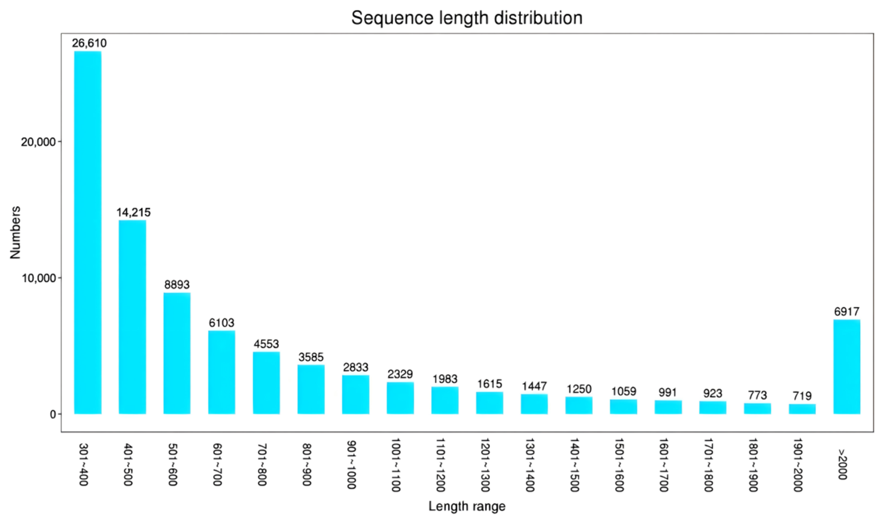
All Unigenes were arranged in ascending order according to their sequence length (Figure 2). The sequence length is highest in the range of 301–400 bp, with 26,610 sequences, accounting for 30.7%. Subsequently, the overall distribution trend gradually decreases from 401 to 20.00 bp with increasing sequence length, with a total of 6917 large fragments longer than 20.00 bp, accounting for 8%.

3.1.2. DEGs Analysis

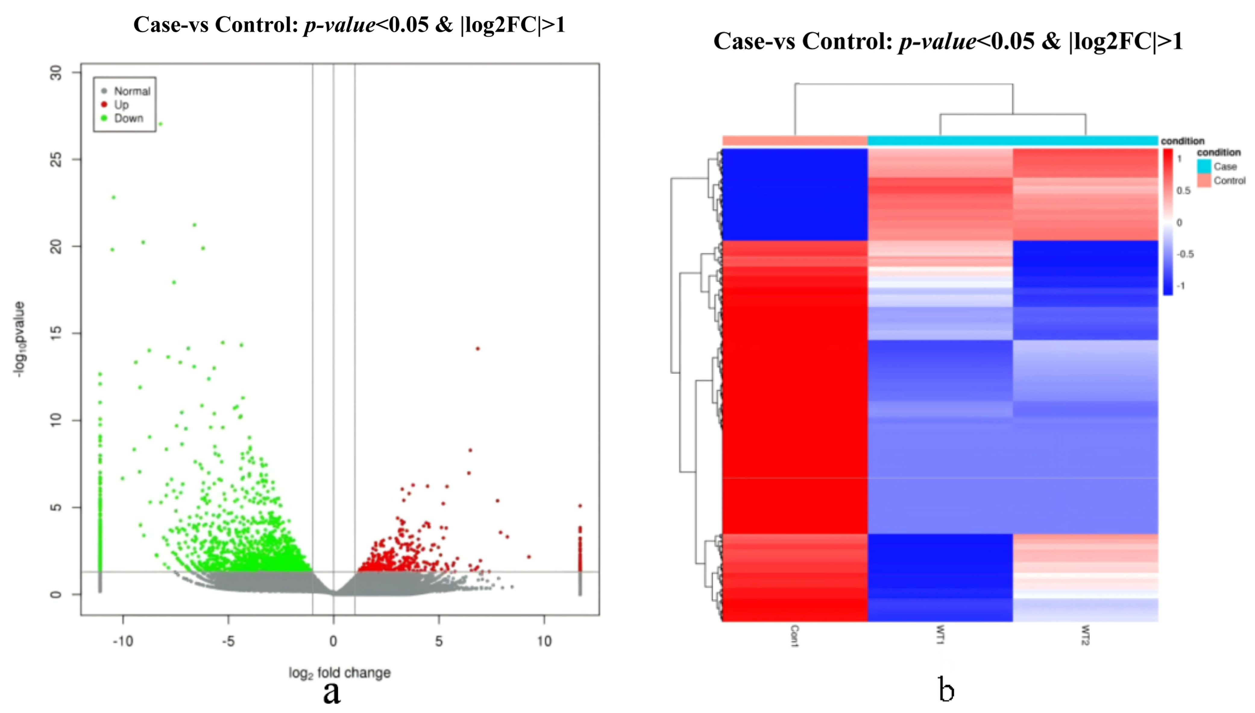
One differential subgroup was constructed to analyze DEGs using p < 0.05 and |log2FC| > 1. It was identified to have 2004 DEGs, containing 389 upregulated and 1615 downregulated genes. The volcano map (Figure 3a) and heat map (Figure 3b) of the DEGs are shown below.

3.1.3. GO Annotation

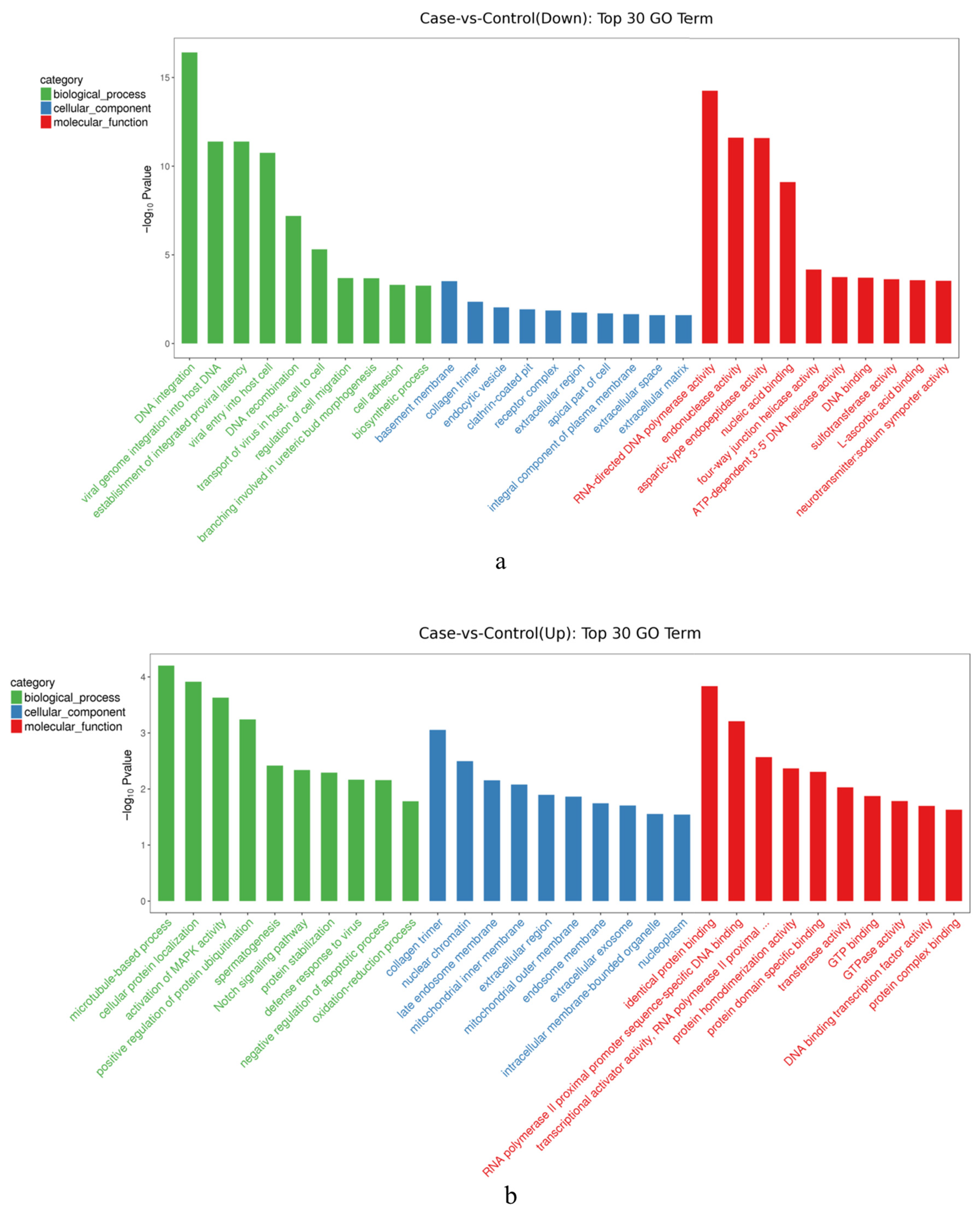
Downregulated DEGs (Figure 4a) were mainly enriched within biological processes such as DNA integration and viral genome integration into the host DNA. In terms of molecular function, downregulated DEGs were enriched basement membrane and collagen trimers. Most of the functions of upregulated DEGs (Figure 4b) were enriched within the collagen timer, nuclear chromatin, and late endosome membrane in the cellular component. In terms of molecular function, identical protein binding and RNA polymerase II promoter sequence-specific DNA binding contributed the largest proportion. With regard to biological processes, the cellular protein localization and spermatogenesis represented were the most prevalent.
Among the above DEGs related to gender gonadal differentiation were those mainly enriched in spermatogenesis. In addition to the top30, there were many other DEGs associated with sex determination and gonad differentiation mainly enriched in steroid hormone synthesis, gonadal development, and the maintenance of ovarian function.

3.1.4. KEGG Annotation

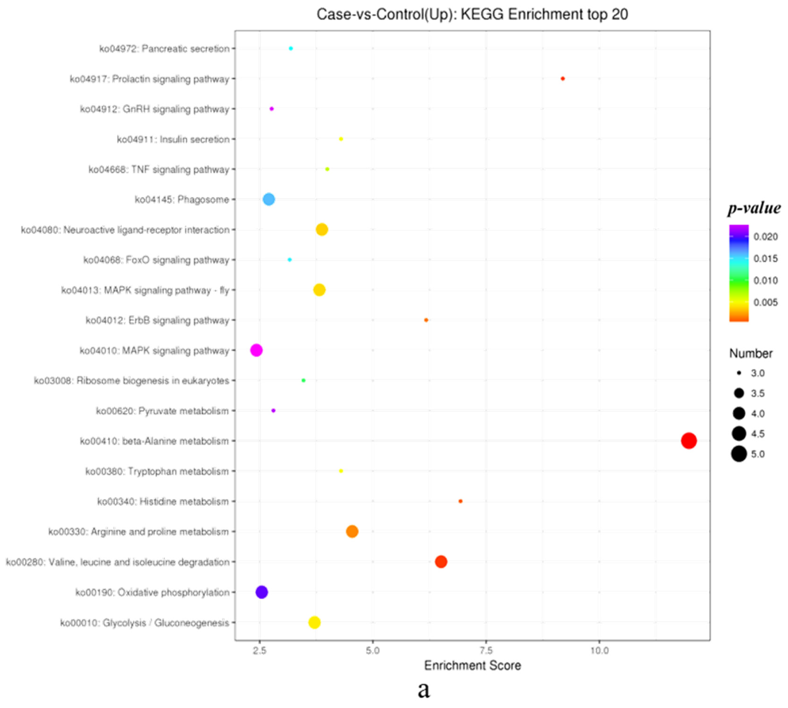
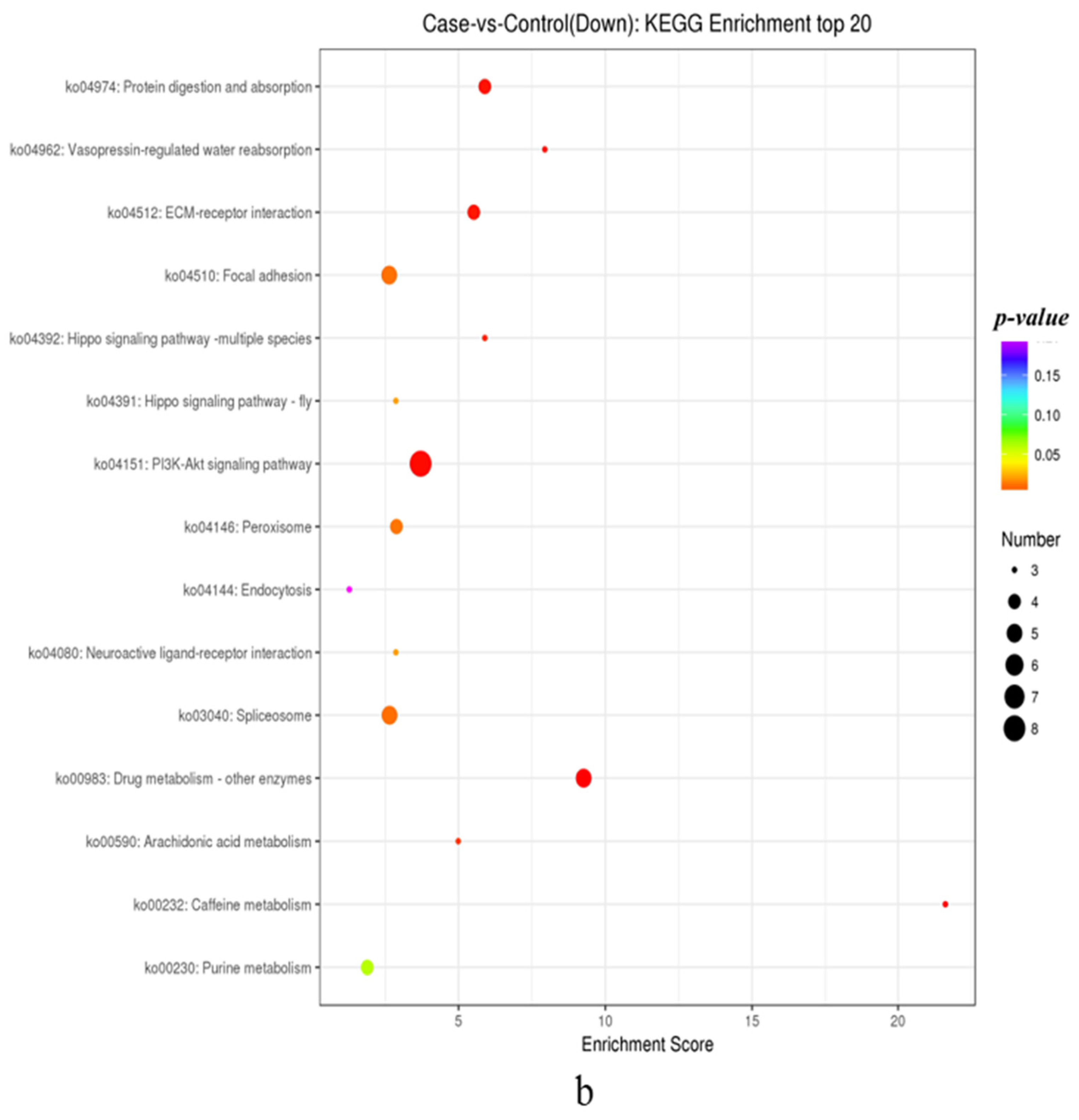
KEGG analysis showed that the MAPK signaling pathway, phagosome, and oxidative phosphorylation were the most enriched in upregulated DEGs (Figure 5a). The PI3k-Akt signaling pathway, protein digestion and absorption, and spliceosome were the most enriched in downregulated DEGs (Figure 5b). Among the top20 pathways, DEGs related to sex determination and gonad differentiation were mainly enriched in the FoxO signaling pathway. In addition to the top20 KEGG pathways, there were a large number of DEGs enriched in the Estrogen Pathway.
Combining the GO functional analysis of DEGs and KEGG signaling pathway analysis, genes related to steroid hormones, spermatogenesis, gonadal development, and oogenesis, as well as genes related to maintaining ovarian function and preventing premature ovarian failure, are all labeled in the caption of Figure 5.
Combined GO and KEGG analysis identified 20 genes associated with steroid hormones (HSD17b8, StARD3, CYP3A, CYP2J, CYP20A, STS, EST, JUN, cPLA2, ADCY9, IP3R1, PLCB4, HSD11B1, CYP17A1, CYP17A2, ST1A1, SERK2, S5AR1, CaM, and Hras), 13 genes associated with spermatogenesis were identified (Dmrt1, Sox9, Nup62, GABABR1, BmHP21, Dhm1, LRRK2, DYNLL, ING2, PAIP-2, Tspan-8, LAMP-1, and CD107), 6 genes related to gonadal development were identified (FST, DAX-1, NOTCH1, Wnt4, MMP-17, and LRP-2), 1 gene related to oogenesis (MARF1), and 1 gene related to the maintenance of ovarian function and the prevention of premature ovarian failure (SIRT1).

3.2. RT-qPCR Validation

Seven genes, STS, EST, CYP17A1, CYP17A2, FTZ-F1, STARD3, and NOTCH1, were verified for expression pre- and post-RNAi; the results (Figure 1) showed that the expressions of those genes were not significantly different between the negative and blank control groups, indicating that the factor of injection had little effect on the results of transcriptome sequencing. There were significant differences between the experimental group and the two control groups, indicating that experimental results were consistent with the trend of the transcriptome data, which verified the accuracy of the sequencing results.

3.3. ChIP-Seq Analysis

3.3.1. ChIP-Seq Peak Analysis

The overview of the ChIP-Seq data of FOXL2 is shown in Table 3, and the raw reads obtained from the two sequenced samples of FOXL2-IP and the input were the average of 23,747,152. The input was the control, which was the genomic nuclease-digested DNA. Without immunoprecipitation treatment, the DNA was directly delinked, purified, and analyzed. After quality filtration, 23,524,839 and 23,700,286 clean reads were obtained for FOXL2-IP and the input, respectively. Subsequently, the obtained clean reads were aligned to the C. farreri genome, and the mapped reads of FOXL2-IP and the input were 17,106,484 and 10,424,229, respectively.
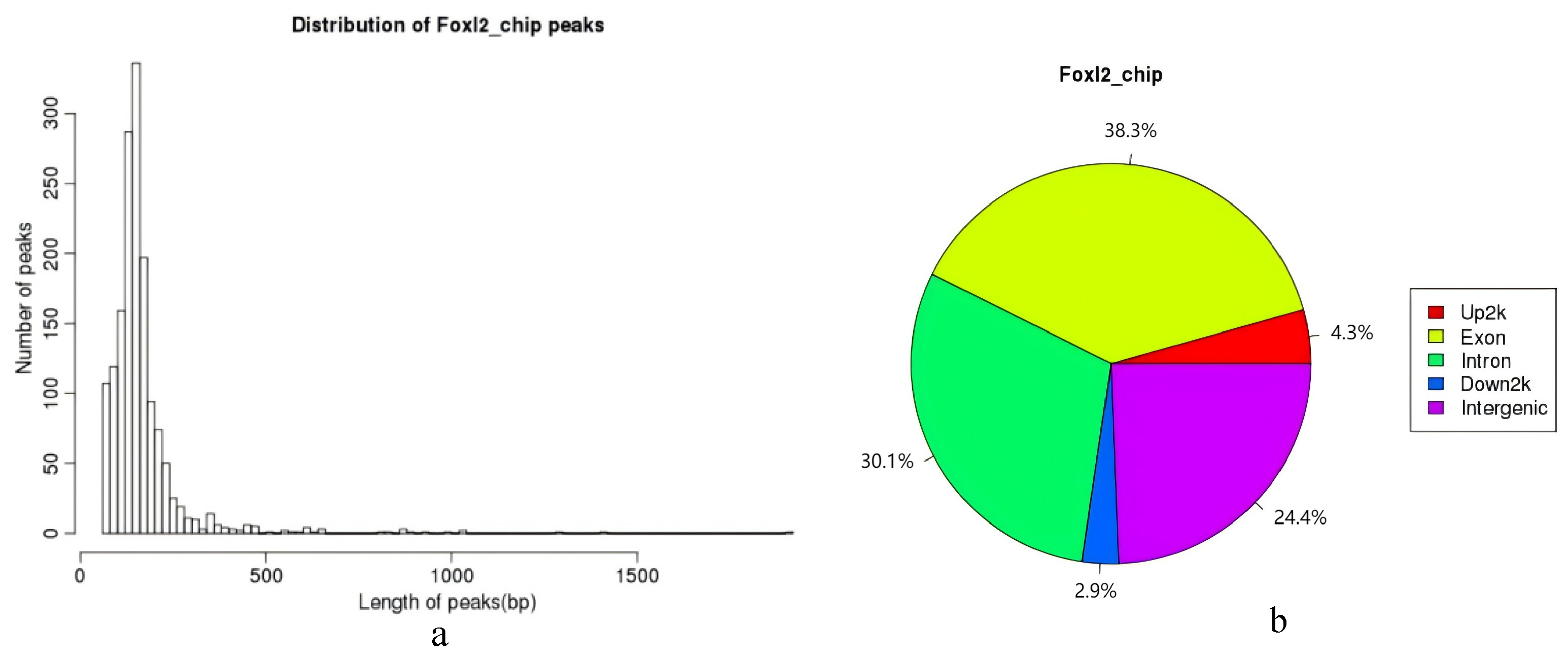
A total of 1557 peaks were enriched and identified in the C. farreri ovary. The average length of the peak is 166 bp, and the peak width–length distribution is shown in Figure 6a, and the results show that most of the peak widths are around 200 bp. The distribution of the FOXL2 target sequences on the gene functional elements was as follows: 24.4% in the intergenic region, 38.3% in the exon region, 30.1% in the intron region, 2.9% in the 3′-UTR end (Down2k), and 4.3% in the 5′-UTR end (Up2k) (Figure 6b).

3.3.2. GO and KEGG Analysis of FOXL2 Target Sequences

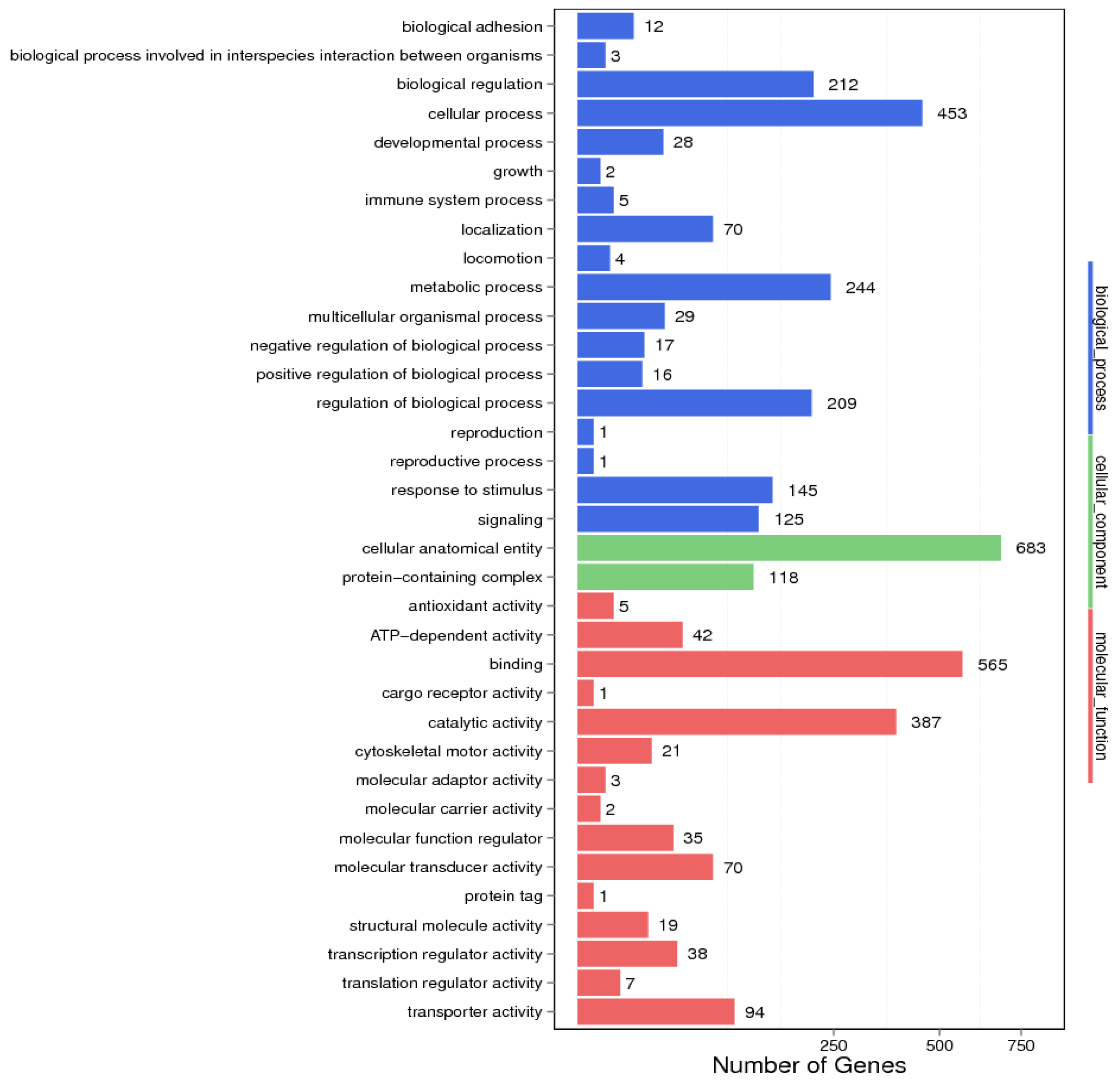
GO annotation analysis showed that the target sequences of FOXL2 were mainly involved in biological processes such as the cellular process, reproduction, development, and metabolic process (Figure 7). In terms of cell components, they were mainly related to the cellular anatomical entity and protein-containing complex. For the molecular functions, the main functional annotations were involved in catalytic activity and transporter activity. The genes involved in the reproductive and developmental pathways related to the development of the gonads are mainly concentrated in the physiological processes of steroid hormone synthesis, gonadal development, and spermatogenesis.
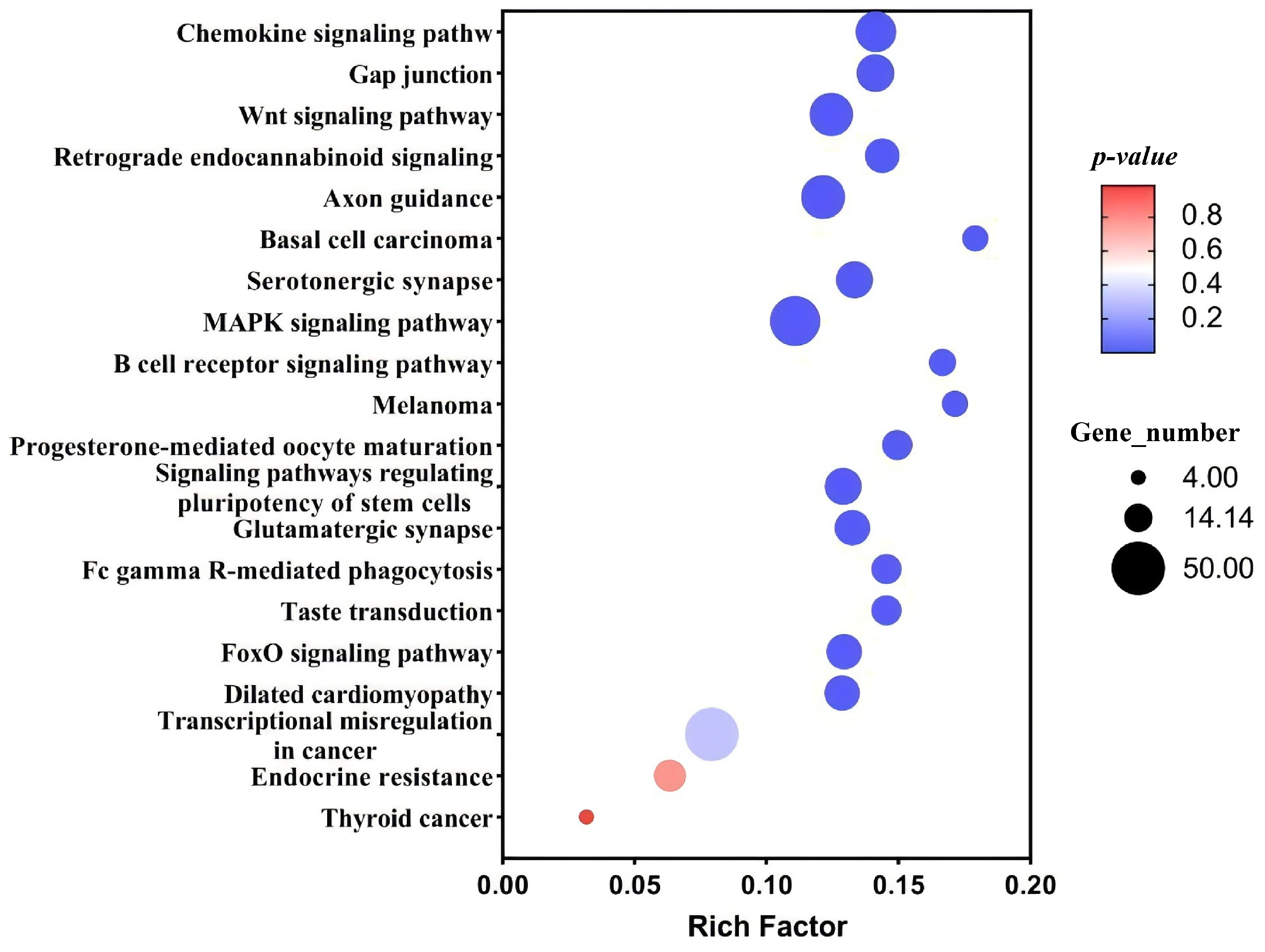
The KEGG pathway analysis provided insights into the metabolic pathways and the specific distribution of the FOXL2 target sequences. The top20 metabolic pathways, as illustrated in Figure 8, revealed that the FOXL2 target sequence was predominantly distributed in the chemokine, FoxO, and MAPK signaling pathways.

3.3.3. Regulatory Candidate Genes of FOXL2

For the 1557 peak genes obtained above, the relevant information of these genes was analyzed by matching the C. farreri genome [26]. There were some genes related to sex gonadal development, as shown in Table 4, 16 steroid hormone-related genes, 2 spermatogenesis-related genes, 2 gonadal development-related genes, and 1 prevention of premature ovarian failure-related genes were identified. More detailed information for all the regulatory candidate genes is listed in Table 4.

3.3.4. ChIP-qPCR

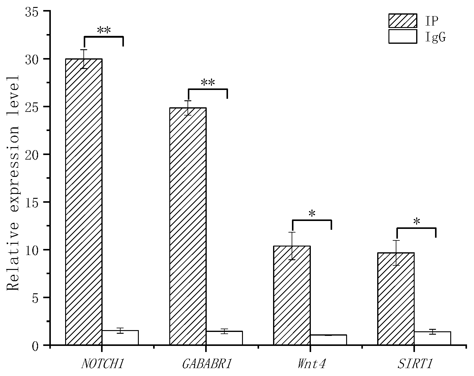
Four randomly selected genes in Table 4 were validated using ChIP-qPCR, as shown in Figure 9; results showed that the expression level of the experimental group (IP) was significantly higher than that of the control group (IgG).

3.3.5. Motif Analysis

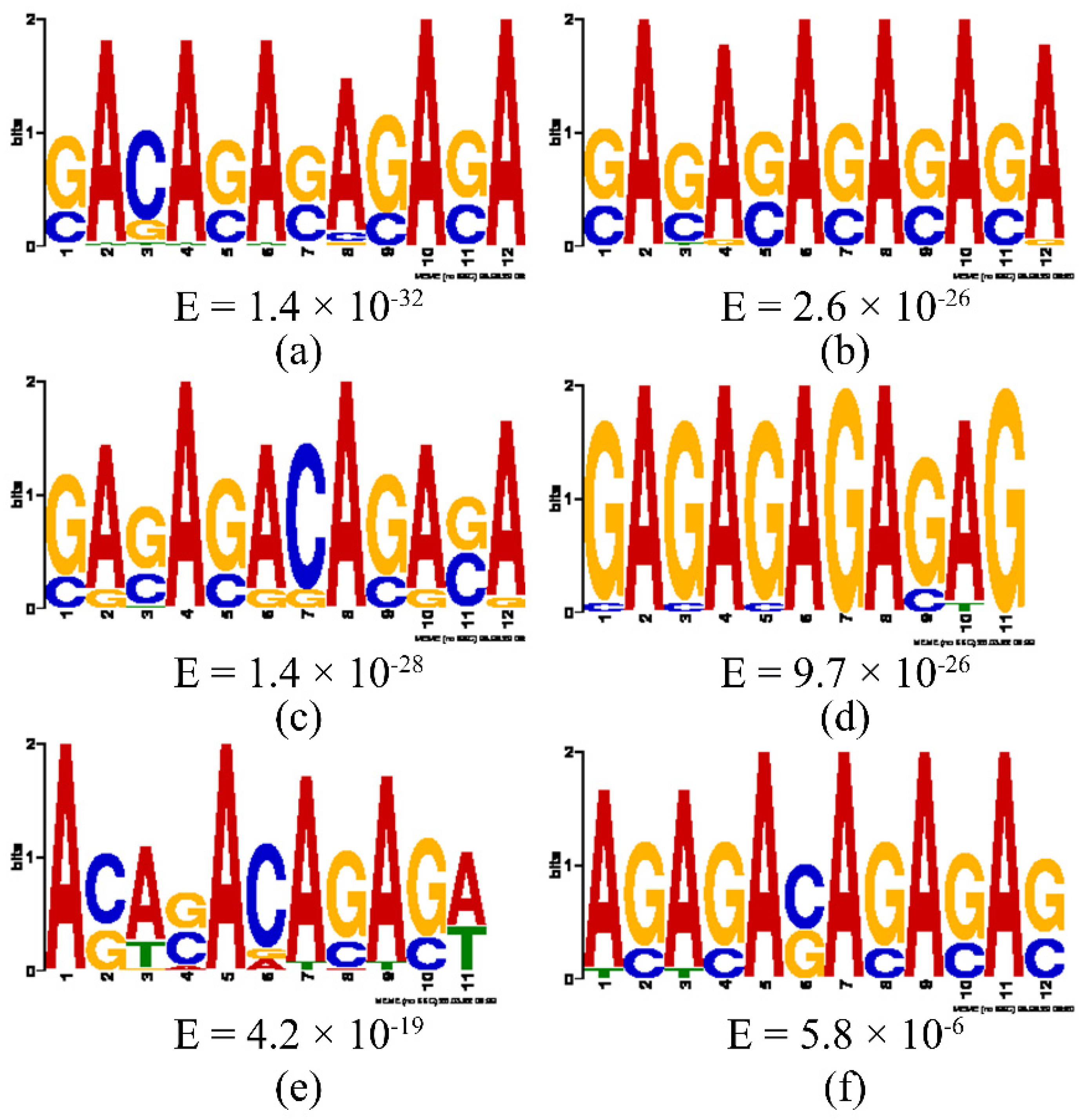
Based on the peak sequences, the following six motifs were predicted using the MEME software (version: 5.4.1) (Figure 10).

4. Discussion

Our laboratory has been studying the gonadal development of C. farreri for many years. Initially, we studied the expression of FOXL2 in different tissues and found that FOXL2 has significant ovarian expression specificity. We considered FOXL2 to be a female-related gene and conducted a series of studies (e.g., we have studied the expression of FOXL2 during gonadal differentiation, the expression pattern of the adult gonadal cycle, the expression pattern of embryonic development, and the function of FOXL2, etc.) [11,18,28,29].
The transcriptome differential expression showed 2004 DEGs, of which 389 genes were upregulated, and 1615 genes were downregulated; the number of downregulated genes was far greater than the upregulated genes, suggesting that FOXL2 mainly upregulates genes’ expressions in scallop ovaries. This is consistent with the research on the regulation of FOXL2 in chicken follicular granulosa cells [30].
Combining the transcriptome differential expression and ChIP omics analysis, it is considered that FOXL2 can directly or indirectly regulate genes in multiple pathways to exert its role in the ovary. Firstly, target genes are included in the steroid hormone synthesis pathway, such as HSD17B8StARD3, CYP3A, CYP2J, CYP20A, STS, EST, JUN, cPLA2, ADCY9, IP3R1, PLCB4, HSD11B1, CYP17A1, CYP17A2, ST1A1, KRAS, PGE2, YPEL1, SOS2, EFCAB11, AKT2, and SHC4.
In invertebrates, little is known about the physiological sources of sex steroids, especially their biosynthetic pathways. Thitiphuree et al. state that CYP17A, HSD3B, HSD17A, HSD17B8, and StAR3 genes are all important genes for steroid hormone synthesis in Mizuhopecten yessoensis. They suggest that CYP17A possesses both 17 α-hydroxylase and 17,20 lyase and is crucial for the production of sex steroids [31]. Previous research in our laboratory has found that there are two types of CYP17 genes in C. farreri; CYP17A1 was most expressed in the mature testis and growing ovary, suggesting that the gene may play a role in C. farreri testis development by participating in testosterone production and by also affecting oocyte growth. CYP17A2 was expressed higher at the mature gonadal stage than at the other stages, suggesting a correlation with sex cell maturation or discharge [32]. Guo et al. also discovered two CYP17A genes in Stronghlocentrotus intermedius; the researchers suggested that CYP17A1 plays an important role in the testis, and CYP17A2 may play a role in the maturation process of oocytes by regulating the production of 17 α, 20 β-dihydroxy-4-pregnen-3-one (DHP, a progesterone) [33]. This paper analyzes the transcriptome differences pre- and post-FOXL2 knockdown and suggests that FOXL2 negatively regulates the expression of CYP17A1 and positively regulates the expression of CYP17A2.
In mammals, StAR can promote the transport of cholesterol from the outer mitochondrial membrane to the inner mitochondrial membrane in steroid hormone synthesis cells, playing a key role in cholesterol metabolism and steroid hormone synthesis. So far, StAR genes have not been found in mollusks, but StARD3 has been found to exist in some shellfish species [31,34]. StARD3 was first found in the metastatic axillary lymph nodes of human breast cancer cells [35]; it has a START domain and belongs to the same subfamily as StAR in the START domain protein family. StARD3 plays a role in the specific binding and targeting of cholesterol to specific organelle membranes and is involved in the synthesis of steroid hormones [36]. Studies have shown that STARD3, homologous to STARD1, can promote steroid hormone synthesis in mammalian tissues that do not express STARD1 [37,38]. Thitiphuree et al. detected StAR3 (StARD3) expression in M. yessoensis and speculated that STARD3 in the steroid hormone synthesis pathway of M. yessoensis, like StAR in vertebrates, plays a role in transporting cholesterol to the mitochondrial inner membrane [31]. In our previous work, StARD3 could also be found in C. farreri, and it was preliminarily speculated that StARD3 may replace StAR in sex hormone synthesis [34]. In this paper, we found that FOXL2 RNAi would upregulate StARD3 expression (Figure 1), suggesting that FOXL2 can inhibit StARD3 expression, reduce progesterone synthesis, and prevent premature ovarian failure. After knocking down FOXL2, the expression of STS is downregulated, while the expression of EST is upregulated. It is considered that FOXL2 positively regulates STS and negatively regulates EST, thereby affecting the activation or inactivation of estrogen production and participating in the regulation of steroid hormone balance in C. farreri.
Previous research in our laboratory has found that Fushi-tarazu factor-1 (FTZ-F1) is mainly expressed in testes at the mature stage, with a significantly higher expression than in the male and female gonads at other stages. It is speculated that FTZ-F1 is involved in testosterone production to regulate testicular development in scallops [32]. In this paper, after knocking down FOXL2, the FTZ-F1 expression was upregulated (Figure 1). It is considered that FOXL2 negatively regulates FTZ-F1, affecting testosterone production in the ovaries and avoiding testicular development. This is consistent with the regulation of Sf-1 (a subfamily of FTZ-F1 genes in mammals) by FOXL2 in mammals [39]. CYP19 encodes aromatase, which catalyzes the conversion of testosterone to estradiol and is a key enzyme in the conversion of testosterone to estrogen. Data suggest that the CYP19 gene appears after chordates and has not been found in mollusks [40]. Thitiphuree et al. suggest that there may be other aromatase genes replacing CYP19 in invertebrates [31]. In this study, we found that the target genes of FOXL2 contain multiple P450 family genes, such as CYP3A, CYP2J, CYP20A, etc., and there is a significant difference in expression pre- and post-FOXL2 RNAi. Whether other aromatase genes replace CYP19 in scallops still needs to be further researched and explored.
Shao et al. suggested that GABAR may be closely related to human sperm production [41]. Wang Yi et al. found that the expression of GABABR in the testes of gibel carp (Carassius auratus gibelio) was significantly higher than that in the ovaries, the researchers suggested that it may play an important role in testicular development or spermatogenesis [42]. Saito et al. found that male mice lacking ING2 exhibited abnormal sperm production and infertility, indicating that ING2 plays an important role in mammalian spermatogenesis [43]. In mammals, autophagy is a process that maintains cellular homeostasis and plays an important regulatory role in spermatogenesis [44]. Moreover, it was found that lysosome-associated membrane protein 1 (LAMP1) is involved in the autophagy of Chinese soft-shelled turtle (Pelodiscus sinensis) sperm [45]. In this research, we found that after FOXL2 RNAi, the expression of genes related to spermatogenesis, such as GABABR, ING2, and LAMP1, were all upregulated. It is considered that FOXL2 prevents spermatogenesis-related genes from differentiating and developing towards the testis by negatively regulating them.
In addition, we found that knocking down FOXL2 downregulated the expression of SIRT1, indicating that FOXL2 positively regulates the expression of this gene. A study has found that after FOXL2 mutation in mice, the promotion of SIRT1 is lost, accelerating follicular development and activating a large number of primordial follicles, leading to premature ovarian failure [6]. SIRT1 is a key gene in the FoxO signaling pathway, which can directly or indirectly deacetylate FoxO to inhibit FoxO-mediated cell apoptosis and delay the aging process [46]. The positive regulation of SIRT1 by FOXL2 in C. farreri may be related to slowing down ovarian aging.
We found that the FOXL2 target included genes related to ovarian development, as FOXL2 can positively regulate FST expression, which in turn affects ovarian development. Ni et al. found that the expression of FST in the ovaries of Crassostrea angulata increases continuously with development and reaches its highest level at the mature stage, whereas the expression of FST in the testis remains at a low level throughout development and significantly decreases at the mature stage [47]. Therefore, it is considered that FST plays an important role in the development of oyster ovaries. Another author found that Wnt4 has the highest expression level at the mature stage of the C. farreri testes and ovaries, and the expression level in the testes is significantly higher than in ovaries. It is speculated that Wnt4 may be involved in the regulation of the development and maturation process of the gonads in both sexes, and its role in the testes is more significant than that in the ovaries [48]. On the other hand, it was found that the expression level of DAX1 in the testes of C. farreri is significantly higher than in the ovaries, and DAX1 is also highly expressed at the proliferative and growing stage of the ovaries. It is speculated that DAX1 may be involved in the development of C. farreri testes and early ovaries. This study found that knocking down FOXL2 upregulated the expression of Wnt4 and DAX-1, suggesting that FOXL2 has a negative regulatory effect on them. Previous research has found that MARF1 can control meiosis in oocytes, and mutations in MARF1 can lead to infertility in female mice because oocytes fail to undergo meiosis and release immature eggs, indicating that this gene plays an important role in the process of oogenesis [49,50]. This study found that after FOXL2 RNAi, the MARF1 expression was downregulated. It is believed that FOXL2 positively regulates the expression of MARF1 to assist in the normal development of ovum into mature ovum.
However, there is relatively little information on the binding sequence of target genes by FOXL2. Pisarska et al. found the sequence of the binding site between FOXL2 and the target gene StAR in mice, which is similar to the motif discovered in this study [1]. In addition, through analysis of FOXL2 binding sites, it was found that the vast majority of binding sites are located within genes, with 30.1% of binding sites in intronic regions and 38.3% in exon regions. This is similar to the study of FOXL2 binding sites carried out by Barbara et al. in fetal ovaries [20]. Through the verification of CHIP-seq results, this study found that some binding sequences located inside genes (such as Figure 10e) have the characteristic sequences of enhancers. The function of intronic enhancers has not been explored to a large extent but has attracted more and more attention from researchers in recent years. The FOXL2 binding sequence data suggest that transcriptional regulation may not exclusively focus on the promoter and its upstream regulatory region, as traditional research has done, and that the focus on introns and intron-type enhancers may also be important in regulatory studies.

5. Conclusions

In the process of oogenesis in Chlamys farreri, FOXL2 mainly functions through the regulation of upregulated genes, and it can regulate genes in multiple pathways to exert its role in the ovary. This paper screened out some key target genes of FOXL2 from the above pathways, laying an omics foundation for future, detailed research on the ovarian development mechanism of Chlamys farreri.

Author Contributions

X.L. designed the study, wrote the manuscript, and acquired the funding. H.Y. performed the experiments, analyzed the data, and wrote the manuscript. Y.X. analyzed the data and wrote the manuscript. S.W. analyzed the data. X.Z. and J.Z. reviewed the manuscript. All authors have read and agreed to the published version of the manuscript.

Funding

This work was supported by project ZR2022MC126 supported by Shandong Provincial Natural Science Foundation and the Technical System of Shellfish Industry in Shandong Province (SDAIT-14-05).

Institutional Review Board Statement

Animal materials of C. farreri used in this study were obtained from Aquatic Product Market near Yantai University, Shandong Province, China. No field permissions were necessary to collect the animal samples for this study. The authors declared that the experimental research on the animals described in this paper was in compliance with institutional, national, and international guidelines.

Informed Consent Statement

Not applicable.

Data Availability Statement

In the next few years, several graduate students in our laboratory will conduct further research based on these omics data. If the total data are leaked in advance, it will affect our laboratory follow-up research. So, based on the above considerations and the policies and confidentiality agreements adhered to in our laboratory, we cannot provide the total raw data. If anyone has questions about specific data, they can contact the corresponding author, and we will do our best to provide more detailed elaboration.

Conflicts of Interest

The authors declare no conflicts of interest.

References

  1. Pisaraka, M.D.; Bae, J.; Klein, C.; Hsueh, A.J.W. Forkhead L2 is expressed in the ovary and represses the promoter activity of the steroidogenic actute regulatory gene. Endocrinology 2004, 145, 3424–3433. [Google Scholar] [CrossRef] [PubMed]
  2. Wang, D.S.; Kobayashi, T.; Zhou, L.Y.; Paul-Prasanth, B.; Ijiri, S.; Sakai, F.; Okubo, K.; Morohashi, K.I.; Nagahama, Y. Foxl2 up-regulates aromatase gene transcription in a female-specific manner by binding to the promoter as well as interacting with ad4bindingprotein/steroidogenic factor 1. Mol. Endocrinol. 2007, 21, 712–725. [Google Scholar] [CrossRef] [PubMed]
  3. Jakob, S. FOXL2 Represses the Testis-Specific Enhancer of Sox9 to Maintain Ovary Differentiation. Ph.D. Thesis, UCL (University College London), London, UK, 2010. [Google Scholar]
  4. Caburet, S.; Georges, A.; L’Hôte, D.; Veitia, R.A. The transcription factor FOXL2: At the crossroads of ovarian physiology and pathology. Mol. Cell. Endocrinol. 2011, 356, 55–64. [Google Scholar] [CrossRef] [PubMed]
  5. Kashimada, K.; Pelosi, E.; Chen, H.; Schlessinger, D.; Wilhelm, D.; Koopman, P. FOXL2 and BMP2 Act Cooperatively to Regulate Follistatin Gene Expression during Ovarian Development. Endocrinology 2011, 152, 272–280. [Google Scholar] [CrossRef]
  6. Yang, X.W.; He, W.B.; Gong, F.; Li, W.; Li, X.R.; Zhong, C.G.; Lu, G.X.; Lin, G.; Du, J.; Tan, Y.Q. Novel FOXL2 mutations cause blepharophimosis-ptosis-epicanthus inversus syndrome with premature ovarian insufficiency. Mol. Genet. Genom. Med. 2017, 6, 261–267. [Google Scholar] [CrossRef]
  7. Hudson, Q.J.; Smith, C.A.; Sinclair, A.H. Aromatase inhibition reduces expression of FOXL2 in the embryonic chicken ovary. Dev. Dyn. 2005, 233, 1052–1055. [Google Scholar] [CrossRef]
  8. Kuo, F.T.; Fan, K.; Bentsi-Barnes, I.; Barlow, G.M.; Pisarska, M.D. Mouse forkhead L2 maintains repression of FSH-dependent genes in the granulosa cell. Reproduction 2012, 144, 485–494. [Google Scholar] [CrossRef]
  9. Pannetier, M. FOXL2 activates P450 aromatase gene transcription: Towards a better characterization of the early steps of mammalian ovarian development. J. Mol. Endocrinol. 2006, 36, 399–413. [Google Scholar] [CrossRef]
  10. Guiguen, Y.; Fostier, A.; Piferrer, F.; Chang, C.F. Ovarian aromatase and estrogens: A pivotal role for gonadal sex differentiation and sex change in fish. Gen. Comp. Endocrinol. 2010, 165, 352–366. [Google Scholar] [CrossRef]
  11. Liu, X.L.; Zhang, Z.F.; Shao, M.Y.; Liu, J.G.; Muhammad, F. Sexually dimorphic expression of foxl2 during gametogenesis in scallop Chlamys farreri, conserved with vertebrates. Dev. Genes. Evol. 2012, 222, 279–286. [Google Scholar] [CrossRef]
  12. Ye, R.H.; Ren, H.B. Molecular cloning and gene expression of foxl2 and sox14 gene from Hyriopsis cumingii. Oceanol. Limnol. Sin. 2018, 49, 160–167. [Google Scholar]
  13. Tang, L.; Hu, Q.; Zhao, R.Y.; Li, J.; Gulipari, A.; Zhang, J.J. Cloning and expression of foxl2 gene in Esox lucius. Guizhou Agric. Sci. 2020, 48, 97–101. [Google Scholar]
  14. He, J.Q.; Zhang, H.; She, Z.C.; Zhen, W.Q.; You, S.Y.; Wang, P.L.; Xu, Y.H.; Zhu, P. Cloning and expression pattern investigation of Crassostrea hongkongensis FOXL2 gene. Genom. Appl. Biol. 2020, 39, 1519–1528. [Google Scholar]
  15. Ren, X.L.; Liang, S.Q.; Zhong, Y.F.; Wang, Y.R.; Lao, J.T.; Li, G.L.; Huang, H.; Chen, H.P. Molecular cloning and tissue expression patterns of foxl2 gene in Bettas plendens. Genom. Appl. Biol. 2021, 40, 109–116. [Google Scholar]
  16. Zhao, D.; Zhou, L.Q.; Zheng, Y.X.; Sun, X.J.; Wu, B.; Liu, Z.H.; Wu, Z.; Wu, L. Expression pattern of the foxl2 gene in the scallop Patinopecten yessoensis during development. Prog. Fish. Sci. 2022, 43, 138–145. [Google Scholar]
  17. Liao, X.T. Study on the Gonadal Development and Expression of Sex Related Genes dmrt1 and foxl2 in clam Cyclina sinensis. Master’s Thesis, Jiangsu Ocean University, Lianyungang, China, 2022. [Google Scholar]
  18. Liu, X.L.; Liu, J.; Wang, Z.D.; Wang, Z.H.; Li, Y.S.; Cui, L.B. Necessity of foxl2 in the oogenesis of the scallop Chlamys farreri. Mar. Sci. 2018, 42, 127–131. [Google Scholar]
  19. Ning, J.H.; Cao, W.A.; Lu, X.; Chen, M.; Liu, B.; Wang, C.D. Identification and functional analysis of a sex-biased transcriptional factor Foxl2 in the bay scallop Argopecten irradians. Comp. Biochem. Physiol. B-Biochem. Mol. Biol. 2012, 256, 110638. [Google Scholar] [CrossRef]
  20. Barbara, N.; Grimm, S.A.; Artiom, G.; Scott, G.J.; Ray, M.K.; Yao, H.C. Genome-wide identification of FOXL2 binding and characterization of FOXL2 feminizing action in the fetal gonads. Hum. Mol. Genet. 2018, 27, 4273–4287. [Google Scholar] [CrossRef]
  21. Yang, D.D. Expression Characteristics and Functional Analysis of Krüppel-like Factor 4 in Chlamys farreri Spermatogenesis and Muscle Tissue. Master’s Thesis, Ocean University of China, Qingdao, China, 2018. [Google Scholar]
  22. Liu, X.L.; Wang, Z.D.; Sun, H.T.; Wang, Z.H.; Zhao, Z.J.; Li, J. Prokaryotic expression and purification of Foxl2 protein, preparation and detection of polyclonal antibody in Chlamys farreri. Mar. Fish. 2018, 40, 447–453. [Google Scholar]
  23. Sambrook, J.; Russell, D.W. Molecular Cloning: A Laboratory Manual, 3rd ed.; Science Press: Beijing, China, 2002; pp. 518–531. [Google Scholar]
  24. Roberts, A.; Trapnell, C.; Donaghey, J.; Rinn, J.; Pachter, L. Improving RNA-Seq expression estimates by correcting for fragment bias. Genome Biol. 2011, 12, R22. [Google Scholar] [CrossRef]
  25. Zhou, Q.R.; Shao, M.Y.; Qin, Z.K.; Kyoung, H.K.; Zhang, Z.F. Cloning, characterization, and expression analysis of the DEAD-box family genes, Fc-vasa and Fc-PL10a, in Chinese shrimp (Fenneropenaeus chinensis). Chin. J. Oceanol. Limnol. 2010, 28, 37–45. [Google Scholar] [CrossRef]
  26. Liu, F.Y.; Li, Y.L.; Yu, H.W.; Zhang, L.L.; Hu, J.J.; Bao, Z.M.; Wang, S. MolluscDB: An integrated functional and evolutionary genomics database for the hyper-diverse animal phylum Mollusca. Nucleic Acids Res. 2021, 49, 988–997. [Google Scholar] [CrossRef]
  27. Zhang, Y.; Liu, T.; Meyer, C.A.; Eeckhoute, J.; Johnson, D.S.; Bernstein, B.E.; Nusbau, C.; Myers, R.M.; Browm, M.; Li, W.; et al. Model-based analysis of ChIP-Seq (MACS). Genome Biol. 2008, 9, R137. [Google Scholar] [CrossRef]
  28. Liu, X.L.; Liu, J.G.; Wang, D.; Zhang, Z.Y.; Zhang, Z.F. Expression pattern of the foxl2 gene in the scallop Chlamys farreri during development. JFSC 2013, 20, 205–210. [Google Scholar] [CrossRef]
  29. Liu, X.L.; Li, Y.; Liu, J.G.; Cui, L.B.; Zhang, Z.F. Gonadogenesis in scallop Chlamys farreri and Cf-foxl2 expression pattern during gonadal sex differentiation. Aquac. Res. 2016, 47, 1605–1611. [Google Scholar] [CrossRef]
  30. Wang, J. Study on the Molecular Mechanism of foxl2 in Chicken Geanulosa Cell Differentiation. Master’s Thesis, Huazhong Agricultural University, Wuhan, China, 2017. [Google Scholar]
  31. Thitiphuree, T.; Nagasawa, K.; Osada, M. Molecular identification of steroidogenesis-related genes in scallops and their potential roles in gametogenesis. J. Steroid Biochem. Mol. Biol. 2019, 186, 22–33. [Google Scholar] [CrossRef] [PubMed]
  32. Mu, X.J. Sequence Characteristics and Expression Analysis of FTZ-F1, CYP17A1 and CYP17A2 Genes in Chlamys farreri. Master’s Thesis, Yantai University, Yantai, China, 2021. [Google Scholar]
  33. Guo, X.L.; Qiu, X.M.; Chang, Y.Q.; Zhang, W.J.; Cui, J.; Cong, Y.T.; Wu, L.Z.; Liu, Y.; Wang, X.L. Cloning and differential expression of CYP17 Gene in sea urchin Strongylocentrous intermedius. J. Dalian Ocean. Univ. 2013, 28, 1–6. [Google Scholar]
  34. Watari, H.; Arakane, F.; Moog-Lutz, C.; Kallen, C.B.; Tomasetto, C.; Gerton, G.L.; Rio, M.C.; Baker, M.E.; Strauss, J.F., III. MLN64contains a domain with homology to the steroidogenic acute regulatory protein (StAR) that stimulates steroidogenesis. Proc. Natl. Acad. Sci. USA 1997, 94, 8462–8467. [Google Scholar] [CrossRef]
  35. Tomasetto, C.; Régnier, C.; Moog-Lutz, C.; Mattei, M.G.; Chenard, M.P.; Lidereau, R.; Basset, P.; Rio, M.C. Identification of four novel human genes amplified and overexpressed in breast carcinoma and localized to the q11-q21.3 region of chromosome 17. Genomics 1995, 28, 367–376. [Google Scholar] [CrossRef]
  36. Akiyama, N.; Sasaki, H.; Ishizuka, T.; Kishi, T.; Sakamoto, H.; Onda, M.; Hirai, H.; Yazaki, Y.; Sugimura, T.; Terada, M. Isolation of a candidate gene, CAB1, for cholesterol transport to mitochondria from the c-ERBB-2 amplicon by a modified cDNA selection method. Cancer Res. 1997, 57, 3548–3553. [Google Scholar]
  37. Sugawara, T.; Holt, J.A.; Driscoll, D.; Strauss, J.F., 3rd; Lin, D.; Miller, W.L.; Patterson, D.; Clancy, K.P.; Hart, I.M.; Clark, B.J. Human steroidogenic acute regulatory protein: Functional activity in COS-1 cells, tissue-specific expression, and mapping of the structural gene to 8p11.2 and a pseudogene to chromosome13. Proc. Natl. Acad. Sci. USA 1995, 92, 4778–4782. [Google Scholar] [CrossRef]
  38. Luo, D.Y.; Zhai, X.T.; Liu, X.L.; Cui, L.B. Sequence analysis and expression pattern of STARD3 cDNA in Chlamys farreri. Mar. Fish. 2024, 46, 579–585. [Google Scholar]
  39. Takasawa, K.; Pelosi, E.; Takagi, M.; Morio, T.; Asahara, H.; Schlessinger, D.; Mizutani, S.; Koopman, P.; Kashimada, K. FOXL2 transcriptionally represses Sf1 expression by antagonizing WT1 during ovarian development in mice. FASEB J. 2014, 28, 2020–2028. [Google Scholar] [CrossRef]
  40. Zhang, Y.; Zhang, L.L.; Li, R.J.; Xie, X.R.; Li, Y.P.; Li, W.R.; Bao, Z.M. Screening of interacting proteins of FOXL2 in scallop Patinopecten yessoensis by Yeast Two-Hybrid System. Period. Ocean. Univ. China 2019, 49, 54–59. [Google Scholar]
  41. Shao, C.; Yang, O.; Wang, H.; Gao, X.K.; Chen, B.Q.; Shao, G.X. Research on GABAR related proteins and ENOS responsive proteins in human spermatogenesis disorders. In Proceedings of the 5th National Male Science Academic Conference of the Chinese Medical Association, Nanjing, China, August 2004. Male Science Branch of the Chinese Medical Association, 21st Century Male Science. [Google Scholar]
  42. Wang, W.; Ruan, J.M.; Zhou, A.L.; Zhao, Y.N.; Cao, H.P.; Hu, K.; Yang, X.L. Cloning and Expression of Partial cDNA Encoding GABAB Receptor Subunit1 in Carassius auratus gibelio. Chin. J. Zool. 2013, 48, 905–911. [Google Scholar]
  43. Saito, M.; Kumamoto, K.; Robles, A.I.; Horikawa, I.; Furusato, B.; Okamura, S.; Goto, A.; Yamashita, T.; Nagashima, M.; Lee, T.L.; et al. Targeted disruption of Ing2 results in defective spermatogenesis and development of soft-tissue sarcomas. PLoS ONE 2010, 5, 0015541. [Google Scholar] [CrossRef] [PubMed]
  44. Yin, J.; Ni, B.; Tian, Z.; Yang, F.; Gao, Y. Regulatory effects of autophagy on spermatogenesis. Biol. Reprod. 2017, 96, 525–530. [Google Scholar] [CrossRef] [PubMed]
  45. Tarique, I. Cellular Charactieristics of Autophagy and Secretions in the Make Reproductive System of the Chinese Soft-Shelled Turtle, Pelodiscus sinensis. Ph.D. Thesis, Nanjing Agricultural University, Nanjing, China, 2020. [Google Scholar]
  46. Tao, X.H.; Liu, M.; Qiu, H. Research progress on Sirt1 regulation of aging in mammals. Gansu Sci. Technol. 2016, 32, 77–80. [Google Scholar]
  47. Ni, J.B.; Zeng, Z.; Han, G.D.; Huang, H.Q.; Ke, C.H. Cloning and characterization of the follistatin gene from Crassostrea angulata and its expression during the reproductive cycle. Comp. Biochem. Physiol. B-Biochem. Mol. Biol. 2012, 163, 246–253. [Google Scholar] [CrossRef]
  48. Li, H.L. Cloning of Gene Wnt4, β-catenin, Dax1, and Their Correlation to the Gonadal Development in Scallop Chlamys farreri. Ph.D. Thesis, Ocean University of China, Qingdao, China, 2013. [Google Scholar]
  49. Su, Y.Q.; Sun, F.; Handel, M.A.; Schimenti, J.C.; Eppig, J.J. Meiosis arrest female 1 (MARF1) has nuage-like function in mammalian oocytes. Proc. Natl. Acad. Sci. USA 2012, 109, 18653–18660. [Google Scholar]
  50. Yao, Q.Q.; Cao, G.Y.; Li, M.Z.; Su, Y.Q. Ribonuclease activity of MARF1 controls oocyte RNA homeostasis and genome integrity in mice. Proc. Natl. Acad. Sci. USA 2018, 115, 11250–11255. [Google Scholar] [CrossRef]
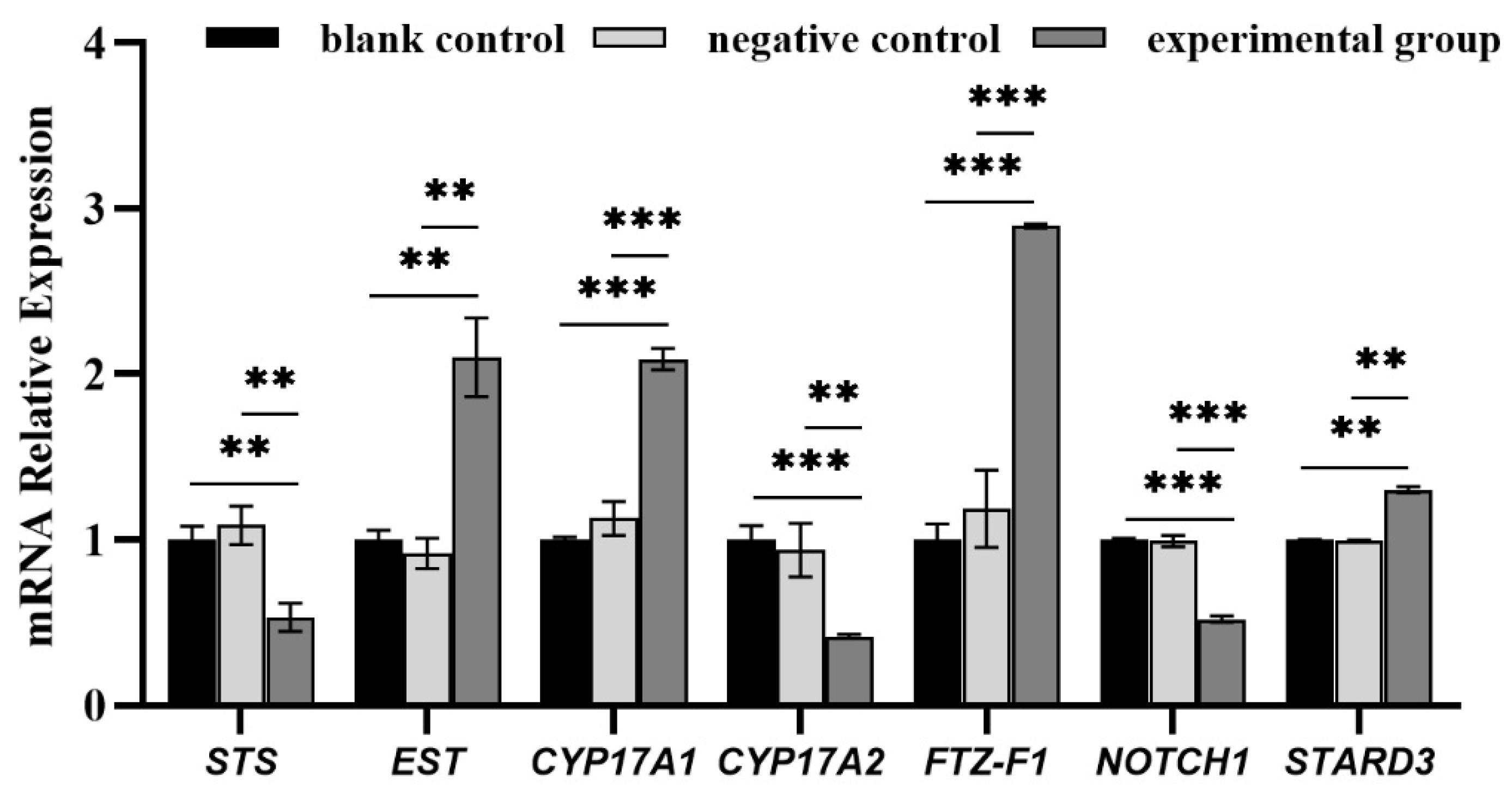
Figure 1. The mRNA expression of key DEGs. Note: * indicates significant difference (** p < 0.01, *** p < 0.001); the blank control group gene expression of each gene was, respectively, set as 1.00.
Biology 14 01259 g001
Figure 2. Unigene length distribution map.
Biology 14 01259 g002
Figure 3. The volcano and heat maps of the DEGs. (a). Volcano plot of differentially expressed genes. Note: Red, green and gray represent significantly up—regulated genes, significantly down- regulated genes and non—significantly genes. (b). Cluster analysis of differential expression profiles. Note: Red indicates high expression genes and blue indicates low expression genes.
Biology 14 01259 g003
Figure 4. Up/downregulated DEGs GO enrichment top30 entries graph. (a). Down—regulated DEGs GO enrichment top30 entries graph; (b). Up—regulated DEGs GO enrichment top30 entries graph. Note: The Go categories are color-coded: green for biological process, blue for cellular component, and red for molecular function.
Biology 14 01259 g004
Figure 5. Up/downregulated DEGs KEGG enrichment top20 bubble map. (a). Up—regulated DEGs KEGG enrichment top20 bubble map; (b). Down—regulated DEGs KEGG enrichment top20 bubble map.
Biology 14 01259 g005aBiology 14 01259 g005b
Figure 6. Length of peaks and peak distribution of gene elements. (a). Length of peaks of gene elements; (b). Peak distribution of gene elements.
Biology 14 01259 g006
Figure 7. GO analysis of peak-related genes.
Biology 14 01259 g007
Figure 8. KEGG analysis of peak-related genes.
Biology 14 01259 g008
Figure 9. ChIP-qPCR analysis of four randomly selected target genes of FOXL2. Note: *: p < 0.05, **: p < 0.01.
Biology 14 01259 g009
Figure 10. Motif analysis of peak sequences. (a) Motif 1; (b) Motif 2; (c) Motif 3; (d)Motif 4; (e) Motif 5; (f) Motif 6.
Biology 14 01259 g010
Table 1. The sequences of the primers used in the experiment.
Gene NamePrimer Sequence (5′→3′)Product
Length
Melting
Temperature
Usage
Steroid Sulfatase (STS)-FGGGTTCTTTTGTTCGTCTGGC 146 bp58.97RT-qPCR
Steroid Sulfatase (STS)-RTTTCCCGGGCTTCCAAGAATT59.35
Estrogen Sulfotransferase (EST)-FGGTGTGATGAAGGTCAAGGG105 bp60.67RT-qPCR
Estrogen Sulfotransferase (EST)-RTACACATCGCCACAGAAGCA59.78
Cytochrome P450c17A1 (CYP17A1)-FCCAAGTAGCCGATTCAAAAAAGTGT144 bp59.81RT-qPCR
Cytochrome P450c17A1 (CYP17A1)-RTCCAGCAAAGAAAATGTCAGCA 60.87
Cytochrome P450c17A2 (CYP17A2)-FGATGTGGACGATGCTTTTCTC114 bp60.03RT-qPCR
Cytochrome P450c17A2 (CYP17A2)-RTGTTTTGCCTGTTGCTGTTC 59.73
Fushi-Tarazu Factor-1 (FTZ-F1)-FTAGAGGCAGTGAGACAGGATAGAA139 bp59.65RT-qPCR
Fushi-Tarazu Factor-1 (FTZ-F1)-R
StAR-Related Lipid Transfer Protein 3 (STARD3)-F
StAR-Related Lipid Transfer Protein 3 (STARD3)-R
GATTGCTGGGTCGGGTTTA
GCGGGGTAGACAAACGGA
ATGTCTCCTGTTCGCCGT
128 bp60.78
60.54
60.02
RT-qPCR
Neurogenic Locus Notch Homolog Protein 1 (NOTCH1)-F1GTTTACATCTGCTGAAGTGTGGAT155 bp59.59RT-qPCR
Neurogenic Locus Notch Homolog Protein 1 (NOTCH1)-R1CTCTGTGTCTTTTCTAGCCGTGTA 60.15
β-actin-FTTCTTGGGAATGGAATCTGC119 bp58.89RT-qPCR
β-actin-RGCCAGACTCGTCGTATTCCT 58.12
Neurogenic Locus Notch Homolog Protein 1 (NOTCH1)-F2GGAGAGGGACAACCAACACC117 bp58.7ChIP-qPCR
Neurogenic Locus Notch Homolog Protein 1 (NOTCH1)-R2TCACATTTGGATGGTTTCTGGA 60.2
γ-Aminobutyric Acid Type B Receptor Subunit 1 (GABABR1)-FATCAAGTGGTCCGCAACTCT49 bp56.8ChIP-qPCR
γ-Aminobutyric Acid Type B Receptor Subunit 1 (GABABR1)-RTCATTGCATGACCTGTTGCC 59.7
Wingless-Type MMTV Integration Site Family, Member 4 (Wnt4)-FAGGTTGGGAAACCCTTGC55 bp 57ChIP-qPCR
Wingless-Type MMTV Integration Site Family, Member 4 (Wnt4)-RCACAACTTGGCAGCACCA 56.1
Silent Mating Typeinformation Regulation 2 Homolog 1 (SIRT1)-FGCCAAGCAGTTCAACATCAA74 bp56.8ChIP-qPCR
Silent Mating Typeinformation Regulation 2 Homolog 1 (SIRT1)-RCTCCTGATGTTCCACAAATCC 56.7
Note: The amplification efficiency of the genes in the table has reached around 95%.
Table 2. Sequencing data quality pre-processing results.
SampleRaw_ReadsClean_ReadsValid_Reads (%)Q30 (%)GC (%)
Con149,024,14845,647,37489.5392.3140.81
KD144,400,93442,162,16691.9793.6241.92
KD249,725,36846,588,67290.1692.6840.45
Table 3. Statistical analysis of raw data.
SampleClean ReadsClean RatioMapped ReadsMapped
FOXL2-IP23,700,28699.80%10,424,22943.98%
Input23,524,83999.06%17,106,48472.72%
Note: Clean reads: the number of reads obtained by filtering raw reads; mapped reads: the total number of reads on the alignment; and mapped rate: the proportion of the total number of reads on the alignment.
Table 4. Target genes of FOXL2.
Gene IDPeak StartPeak EndPeak AnnotationGene Name
Steroid Hormone
1104417801,660,5941,660,681estradiol 17-beta-dehydrogenase 8-likeHSD17b8
1104470982,005,8512,005,913cytochrome P450 2J6-likeCYP2J6
110451342431,036431,098cytosolic phospholipase A2-likecPLA2
110451344520,879520,970prostaglandin G/H synthase 2-likePGE2
110461839815,032815,194adenylate cyclase type 9-likeADCY9
110445220505,918506,027protein yippee-like 1YPEL1
110447714109,981110,076son of sevenless homolog 2-likeSOS2
110445266925,477925,554EF-hand calcium-binding domain-containing protein 11-likeEFCAB11
110459812161,479161,605RAC-beta serine/threonine-protein kinase B-likeAKT2
11046007625,29225,362SHC-transforming protein 4-likeSHC4
110460372108,457108,539inositol 1,4,5-trisphosphate receptor type 1-likeIP3R1
1104614411,140,0801,140,2621-phosphatidylinositol 4,5-bisphosphate
2-phosphodiesterase beta-4-like
PLCB4
1104455011,702,4101,702,520GTPase HRasHras1
110463419101,557101,621hydroxysteroid 11-beta-dehydrogenase 1-like proteinHSD11B1
110464786243,337243,491lysosome membrane protein 2-likeLAMP2
110449851223,863224,009neurogenic locus notch homolog protein 1-likeNOTCH1
Spermatogenesis
1104593751,049,9381,050,020gamma-aminobutyric acid type B receptor subunit 1-likeGABABR1
110456833188,225188,363golgin subfamily A member 3-likeGOLGA3
Gonadal Development
11045100272,92973,016protein Wnt-4-likeWnt4
1104550121,822,0371,822,131insulin-like peptide receptorILPR
Prevention of Premature Ovarian Failure
110460060130,987131,077NAD-dependent protein deacetylase sirtuin-1-likeSIRT1
Note: The bold are different pathways of action related to sex gonadal development in FOXL2 target genes.
Disclaimer/Publisher’s Note: The statements, opinions and data contained in all publications are solely those of the individual author(s) and contributor(s) and not of MDPI and/or the editor(s). MDPI and/or the editor(s) disclaim responsibility for any injury to people or property resulting from any ideas, methods, instructions or products referred to in the content.