Article

Characterization of Two Novel Heat Shock Protein 70 Transcripts from Sitodiplosis mosellana and Their Response to Larval Diapause and Thermal Stress

1
Shandong Institute of Sericulture, Shandong Academy of Agricultural Sciences, Yantai 265503, China
2
Key Laboratory of Plant Protection Resources and Pest Management of Ministry of Education, College of Plant Protection, Northwest A&F University, Yangling 712100, China
3
Department of Entomology, Texas A&M University, College Station, TX 77843, USA
*
Authors to whom correspondence should be addressed.
Biology 2025, 14(9), 1147; https://doi.org/10.3390/biology14091147
Submission received: 7 August 2025 / Revised: 28 August 2025 / Accepted: 29 August 2025 / Published: 30 August 2025

Simple Summary

The heat shock protein 70 (Hsp70) family plays key roles not only in heat/cold stress tolerance but also in insect diapause. In this study, we characterized two cytoplasmic Hsp70 genes (SmHsp70A1-1 and SmHsp70A1-2) from Sitodiplosis mosellana, a major wheat pest undergoing obligatory diapause as third-instar larvae in the soil, and analyzed their expression profiles and functions in relation to diapause and thermal stress. The results showed that the expression of SmHsp70A1-1 and SmHsp70A1-2 is developmentally and environmentally regulated, with increased expression being essential for enhancing cold tolerance in S. mosellana. The findings provide crucial insights into the mechanisms underlying thermal tolerance in this pest.

Abstract

The heat shock protein 70 (Hsp70) family mediates responses to environmental stress in insects. The wheat midge Sitodiplosis mosellana, a worldwide pest, avoids summer and winter temperature extremes by diapause of the third-instar larvae in the soil. To explore the functions of Hsp70s in this process, we characterized two cytoplasmic Hsp70 genes (SmHsp70A1-1 and SmHsp70A1-2) from this insect. Both SmHsp70s contained three signature motifs of the family and lacked introns. Developmental expression profiling revealed maximal SmHsp70A1-1 expression during early larval stages, while the expression of SmHsp70A1-2 was highest in the pupal stages. The expression of SmHsp70A1-1 was significantly upregulated during diapause, particularly during summer and winter, whereas SmHsp70A1-2 showed marked downregulation and dose-dependent induction by 20-hydroxyecdysone (20E). Furthermore, both genes exhibited similar expression patterns in over-summering and over-wintering larvae under thermal stress, with maximal expression at 40 °C and −10 °C, respectively, but were not significantly induced at prolonged extreme temperatures (50 °C or −15 °C). Knockdown of the two SmHsp70 genes by RNA interference (RNAi) significantly increased the susceptibility of the larvae to cold stress. These results suggest the important role of both SmHsp70 genes in diapause-associated stress tolerance and provide crucial insights into the mechanisms underlying thermal adaptation in S. mosellana.

Graphical Abstract

1. Introduction

Heat shock proteins (Hsps) are a superfamily of molecular chaperones whose expression is upregulated under stress conditions [1,2]. They were originally discovered in connection with the heat shock response in Drosophila [3] but are now recognized as being inducible by various stressors, including cold shock, chemical pesticides, and pathogenic invasion [4,5,6]. The primary function of Hsps is to preserve proteostasis by preventing aberrant protein aggregation and promoting clearance of misfolded or aggregated proteins [7,8]. Six distinct Hsp families are recognized based on evolutionary lineage and molecular weight, namely, the Hsp100, Hsp90, Hsp70, Hsp60, Hsp40, and small Hsp families [9,10,11].
Among Hsps, Hsp70s represent a group of structurally and evolutionarily conserved multifunctional proteins that fall into two subtypes, namely, the stress-inducible heat shock protein (Hsp70) and heat shock cognate protein 70 (Hsc70) subtypes [12,13]. The mRNA expression of Hsp70s is induced rapidly from basal levels in response to various stress stimuli, whereas Hsc70 proteins are expressed constitutively at relatively high levels with minimal variation under stress conditions [14,15]. Both protein types are approximately 70 kDa in molecular weight and share several common structural features. In insects, Hsp70s are suggested to participate in multiple physiological processes, including enhancing resistance to extreme temperatures to protect against thermal stress-induced injury and death [16,17]. In addition, Hsp70s have been shown to modulate larval development in Spodoptera litura, with their expression being induced by ecdysone (20-hydroxyecdysone [20E]) [18].
An emerging body of evidence also indicates the involvement of Hsp70s in insect diapause, an adaptive physiological tactic employed by insects to withstand adverse environmental conditions, including extreme temperatures associated with seasonal changes [19,20]. For instance, in the cabbage butterfly Pieris melete, expression of PmHsp70b is induced during diapause, whereas that of PmHsp70a is unaffected [21]. Similarly, increased expression of Hsp70 genes during diapause has been reported in various insect taxa, such as Sarcophaga crassipalpis Hsp70a and Hsp70b [22], Leptinotarsa decemlineata Hsp70a [23], Bombyx mori Hsp70a [24], and Ostrinia nubilalis Hsp70 [25]. In contrast, Hsp70 expression is either downregulated or maintained at low levels during diapause in Omphisa fuscidentalis [26], Sesamia nonagrioides [27], and Helicoverpa zea [28]. These variations in expression suggest that the functions of Hsp70s during diapause differ according to the insect species.
The orange wheat blossom midge, Sitodiplosis mosellana Géhin (Diptera: Cecidomyiidae), is a cosmopolitan and destructive pest of wheat crops, causing significant reductions in yield and economic repercussions during outbreak years [29,30,31]. Typically, this species completes a univoltine life cycle annually. In most wheat-producing regions of northern China, adult emergence and subsequent egg-laying take place from late April to early May [32]. Upon hatching, the larvae feed on the developing grains until the third instar. By mid-to-late May, the mature larvae exit the wheat heads and descend to the ground, where they burrow into the soil to spin cocoons for obligatory diapause. Although the larvae remain inside their protective cocoons until mid-March of the following year, diapause is effectively terminated by low temperatures in early December [33]. The larvae then enter a quiescent post-diapause stage that does not differ morphologically from diapause. The initiation of post-diapausal development is triggered by the rise in ambient temperature during the spring, prompting the larvae to exit their cocoons and proceed to pupation on the soil surface. These prolonged periods of diapause and quiescence thus shield the pest from seasonal temperature extremes, enabling synchronization of its lifecycle with wheat phenology.
In our previous studies, an Hsp70 gene from S. mosellana was identified and characterized, and its expression patterns during diapause were elucidated [34]. Despite this, the potential involvement and functional roles of other Hsp70 family members in the diapause response remain unknown. In this study, we identified two additional Hsp70 genes (SmHsp70A1-1 and SmHsp70A1-2) in S. mosellana larvae and analyzed their transcriptional profiles during direct development and diapause, as well as their responsiveness to extreme temperatures or 20E treatment. Furthermore, RNA interference (RNAi) was used to knock down the expression of these two SmHsp70 genes and assess cold tolerance between the knockdown larvae and the control group. The findings provide crucial insights into the molecular processes underlying stress resistance in diapausing S. mosellana.

2. Materials and Methods

2.1. Insect Source

S. mosellana individuals at all post-embryonic stages (excluding eggs) were collected from natural habitats using an established method [35]. Specifically, first- and second-instar larvae (n = 50) and pre-diapausing third-instar larvae (n = 20), characterized by phenotypic traits (coloration and body size) and the presence of a Y-shaped sclerite, were obtained through the dissection of S. mosellana-infested wheat spikes during the grain-filling period in May 2022. Concurrently, large amounts of wheat spikes infested with third-stage larvae were pooled and translocated to soil within an experimental field insectary in Yangling, China (34°16′ N, 108°4′ E). Soil moisture was maintained through intermittent watering to stimulate diapause initiation and termination. Successful transition into diapause was confirmed by the formation of larval cocoons. Cocooned larvae at the diapause (June–November, 2022) and post-diapause quiescent (December 2022–February 2023) phases were collected monthly by sieving the soil in the field insectary. Post-diapause developing larvae, pre-pupae, early-, mid-, and late-stage pupae, and adults were collected in succession from mid-March onward. All collected samples were immediately frozen in liquid nitrogen, followed by storage at −80 °C for later analyses.

2.2. RNA Extraction, cDNA Synthesis, and gDNA Isolation

Total RNA was isolated from pre-diapause S. mosellana larvae (n = 20) using the RNAsimple Total RNA Kit (Tiangen, Beijing, China) according to the manufacturer’s protocol. RNA integrity was checked by electrophoresis on 1% agarose gels and quantified using the BioSpec-nano spectrophotometer (Shimadzu, Kyoto, Japan). High-quality RNA (1 μg) was then reverse-transcribed to cDNA using the PrimeScript™ RT Reagent Kit with gDNA Eraser (TaKaRa, Dalian, China). For the isolation of genomic DNA, pooled larval samples (n = 20) were processed with the Biospin Insect Genomic DNA Extraction Kit (Bioer Technology Co., Ltd., Hangzhou, China), as directed.

2.3. Gene Cloning

Primers targeting the open reading frames (ORFs) of SmHsp70A1-1-F/R and Sm70A1-2-F/R (Table 1) were designed based on transcriptome sequencing data from S. mosellana larvae. For amplification, each 25 µL reaction volume contained 12.5 μL of PrimeSTAR Max Premix (Takara, Beijing, China), 1.0 μL of cDNA template, 1.0 μL of each specific primer (10 μM), and 9.5 μL of RNase-free water. The thermal cycling conditions used for PCR are as follows: 38 cycles at 98 °C for 10 s, 54 °C for 15 s, and 72 °C for 40 s. The PCR amplicons were gel-purified using a gel extraction kit (CWBIO, Beijing, China). The purified DNAs were cloned into a pEASY®-Blunt Zero vector and transferred into Trans1-T1 competent cells (TransGen, Beijing, China). Three positive clones per PCR amplicon were randomly selected for sequencing by AuGCT DNA-SYN Biotech (Beijing, China).

2.4. Bioinformatic Analysis of SmHsp70s

The molecular weights and isoelectric points of the SmHsp70A1-1 and SmHsp70A1-2 proteins were calculated using the Compute pI/Mw tool (https://web.expasy.org/compute_pi/, accessed on 10 March 2023). Conserved functional domains in the deduced protein sequences were identified using the NCBI Conserved Domain Database (https://www.ncbi.nlm.nih.gov/Structure/cdd/wrpsb.cgi, accessed on 10 March 2023). Subcellular localization of the proteins was predicted using the CELLO v2.5 program (http://cello.life.nctu.edu.tw/, accessed on 10 March 2023). Multiple sequence alignments of S. mosellana Hsp70A1-1 and Hsp70A1-2 with their homologs from other insects were performed using DNAMAN v.7.0.2 (Lynnon Biosoft, San Ramon, CA, USA). A neighbor-joining phylogenetic tree was constructed using MEGA X version 10.0.1 (Temple University, Philadelphia, PA, USA) with 1000 bootstrap replicates, excluding bootstrap values less than 50.

2.5. 20E Treatment

To investigate the influence of 20E on the transcriptional levels of the two SmHsp70 genes in diapausing larvae, 23 nl of 20E (0.1–0.6 pg/nL in 50% ethanol; Sigma-Aldrich, St. Louis, MO, USA) was injected into the abdomens of each October-collected cocooned larvae using a Nanoject II microinjection device (Drummond Scientific Company, Broomall, PA, USA). These larval stages were chosen due to their low expression of SmHsp70A1-1 and SmHsp70A1-2 (refer to Section 3). The doses used for injection reflected the endogenous 20E levels reported for this species [36]. Following injection, the larvae were maintained in Petri dishes containing moist filter paper at 25 °C for 3, 6, or 12 h in a controlled climate chamber, followed by flash-freezing in liquid nitrogen and storage at −80 °C for subsequent RNA extraction. The control groups consisted of insects treated with an equivalent volume of 50% ethanol. Each treatment was replicated three times with at least 20 individuals per replicate.

2.6. Temperature Treatments

Sitodiplosis mosellana typically undergoes diapause as cocooned larvae at a soil depth of 3–10 cm to survive summer and winter [37]. In the Yangling zone, Shaanxi Province, China (34°16′ N, 108°4′ E), this soil layer is subjected to maximum temperatures of around 35 °C in summer and minimum temperatures close to 0 °C in winter. However, surface extremes routinely exceed 50 °C in summer and plunge to −15 °C in winter [38]. To clarify the prospective roles of SmHsp70A1-1 and SmHsp70A1-2 under these circumstances, over-summering and over-wintering larvae were subjected to short-term heat and cold treatments. In brief, 20 August-collected cocooned larvae or December-collected cocooned larvae were placed in sterile cryotubes. To elicit a heat shock response, the tubes containing August-collected larvae were submerged in water baths at varying temperatures (35–50 °C in increments of 5 °C) for 1 h and at temperatures of 40 °C and 45 °C for different durations (15–120 min). The December-collected larvae were treated in a similar way, with incubation in temperature-controlled chambers at varying temperatures (−15–0 °C) for 1 h and at −5 °C and −10 °C for different durations (15–120 min). Larvae collected before the temperature treatments were used as the controls. At each time point post-treatment, the larvae were flash-frozen in liquid nitrogen and stored at −80 °C for analysis by qRT-PCR. All treatments were replicated a minimum of three times.

2.7. Quantitative Real-Time Polymerase Chain Reaction (qRT-PCR)

To examine the developmental expression profiles of the two SmHsp70 genes, total RNA was isolated from first- to second-instar larvae (n = 50), third-instar larvae at each diapause stage (n = 20, per stage), pupae at early, mid-, and late developmental stages (n = 20, per stage), and adult females and males (n = 20, per sex). For the analysis of transcript abundance in 20E-treated larvae, RNA was isolated from 20 individuals per treatment group. Total RNA isolation and cDNA synthesis were performed as described above.
qRT-PCR was performed with SuperReal PreMix Plus (SYBR Green) (Tiangen, Beijing, China) on a Bio-Rad CFX96 PCR system (Hercules, CA, USA). Each reaction was made up to 20 µL, comprising 10 μL of 2 × SuperReal PreMix Plus (Tiangen, Beijing, China), 1.0 μL of cDNA template, 1.2 μL of primer mix (10 μM of each sense and antisense primer) (Table 1), and 7.8 μL of double-distilled water. The following cycling conditions were used: 95 °C/30 s; 35 cycles of 95 °C/5 s; and 60 °C/30 s. A melting curve analysis was then conducted for each reaction from 60 to 95 °C to assess the specificity and homogeneity of the PCR products. RNase-free water replaced the cDNA template in no-template control reactions to confirm assay specificity and validity. Gapdh (GenBank accession number: KR733066) was used as the internal reference for normalization. Each sample underwent three independent tests with three replicates, and the relative expression levels of each transcript were quantified using the 2−ΔΔCT method.

2.8. Double-Stranded RNA (dsRNA) Synthesis and Delivery

dsRNA primers incorporating a T7 promoter sequence (TAATACGACTCACTATAGGG) at the 5′ end of both the forward and reverse strands were designed using the SnapDragon dsRNA Design tool (https://www.flyrnai.org/snapdragon, accessed on 10 November 2022) to amplify target fragments of SmHsp70A1-1 (393 bp), SmHsp70A1-2 (221 bp), and the green fluorescent protein gene GFP (315 bp, nonspecific negative control). The resulting PCR products were used as templates for the in vitro transcription of dsRNA using the T7 RiboMAX Express RNAi system (Promega, Madison, WI, USA), following the provided instructions. The synthesized dsRNA was checked by electrophoresis on 1% agarose gels and quantified using the BioSpec-nano spectrophotometer (Shimadzu, Kyoto, Japan). Before storage, the dsRNA was diluted to 10 μg/μL with RNase-free water and stored at −80 °C until further use.
For RNAi assays, cocooned larvae collected in January (post-diapause quiescent larvae) were selected due to their high levels of SmHsp70A1-1 and SmHsp70A1-2 expression at this particular stage (refer to Section 3). Initial in vitro assessments of gene knockdown efficiency and organismal viability following RNAi treatment led to the selection of the optimal delivery parameters as 30 nL of dsRNA solution at 10 μg/μL concentration. Accordingly, each test subject was microinjected with 30 nL dsRNA (300 ng) between the fifth and sixth abdominal segments using a Nanoject II microinjection device (Drummond Scientific Company, Broomall, PA, USA). The control groups received equivalent quantities of DEPC-water and dsGFP injections, respectively. Post-injection, the larvae were transferred to Petri dishes lined with moistened filter paper and were incubated under controlled conditions at 24 ± 1 °C and 70% ± 5% relative humidity (RH), with a 16:8 h light/dark photoperiod for 24 h. Twenty larvae per treatment group were sampled for qRT-PCR assessment of the gene knockdown efficiency, with three biological replicates per time point.

2.9. Effects of RNAI on Larval Cold Tolerance

To evaluate the effects of gene knockdown on cold tolerance in S. mosellana larvae, cocooned larvae (n = 3 replicates of 40 individuals each) were collected 24 h post-dsRNA injection and were exposed to −10 °C for 2 h in a controlled low-temperature incubator, as both target genes showed maximal expression at this temperature (refer to Section 3). After treatment, the larvae were returned to the controlled environmental conditions (24 ± 1 °C, 70 ± 5% RH, 16:8 light:dark cycle), and their status was monitored daily for six days to assess mortality rates. The larvae were classified as living only if they concurrently satisfied two conditions: (1) the ability to crawl after egress from the cocoon and (2) an observable response to gentle tactile stimulation when their motility was uncertain.

2.10. Data Analysis

All datasets were subjected to one-way ANOVA and Tukey’s post hoc analysis using SPSS 22.0 software (SPSS Inc., Chicago, IL, USA) to determine intergroup differences. Larval survival over time was assessed using the Kaplan–Meier estimator, with statistical comparisons conducted via the log-rank test in GraphPad Prism 9.5.0 (GraphPad Inc., San Diego, CA, USA). A significance threshold of p < 0.05 was applied for all statistical evaluations.

3. Results

3.1. Characterization of SmHsp70 Genes

The ORFs of SmHsp70A1-1 (GenBank accession no: PV849653) and SmHsp70A1-2 (GenBank accession no: PV849654) obtained by PCR were found to be 1923 and 1917 bp in length (Figure S1), respectively, encoding polypeptides of 640 and 638 amino acids with predicted molecular masses of 70.45 and 70.02 kDa and theoretical isoelectric points of 5.45 and 5.49, respectively. The genomic DNA analysis showed that both genes lacked introns, while the encoded proteins shared three canonical Hsp70 signature motifs: IDLGTTYS (a.a. 7–13), IFDLGGGTFDVSIL (a.a. 194–206), and VVLVGGSTRIPKIQKS (a.a. 332–346) (Figure 1). In addition, the conserved cytosolic EEVD motif was identified at their C-termini (Figure 1). Subcellular localization predictions suggested cytoplasmic localization for both proteins.
Homology analysis indicated that SmHsp70A1-1 (XXH62669.1) and SmHsp70A1-2 (XXH62668.1) shared the highest degree of sequence identity with Contarinia nasturtii Hsp70A (XP_031624417.1), with 92% and 90% identity, respectively, and 78–82% identity with Bombyx mori Hsp70A1 (NP_001296526.1), Tribolium castaneum Hsp70-1 (QCI56580.1), and Drosophila melanogaster Hsp70A (AAG26887.1). The two SmHsp70s were 89% identical (Figure 1).
The phylogenetic analysis revealed that insect Hsp70s were segregated into three primary clades corresponding to specific subcellular localizations, namely, cytosol, endoplasmic reticulum, and mitochondria (Figure 2). SmHSP70A1-1 and SmHsp70A1-2 were clustered into the cytosolic clade and were most closely related to Hsp70 from C. nasturtii (Figure 2).

3.2. Expression Patterns of SmHsp70 Genes at Different Developmental Stages

The transcription patterns of SmHsp70A1-1 and SmHsp70A1-2 were determined in S. mosellana at various developmental stages, including 1st- to 3rd-instar larvae, pre-pupae, early- to late-stage pupae, and adult males and females (Figure 3). The expression levels of SmHsp70A1-1 peaked in 1st- and 2nd-instar larvae, followed by the pupal stage, with no significant difference observed between the 3rd-instar larvae and pre-pupae (Figure 3A). In contrast, SmHsp70A1-2 expression in 1st- and 2nd-instars was lower than in pre-pupae and pupae but higher than that in adult males and females (Figure 3B).

3.3. Expression Patterns of SmHsp70 Genes During Diapause

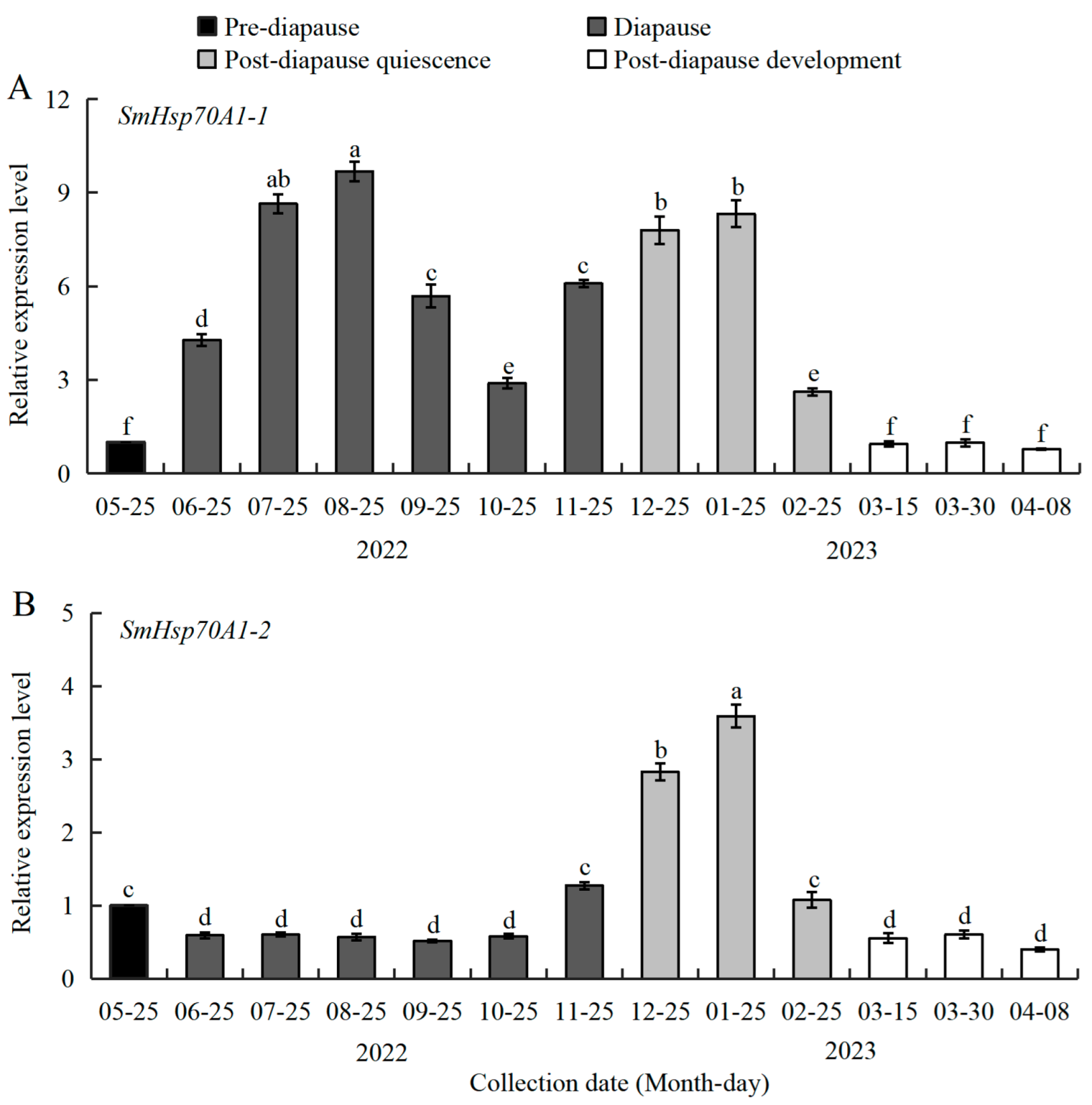
To clarify the association between diapause and SmHsp70A1-1/SmHsp70A1-2 expression, the mRNA transcriptional abundance of both genes was quantified in 3rd-instar S. mosellana larvae across four diapause-associated physiological stages: pre-diapause, diapause, post-diapause quiescence, and post-diapause development (Figure 4). SmHsp70A1-1 exhibited relatively low transcript abundance at the pre-diapause phase (May), with significant upregulation at diapause entry (June). High expression was maintained throughout diapause (June–November) and post-diapause quiescence (December to the following February), followed by a rapid decline to baseline pre-diapausal levels after the onset of post-diapausal development (March to early April in the following spring). The apex of expression was observed during July and August (summer), with a secondary peak in December and January (winter) (Figure 4A).
In contrast, the expression of SmHsp70A1-2 exhibited significant downregulation at the start of diapause, with persistent suppression during diapause (June–October), followed by marked upregulation from late November as the populations entered post-diapause quiescence [33]. The expression levels reached their peak during mid-quiescence (January) before rapidly declining to diapause-equivalent levels during the post-diapausal developmental phases (Figure 4B).

3.4. 20E Regulation of SmHsp70 Genes During Diapause

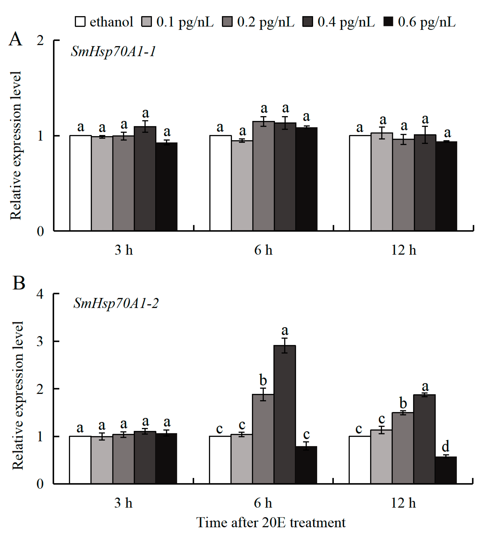
The distinct expression patterns of the two SmHsp70s were observed in the diapausing larvae collected during October, which had been treated with 20E (0.1–0.6 pg/nL). The expression of SmHsp70A1-1 remained unchanged, while that of SmHsp70A1-2 exhibited significant dose-time-dependent induction at 6 and 12 h post-treatment (Figure 5). At 0.2 and 0.4 pg/nL of 20E, SmHsp70A1-2 expression increased by 1.88- and 2.91-fold at 6 h and by 1.50- and 1.87-fold at 12 h, respectively, compared to the control (ethanol) group (Figure 5B). No significant changes in expression were detected for either gene at 3 h post-injection at any of the tested 20E concentrations (Figure 5A,B).

3.5. Patterns of SmHsp70 Gene Expression in Response to Heat and Cold Temperature Stress During Diapause

The heat-responsive expression patterns of SmHsp70A1-1 and SmHsp70A1-2 were very similar in the over-summering diapause larvae. When compared to the untreated group (CK), both SmHsp70A1-1 and SmHsp70A1-2 exhibited pronounced upregulation at temperatures between 35 °C and 45 °C, reaching a maximum at 40 °C, with 22.07-fold and 6.39-fold increases, respectively. No notable changes in the expression of either gene were observed at 50 °C (Figure 6A,B).
The effects of varying exposure durations at 40 °C and 45 °C on the mRNA expression profiles of both genes were then investigated. Transcriptional upregulation of both genes was observed after exposure to heat stress for 15 to 60 min at 40 °C, with peak expression observed at 60 min. At 45 °C, the highest gene expression levels occurred between 15 and 30 min, with SmHsp70A1-1 reaching peak transcription at 15 min and SmHsp70A1-2 at 30 min (Figure 6C,D).
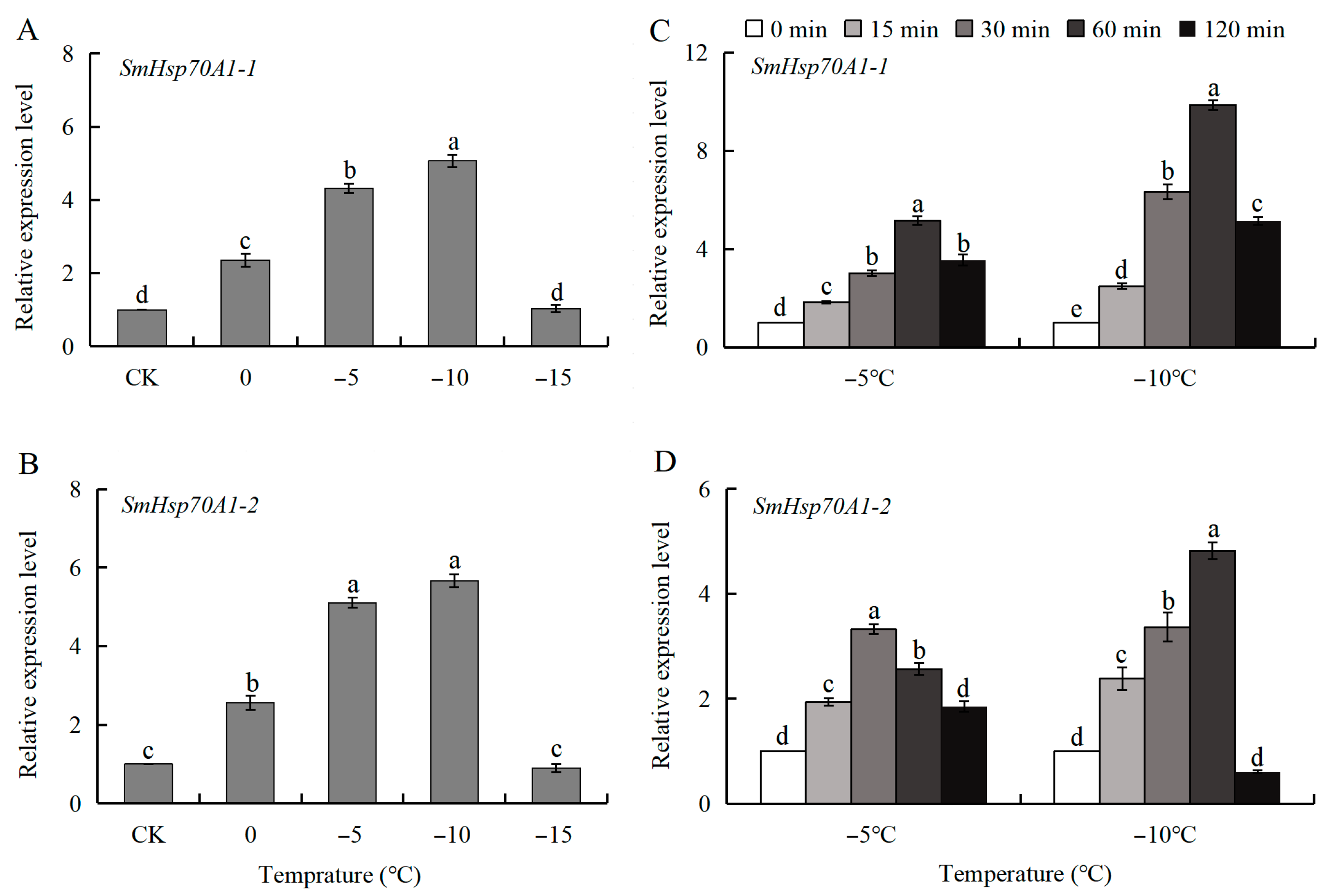
Similarly, the transcriptional expression levels of SmHsp70A1-1 and SmHsp70A1-2 were quantified in over-wintering larvae after an hour of exposure to different low temperatures. Both genes showed significant cold-induced expression above −15 °C, reaching maximum expression at −10 °C, with an 8.53-fold increase in SmHsp70A1-1 levels and a 5.66-fold increase in SmHsp70A1-2 levels relative to the control group (Figure 7A,B).

3.6. Silencing of SmHsp70 Genes by RNAI and Assessment of Susceptibility to Cold Stress

To assess the efficacy of RNAi-mediated gene silencing, the expression of SmHsp70A1-1, SmHsp70A1-2, and SmHsp70 (GenBank: KJ813013) was determined after dsRNA injection. Significant knockdown of SmHsp70A1-1 and SmHsp70A1-2 was observed at 24 h and 48 h post-injection relative to the DEPC-treated water and dsGFP controls. Specifically, 24 h after injection with dsSmHsp70A1-1, the expression of SmHsp70A1-1 had decreased by 52% and 56% compared to the DEPC-treated water and dsGFP control, respectively, whereas that of SmHsp70A1-2 and SmHsp70 remained unaffected. After injection with dsSmHsp70A1-2, the expression levels of SmHsp70A1-2 were reduced by 50% and 52%, respectively, compared to the DEPC-treated water and dsGFP controls. Although no effect on SmHsp70A1-1 was observed, SmHsp70 expression was upregulated by 79% and 83% compared to DEPC-treated water and dsGFP control, respectively (Figure 8A).
Similarly, 48 h after dsSmHsp70A1-2 injection, respective reductions of 59% and 61% were observed in SmHsp70A1-2 expression compared to the DEPC-treated water and dsGFP controls, with no effects on SmHsp70A1-1 and 70% and 73% increases in SmHsp70 expression, respectively. After dsSmHsp70A1-1 administration, the expression of SmHsp70A1-1 decreased by 61% and 60%, respectively, relative to the DEPC-treated water and dsGFP controls, whereas that of SmHsp70A1-2 and SmHsp70 remained unchanged (Figure 8B).
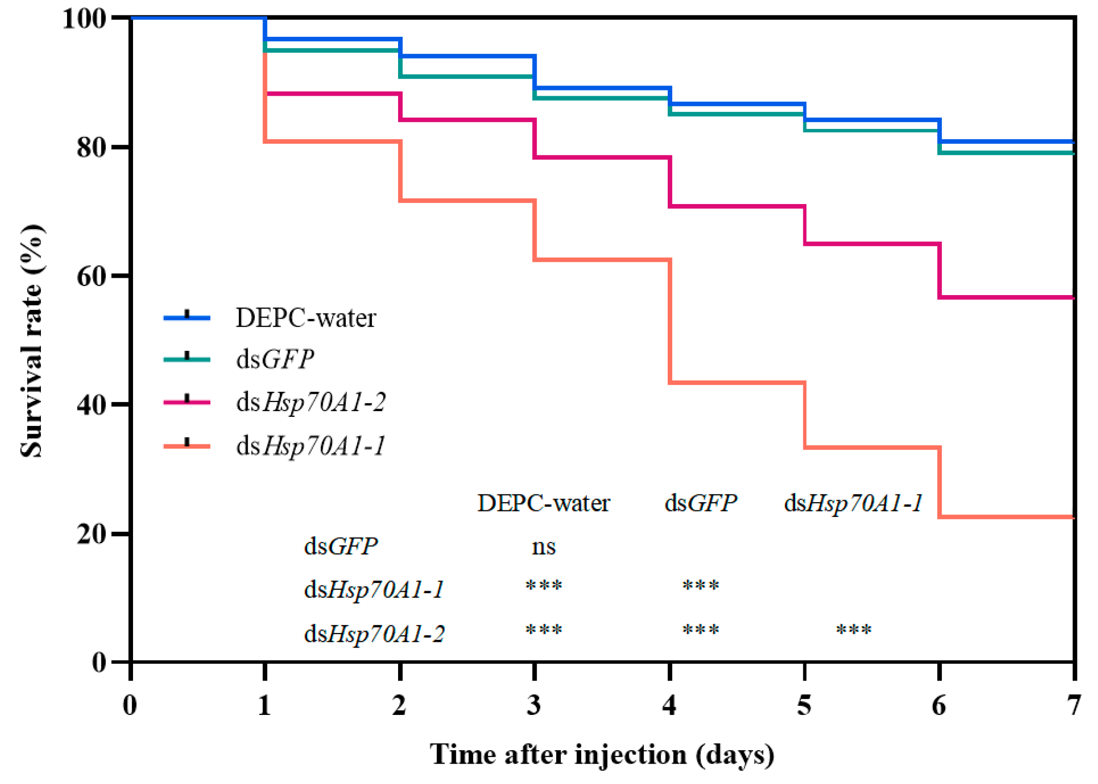
Twenty-four hours after knockdown, the larvae were subjected to a −10 °C cold shock for 2 h in a controlled low-temperature incubator, followed by monitoring of subsequent mortality for six days. The mortality rates of the SmHsp70A1-1 and SmHsp70A1-2-silenced larvae were found to increase significantly compared to the DEPC-water or dsGFP-treated groups. Each experimental cohort (DEPC-water, dsGFP, dsHsp70A1-1, and dsHsp70A1-2; n = 120 per group) showed cumulative six-day mortality rates of 19% (23/120), 22% (26/120), 78% (93/120), and 43% (52/120), respectively. Furthermore, the mortality rate in the SmHsp70A1-1-silenced group was significantly higher than that observed in the SmHsp70A1-2-silenced group. These findings indicate that the suppression of SmHsp70A1-1 and SmHsp70A1-2 significantly increased susceptibility to cold stress in S. mosellana larvae (Figure 9).

4. Discussion

Members of the Hsp70 family are pivotal components of the molecular chaperone network in cells [39]. The number of Hsp70 genes varies according to the insect species [2,40,41]. In S. mosellana larvae, one Hsp70 had been previously identified and was believed to play a crucial role in thermal stress responses during diapause [34]. Here, we isolated two additional Hsp70 transcripts (SmHsp70A1-1 and SmHsp70A1-2) from this species, which shared high sequence homology with orthologs in other taxonomic groups. The comparison of the cDNA and genomic DNA nucleotide sequences indicated that the two genes are intronless. The absence of introns is posited to enhance the expression of stress-responsive genes, as the lack of mRNA splicing enables rapid accumulation of the transcripts and subsequent responses to stress [19,42].
As anticipated, each deduced SmHsp70 protein sequence contained three Hsp70 family signature motifs, namely, IDLGTTYS, FDLGGGTFDVSV/IL, and IVLVGGSTRIPKV/IQ, which are distinctive hallmarks of this family (Figure 1) [43,44]. In addition, highly conserved EEVD motifs, shown to be essential for chaperone-binding activity [45,46], were identified in the C-terminal regions of both SmHsp70s (Figure 1), suggesting their localization within the cytoplasm. Predictions of subcellular localization and phylogenetic analyses demonstrated the classification of the two SmHsp70s within cytosolic subfamilies (Figure 2). While the SmHsp70s showed extensive regions of conservation, greater variability was observed in the C-terminal domains, suggesting that these differences may determine the functional specificities of individual Hsps [47,48].
Insect Hsps have been found to be associated with the regulation of insect growth, development, and metamorphosis [18,49]. Typically, the expression levels of most insect Hsps vary considerably across different developmental stages within a species. For instance, TcHsp70III in Tribolium castaneum is highly expressed in adults and young larvae but at lower levels in older larvae and pupae [50], while in Galeruca daurica, GdHsp70 exhibits elevated expression in eggs and first-instar larvae [51]. In the present study, the highest levels of SmHsp70A1-1 were observed in the first- and second-instar larvae, followed by the pupae, whereas SmHsp70A1-2 exhibited its highest expression during the pre-pupal and pupal stages (Figure 3). These developmental stages correspond to periods of elevated basal metabolic activity, as feeding occurs during the early larval stage, while metamorphosis is associated with the pupal stage. Similar developmental stage-preferred expression patterns have been documented in Cydia pomonella [1], Nilaparvata lugens [52], and Myzus persicae [53]. Moreover, the pupal stage involves the degradation and reconstruction of multiple organs and tissues, which may contribute to the increased expression of Hsps, as observed with Hsp70 in Grapholitha molesta [54] and Hsp71.3 in Glyphodes pyloalis [55].
As described in the Introduction section, many studies have shown that the roles of Hsp70s in mediating diapause vary considerably across different insect taxonomic groups. The present research on S. mosellana revealed pronounced upregulation of SmHsp70A1-1 during diapause, especially in summer and winter (Figure 4A), consistent with the expression patterns of SmHsp70/90 [34]. The current evidence suggests that diapause-induced Hsps may directly induce cell cycle arrest [56], as exemplified in Drosophila melanogaster, where elevated levels of small Hsps and Hsp70 are correlated directly with cell cycle arrest or retardation [57,58]. It is possible that Hsp70A1-1 in S. mosellana may also have similar regulatory roles during diapause, although this specific role warrants further molecular investigation.
In contrast to SmHsp70A1-1, SmHsp70A1-2 expression was found to be downregulated upon entry into diapause but increased markedly during the shift to post-diapause quiescence (Figure 4B), correlating with fluctuations in 20E titers documented in this species [36]. Functional crosstalk has been reported between 20E signaling pathways and heat shock protein regulatory mechanisms [59,60]. In Bombyx mori Bm12 cell lines, 20E induces the expression of Hsp70s, which enhances 20E signal transduction by promoting EcR nuclear localization [18]. The observed 20E-mediated transcriptional induction of S. mosellana Hsp70A1-2 in diapausing larvae provides additional evidence of the linkage between 20E signaling and Hsp70A1-2 expression (Figure 5B). This pronounced upregulation at the transition of diapause to post-diapause quiescence suggests that SmHsp70A1-2 may represent a dependable molecular indicator for predicting this developmental shift.
Temperature has a profound influence on insect dispersal and population dynamics. Extreme temperatures lead to increased synthesis of Hsps in insects as an adaptive mechanism [19,61]. Similar to patterns observed in Bemisia tabaci for Hsp70 [43], Bombyx mori for Hsp70-1, Hsp70-2, and Hsp70-3 [62], and Pieris melete for Hsp70a and Hsp70b [21], the two S. mosellana Hsp70 transcripts were significantly induced by heat and cold stress within a brief timeframe during larval diapause, although this induction became less pronounced with prolonged exposure and further increases or decreases in temperature (Figure 6 and Figure 7). Notably, exposure to 47.5 °C for 1 h was found to be lethal to cocooned S. mosellana larvae [63], indicating the limitations of Hsp-mediated thermoprotection under extreme heat stress. This may explain why agronomic practices that elevate soil temperature are effective in pest management; during the fallow summer period, increased solar radiation is absorbed by the surface soils, while tillage redistributes the larvae into the warmer upper-soil strata, with both factors contributing to pest mortality [64,65].
In general, the upregulation of Hsp gene transcription in insects is closely associated with enhanced tolerance to low-temperature stress [19,66]. For instance, in the flesh fly Sarcophaga crassipalpis, Hsp70 exhibits a developmentally upregulated response during the over-wintering pupal diapause [67]. While RNAi inhibition of Hsp70 upregulation in response to cold in S. crassipalpis did not affect diapause initiation or duration, it significantly reduced low-temperature tolerance in the pupae [22]. Similarly, Spodoptera exigua larvae exhibited increased sensitivity to temperature around 0 °C and higher mortality after Hsp70-specific dsRNA injection [17]. In this study, the knockdown of SmHsp70A1-1 and SmHsp70A1-2 significantly reduced the survival rate of S. mosellana larvae at −10 °C, indicating that both genes confer protective effects that enhance cold tolerance in this pest. Notably, larval mortality was significantly higher in the SmHsp70A1-1-knockdown group compared to the SmHsp70A1-2 group at −10 °C (Figure 9), suggesting compensatory effects, as the suppression of SmHsp70A1-2 resulted in a significant upregulation of SmHsp70 expression (Figure 8), and vice versa (Figure S2). However, due to the lack of specific monoclonal antibodies against SmHsp70A1-1 and SmHsp70A1-2, this study was limited to mRNA-level analysis after gene knockout; thus, protein-level verification post-knockout requires further investigation.

5. Conclusions

The study findings demonstrate that both SmHsp70A1-1 and SmHsp70A1-2 are developmentally and environmentally regulated. The expression of SmHsp70A1-1 was upregulated during diapause, whereas that of SmHsp70A1-2 showed downregulation and dose-dependent induction by 20E. Their induction in response to heat (35–45 °C) and cold (0–−10 °C) stress, as well as the cold sensitivity induced by gene-specific RNA interference, indicated that both genes play crucial roles in conferring resistance or tolerance to environmental stresses in S. mosellana. Further investigation into more SmHsp70s and their interactions is essential for elucidating the adaptive mechanisms of S. mosellana and promoting efficient integrated pest management under environmental selection pressures.

Supplementary Materials

The following supporting information can be downloaded at: https://www.mdpi.com/article/10.3390/biology14091147/s1, Figure S1. Nucleotide and deduced amino acid sequences of SmHsp70A1-1 and SmHsp70A1-2 in Sitodiplosis mosellana. Initiation codons (ATG) and termination codons (TGA/TAA) were marked with ellipses. Three signature motifs characteristic of the Hsp70 family were highlighted by shading, and the cytosolic-specific consensus motif (EEVD) was boxed, Figure S2. Expression patterns of three SmHsp70s at 24 h (A) and 48h (B) after dsSmHsp70 injection. Relative mRNA levels (mean ± SE) at each gene are quantified against the DEPC-water control (value = 1). Different letters indicate statistically significant differences (Tukey’s multiple range test, p < 0.05).

Author Contributions

Conceptualization, W.C. and K.Z.-S.; methodology, Q.H.; investigation, Q.H.; formal analysis, Q.H., W.T., X.L., Q.M. and W.C.; writing—original draft preparation, Q.H. and W.C.; writing—review and editing, W.C. and K.Z.-S. All authors have read and agreed to the published version of the manuscript.

Funding

This work was supported by the Natural Science Foundation of Shandong Province (ZR2023QC051), the Shandong Academy of Agricultural Sciences Agricultural Science and Technology Innovation Engineering (CXGC2024F19, CXGC2025F18), the National Natural Science Foundation of China (31371933), and the Open Project of Shaanxi Laboratory for Agriculture in Arid Areas (2024ZY-JCYJ-02-39).

Institutional Review Board Statement

Not applicable.

Informed Consent Statement

Not applicable.

Data Availability Statement

The original contributions presented in this study are included in the article/Supplementary Material. Further inquiries can be directed to the corresponding author(s).

Conflicts of Interest

The authors declare no conflicts of interest.

References

  1. Yang, X.Q.; Zhang, Y.L.; Wang, X.Q.; Dong, H.; Gao, P.; Jia, L.Y. Characterization of multiple heat-shock protein transcripts from Cydia pomonella: Their response to extreme temperature and insecticide exposure. J. Agric. Food Chem. 2016, 64, 4288–4298. [Google Scholar] [CrossRef]
  2. Tan, S.Y.; Feng, K.; Zhai, X.D.; Wang, J.J.; Wei, D. Expression and function analysis of two heat shock protein 70 genes (Hsp70s) involved in different high-temperature stresses in Zeugodacus cucurbitae (Diptera: Tephritidae). Entomol. Gen. 2023, 43, 481–490. [Google Scholar] [CrossRef]
  3. Ritossa, F. A new puffing pattern induced by temperature shock and DNP in drosophila. Experientia 1962, 18, 571–573. [Google Scholar] [CrossRef]
  4. Lubkowska, A.; Pluta, W.; Strońska, A.; Lalko, A. Role of heat shock proteins (HSP70 and HSP90) in viral infection. Int. J. Mol. Sci. 2021, 22, 9366. [Google Scholar] [CrossRef]
  5. Pei, T.W.; Zhang, M.; Nwanade, C.F.; Meng, H.; Bai, R.W.; Wang, Z.H.; Wang, R.T.; Zhang, T.A.; Liu, J.Z.; Yu, Z.J. Sequential expression of small heat shock proteins contributing to the cold response of Haemaphysalis longicornis (Acari: Ixodidae). Pest Manag. Sci. 2024, 80, 2061–2071. [Google Scholar] [CrossRef]
  6. Liu, J.P.; Liu, Y.; Wang, W.; Liang, G.M.; Lu, Y.H. Characterizing three heat shock protein 70 genes of Aphis gossypii and their expression in response to temperature and insecticide stress. J. Agric. Food. Chem. 2025, 73, 2842–2852. [Google Scholar] [CrossRef]
  7. Rosenzweig, R.; Nillegoda, N.B.; Mayer, M.P.; Bukau, B. The Hsp70 chaperone network. Nat. Rev. Mol. Cell Biol. 2019, 20, 665–680. [Google Scholar] [CrossRef]
  8. Hu, C.; Yang, J.; Qi, Z.P.; Wu, H.; Wang, B.L.; Zou, F.M.; Mei, H.S.; Liu, J.; Wang, W.C.; Liu, Q.S. Heat shock proteins: Biological functions, pathological roles, and therapeutic opportunities. Med. Comm. 2022, 3, e161. [Google Scholar] [CrossRef]
  9. Huang, L.H.; Wang, H.S.; Kang, L. Different evolutionary lineages of large and small heat shock proteins in eukaryotes. Cell Res. 2008, 18, 1074–1076. [Google Scholar] [CrossRef]
  10. Zhang, X.Z.; Yu, W. Heat shock proteins and viral infection. Front. Immunol. 2022, 13, 947789. [Google Scholar] [CrossRef]
  11. Sørensen, J.G.; Kristensen, T.N.; Loeschcke, V. The evolutionary and ecological role of heat shock proteins. Ecol. Lett. 2023, 6, 1025–1037. [Google Scholar] [CrossRef]
  12. Mayer, M.P.; Bukau, B. Hsp70 chaperones: Cellular functions and molecular mechanism. Cell. Mol. Life Sci. 2005, 62, 670–684. [Google Scholar] [CrossRef]
  13. Paim, R.M.M.; Araujo, R.N.; Leis, M.; Sant’anna, M.R.V.; Gontijo, N.F.; Lazzari, C.R.; Pereira, M.H. Functional evaluation of heat shock proteins 70 (HSP70/HSC70) on Rhodnius prolixus (Hemiptera, Reduviidae) physiological responses associated with feeding and starvation. Insect Biochem. Mol. Biol. 2016, 77, 10–20. [Google Scholar] [CrossRef]
  14. Qin, W.S.; Tyshenko, M.G.; Wu, B.S.; Walker, V.K.; Robertson, R.M. Cloning and characterization of a member of the Hsp70 gene family from Locusta migratoria, a highly thermotolerant insect. Cell Stress Chaperones 2003, 8, 144–152. [Google Scholar] [CrossRef]
  15. Sun, Y.; Zhao, J.; Sheng, Y.; Xiao, Y.F.; Zhang, Y.J.; Bai, L.X.; Tan, Y.A.; Xiao, L.B.; Xu, G.C. Identification of heat shock cognate protein 70 gene (Alhsc70) of Apolygus lucorum and its expression in response to different temperature and pesticide stresses. Insect Sci. 2016, 23, 37–49. [Google Scholar] [CrossRef]
  16. Gehring, W.J.; Wehner, R. Heat shock protein synthesis and thermotolerance in Cataglyphis, an ant from the Sahara desert. Proc. Natl. Acad. Sci. USA 1995, 92, 2994–2998. [Google Scholar] [CrossRef]
  17. Choi, B.G.; Hepat, R.; Kim, Y. RNA interference of a heat shock protein, Hsp70, loses its protection role in indirect chilling injury to the beet armyworm, Spodoptera exigua. Comp. Biochem. Physiol. A Mol. Integr. Physiol. 2014, 168, 90–95. [Google Scholar] [CrossRef]
  18. Gu, J.; Ye, Y.; Zheng, Z.W.; Luo, W.; Gong, Y.J.; Feng, Q.L.; Li, S.; Huang, L.H. Cytoplasmic Hsp70s promote EcR transport into the nucleus by responding to various stimuli. Insect Biochem. Mol. Biol. 2023, 157, 103964. [Google Scholar] [CrossRef]
  19. King, A.M.; MacRae, T.H. Insect heat shock proteins during stress and diapause. Annu. Rev. Entomol. 2015, 60, 59–75. [Google Scholar] [CrossRef]
  20. Schebeck, M.; Lehmann, P.; Laparie, M.; Bentz, B.J.; Ragland, G.J.; Battisti, A.; Hahn, D.A. Seasonality of forest insects: Why diapause matters. Trends Ecol. Evol. 2024, 39, 757–770. [Google Scholar] [CrossRef]
  21. Zhang, J.; Miano, F.N.; Jiang, T.; Peng, Y.C.; Zhang, W.N.; Xiao, H.J. Characterization of three heat shock protein genes in Pieris melete and their expression patterns in response to temperature stress and pupal diapause. Insects 2022, 13, 430. [Google Scholar] [CrossRef]
  22. Rinehart, J.P.; Li, A.Q.; Yocum, G.D.; Robich, R.M.; Hayward, S.A.L.; Denlinger, D.L. Up-regulation of heat shock proteins is essential for cold survival during insect diapause. Proc. Natl. Acad. Sci. USA 2007, 104, 11130–11137. [Google Scholar] [CrossRef]
  23. Yocum, G.D. Differential expression of two HSP70 transcripts in response to cold shock, thermoperiod, and adult diapause in the Colorado potato beetle. J. Insect Physiol. 2001, 47, 1139–1145. [Google Scholar] [CrossRef]
  24. Moribe, Y.; Oka, K.; Niimi, T.; Yamashita, O.; Yaginuma, T. Expression of heat shock protein 70a mRNA in Bombyx mori diapause eggs. J. Insect Physiol. 2010, 56, 1246–1252. [Google Scholar] [CrossRef]
  25. Uzelac, I.; Avramov, M.; Knežić, T.; Tatić, V.; Gošić-Dondo, S.; Popović, Ž.D. Prolonged heat stress during winter diapause alters the expression of stress-response genes in Ostrinia nubilalis (Hbn.). Int. J. Mol. Sci. 2024, 25, 3100. [Google Scholar] [CrossRef] [PubMed]
  26. Tungjitwitayakul, J.; Tatun, N.; Singtripop, T.; Sakurai, S. Characteristic expression of three heat shock-responsive genes during larval diapause in the bamboo borer Omphisa fuscidentalis. Zool. Sci. 2008, 25, 321–333. [Google Scholar] [CrossRef] [PubMed]
  27. Gkouvitsas, T.; Kontogiannatos, D.; Kourti, A. Cognate Hsp70 gene is induced during deep larval diapause in the moth Sesamia nonagrioides. Insect Mol. Biol. 2009, 18, 253–264. [Google Scholar] [CrossRef]
  28. Zhang, Q.R.; Denlinger, D.L. Molecular characterization of heat shock protein 90, 70 and 70 cognate cDNAs and their expression patterns during thermal stress and pupal diapause in the corn earworm. J. Insect Physiol. 2010, 56, 138–150. [Google Scholar] [CrossRef]
  29. Miao, J.; Huang, J.R.; Wu, Y.Q.; Gong, Z.J.; Li, H.L.; Zhang, G.Y.; Duan, Y.; Li, T.; Jiang, Y.L. Climate factors associated with the population dynamics of Sitodiplosis mosellana (Diptera: Cecidomyiidae) in central China. Sci. Rep. 2019, 9, 12361. [Google Scholar] [CrossRef]
  30. Shrestha, G.; Reddy, G.V.P. Field efficacy of insect pathogen, botanical, and jasmonic acid for the management of wheat midge Sitodiplosis mosellana and the impact on adult parasitoid Macroglenes penetrans populations in spring wheat. Insect Sci. 2019, 26, 523–535. [Google Scholar] [CrossRef] [PubMed]
  31. Wang, Q.; Liu, X.B.; Liu, H.; Fu, Y.; Cheng, Y.M.; Zhang, L.J.; Shi, W.P.; Zhang, Y.; Chen, J.L. Transcriptomic and metabolomic analysis of wheat kernels in response to the feeding of orange wheat blossom midges (Sitodiplosis mosellana) in the field. J. Agric. Food Chem. 2022, 70, 1477–1493. [Google Scholar] [CrossRef]
  32. Wang, Y.; Long, Z.R.; Feng, A.R.; Cheng, W.N. Effects of initial population number, wheat varieties and precipitation on infestation of Sitodiplosis mosellana (Diptera: Cecidomyiidae). Acta Agric. Boreali-Occident. Sin. 2015, 24, 165–171. [Google Scholar]
  33. Cheng, W.N.; Long, Z.R.; Zhang, Y.D.; Liang, T.T.; Zhu-Salzman, K.Y. Effects of temperature, soil moisture and photoperiod on diapause termination and post-diapause development of the wheat blossom midge, Sitodiplosis mosellana (Géhin) (Diptera: Cecidomyiidae). J. Insect Physiol. 2017, 103, 78–85. [Google Scholar] [CrossRef] [PubMed]
  34. Cheng, W.N.; Li, D.; Wang, Y.; Liu, Y.; Zhu-Salzman, K.Y. Cloning of heat shock protein genes (hsp70, hsc70 and hsp90) and their expression in response to larval diapause and thermal stress in the wheat blossom midge, Sitodiplosis mosellana. J. Insect Physiol. 2016, 95, 66–77. [Google Scholar] [CrossRef] [PubMed]
  35. Cheng, W.N.; Li, X.J.; Zhao, J.J.; Zhu-Salzman, K.Y. Cloning and characterization of methoprene-tolerant (Met) and krüppel homolog 1 (Kr-h1) genes in the wheat blossom midge, Sitodiplosis mosellana. Insect Sci. 2020, 27, 292–303. [Google Scholar] [CrossRef]
  36. Cheng, W.N.; Li, J.J.; Li, Y.P.; Li, X.L.; Wu, J.X.; Wang, H.L. Quantitative analysis of ecdysteroid in adults and the pre-diapause, diapause and post-diapause larvae of wheat blossom midge, Sitodiplosis mosellana Gehin. Acta Phytophy. Sin. 2009, 36, 163–167. [Google Scholar]
  37. Doane, J.F.; Olfert, O. Seasonal development of wheat midge, Sitodiplosis mosellana (Géhin) (Diptera: Cecidomyiidae), in Saskatchewan, Canada. Crop Prot. 2008, 27, 951–958. [Google Scholar] [CrossRef]
  38. Ni, H.X.; Ding, H.J.; Sun, J.R. Occurrence dynamic and integrated control strategies of Sitodiplosis mosellana. Chin. Agric. Sci. Bull. 1994, 10, 20–23. [Google Scholar]
  39. Qiu, X.B.; Shao, Y.M.; Miao, S.; Wang, L. The diversity of the DnaJ/Hsp40 family, the crucial partners for Hsp70 chaperones. Cell. Mol. Life Sci. 2006, 63, 2560–2570. [Google Scholar] [CrossRef]
  40. Nguyen, A.D.; Gotelli, N.J.; Cahan, S.H. The evolution of heat shock protein sequences, cis-regulatory elements, and expression profiles in the eusocial Hymenoptera. BMC Evol. Biol. 2016, 16, 15. [Google Scholar] [CrossRef] [PubMed]
  41. Goyal, B.; Tushir, S.; Sharma, A.; Singh, S.; Tatu, U.; Pandey, K.; Chakraborti, S. Unveiling role of HSP70 genes for development and survival of Indian malaria vector Anopheles culicifacies. Int. J. Biol. Macromol. 2025, 308, 142173. [Google Scholar] [CrossRef]
  42. Silver, J.T.; Noble, E.G. Regulation of survival gene hsp70. Cell Stress Chaperones 2012, 17, 1–9. [Google Scholar] [CrossRef]
  43. Wang, X.R.; Wang, C.; Ban, F.X.; Zhu, D.T.; Liu, S.S.; Wang, X.W. Genome-wide identification and characterization of HSP gene superfamily in whitefly (Bemisia tabaci) and expression profiling analysis under temperature stress. Insect Sci. 2019, 26, 44–57. [Google Scholar] [CrossRef] [PubMed]
  44. Chen, X.; Li, Z.D.; Li, D.T.; Jiang, M.X.; Zhang, C.X. HSP70/DNAJ family of genes in the brown planthopper, Nilaparvata lugens: Diversity and function. Genes 2021, 12, 394. [Google Scholar] [CrossRef]
  45. Bukau, B.; Weissman, J.; Horwich, A. Molecular chaperones and protein quality control. Cell 2006, 125, 443–451. [Google Scholar] [CrossRef] [PubMed]
  46. Daugaard, M.; Rohde, M.; Jäättelä, M. The heat shock protein 70 family: Highly homologous proteins with overlapping and distinct functions. FEBS Lett. 2007, 581, 3702–3710. [Google Scholar] [CrossRef]
  47. Fuertes, M.A.; Pérez, J.M.; Soto, M.; Menéndez, M.; Alonso, C. Thermodynamic stability of the C-terminal domain of the human inducible heat shock protein 70. Biochim. Biophys. Acta 2004, 1699, 45–56. [Google Scholar] [CrossRef]
  48. Park, H.; Ahn, I.Y.; Lee, H.E. Expression of heat shock protein 70 in the thermally stressed antarctic clam Laternula elliptica. Cell Stress Chaperones 2007, 12, 275–282. [Google Scholar] [CrossRef] [PubMed]
  49. Kanakala, S.; Kontsedalov, S.; Lebedev, G.; Ghanim, M. Plant-mediated Silencing of the whitefly Bemisia tabaci cyclophilin B and heat shock protein 70 impairs insect development and virus transmission. Front. Physiol. 2019, 10, 557. [Google Scholar] [CrossRef]
  50. Mahroof, R.; Zhu, K.Y.; Neven, L.; Subramanyam, B.; Bai, J.F. Expression patterns of three heat shock protein 70 genes among developmental stages of the red flour beetle, Tribolium castaneum (Coleoptera: Tenebrionidae). Comp. Biochem. Physiol. A Mol. Integr. Physiol. 2005, 141, 247–256. [Google Scholar] [CrossRef]
  51. Tan, Y.; Zhang, Y.; Huo, Z.J.; Zhou, X.R.; Shan, Y.M.; Pang, B.P. Molecular cloning and expression profiling of the heat shock protein gene GdHsp70 in Galeruca daurica (Coleoptera: Chrysomelidae). Acta Entomol. Sin. 2017, 60, 865–875. [Google Scholar]
  52. Lu, K.; Chen, X.; Liu, W.T.; Zhang, Z.C.; Wang, Y.; You, K.K.; Li, Y.; Zhang, R.B.; Zhou, Q. Characterization of heat shock protein 70 transcript from Nilaparvata lugens (Stål): Its response to temperature and insecticide stresses. Pestic. Biochem. Physiol. 2017, 142, 102–110. [Google Scholar] [CrossRef]
  53. Dong, B.; Liu, X.Y.; Li, B.; Li, M.Y.; Li, S.G.; Liu, S. A heat shock protein protects against oxidative stress induced by lambda-cyhalothrin in the green peach aphid Myzus persicae. Pestic. Biochem. Physiol. 2022, 181, 104995. [Google Scholar] [CrossRef]
  54. Zhang, B.; Peng, Y.; Zheng, J.C.; Liang, L.N.; Hoffmann, A.A.; Ma, C.S. Response of heat shock protein genes of the oriental fruit moth under diapause and thermal stress reveals multiple patterns dependent on the nature of stress exposure. Cell Stress Chaperones 2016, 21, 653–663. [Google Scholar] [CrossRef]
  55. Ding, J.H.; Zheng, L.X.; Chu, J.; Liang, X.H.; Wang, J.; Gao, X.W.; Wu, F.A.; Sheng, S. Characterization, and functional analysis of Hsp70 and Hsp90 gene families in Glyphodes pyloalis Walker (Lepidoptera: Pyralidae). Front. Physiol. 2021, 12, 753914. [Google Scholar] [CrossRef]
  56. Denlinger, D.L. Regulation of diapause. Annu. Rev. Entomol. 2002, 47, 93–122. [Google Scholar] [CrossRef]
  57. Feder, J.H.; Rossi, J.M.; Solomon, J.; Solomon, N.; Lindquist, S. The consequences of expressing hsp70 in Drosophila cells at normal temperatures. Genes Dev. 1992, 6, 1402–1413. [Google Scholar] [CrossRef] [PubMed]
  58. Krebs, R.A.; Feder, M.E. Deleterious consequences of Hsp70 overexpression in Drosophila melanogaster larvae. Cell Stress Chaperones 1997, 2, 60–71. [Google Scholar] [CrossRef] [PubMed]
  59. Lezzi, M. Chromosome puffing: Supramolecular aspects of ecdysone action. In Metamorphosis; Academic Pres: San Diego, CA, USA, 1996; pp. 145–173. [Google Scholar] [CrossRef]
  60. Liu, W.; Zhang, F.X.; Cai, M.J.; Zhao, W.L.; Li, X.R.; Wang, J.X.; Zhao, X.F. The hormone-dependent function of Hsp90 in the crosstalk between 20-hydroxyecdysone and juvenile hormone signaling pathways in insects is determined by differential phosphorylation and protein interactions. Biochim. Biophys. Acta 2013, 1830, 5184–5192. [Google Scholar] [CrossRef] [PubMed]
  61. Liu, J.P.; Liu, Y.; Li, Q.; Lu, Y.H. Heat shock protein 70 and Cathepsin B genes are involved in the thermal tolerance of Aphis gossypii. Pest Manag. Sci. 2023, 79, 2075–2086. [Google Scholar] [CrossRef]
  62. Fang, S.M.; Zhang, Q.; Zhang, Y.L.; Zhang, G.Z.; Zhang, Z.; Yu, Q.Y. Heat shock protein 70 family in response to multiple abiotic stresses in the silkworm. Insects 2021, 12, 928. [Google Scholar] [CrossRef]
  63. Sokhansanj, S.; Venkatesam, V.S.; Wood, H.C.; Doane, J.F.; Spurr, D.T. Thermal kill of wheat midge and Hessian fly. Postharvest Biol. Technol. 1992, 2, 65–71. [Google Scholar] [CrossRef]
  64. Barnes, H.F. Studies of fluctuations in insect populations. VIII. The wheat blossom midge on broadbalk, 1932–1940, with a discussion of the results obtained 1927–40. J. Anim. Ecol. 1941, 10, 94–120. [Google Scholar] [CrossRef]
  65. Yuan, F. The Wheat Blossom Midges Sitodiplosis mosellana (Gehin) and Contarinia tritici (Kirby): Their Plague Principle and Control; Science Press: Beijing, China, 2004. [Google Scholar]
  66. Jiang, F.; Chang, G.F.; Li, Z.Z.; Abouzaid, M.; Du, X.Y.; Hull, J.J.; Ma, W.H.; Lin, Y.J. The HSP/co-chaperone network in environmental cold adaptation of Chilo suppressalis. Int. J. Biol. Macromol. 2021, 187, 780–788. [Google Scholar] [CrossRef] [PubMed]
  67. Rinehart, J.P.; Yocum, G.D.; Denlinger, D.L. Developmental upregulation of inducible hsp70 transcripts, but not the cognate form, during pupal diapause in the flesh fly, Sarcophaga crassipalpis. Insect Biochem. Mol. Biol. 2000, 30, 515–521. [Google Scholar] [CrossRef]
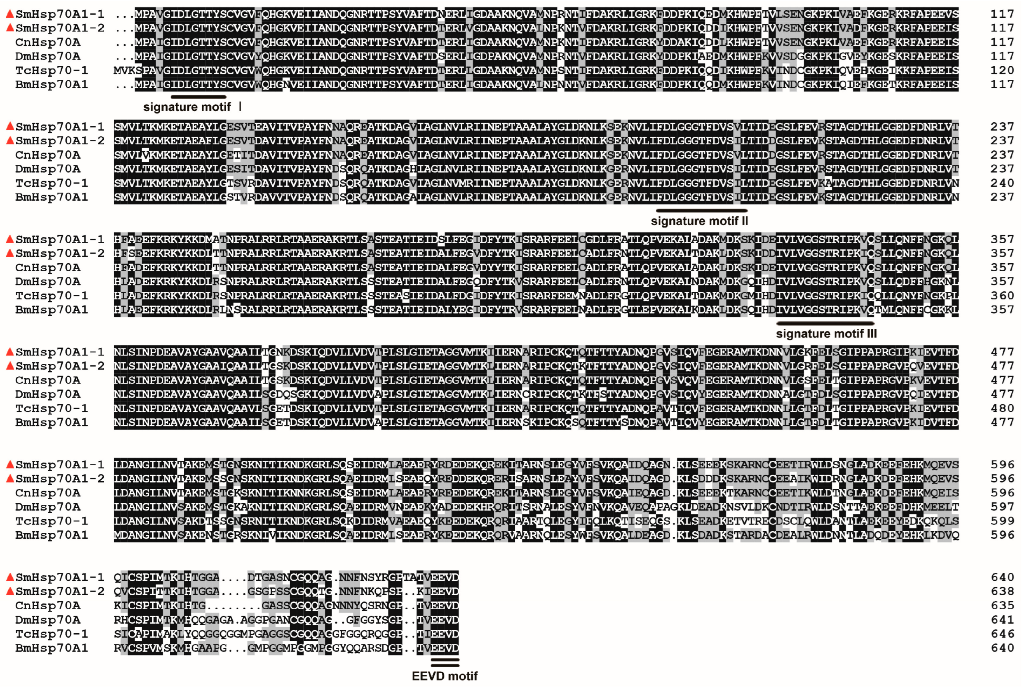
Figure 1. Sequence alignments of Sitodiplosis mosellana SmHsp70A1-1 (GenBank accession no. XXH62668.1) and SmHsp70A1-2 (XXH62669.1) with homologous proteins from other insect taxa, including Contarinia nasturtii (CnHsp70A, XP_031624417.1), Drosophila melanogaster (DmHsp70A, AAG26887.1), Tribolium castaneum (TcHsp70-1, QCI56580.1), and Bombyx mori (BmHsp70A, NP_001296526.1). SmHsp70s are marked by red triangles, with identical residues shown in black and similar ones in gray. Three signature motifs characteristic of the Hsp70 family are delineated with single underlining in black, and the cytosolic-specific consensus motif (EEVD) is emphasized with double underlining in black.
Biology 14 01147 g001
Figure 2. Phylogenetic tree of SmHsp70A1-1 and SmHsp70A1-2 (red triangles) with other known insect Hsp70s based on the neighbor-joining method.
Biology 14 01147 g002
Figure 3. Expression patterns of SmHsp70A1-1 (A) and SmHsp70A1-2 (B) at different developmental stages. Relative mRNA levels (mean ± SE) at different developmental stages are quantified against the first-instar larvae (value = 1). Different letters denote statistically significant differences at each developmental stage (Tukey’s multiple range test, p < 0.05).
Biology 14 01147 g003
Figure 4. Expression patterns of SmHsp70A1-1 (A) and SmHsp70A1-2 (B) in pre-diapausal, diapausal, and post-diapausal larvae of Sitodiplosis mosellana. Relative mRNA levels (mean ± SE) at different diapause stages are quantified against pre-diapausing larvae (value = 1). Different letters denote statistically significant differences at different diapause stages (Tukey’s multiple range test, p < 0.05).
Biology 14 01147 g004
Figure 5. Expression patterns of Hsp70A1-1 (A) and Hsp70A1-2 (B) at 3, 6, and 12 h post-injection of 20E (0.1–0.6 pg/nL) into October-collected diapausing larvae of Sitodiplosis mosellana. Relative mRNA levels (mean ± SE) at each time point are quantified against the ethanol control (value = 1). Different letters denote statistically significant differences at each time point (Tukey’s multiple range test, p < 0.05).
Biology 14 01147 g005
Figure 6. Heat-induced expression profiles of Sitodiplosis mosellana Hsp70A1-1 and Hsp70A1-2 in over-summering diapause larvae treated with various high temperatures (35–50 °C) for 1 h (A,B) or 40–45 °C for varying durations (0–120 min) (C,D). Relative mRNA levels (mean ± SE) at each treatment are quantified against the untreated control (CK, value = 1). Different letters denote statistically significant differences (Tukey’s multiple range test, p < 0.05).
Biology 14 01147 g006
Figure 7. Cold-induced expression profiles of Sitodiplosis mosellana Hsp70A1-1 and Hsp70A1-2 in over-wintering diapause larvae treated with various low temperatures (0–−15 °C) for 1 h (A,B) or −5–−10 °C for varying durations (0–120 min) (C,D). Relative mRNA levels (mean ± SE) at each treatment are quantified against the untreated control (CK, value = 1). Different letters denote statistically significant differences (Tukey’s multiple range test, p < 0.05).
Biology 14 01147 g007
Figure 8. Expression patterns of three SmHsp70s at 24 h (A) and 48 h (B) post-dsRNA injection. Relative mRNA levels (mean ± SE) at each gene are quantified against the DEPC-water control (value = 1). Data are presented as means (n = 3) ± standard error (SE). Different letters indicate statistically significant differences (Tukey’s multiple range test, p < 0.05).
Biology 14 01147 g008
Figure 9. Kaplan–Meier survival curves of Sitodiplosis mosellana larvae after dsRNA injection. Different Kaplan–Meier survival curves correspond to different experimental groups (n = 120 per group), with DEPC-water as the blank control and dsGFP as the nonspecific dsRNA control. Statistical comparisons were performed using log-rank tests, with significance levels indicated as ***, p < 0.001; ns, p > 0.05.
Biology 14 01147 g009
Table 1. Primer sequences used in this study.
Primer NameSequence (5′ to 3′)Purpose
Hsp70A1-1 senseGCGAAAGAAAGGAGGAGAAGORF and
gDNA cloning
Hsp70A1-1 antisenseCAGGCTCTCTTATTGTACGAG
Hsp70A1-2 senseATGCCTGCAGTTGGAATTG
Hsp70A1-2 antisenseGCAAAGAGTGATTTCTTCCTCG
dsHsp70A1-1 sensetaatacgactcactatagggAAAACCAAGTCGCCATGAACdsRNA
synthesis
dsHsp70A1-1 antisensetaatacgactcactatagggTATCCAATCCGTAGGCCAAG
dsHsp70A1-2 sensetaatacgactcactatagggGAAGGCGAACGAGCTATGAC
dsHsp70A1-2 antisensetaatacgactcactatagggCGATCGATTTCTGCTTGTGA
dsGFP sensetaatacgactcactatagggGTGTTCAATGCTTTTCCCGT
dsGFP antisensetaatacgactcactatagggCAATGTTGTGGCGAATTTTG
Hsp70A1-1 senseAATCAACCCAGACGAGGCAGqPCR
Hsp70A1-1 antisenseGCCCAATGAGAGTGGTGTCA
Hsp70A1-2 senseCGTAATTGCTGCGAGGAAGC
Hsp70A1-2 antisenseGTGATTGGCGAGCAAACCTG
GAPDH senseCCATCAAAGCAAGCAAGA
GAPDH antisenseCAGCACGGAGCACAAGAC
Disclaimer/Publisher’s Note: The statements, opinions and data contained in all publications are solely those of the individual author(s) and contributor(s) and not of MDPI and/or the editor(s). MDPI and/or the editor(s) disclaim responsibility for any injury to people or property resulting from any ideas, methods, instructions or products referred to in the content.