Article

Host Shaping Associated Microbiota in Hydrothermal Vent Snails from the Indian Ocean Ridge

1
Key Laboratory of Marine Biogenetic Resources, Third Institute of Oceanography, Ministry of Natural Resources, Xiamen 361005, China
2
Faculty of Marine Biology, Xiamen Ocean Vocational College, Xiamen 361100, China
3
BGI-Qingdao, BGI-Shenzhen, Qingdao 266555, China
4
Laboratory of Genomics and Molecular Biomedicine, Department of Biology, University of Copenhagen, Universitetsparken 13, 2100 Copenhagen, Denmark
5
Key Laboratory of Marine Ecosystem and Biogeochemistry, Second Institute of Oceanography, Ministry of Natural Resources, Hangzhou 310012, China
6
Hainan Deep-Sea Technology Laboratory, Institution of Deep-Sea Life Sciences, IDSSE-BGI, IDSTI-CAS, Sanya 572000, China
*
Authors to whom correspondence should be addressed.
These authors contributed equally to this work.
Biology 2025, 14(8), 954; https://doi.org/10.3390/biology14080954
Submission received: 30 May 2025 / Revised: 19 July 2025 / Accepted: 21 July 2025 / Published: 29 July 2025

Simple Summary

Snails are a dominant member of the fauna in hydrothermal vents in terms of abundance and biomass. Two species of hydrothermal vent snails, Chrysomallon squamiferum (scale snail) and Gigantopelta aegis (scaleless snail), are found along the Indian Ocean Ridge and inhabit areas of low-temperature hydrothermal diffuse flow. Although it is generally assumed that they rely on autotrophic endosymbionts to utilize organic substrates and energy for growth, little is known about their associations with microbiota, particularly regarding community structure, niche acclimation, and metabolic potential. Here, we focused on the diversity and roles of bacterial ectosymbionts on snail feet and endosymbionts in digestive glands of these two species to understand the role of symbiotic microbiota in niche adaptation of the host to harsh hydrothermal environments with steep physical and chemical gradients.

Abstract

Snails at hydrothermal vents rely on symbiotic bacteria for nutrition; however, the specifics of these associations in adapting to such extreme environments remain underexplored. This study investigated the community structure and metabolic potential of bacteria associated with two Indian Ocean vent snails, Chrysomallon squamiferum and Gigantopelta aegis. Using microscopic, phylogenetic, and metagenomic analyses, this study examines bacterial communities inhabiting the foot and gland tissues of these snails. G. aegis exhibited exceptionally low bacterial diversity (Shannon index 0.14–0.18), primarily Gammaproteobacteria (99.9%), including chemosynthetic sulfur-oxidizing Chromatiales using Calvin–Benson–Bassham cycle and methane-oxidizing Methylococcales in the glands. C. squamiferum hosted significantly more diverse symbionts (Shannon indices 1.32–4.60). Its black variety scales were dominated by Campylobacterota (67.01–80.98%), such as Sulfurovum, which perform sulfur/hydrogen oxidation via the reductive tricarboxylic acid cycle, with both Campylobacterota and Gammaproteobacteria prevalent in the glands. The white-scaled variety of C. squamiferum had less Campylobacterota but a higher diversity of heterotrophic bacteria, including Delta-/Alpha-Proteobacteria, Bacteroidetes, and Firmicutes (classified as Desulfobacterota, Pseudomomonadota, Bacteroidota, and Bacillota in GTDB taxonomy). In C. squamiferum, Gammaproteobacteria, including Chromatiales, Thiotrichales, and a novel order “Endothiobacterales,” were chemosynthetic, capable of oxidizing sulfur, hydrogen, or iron, and utilizing the Calvin–Benson–Bassham cycle for carbon fixation. Heterotrophic Delta- and Alpha-Proteobacteria, Bacteroidetes, and Firmicutes potentially utilize organic matter from protein, starch, collagen, amino acids, thereby contributing to the holobiont community and host nutrition accessibility. The results indicate that host species and intra-species variation, rather than the immediate habitat, might shape the symbiotic microbial communities, crucial for the snails’ adaptation to vent ecosystems.

1. Introduction

Symbioses between animal groups and chemosynthetic bacteria occur in a wide range of habitats worldwide [1]. In deep-sea chemoautosynthetic ecosystems such as hydrothermal ecosystems, vent fauna thrives in dense populations, largely through symbiotic relationships with bacteria [2]. Environmental factors shape the free-living microbial community structure in hydrothermal vents [3,4]. However, we know little about how vent animals acquire their symbionts from the surrounding environment. This acquisition is a critical life-history step, generally following one of two pathways: vertical transmission, where symbionts are passed directly from parent to offspring, or horizontal transmission, where hosts acquire their symbionts anew from a free-living population in the environment with each generation [5]. Understanding the transmission mode is key to deciphering how these symbioses are established and maintained. The resulting holobiont (host plus its microbiota) then populates a specific niche, suggesting that these partnerships promote the successful propagation of hosts and lead to distinct habitat-utilization patterns [6,7].
Three species of hydrothermal snail, Alviniconcha, co-occur at several vent localities in the Eastern Lau Spreading Center (ELSC), which harbor different lineages of bacterial symbionts, such as Alviniconcha boucheti, associated with Campylobacteria, and Alviniconcha kojimai and Alviniconcha strummeri, which harbor two distinct lineages of Gammaproteobacteria (γ-1 and γ-lau) [7]. Their niche separation is presumably ascribed to variations in environmental parameters such as oxygen and sulfide concentrations [8]. In contrast, several chemosymbiotic bivalve species in the Lucinidae family are distributed across the globe and are all associated with a single cosmopolitan bacterial symbiont [9]. To date, the interaction between niche, host, and symbiont is still unclear, especially for hydrothermal animals.
Snails are an important component of the fauna in hydrothermal vents in terms of abundance and biomass [10]. The scaly-foot snail, Chrysomallons quamiferum, is found only at hydrothermal vents at ~3000 m depths in the Indian Ocean. The scaly-foot snail has two varieties without detectable genetic differences: black scaly-foot snails that were found in the Kairei field on the Central Indian Ridge and the Longqi field on the Southwest Indian Ridge (abbreviated as SWIR) and white scaly-foot individual snails that were found in the Solitaire field on the Central Indian Ridge [10] and the Wocan field on the Carlsberg Ridge (abbreviated as CR) of the Northwest Indian Ocean [11]. The color difference of scaly-foot snails may be due to the iron in the surrounding water from the endmember hydrothermal fluids by forming a greigite—an iron sulfide mineral covering the exterior of the black scaly-foot snail [12]. The snail of Gigantopelta without scales on its foot inhabits mainly the Indian Ocean and Southern Ocean, including two species, Gigantopelta chessoia found in the East Scotia Ridge and Gigantopelta aegis found in SWIR [10]. Genome analysis of the hydrothermal snails Chrysomallon and Gigantopelta found that these 2 deep-sea snails evolved independently, and their divergence from each other occurred ∼66.3 million years ago [11]. Scaly-foot snail C. squamiferum revealed significant enrichment for metabolism in the glycolysis pathway and citrate cycle (TCA cycle). In the G. aegis genome, we found unique and expanding histocompatibility complex (MHC) genes for immunity [11]. Both the C. squamiferum and G. aegis host genomes lack the biosynthetic capabilities of the same nutrients, including six amino acids, six vitamins, and coenzyme A, which could be supplied by symbionts [13]. These two genera (Chrysomallon and Gigantopelta) belong to the family Peltospiridae and presumably rely on thioautotrophic endosymbionts living in bacteriocytes inside the esophageal gland of the snail [14]. Previous studies have found that in scaly-foot snails, a dense population of Gammaproteobacteria populates the gland cells, and Epsilon- and Deltaproteobacteria colonize the scale surface, as determined by a 16S rDNA clone library [15]; additionally, metagenomic analysis revealed that one symbiont genome belongs to the Gammaproteobacteria (order Chromatiales) [16]. Lan et al. described two endosymbionts of Gammaproteobacteria MAGs of G. aegis [13].
At present, knowledge of the relationship between symbiosis and vent snails is limited. In this study, we compared black scaly-foot and white scaly-foot snails of Chrysomallon squamiferum and Gigantopelta aegis in the diversity and metabolism of epi- and endosymbionts. Insights into the associations of symbiont microbiota with vent snails will help us to better understand host adaptation to hydrothermal vent environments and the mechanisms underlying niche–host–symbiont interactions.

2. Materials and Methods

2.1. Sample Collection

Individuals of Gigantopelta aegis (abbreviated as G) and the black scaly-foot snail Chrysomallon squamiferum (abbreviated as BC) were collected from the Longqi hydrothermal field (49.9° E, 37.8° S, 2780 m depth) on the SWIR in March 2015 during the Chinese DY35th cruise of R/V Xiangyanghong 9. The white scaly-foot snail Chrysomallon squamiferum (abbreviated as WC) was from the Wocan hydrothermal field (60.5° E, 6.4° N, 2926 m depth) on the CR in March 2017 during the Chinese DY38th cruise of R/V Xiangyanghong 9 (Figure 1; Additional File S1: Table S1). Using the port manipulator of the Jiaolong HOV submersible, snail samples were recovered in an insulated container and delivered to the surface. On board, samples were immediately thoroughly washed with sterile seawater and frozen in liquid nitrogen. Frozen tissues, including foot tissue (abbreviated as F) and esophageal gland (abbreviated as G), from 9 snails were weighed, washed, ground in liquid nitrogen, and used to extract DNA using a DNeasy Blood and Tissue Kit (Qiagen, Hilden, Germany) according to the manufacturer’s instructions (Additional File S1: Table S1).
Two scaly sclerites from C. squamiferum were examined for the presence of bacterial ectosymbionts. Samples for scanning electron microscopy were dehydrated in 70% ethanol and then air-dried and examined under an FEI Quanta 450 Field Emission Scanning Electron Microscope (Hillsboro, OR, USA) equipped with Quorum PP3000T (Quorum Technologies Ltd., Laughton, UK). Temperature and salinity of the fluids surrounding the snails were measured in situ using electrochemical sensors deployed by a manipulator arm (HOV). The fluid was filtered for pH determination on board at room temperature by a pH meter. The concentrations of CH4, H2, and H2S in the fluid samples were detected by gas chromatography [17].

2.2. 16S rRNA Gene Library Preparation, Sequencing and Analysis for 17 Samples from C. squamiferum (BC, WC) and G. aegis (G)

The concentration of the DNA was determined with a NanoDrop ND-2000 (Thermo Scientific, Wilmington, NC, USA). The V4 regions of the 16S rRNA gene were amplified from each sample using universal prokaryotic primers (515F: GTGCCAGCMGCCGCGGTAA, 806R: GGACTACHVGGGTWTCTAAT) with barcodes [18]. The PCR products were mixed in equal amounts, and the target bank was gel-cut by electrophoresis in a 2% agarose gel. Then, a single-stranded circular DNA library was generated. After assessment with a Qubit@2.0 Fluorometer (Thermo Scientific, USA) and Agilent Bioanalyzer 2100 system, the library was sequenced on the MGISEQ-2000 platform at BGI-Qingdao (Qingdao, China), and raw paired-end sequencing reads (2 × 150 bp) were generated.
The adapter contamination and low-quality reads of raw data were filtered out by SOAPnuke (v1.5.6). The paired-end clean reads were combined into tags by FLASH (v1.2.11), and then the denoising ASVs were generated by USEARCH (v10.0.240) using the unoise3 algorithm with the parameter “-minsize 8” to obtain the ASV abundance profiles [19]. The ASV taxonomic assignment was carried out by RDP-Classifier (v2.2) against the Greengene (v201305) database with a 0.8 confidence cutoff for microbial community structure analysis. The alpha diversity index, including observed ASVs, Shannon indices, Chao indices, and Bray_CurtisBeta diversity distance and principal coordinate analysis (PCoA), was analyzed using the microbial ecology software package QIIME (v1.9.1) [20]. All significant analyses, including the Shannon–Wiener index, species abundance, and ASV abundance, were determined by the Wilcoxon test using R software (v3.4.1).

2.3. Metagenomic Sequencing, Assembly and Binning for Six Samples from C. squamiferum (BC, WC) and G. aegis (G)

DNA was sheared to 400–600 bp using a Covaris S-series sonicator, and metagenomic library construction was completed using the MGIEasy DNA Rapid Library Prep Kit (MGI-Shenzhen, catalog no. 1000006985, China) following the manufacturer’s instructions. Metagenomic sequencing was performed on six samples: two snail foot metagenomes from C. squamiferum (WC3F, BC1F) and two gland metagenomes from C. squamiferum (WC1G, BC1G). Additionally, two snail foot metagenomes were analyzed from G. aegis (G1F, G3F). Despite repeated attempts, metagenomes could not be successfully constructed from the gland tissues of G. aegis. Refer to Table S2 for sample details (Additional File S1: Table S2). Metagenomic sequencing was performed on BGISEQ-500 platforms at BGI-Qingdao (Qingdao, China) in a 100 bp paired-end read model. After filtering low-quality data, duplication reads, adapter contamination reads, and host genome sequences by KneadData (v0.7.2, http://huttenhower.sph.harvard.edu/kneaddata, accessed on 5 January 2023), we obtained high-quality data for all foot and gland metagenomics samples. The metagenomics data were assembled using idbaud (v1.1.2) [21] with the parameters “--mink 23 --maxk 83 --step 20”, discarding contigs smaller than 300 bp.
Subsequent binning analyses were performed in a supervised fashion using both tetranucleotide frequency and coverage for clustering by MetaWRAP (v1.1.5) [22]. The “Binning” module using methods metabat2, concoct, and maxbin2 and the “Bin_refinement” module with parameters “contamination < 5% and completeness > 80%” were used to generate the metagenome-assembled genomes (MAGs) for every assembled genome. We used GTDBsoftware (v2.3.2) [23] to confirm the taxonomic assignment of the identified MAGs. The completeness and quality of the final MAGs were assessed by CheckM (v1.0.7) [24].

2.4. Gene Annotation and Metabolic Analysis of MAGs and Metagenomes

To distinguish microbial contigs from host-derived sequences within the metagenomic assemblies, all assembled contigs were aligned against the published host genomes [11] using BLASTn(v2.13.0). Contigs with high sequence similarity (e.g., >95% identity over >80% of the contig length) to the host genome were classified as host contamination and removed. We also examined the GC content of the remaining contigs, as symbiont genomes often exhibit a distinct GC percentage from their host.
Genes were identified using Glimmer (v3.02) for MAGs and MetaGenemark (v3.26) for metagenomics assembly contigs, followed by manual screening. The genes from MAGs and the nonredundant gene set were compared with the nonredundant protein database of the NCBI (Nr) (v20200204) and Kyoto Encyclopedia of Genes and Genomes (KEGG) (v87.0) databases using BLASTP with an e-value < 1 × 10⁻⁵. The metabolic pathway was reconstructed using the KEGG [25] and MetaCyc [26] databases. Gene prediction and annotation of MAGs were also performed with RASTtk [27]. Hydrogenase and iron oxidase/reductase analyses were further analyzed by HydDB [28] and FeGenie [29]. Additionally, all metagenomics gene sets were merged, and the nonredundant gene set was constructed by CD-Hit (v4.6.6) with 95% identity. To determine the differential abundance of functional features between different snails, Wilcoxon-test Metastats analysis was applied, and the heatmap of significant KO abundance was drawn by the R “pheatmap” package.

2.5. Phylogenomic Analysis of MAGs

Marker genes of MAGs and reference genomes were classified, and consensus alignment sequences were generated according to the latest version of GTDB [23]. RNAmmer (v1.2) [30] was used to retrieve 16S rRNA sequences from metagenomic assemblies, and all predicted and reference 16S rRNA sequences were aligned by the QIIME (v1.9.1) software package “align_seqs.py” using the PyNAST method. The phylogenetic trees of 16S rRNA and marker genes were constructed by FastTree in the “phyml” model. Average nucleotide identities (ANIs) to the next sequenced relative and between the assemblies were calculated using OrthoANI software (v0.6.0) [31].

2.6. Data Availability

The 16S rRNA genes and metagenomics sequencing data that support the findings of this study have been deposited in the CNSA (https://db.cngb.org/cnsa/, accessed on 13 October 2021) of CNGBdb with accession code CNP0001245. The assembled and annotated symbiont genomes are also publicly available on the RAST server (http://rast.theseed.org/, accessed on 10 May 2022) using the guest login with IDs 6666666.654542-6666666.654548, 6666666.654559, and 6666666.654594-6666666.654598.

3. Results

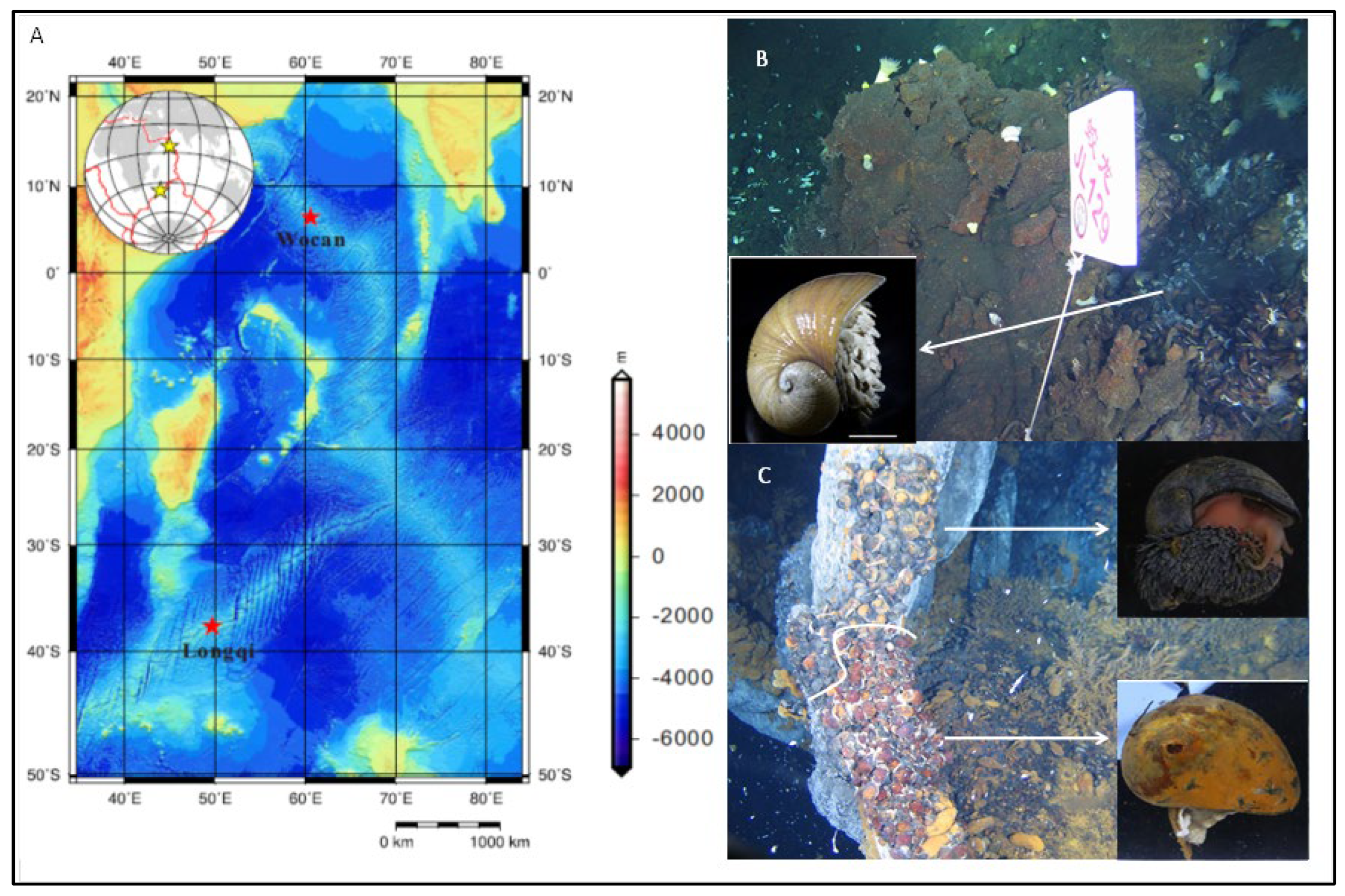
3.1. Distributions of Snails on Indian Ocean Ridges and Ultrastructural Characterization of Scaly-Foot Snails by Scanning Electron Microscopy (SEM)

In the Longqi hydrothermal field of the SWIR ridge, black scaly-foot C. squamiferum (BC) and G. aegis (G) colonize together in dense populations on vent chimney basals close to visible diffuse flow of vent fluid, and at sampling site DFF11, the vent chimney basals are occupied solely by these two species (Figure 1C). The maximum temperature at these diffusing areas was 13.3 °C. The diffuse flow has a high concentration of methane (12.68 mM) and hydrogen (8.197 mM). The white scaly-foot C. squamiferum inhabits the collapsed sulfide chimney with low-temperature diffusing flow around the high-temperature black smoking vent of the Carlsberg Ridge of the Northwest Indian Ocean in the hydrothermal field of Wocan (Figure 1B). As previously reported, Gigantopelta aegis has not been found in CR [32].
Scanning electron microscopy revealed the difference in sclerites between white (WC) and black scaly-foot snails (BC) of C. squamiferum. On the foot of WC, the sclerites were colonized by long microbial filaments with short rods attached to the filament surface, while on the sclerites of the BC snail, iron sulfide accumulation was observed sticking to cocci- and filamentous bacteria (Figure S1).

3.2. Characterization of Microbial Communities Associated with Hydrothermal Snails

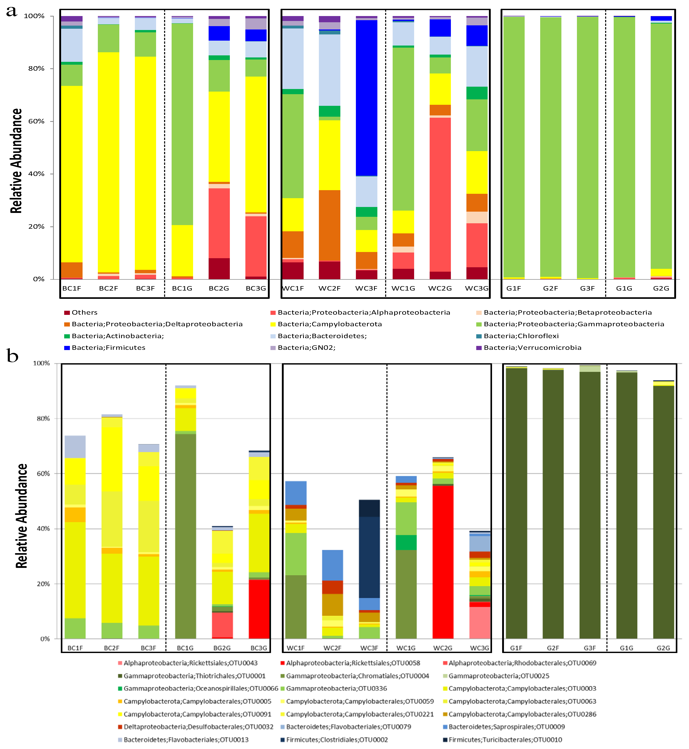
3.2.1. Microbial Community Structure Revealed by High Throughput Sequencing

Seventeen 16S rRNA gene amplicon libraries (three individuals per sample) were constructed from the foot and gland of C. squamiferum and G. aegis (Table 1). Raw sequencing reads were obtained from these libraries on the MGISEQ-2000 platform. After merging and processing the raw reads, 1,265,697 filtered tags with an average length of >252 bp of the 16S rRNA gene spanning the variable region V4 were clustered into denoising ASVs (also called zero-radius ASVs, or ZOTUs (Operational Taxonomic Units)) at 100% sequence similarity, of which 34.72%, 28.14%, and 37.14% were from the three individuals of BC, WC, and G, respectively (Table 1). A total of 1014 ASVs were obtained. Nearly all sequence tags (99.9982%) were assigned to the bacterial domain. Rarefaction curves approached a plateau (Additional File S2: Figure S2A), which indicated that sequences adequately represented bacterial composition in snail samples. Rank-abundance curves showed that the scaly-foot snail C. squamiferum, especially gland samples, contained more diverse sequence tags and relatively lower abundance than G. aegis (Additional File S2: Figure S2A).
The diversity indices, including the Shannon, Simpson, and community richness Modified itModified itChao1 and ACE indices, all revealed microbiota variations between C. squamiferum and G. aegis and between the scaly-foot and gland samples in C. squamiferum (Table 1). The Shannon index of snail C. squamiferum samples ranged from 1.32 to 4.60, whereas the Shannon index of the G. aegis samples ranged from 0.14 to 0.18. The Chao1 index ranged from 202.79 to 508.00 in C. squamiferum samples and from 140.25 to 246.86 in G. aegis. This result indicated that obviously higher bacterial diversity occurred in C. squamiferum than in G. aegis. For C. squamiferum, the diversity of WC was higher than the diversity of BC (Table 1). The mean Shannon indices of bacterial communities in the glands (2.49) were slightly higher than the mean Shannon indices on the foot (2.17). Likewise, the mean Chao1 index in glands (341.04) was higher than the mean Chao1 index in the foot (284.07).
Then, PCoA analysis (Figure S3A) and the Bray–Curtis cluster tree (Figure S3B) based on 16S rRNA gene ASVs were performed to examine the differences in bacterial communities. Two principal components (PC1 41.25% and PC2 18.83%) explained 60.08% of the total variation in the bacterial community in the samples. Both BC and WC were obviously separated from G. aegis along axis PC1. The minor factor PC2 impacted the communities of BC and WC. The BC and WC samples had 455 identical symbiont ASVs, which accounted for ~96.36% and ~80.53% of the ASV abundance of the two species, respectively (Figure S4). Only 237 ASVs were shared among the three snails, accounting for ~83.64% of BC samples, ~48.39% of WC samples, and 99.46% of G samples (Figure S4). The Bray–Curtis cluster tree (Figure S3B) indicated that 17 populations could be divided into two major clusters: Cluster 1 included mainly the populations of G. aegis (GG and GF), with Cluster 2 consisting of the remaining 12 populations of C. squamiferum (C), which were further divided into two short branches in accordance with snail hosts of white (WC) and black (BC). The results implied that the microbial community structure of these snails was relevant mainly to the host and less relevant to the adjacent geochemistry of the habitat environments.
The microbial community structures revealed striking and distinct patterns across the two snail species and between the C. squamiferum ecotypes (Figure 2a,b).
The black scaly-foot snail (BC) displayed a stable epibiotic community with a variable endosymbiotic core. Its foot surface (epibionts) was characterized by a low-diversity community overwhelmingly dominated by the phylum Campylobacterota (67.01–83.57%). Specifically, the family Helicobacteraceae, driven by key taxa such as ASV0003, was the principal component. This is consistent with previous findings that genera in this family are key sulfur-oxidizing bacteria (SOB) in hydrothermal environments [8]. In contrast, the gland community (endobionts) was more variable. Some individuals were dominated by Gammaproteobacteria, with a single taxon from the order Chromatiales (ASV0004) comprising over 74% of the community, while others hosted a more mixed assemblage of Campylobacterota, Alphaproteobacteria, Bacteroidetes, and Firmicutes (Figure 2a).
In contrast, the white scaly-foot snail (WC) harbored a significantly more diverse and heterogeneous microbiome with high inter-individual variability. The strong dominance of a single Campylobacterota lineage seen on the black ecotype was absent. Instead, WC communities comprised a shifting mixture of several major phyla, including Gammaproteobacteria, Bacteroidetes, Deltaproteobacteria, Alphaproteobacteria, and Firmicutes. The dominant taxa frequently differed not only between individuals but also between the foot and gland within the same animal, indicating a less specialized microbial association.
G. aegis presented a highly specialized gammaproteobacterial symbiosis. Both its foot and gland communities were almost entirely composed of Gammaproteobacteria, with the order Thiotrichales, represented almost entirely by a single amplicon sequence variant (ASV0001), accounting for over 96% of total sequences (Figure 2). Genera within Thiotrichales are known to thrive in vent environments [33]. This Thiotrichales-dominated symbiosis is notably different from the Chromatiales-based symbioses reported in other vent fauna [7,34,35].
In summary, these snails exhibit three distinct symbiotic signatures. G. aegis relies on a highly specialized Thiotrichales (Gammaproteobacteria) symbiosis. C. squamiferum, on the other hand, hosts a more complex suite of symbionts, with the black ecotype (BC) maintaining a consistent Campylobacterota-dominated epibiome, while the white ecotype (WC) is characterized by a more generalized and variable microbial community. The remarkably lower concentrations of methane and hydrogen in the Wocan field vent fluids compared to those in the Longqi field (DFF11) of SWIR (Table S1), coupled with the relatively low H2S concentrations (1400–2200 nM) compared to other hydrothermal plumes, likely results in reduced sulfide availability. This limitation may be a key factor in the reduced colonization of the white scaly-foot snail scales by hydrogen- and sulfur-oxidizing Campylobacterota.

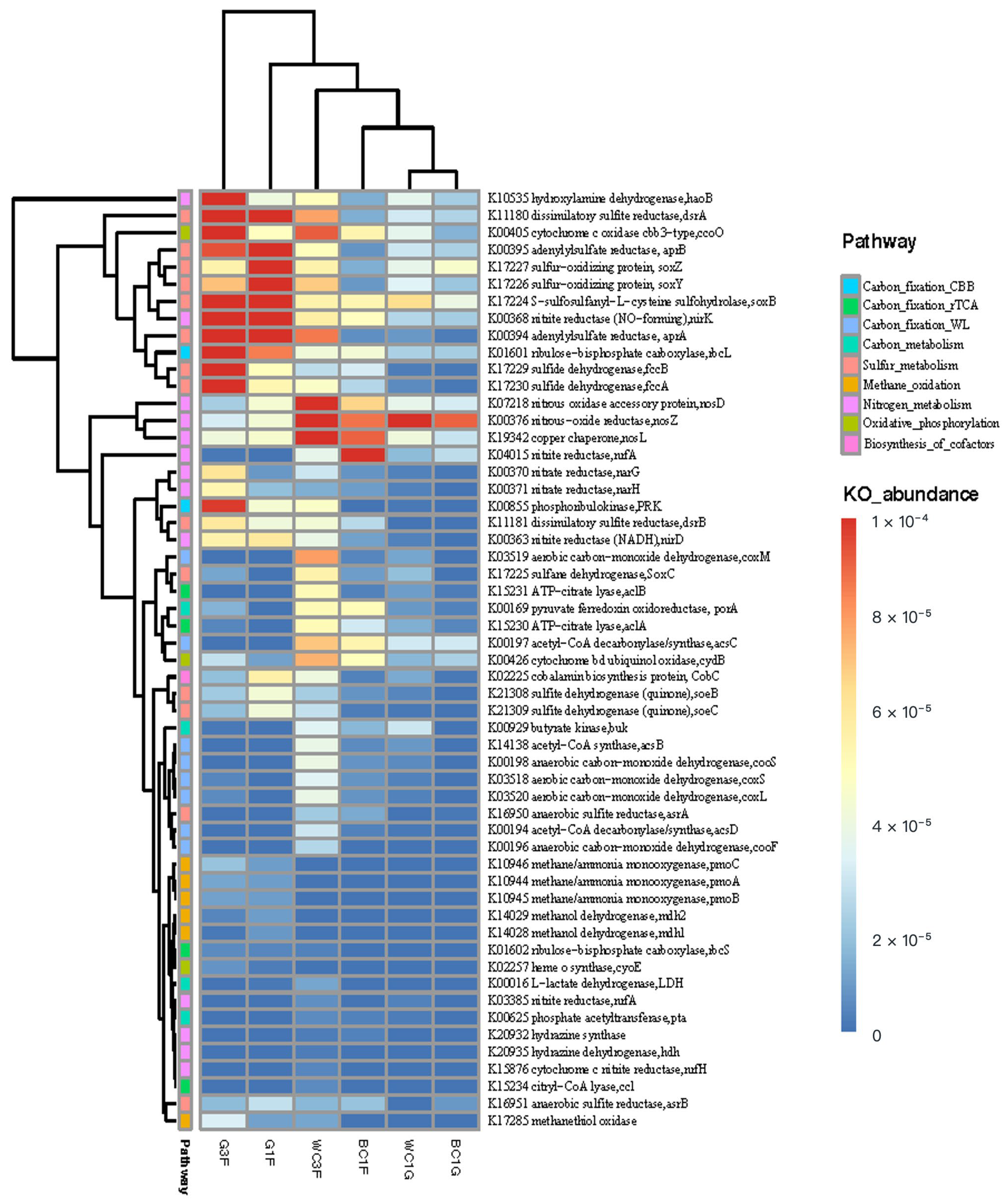
3.2.2. Metabolic Potential of the Microbial Community Based on Metagenomic Analysis

We sequenced and analyzed the metagenomes of six samples from white scaly-foot C. squamiferum (WC3F, WC1G), black scaly-foot C. squamiferum (BC1F, BC1G), and G. aegis (G1F, G3F) (Additional File S1: Table S2). Unfortunately, the gland metagenome from G. aegis was not constructed.
The functional capacity was determined according to the annotation of ORFs predicted from the assembled contigs. The nonredundant gene catalog containing a total of 782,337 ORFs was constructed with an average length of 329.57 bp. The nonredundant predicted genes were then aligned with the KEGG gene database. We identified a total of 195,997 KEGG genes and assigned them to 426 KEGG pathways. PCA based on KEGG annotations of metagenomic data showed obvious differential distributions of KEGG pathways between C. squamiferum and G. aegis (Additional File S1: Figure S5). A heatmap of functional genes based on the KEGG pathway annotation also shows differences between C. squamiferum and G. aegis (Figure 3). The genes that were highly enriched in G. aegis (G1F, G3F) were associated with the sulfur cycle and methane-oxidizing pathway, indicated by the corresponding key genes encoding dissimilatory sulfite reductase (DsrAB, K11180, K11181), adenylylsulfate reductase (AprAB, K00394, K00395), sulfide dehydrogenase (fccAB, K017229, K017230), sulfur-oxidizing protein (SoxBYZ, K017224, K017226, K017227), and methane/ammonia monooxygenase (pmoABC, K10944–10946). Compared with G.aegis, the metabolic pathways in C. squamiferum were enriched with carbon fixation (WL and rTCA), denitrification and glycolysis in addition to sulfur cycle genes (Figure 3), reflected by the corresponding key genes encoding acetyl-CoA synthase (K00194, K00197, K14138), anaerobic carbon-monoxide dehydrogenase (K00196, K00198) in WL pathway, ATP-citrate lyase (aclAB, K15230, K15231) in reductive TCA cycle, nitrous-oxide reductase (nos. DLZ, K07218, K00376, K19342) in denitrification (N2-forming), menaquinol-cytochrome c reductase subunit NrfD (K04015) in dissimilatory nitrate reduction, pyruvate ferredoxin oxidoreductase (PFOR, K00169) and butyrate kinase (K00929) to catalyze the oxidative decarboxylation of pyruvate to acetyl-CoA and produce butyrate, cytochrome d ubiquinol oxidase, subunit II (K00426) in oxygen utilization. Comparing C. squamiferum and G. aegis revealed that they not only host different bacterial symbionts in microbial taxonomic profiles but also significantly vary in metabolic potential.

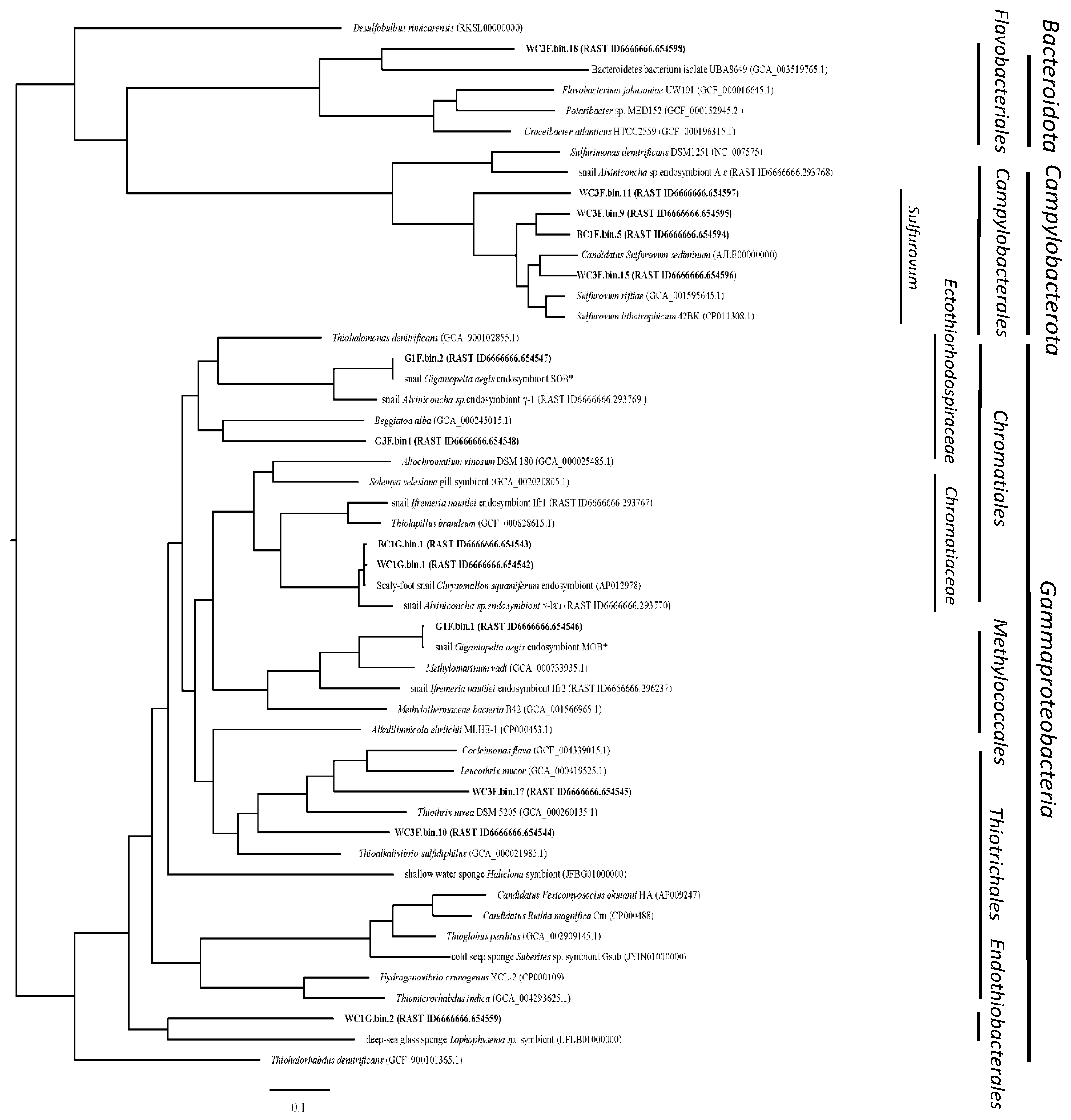
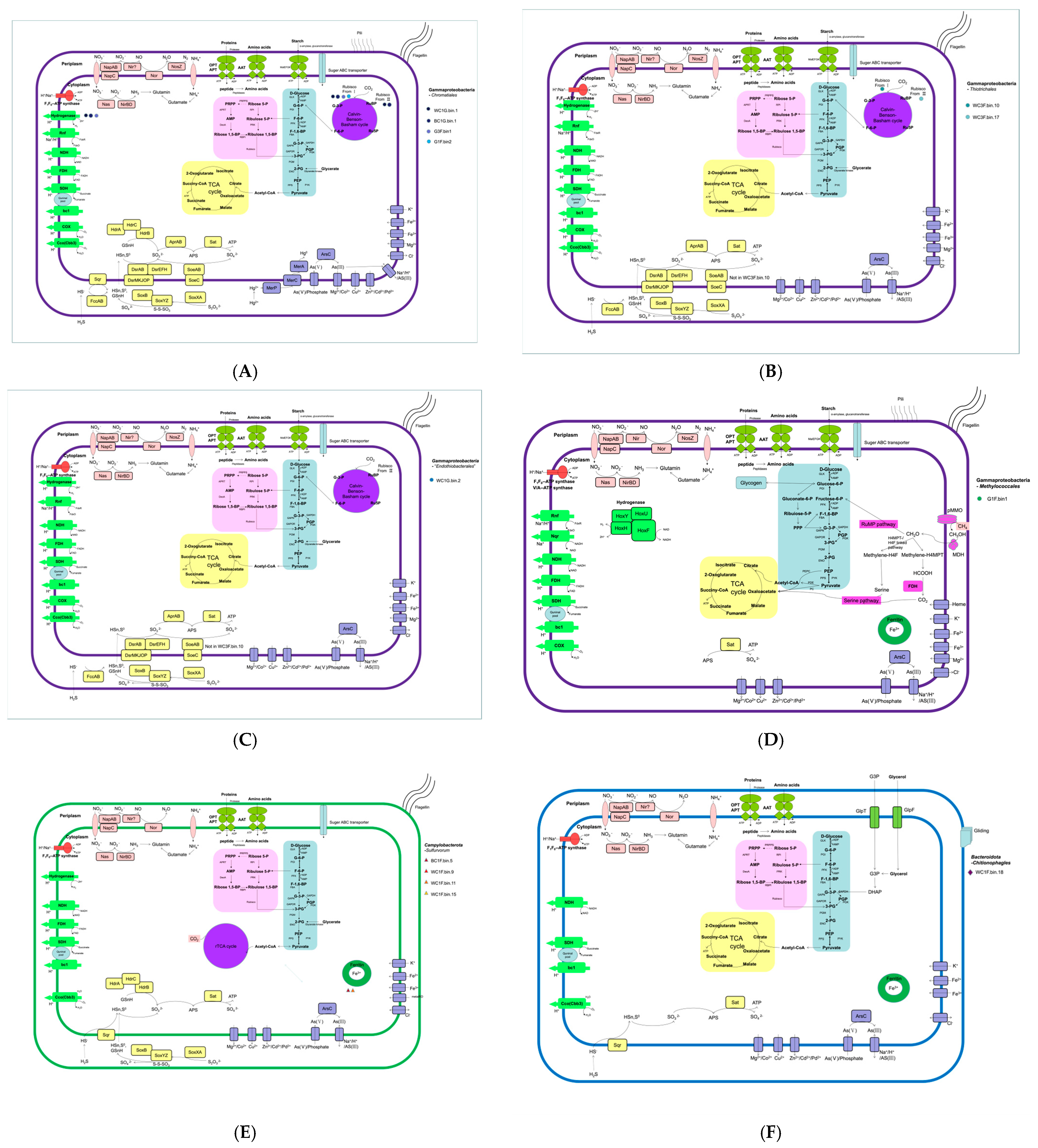
3.2.3. Phylogenomics and Predicted Metabolic Capabilities of Dominant Metagenome-Assembled Genomes (MAGs)

After filtration of low-quality MAGs, 13 MAGs with completeness ≥80% and contamination ≤ 5% were obtained for further analysis (Table 2). The 13 retrieved high-quality MAGs were taxonomically assigned to three phyla, including Gammaproteobacteria (8 MAGs), Campylobacterota (4 MAGs), and Bacteroidota (1 MAG). Additionally, two MAGs affiliated with Firmicutes and Bacteroidota from sample BC1F were also binned out but not analyzed due to their low completeness (71.54% and 60.84%).
Phylogenomic analysis based on 120 marker genes was performed between 13 MAGs and their related genomes, including symbionts from marine animals (Figure 4). The phylogenomic tree of 13 MAGs indicated the presence of multiple symbiont phylotypes of hydrothermal snails from the Indian Ocean in this study. Four distinct lineages in the class Gammaproteobacteria were identified, including four MAGs of the order Chromatiales, two of the order Thiotrichales, one in the order Methylococcales, and one unclassified group distinct from the other lineages. Four MAGs from scaly snails were affiliated with the Sulfurovum genus of Campylobacterota, and one MAG belonged to the phylum Bacteroidota.
To understand the functional roles of the dominant symbionts, we focused on their core metabolic pathways for carbon fixation, energy generation, and key adaptations to the vent environment (Figure 5, Table 2).
Chromatiales of Gammaproteobacteria
The order Chromatiales is frequently found as endosymbionts in hydrothermal vent fauna, including snails, tube worms, and sponges [13,35,36,37]. We recovered four Chromatiales MAGs that segregated into two distinct families. The first group, comprising endosymbionts from the glands of both black (BC1G.bin.1) and white (WC1G.bin.1) C. squamiferum, belonged to the family Chromatiaceae. These two MAGs were nearly identical to a previously reported C. squamiferum endosymbiont from the Kairei field (98.5% ANI) [36], confirming a stable host–symbiont association across different Indian Ocean vent fields. Metabolically, they are versatile chemolithoautotrophs, fixing carbon via the Calvin–Benson–Bassham (CBB) cycle using both Form I and Form II RubisCO, which suggests an ability to adapt to varying CO2/O2 conditions [38]. They possess an extensive repertoire of sulfur-oxidizing pathways (Sox, Hdr, reverse Dsr), genes for hydrogen utilization, and mechanisms for heavy metal detoxification as reported [39,40,41,42]. The second group, recovered from the foot of G. aegis (G3F.bin1, G1F.bin2), belonged to the family Ectothiorhodospiraceae and was phylogenetically distinct from an Alviniconcha symbiont lineage (γ-1) [43] (Figure 4). These epibionts are also sulfur-oxidizing autotrophs but primarily utilize Form II RubisCO for carbon fixation via the CBB cycle as reported [44,45,46,47,48].
Thiotrichales of Gammaproteobacteria
Two epibiotic MAGs from the white scaly-foot snail (WC3F.bin.10, WC3F.bin.17) were affiliated with filamentous sulfur-oxidizers in the order Thiotrichales (e.g., Thiothrix, Leucothrix), which have been described on other vent organisms [49]. These MAGs encode a mixotrophic metabolism, using the CBB cycle for carbon fixation and oxidizing sulfide via sulfide dehydrogenase (FccAB), a pathway efficient in low-sulfide conditions. Notably, both MAGs possess cyc2 genes (cyc2-Cluster 3), suggesting a previously unreported capacity for iron oxidation among these symbionts [50,51,52] (Additional file S2: Figure S7).
Methylococcales of Gammaproteobacteria
A methanotrophic MAG (G1F.bin.1) was recovered from G. aegis and is phylogenetically close to Methylomarinum vadi, a methanotroph from a shallow hydrothermal system [53,54]. Symbiotic Methylococcales are also known from marine mussels and sponges [55]. This MAG harbors the complete genetic toolkit for aerobic methane oxidation, including methane monooxygenase (pmoCAB), and assimilates C1 compounds via the ribulose monophosphate (RuMP) pathway (Figure 5D). Its genome also contains genes for hydrogen utilization and glycogen storage, key adaptations for a symbiotic lifestyle [56].
Candidatus Endothiobacterales of Gammaproteobacteria
A MAG from the white scaly-foot snail gland (WC1G.bin2) represents a previously uncharacterized lineage of Gammaproteobacteria. Phylogenomically, it is highly distinct, clustering only with a sulfur-oxidizing symbiont from a deep-sea glass sponge [57] and sharing low ANI values (<68.3%) with other known orders (Figure 4). Based on this evidence, we propose the new candidate order “Candidatus Endothiobacterales”. Metabolically, this MAG is a sulfur-oxidizing autotroph, utilizing the CBB cycle (Form II RubisCO) for carbon fixation and possessing a comprehensive suite of sulfur oxidation pathways (Figure 5C).
Campylobacterales of Campylobacterota
Four MAGs, primarily from the foot surface of C. squamiferum, were classified within the genus Sulfurovum, a well-known chemosynthetic group in vent environments where its members thrive attached to surfaces [8,58]. These symbionts are chemolithoautotrophs that fix carbon via the reductive tricarboxylic acid (rTCA) cycle (Figure 5E). They oxidize sulfur compounds using sulfide-quinone reductase (Sqr) and the Sox system. Consistent with a symbiotic lifestyle, their genomes were significantly smaller (1.38–1.61 Mbp) than those of their free-living relatives (Table S3; Figure S6) [59,60,61].
Flavobacteriales of Bacteroidetes
A single MAG from the white scaly-foot snail (WC3F.bin18) belongs to the order Flavobacteriales (phylum Bacteroidetes). This MAG exhibits features of a heterotrophic, surface-associated lifestyle, a common strategy for this phylum, which often degrades polymers on particles [62]. Its genome encodes numerous carbohydrate-active enzymes (CAZymes) for degrading complex organic matter, as well as genes for gliding motility (Figure 5F). This suggests a role in utilizing organic polymers produced by the primary chemosynthetic symbionts on the snail’s scales, a niche also observed for Bacteroidetes ectobionts on Sulfurovum filaments [63].
SOX, sulfur-oxidizing protein; SQR, sulfide:quinone oxidoreductase; FCC, flavocytochrome-c sulfide dehydrogenase; DsrEF intracellular sulfur oxidation protein; HdrABC, heterodisulfide reductase; DsrAB, dissimilatory sulfite reductase, subunit AB; AprAB, adenylylsulfate reductase, subunit AB; Sat, sulfate adenylyltransferase; SoeABC, membrane-bound sulfite dehydrogenase; MBHL, membrane-bound hydrogenase; Hox, cytoplasmic NiFe hydrogenases; pMMO methane monooxygenase; CODH, carbon monoxide dehydrogenase; Cyc2, cyctochrome C oxidase; ArsC, arsenate reductase; MerA, mercury reductase; Cox, cytochrome c oxidase; Cco, cytochrome c oxidoreductase; Glc, glycolate oxidase; NapAB, periplasmic nitrate reductase; NasA, Nitrate reductase; NirBD, nitrite reductase; Nuo, NADH ubiquinone oxidoreductase; Fdh, formate dehydrogenase; Sdh, succinate dehydrogenase; Nqr, sodium-dependent NADH dehydrogenase.

4. Discussion

Nutritional symbioses between eukaryotic organisms and autotrophic microbes are ubiquitous throughout the oceans of the Earth. These associations have allowed marine organisms to flourish in nutrient-limited or extreme environments where they reach population densities unmatched by their nonsymbiotic relatives [64]. In this study, the diversity and host specificity of host–microbe relationships in hydrothermal snails were analyzed by comparing the microbiota of different snail species in the same niche and two varieties of the same species in different hydrothermal inhabitants (Figure 6).
CBB, Calvin–Benson–Bassham cycle; rTCA, the reductive tricarboxylic acid cycle; WL, reductive acetyl-CoA pathway (Wood-Ljungdahl pathway); SOX, sulfur-oxidizing protein; SQR, sulfide:quinone oxidoreductase; FCC, flavocytochrome-c sulfide dehydrogenase; DsrEF Intracellular sulfur oxidation protein; HdrABC, heterodisulfide reductase; DsrAB, dissimilatory sulfite reductase, subunit AB; AprAB, adenylylsulfate reductase, subunit AB; Sat, sulfate adenylyltransferase; SoeABC, membrane-bound sulfite dehydrogenase; MBHL, membrane-bound hydrogenase; Hox, cytoplasmic NiFe hydrogenases; pMMO, methane monooxygenase; CODH, carbon monoxide dehydrogenase; Cyc2, cytochrome C oxidase; ArsC, arsenate reductase; MerA, mercury reductase.

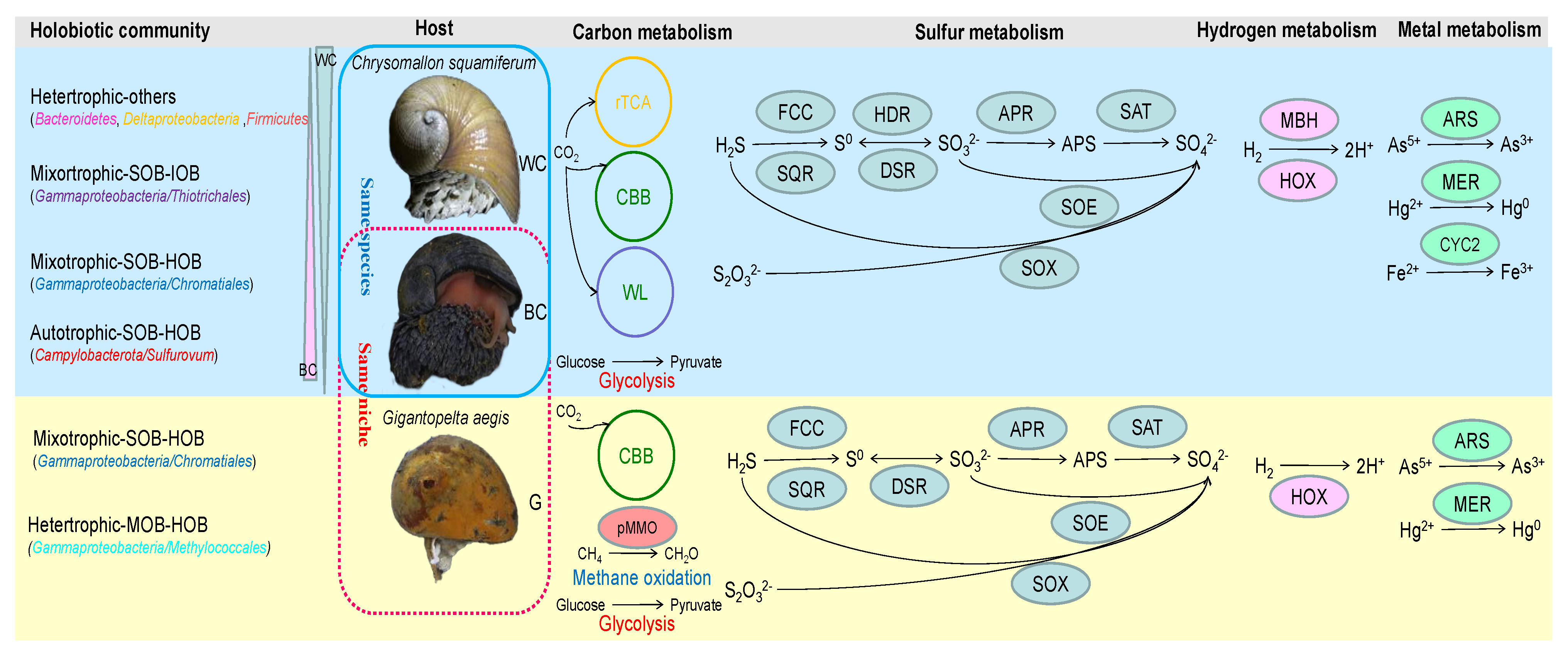
4.1. Host-Specific Symbiont Type in Hydrothermal Snails

The hydrothermal snail G. aegis and the scaly-foot snail C. squamiferum were affiliated with the same family (Peltospiridae), restricted to chemosynthetic ecosystems [10], and phylogenetically independent of other shallow-water gastropods [11]. In this study, these two snail species, C. squamiferum and G. aegis, live together in the same niche in the Longqi vent, SWIR. G. aegis harbors extremely overabundant Gammaproteobacteria, belonging mainly to the order Chromatiales and Methyloccales. As reported for two MAGs in G. aegis [13], G1F.bin2 was close to abundant sulfur-oxidizing bacteria, and G1F.bin1 was close to the less common methane-oxidizing bacteria. Another sulfur-oxidizing bacterium, G3F.bin1, which was close to the filamentous SOB Beggiatoa alba, has never been reported before. Compared with SOB G1F.bin2, G3F.bin1 can use hydrogen and sulfur species as electron donors but cannot produce pyridoxine (vitamin B6) and cobalamin (vitamin B12). Based on the low bacterial diversity in G. aegi, we assumed that powerful host-immune responses and disturbance may contribute to it according to host genome analysis. The black-scale snail C. squamiferum resides on an active chimney neighboring G. aegi but houses Campylobacterota and Gammaproteobacteria as dominant members in the symbiotic community, in addition to diverse bacteria of Delta-Alphaproteobacteria, Bacteroidetes, and Firmicutes. Their symbiotic bacteria can use versatile energy sources by H2 oxidation, sulfur oxidation, and iron oxidation coupled with the reduction of sulfur and multiple oxygen receptors. The host C. squamiferum also displayed significant enrichment of metabolism in the glycolysis pathway and citrate cycle (TCA cycle) [11], which helped to maintain the levels of ATP and metabolic pathways to provide additional nutrients for diverse symbionts. Therefore, the metabolic versatility of the microbiome, along with other key life-history traits such as dispersal potential, likely contributes to the broader distribution of C. squamiferum compared to G. aegis along the Indian Ocean Ridge.
In summary, even when living in the same niche on a hydrothermal vent chimney, different snail species harbor totally different symbionts. The phylosymbiosis found in the hydrothermal gastropod family Peltospiridae (this study) and chemosymbiotic bivalve family Lucinidae [9] implied that it could be a trait in some chemosymbiotic animals.

4.2. “Core” Microbial Community with Abundance Variation in the Hydrothermal Snail C. squamiferum

Black scaly-foot and white scaly-foot varieties of snail C. squamiferum spread across hydrothermal fields from SWIR to NWIR via CR in the Indian Ocean. As previously reported, the snail C. squamiferum can biologically control the mineralization of iron sulfide nanoparticles through channel-like columnar structures within the scales to enrich sulfur. Then, sulfur/iron diffusion further occurs in seawater [12]. Due to the low iron content in the fluid, the white scaly-foot snail without sulfides on the scale spread in the Solitaire Field in CIR Ridge [65] and Wocan Field in the Carlsberg Ridge (this study). Sulfur isotope analysis evidenced the accumulation of 32S in the scaly-foot snail by bacteria. In this study, black scaly-foot (BC) snails live in H2- and CH4-abundant sites, while white-scaly (WC) snails grow with depleted H2 and CH4 (Additional File S1: Table S1). We found that black scaling (BC) was obviously different from white scaling (WC) in the abundance of the bacterial community by amplicon sequencing (Figure 2) and microscopy analysis (Additional file S2: Figure S1). Black scaly-foot hosts harbor mainly 67.01–83.57% Campylobacterota bacteria, among which the sulfur-oxidizing bacteria Sulfurovum spp. are the predominant key member. The predominance of Sulfurovum spp. occurs on the surfaces of vent animals such as snails and shrimp [15,66]. The bacteria associated with white scaly-foot WC were more diverse, composed of Campylobacterota, Gammaproteobacteria, Deltaproteobacteria, Bacteroidetes, and Firmicutes (Figure 2); intriguingly, WC contained a higher content of heterotrophic groups and less autotrophic Campylobacterota. Normally, the proportion of heterotrophs, including Deltaproteobacteria and Bacteroidetes, is greater in inactive hydrothermal sulfides than in active hydrothermal sulfides [67,68]. Firmicutes are also always found in nonhydrothermal sediments [69,70]. Deltaproteobacterial symbionts may also act as sulfate reducers on scales, indicating that the white scaly-foot snail C. squamiferum seems adaptive to inactive hydrothermal environments such as sulfides and sediments, in which reducing gases such as H2S, H2, and CH4 are not effluent but the content of organic carbons may be higher.
Although the two subtypes of scaly-foot snails of C. squamiferum are distributed across a large geographical distance, they contain the same sulfur-oxidizing endosymbiont, which formed a small cluster with high similarity (ANI value > 98.5%, Figure 3), including WC1G.bin.1 from white scaly-foot snails from the Wocan field in the Carlsberg Ridge, BC1G.bin.1 from black scaly-foot snails in the Longqi field of the Southwest Indian Ridge, and one black scaly-foot snail from the Kairei hydrothermal field in the central Indian Ocean [37], indicating that this endosymbiont belongs to the family Chromatiaceae in the order Chromatiales of Gammaproteobacteria, which tightly evolved with C. squamiferum and played an important role in symbiosis. This endosymbiont is another host-specific symbiont type identified in the hydrothermal gastropod family Peltospiridae. The transmission mode of this symbiont, whether vertical, horizontal, or mixed, requires further investigation. Lan et al. (2021) provide evidence suggesting a mixed transmission mode for C. squamiferum symbionts, highlighting the complexity of symbiont acquisition in these systems [13].
The “core” microbial community with key endosymbionts was transmittable among individuals of C. squamiferum spreading along the Indian Ocean Ridge, but variations in microbial abundance showed a microbial community in response to environmental stressors. The symbionts respond sensitively to the environment and have functions in nutrient supply and sulfide detoxification for the host, which is an important part of a complex adaptive system of C. squamiferum.

4.3. The Association of Hydrothermal Snails and Their Symbionts to Adapt to the Environment

The dominant macrofaunal species in hydrothermal vents are typically symbiotic primary consumers. Most chemosynthetic holobionts (host–symbiont associations) are sustained by energy metabolism of either sulfur or methane or hydrogen oxidation [1,13,71]. We compared the four studied species of hydrothermal chemosynthetic snails and their symbionts (Table 3). Snails Gigantopelta spp. and Ifremeria nautilei are distributed in a limited area, whereas Alviniconcha spp. and Chrysomallon squamiferum spread wider in the Pacific and Indian oceans (Table 3). Gammaproteobacteria, especially the order Chromatiales, are the main and transmittable endosymbionts in these four species, which encoded carbon fixation and a hybrid Sox-reverse Dsr pathway, which would allow carbon fixation and the oxidation of thiosulfate, elemental sulfur, and sulfide as energy sources. Campylobacterota could be symbionts in the gills of Alviniconcha spp. (mainly Sulfurimonas and Sulfurovum) and on the scale of C. squamiferum (mainly Sulfurovum). They showed high similarity with free-living Sulfurimonas spp. and Sulfurovum spp., indicating their acquisition from environments. Compared with only the CBB cycle in G. aegis and Ifremeria nautilei, three major metabolic pathways for carbon fixation were found in symbiotic communities of hydrothermal snail C. squamiferum, including the CBB cycle, reductive tricarboxylic acid (rTCA) cycle, and reductive acetyl-CoA pathway (Wood-Ljungdahl pathway) (Figure 3 and Figure 5). A high proportion of heterotrophic bacteria in the gland of C. squamiferum are devoted to degrading and transporting various carbohydrates, amino acids, and peptides, suggesting a broad substrate spectrum for both carbon and nitrogen gain. Notably, most symbiont MAGs contain arsenate reductase (AsrC), mercury reductase (MerA), and copper resistance-related genes, indicating that those symbionts were adapted and helped the host develop metal resistance.
Collectively, these patterns in Chrysomallon might suggest that different host tissues represent distinct ecological niches that select for specific microbial partners. External surfaces, exposed to the dynamic chemistry of the surrounding water, favor a more diverse community adapted to environmental fluctuations, whereas stable, internal organs select for highly specialized endosymbionts optimized for nutrient provisioning. Although the metabolic flexibility of symbionts may contribute to the distribution and abundance of hydrothermal chemosynthetic snails, further research is needed to disentangle its effects from those of other factors, such as dispersal potential, habitat conditions, and interspecies competition.

5. Conclusions

The microbial symbionts of two snail species (black scale/white scale Chrysomallon squamiferum and Gigantopelta aegis) were characterized to understand their relationship with hydrothermal snails. Two snail species in the Indian Ocean harbor phylogenetically and functionally distant symbionts. G. aegis has strict selection on bacterial symbionts of sulfur-oxidizing bacteria of the family Ectothiorhodospiraceae and methane-oxidizing bacteria of the family Methylococcaceae. In contrast, snails of C. squamiferum adapt to a wider habitation range by recruiting a “core” community with changeable abundance in accordance with inhabitant spreading. The diverse “core” community includes Campylobacterota, Gamma-, Delta-, and Alpha-Proteobacteria, Bacteroidetes, and Firmicutes. These data highlight that the host largely shapes the associated microbiota in hydrothermal vent snails and that the local environment has a selective impact on the host.

Supplementary Materials

The following supporting information can be downloaded at https://www.mdpi.com/article/10.3390/biology14080954/s1: Table S1: Geophysical features of sampling sites where deep-sea snails were collected; Figure S1: Microscopic and scanning electron micrograph showing sclerites of the scaly snails; Table S2. Summary of the specimens processed in this study for 16S rRNA amplicon sequence and metagenomics; Table S3. Symbiont Sulfurovum bins compared with free-living Sulfurovum isolates; Figure S2: Characteristics of microbiota between the three groups; Figure S3: PCoA analysis (A) and the Bray–Curtis cluster tree (B), showing results of beta diversity analysis of hydrothermal snails; Figure S4: Venn diagram describing the ASV distribution among three snails; Figure S5: PCA analysis results based on functional abundance of level 3 KEGG pathways; Figure S6: Heatmap of pairwise amino acid identity (ANI) values and phylogenomic tree in the genus Sulfurvum; Figure S7: Cyc2 maximum likelihood phylogenetic tree (300 bootstraps) with all (A) and the neighbors’ (B) sequences showing the relative placement of Cyc2 belonging to three MAGs identified in this study.

Author Contributions

Conceptualization, X.Z.; formal analysis and investigation, X.Z., J.C., G.L. and Y.Z. (Yaolei Zhang); resources, X.Z., L.W. and Y.Z. (Yadong Zhou); writing—original draft preparation, X.Z.; writing—review and editing, X.Z., J.C. and Z.S.; project administration, X.Z., Z.S. and S.L.; funding acquisition, X.Z. and Z.S. All authors have read and agreed to the published version of the manuscript.

Funding

This research was funded by the National Key R&D Program of China, grant numbers 2023YFC2812903, 2021YFF0501304, and 2018YFC0310702.

Institutional Review Board Statement

Not applicable.

Informed Consent Statement

Not applicable.

Data Availability Statement

The 16S rRNA genes and metagenomics sequencing data that support the findings of this study have been deposited in the CNSA (https://db.cngb.org/cnsa/) of CNGBdb with accession code CNP0001245. The assembled and annotated symbiont genomes are also publicly available on the RAST server (http://rast.theseed.org/) using the guest login with IDs 6666666.654542-6666666.654548, 6666666.654559, and 6666666.654594-6666666.654598.

Acknowledgments

We are very grateful to the whole team of the oceanic cruises of COMRA DY35-leg Ⅲ and COMRA DY38-leg I via the R/V ‘Xiangyanghong 9’and HOV ‘Jiaolong’ for helping us to collect the snail samples from the deep-sea hydrothermal vent field and geochemical composition determinations. We are also grateful to Li GU (The Third Institute of Oceanography) for her help with maintenance and technical aspects of SEM.

Conflicts of Interest

Authors Jianwei Chen, Guilin Liu, Yaolei Zhang, and shanshan Liu were employed by the company “BGI-Qingdao; BGI-Shenzhen”. The remaining authors declare that the research was conducted in the absence of any commercial or financial relationships that could be construed as potential conflicts of interest.

Abbreviations

The following abbreviations are used in this manuscript:
KEGGKyoto Encyclopedia of Genes and Genomes
ANIAverage Nucleotide Identity
PCAPrincipal Component Analysis
MAGMetagenome-Assembled Genome
ASVAmplicon Sequence Variant

References

  1. Dubilier, N.; Bergin, C.; Lott, C. Symbiotic diversity in marine animals: The art of harnessing chemosynthesis. Nat. Rev. Microbiol. 2008, 6, 725–740. [Google Scholar] [CrossRef]
  2. Dick, G.J. The microbiomes of deep-sea hydrothermal vents: Distributed globally, shaped locally. Nat. Rev. Microbiol. 2019, 17, 271–283. [Google Scholar] [CrossRef]
  3. Ramírez, G.A.; Mara, P.; Sehein, T.; Wegener, G.; Chambers, C.R.; Joye, S.B.; Peterson, R.N.; Philippe, A.; Burgaud, G.; Edgcomb, V.P.; et al. Environmental factors shaping bacterial, archaeal and fungal community structure in hydrothermal sediments of Guaymas Basin, Gulf of California. PLoS ONE 2021, 16, e0256321. [Google Scholar] [CrossRef]
  4. Olins, H.C.; Rogers, D.R.; Preston, C.; Ussler, W.; Pargett, D.; Jensen, S.; Roman, B.; Birch, J.M.; Scholin, C.A.; Haroon, M.F.; et al. Co-registered geochemistry and metatranscriptomics reveal unexpected distributions of microbial activity within a hydrothermal vent field. Front. Microbiol. 2017, 8, 1042. [Google Scholar] [CrossRef]
  5. Chomicki, G.; Kiers, E.T.; Renner, S.S. The evolution of mutualistic dependence. Annu. Rev. Ecol. Evol. Syst. 2020, 51, 409–432. [Google Scholar] [CrossRef]
  6. Prazeres, M.A.T.; Roberts, T.E.; Pandolfi, J.M.; Leggat, W. Symbiosis and microbiome flexibility in calcifying benthic foraminifera of the Great Barrier Reef. Microbiome 2017, 5, 38. [Google Scholar] [CrossRef] [PubMed]
  7. Beinart, R.A.; Sanders, J.G.; Faure, B.; Sylva, S.P.; Lee, R.W.; Becker, E.L.; Gartman, A.; Luther, G.W.; Seewald, J.S.; Fisher, C.R.; et al. Evidence for the role of endosymbionts in regional-scale habitat partitioning by hydrothermal vent symbioses. Proc. Natl. Acad. Sci. USA 2012, 109, E3241–E3250. [Google Scholar] [CrossRef] [PubMed]
  8. Meier, D.P.P.; Bach, W.; Hourdez, S.; Girguis, P.R.; Vidoudez, C.; Amann, R.; Meyerdierks, A. Niche partitioning of diverse sulfur-oxidizing bacteria at hydrothermal vents. ISME J. 2017, 11, 1545–1558. [Google Scholar] [CrossRef]
  9. Osvatic, J.T.; Wilkins, L.G.E.; Leibrecht, L.; Leray, M.; Zauner, S.; Polzin, J.; Camacho, Y.; Gros, O.; van Gils, J.A.; Eisen, J.A.; et al. Global biogeography of chemosynthetic symbionts reveals both localized and globally distributed symbiont groups. Proc. Natl. Acad. Sci. USA 2021, 118, e2104378118. [Google Scholar] [CrossRef] [PubMed]
  10. Chen, C.; Linse, K.; Copley, J.; Rogers, A.D. The ‘scaly-foot gastropod’: A new genus and species of hydrothermal vent-endemic gastropod (Neomphalina: Peltospiridae) from the Indian Ocean. J. Molluscan Stud. 2015, 81, 322–334. [Google Scholar] [CrossRef]
  11. Zeng, X.; Zhang, Y.; Meng, L.; Fan, G.; Bai, J.; Chen, J.; Song, Y.; Seim, I.; Wang, C.; Shao, Z.; et al. Genome sequencing of deep-sea hydrothermal vent snails reveals adaptions to extreme environments. Gigascience 2020, 9, giaa139. [Google Scholar] [CrossRef] [PubMed]
  12. Okada, S.; Chen, C.; Watsuji, T.O.; Nishizawa, M.; Suzuki, Y.; Sano, Y.; Bissessur, D.; Deguchi, S.; Takai, K. The making of natural iron sulfide nanoparticles in a hot vent snail. Proc. Natl. Acad. Sci. USA 2019, 116, 20376–20381. [Google Scholar] [CrossRef] [PubMed]
  13. Lan, Y.; Sun, J.; Chen, C.; Sun, Y.; Zhou, Y.; Yang, Y.; Zhang, W.; Li, R.; Zhou, K.; Wong, W.C.; et al. Hologenome analysis reveals dual symbiosis in the deep-sea hydrothermal vent snail Gigantopelta aegis. Nat. Commun. 2021, 12, 1165. [Google Scholar] [CrossRef]
  14. Chen, C.; Uematsu, K.; Linse, K.; Sigwart, J.D. By more ways than one: Rapid convergence at hydrothermal vents shown by 3D anatomical reconstruction of Gigantopelta (Mollusca: Neomphalina). BMC Evol. Biol. 2017, 17, 62. [Google Scholar] [CrossRef]
  15. Goffredi, S.K.; Waren, A.; Orphan, V.J.; Van Dover, C.L.; Vrijenhoek, R.C. Novel forms of structural integration between microbes and a hydrothermal vent gastropod from the indian ocean. Appl. Environ. Microbiol. 2004, 70, 3082–3090. [Google Scholar] [CrossRef]
  16. Lan, Y.; Sun, J.; Chen, C.; Wang, H.; Xiao, Y.; Perez, M.; Yang, Y.; Kwan, Y.H.; Sun, Y.; Zhou, Y.; et al. Endosymbiont population genomics sheds light on transmission mode, partner specificity, and stability of the scaly-foot snail holobiont. ISME J. 2022, 16, 2132–2143. [Google Scholar] [CrossRef]
  17. Ji, F.; Zhou, H.; Yang, Q.; Gao, H.; Wang, H.; Lilley, M.D. Geochemistry of hydrothermal vent fluids and its implications for subsurface processes at the active Longqi hydrothermal field, Southwest Indian Ridge. Deep Sea Res. Part I Oceanogr. Res. Pap. 2017, 122, 41–47. [Google Scholar] [CrossRef]
  18. Li, C.; Tan, X.; Bai, J.; Xu, Q.; Liu, S.; Guo, W.; Yu, C.; Fan, G.; Lu, Y.; Zhang, H.; et al. A survey of the sperm whale (Physeter catodon) commensal microbiome. Peer J. 2019, 7, e7257. [Google Scholar] [CrossRef] [PubMed]
  19. Edgar, R.C.; Flyvbjerg, H. Error filtering, pair assembly and error correction for next-generation sequencing reads. Bioinformatics 2015, 31, 3476–3482. [Google Scholar] [CrossRef] [PubMed]
  20. Kuczynski, J.; Stombaugh, J.; Walters, W.A.; Gonzalez, A.; Caporaso, J.G.; Knight, R. Using QIIME to analyze 16S rRNA gene sequences from microbial communities. Curr. Protoc. Microbiol. 2012, 27, 10.7.1–10.7.20. [Google Scholar] [CrossRef]
  21. Peng, Y.; Leung, H.C.M.; Yiu, S.M.; Chin, F.Y.L. IDBA-UD: A de novo assembler for single-cell and metagenomic sequencing data with highly uneven depth. Bioinformatics 2012, 28, 1420–1428. [Google Scholar] [CrossRef]
  22. Uritskiy, G.V.; DiRuggiero, J.; Taylor, J. MetaWRAP-a flexible pipeline for genome-resolved metagenomic data analysis. Microbiome 2018, 6, 158. [Google Scholar] [CrossRef]
  23. Parks, D.H.; Chuvochina, M.; Chaumeil, P.A.; Rinke, C.; Mussig, A.J.; Hugenholtz, P. A complete domain-to-species taxonomy for Bacteria and Archaea. Nat. Biotechnol. 2020, 38, 1079–1086. [Google Scholar] [CrossRef]
  24. Parks, D.H.; Imelfort, M.; Skennerton, C.T.; Hugenholtz, P.; Tyson, G.W. CheckM: Assessing the quality of microbial genomes recovered from isolates, single cells, and metagenomes. Genome Res. 2015, 25, 1043–1055. [Google Scholar] [CrossRef]
  25. Kanehisa, M.; Goto, S. KEGG: Kyoto encyclopedia of genes and genomes. Nucleic Acids Res. 2000, 28, 27–30. [Google Scholar] [CrossRef]
  26. Caspi, R.; Billington, R.; Ferrer, L.; Foerster, H.; Fulcher, C.A.; Keseler, I.M.; Kothari, A.; Krummenacker, M.; Latendresse, M.; Mueller, L.A.; et al. The MetaCyc database of metabolic pathways and enzymes and the BioCyc collection of pathway/genome databases. Nucleic Acids Res. 2016, 44, D471–D480. [Google Scholar] [CrossRef]
  27. Brettin, T.; Davis, J.J.; Disz, T.; Edwards, R.A.; Gerdes, S.; Olsen, G.J.; Olson, R.; Overbeek, R.; Parrello, B.; Pusch, G.D. RASTtk: A modular and extensible implementation of the RAST algorithm for building custom annotation pipelines and annotating batches of genomes. Sci. Rep. 2015, 5, 8365. [Google Scholar] [CrossRef]
  28. Sondergaard, D.; Pedersen, C.N.S.; Greening, C. HydDB: A web tool for hydrogenase classification and analysis. Sci. Rep. 2016, 6, 34212. [Google Scholar] [CrossRef] [PubMed]
  29. Garber, A.I.; Nealson, K.H.; Okamoto, A.; McAllister, S.M.; Merino, N. FeGenie: A comprehensive tool for the identification of iron genes and iron gene neighborhoods in genome and metagenome assemblies. Front. Microbiol. 2020, 11, 37. [Google Scholar] [CrossRef] [PubMed]
  30. Lagesen, K.; Hallin, P.F.; Rodland, E.A.; Staerfeldt, H.; Rognes, T.; Ussery, D.W. RNAmmer: Consistent and rapid annotation of ribosomal RNA genes. Nucleic Acids Res. 2007, 35, 3100–3108. [Google Scholar] [CrossRef] [PubMed]
  31. Lee, I.O.K.Y.; Park, S.C.; Chun, J. OrthoANI: An improved algorithm and software for calculating average nucleotide identity. Int. J. Syst. Evol. Microbiol. 2016, 66, 1100–1103. [Google Scholar] [CrossRef] [PubMed]
  32. Zhou, Y.; Zhang, D.; Zhang, R.; Liu, Z.; Tao, C.; Lu, B.; Sun, D.; Xu, P.; Lin, R.; Wang, J.; et al. Characterization of vent fauna at three hydrothermal vent fields on the Southwest Indian Ridge: Implications for biogeography and interannual dynamics on ultraslow-spreading ridges. Deep Sea Res. Part I Oceanogr. Res. Pap. 2018, 137, 1–12. [Google Scholar] [CrossRef]
  33. Zeng, X.; Alain, K.; Shao, Z. Microorganisms from deep-sea hydrothermal vents. Mar. Life Sci. Technol. 2021, 3, 204–230. [Google Scholar] [CrossRef]
  34. Tian, R.; Wang, Y.; Bougouffa, S.; Gao, Z.; Cai, L.; Bajic, V.B.; Qian, P. Genomic analysis reveals versatile heterotrophic capacity of a potentially symbiotic sulfur-oxidizing bacterium in sponge. Environ. Microbiol. 2014, 16, 3548–3561. [Google Scholar] [CrossRef]
  35. Lavy, A.; Keren, R.; Yu, K.; Thomas, B.C.; Alvarez-Cohen, L.; Banfield, J.F.; Ilan, M. A novel Chromatiales bacterium is a potential sulfide oxidizer in multiple orders of marine sponges. Environ. Microbiol. 2018, 20, 800–814. [Google Scholar] [CrossRef] [PubMed]
  36. Nakagawa, S.; Shimamura, S.; Takaki, Y.; Suzuki, Y.; Murakami, S.; Watanabe, T.; Fujiyoshi, S.; Mino, S.; Sawabe, T.; Maeda, T.; et al. Allying with armored snails: The complete genome of gammaproteobacterial endosymbiont. ISME J. 2014, 8, 40–51. [Google Scholar] [CrossRef]
  37. Zvi-Kedem, T.; Shemesh, E.; Tchernov, D.; Rubin-Blum, M. The worm affair: Fidelity and environmental adaptation in symbiont species that co-occur in vestimentiferan tubeworms. Environ. Microbiol. Rep. 2021, 13, 744–752. [Google Scholar] [CrossRef]
  38. Badger, M.R.; Bek, E.J. Multiple Rubisco forms in proteobacteria: Their functional significance in relation to CO2 acquisition by the CBB cycle. J. Exp. Bot. 2008, 59, 1525–1541. [Google Scholar] [CrossRef]
  39. Dahl, C.; Franz, B.; Hensen, D.; Kesselheim, A.L.; Zigann, R. Sulfite oxidation in the purple sulfur bacterium Allochromatium vinosum: Identification of SoeABC as a major player and relevance of SoxYZ in the process. Microbiology 2013, 159, 2626–2638. [Google Scholar] [CrossRef] [PubMed]
  40. Quatrini, R.; Appiaayme, C.; Denis, Y.; Jedlicki, E.; Holmes, D.S.; Bonnefoy, V. Extending the models for iron and sulfur oxidation in the extreme acidophile Acidithiobacillus ferrooxidans. BMC Genom. 2009, 10, 394. [Google Scholar] [CrossRef]
  41. Wang, R.; Lin, J.Q.; Liu, X.M.; Pang, X.; Zhang, C.J.; Yang, C.L.; Gao, X.Y.; Lin, C.M.; Li, Y.Q.; Li, Y.; et al. Sulfur oxidation in the acidophilic autotrophic Acidithiobacillus spp. Front. Microbiol. 2019, 9, 3290. [Google Scholar]
  42. Nunoura, T.; Takaki, Y.; Kazama, H.; Kakuta, J.; Shimamura, S.; Makita, H.; Hirai, M.; Miyazaki, M.; Takai, K. Physiological and genomic features of a novel sulfur-oxidizing gammaproteobacterium belonging to a previously uncultivated symbiotic lineage isolated from a hydrothermal vent. PLoS ONE 2014, 9, e104959. [Google Scholar] [CrossRef] [PubMed]
  43. Beinart, R.A.; Luo, C.; Konstantinidis, K.T.; Stewart, F.J.; Girguis, P.R. The Bacterial symbionts of closely related hydrothermal vent snails with distinct geochemical habitats show broad similarity in chemoautotrophic gene content. Front. Microbiol. 2019, 10, 1818. [Google Scholar] [CrossRef] [PubMed]
  44. Yu, G.M.; Muntyan, M.S.; Yu, L.V.; Ustiyan, V.S.; Dubinina, G.A. Lithoheterotrophic growth and electron transfer chain components of the filamentous gliding bacterium Leucothrix mucor DSM 2157 during oxidation of sulfur compounds. FEMS Microbiol. Lett. 1999, 175, 155–161. [Google Scholar] [CrossRef]
  45. Tanaka, N.; Romanenko, L.A.; Iino, T.; Frolova, G.M.; Mikhailov, V.V. Cocleimonas flava gen. nov., sp. nov., a gammaproteobacterium isolated from sand snail (Umbonium costatum). Int. J. Syst. Evol. Microbiol. 2011, 61, 412. [Google Scholar] [CrossRef]
  46. Larkin, J.M.; Shinabarger, D.L. Characterization of Thiothrix nivea. Int. J. Syst. Bacteriol. 1983, 33, 841–846. [Google Scholar] [CrossRef]
  47. Rossetti, S.; Blackall, L.L.; Levantesi, C.; Uccelletti, D.; Tandoi, V. Phylogenetic and physiological characterization of a heterotrophic, chemolithoautotrophic Thiothrix strain isolated from activated sludge. Int. J. Syst. Evol. Microbiol. 2003, 53, 1271–1276. [Google Scholar] [CrossRef]
  48. Sorokin, D.Y.; Muntyan, M.S.; Panteleeva, A.N.; Muyzer, G. Thioalkalivibrio sulfidiphilus sp. nov., a haloalkaliphilic, sulfur-oxidizing gammaproteobacterium from alkaline habitats. Int. J. Syst. Evol. Microbiol. 2012, 62, 1884–1889. [Google Scholar] [CrossRef]
  49. Brigmon, R.L.; De Ridder, C. Symbiotic relationship of Thiothrix spp. with an Echinoderm. Appl. Environ. Microbiol. 1998, 64, 3491–3495. [Google Scholar] [CrossRef]
  50. McAllister, S.M.; Polson, S.W.; Butterfield, D.A.; Glazer, B.T.; Sylvan, J.B.; Chan, C.S. Validating the Cyc2 neutrophilic iron oxidation pathway using meta-omics of Zetaproteobacteria iron mats at marine hydrothermal vents. mSystems 2020, 5, e00553-19. [Google Scholar] [CrossRef]
  51. Barco, R.A.H.C.; Ramírez, G.A.; Toner, B.M.; Edwards, K.J.; Sylvan, J.B. In-situ incubation of iron-sulfur mineral reveals a diverse chemolithoautotrophic community and a new biogeochemical role for Thiomicrospira. Environ. Microbiol. 2017, 19, 1322–1337. [Google Scholar] [CrossRef] [PubMed]
  52. Neely, C.; Bou Khalil, C.; Cervantes, A.; Diaz, R.; Escobar, A.; Ho, K.; Hoefler, S.; Smith, H.H.; Abuyen, K.; Savalia, P.; et al. Genome sequence of Hydrogenovibrio sp. strain SC-1, a chemolithoautotrophic sulfur and iron oxidizer. Genome Announc. 2018, 6, e01581-17. [Google Scholar] [CrossRef]
  53. Hirayama, H.; Fuse, H.; Abe, M.; Miyazaki, M.; Nakamura, T.; Nunoura, T.; Furushima, Y.; Yamamoto, H.; Takai, K. Methylomarinum vadi gen. nov., sp. nov., a methanotroph isolated from two distinct marine environments. Int. J. Syst. Evol. Microbiol. 2013, 63, 1073–1082. [Google Scholar] [CrossRef] [PubMed]
  54. Orata, F.D.; Meier-Kolthoff, J.P.; Sauvageau, D.; Stein, L.Y. Phylogenomic analysis of the Gammaproteobacterial Methanotrophs (Order Methylococcales) calls for the reclassification of members at the genus and species levels. Front. Microbiol. 2018, 9, 3162. [Google Scholar] [CrossRef] [PubMed]
  55. Rubin-Blum, M.; Antony, C.P.; Sayavedra, L.; Martinez-Perez, C.; Birgel, D.; Peckmann, J.L.; Wu, Y.; Cardenas, P.; Macdonald, I.R.; Marcon, Y. Fueled by methane: Deep-sea sponges from asphalt seeps gain their nutrition from methane-oxidizing symbionts. ISME J. 2019, 13, 1209–1225. [Google Scholar] [CrossRef]
  56. Flynn, J.D.; Hirayama, H.; Sakai, Y.; Dunfield, P.F.; Klotz, M.G.; Knief, C.; Op den Camp, H.J.M.; Jetten, M.S.M.; Khmelenina, V.N.; Trotsenko, Y.A.; et al. Draft Genome sequences of gammaproteobacterial methanotrophs isolated from marine ecosystems. Microbiol. Resour. Announc. 2016, 4, e01629-15. [Google Scholar] [CrossRef]
  57. Tian, R.M.; Sun, J.; Cai, L.; Zhang, W.P.; Zhou, G.W.; Qiu, J.W.; Qian, P.Y. The deep-sea glass sponge Lophophysema eversa harbours potential symbionts responsible for the nutrient conversions of carbon, nitrogen and sulfur. Environ. Microbiol. 2016, 18, 2481–2494. [Google Scholar] [CrossRef]
  58. Nakagawa, S.; Takai, K. Deep-sea vent chemoautotrophs: Diversity, biochemistry and ecological significance. FEMS Microbiol. Ecol. 2008, 65, 1–14. [Google Scholar] [CrossRef]
  59. Inagaki, F.; Takai, K.; Nealson, K.H.; Horikoshi, K. Sulfurovum lithotrophicum gen. nov., sp. nov., a novel sulfur-oxidizing chemolithoautotroph within the ε-Proteobacteria isolated from Okinawa Trough hydrothermal sediments. Int. J. Syst. Evol. Microbiol. 2004, 54, 1477–1482. [Google Scholar] [CrossRef]
  60. Park, S.; Ghai, R.; Martin-Cuadrado, A.; Rodriguez-Valera, F.; Jung, M.; Kim, J.; Rhee, S. Draft genome sequence of the sulfur-oxidizing bacterium “Candidatus Sulfurovum sediminum” AR, which belongs to the Epsilonproteobacteria. J. Bacteriol. 2012, 194, 4128–4129. [Google Scholar] [CrossRef]
  61. Jeon, W.; Priscilla, L.; Park, G.; Lee, H.; Lee, N.; Lee, D.; Kwon, H.; Ahn, I.S.; Lee, C.; Lee, H. Complete genome sequence of the sulfur-oxidizing chemolithoautotrophic Sulfurovum lithotrophicum 42BKTT. Stand. Genom. Sci. 2017, 12, 54. [Google Scholar] [CrossRef]
  62. Fernandez-Gomez, B.; Richter, M.; Schuler, M.; Pinhassi, J.; Acinas, S.G.; Gonzalez, J.M.; Pedros-Alio, C. Ecology of marine Bacteroidetes: A comparative genomics approach. ISME J. 2013, 7, 1026–1037. [Google Scholar] [CrossRef] [PubMed]
  63. Stokke, R.; Dahle, H.; Roalkvam, I.; Wissuwa, J.; Daae, F.L.; Tooming-Klunderud, A.; Thorseth, I.H.; Pedersen, R.B.; Steen, I.H. Functional interactions among filamentous Epsilonproteobacteria and Bacteroidetes in a deep-sea hydrothermal vent biofilm. Environ. Microbiol. 2015, 17, 4063–4077. [Google Scholar] [CrossRef]
  64. Cavanaugh, C.M. Microbial symbiosis: Patterns of diversity in the marine environment. Integr. Comp. Biol. 2015, 34, 79–89. [Google Scholar] [CrossRef]
  65. Chen, C.; Copley, J.T.; Linse, K.; Rogers, A.D. Low connectivity between ‘scaly-foot gastropod’ (Mollusca: Peltospiridae) populations at hydrothermal vents on the Southwest Indian Ridge and the Central Indian Ridge. Org. Divers. Evol. 2015, 15, 663–670. [Google Scholar] [CrossRef]
  66. Tokuda, G.; Yamada, A.; Nakano, K.; Arita, N.O.; Yamasaki, H. Colonization of Sulfurovum sp. on the gill surfaces of Alvinocaris longirostris, a deep-sea hydrothermal vent shrimp. Mar. Ecol. 2010, 29, 106–114. [Google Scholar] [CrossRef]
  67. Sylvan, J.B.; Toner, B.M.; Edwards, K.J. Life and death of deep-sea vents: Bacterial diversity and ecosystem succession on inactive hydrothermal sulfides. mBio 2012, 3, e00279-11. [Google Scholar] [CrossRef] [PubMed]
  68. Hou, J.; Sievert, S.M.; Wang, Y.; Seewald, J.S.; Xiao, X. Microbial succession during the transition from active to inactive stages of deep-sea hydrothermal vent sulfide chimneys. Microbiome 2020, 8, 102. [Google Scholar] [CrossRef]
  69. Cao, W.; Wang, L.; Saren, G.; Yu, X.; Li, Y. Variable microbial communities in the non-hydrothermal sediments of the Mid-Okinawa Trough. Geomicrobiol. J. 2020, 37, 881–889. [Google Scholar] [CrossRef]
  70. Zhang, L.; Kang, M.; Xu, J.; Xu, J.; Shuai, Y.; Zhou, X.; Yang, Z.; Ma, K. Bacterial and archaeal communities in the deep-sea sediments of inactive hydrothermal vents in the Southwest India Ridge. Sci. Rep. 2016, 6, 25982. [Google Scholar] [CrossRef]
  71. Miyazaki, J.; Ikuta, T.; Watsuji, T.O.; Abe, M.; Yamamoto, M.; Nakagawa, S.; Takaki, Y.; Nakamura, K.; Takai, K. Dual energy metabolism of the Campylobacterota endosymbiont in the chemosynthetic snail Alviniconcha marisindica. ISME J. 2020, 14, 1273–1289. [Google Scholar] [CrossRef] [PubMed]
Figure 1. The sampling sites of snails living in the deep-sea hydrothermal field. (A) Map showing the sampling locations; (B) Chrysomallon squamiferum (white scaly) habit in Wocan Vent, NWIR; (C) Zonation of Chrysomallon squamiferum (black scaly) and Gigantopelta aegis in Longqi Vent, SWIR.
Biology 14 00954 g001
Figure 2. Bacterial communities based on relative abundance of ASVs from 16S rRNA gene amplicon sequences at class (a) and species (b) levels in the foot (F) and glands (G) of hydrothermal snails. In panel (b), the uncolored portion of each bar represents the cumulative abundance of taxa that could not be classified to the species level. BC, Chrysomallon squamiferum (black scaly); WC, Chrysomallon squamiferum (white scaly); G, Gigantopelta aegis.
Biology 14 00954 g002
Figure 3. Heatmap and clustering of functional capacity profiles showing different enrichment in hydrothermal snails by metagenomic sequencing analysis. BC, Chrysomallon squamiferum (black scaly); WC, Chrysomallon squamiferum (white scaly); G, Gigantopelta aegis; F, foot; G, glands.
Biology 14 00954 g003
Figure 4. Phylogenomic tree of snail symbiont MAGs based on 120 marker genes, compared with other typical species and animal symbionts. * Indicates the same species as previously reported.
Biology 14 00954 g004
Figure 5. The major metabolisms of symbionts (13 MAGs) of orders Chromatiales (A) and Thiotrichales (B), “Endothiobacterales” (C) and Methylococcales (D) in Gammaproteobacteria, and Campylobacterota (E) and Bacteroidota (F) from the hydrothermal snails Chrysomallon squamiferum and Gigantopelta aegis.
Biology 14 00954 g005
Figure 6. The schematic diagram showing the differences among the holobionts of Chrysomallon squamiferum and Gigantopelta aegis, in live style and symbiont metabolisms.
Biology 14 00954 g006
Table 1. 16S rRNA amplicon sequence information and diversity estimates for 17 samples from C. squamiferum (BC, WC) and G. aegis (G).
SpeciesThe Black Scaly Chrysomallon squamiferum (BC)The White Scaly Chrysomallon squamiferum (WC)Gigantopelta aegis (G)
SamplesScale FootGlandScale FootGlandFootGland
IDBC1FBC2FBC3FBC1GBC2GBC3GWC1FWC2FWC3FWC1GWC2GWC3GG1FG2FG3FG1GG2G
Effective sequence tags83,41341,70548,417102,47886,26277,15272,32915,54271,88372,32958,50365,64195,14389,07384,091101,179100,557
ASV1872843262064172642911973234693443369814599126204
Shannon (H’)2.672.452.771.323.403.273.393.683.383.452.814.600.140.190.190.250.84
Simpson0.150.160.130.560.050.110.090.040.100.130.310.030.970.950.940.940.84
Chao1202.79387.46475.24226.04458.17277.00340.40230.91395.55508.00378.50493.50152.09224.69147.46140.25246.86
Ace206.55386.90546.24233.05541.17273.87318.05232.01382.70500.82358.77504.86143.58201.53171.82134.41224.84
Table 2. Comparison of genomic features among hydrothermal snail symbionts (13 MAGs).
TaxonomyGammaproteobacteriaCampylobacterotaBacteroidota
Chromatiales ThiotrichalesMethy-
Lococcales
Endothio-
Bacterales
Campylobacterales
/Sulfurovum
Chitin-
Ophagales
MAGsWC1G.
bin.1
BC1G.
bin.1
G3F.
bin1
G1F.
bin2
WC3F.
bin.10
WC3F.
bin.17
G1F.
bin.1
WC1G.
bin.2
BC1F.
bin.5
WC3F.
bin.9
WC3F.
bin.15
WC3F.
bin.11
WC3F.
bin.18
Completeness97.5999.7492.7698.5580.2880.1698.1388.4882.7787.8985.3889.9595.32
Genome size (bp)2,472,2182,782,0744,909,3763,525,9703,222,5822,694,1312,407,9892,093,7041,424,1521,383,7531,614,0012,030,8183,087,810
GC (%)65.5464.8961.0540.2453.6042.3745.4357.744.7746.1038.9131.7129.11
No. protein coding gene2468266842893256303123692409218714781438164019062503
Coding density (%)89.5089.6676.2585.7477.2676.3788.3686.8879.7788.0183.2977.0376.98
Carbon Fixation
CBB
(form I)
++--+--------
CBB
(form II)
++++-+-+-----
rTCA--------++++-
Sulfur oxidation
SoxBAZYX++++++-+---+-
SoxCDYZ--------++++-
Sqr++++++-++++++
Fcc-+++++-+-----
HdrABC++-----------
DsrAB++++++-+-----
AprAB+++-++-+-----
sat+++++++++++++
SoeABC++-++--------
Hydrogen oxidation
MBHL+++--+-------
Hox+-+-+-+------
Methano oxidation
pMMO------+------
CO oxidation
Coo-------------
Metal utilization and resistance
Iron oxidase Cyc2----++---+---
Iron reduction genes-------------
Iron storage genes------+++-+-+
Arsenite oxidation genes-------------
ArsC+++++++++++++
MerA+++++--------
Copper resistance+++++++++++++
Oxygen respiration
Cox++++--++----+
Cco+++-++--+++++
Glc+++-++-------
Nitrate and nitrite ammonification
NapAB++++++++-++++
NasA+++++-+++-+-+
NirBD+++++-+++-+-+
Electron transport chain
ATP synthaseF-typeF-typeF-type;
V-type
F-typeF-typeF-typeF-type;
V-type
F-typeF-typeF-typeF-typeF-typeF-type
Nuo++++++-++++++
Fdh+++---+------
Sdh+++++++++++++
Nqr------+------
Rnf++++-+++-----
Motility
Flagellum++-++-+------
Pili++++++++-----
Gliding------------+
Vitamin and cofactor
Thiamine (Vitamin B1)++++-+++++++-
Riboflavin (Vitamin B2)+++--+-++++++
pyridoxine (Vitamin B6)++-+-++-++-+-
Biotin (Vitamin B7)+++++-+++--+-
Folic acid (Vitamin B9)+++++-+++++++
Cobalamin (vitamin B12)---++-++-----
Table 3. Diverse symbiotic metabolisms in hydrothermal snails belong to the family Provannidae and the family Peltospiridae, class Gastropoda in the phylum Mollusca. SOB, sulfur-oxidizing bacteria; HOB, hydrogen-oxidizing bacteria; MOB, methane-oxidizing bacteria; HB, heterotrophic bacteria.
HostHabitatSymbiont LocationSymbiont TypeMain Symbiont CommunityRefs
Alviniconcha spp.
/Provannidae
SWIR, CR,
Indian Ocean; Western Pacific Ocean
Intracellular;
Gill;
SOB; HOB;Sulfurimonas/Sulfurovum, Helicobacteraceae, Campylobacterales, Campylobacterota[7,71]
SOB; HOB;Ectothiorhodospiraceae, Chromatiales, Gammaproteobacteria
Chromatiaceae, Chromatiales, Gammaproteobacteria
Gigantopelta spp.
/Peltospiridae
SWIR,
Indian Ocean;
Southern Ocean
Intracellular; Gland;SOB;
MOB
Ectothiorhodospiraceae, Chromatiales, Gammaproteobacteria
Methylococcaceae, Methylococcales, Gammaproteobacteria
[13]
This study
Chrysomallon squamiferum
/Peltospiridae
SWIR, CIR, CR,
Indian Ocean
Scale;SOB; HOB;
HB
Sulfurovum, Helicobacteraceae, Campylobacterales, Campylobacterota
Thiotrichaceae, Thiotrichales, Gammaproteobacteria
Flavobacteriales, Bacteroidota
[16,36]
This study
Intracellular;
Gland;
SOB; HOB;Chromatiaceae, Chromatiales, Gammaproteobacteria
Thiotrichaceae, Thiotrichales, Gammaproteobacteria
Endothiobacterales”, Gammaproteobacteria
Disclaimer/Publisher’s Note: The statements, opinions and data contained in all publications are solely those of the individual author(s) and contributor(s) and not of MDPI and/or the editor(s). MDPI and/or the editor(s) disclaim responsibility for any injury to people or property resulting from any ideas, methods, instructions or products referred to in the content.

