Review

Harnessing the Power of Microbiota: How Do Key Lactobacillus Species Aid in Clearing High-Risk Human Papilloma Virus Infection and Promoting the Regression of Cervical Dysplasia?

by
Edyta Kęczkowska
1,
Joanna Wrotyńska-Barczyńska
2,
Aneta Bałabas
3,
Magdalena Piątkowska
3,
Michalina Dąbrowska
3,
Paweł Czarnowski
3,
Ewa E. Hennig
3,4,
Maciej Brązert
2,
Piotr Olcha
5,
Michał Ciebiera
1,6 and
Natalia Zeber-Lubecka
3,4,*
1
Warsaw Institute of Women’s Health, 00-189 Warsaw, Poland
2
Department of Diagnostics and Treatment of Infertility, Poznan University of Medical Sciences, 60-535 Poznan, Poland
3
Department of Genetics, Maria Sklodowska-Curie National Research Institute of Oncology, 02-781 Warsaw, Poland
4
Department of Gastroenterology, Hepatology and Clinical Oncology, Center of Postgraduate Medical Education, 02-781 Warsaw, Poland
5
Department of Gynaecology and Gynaecological Endocrinology, Medical University of Lublin, 20-049 Lublin, Poland
6
Second Department of Obstetrics and Gynecology, Center of Postgraduate Medical Education, 00-189 Warsaw, Poland
*
Author to whom correspondence should be addressed.
Biology 2025, 14(8), 1081; https://doi.org/10.3390/biology14081081
Submission received: 23 June 2025 / Revised: 11 August 2025 / Accepted: 16 August 2025 / Published: 19 August 2025

Simple Summary

This review explores the potential mechanisms through which Lactobacillus species contribute to high-risk human papillomavirus clearance and the regression of cervical dysplasia. We discuss their role in modulating local immune responses, producing antiviral metabolites, and restoring microbiota balance—factors that collectively enhance the host’s ability to combat infection and reverse precancerous lesions.

Abstract

Lactobacillus species play a fundamental role in maintaining a healthy vaginal microbiota and have been increasingly recognized for their protective effects against high-risk human papillomavirus (HR-HPV) infection and the progression of cervical intraepithelial neoplasia (CIN). These beneficial bacteria contribute to host defense through multiple mechanisms, including the production of lactic acid that sustains a low vaginal pH, enhancement of epithelial barrier integrity via E-cadherin regulation, and modulation of immune signaling pathways such as interferon responses and NF-κB activity. Lactobacillus strains exert anti-inflammatory effects by downregulating pro-inflammatory cytokines and interfering with oncogenic pathways including Wnt/β-catenin and the expression of HPV E6 and E7 proteins. Additionally, they may regulate tumor-suppressor microRNAs and modulate dendritic cell and macrophage activity, supporting antiviral immunity. Recent studies have explored their potential influence on CIN regression and HR-HPV clearance, particularly the strains Lactobacillus crispatus and L. gasseri, which are associated with favorable microbial community states. This review explores the potential mechanisms through which Lactobacillus species contribute to HR-HPV clearance and the regression of cervical dysplasia, integrating evidence from molecular studies, in vivo models, and clinical trials. The emerging role of probiotic interventions as adjunctive strategies in HPV management is also discussed, highlighting their possible synergy with conventional treatments and prophylactic vaccination.

Graphical Abstract

1. Introduction

Cervical cancer remains a significant global health burden, ranking among the leading causes of cancer-related death in women, particularly in low- and middle-income countries. A persistent infection with high-risk human papillomavirus (HR-HPV), most notably types 16 and 18, is a central etiological factor in the development of cervical intraepithelial neoplasia (CIN) and cervical cancer [1]. While prophylactic HPV vaccines have substantially reduced infection rates in vaccinated populations, there is a critical lack of effective therapeutic strategies for those already infected [2]. Although most HR-HPV infections resolve spontaneously, a subset of them persist and progress, underscoring the need for novel, non-invasive interventions that can promote viral clearance and prevent a malignant transformation [3].
In recent years, mounting evidence has revealed the pivotal role of the cervicovaginal microbiota in modulating susceptibility to an HR-HPV infection, persistence, and the progression of cervical dysplasia [4]. A microbiota dominated by Lactobacillus species—particularly Lactobacillus crispatus, L. gasseri, L. jensenii, and, to a more complex degree, L. iners—is characterized by a protective mucosal environment that fosters immune regulation, epithelial integrity, and pathogen resistance [5]. Conversely, numerous studies showed that a vaginal microbiota dominated by Lactobacillus iners or non-lactobacilli was linked to a 3–5 times higher risk of an HPV infection, a 2–3 times higher risk of an HR-HPV infection, and an increased likelihood of CIN progression and carcinogenesis compared to a microbiota dominated by L. crispatus [6]. In contrast, microbial dysbiosis, often characterized by increased microbial diversity and the depletion of Lactobacillus, was correlated with HR-HPV persistence and a greater risk of high-grade lesions [7]. However, the relationship between the microbiota and HR-HPV is far from uniform. Substantial differences in microbiota composition occur across geographic, ethnic, and sociodemographic lines [8]. For example, non-Lactobacillus-dominant profiles—more frequently observed in women of African descent—were linked with a higher HR-HPV burden and might necessitate tailored therapeutic approaches [9]. These disparities challenge the one-size-fits-all model of microbiota modulation and call for a more nuanced, equity-informed approach.
Concurrently, the field is witnessing a paradigm shift from generic probiotic supplementation to precision microbiota strategies that leverage host–microbiota–virus interactions. Advances in omics technologies, such as metagenomics and metabolomics, are uncovering functional insights that could enable individualized interventions based on microbial biomarkers, host immune status, and ecological context [10]. Such translational developments offer a promising frontier for non-invasive, microbiota-centered therapeutics.

2. HR-HPV Infection and Cervical Dysplasia: Virological and Oncogenic Foundations

HPVs are small, non-enveloped viruses, approximately 55 nm in diameter, belonging to the Papillomaviridae family. Their genome consists of circular, double-stranded DNA enclosed within an icosahedral capsid [11]. HPVs are classified into five genera (i.e., Alpha-, Beta-, Gamma-, Mu-, and Nupapillomaviruses) based on genomic features and tropism. The Mu and Nu types typically cause benign cutaneous lesions, while Beta and Gamma HPVs infect the skin [12]. As regards the Alpha types, some are mucosotropic and are strongly associated with oncogenesis. To date, more than 230 HPV genotypes have been described and are broadly categorized into low-risk (LR)-HPV and HR-HPV types, depending on their oncogenic potential [13]. A persistent infection with HR-HPV genotypes is the principal etiological factor in the development of CIN, a spectrum of premalignant lesions in the cervical epithelium. If unresolved, such dysplastic lesions may progress to invasive cervical cancer, although the process may span many years [14]. Seventeen genotypes are implicated in the precancerous lesions, with HPV 16 and 18 being the most prevalent, followed by types such as HPV 31, 33, 45, 52, and 58, as well as a group of less common HR types [15]. LR-HPV types, notably 6 and 11, are typically associated with benign lesions such as genital warts [15].
The HPV genome, approximately 7000 to 8000 base pairs in length, comprises three functional regions, as follows: one encoding early proteins (E1–E7) involved in viral replication and host cell manipulation; another one encoding structural capsid proteins (L1 and L2); and a regulatory long control region responsible for the initiation of transcription and replication [11]. Productive HPV infection refers to the stage at which the virus actively replicates and produces viral particles, typically occurring in differentiated epithelial cells [16]. In contrast, non-productive infection is characterized by the presence of viral DNA without active replication or virion production, often associated with viral persistence and potential progression to neoplasia [17]. An HPV infection begins when viral particles gain access to the basal epithelial layer via microabrasions and bind to cell surface receptors, including heparan sulfate proteoglycans and integrins, facilitating endocytic uptake [18]. Once inside the host cell, the viral genome is delivered to the nucleus, where early genes are expressed to establish and maintain infection in proliferating basal cells. As infected cells migrate and differentiate, viral replication intensifies and late gene expression ensues, leading to virion assembly and eventual release through epithelial desquamation [19].
The oncogenic potential of HR-HPV lies in the expression of the following three key early proteins: E6, E7, and E5 [20]. These oncoproteins are often overexpressed following the integration of viral DNA into the host genome, a process that disrupts the normal viral life cycle and typically results in the deletion of regulatory regions such as E1 and E2, while preserving E6 and E7 coding sequences [21]. E6 promotes the degradation of p53, thereby impairing cell cycle arrest and apoptosis and, additionally, modulates immune evasion, autophagy, and cellular metabolism. E7 inactivates the retinoblastoma protein (pRb), enabling uncontrolled cell cycle progression and enhancing cellular proliferation [22]. It also affects immune recognition and increases cellular migration. E5 contributes to oncogenesis by enhancing epidermal growth factor receptor (EGFR) signaling, promoting angiogenesis through vascular endothelial growth factor (VEGF) upregulation, and suppressing immune responses [20]. Together, these proteins manipulate host cell pathways to promote immune evasion, sustained proliferation, resistance to apoptosis, and, ultimately, cellular transformation (Figure 1).
Cytological hallmarks of an HPV infection include the presence of koilocytes, i.e., squamous epithelial cells with enlarged, hyperchromatic nuclei surrounded by a perinuclear halo [23]. In dysplastic cells, morphological features become more pronounced, including cellular pleomorphism, an increased nuclear-to-cytoplasmic ratio, irregular nuclear contours, thin nuclear membranes, nuclear indentations and vacuolization, prominent nucleoli, abnormal mitotic figures, disorganized epithelial stratification, the loss of polarity, and an increased mitotic index [24,25]. In line with WHO and ASCCP criteria, CIN is categorized by epithelial involvement depth, as follows: CIN1 (low-grade squamous intraepithelial lesion, LSIL) presents as mild dysplasia affecting only the basal third of the squamous epithelium and is typically transient; CIN2 (high-grade squamous intraepithelial lesion, HSIL) represents moderate dysplasia involving up to two-thirds of the epithelial thickness, with reduced chances of spontaneous regression; and CIN3 (HSIL) constitutes severe dysplasia or carcinoma in situ, where atypical cells span the entire epithelial layer without breaching the basement membrane [26].
While CIN1 and a significant proportion of CIN2 lesions may regress within 24 months, particularly in HR-HPV-negative women under 30, the regression is influenced by several protective factors [27]. These include nonsmoking status, nulliparity, and a vaginal microbiota dominated by Lactobacillus spp. A meta-analysis from 2021 revealed regression rates of 60% for CIN1, 55% for CIN2, and 28% for CIN3. The corresponding progression rates to higher-grade lesions were 11%, 19%, and 2%, respectively, with invasive cancer developing in <0.5% of CIN1/2 cases [28]. Progression is more likely when an HR-HPV infection, especially with type 16, coexists with immunosuppression (an HIV infection and immunosuppressive therapy), behavioral and environmental exposures (smoking and long-term oral contraceptive use), or certain host genetic factors. For instance, a co-infection with Chlamydia trachomatis, Mycoplasma hominis, Trichomonas vaginalis, and herpesviruses (HSV and EBV) may act as a cofactor by impairing HPV clearance or promoting dysplastic transformation [29,30,31,32,33]. Although EBV does not share the same primary infection site as HPV, it establishes lifelong latent infection and modulates host immune responses. EBV oncogenic proteins, including latent membrane protein 1 (LMP1), activate multiple signaling pathways and epigenetic mechanisms that contribute to immune evasion and chronic inflammation, indirectly facilitating HPV persistence and neoplastic progression [34,35].

3. Vaginal Microbiota: Community State Types and Dysbiotic Conditions

The vaginal environment constitutes a unique microecosystem predominantly colonized by Gram-positive bacteria [36]. Lactobacillus species, members of the Lactobacillaceae family, are the predominant constituents of this microbiota and play a crucial role in maintaining vaginal health [37]. The key species include L. crispatus, L. iners, L. jensenii, and L. gasseri, which act as natural probiotics employing both direct and indirect mechanisms that inhibit the overgrowth of pathogens and preserve microbial balance [37]. These bacteria produce lactic acid, which lowers the vaginal pH to below 4.5, creating an acidic environment that is inhospitable to many pathogenic microorganisms. Certain Lactobacillus strains also synthesize hydrogen peroxide (H2O2), a potent antimicrobial compound that further contributes to microbial homeostasis [38]. Additionally, Lactobacillus spp. secrete bacteriocins (i.e., antimicrobial peptides that inhibit the growth of competing bacteria), thereby reinforcing the dominance of beneficial microbes [39]. Another important mechanism involves the adherence of Lactobacillus to the vaginal epithelial cells, forming a protective biofilm that prevents colonization by pathogenic species [40]. Moreover, these bacteria modulate local immune responses by promoting the production of host-derived antimicrobial peptides and influencing immune cell activity, thereby supporting immune equilibrium [41]. Disruptions to these protective mechanisms, caused by factors such as menstruation, pregnancy, sexual activity, antibiotic use, or vaginal douching, may disturb the microbial balance and predispose individuals to conditions such as BV, sexually transmitted infections, and vulvovaginal candidiasis [42,43,44]. Thus, preserving the functional integrity of Lactobacillus species is essential for maintaining vaginal health. In clinical settings, BV is primarily diagnosed using the Amsel criteria, which involve assessing the following four key factors: vaginal pH, vaginal discharge characteristics, the presence of clue cells (vaginal epithelial cells covered with bacteria), and the result of the “whiff test” (a fishy odor detected when 10% potassium hydroxide is added to a vaginal discharge sample) [45,46]. Among these, vaginal pH > 4.5 and the presence of a thin, homogeneous, and milky discharge are the most sensitive indicators (97%), though the latter has a low specificity (26%) and a positive predictive value (27%) [46,47]. The presence of clue cells is the most specific criterion (86%). When at least three of these criteria are met, BV diagnosis becomes more reliable, with a sensitivity of 97% and specificity of 90% [46,47]. BV was associated with an increased risk of sexually transmitted infections caused by pathogens such as Neisseria gonorrhoeae, Chlamydia trachomatis, Trichomonas vaginalis, HPV, herpes simplex virus, and human immunodeficiency virus [48]. Aerobic vaginitis (AV) is another form of vaginal dysbiosis. It also disrupts the normal Lactobacillus-dominated microbiota but presents with distinct characteristics [49]. Unlike BV, AV is marked by an overt inflammatory response, infiltration of leukocytes and parabasal cells, and the overgrowth of aerobic bacteria, such as Escherichia coli, Enterococcus, Staphylococcus aureus, and group B Streptococcus [50]. AV was described as the aerobic counterpart of BV, as it also involves reduced lactic acid levels due to Lactobacillus depletion [47].
Advances in molecular techniques, particularly next-generation sequencing, have enabled the identification of previously uncultivable and fastidious bacterial species, leading to the classification of distinct vaginal microbial community state types (CSTs) [51,52,53]. In reproductive-aged women, these CSTs are typically categorized into five major groups based on the composition and relative abundance of vaginal bacterial species. CST I, CST II, CST III, and CST V are defined by the dominance of Lactobacillus crispatus, L. gasseri, L. iners, and L. jensenii, respectively. In contrast, CST IV is characterized by a diverse community of facultative anaerobes and a reduced presence of Lactobacillus spp. [51]. CST IV is further subdivided into the following two subtypes: CST IV-A, comprising genera such as Anaerococcus, Peptoniphilus, Corynebacterium, Prevotella, Finegoldia, and Streptococcus, and CST IV-B, which includes taxa such as Atopobium, Gardnerella, Sneathia, Mobiluncus, Megasphaera, and other members of the order Clostridiales [54]. According to the Nugent scoring system, CST IV is commonly associated with bacterial vaginosis (BV), a dysbiotic condition [49]. However, this CST was also observed in some asymptomatic individuals, particularly among Black and Hispanic women, where it accounted for approximately 40% of cases [55]. This observation raises a question whether CST IV represents a non-pathogenic variation in the vaginal microbiota or an asymptomatic form of BV, underscoring the need to refine current diagnostic and classification criteria.
Building on the earlier discussion of BV and CSTs, growing evidence suggests that vaginal dysbiosis may also contribute to the development of cervical cancer [56,57,58]. A study investigating cervicovaginal metabolomic profiles revealed three distinct clusters associated with inflammation-driven, dysbiotic cervical carcinogenesis [59]. These metabolic signatures were correlated with vaginal pH and an HPV infection status. Elevated concentrations of 3-hydroxybutyrate, eicosenoate, and oleate/vaccenate were linked to disease progression, whereas increased levels of sphingolipids, plasmalogens, and linoleate reflected an inflammatory state [59]. A microbiota dominated by non-Lactobacillus species was associated with altered amino acid and nucleotide metabolism, promoting a microenvironment favorable to HPV persistence and inflammation. Conversely, Lactobacillus-dominant communities were linked to higher levels of adenosine and cytosine and lower levels of inflammatory markers, indicating a more protective profile. Additionally, the presence of specific metabolites such as glycochenodeoxycholate and carnitine in the non-Lactobacillus-dominated microbiota appeared to exacerbate inflammation, further contributing to carcinogenic processes. This area of research continues to evolve, offering new insights into how microbial communities might influence disease progression [59].

4. The Role of Lactobacillus in HR-HPV Clearance and CIN Regression

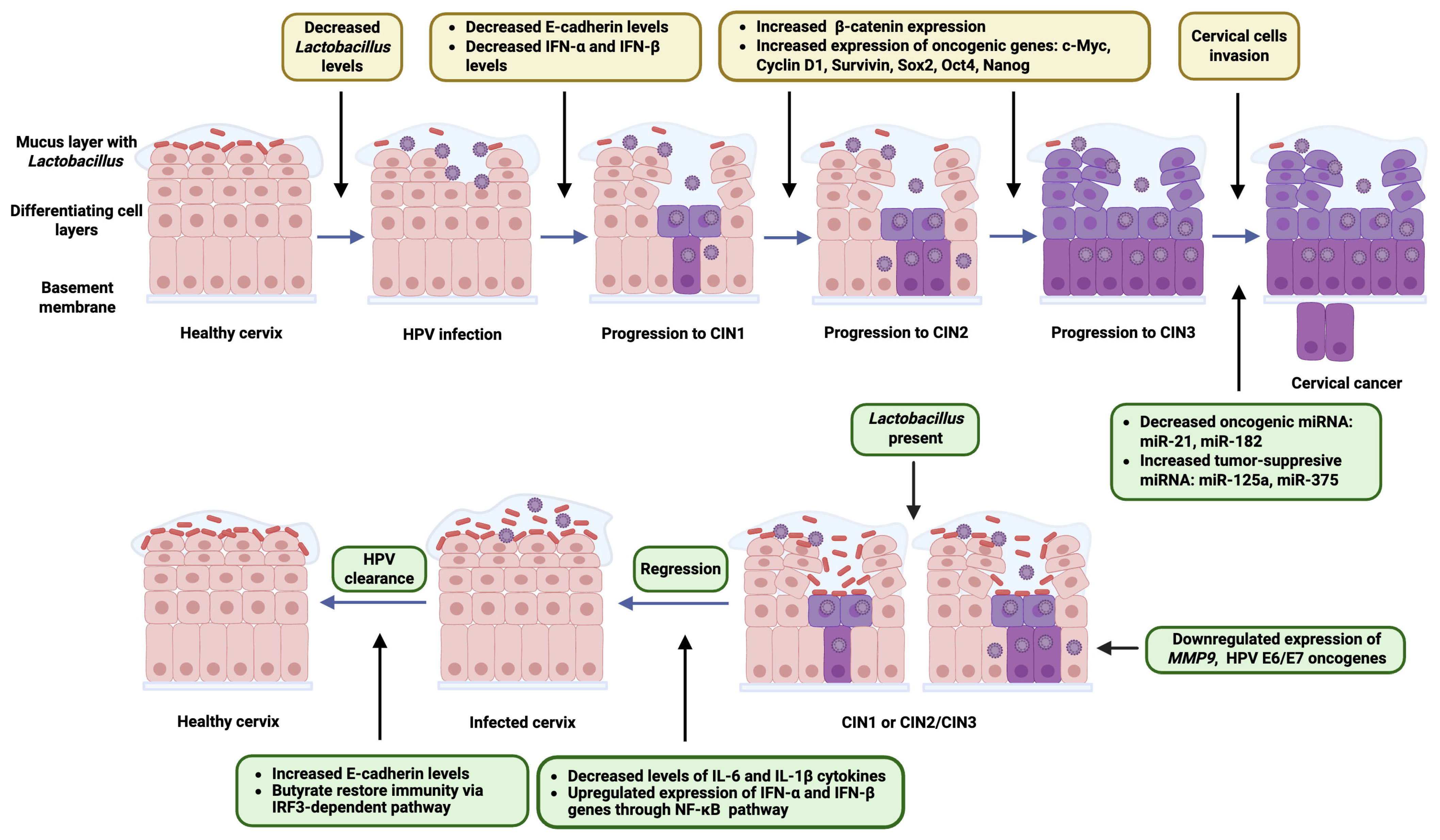
Lactobacillus species are central to the maintenance of a healthy vaginal microbiota and have been increasingly recognized for their role in natural defense against persistent HR-HPV infection and the progression of CIN. One of the primary mechanisms by which Lactobacillus exerts its protective effect is through the production of lactic acid, which maintains a low vaginal pH [60]. This acidic environment is hostile to many pathogens and prevents the overgrowth of anaerobic bacteria that are associated with dysbiosis and chronic inflammation [61]. The mechanisms by which Lactobacillus species protect against an HPV infection, persistence, and progression to cervical cancer are multifaceted and involve several pathways. Lactobacillus species promote the integrity of the epithelial mucosal barrier by increasing E-cadherin levels, a biomarker of epithelial health [62,63]. This reduces the risk of HPV’s penetration through abrasions or epithelial disruptions. Lactobacillus enhances the antiviral innate immunity by upregulating interferon (IFN)-α and IFN-β expression through pathways like NF-κB [64,65]. These bacteria decrease the levels of pro-inflammatory cytokines such as interleukin (IL)-6 and IL-1β, reducing chronic inflammation and the associated HPV persistence risks [66]. Butyrate production by Lactobacillus species helps restore immunity via interferon regulatory factor 3 (IRF3)-dependent pathways, aiding HPV clearance [60]. Certain Lactobacillus species (L. crispatus and L. gasseri) downregulate the expression of HPV E6 and E7 oncogenes [60,67]. Lactobacillus reduces the expression of matrix metalloproteinase-9 (MMP9), an enzyme that contributes to extracellular matrix degradation, cancer cell invasion, and metastasis [68]. By regulating key markers such as E-cadherin and β-catenin, it also helps inhibit the epithelia–mesenchymal transition, a critical process in cancer metastasis [69]. Research has demonstrated that the Wnt/β-catenin signaling pathway plays a critical role in the progression of CIN [70,71]. As CIN advances from low- to high-grade lesions, there is a notable increase in β-catenin expression and a shift in its localization from the cell membrane to the cytoplasm and nucleus [71]. This nuclear accumulation of β-catenin is a hallmark of active Wnt signaling. Once inside the nucleus, β-catenin acts as a transcriptional co-activator, promoting the expression of oncogenic target genes, such as the c-Myc, Cyclin D1, Survivin, Sox2, Oct4, and Nanog [71]. These genes support cellular proliferation, inhibit apoptosis, and enhance the stem-like characteristics of dysplastic cervical cells contributing to lesion persistence and progression toward cervical cancer.
Several studies have highlighted the influence of Lactobacillus strains on epithelial health and signaling pathways relevant to cancer and inflammation. For instance, L. rhamnosus GG (LGG) demonstrated protective effects in neonatal necrotizing enterocolitis by modulating intestinal stem cell activity, restoring tight junction integrity, and activating the Wnt/β-catenin signaling pathway [72]. Similarly, in colorectal cancer models, both Lactobacillus cocktails and L. acidophilus postbiotics were shown to interfere with proliferative signaling, including the Wnt, Notch, and BMP pathways, also exerting anti-proliferative and anti-migratory effects on cancer cells [73]. While these studies pertain to intestinal and colorectal models, they raise the question whether similar molecular interactions might be relevant in the context of CIN. Given the importance of the Wnt pathway in epithelial homeostasis and neoplastic progression, exploring the potential of specific Lactobacillus strains to influence CIN regression through similar mechanisms could be of interest. Some evidence also suggests that Lactobacillus strains might contribute to β-catenin regulation by modulating upstream components such as glycogen synthase kinase-3β, which facilitates β-catenin phosphorylation and subsequent degradation [74,75]. The exact relevance of this interaction in cervical tissue remains to be elucidated. However, it may offer a potential mechanistic explanation for the observed anti-proliferative effects of Lactobacillus in epithelial models.
Several studies have demonstrated that Lactobacillus could modulate host gene expression at the post-transcriptional level through the regulation of tumor-suppressor microRNAs (miRNAs), small non-coding RNAs that play a key role in cell cycle control, apoptosis, and oncogenesis [76,77,78]. In germ-free mice, microbial colonization altered the expression of intestinal miRNAs—miR-146, miR-375, and miR-10a, which regulate immune signaling pathways, including the Toll-like receptor (TLR)/MyD88 and cytokine balance (IL-12/IL-23) [79,80,81]. The dysregulation of these miRNAs contributes to inflammation and carcinogenesis. In colorectal cancer, certain lncRNAs (HOTAIR) were found to suppress tumor-suppressor miRNAs, promoting proliferation and therapy resistance [82]. Probiotic strains, including various Lactobacillus spp., were shown to modulate host miRNA profiles by decreasing oncogenic miRNAs (i.e., miR-21 and miR-182) and increasing tumor-suppressive ones (miR-125a-5p and miR-375), leading to enhanced apoptosis and reduced inflammation via targeting the Bcl-2, COX-2, and iNOS pathways [78,83]. Some Lactobacillus-derived components, including exopolysaccharides, induced pro-apoptotic gene expression and suppressed anti-apoptotic signals in cancer cells [84,85]. Although these findings were primarily demonstrated in colorectal cancer models, it is plausible that similar Lactobacillus–miRNA interactions could influence the cervical microenvironment. Hypothetically, Lactobacillus-induced modulation of miRNA expression may affect local immune tolerance, epithelial integrity, or viral persistence—mechanisms critical to HR-HPV clearance and CIN regression. For instance, the downregulation of pro-inflammatory or oncogenic miRNAs (miR-21) or the enhancement of miRNAs involved in epithelial differentiation and immune activation might support viral clearance and limit neoplastic progression. However, this remains speculative and warrants direct investigation in HPV-related cervical disease. Emerging evidence from vaginal microbiota studies supported a potential regulatory relationship between Lactobacillus dominance and host miRNA expression [86]. A study profiling miRNA expression in vaginal samples from young women revealed that non-Lactobacillus-dominated communities were associated with a broad upregulation of host miRNAs, including miR-23a-3p and miR-130a-3p. These miRNAs not only discriminated microbial community types with high accuracy, but they might also reflect or influence host immune or epithelial responses. Notably, L. crispatus dominance correlated with the most distinct miRNA profile, further supporting a functional link between beneficial microbes and local gene regulation. The Lactobacillus-associated suppression of specific miRNAs could contribute to a protective mucosal environment, potentially favoring HPV clearance or limiting progression to CIN. Although the causality remains to be established, this study offers a valuable molecular framework for exploring how vaginal commensals might shape host gene expression through miRNA modulation [86].
Modulating the immune response, through the activation of macrophages and dendritic cells, plays a crucial role in combating HPV infections [87]. Dendritic cells (DCs) are pivotal for initiating the host’s immune defense against HPV. Upon encountering the virus, DCs mature and produce type I IFNs and IL-12, essential for activating T cells and orchestrating an effective immune response [88]. However, HPV has developed mechanisms to evade this response, leading to persistent infections. Immunotherapeutic strategies aim to enhance DC function to counteract this evasion [89]. Macrophages also significantly contribute to the immune response against HPV. Studies show that HPV-positive head and neck squamous cell carcinomas contained a higher proportion of M1 macrophages, which are associated with anti-tumor activity, compared to HPV-negative tumors [90]. This suggests that modulating macrophage activity could influence disease outcomes. Macrophages play a key role in immune defense, including HPV infections. While some studies show a correlation between macrophage presence and HPV lesion grade, their exact role remains unclear [90]. Tumor-associated macrophages can suppress immune responses in cancer, and similar mechanisms may affect HPV infections [91].
HPV can interfere with innate immune mechanisms, such as downregulating TLR9 expression in infected cells, thereby evading immune detection [92]. Understanding these interactions has led to the exploration of therapies targeting TLR9 and other pathways to restore effective immune surveillance. Research demonstrated that cervical epithelial immune responses differed significantly between women with persistent HPV infections and those who cleared the virus [67]. The study revealed that type I IFNs and TLR3 were downregulated in persistent cases. Certain Lactobacillus strains, particularly L. gasseri LGV03 isolated from women who had cleared HPV, modulated immune signaling pathways. L. gasseri LGV03 enhanced antiviral signaling by increasing IRF3 phosphorylation in response to poly (I:C) stimulation and simultaneously attenuated the inflammatory response by downregulating NF-κB pathway activation. Additionally, L. gasseri LGV03 suppressed cervical cell proliferation in a zebrafish model, suggesting a potential role in supporting antiviral immunity and limiting pathogen-driven inflammation [67].
Long-term HPV persistence leads to viral DNA integration into the host genome, disrupting key tumor suppressor genes and promoting carcinogenesis. Vaginal lactobacilli protect against HPV through mechanisms, such as epithelial barrier reinforcement, anti-inflammatory effects, and the modulation of immune responses [60]. HPV E6 expression was reduced by L. crispatus and L. iners and increased by Gardnerella vaginalis, Megasphaera micronuciformis, and Fannyhessea vaginae [93,94]. Similarly, E7 protein production was suppressed by L. crispatus and L. gasseri and enhanced by L. iners, G. vaginalis, and M. micronuciformis [60,95]. The latter species also reduced p53 and pRb tumor suppressor levels, potentially promoting cell cycle deregulation and neoplastic initiation [96]. Additionally, non-vaginal Lactobacillus strains showed promise as anti-cervical cancer probiotics. L. casei LH23 inhibited HeLa cell proliferation, reduced cell migration, and suppressed HPV E6 and E7 expression [97]. L. casei and L. paracasei also enhanced apoptosis-related gene expression while downregulating anti-apoptotic Bcl-2, suggesting a general anticancer effect [98,99]. Probiotic interventions, including vaginal Lactobacillus strains and non-vaginal Lacticaseibacillus casei, showed promise in reducing HPV infection and cervical cancer risk [60] (Figure 2).

5. Current Evidence Linking Vaginal Microbiota Composition to HR-HPV Persistence and CIN Outcomes

Several studies explored the association between the Lactobacillus-rich microbiota and the efficiency of HR-HPV elimination. The analysis of multiple studies revealed that the presence of Lactobacillus species in the cervicovaginal environment was associated with a decreased detection of HR-HPV infections, CIN, and cervical cancer. Specifically, L. crispatus was identified as a critical protective factor, correlating with lower rates of HR-HPV and CIN [100,101]. In a cohort of 73 women analyzed, 58.9% experienced HPV clearance within a 12-month period [102]. There were no statistically significant differences between those who cleared the virus and those who did not in terms of the age, stage of disease, HPV subtype, vaginal microbiota CSTs, or vaginal microbiota diversity (both α and β metrics). At baseline, participants with a lower presence of Enterococcus ASV 62 and higher levels of L. iners were less likely to eliminate the virus by the 12-month mark. Additional analysis indicated a notable inverse correlation between elevated L. iners abundance and HPV clearance in individuals undergoing non-surgical treatment, whereas no such association was found among those who received surgical interventions [102].
A pilot study by Di Paola et al. [6] evaluated HPV testing in a primary screening program involving 1029 women aged 26–64. Multiple HPV genotypes were detected in 13 cases. After one year, all HR-HPV-positive women underwent follow-up testing and were classified into the clearance group (n = 27) (i.e., women who had cleared the infection (no HR-HPV DNA detected)) and the persistence group (n = 28), i.e., women with a persistent HR-HPV infection (at least one HPV genotype remained). Lactobacillus was the dominant genus across all groups (clearance, persistence, and control), but differences in species distribution were observed. L. crispatus was the most abundant species in both the control and clearance groups, suggesting a potential role in HPV clearance [6] (Table 1).
Zeng et al. [103] investigated the relationship between vaginal microbiota composition and the clearance of HR-HPV infection. Among 135 participants, including 45 HPV-negative and 90 patients with an HPV infection, 26 patients experienced HPV clearance within a year, with most turning negative within six months. The analysis revealed that women with HPV clearance had significantly lower bacterial alpha diversity. The lower diversity of the vaginal microbiota, characterized by a predominance of Lactobacillus species, may be associated with natural HPV clearance. In contrast, persistent HPV infection could be linked to the presence of specific bacterial taxa, including Fusobacterium, Neisseria, Helicobacter, and Bacteroides (genus level), as well as Streptococcaceae, Neisseriaceae, Helicobacteraceae, Erysipelotrichaceae, and Bacteroidaceae (family level) [103]. Brotman et al. [104] explored the relationship between vaginal microbiota compositions and the detection of HPV. Thirty-two women of reproductive age self-collected mid-vaginal swabs twice a week over a 16-week period, yielding a total of 937 samples. By sequencing barcoded 16S rRNA genes, the study identified six distinct CSTs within the vaginal bacterial communities. Each swab was also tested for the presence of 37 different HPV types. The study revealed that HPV clearance was strongly associated with the structure of the vaginal microbiota. Microbial communities dominated by L. gasseri were linked to a faster clearance of the virus, whereas those characterized by low Lactobacillus abundance and a higher presence of Atopobium were associated with a delayed clearance. Additionally, certain low-Lactobacillus community types appeared to increase the likelihood of acquiring new HPV infections [104].
Table 1. Overview of the research on cervicovaginal microbiota composition and its impact on HPV infection dynamics and cervical neoplasia.
Author, Year; Country [Ref]Study AimGroups: Cases and HPV-Negative ControlsMaterial and Detection MethodResultsChanges in Microbiota AbundanceConclusions
Brotman et al., 2014; USA [104]Assess temporal links between microbiota dynamics and HPV detection32 women, 937 self-collected vaginal samples over 16 weeksVaginal swabs; 16S rRNA sequencing; HPV DNA testingCST II (L. gasseri) linked to HPV clearance; CST IV-B (anaerobes) linked to persistence; the most abundant Lactobacillus species in the control and clearance groupL. gasseri enriched in clearance; Atopobium in slow clearance statesMicrobiota state changes HPV detection and outcomes; longitudinal sampling is key
Di Paola et al., 2017; Italy [6]Characterize cervicovaginal microbiota in persistent HR-HPV55 HPV-positive (from 1029 screened), 17 HPV-negative controlsCervicovaginal swabs; 16S rRNA pyrosequencingCST IV more frequent in persistent HPV (72.7%) vs. controls (16.6%)Decreased Lactobacillus, increased Gardnerella, and Atopobium in the persistent groupLactobacillus-dominated microbiota linked to HPV clearance; dysbiosis favors persistence
Mitra et al., 2020;
UK [31]
Study how vaginal microbiota influences CIN2 lesion regression87 women (16–26 years) with CIN2, followed-up for 24 monthsVaginal swabs; 16S rRNA sequencingLactobacillus-dominant microbiota associated with lesion regressionIncreased Gardnerella, Prevotella, and Megasphaera in the persistent groupVaginal microbiota could predict CIN2 outcome and serve as a therapeutic target
Zeng et al., 2023; China [103]Explore vaginal microbiota’s role in HPV infection, persistence, and clearance90 HPV-positive (persistent vs. cleared), 45 HPV-negative controlsVaginal swabs; 16S rRNA sequencingHigher alpha diversity in persistent HPV; L. iners prevalent in infectionsIncreased Sneathia amnii, Bacteroidaceae in persistence; L. crispatus in clearanceMicrobiota composition affects HPV outcomes;
L. crispatus dominance may promote clearance
CIN, cervical intraepithelial neoplasia; CSTs, community state types, for classification of distinct vaginal microbial communities.
A study conducted by Mitra et al. [31] provided detailed scientific data on the association between vaginal microbiota composition and the regression of CIN2. The study included 87 participants. They were 16–26 years old, with histologically confirmed CIN2. The follow-up period was 24 months. At baseline, CST III (L. iners-dominant; 41.4%) was the most common, followed by CST IV (Lactobacillus-depleted; 34.5%) and CST I (L. crispatus-dominant; 24.1%). CST II (L. gasseri) and CST V (L. jensenii) were absent at baseline but occurred in several samples later on. Vaginal microbiota composition did not significantly differ based on HPV status or subtype at baseline. Lactobacillus depletion and the presence of specific anaerobic bacteria at CIN2 diagnosis were linked to a significantly lower likelihood of regression at 12 and 24 months. When regression occurred, it was slower in the absence of a Lactobacillus-dominant vaginal microbiota. A similar trend was observed at 12 months in women with persistent disease, though the statistical power is limited due to the reduced sample size. The evidence suggests that interventions aimed at restoring and maintaining a healthy vaginal microbiota, including probiotic therapies, could be beneficial in HPV infection management and cervical dysplasia treatment.
Liu et al. [105] showed the effects of the intravaginal administration of L. crispatus chen-01 on HR-HPV infections. In this study, 100 women with HR-HPV were randomly assigned to receive either the probiotic treatment or a placebo. After six months, the group treated with L. crispatus chen-01 showed a significant reduction in HPV viral load, an increased clearance rate of the virus, and improved vaginal health without notable adverse effects. Additionally, 16S rRNA sequencing indicated that the probiotic effectively restored a healthy vaginal microbiota composition in these women. These findings suggest that L. crispatus chen-01 could be a promising treatment option for HR-HPV infections [105].

6. Therapeutic Perspectives

The modulation of the vaginal microbiota using probiotics and prebiotics, particularly those containing immunomodulatory and acid-producing Lactobacillus species, has emerged as a promising adjunctive strategy in the management of HR-HPV infections. Several clinical studies investigated the therapeutic potential of probiotics in the context of HR-HPV infections and cervical dysplasia, employing diverse formulations, strains, and routes of administration (Table 2). Interventions included oral and vaginal deliveries of specific Lactobacillus strains administered either as a monotherapy or as adjuncts to the standard treatment. Probiotics may offer several therapeutic benefits when used alongside conventional treatments for CIN, such as the loop electrosurgical excision procedure [106,107]. These benefits include promoting tissue repair, reducing post-treatment inflammation, restoring healthy microbial balance, and potentially decreasing the risk of HR-HPV reinfection or disease progression. Moreover, microbiota restoration may also contribute to a reduction in dysbiosis-associated symptoms such as vaginal discomfort, discharge, or an increased susceptibility to secondary infections [61]. The integration of probiotics into treatment regimens may be particularly valuable in patients with persistent HPV infections or incomplete immune clearance following the standard therapy. Probiotic formulations administered orally or intravaginally are being evaluated in ongoing trials to assess their role in accelerating viral clearance and reducing treatment-related complications.
Table 2. Summary of clinical studies on probiotic interventions in HR-HPV clearance and cytological outcomes.
Number of Clinical TrialStudy Period, Time Frame (Months); and LocationIntervention/TreatmentInclusion CriteriaExclusion CriteriaStudy Design and MaskingSample SizeStudy GroupControl GroupPrimary Outcome MeasuresSecondary Outcome Measures
NCT010973562010–2011 (6); BelgiumProbiotic drink vs. no interventionWomen; age 18–65; LSIL+ HPV+ in the Papanicolaou testAge >65; immuno-compromisedRandomized, parallel;
single-blind (participant)
60Probiotic drink daily (6 months)No intervention (6-months observation)HPV positivity; LSIL regressionNo data
NCT015994162011–2013 (12); TaiwanOral U-relax (L. rhamnosus GR-1 + L. reuteri RC-14) vs. placeboWomen; age 30–65; HPV+ 6 months after conization; negative for intraepithelial lesion or malignancy in the Papanicolaou testCIN before conization, cervical cancer, genital infection issues, long-term antibioticsRandomized, parallel;
triple-blind (participant, care provider, investigator)
80U-relax: 2 caps/day → 1/day until day 360Placebo (identical schedule)Vaginal health statusHPV DNA index change
NCT033723952015–2016 (9); ItalyVaginal L. rhamnosus BMX 54 + standard treatmentAge >18, bacterial vaginosis/yeast vaginitis + HPV; (ASCUS, LSIL, CIN1, HPV DNA+)Pregnancy; CIN2/3, cancer, immuno-deficiency, corticosteroidsRandomized, sequential (pilot);
open label
117 (60 short, 57 long)Long-term probiotics (6 months)Short-term probiotics (3 months)Infection symptom resolutionAdverse events (CTCAE v4.0)
NCT051095332018–2021 (12); ItalyVaginal + oral probiotics: L. rhamnosus BMX 54, L. reuteri RC-14, L. rhamnosus GR-1 + standard treatmentAge >18, positive vaginal infection swabs, HPV+Pregnancy or breast-feeding, malignancies, immunological diseases, comorbidities, corticosteroidsRandomized, parallel;
open label
483 (252 probiotic, 231 controls)Standard treatment + 9-months probioticsStandard treatmentHPV clearance; vaginal infection resolutionNo data
NCT068028092022–2024 (4); ItalyOral L. crispatus M247 (Crispact®) vs. placeboWomen; age 18–69, HR-HPV+, ASCUS or LSIL; negative colposcopy (biopsy)Prior HPV vaccination, HSIL, immunotherapy, neoplasia, pregnancy, allergyRandomized, parallel; placebo-controlled,
single-blind (participant)
66L. crispatus M247, 20 billion CFU/day for 4 monthsPlacebo, 1 stick/day for 4 monthsHR-HPV clearanceMicrobiota change,
cytology normalization,
side effects
ASCUS, atypical squamous cells of undetermined significance; CTCAE, Common Terminology Criteria for Adverse Events; CIN, cervical intraepithelial neoplasia; HSIL, high-grade squamous intraepithelial lesion; LSIL, low-grade squamous intraepithelial lesion.
Dellino et al. [108] noted that the oral administration of L. crispatus M247 was associated with a higher probability of resolving HPV-related cytological abnormalities compared to those who only underwent follow-up (60.5% vs. 41.3%). The median follow-up was 12 months (ranging from 10 to 30 months). Complete HPV clearance was observed in 9.3% of the patients in the follow-up-only group, whereas 15.3% of those taking long-term oral L. crispatus M247 achieved clearance. However, by the end of the study, the proportion of HPV-negative patients, as determined with the HPV-DNA test, was not significantly different between those two groups [108].
A randomized study included 117 women divided into two groups, as follows: one received standard antimicrobial treatment plus a short-term (3-month) course of vaginal probiotics, while the other received the same treatment followed by a long-term (6-month) course of L. rhamnosus BMX 54 [109]. The results showed that the long-term probiotic group experienced a higher rate of HPV clearance (31.2%) compared to the short-term group (11.6%). This suggests that extended use of vaginal L. rhamnosus BMX 54 may help restore a balanced vaginal microbiota and aid in resolving HPV infections. Another study included 121 women with a genital HR-HPV infection, divided into a study group (62 participants) and a control group (59 participants) [110]. There was no significant difference in the HR-HPV clearance rates between the groups (58.1% vs 54.2%). A lower initial viral load was the only factor predicting HR-HPV clearance. Twenty-two women had a mildly abnormal initial cervical smear and nine had an unsatisfactory smear. At a 6-month follow-up, both mildly abnormal cervical smear and unsatisfactory smear rates decreased significantly in the study group compared to the control group. The application of probiotic strains (i.e., L. rhamnosus GR-1 and L. reuteri RC-14) did not influence genital HR-HPV clearance but may have decreased the rates of mildly abnormal and unsatisfactory cervical smears.
In parallel, the impact of prophylactic HPV vaccination on the composition and function of the vaginal microbiota is gaining increasing scientific attention [111,112]. Although prophylactic HPV vaccines primarily target viral infection and lesion development, emerging evidence suggests that vaccination may also influence the vaginal microenvironment. By reducing chronic-HPV-related inflammation and epithelial disruption, vaccination could indirectly support the restoration or maintenance of a healthy Lactobacillus-dominated microbiota [43]. Furthermore, the interplay between host immunity and microbial composition can be modulated by vaccine-induced immune responses, which affect microbial dynamics and resilience against dysbiosis [113]. While vaccines targeting oncogenic HPV types have proven highly effective at reducing infection rates and associated lesions, their influence on the local microbial ecosystem remains incompletely understood. Lactobacillus-based strategies may serve as complementary tools to vaccination by enhancing mucosal resilience, supporting a balanced immune response, and preventing chronic, subclinical infections. Combining prophylactic vaccination with microbiota-supportive interventions could offer a synergistic approach to preventing both primary HPV infections and their recurrence in at-risk populations. Future research should aim to clarify the interplay between vaccination, host immunity, and microbial dynamics to optimize long-term protective strategies against HPV-related disease.

7. Conclusions

The effective clearance of HR-HPV is essential for interrupting the pathway to cervical carcinogenesis, and the modulation of the vaginal microbiota through Lactobacillus-dominated communities may play a crucial role in achieving this outcome. Key findings from studies on the interactions between the vaginal microbiota and HR-HPV infections highlight the pivotal role of specific Lactobacillus species in facilitating viral clearance and supporting the regression of cervical dysplastic lesions. Species such as L. crispatus and L. gasseri exhibit protective effects through the modulation of local immune responses, the maintenance of acidic vaginal pH, and the competitive inhibition of pathogenic microorganisms. Given the current evidence, the use of probiotics containing selected Lactobacillus strains emerges as a promising adjunctive strategy in the management and prevention of HPV-related cervical abnormalities. However, further research, including randomized clinical trials, is essential to validate their efficacy, safety, and to establish optimal treatment protocols. Moreover, the individualization of probiotic-based interventions, tailored to the specific microbial profile of each patient, may significantly enhance therapeutic outcomes. Integrating microbiological, gynecological, and immunological insights opens new avenues for the development of innovative, microbiota-targeted strategies in the fight against HR-HPV and cervical dysplasia.

Author Contributions

Conceptualization, N.Z.-L.; writing—original draft preparation, E.K., J.W.-B., A.B., M.P., M.D., and N.Z.-L.; writing—review and editing, E.K., J.W.-B., E.E.H., P.C., M.B., P.O., M.C., and N.Z.-L.; visualization, P.C., E.E.H., and N.Z.-L.; supervision, N.Z.-L.; funding acquisition, E.E.H. and N.Z.-L. All authors have read and agreed to the published version of the manuscript.

Funding

This work was supported by the Centre of Postgraduate Medical Education (grant numbers: 501-1-009-12-25, 501-1-022-26-25 and 501-1-009-12-24/MG2 (N.Z.-L.)).

Institutional Review Board Statement

Not applicable.

Informed Consent Statement

Not applicable.

Data Availability Statement

Not applicable.

Conflicts of Interest

The authors declare no conflicts of interest. The funders had no role in the design of the study; in the collection, analyses, or interpretation of data; in the writing of the manuscript; or in the decision to publish the results.

Abbreviations

The following abbreviations are used in this manuscript:
HRHigh risk
HPVHuman papillomavirus
CINCervical intraepithelial neoplasia
LRLow risk
pRBRetinoblastoma protein
CSTCommunity state type
BVBacterial vaginosis
AVAerobic vaginitis
IFNInterferon
ILInterleukin
TLRToll-like receptor
IRFInterferon regulatory factor
miRNAmicroRNA
DCDendritic cell

References

  1. Hull, R.; Mbele, M.; Makhafola, T.; Hicks, C.; Wang, S.; Reis, R.; Mehrotra, R.; Mkhize-Kwitshana, Z.; Kibiki, G.; Bates, D.; et al. Cervical Cancer in Low and Middle-income Countries (Review). Oncol. Lett. 2020, 20, 2058–2074. [Google Scholar] [CrossRef]
  2. Mo, Y.; Ma, J.; Zhang, H.; Shen, J.; Chen, J.; Hong, J.; Xu, Y.; Qian, C. Prophylactic and Therapeutic HPV Vaccines: Current Scenario and Perspectives. Front. Cell. Infect. Microbiol. 2022, 12, 909223. [Google Scholar] [CrossRef] [PubMed]
  3. Yousaf, S.; Shehzadi, A.; Ahmad, M.; Asrar, A.; Ahmed, I.; Iqbal, H.M.N.; Hussen Bule, M. Recent Advances in HPV Biotechnology: Understanding Host-Virus Interactions and Cancer Progression—A Review. Int. J. Surg. 2024, 110, 8025–8036. [Google Scholar] [CrossRef]
  4. Molina, M.A.; Coenen, B.A.; Leenders, W.P.J.; Andralojc, K.M.; Huynen, M.A.; Melchers, W.J.G. Assessing the Cervicovaginal Microbiota in the Context of hrHPV Infections: Temporal Dynamics and Therapeutic Strategies. mBio 2022, 13, e01619-22. [Google Scholar] [CrossRef]
  5. Chee, W.J.Y.; Chew, S.Y.; Than, L.T.L. Vaginal Microbiota and the Potential of Lactobacillus Derivatives in Maintaining Vaginal Health. Microb. Cell Factories 2020, 19, 203. [Google Scholar] [CrossRef]
  6. Di Paola, M.; Sani, C.; Clemente, A.M.; Iossa, A.; Perissi, E.; Castronovo, G.; Tanturli, M.; Rivero, D.; Cozzolino, F.; Cavalieri, D.; et al. Characterization of Cervico-Vaginal Microbiota in Women Developing Persistent High-Risk Human Papillomavirus Infection. Sci. Rep. 2017, 7, 10200. [Google Scholar] [CrossRef]
  7. Schellekens, H.C.J.; Schmidt, L.M.S.; Morré, S.A.; Van Esch, E.M.G.; De Vos Van Steenwijk, P.J. Vaginal Microbiota and Local Immunity in HPV-Induced High-Grade Cervical Dysplasia: A Narrative Review. Int. J. Mol. Sci. 2025, 26, 3954. [Google Scholar] [CrossRef]
  8. Dwiyanto, J.; Hussain, M.H.; Reidpath, D.; Ong, K.S.; Qasim, A.; Lee, S.W.H.; Lee, S.M.; Foo, S.C.; Chong, C.W.; Rahman, S. Ethnicity Influences the Gut Microbiota of Individuals Sharing a Geographical Location: A Cross-Sectional Study from a Middle-Income Country. Sci. Rep. 2021, 11, 2618. [Google Scholar] [CrossRef] [PubMed]
  9. Onywera, H.; Mbulawa, Z.Z.A.; Brink, A.; Williamson, A.-L.; Mwapagha, L.M. Unravelling the Biological Interplay Between Genital HPV Infection and Cervicovaginal Microbiota in Sub-Saharan Africa: Implications for Cervical (Pre)Cancer Prevention. Venereology 2024, 3, 211–231. [Google Scholar] [CrossRef]
  10. Głowienka-Stodolak, M.; Bagińska-Drabiuk, K.; Szubert, S.; Hennig, E.E.; Horala, A.; Dąbrowska, M.; Micek, M.; Ciebiera, M.; Zeber-Lubecka, N. Human Papillomavirus Infections and the Role Played by Cervical and Cervico-Vaginal Microbiota—Evidence from Next-Generation Sequencing Studies. Cancers 2024, 16, 399. [Google Scholar] [CrossRef] [PubMed]
  11. IARC Working Group on the Evaluation of Carcinogenic Risks to Humans. Human Papillomaviruses; International Agency for Research on Cancer: Lyon, France, 2007. [Google Scholar]
  12. Gheit, T. Mucosal and Cutaneous Human Papillomavirus Infections and Cancer Biology. Front. Oncol. 2019, 9, 355. [Google Scholar] [CrossRef] [PubMed]
  13. Glinska, P.; Macios, A.; Jaworski, R.; Bobinski, M.; Pruski, D.; Przybylski, M.; Zielinska, A.; Sawicki, W.; Nowakowski, A. Baseline Data on Distribution of Human Papillomavirus (HPV) Genotypes in Cervical Samples of Gynecological Patients before Implementation of Population-Based HPV Vaccination Program in Poland. Ginekol. Pol. 2024, 95, 870–878. [Google Scholar] [CrossRef]
  14. The Cancer Genome Atlas Research Network. Integrated Genomic and Molecular Characterization of Cervical Cancer. Nature 2017, 543, 378–384. [Google Scholar] [CrossRef]
  15. Wei, F.; Georges, D.; Man, I.; Baussano, I.; Clifford, G.M. Causal Attribution of Human Papillomavirus Genotypes to Invasive Cervical Cancer Worldwide: A Systematic Analysis of the Global Literature. Lancet 2024, 404, 435–444. [Google Scholar] [CrossRef]
  16. Kajitani, N.; Satsuka, A.; Kawate, A.; Sakai, H. Productive Lifecycle of Human Papillomaviruses That Depends Upon Squamous Epithelial Differentiation. Front. Microbiol. 2012, 3, 152. [Google Scholar] [CrossRef]
  17. Harden, M.E.; Munger, K. Human Papillomavirus Molecular Biology. Mutat. Res. Mutat. Res. 2017, 772, 3–12. [Google Scholar] [CrossRef]
  18. Jain, M.; Yadav, D.; Jarouliya, U.; Chavda, V.; Yadav, A.K.; Chaurasia, B.; Song, M. Epidemiology, Molecular Pathogenesis, Immuno-Pathogenesis, Immune Escape Mechanisms and Vaccine Evaluation for HPV-Associated Carcinogenesis. Pathogens 2023, 12, 1380. [Google Scholar] [CrossRef]
  19. Hong, S.; Laimins, L.A. Regulation of the Life Cycle of HPVs by Differentiation and the DNA Damage Response. Future Microbiol. 2013, 8, 1547–1557. [Google Scholar] [CrossRef]
  20. Haręża, D.A.; Wilczyński, J.R.; Paradowska, E. Human Papillomaviruses as Infectious Agents in Gynecological Cancers. Oncogenic Properties of Viral Proteins. Int. J. Mol. Sci. 2022, 23, 1818. [Google Scholar] [CrossRef] [PubMed]
  21. Scarth, J.A.; Patterson, M.R.; Morgan, E.L.; Macdonald, A. The Human Papillomavirus Oncoproteins: A Review of the Host Pathways Targeted on the Road to Transformation. J. Gen. Virol. 2021, 102, 001540. [Google Scholar] [CrossRef] [PubMed]
  22. Zhang, Y.; Qiu, K.; Ren, J.; Zhao, Y.; Cheng, P. Roles of Human Papillomavirus in Cancers: Oncogenic Mechanisms and Clinical Use. Signal Transduct. Target. Ther. 2025, 10, 44. [Google Scholar] [CrossRef]
  23. Sivakumar, N.; Narwal, A.; Kumar, S.; Kamboj, M.; Devi, A.; Pandiar, D.; Bhardwaj, R. Application of the Bethesda System of Reporting for Cervical Cytology to Evaluate Human Papilloma Virus Induced Changes in Oral Leukoplakia, Oral Squamous Cell Carcinoma, and Oropharyngeal Squamous Cell Carcinoma: A Cytomorphological and Genetic Study. Diagn. Cytopathol. 2021, 49, 1036–1044. [Google Scholar] [CrossRef] [PubMed]
  24. Kamal, M. Cervical Pre-Cancers: Biopsy and Immunohistochemistry. Cytojournal 2022, 19, 38. [Google Scholar] [CrossRef]
  25. Alrajjal, A.; Pansare, V.; Choudhury, M.S.R.; Khan, M.Y.A.; Shidham, V.B. Squamous Intraepithelial Lesions (SIL: LSIL, HSIL, ASCUS, ASC-H, LSIL-H) of Uterine Cervix and Bethesda System. Cytojournal 2021, 18, 16. [Google Scholar] [CrossRef]
  26. Bentley, J.; Executive Council of the Society of Canadian Colposcopists; Special contributors. Colposcopic Management of Abnormal Cervical Cytology and Histology. J. Obstet. Gynaecol. Can. 2012, 34, 1188–1202. [Google Scholar] [CrossRef]
  27. Li, X.; Chen, Y.; Xiong, J.; Chen, P.; Zhang, D.; Li, Q.; Zhu, P. Biomarkers Differentiating Regression from Progression among Untreated Cervical Intraepithelial Neoplasia Grade 2 Lesions. J. Adv. Res. 2024, 74, 391–402. [Google Scholar] [CrossRef]
  28. Loopik, D.L.; Bentley, H.A.; Eijgenraam, M.N.; IntHout, J.; Bekkers, R.L.M.; Bentley, J.R. The Natural History of Cervical Intraepithelial Neoplasia Grades 1, 2, and 3: A Systematic Review and Meta-Analysis. J. Low. Genit. Tract Dis. 2021, 25, 221–231. [Google Scholar] [CrossRef]
  29. Hatano, Y.; Ideta, T.; Hirata, A.; Hatano, K.; Tomita, H.; Okada, H.; Shimizu, M.; Tanaka, T.; Hara, A. Virus-Driven Carcinogenesis. Cancers 2021, 13, 2625. [Google Scholar] [CrossRef] [PubMed]
  30. Koeneman, M.M.; Hendriks, N.; Kooreman, L.F.; Winkens, B.; Kruitwagen, R.F.; Kruse, A.J. Prognostic Factors for Spontaneous Regression of High-Risk Human Papillomavirus-Positive Cervical Intra-Epithelial Neoplasia Grade 2. Int. J. Gynecol. Cancer 2019, 29, 1003–1009. [Google Scholar] [CrossRef] [PubMed]
  31. Mitra, A.; MacIntyre, D.A.; Ntritsos, G.; Smith, A.; Tsilidis, K.K.; Marchesi, J.R.; Bennett, P.R.; Moscicki, A.-B.; Kyrgiou, M. The Vaginal Microbiota Associates with the Regression of Untreated Cervical Intraepithelial Neoplasia 2 Lesions. Nat. Commun. 2020, 11, 1999. [Google Scholar] [CrossRef]
  32. Wang, K.; Muñoz, K.J.; Tan, M.; Sütterlin, C. Chlamydia and HPV Induce Centrosome Amplification in the Host Cell through Additive Mechanisms. Cell. Microbiol. 2021, 23, e13397. [Google Scholar] [CrossRef]
  33. Akbari, E.; Milani, A.; Seyedinkhorasani, M.; Bolhassani, A. HPV Co-infections with Other Pathogens in Cancer Development: A Comprehensive Review. J. Med. Virol. 2023, 95, e29236. [Google Scholar] [CrossRef]
  34. Cao, Y. EBV Based Cancer Prevention and Therapy in Nasopharyngeal Carcinoma. npj Precis. Oncol. 2017, 1, 10. [Google Scholar] [CrossRef] [PubMed]
  35. Münz, C. Latency and Lytic Replication in Epstein–Barr Virus-Associated Oncogenesis. Nat. Rev. Microbiol. 2019, 17, 691–700. [Google Scholar] [CrossRef]
  36. Auriemma, R.S.; Scairati, R.; del Vecchio, G.; Liccardi, A.; Verde, N.; Pirchio, R.; Pivonello, R.; Ercolini, D.; Colao, A. The Vaginal Microbiome: A Long Urogenital Colonization Throughout Woman Life. Front. Cell. Infect. Microbiol. 2021, 11, 686167. [Google Scholar] [CrossRef] [PubMed]
  37. Heczko, P.B.; Giemza, M.; Ponikiewska, W.; Strus, M. Importance of Lactobacilli for Human Health. Microorganisms 2024, 12, 2382. [Google Scholar] [CrossRef]
  38. Miko, E.; Barakonyi, A. The Role of Hydrogen-Peroxide (H2O2) Produced by Vaginal Microbiota in Female Reproductive Health. Antioxidants 2023, 12, 1055. [Google Scholar] [CrossRef]
  39. Darbandi, A.; Asadi, A.; Mahdizade Ari, M.; Ohadi, E.; Talebi, M.; Halaj Zadeh, M.; Darb Emamie, A.; Ghanavati, R.; Kakanj, M. Bacteriocins: Properties and Potential Use as Antimicrobials. J. Clin. Lab. Anal. 2021, 36, e24093. [Google Scholar] [CrossRef] [PubMed]
  40. Patel, M.; Siddiqui, A.J.; Hamadou, W.S.; Surti, M.; Awadelkareem, A.M.; Ashraf, S.A.; Alreshidi, M.; Snoussi, M.; Rizvi, S.M.D.; Bardakci, F.; et al. Inhibition of Bacterial Adhesion and Antibiofilm Activities of a Glycolipid Biosurfactant from Lactobacillus Rhamnosus with Its Physicochemical and Functional Properties. Antibiotics 2021, 10, 1546. [Google Scholar] [CrossRef]
  41. Duarte-Mata, D.I.; Salinas-Carmona, M.C. Antimicrobial Peptides’ Immune Modulation Role in Intracellular Bacterial Infection. Front. Immunol. 2023, 14, 1119574. [Google Scholar] [CrossRef]
  42. Holdcroft, A.M.; Ireland, D.J.; Payne, M.S. The Vaginal Microbiome in Health and Disease—What Role Do Common Intimate Hygiene Practices Play? Microorganisms 2023, 11, 298. [Google Scholar] [CrossRef]
  43. Han, Y.; Liu, Z.; Chen, T. Role of Vaginal Microbiota Dysbiosis in Gynecological Diseases and the Potential Interventions. Front. Microbiol. 2021, 12, 643422. [Google Scholar] [CrossRef]
  44. Graziottin, A. Maintaining Vulvar, Vaginal and Perineal Health: Clinical Considerations. Womens Health 2024, 20, 17455057231223716. [Google Scholar] [CrossRef]
  45. Zheng, N.; Guo, R.; Wang, J.; Zhou, W.; Ling, Z. Contribution of Lactobacillus Iners to Vaginal Health and Diseases: A Systematic Review. Front. Cell. Infect. Microbiol. 2021, 11, 792787. [Google Scholar] [CrossRef]
  46. Colonna, C.; Steelman, M. Amsel Criteria. In StatPearls; StatPearls Publishing: Treasure Island, FL, USA, 2025. [Google Scholar]
  47. Simoes, J.A.; Discacciati, M.G.; Brolazo, E.M.; Portugal, P.M.; Dini, D.V.; Dantas, M.C.M. Clinical Diagnosis of Bacterial Vaginosis. Int. J. Gynaecol. Obstet. Off. Organ Int. Fed. Gynaecol. Obstet. 2006, 94, 28–32. [Google Scholar] [CrossRef] [PubMed]
  48. Amabebe, E.; Anumba, D.O.C. The Vaginal Microenvironment: The Physiologic Role of Lactobacilli. Front. Med. 2018, 5, 181. [Google Scholar] [CrossRef]
  49. Abou Chacra, L.; Ly, C.; Hammoud, A.; Iwaza, R.; Mediannikov, O.; Bretelle, F.; Fenollar, F. Relationship between Bacterial Vaginosis and Sexually Transmitted Infections: Coincidence, Consequence or Co-Transmission? Microorganisms 2023, 11, 2470. [Google Scholar] [CrossRef] [PubMed]
  50. Valeriano, V.D.; Lahtinen, E.; Hwang, I.-C.; Zhang, Y.; Du, J.; Schuppe-Koistinen, I. Vaginal Dysbiosis and the Potential of Vaginal Microbiome-Directed Therapeutics. Front. Microbiomes 2024, 3, 1363089. [Google Scholar] [CrossRef]
  51. Fan, A.; Yue, Y.; Geng, N.; Zhang, H.; Wang, Y.; Xue, F. Aerobic Vaginitis and Mixed Infections: Comparison of Clinical and Laboratory Findings. Arch. Gynecol. Obstet. 2013, 287, 329–335. [Google Scholar] [CrossRef] [PubMed]
  52. Maduta, C.S.; McCormick, J.K.; Dufresne, K. Vaginal Community State Types (CSTs) Alter Environmental Cues and Production of the Staphylococcus Aureus Toxic Shock Syndrome Toxin-1 (TSST-1). J. Bacteriol. 2024, 206, e00447-23. [Google Scholar] [CrossRef]
  53. De Seta, F.; Campisciano, G.; Zanotta, N.; Ricci, G.; Comar, M. The Vaginal Community State Types Microbiome-Immune Network as Key Factor for Bacterial Vaginosis and Aerobic Vaginitis. Front. Microbiol. 2019, 10, 2451. [Google Scholar] [CrossRef] [PubMed]
  54. Lee, S.; Oh, K.Y.; Hong, H.; Jin, C.H.; Shim, E.; Kim, S.H.; Kim, B.-Y. Community State Types of Vaginal Microbiota and Four Types of Abnormal Vaginal Microbiota in Pregnant Korean Women. Front. Public Health 2020, 8, 507024. [Google Scholar] [CrossRef]
  55. Cocomazzi, G.; De Stefani, S.; Del Pup, L.; Palini, S.; Buccheri, M.; Primiterra, M.; Sciannamè, N.; Faioli, R.; Maglione, A.; Baldini, G.M.; et al. The Impact of the Female Genital Microbiota on the Outcome of Assisted Reproduction Treatments. Microorganisms 2023, 11, 1443. [Google Scholar] [CrossRef]
  56. Ravel, J.; Gajer, P.; Abdo, Z.; Schneider, G.M.; Koenig, S.S.K.; McCulle, S.L.; Karlebach, S.; Gorle, R.; Russell, J.; Tacket, C.O.; et al. Vaginal Microbiome of Reproductive-Age Women. Proc. Natl. Acad. Sci. USA 2011, 108 (Suppl. S1), 4680–4687. [Google Scholar] [CrossRef]
  57. Kyrgiou, M.; Mitra, A.; Moscicki, A.-B. Does the Vaginal Microbiota Play a Role in the Development of Cervical Cancer? Transl. Res. J. Lab. Clin. Med. 2017, 179, 168–182. [Google Scholar] [CrossRef]
  58. Kyrgiou, M.; Moscicki, A.-B. Vaginal Microbiome and Cervical Cancer. Semin. Cancer Biol. 2022, 86, 189–198. [Google Scholar] [CrossRef]
  59. Loonen, A.J.M.; Verhagen, F.; Luijten-de Vrije, I.; Lentjes-Beer, M.; Huijsmans, C.J.; van den Brule, A.J.C. Vaginal Dysbiosis Seems Associated with hrHPV Infection in Women Attending the Dutch Cervical Cancer Screening Program. Front. Cell. Infect. Microbiol. 2024, 14, 1330844. [Google Scholar] [CrossRef] [PubMed]
  60. Ilhan, Z.E.; Łaniewski, P.; Thomas, N.; Roe, D.J.; Chase, D.M.; Herbst-Kralovetz, M.M. Deciphering the Complex Interplay between Microbiota, HPV, Inflammation and Cancer through Cervicovaginal Metabolic Profiling. eBioMedicine 2019, 44, 675–690. [Google Scholar] [CrossRef]
  61. Avitabile, E.; Menotti, L.; Croatti, V.; Giordani, B.; Parolin, C.; Vitali, B. Protective Mechanisms of Vaginal Lactobacilli against Sexually Transmitted Viral Infections. Int. J. Mol. Sci. 2024, 25, 9168. [Google Scholar] [CrossRef]
  62. Qian, G.; Zang, H.; Tang, J.; Zhang, H.; Yu, J.; Jia, H.; Zhang, X.; Zhou, J. Lactobacillus Gasseri ATCC33323 Affects the Intestinal Mucosal Barrier to Ameliorate DSS-Induced Colitis through the NR1I3-Mediated Regulation of E-Cadherin. PLoS Pathog. 2024, 20, e1012541. [Google Scholar] [CrossRef] [PubMed]
  63. Huang, X.; Lin, R.; Mao, B.; Tang, X.; Zhao, J.; Zhang, Q.; Cui, S. Lactobacillus Crispatus CCFM1339 Inhibits Vaginal Epithelial Barrier Injury Induced by Gardnerella Vaginalis in Mice. Biomolecules 2024, 14, 240. [Google Scholar] [CrossRef]
  64. Gutierrez-Merino, J.; Isla, B.; Combes, T.; Martinez-Estrada, F.; Maluquer De Motes, C. Beneficial Bacteria Activate Type-I Interferon Production via the Intracellular Cytosolic Sensors STING and MAVS. Gut Microbes 2020, 11, 771–788. [Google Scholar] [CrossRef]
  65. Kim, D.H.; Jeong, M.; Kim, J.H.; Son, J.E.; Lee, J.J.Y.; Park, S.; Lee, J.; Kim, M.; Oh, J.-W.; Park, M.S.; et al. Lactobacillus Salivarius HHuMin-U Activates Innate Immune Defense against Norovirus Infection through TBK1-IRF3 and NF-κB Signaling Pathways. Research 2022, 2022, 0007. [Google Scholar] [CrossRef]
  66. Xia, Q.; Pierson, S. HPV Infection and Oral Microbiota: Interactions and Future Implications. Int. J. Mol. Sci. 2025, 26, 1424. [Google Scholar] [CrossRef] [PubMed]
  67. Gao, Q.; Fan, T.; Luo, S.; Zheng, J.; Zhang, L.; Cao, L.; Zhang, Z.; Li, L.; Huang, Z.; Zhang, H.; et al. Lactobacillus Gasseri LGV03 Isolated from the Cervico-Vagina of HPV-Cleared Women Modulates Epithelial Innate Immune Responses and Suppresses the Growth of HPV-Positive Human Cervical Cancer Cells. Transl. Oncol. 2023, 35, 101714. [Google Scholar] [CrossRef]
  68. Escamilla, J.; Lane, M.A.; Maitin, V. Cell-Free Supernatants from Probiotic Lactobacillus Casei and Lactobacillus Rhamnosus GG Decrease Colon Cancer Cell Invasion in Vitro. Nutr. Cancer 2012, 64, 871–878. [Google Scholar] [CrossRef]
  69. Liu, J.; Chen, X.; Zhou, X.; Yi, R.; Yang, Z.; Zhao, X. Lactobacillus Fermentum ZS09 Mediates Epithelial–Mesenchymal Transition (EMT) by Regulating the Transcriptional Activity of the Wnt/β-Catenin Signalling Pathway to Inhibit Colon Cancer Activity. J. Inflamm. Res. 2021, 14, 7281–7293. [Google Scholar] [CrossRef]
  70. Shiri Aghbash, P.; Hemmat, N.; Baradaran, B.; Mokhtarzadeh, A.; Poortahmasebi, V.; Ahangar Oskuee, M.; Bannazadeh Baghi, H. The Effect of Wnt/β-Catenin Signaling on PD-1/PDL-1 Axis in HPV-Related Cervical Cancer. Oncol. Res. 2022, 30, 99–116. [Google Scholar] [CrossRef] [PubMed]
  71. Feng, D.; Lin, J.; Wang, W.; Yan, K.; Liang, H.; Liang, J.; Yu, H.; Ling, B. Wnt3a/β-Catenin/CBP Activation in the Progression of Cervical Intraepithelial Neoplasia. Pathol. Oncol. Res. 2021, 27, 609620. [Google Scholar] [CrossRef] [PubMed]
  72. Li, Y.; Chen, J.; Sun, D.; Liu, J.; Wang, Z.; Li, A. Lactobacillus GG Regulates the Wnt/β-Catenin Pathway to Reinforce Intestinal Barrier Function and Alleviate Necrotizing Enterocolitis. J. Funct. Foods 2022, 97, 105243. [Google Scholar] [CrossRef]
  73. Sepehr, A.; Aghamohammad, S.; Ghanavati, R.; Bavandpour, A.K.; Talebi, M.; Rohani, M.; Pourshafie, M.R. The Inhibitory Effects of the Novel Lactobacillus Cocktail on Colorectal Cancer Development through Modulating BMP Signaling Pathway: In Vitro and in Vivo Study. Heliyon 2024, 10, e36554. [Google Scholar] [CrossRef]
  74. Abu-Elfotuh, K.; Selim, H.M.R.M.; Riad, O.K.M.; Hamdan, A.M.E.; Hassanin, S.O.; Sharif, A.F.; Moustafa, N.M.; Gowifel, A.M.H.; Mohamed, M.Y.A.; Atwa, A.M.; et al. The Protective Effects of Sesamol and/or the Probiotic, Lactobacillus Rhamnosus, against Aluminum Chloride-Induced Neurotoxicity and Hepatotoxicity in Rats: Modulation of Wnt/β-Catenin/GSK-3β, JAK-2/STAT-3, PPAR-γ, Inflammatory, and Apoptotic Pathways. Front. Pharmacol. 2023, 14, 1208252. [Google Scholar] [CrossRef] [PubMed]
  75. Ali, M.S.; Hussein, R.M.; Gaber, Y.; Hammam, O.A.; Kandeil, M.A. Modulation of JNK-1/β-Catenin Signaling by Lactobacillus casei, Inulin and Their Combination in 1,2-Dimethylhydrazine-Induced Colon Cancer in Mice. RSC Adv. 2019, 9, 29368–29383. [Google Scholar] [CrossRef] [PubMed]
  76. Staedel, C.; Darfeuille, F. MicroRNAs and Bacterial Infection. Cell. Microbiol. 2013, 15, 1496–1507. [Google Scholar] [CrossRef]
  77. Khodaii, Z.; Mehrabani Natanzi, M.; Khalighfard, S.; Ghandian Zanjan, M.; Gharghi, M.; Khori, V.; Amiriani, T.; Rahimkhani, M.; Alizadeh, A.M. Novel Targets in Rectal Cancer by Considering lncRNA–miRNA–mRNA Network in Response to Lactobacillus Acidophilus Consumption: A Randomized Clinical Trial. Sci. Rep. 2022, 12, 9168. [Google Scholar] [CrossRef]
  78. Şahin, T.Ö.; Yılmaz, B.; Yeşilyurt, N.; Cicia, D.; Szymanowska, A.; Amero, P.; Ağagündüz, D.; Capasso, R. Recent Insights into the Nutritional Immunomodulation of Cancer-related microRNAs. Phytother. Res. 2023, 37, 4375–4397. [Google Scholar] [CrossRef]
  79. Bi, K.; Zhang, X.; Chen, W.; Diao, H. MicroRNAs Regulate Intestinal Immunity and Gut Microbiota for Gastrointestinal Health: A Comprehensive Review. Genes 2020, 11, 1075. [Google Scholar] [CrossRef]
  80. Runtsch, M.C.; Hu, R.; Alexander, M.; Wallace, J.; Kagele, D.; Petersen, C.; Valentine, J.F.; Welker, N.C.; Bronner, M.P.; Chen, X.; et al. MicroRNA-146a Constrains Multiple Parameters of Intestinal Immunity and Increases Susceptibility to DSS Colitis. Oncotarget 2015, 6, 28556–28572. [Google Scholar] [CrossRef]
  81. Belcheva, A. MicroRNAs at the Epicenter of Intestinal Homeostasis. BioEssays 2017, 39, 1600200. [Google Scholar] [CrossRef]
  82. Alshahrani, S.H.; Al-Hadeithi, Z.S.M.; Almalki, S.G.; Malviya, J.; Hjazi, A.; Mustafa, Y.F.; Alawady, A.H.R.; Alsaalamy, A.H.; Joshi, S.K.; Alkhafaji, A.T. LncRNA-miRNA Interaction Is Involved in Colorectal Cancer Pathogenesis by Modulating Diverse Signaling Pathways. Pathol.-Res. Pract. 2023, 251, 154898. [Google Scholar] [CrossRef] [PubMed]
  83. Nikolaieva, N.; Sevcikova, A.; Omelka, R.; Martiniakova, M.; Mego, M.; Ciernikova, S. Gut Microbiota–MicroRNA Interactions in Intestinal Homeostasis and Cancer Development. Microorganisms 2022, 11, 107. [Google Scholar] [CrossRef]
  84. Zhou, X.; Hong, T.; Yu, Q.; Nie, S.; Gong, D.; Xiong, T.; Xie, M. Exopolysaccharides from Lactobacillus Plantarum NCU116 Induce C-Jun Dependent Fas/Fasl-Mediated Apoptosis via TLR2 in Mouse Intestinal Epithelial Cancer Cells. Sci. Rep. 2017, 7, 14247. [Google Scholar] [CrossRef]
  85. Di, W.; Zhang, L.; Yi, H.; Han, X.; Zhang, Y.; Xin, L. Exopolysaccharides Produced by Lactobacillus Strains Suppress HT-29 Cell Growth via Induction of G0/G1 Cell Cycle Arrest and Apoptosis. Oncol. Lett. 2018, 16, 3577–3586. [Google Scholar] [CrossRef]
  86. Cheng, L.; Kaźmierczak, D.; Norenhag, J.; Hamsten, M.; Fransson, E.; Schuppe-Koistinen, I.; Olovsson, M.; Engstrand, L.; Hydbring, P.; Du, J. A MicroRNA Gene Panel Predicts the Vaginal Microbiota Composition. mSystems 2021, 6, e00175-21. [Google Scholar] [CrossRef]
  87. Senba, M.; Mori, N. Mechanisms of Virus Immune Evasion Lead to Development from Chronic Inflammation to Cancer Formation Associated with Human Papillomavirus Infection. Oncol. Rev. 2012, 6, e17. [Google Scholar] [CrossRef]
  88. A. Rahman, N.A.; Balasubramaniam, V.R.M.T.; Yap, W.B. Potential of Interleukin (IL)-12 Group as Antivirals: Severe Viral Disease Prevention and Management. Int. J. Mol. Sci. 2023, 24, 7350. [Google Scholar] [CrossRef] [PubMed]
  89. Zanotta, S.; Galati, D.; De Filippi, R.; Pinto, A. Enhancing Dendritic Cell Cancer Vaccination: The Synergy of Immune Checkpoint Inhibitors in Combined Therapies. Int. J. Mol. Sci. 2024, 25, 7509. [Google Scholar] [CrossRef] [PubMed]
  90. Wang, H.; Wang, S.; Tang, Y.-J.; Chen, Y.; Zheng, M.; Tang, Y.; Liang, X. The Double-Edged Sword—How Human Papillomaviruses Interact With Immunity in Head and Neck Cancer. Front. Immunol. 2019, 10, 653. [Google Scholar] [CrossRef] [PubMed]
  91. Chen, Z.; Zhao, B. The Role of Tumor-Associated Macrophages in HPV Induced Cervical Cancer. Front. Immunol. 2025, 16, 1586806. [Google Scholar] [CrossRef]
  92. Zhou, C.; Tuong, Z.K.; Frazer, I.H. Papillomavirus Immune Evasion Strategies Target the Infected Cell and the Local Immune System. Front. Oncol. 2019, 9, 682. [Google Scholar] [CrossRef]
  93. Lorusso, M.; D’aMbrosio, M.; Nesta, D.; Triggiano, F.; Diella, G.; Veneziani, P.; Santacroce, L. Assessing the Relationship between Lactobacilli and HPV: A Decade of Research. Biocell 2025, 49, 199–220. [Google Scholar] [CrossRef]
  94. Sousa, L.G.V.; Castro, J.; França, A.; Almeida, C.; Muzny, C.A.; Cerca, N. A New PNA-FISH Probe Targeting Fannyhessea Vaginae. Front. Cell. Infect. Microbiol. 2021, 11, 779376. [Google Scholar] [CrossRef] [PubMed]
  95. Frąszczak, K.; Barczyński, B.; Kondracka, A. Does Lactobacillus Exert a Protective Effect on the Development of Cervical and Endometrial Cancer in Women? Cancers 2022, 14, 4909. [Google Scholar] [CrossRef] [PubMed]
  96. Sionov, R.V.; Hayon, I.L.; Haupt, Y. The Regulation of P53 Growth Suppression. In Madame Curie Bioscience Database [Internet]; Landes Bioscience: Austin, TX, USA, 2013. [Google Scholar]
  97. Hu, S.; Hao, Y.; Zhang, X.; Yang, Y.; Liu, M.; Wang, N.; Zhang, T.-C.; He, H. Lacticaseibacillus Casei LH23 Suppressed HPV Gene Expression and Inhibited Cervical Cancer Cells. Probiotics Antimicrob. Proteins 2023, 15, 443–450. [Google Scholar] [CrossRef]
  98. Tiptiri-Kourpeti, A.; Spyridopoulou, K.; Santarmaki, V.; Aindelis, G.; Tompoulidou, E.; Lamprianidou, E.E.; Saxami, G.; Ypsilantis, P.; Lampri, E.S.; Simopoulos, C.; et al. Lactobacillus Casei Exerts Anti-Proliferative Effects Accompanied by Apoptotic Cell Death and Up-Regulation of TRAIL in Colon Carcinoma Cells. PLoS ONE 2016, 11, e0147960. [Google Scholar] [CrossRef]
  99. Lee, Y.Z.; Cheng, S.-H.; Lin, Y.-F.; Wu, C.-C.; Tsai, Y.-C. The Beneficial Effects of Lacticaseibacillus Paracasei Subsp. Paracasei DSM 27449 in a Letrozole-Induced Polycystic Ovary Syndrome Rat Model. Int. J. Mol. Sci. 2024, 25, 8706. [Google Scholar] [CrossRef]
  100. Norenhag, J.; Du, J.; Olovsson, M.; Verstraelen, H.; Engstrand, L.; Brusselaers, N. The Vaginal Microbiota, Human Papillomavirus and Cervical Dysplasia: A Systematic Review and Network Meta-Analysis. BJOG Int. J. Obstet. Gynaecol. 2020, 127, 171–180. [Google Scholar] [CrossRef]
  101. Wang, H.; Ma, Y.; Li, R.; Chen, X.; Wan, L.; Zhao, W. Associations of Cervicovaginal Lactobacilli With High-Risk Human Papillomavirus Infection, Cervical Intraepithelial Neoplasia, and Cancer: A Systematic Review and Meta-Analysis. J. Infect. Dis. 2019, 220, 1243–1254. [Google Scholar] [CrossRef]
  102. Shi, W.; Zhu, H.; Yuan, L.; Chen, X.; Huang, X.; Wang, K.; Li, Z. Vaginal Microbiota and HPV Clearance: A Longitudinal Study. Front. Oncol. 2022, 12, 955150. [Google Scholar] [CrossRef]
  103. Zeng, M.; Li, X.; Jiao, X.; Cai, X.; Yao, F.; Xu, S.; Huang, X.; Zhang, Q.; Chen, J. Roles of Vaginal Flora in Human Papillomavirus Infection, Virus Persistence and Clearance. Front. Cell. Infect. Microbiol. 2023, 12, 1036869. [Google Scholar] [CrossRef]
  104. Brotman, R.M.; Shardell, M.D.; Gajer, P.; Tracy, J.K.; Zenilman, J.M.; Ravel, J.; Gravitt, P.E. Interplay Between the Temporal Dynamics of the Vaginal Microbiota and Human Papillomavirus Detection. J. Infect. Dis. 2014, 210, 1723–1733. [Google Scholar] [CrossRef]
  105. Liu, Y.; Zhao, X.; Wu, F.; Chen, J.; Luo, J.; Wu, C.; Chen, T. Effectiveness of Vaginal Probiotics Lactobacillus crispatus Chen-01 in Women with High-Risk HPV Infection: A Prospective Controlled Pilot Study. Aging 2024, 16, 11446–11459. [Google Scholar] [CrossRef]
  106. Giovannetti, O.; Tomalty, D.; Velikonja, L.; Gray, G.; Boev, N.; Gilmore, S.; Oladipo, J.; Sjaarda, C.; Sheth, P.M.; Adams, M.A. Pre- and Post-LEEP: Analysis of the Female Urogenital Tract Microenvironment and Its Association with Sexual Dysfunction. Sex. Med. 2023, 11, qfad039. [Google Scholar] [CrossRef]
  107. Li, H.; Xu, Z.; Liu, C.; Deng, J.; Li, Z.; Zhou, Y.; Li, Z. Probiotics: A New Approach for the Prevention and Treatment of Cervical Cancer. Probiotics Antimicrob. Proteins 2025. [Google Scholar] [CrossRef]
  108. Dellino, M.; Cascardi, E.; Laganà, A.S.; Di Vagno, G.; Malvasi, A.; Zaccaro, R.; Maggipinto, K.; Cazzato, G.; Scacco, S.; Tinelli, R.; et al. Lactobacillus Crispatus M247 Oral Administration: Is It Really an Effective Strategy in the Management of Papillomavirus-Infected Women? Infect. Agent. Cancer 2022, 17, 53. [Google Scholar] [CrossRef]
  109. Palma, E.; Recine, N.; Domenici, L.; Giorgini, M.; Pierangeli, A.; Panici, P.B. Long-Term Lactobacillus Rhamnosus BMX 54 Application to Restore a Balanced Vaginal Ecosystem: A Promising Solution against HPV-Infection. BMC Infect. Dis. 2018, 18, 13. [Google Scholar] [CrossRef]
  110. Ou, Y.-C.; Fu, H.-C.; Tseng, C.-W.; Wu, C.-H.; Tsai, C.-C.; Lin, H. The Influence of Probiotics on Genital High-Risk Human Papilloma Virus Clearance and Quality of Cervical Smear: A Randomized Placebo-Controlled Trial. BMC Womens Health 2019, 19, 103. [Google Scholar] [CrossRef] [PubMed]
  111. Giraldo, P.C.; Sanches, J.M.; Sparvolli, L.G.; Amaral, R.; Migliorini, I.; Gil, C.D.; Taddei, C.R.; Witkin, S.S.; Discacciati, M.G. Relationship between Papillomavirus Vaccine, Vaginal Microbiome, and Local Cytokine Response: An Exploratory Research. Braz. J. Microbiol. 2021, 52, 2363–2371. [Google Scholar] [CrossRef] [PubMed]
  112. Taghinezhad-S, S.; Keyvani, H.; Bermúdez-Humarán, L.G.; Donders, G.G.G.; Fu, X.; Mohseni, A.H. Twenty Years of Research on HPV Vaccines Based on Genetically Modified Lactic Acid Bacteria: An Overview on the Gut-Vagina Axis. Cell. Mol. Life Sci. 2021, 78, 1191–1206. [Google Scholar] [CrossRef] [PubMed]
  113. Ciabattini, A.; Olivieri, R.; Lazzeri, E.; Medaglini, D. Role of the Microbiota in the Modulation of Vaccine Immune Responses. Front. Microbiol. 2019, 10, 1305. [Google Scholar] [CrossRef]
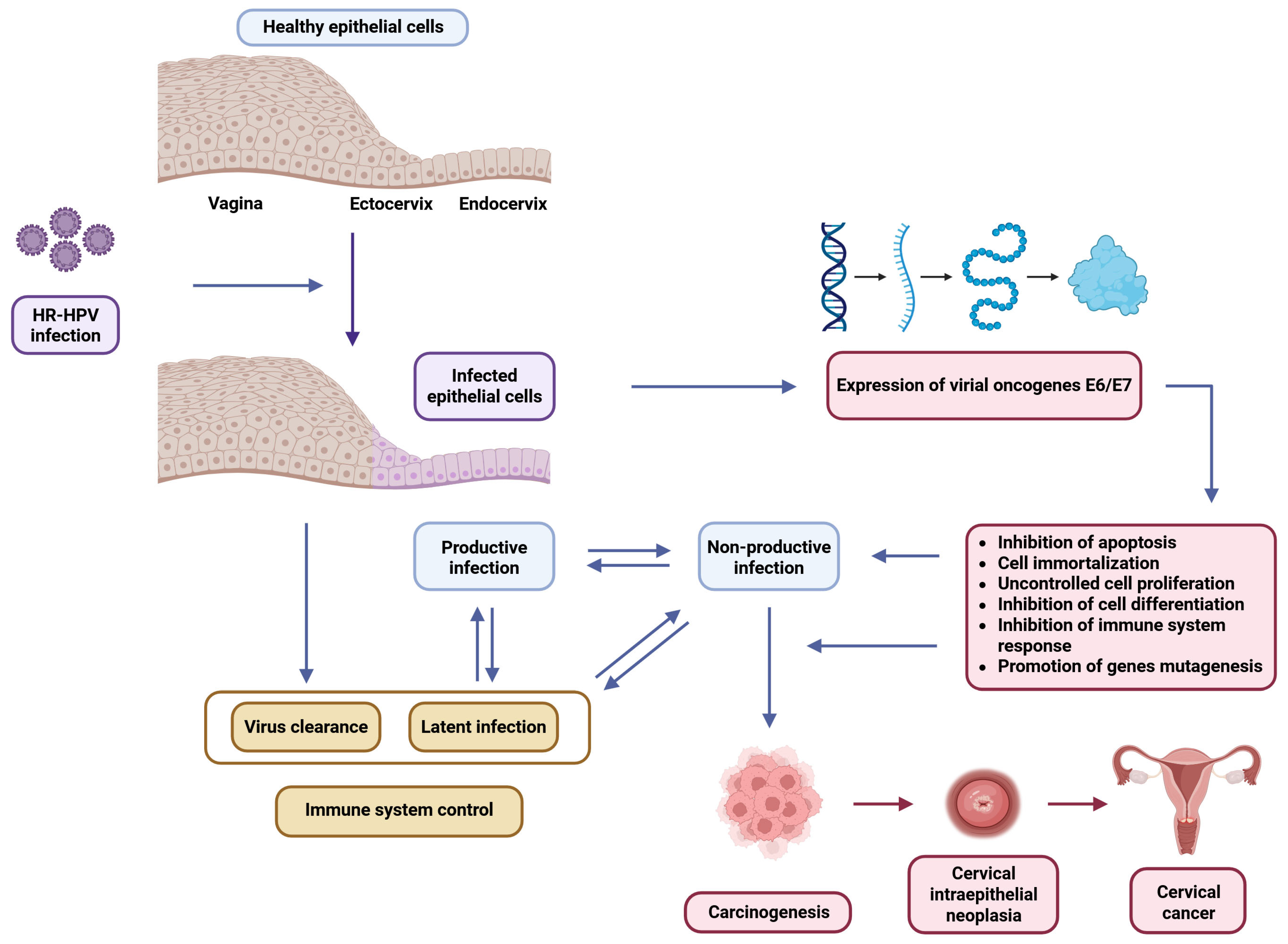
Figure 1. Schematic representation of the progression of a high-risk (HR)-HPV infection in epithelial cells. Following the initial infection, the virus may cause a subclinical infection leading to either productive or non-productive lesions. Outcomes depend on immune control and may include viral clearance, a latent infection, or progression to carcinogenesis. The dysregulation of E6/E7 oncogene expression plays a key role by promoting cell immortalization, abnormal proliferation, the inhibition of apoptosis and differentiation, immune evasion, and gene mutations.
Biology 14 01081 g001
Figure 2. The figure illustrates the protective mechanisms by which Lactobacillus species act against HPV infection and cervical cancer progression. Lactobacillus enhances epithelial barrier integrity by increasing E-cadherin levels and promotes antiviral immunity by upregulating IFN-α and IFN-β through NF-κB signaling. It reduces chronic inflammation by lowering IL-6 and IL-1β levels and supports immune restoration via butyrate and IRF3 pathways. Certain strains suppress HPV oncogenes E6/E7 and inhibit MMP9 expression, limiting invasion and metastasis. Lactobacillus also modulates Wnt/β-catenin signaling, preventing nuclear β-catenin accumulation and oncogene activation. Additionally, it regulates host miRNA expression—downregulating oncogenic miRNAs and upregulating tumor-suppressive ones—enhancing apoptosis and reducing inflammation.
Biology 14 01081 g002
Disclaimer/Publisher’s Note: The statements, opinions and data contained in all publications are solely those of the individual author(s) and contributor(s) and not of MDPI and/or the editor(s). MDPI and/or the editor(s) disclaim responsibility for any injury to people or property resulting from any ideas, methods, instructions or products referred to in the content.