Article

Integrative Transcriptomic Profiling Identifies TNF and IL1B as Candidate Key Early-Response Genes in Macrophages Infected with Smooth Brucella Using a Comprehensive Bioinformatic Approach

Faculty of Animal Science and Technology, Yunnan Agricultural University, Kunming 650201, China
*
Author to whom correspondence should be addressed.
Biology 2025, 14(5), 579; https://doi.org/10.3390/biology14050579
Submission received: 8 April 2025 / Revised: 1 May 2025 / Accepted: 19 May 2025 / Published: 21 May 2025
(This article belongs to the Special Issue Zoonotic Diseases)

Simple Summary

Brucella spp. are classic zoonotic intracellular pathogens. Infection in livestock such as cattle, sheep, and pigs often causes abortion and infertility, while in humans, it manifests as arthritis or Mediterranean fever, posing serious threats to both public health and livestock industry development. The smooth phenotype Brucella represents a crucial virulent form that establishes chronic infections in hosts. Therefore, elucidating the invasion mechanisms of smooth Brucella holds significant value for brucellosis diagnosis and treatment, particularly in deciphering the critical molecular events triggered during early infection. Our study employed a cross-Brucella species analysis strategy, integrating gene expression profiles from macrophages infected with smooth phenotype B. melitensis and B. suis strains through comprehensive bioinformatics approaches. We identified key early transcriptional response events in infected macrophages, including immune-related pathways and genes. These findings provide valuable insights into the transcriptional regulatory mechanisms during initial Brucella–macrophage interaction, advancing our understanding of brucellosis pathogenesis.

Abstract

Smooth Brucella are the main pathogenic bacteria that threaten human health and food safety. The early stage of smooth Brucella and macrophage interaction is an important phase, and smooth Brucella species elicit a dramatic transcriptional response in infected macrophages. However, the key transcriptional events are still obscure. This study aimed to identify key candidate response pathways and genes in macrophages infected with smooth Brucella at the early interaction stage. Three gene expression profiles including GSE21117, GSE5202, and GSE8385 were retrieved from the NCBI GEO database, and were integrated using comprehensive bioinformatics methods including gene set enrichment analysis, differentially expressed gene analysis, protein and protein interaction (PPI) network construction, and transcription factor prediction. The results showed that 16 up-regulated and 22 down-regulated pathways were identified, including six up-regulated immune-related pathways. A total of 41 up-regulated and four down-regulated genes were identified, and a PPI network including 31 nodes and 134 edges was constructed based on the interactive information of 45 dysregulated genes. A highly correlated module comprising 19 nodes and 103 edges was identified based on the topological features of the whole PPI network. Seven centrality analyses revealed that Tnf and Il1b were essential genes in the highly correlated module, and that the two essential genes were simultaneously enriched in eight significantly up-regulated pathways (including two immune-related pathways). Bcl3 was predicted as a transcription factor in the highly correlated module, and may play regulatory roles in the transcription of Tnf and Il1b genes. The present study identified Tnf and IL1b as candidate key response genes in infected macrophages at the early stage of smooth Brucella and macrophage interaction, which contributes to a deeper understanding of the early key transcriptional events in macrophages infected with smooth Brucella species.

1. Introduction

Brucella spp. are Gram-negative facultative intracellular bacteria, and produce brucellosis in a variety of animal species including humans, pigs, sheep, goats, and cattle [1,2]. In humans, brucellosis causes a spectrum of clinical manifestations, ranging from common symptoms such as fever, fatigue, and joint pain to severe complications including endocarditis and neurological disorders [3]. Brucellosis has been globally acknowledged as a substantial public health concern, yet the precise annual incidence rate remains undetermined [4]. Recent studies demonstrate a higher annual incidence, with 1.6–2.1 million new brucellosis cases occurring worldwide [2,5], especially in the Middle East, Mediterranean, Asia, Africa, and America [2]. In domestic animals, brucellosis is closely associated with reproductive losses, often causing abortion and sterility in animals [6,7]. Annually, brucellosis imposes substantial economic burdens on both the healthcare and agricultural sectors in many countries, such as China and India [8,9]. Brucellosis seriously threatens human health and impedes agricultural economic development; elucidating its pathogenesis is crucial for treatment and prevention and the advancement of livestock production.
Currently, more than ten Brucella species have been reported [7], including Brucella suis (B. suis), Brucella ovis (B. ovis), Brucella abortus (B. abortus), Brucella canis (B. canis), Brucella melitensis (B. melitensis), Brucella neotomae (B. neotomae), Brucella ceti (B. ceti), Brucella pinnipedialis (B. pinnipedialis), Brucella microti (B. microti), Brucella inopinata (B. inopinata), Brucella papionis (B. papionis), and Brucella vulpis (B. vulpis). Among these Brucella species, four are pathogenic to humans, including B. suis, B. melitensis, B. abortus, and B. canis [10]. Their primary animal hosts are swine (B. suis), sheep and goats (B. melitensis), cattle (B. abortus), and dogs (B. canis), respectively [11,12]. Among the four Brucella species, B. melitensis and B. suis exhibit the highest pathogenicity, responsible for the most severe and moderately severe cases of brucellosis, respectively, whereas B. abortus and B. canis show moderate virulence and typically cause milder symptoms of the disease [10]. Given the significant health impact of brucellosis, particularly in patients with underlying cardiopulmonary conditions, the cornerstone of brucellosis management lies in rapid disease control to prevent complications and relapse. Consequently, accurate diagnosis, especially during the early (acute) phase, is of paramount importance. However, the non-specific clinical manifestations of brucellosis, which mimic other bacterial and viral infections, frequently lead to misdiagnosis in clinical practice. This often results in delayed treatment during the acute phase, allowing progression to chronic infection with multiple complications, and consequently, poorer prognosis [3]. Presently, the prevention and treatment of brucellosis primarily rely on vaccination and antibiotic therapy [2]. However, available vaccines exhibit inadequate protective efficacy and safety concerns, necessitating the development of safer and more effective vaccines. The establishment of early, rapid, and accurate diagnostic methods would enable combined vaccine–antibiotic therapy to serve as an effective strategy against brucellosis [10], but this strategy first demands the elucidation of Brucella’s infection mechanisms.
B. melitensis and B. suis are the two most pathogenic Brucella species, and exhibit strikingly similar genomic characteristics, with nearly identical genetics and gene organization [13]. Previous studies have shown that the open reading frames (ORFs) have 99% or higher sequence identity between B. melitensis and B. suis [13]. Despite their high genomic similarity, the two Brucella species exhibit distinct host tropisms. Genomic variations, including some insertion–deletion (indels) and polymorphic events encoding putative outer membrane proteins, may account for these host specificity differences. On the other hand, while the two Brucella species display different host specificities in animals, they share the common capability to cause human infections, which indicates that both B. melitensis and B. suis possess not only species-specific infection mechanisms, but also shared pathogenic strategies. The pathogenesis of Brucella species primarily relies on its virulence determinants, such as lipopolysaccharide (LPS), the type IV secretion system (T4SS), and the BvrR/Bvrs system [7]. Some studies have revealed that B. melitensis and B. suis share common virulence mechanisms, such as the T4SS and LPS [14,15,16,17], while exhibiting potential distinct pathogenic strategies [1,18]. Despite advances in understanding the pathogenesis of Brucella species, the pathogen’s unique absence of classical virulence factors such as exotoxins, cytolysins, and exoenzymes [2] results in exceptionally intricate infection mechanisms, with numerous unresolved questions remaining. It is vital to uncover the consistent infection characteristics for a greater understanding of the infection mechanisms. Therefore, further mechanistic studies are urgently needed.
Macrophages are the main targets of Brucella species, and attempt to eliminate the invading bacteria by triggering defense mechanisms. Once the target macrophages are infected, Brucella species enter the infected macrophages [19]. When Brucella species circumvent macrophage killing, Brucella species enter the intracellular life cycle and dramatically replicate in infected macrophages. Smooth Brucella species with an intact lipopolysacharide (LPS) O side chain is one major phenotype of Brucella species [20]. Unlike rough Brucella species, smooth Brucella species have been implicated in the virulence of target macrophages, producing enormous effects on animal immune systems and affecting disease processes [21]. Previous studies have demonstrated that smooth Brucella species inhibited macrophage apoptosis and impaired cytokine production [18,22,23], which facilitates the survival and replication of smooth Brucella species in infected macrophages. Furthermore, in vitro studies have found that the early stage of smooth Brucella species and macrophage interaction was the key phase, and that the majority of smooth Brucella species were killed and caused a more dramatic transcriptional response in the infected macrophages [20,24]. However, a small subset of Brucella species can evade immune surveillance, utilizing macrophages as host cells for intracellular survival and reproduction [25]. The persistent interaction forces the host to enter a chronic infection stage [20,25]. Studies have partially demonstrated that Brucella survive in macrophages by inhibiting the fusion of Brucella-containing vacuoles (BCVs) with lysosomes and by altering the maturation process of BCVs along the endocytic pathway [26,27]. However, the nature of the replicative compartment of Brucella in macrophages remains controversial [28]. Some evidence suggests that Brucella replicates within compartments exhibiting phagolysosomal characteristics [27], but this would imply the completion of phagosome maturation—a notion that appears contradictory to the observed inhibition of BCV–lysosome fusion. Thus, it is critical to clarify the interaction mechanism for a better understanding of the establishment of a chronic Brucella species infection at the early infection stage. Several independent studies have used gene expression profiles to identify key genes in the infected macrophages at the early stage of macrophage and smooth Brucella species interaction [1,18], and hundreds of differentially expressed genes (DEGs) were identified. However, comparative analysis is still lacking, and identifying consistent DEGs via integrative analysis is urgently required.
Increasing datasets enable the systematic identification of conserved key genes inside the macrophages infected with smooth Brucella species at the early infection stage. In this study, we integrated three gene expression profiles obtained from the Gene Expression Omnibus (GEO) database of the National Center for Biotechnology Information (NCBI) to investigate the response mechanism of macrophage and smooth Brucella species interaction at the transcriptional level using comprehensive bioinformatic methods including gene set enrichment analysis (GSEA), differentially expressed gene analysis (DEGA), gene ontology (GO) analysis, protein and protein interaction (PPI) network analysis, highly correlated module analysis, and centrality analysis. The present study identified candidate key response genes in macrophages infected with smooth Brucella species at the early interaction stage, which contributes to a deeper understanding of the early response mechanisms of macrophages against smooth Brucella species infection.

2. Materials and Methods

2.1. Data Collection

The gene expression profiles were obtained from the NCBI GEO database as follows (http://www.ncbi.nlm.nih.gov/geo/, (accessed on 10 March 2018)): (1) phenotype for smooth Brucella species; (2) target cell line for macrophage; and (3) data with the same interaction stage. Finally, two gene expression profiles with accession numbers GSE21117 and GSE5202 were retrieved [1,18]. Two microarray data were separately produced using the GPL1261 platform (Affymetrix Mouse Genome 430 2.0 Array). In this study, two subsets related to the early interaction stage from GSE21117 and GSE5202 were selected to be integrated and reanalyzed. Each subset contained 3 infected macrophages samples (4 h time point post infection) and 3 uninfected macrophages samples (normal). The data subset related to B. melitensis in the GSE8385 dataset was retrieved to validate the results [22]. The main information about these subsets, such as the cell line, sample size, and infection time, was listed in Table 1.

2.2. Data Preprocessing

To improve the efficiency of data reanalyzing, all data were reprocessed using the same criteria. The data reprocessing was performed using the packages in the Bioconductor project (version 3.6, http://www.bioconductor.org/, (accessed on 16 March 2018)) based on R language [29]. All datasets were background-adjusted and normalized, and log2 probe-set intensities were calculated using the Robust Multichip Averaging (RMA) algorithm in the affy package (version 1.56.0) [30,31]. The interquartile range (IQR) was used to measure the data variability. In order to optimally preserve the functionally relevant low-abundance transcripts while ensuring analytical robustness, an IQR threshold of 0.5 was set based on the resulting distribution of the IQR values for all genes. Genes exhibiting IQR values < 0.5 were filtered out. The preprocessed data were used to perform the DEGA and GSEA.

2.3. DEGA of DEGs

The DEGs were identified using the limma package (version 3.32.7) in the Bioconductor project [32]. The limma package employs the voom method, liner modeling, and empirical Bayes moderation to assess the differential gene expression and can acquire more robust results, even in fewer microarrays. A false discovery rate (FDR) < 5% and a linear fold change >2 or <0.5(|logFC| > 1) were used as the cutoff criteria. The functional analyses of identified DEGs, including the GO and Reactome pathway (RP), were investigated using the online STRING database (version 11.0, https://string-db.org/, (accessed on 8 June 2018)) [33].

2.4. GSEA of KEGG Pathways

The GSEA of KEGG pathways was performed using the category package (version 2.34.2) in the Bioconductor project [34]. The purpose of performing GSEA was to determine whether the members of a gene set S were randomly distributed throughout the entire reference gene list L or were principally found at the top or bottom. An obvious merit of GSEA was the relative robustness to the noise and outliers in the data. For the GSEA of pathways, when multiple probe sets targeted the same gene, the probe set with the largest variability was kept for the next analysis, and the other probe sets were discarded. The gene sets with less than 10 genes were removed from the GSEA results. The t-statistic mean of the genes was computed in each KEGG pathway. A permutation test with 1000 times was implemented, and the KEGG pathways with p < 0.05 were identified to significantly change in the macrophages infected with smooth Brucella species [34].
To elucidate the interactive relationships between pathways and genes, a pathway–gene network was graphically represented using the open-source software platform Cytoscape software (version 3.7.0, http://www.cytoscape.org/, (accessed on 20 December 2018)) [35].

2.5. PPI Network and Analysis

The interaction relationships among DEGs encoding proteins were analyzed using a PPI network. PPI information was obtained using the online STRING database (version 11.0, https://string-db.org/, (accessed on 8 June 2018)) [33]. To evaluate the reliability of the predicted associations, the interaction score integrating multiple types of interaction evidence was utilized. To maintain prediction confidence and reduce false positive rates, we established an interaction score threshold of 0.4 (false positive rates < 0.15) based on the benchmarking standards specified in the published article [33]. A PPI network was constructed using Cytoscape software [35]. The PPI subnetwork (highly correlated module) was extracted from the whole PPI network using a Molecular COmplex DEtection (MCODE) algorithm based on the topological properties of whole PPI network, and a plugin MCODE (version 1.5.1) in the Cytoscape software was used to perform the MCODE analysis [36]. The threshold parameters were severely set for the Degree Cutoff = 6, Node Score Cutoff = 0.6, K-Core = 2, and Max. Depth = 100. The PPI subnetwork was used to perform a centrality analysis and transcription factor analysis. Seven centrality analyses were used to identify the core genes, including the Subgraph centrality, Degree centrality, Eigenvector centrality, Betweenness centrality, Network centrality, Information centrality, and Closeness centrality. Seven centrality metrics for each gene were computed based on their topological characteristics within the network, with detailed calculation methods for each metric provided in the published article [37]. Higher centrality scores indicate the greater functional importance of genes in the network, and all centrality analyses were performed using a plugin CytoNCA (version 2.1.6) in the Cytoscape software [37]. The genes with higher scores from each centrality method were identified as key genes, and the intersecting genes of key genes obtained via seven centrality methods were identified as essential genes. Transcription factor analysis was performed using an iRegulon plugin in the Cytoscape software and was used to identify the transcription factor in the highly correlated PPI subnetwork [38].

3. Results

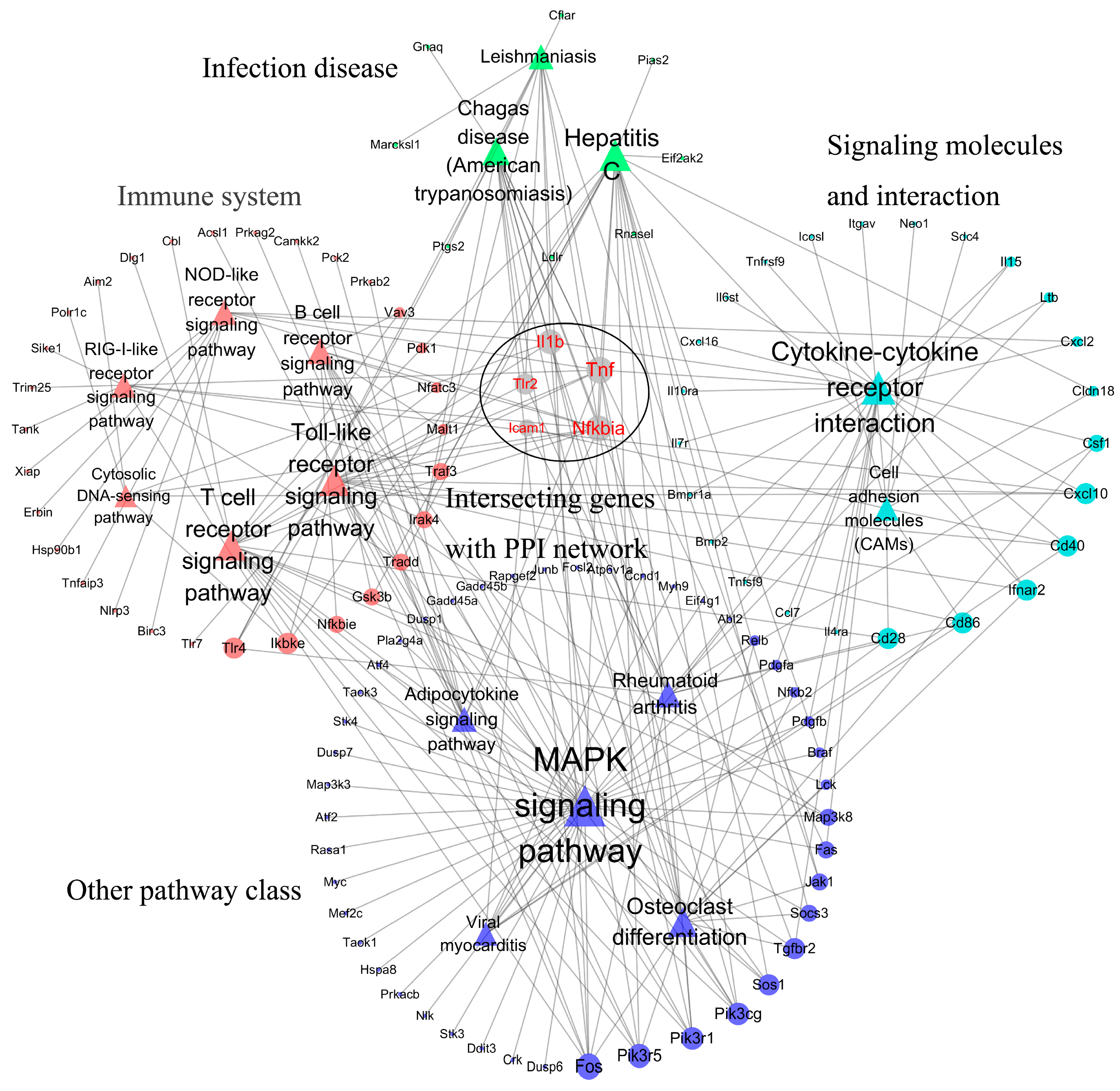
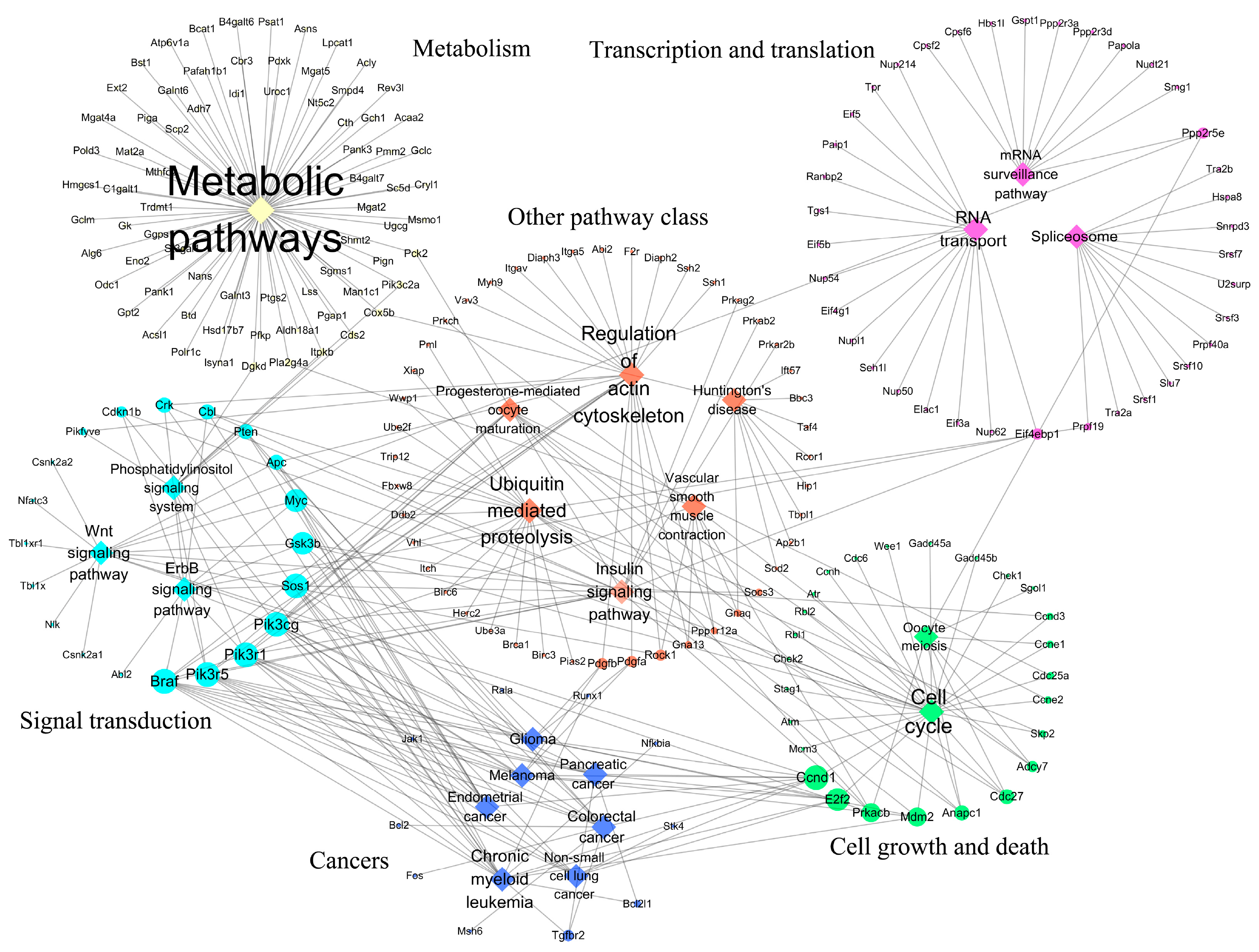
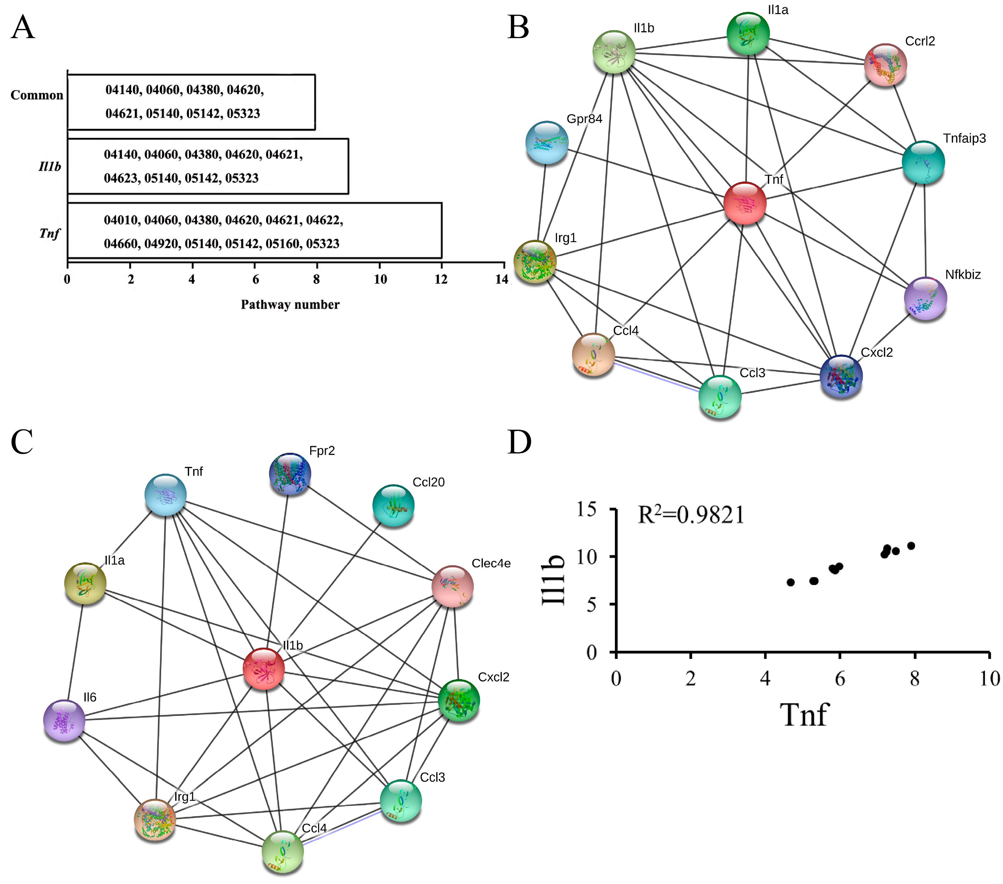
3.1. KEGG Pathways Identification

According to p < 0.05, significantly regulated KEGG pathways were identified inside the infected macrophages at an early stage. In GSE21117, 21 up-regulated and 36 down-regulated pathways were identified (Table S1), and 57 up-regulated and 87 down-regulated pathways were identified in GSE5202 (Table S2). An overlap analysis showed that 16 common up-regulated and 22 common down-regulated pathways were identified as key response KEGG pathways (Figure 1). The common pathways were listed in Table 2.
The 16 up-regulated pathways were primarily associated with the immune system (including six pathways) and infectious diseases (including three pathways). The six up-regulated immune-related pathways were, separately, the toll-like receptor signaling pathway, NOD-like receptor signaling pathway, RIG-I-like receptor signaling pathway, cytosolic DNA-sensing pathway, T cell receptor signaling pathway, and B cell receptor signaling pathway. Importantly, four of these pathways were identified to significantly up-regulate in the GSE8385 dataset, including the toll-like receptor signaling pathway, NOD-like receptor signaling pathway, RIG-I-like receptor signaling pathway, and cytosolic DNA-sensing pathway.
The 22 key down-regulated pathways were mainly related to metabolism (including one global metabolic pathway), cancers (including seven pathways), cell growth and death (including two pathways), signal transduction (including three pathways), and translation (including two pathways). Two down-regulated pathways belonging to cell growth and death were separately cell cycle and oocyte meiosis.
To elucidate the interactive relationships among pathways and genes, the relational network among pathways and genes was established. Two relational networks including up-regulated and down-regulated pathways are shown in Figure 2 and Figure 3. In the up-regulated pathway–gene network, the immune-related pathways included most of the genes retrieved. In addition, five immune-related genes including Tnf, Nfkbia, Il1b, Tlr2, and Icam1 participated in many of the pathways retrieved. In the down-regulated pathway–gene network, five pathway classes were identified, and metabolic pathways contained most of the genes retrieved.

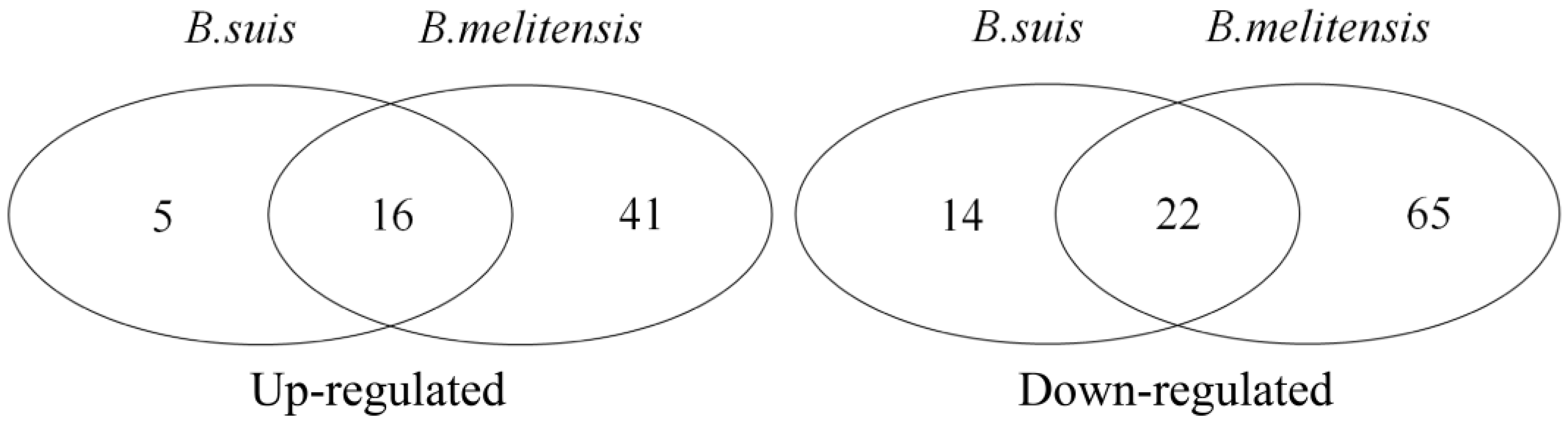
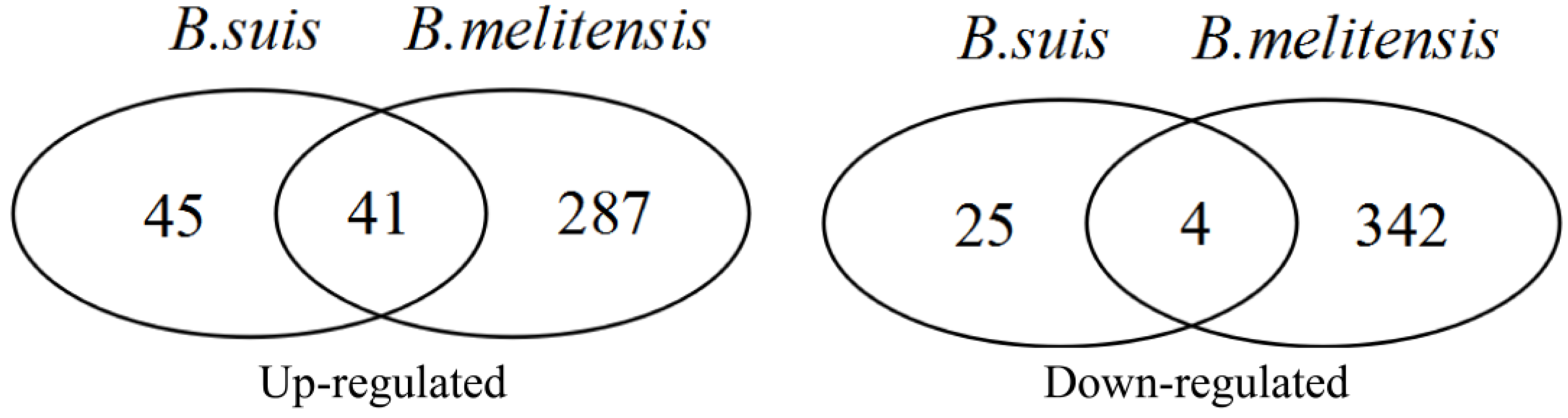
3.2. DEG Identification and Functional Analyses

DEGAs of two expression profiles including GSE21117 and GSE5202 were separately implemented using the limma package. According to FDR < 0.05 and |logFC| > 1 cutoff criteria, differentially expressed probes (DEPs) and DEGs corresponding to DEPs were identified. In the GSE21117 dataset, 117 up-regulated and 35 down-regulated DEPs were identified, and 86 up-regulated and 29 down-regulated DEGs were identified (Table S3). In the GSE5202 dataset, 444 up-regulated and 400 down-regulated DEPs corresponding to 328 up-regulated and 346 down-regulated DEGs were identified (Table S4). An overlap analysis showed that 54 common up-regulated and four common down-regulated DEPs (Table 3), corresponding to 41 common up-regulated and four common down-regulated DEGs were identified (Table 3, Figure 4). Among 45 common dysregulated genes (41 up- and four down-regulated), a transcription factor Bcl3 (B-cell lymphoma 3) was identified.
GO analysis showed that 45 dysregulated genes were significantly enriched in 797 GO terms related to the biological process (BP) (Table S5), and the majority of significantly enriched BPs were associated with the biological process and regulation. The top five BPs with the most significant p value were cellular processes (GO:0009987, p = 4.50 × 10−115), the regulation of biological processes (GO:0050789, p = 3.30 × 10−108), the regulation of cellular processes (GO:0050794, p = 3.95 × 10−105), response to stimulus (GO:0050896, p = 8.62 × 10−102), and the positive regulation of biological processes (GO:0048518, p = 1.27 × 10−90).
RP analysis showed that 45 dysregulated genes were significantly enriched in 45 RPs (Table S6), and that the top five RPs with the most significant p value were mainly related to the immune system. The top five RPs were, separately, signal transduction (MMU-162582, p = 2.32 × 10−37), immune system (MMU-168256, p = 5.70 × 10⁻³²), death receptor signaling (MMU-73887, p = 2.22 × 10⁻¹⁵), adaptive immune system (MMU-1280218, p = 2.22 × 10⁻¹⁵), and cytokine signaling in the immune system (MMU-1280215, p = 2.22 × 10⁻¹⁵).

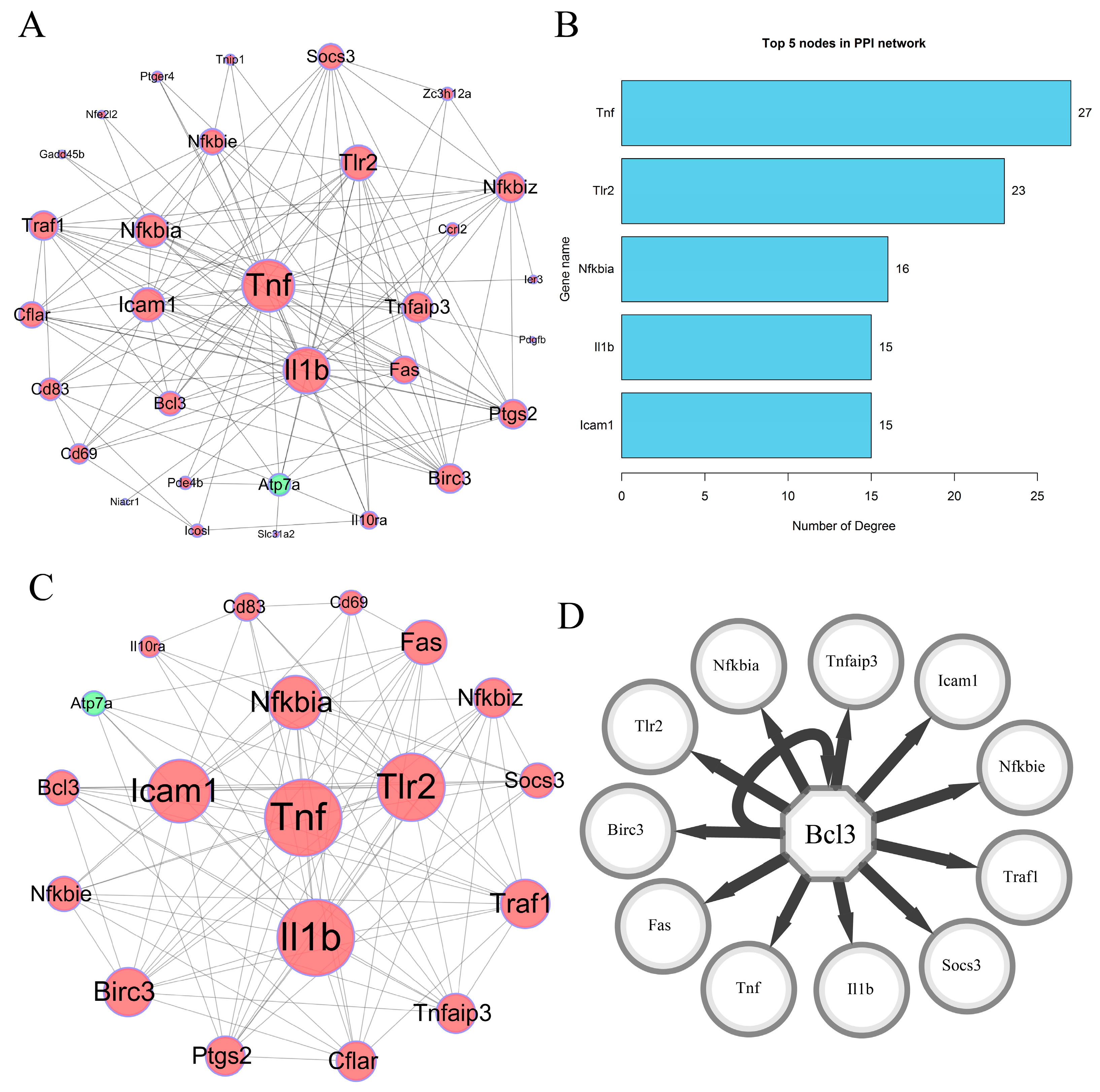
3.3. PPI Network Construction

To reveal the interactive relationships of 45 dysregulated DEGs, a PPI network including 45 dysregulated DEGs was constructed according to the interactive information from the STRING database. At a combined score > 0.4, 31 DEGs (including 30 up-regulated genes and one down-regulated gene) had 134 interactive relationships, and a PPI network including 31 nodes and 134 edges was established (Figure 5A). Node degree analysis showed that the top five genes with the most links were, separately, Tnf-α (logFC = 2.94, p = 2.89 × 10⁻³ in GSE21117 and logFC = 1.79, p = 2.33 × 10⁻⁸ in GSE5202), Il1b (logFC = 1.96, p = 1.26 × 10⁻² in GSE21117 and logFC = 1.47, p = 6.80 × 10⁻⁸ in GSE5202), Tlr2 (logFC = 1.81, p = 1.02 × 10⁻⁵ in GSE21117 and logFC = 2.17, p = 2.36 × 10⁻⁹ in GSE5202), Icam1(logFC = 1.95, p = 1.10 × 10⁻⁴ in GSE21117 and logFC = 1.57, p = 1.43 × 10⁻⁷ in GSE5202), and Nfkbiα (logFC = 1.56, p = 1.02 × 10⁻³ in GSE21117 and logFC = 1.57, p = 1.66 × 10⁻⁸ in GSE5202). The degrees of these five genes were 27 (Tnf-α), 23 (Il1b), 16 (Tlr2), 15 (Icam1), and 15 (Nfkbiα) in the PPI network, respectively (Figure 5B).

3.4. Highly Correlated Module Analysis and Essential Gene Identification

To identify the key response genes at the early stage of smooth Brucella species and macrophage interaction, highly correlated module and centrality analyses were performed. The results showed that one highly correlated module with 19 nodes and 103 edges (Score = 11.444) was identified (Figure 5C). Centrality analysis showed that up-regulated Tnf and Il-1b were the top two genes based on comprehensive scores across seven centrality methods (Table 4), and were identified as candidate key response genes. The GSE8385 dataset showed that the two genes were significantly up-regulated. Transcription factor analysis showed that Bcl3 (NES = 12.212) was predicted as a transcription factor of 12 target genes, including Tnf and Il1b (Figure 5D).
Furthermore, two essential genes, Tnf and Il-1b, were found to be enriched in twelve and nine pathways among sixteen common up-regulated pathways, respectively (Figure 6A). An overlap analysis showed that eight pathways were common (Figure 6A) and two pathways were related to the immune system. The two immune-related pathways were the toll-like receptor signaling pathway and NOD-like receptor signaling pathway. In addition, the two essential response genes were observed to be significantly enriched in the top five GO terms (Table S5), including cellular processes (GO:0009987), the regulation of biological processes (GO: 0050789), the regulation of cellular processes (GO: 0050794), response to stimulus (GO:0050896), and the positive regulation of biological processes (GO:0048518).
Network analysis on the basis of single genes showed that the two essential response genes had a stronger interactive relationship (Figure 6B,C). Pearson correlation analysis showed that the two essential response genes had an extremely strong positive correlation in expression in the normal and infected J774.A1murine cell lines (R2 = 0.9821, Figure 6D).

4. Discussion

The interaction of smooth Brucella species and macrophages is a dynamic process [1]. Studying molecular events is critical for understanding the pathogenesis of brucellosis in the process of smooth Brucella species and macrophage interaction. Especially, it is key to elucidate the early macrophage transcriptional response elicited by smooth Brucella species for understanding how a chronic infection is established [1]. B. melitensis and B. suis, as the two most detrimental Brucella species to both human health and the livestock industry, necessitate the urgent elucidation of their infection mechanisms. Previous single-species studies have identified some DEGs at the early stage of smooth Brucella species and macrophage interaction, advancing our understanding of brucellosis pathogenesis [1,18]. Nevertheless, the remarkable diversity and unique infectious strategies of Brucella species necessitate the elucidation of conserved infection mechanisms to fully decipher their distinct pathogenicity. Integrated cross-species analysis represents a powerful yet underutilized approach to address this knowledge gap. Currently, such comparative studies remain scarce. To bridge this gap, we performed a comprehensive bioinformatics analysis of two gene expression profiles, combining DEGA, GSEA, and GO analysis, PPI network construction, and highly correlated module and centrality analyses. Through a multi-dimensional bioinformatics analytical framework (differentially expressed gene identification→Gene functional enrichment analysis→Gene interaction network construction→Network topological feature characterization→Transcription factor prediction), we systematically investigated the translational events during the early-stage interactions between Brucella species and macrophages. Finally, 16 key up-regulated and 22 key down-regulated pathways were identified, including six key immune-related pathways. A total of 41 up-regulated and four down-regulated DEGs were identified. A PPI network with 31 nodes and 134 edges was established, and a highly correlated module with 19 nodes and 103 edges was extracted from the whole PPI network. Tnf and Il1b were identified as the candidate essential early-response genes of smooth Brucella and macrophage interaction. The findings indicate that six key immune-related pathways and two essential genes may play roles at the early stage of smooth Brucella species and macrophage interaction.
The six significantly up-regulated immune-related pathways were, separately, the toll-like receptor signaling pathway, NOD-like receptor signaling pathway, RIG-I-like receptor signaling pathway, cytosolic DNA-sensing pathway, T cell receptor signaling pathway, and B cell receptor signaling pathway. The toll-like receptor signaling pathway is a vital biological pathway generating innate immune responses and developing adaptive immunity [39,40]. In the pathway, the key genes toll-like receptors (TLRs) play critical roles in host resistance to infection, such as Brucella species infection [41,42]. For example, TLR6 was able to independently trigger an innate immune response against B. abortus, and further cooperated with TRL2 to activate the NF-kB signaling pathway [43]. Our study demonstrated that some TLRs, including Tlr2, were actively regulated in response to Brucella species, which indicates the important role of the toll-like receptor signaling pathway. The RIG-I-like receptor signaling pathway is an important pathway triggering an innate immune response by detecting viral pathogens [44,45,46]. Most published results showed that the RIG-I-like receptor signaling pathway plays a vital role in RNA virus recognition [47,48]. Lately, some studies have shown that the RIG-I-like receptor signaling pathway is a universal mechanism in defending against bacterial infection [49,50]. For example, Yersinia pestis infection induced the up-regulation of numerous genes in the pathway, including critical RIG-I-like receptors such as RIG-I and MDA5 [50]. Despite these findings, no published studies reported the role of the pathway in macrophages defending against Brucella species. Our study showed that the RIG-I-like receptor signaling pathway was significantly up-regulated by smooth Brucella species induction, which indicates that the pathway may play an important role in triggering immune responses against smooth Brucella species. For many years, the cytosolic DNA-sensing pathway has been appreciated because exogenous cytosolic DNA could evoke a type I interferon response [51,52]. During host infection, most DNA-containing microbes released DNA into the cytoplasm and induced DNA immunity [53,54]. In this study, the cytosolic DNA-sensing pathway was up-regulated, which indicates that killed smooth Brucella species were disintegrated and released DNA into the infected macrophages to induce DNA immunity at the early interaction stage of smooth Brucella species and macrophages [55].
Many studies have focused on the key response genes in the processes of smooth Brucella species and macrophage interaction, and some DEGs have been identified as being involved in the interaction responses [1,18,22]. Some studies reported that two critical immune genes, Tnf-α and Il1b, were up-regulated at the early stage of smooth Brucella species and macrophage interaction via a DEGA method [1,56]. Our results further confirmed via a comprehensive bioinformatic method that the two genes played key roles as essential response genes between smooth Brucella species and macrophage interaction. As we know, Tnf-α is a cytokine mainly secreted by activated macrophages, and involved in systemic inflammation in an acute phase reaction, inducing cell death [57]. So, Tnf-α is usually used for anti-cancer therapy [57]. Recently, more and more studies have shown that Tnf-α functions in activating defense mechanisms [58,59]. Some studies have shown that Tnf- α was able to control the number of Brucella species in BALB/c mice [60], and that the production of Tnf-α was triggered through a Tlr2-dependent pathway in response to B. abortus [61]. Mentioned above, Tlr2 is a membrane protein playing a key role in innate immunity by recognizing bacterial lipoproteins and other microbial components. In our study, Tlr2 was identified to significantly up-regulate, which further demonstrates that Tnf-α may play a defense role through Tlr2. Il1b, a member of the interleukin 1 family, is an important mediator of the inflammatory response produced by activated macrophages [18]. A large number of studies have proven the critical roles of Il1b in regulating immune responses [62,63]. In this study, Il1b was identified as an essential response gene via systematic analysis, which indicates that Il1b may play a key immune role in macrophages against Brucella species at the early infection stage. Two up-regulated genes related to immunity and inflammation may contribute to clearing smooth Brucella species at the early infection stage [18]. However, it should be noted that these two cytokine genes may exert dual roles during host defense against Brucella infection. While their pro-inflammatory effects contribute to bacterial clearance, sustained overexpression may lead to excessive inflammation and subsequent tissue damage. Therefore, during early infection, vaccines or adjuvants could be utilized to up-regulate their expression to control Brucella replication. Conversely, in later stages, the moderated down-regulation of these cytokines may help to mitigate tissue damage (e.g., arthritis) while maintaining adequate bacterial containment.
Transcription factor Bcl3, belonging to the I kappa B protein family, is the major regulator of the NFκB signaling pathway [64]. Bcl3 protein contains ankyrin repeat domains and transactivation domains, and has the ability to bind and regulate specific NF-κB dimers [65]. Bcl3 has been identified as a candidate proto-oncogene [66], while accumulating evidence suggests its potential tumor-suppressive functions [67]. The dual role of Bcl3 in tumorigenesis depends on the cellular and environmental context [67]. Recent studies have shown that Bcl3 plays roles in regulating immunity and inflammation [65], and has an anti-apoptotic effect [68,69,70]. For example, Bcl3 can reduce the sterile inflammatory response in pancreatic and biliary tissues [71]. Bcl3 mediates cell proliferation by inducing PD-L1 expression in ovarian cancer [72]. Currently, no studies have demonstrated direct associations between Bcl3 and Brucella infection. Several studies have shown regulatory relationships between Bcl3 and Tnf/Il1b [67,73,74]. For example, Bcl3 promotes Tnf-induced hepatocyte apoptosis, and plays a role in regulating Tnf-induced hepatic cell death [73]. The expression of Tnf is oscillatory along the estrous cycle in ICR mice, and its expression level is negatively associated with the presence of Bcl3 [74]. In this study, Bcl3 was predicted as a transcription factor to play roles in regulating Tnf and Il1b expression, but its precise regulatory mechanism remains unclear. Based on existing research evidence [69,70], we hypothesize that up-regulated Bcl3 may inhibit macrophage apoptosis during the early stage of Brucella infection. Future studies should focus on elucidating the precise regulatory role of Bcl3 in Brucella infection mechanisms.
Collectively, on the basis of a comprehensive bioinformatic analysis, we systematically investigated the transcriptional response events in macrophages infected with smooth Brucella species at an early interaction stage. The findings contribute to a better understanding of the transcriptional response mechanisms of macrophages against smooth Brucella species. Nevertheless, some limitations should be mentioned. First, the B. abortus-related gene expression data should have been included in this study. However, the lack of suitable datasets represents a significant limitation. Second, the functional roles of the two key candidate response genes Tnf and Il1b should be further investigated. Next, we will focus on the functional studies of two key candidate genes.

5. Conclusions

The study identified key candidate immune pathways and response genes in macrophages infected with smooth Brucella species during the early infection stage. The findings contribute to a deeper understanding of the early transcriptional response in macrophages infected with smooth Brucella species.

Supplementary Materials

The following supporting information can be downloaded at: https://www.mdpi.com/article/10.3390/biology14050579/s1, Table S1: Dysregulated pathways inside infected macrophages with smooth B. suis. Table S2: Dysregulated pathways inside infected macrophages with smooth B. melitensis. Table S3: Differentially expressed probes and genes inside infected macrophages with smooth B. suis. Table S4: Differentially expressed probes and genes inside infected macrophages with smooth B. melitensis. Table S5: GO terms enriched by 45 common differentially expressed genes. Table S6: Reactome pathways enriched by 45 common differentially expressed genes.

Author Contributions

Conceptualization, Q.C.; methodology, Q.C. and X.Y.; formal analysis, Q.C. and X.Y.; writing—original draft preparation, X.Y.; writing—review and editing, Q.C.; funding acquisition, Q.C. All authors have read and agreed to the published version of the manuscript.

Funding

This research was funded by the Xingdian Talent Support Program Fund (YNWR-QNBJ-2020-219). The funding agency had no role in the study design, data gathering, data analysis, or writing the manuscript.

Institutional Review Board Statement

Not applicable.

Informed Consent Statement

Not applicable.

Data Availability Statement

The gene expression profiles used to support the findings of this study were deposited in the NCBI GEO database, and were freely downloaded according to accession numbers GSE21117, GSE5202, and GSE8385.

Acknowledgments

The authors are grateful to Kunming University of Science and Technology and Yunnan Provincial Science and Technology Department for their financial support of this work.

Conflicts of Interest

The authors declared that the research was conducted in the absence of any commercial or financial relationships that could be construed as a potential conflicts of interest.

References

  1. Chen, F.; Ding, X.; Ding, Y.; Xiang, Z.; Li, X.; Ghosh, D.; Schurig, G.G.; Sriranganathan, N.; Boyle, S.M.; He, Y. Proinflammatory Caspase-2-Mediated Macrophage Cell Death Induced by a Rough Attenuated Brucella Suis Strain. Infect. Immun. 2011, 79, 2460–2469. [Google Scholar] [CrossRef] [PubMed]
  2. Qureshi, K.A.; Parvez, A.; Fahmy, N.A.; Hady, B.H.A.; Kumar, S.; Ganguly, A.; Atiya, A.; Elhassan, G.O.; Alfadly, S.O.; Parkkila, S.; et al. Brucellosis: Epidemiology, Pathogenesis, Diagnosis and Treatment—A Comprehensive Review. Ann. Med. 2023, 55, 2295398. [Google Scholar] [CrossRef] [PubMed]
  3. Jin, M.; Fan, Z.; Gao, R.; Li, X.; Gao, Z.; Wang, Z. Research Progress on Complications of Brucellosis. Front. Cell. Infect. Microbiol. 2023, 13, 1136674. [Google Scholar] [CrossRef]
  4. Laine, C.G.; Scott, H.M.; Arenas-Gamboa, A.M. Human Brucellosis: Widespread Information Deficiency Hinders an Understanding of Global Disease Frequency. PLoS Negl. Trop. Dis. 2022, 16, e0010404. [Google Scholar] [CrossRef]
  5. Laine, C.G.; Johnson, V.E.; Scott, H.M.; Arenas-Gamboa, A.M. Global Estimate of Human Brucellosis Incidence. Emerg. Infect. Dis. 2023, 29, 1789–1797. [Google Scholar] [CrossRef]
  6. Olsen, S.C.; Tatum, F.M. Swine Brucellosis: Current Perspectives. Vet. Med. 2017, 8, 1–12. [Google Scholar] [CrossRef]
  7. Glowacka, P.; Zakowska, D.; Naylor, K.; Niemcewicz, M.; Bielawska-Drozd, A. Brucella—Virulence Factors, Pathogenesis and Treatment. Pol. J. Microbiol. 2018, 67, 151–161. [Google Scholar] [CrossRef]
  8. Singh, B.B.; Kostoulas, P.; Gill, J.P.S.; Dhand, N.K. Cost-Benefit Analysis of Intervention Policies for Prevention and Control of Brucellosis in India. PLoS Negl. Trop. Dis. 2018, 12, e0006488. [Google Scholar] [CrossRef]
  9. He, H.-B.; Lu, Q.-S.; Wang, L.-L.; Aishan, M.; Zhao, J.-S.; Tang, X.-Y.; Zhu, M.-T.; Reheman, M.; Chen, Q.-L.; Zhang, Y.-P. The Economic Burden of Brucellosis Care in China: Socioeconomic Status Inequality. J. Trop. Med. 2024, 2024, 7992287. [Google Scholar] [CrossRef]
  10. Deng, Y.; Liu, X.; Duan, K.; Peng, Q. Research Progress on Brucellosis. Curr. Med. Chem. 2019, 26, 5598–5608. [Google Scholar] [CrossRef]
  11. Bukhari, E.E. Pediatric Brucellosis. An Update Review for the New Millennium. Saudi Med. J. 2018, 39, 336–341. [Google Scholar] [CrossRef] [PubMed]
  12. Brown, V.R.; Bowen, R.A.; Bosco-Lauth, A.M. Zoonotic Pathogens from Feral Swine That Pose a Significant Threat to Public Health. Transbound. Emerg. Dis. 2018, 65, 649–659. [Google Scholar] [CrossRef]
  13. Halling, S.M.; Peterson-Burch, B.D.; Bricker, B.J.; Zuerner, R.L.; Qing, Z.; Li, L.-L.; Kapur, V.; Alt, D.P.; Olsen, S.C. Completion of the Genome Sequence of Brucella Abortus and Comparison to the Highly Similar Genomes of Brucella Melitensis and Brucella Suis. J. Bacteriol. 2005, 187, 2715–2726. [Google Scholar] [CrossRef]
  14. Lei, L.; Wang, X.; Zhang, J.; Yin, J.; Xu, Q.; Wang, T.; Jin, Y.; Wang, A. Lipopolysaccharides of Brucella Suis S2 Impaired the Process of Decidualization in Early Pregnancy in Mice. Toxins 2023, 15, 662. [Google Scholar] [CrossRef]
  15. Sharifahmadian, M.; Arya, T.; Bessette, B.; Lecoq, L.; Ruediger, E.; Omichinski, J.G.; Baron, C. Monomer-to-Dimer Transition of Brucella Suis Type Iv Secretion System Component Virb8 Induces Conformational Changes. FEBS J. 2017, 284, 1218–1232. [Google Scholar] [CrossRef]
  16. Osman, A.Y.; Abdullah, F.F.J.; Kadir, A.A.; Saharee, A.A. Immuno-Pathophysiological Responses of Mouse Model to Experimental Infection with Brucella Melitensis and Its Lipopolysaccharides Via Intraperitoneal Route. Microb. Pathog. 2016, 100, 17–29. [Google Scholar] [CrossRef]
  17. Yin, Y.; Tian, M.; Zhang, G.; Ding, C.; Yu, S. A Novel Brucella T4ss Effector Rs15060 Acts on Bacterial Morphology, Lipopolysaccharide Core Synthesis and Host Proinflammatory Responses, Which Is Beneficial for Brucella Melitensis Virulence. Microbiol. Res. 2025, 292, 128015. [Google Scholar] [CrossRef]
  18. He, Y.; Reichow, S.; Ramamoorthy, S.; Ding, X.; Lathigra, R.; Craig, J.C.; Sobral, B.W.S.; Schurig, G.G.; Sriranganathan, N.; Boyle, S.M. Brucella Melitensis Triggers Time-Dependent Modulation of Apoptosis and Down-Regulation of Mitochondrion-Associated Gene Expression in Mouse Macrophages. Infect. Immun. 2006, 74, 5035–5046. [Google Scholar] [CrossRef]
  19. Celli, J. Surviving inside a Macrophage: The Many Ways of Brucella. Res. Microbiol. 2006, 157, 93–98. [Google Scholar] [CrossRef]
  20. Roop, R.M., 2nd; Bellaire, B.H.; Valderas, M.W.; Cardelli, J.A. Adaptation of the Brucellae to Their Intracellular Niche. Mol. Microbiol. 2004, 52, 621–630. [Google Scholar] [CrossRef]
  21. Dorneles, E.M.; Teixeira-Carvalho, A.; Araújo, M.S.; Sriranganathan, N.; Lage, A.P. Immune Response Triggered by Brucella Abortus Following Infection or Vaccination. Vaccine 2015, 33, 3659–3666. [Google Scholar] [CrossRef] [PubMed]
  22. Covert, J.; Mathison, A.J.; Eskra, L.; Banai, M.; Splitter, G. Brucella Melitensis, B. Neotomae and B. Ovis Elicit Common and Distinctive Macrophage Defense Transcriptional Responses. Exp. Biol. Med. 2009, 234, 1450–1467. [Google Scholar] [CrossRef]
  23. Gross, A.; Terraza, A.; Ouahrani-Bettache, S.; Liautard, J.-P.; Dornand, J. In Vitro Brucella Suis Infection Prevents the Programmed Cell Death of Human Monocytic Cells. Infect. Immun. 2000, 68, 342–351. [Google Scholar] [CrossRef]
  24. Celli, J.; de Chastellier, C.; Franchini, D.-M.; Pizarro-Cerda, J.; Moreno, E.; Gorvel, J.-P. Brucella Evades Macrophage Killing Via Virb-Dependent Sustained Interactions with the Endoplasmic Reticulum. J. Exp. Med. 2003, 198, 545–556. [Google Scholar] [CrossRef]
  25. Guo, X.; Zeng, H.; Li, M.; Xiao, Y.; Gu, G.; Song, Z.; Shuai, X.; Guo, J.; Huang, Q.; Zhou, B.; et al. The Mechanism of Chronic Intracellular Infection with Brucella Spp. Front. Cell. Infect. Microbiol. 2023, 13, 1129172. [Google Scholar] [CrossRef]
  26. Naroeni, A.; Jouy, N.; Ouahrani-Bettache, S.; Liautard, J.-P.; Porte, F. Brucella Suis-Impaired Specific Recognition of Phagosomes by Lysosomes Due to Phagosomal Membrane Modifications. Infect. Immun. 2001, 69, 486–493. [Google Scholar] [CrossRef]
  27. Arenas, G.N.; Staskevich, A.S.; Aballay, A.; Mayorga, L.S. Intracellular Trafficking of Brucella Abortus in J774 Macrophages. Infect. Immun. 2000, 68, 4255–4263. [Google Scholar] [CrossRef]
  28. Gorvel, J.P.; Moreno, E. Brucella Intracellular Life: From Invasion to Intracellular Replication. Vet. Microbiol. 2002, 90, 281–297. [Google Scholar] [CrossRef] [PubMed]
  29. Gentleman, R.C.; Carey, V.J.; Bates, D.M.; Bolstad, B.; Dettling, M.; Dudoit, S.; Ellis, B.; Gautier, L.; Ge, Y.; Gentry, J.; et al. Bioconductor: Open Software Development for Computational Biology and Bioinformatics. Genome Biol. 2004, 5, R80. [Google Scholar] [CrossRef]
  30. Irizarry, R.A.; Hobbs, B.; Collin, F.; Beazer-Barclay, Y.D.; Antonellis, K.J.; Scherf, U.; Speed, T.P. Exploration, Normalization, and Summaries of High Density Oligonucleotide Array Probe Level Data. Biostatistics 2003, 4, 249–264. [Google Scholar] [CrossRef]
  31. Gautier, L.; Cope, L.; Bolstad, B.M.; Irizarry, R.A. Affy–Analysis of Affymetrix Genechip Data at the Probe Level. Bioinformatics 2004, 20, 307–315. [Google Scholar] [CrossRef]
  32. Diboun, I.; Wernisch, L.; Orengo, C.A.; Koltzenburg, M. Microarray Analysis after Rna Amplification Can Detect Pronounced Differences in Gene Expression Using Limma. BMC Genom. 2006, 7, 252. [Google Scholar] [CrossRef]
  33. von Mering, C.; Huynen, M.; Jaeggi, D.; Schmidt, S.; Bork, P.; Snel, B. String: A Database of Predicted Functional Associations between Proteins. Nucleic Acids Res. 2003, 31, 258–261. [Google Scholar] [CrossRef]
  34. Zhao, H.; Huang, M.; Chen, Q.; Wang, Q.; Pan, Y. Comparative Gene Expression Analysis in Mouse Models for Identifying Critical Pathways in Mammary Gland Development. Breast Cancer Res. Treat. 2012, 132, 969–977. [Google Scholar] [CrossRef]
  35. Shannon, P.; Markiel, A.; Ozier, O.; Baliga, N.S.; Wang, J.T.; Ramage, D.; Amin, N.; Schwikowski, B.; Ideker, T. Cytoscape: A Software Environment for Integrated Models of Biomolecular Interaction Networks. Genome Res. 2003, 13, 2498–2504. [Google Scholar] [CrossRef]
  36. Bader, G.D.; Hogue, C.W. An Automated Method for Finding Molecular Complexes in Large Protein Interaction Networks. BMC Bioinform. 2003, 4, 2–27. [Google Scholar] [CrossRef]
  37. Tang, Y.; Li, M.; Wang, J.; Pan, Y.; Wu, F.-X. Cytonca: A Cytoscape Plugin for Centrality Analysis and Evaluation of Protein Interaction Networks. Biosystems 2015, 127, 67–72. [Google Scholar] [CrossRef]
  38. Janky, R.; Verfaillie, A.; Imrichová, H.; Van de Sande, B.; Standaert, L.; Christiaens, V.; Hulselmans, G.; Herten, K.; Sanchez, M.N.; Potier, D.; et al. Iregulon: From a Gene List to a Gene Regulatory Network Using Large Motif and Track Collections. PLoS Comput. Biol. 2014, 10, e1003731. [Google Scholar] [CrossRef]
  39. Chen, L.; Yu, J. Modulation of Toll-Like Receptor Signaling in Innate Immunity by Natural Products. Int. Immunopharmacol. 2016, 37, 65–70. [Google Scholar] [CrossRef]
  40. Uematsu, S.; Akira, S. Toll-Like Receptor and Innate Immunity. Seikagaku 2007, 79, 769–776. [Google Scholar] [CrossRef]
  41. Barquero-Calvo, E.; Chaves-Olarte, E.; Weiss, D.S.; Guzmán-Verri, C.; Chacón-Díaz, C.; Rucavado, A.; Moriyón, I.; Moreno, E. Brucella Abortus Uses a Stealthy Strategy to Avoid Activation of the Innate Immune System During the Onset of Infection. PLoS ONE 2007, 2, e631. [Google Scholar] [CrossRef] [PubMed]
  42. Gao, G.; Xu, J. Important Biology Events and Pathways in Brucella Infection and Implications for Novel Antibiotic Drug Targets. Crit. Rev. Eukaryot. Gene Expr. 2013, 23, 65–76. [Google Scholar] [CrossRef]
  43. de Almeida, L.A.; Macedo, G.C.; Marinho, F.A.V.; Gomes, M.T.R.; Corsetti, P.P.; Silva, A.M.; Cassataro, J.; Giambartolomei, G.H.; Oliveira, S.C. Toll-Like Receptor 6 Plays an Important Role in Host Innate Resistance to Brucella Abortus Infection in Mice. Infect. Immun. 2013, 81, 1654–1662. [Google Scholar] [CrossRef]
  44. Kell, A.M.; Gale, M., Jr. Rig-I in Rna Virus Recognition. Virology 2015, 479–480, 110–121. [Google Scholar] [CrossRef]
  45. Sato, S.; Li, K.; Kameyama, T.; Hayashi, T.; Ishida, Y.; Murakami, S.; Watanabe, T.; Iijima, S.; Sakurai, Y.; Watashi, K.; et al. The RNA Sensor Rig-I Dually Functions as an Innate Sensor and Direct Antiviral Factor for Hepatitis B Virus. Immunity 2015, 42, 123–132. [Google Scholar] [CrossRef]
  46. Fan, X.; Dong, S.; Li, Y.; Ding, S.-W.; Wang, M. Rig-I-Dependent Antiviral Immunity Is Effective against an Rna Virus Encoding a Potent Suppressor of Rnai. Biochem. Biophys. Res. Commun. 2015, 460, 1035–1040. [Google Scholar] [CrossRef]
  47. Yoneyama, M.; Kikuchi, M.; Natsukawa, T.; Shinobu, N.; Imaizumi, T.; Miyagishi, M.; Taira, K.; Akira, S.; Fujita, T. The RNA Helicase Rig-I Has an Essential Function in Double-Stranded Rna-Induced Innate Antiviral Responses. Nat. Immunol. 2004, 5, 730–737. [Google Scholar] [CrossRef]
  48. Chow, K.T.; Gale, M., Jr.; Loo, Y.M. Rig-I and Other RNA Sensors in Antiviral Immunity. Annu. Rev. Immunol. 2018, 36, 667–694. [Google Scholar] [CrossRef]
  49. Li, X.D.; Chiu, Y.-H.; Ismail, A.S.; Behrendt, C.L.; Wight-Carter, M.; Hooper, L.V.; Chen, Z.J. Mitochondrial Antiviral Signaling Protein (Mavs) Monitors Commensal Bacteria and Induces an Immune Response That Prevents Experimental Colitis. Proc. Natl. Acad. Sci. USA 2011, 108, 17390–17395. [Google Scholar] [CrossRef]
  50. Du, Z.M.; Yang, H.; Tan, Y.; Tian, G.; Zhang, Q.; Cui, Y.; Yan, Y.; Wu, X.; Chen, Z.; Cao, S.; et al. Transcriptomic Response to Yersinia Pestis: Rig-I Like Receptor Signaling Response Is Detrimental to the Host against Plague. J. Genet. Genom. 2014, 41, 379–396. [Google Scholar] [CrossRef]
  51. Dempsey, A.; Bowie, A.G. Innate Immune Recognition of DNA: A Recent History. Virology 2015, 479–480, 146–152. [Google Scholar] [CrossRef] [PubMed]
  52. Keating, S.E.; Baran, M.; Bowie, A.G. Cytosolic DNA Sensors Regulating Type I Interferon Induction. Trends Immunol. 2011, 32, 574–581. [Google Scholar] [CrossRef] [PubMed]
  53. Manzanillo, P.S.; Shiloh, M.U.; Portnoy, D.A.; Cox, J.S. Mycobacterium Tuberculosis Activates the DNA-Dependent Cytosolic Surveillance Pathway within Macrophages. Cell Host Microbe 2012, 11, 469–480. [Google Scholar] [CrossRef]
  54. Fernandes-Alnemri, T.; Yu, J.-W.; Juliana, C.; Solorzano, L.; Kang, S.; Wu, J.; Datta, P.; McCormick, M.; Huang, L.; McDermott, E.; et al. The Aim2 Inflammasome Is Critical for Innate Immunity to Francisella Tularensis. Nat. Immunol. 2010, 11, 385–393. [Google Scholar] [CrossRef]
  55. Celli, J. Intracellular Localization of Brucella Abortus and Francisella Tularensis in Primary Murine Macrophages. Methods Mol. Biol. 2008, 431, 133–145. [Google Scholar]
  56. Rossetti, C.A.; Drake, K.L.; Lawhon, S.D.; Nunes, J.S.; Gull, T.; Khare, S.; Adams, L.G. Systems Biology Analysis of Temporal in Vivo Brucella Melitensis and Bovine Transcriptomes Predicts Host:Pathogen Protein-Protein Interactions. Front. Microbiol. 2017, 8, 1275. [Google Scholar] [CrossRef]
  57. Bertazza, L.; Mocellin, S. Tumor Necrosis Factor (Tnf) Biology and Cell Death. Front. Biosci. 2008, 13, 2736–2743. [Google Scholar] [CrossRef]
  58. Mehrad, B.; Strieter, R.M.; Standiford, T.J. Role of Tnf-Alpha in Pulmonary Host Defense in Murine Invasive Aspergillosis. J. Immunol. 1999, 162, 1633–1640. [Google Scholar] [CrossRef]
  59. Rijneveld, A.W.; Florquin, S.; Branger, J.; Speelman, P.; Van Deventer, S.J.H.; van der Poll, T. Tnf-Alpha Compensates for the Impaired Host Defense of Il-1 Type I Receptor-Deficient Mice During Pneumococcal Pneumonia. J. Immunol. 2001, 167, 5240–5246. [Google Scholar] [CrossRef]
  60. Murphy, E.A.; Parent, M.; Sathiyaseelan, J.; Jiang, X.; Baldwin, C.L. Immune Control of Brucella Abortus 2308 Infections in Balb/C Mice. FEMS Immunol. Med. Microbiol. 2001, 32, 85–88. [Google Scholar] [CrossRef]
  61. Giambartolomei, G.H.; Zwerdling, A.; Cassataro, J.; Bruno, L.; Fossati, C.A.; Philipp, M.T. Lipoproteins, Not Lipopolysaccharide, Are the Key Mediators of the Proinflammatory Response Elicited by Heat-Killed Brucella Abortus. J. Immunol. 2004, 173, 4635–4642. [Google Scholar] [CrossRef] [PubMed]
  62. Rachal Pugh, C.; Fleshner, M.; Watkins, L.R.; Maier, S.F.; Rudy, J.W. The Immune System and Memory Consolidation: A Role for the Cytokine Il-1beta. Neurosci. Biobehav. Rev. 2001, 25, 29–41. [Google Scholar] [CrossRef]
  63. Ramos, H.J.; Lanteri, M.C.; Blahnik, G.; Negash, A.; Suthar, M.S.; Brassil, M.M.; Sodhi, K.; Treuting, P.M.; Busch, M.P.; Norris, P.J.; et al. Il-1beta Signaling Promotes Cns-Intrinsic Immune Control of West Nile Virus Infection. PLoS Pathog. 2012, 8, e1003039. [Google Scholar] [CrossRef]
  64. Hoesel, B.; Schmid, J.A. The Complexity of Nf-Kappab Signaling in Inflammation and Cancer. Mol. Cancer 2013, 12, 86. [Google Scholar] [CrossRef]
  65. Herrington, F.D.; Nibbs, R.J.B. Regulation of the Adaptive Immune Response by the Ikappab Family Protein Bcl-3. Cells 2016, 5, 14. [Google Scholar] [CrossRef]
  66. Zhao, H.; Wang, W.; Zhao, Q.; Hu, G.; Deng, K.; Liu, Y. Bcl3 Exerts an Oncogenic Function by Regulating Stat3 in Human Cervical Cancer. OncoTargets Ther. 2016, 9, 6619–6629. [Google Scholar] [CrossRef]
  67. Tang, W.; Wang, H.; Ha, H.L.; Tassi, I.; Bhardwaj, R.; Claudio, E.; Siebenlist, U. The B-Cell Tumor Promoter Bcl-3 Suppresses Inflammation-Associated Colon Tumorigenesis in Epithelial Cells. Oncogene 2016, 35, 6203–6211. [Google Scholar] [CrossRef]
  68. Choi, H.J.; Lee, J.M.; Kim, H.; Nam, H.J.; Shin, H.-J.R.; Kim, D.; Ko, E.; Noh, D.-Y.; Kim, K.I.; Kim, J.H.; et al. Bcl3-Dependent Stabilization of Ctbp1 Is Crucial for the Inhibition of Apoptosis and Tumor Progression in Breast Cancer. Biochem. Biophys. Res. Commun. 2010, 400, 396–402. [Google Scholar] [CrossRef]
  69. Mao, R.; Yang, R.; Chen, X.; Harhaj, E.W.; Wang, X.; Fan, Y. Regnase-1, a Rapid Response Ribonuclease Regulating Inflammation and Stress Responses. Cell. Mol. Immunol. 2017, 14, 412–422. [Google Scholar] [CrossRef]
  70. Daams, R.; Tran, T.T.P.; Jemaà, M.; Sime, W.; Mickeviciute, R.; Ek, S.; Rönnstrand, L.; Kazi, J.U.; Massoumi, R. Enhancing Cell Death in B-Cell Malignancies through Targeted Inhibition of Bcl-3. Cell Death Dis. 2024, 15, 690. [Google Scholar] [CrossRef]
  71. Song, L.; Wörmann, S.; Ai, J.; Neuhöfer, P.; Lesina, M.; Diakopoulos, K.N.; Ruess, D.; Treiber, M.; Witt, H.; Bassermann, F.; et al. Bcl3 Reduces the Sterile Inflammatory Response in Pancreatic and Biliary Tissues. Gastroenterology 2016, 150, 499–512.e20. [Google Scholar] [CrossRef] [PubMed]
  72. Zou, Y.; Uddin, M.M.; Padmanabhan, S.; Zhu, Y.; Bu, P.; Vancura, A.; Vancurova, I. The Proto-Oncogene Bcl3 Induces Immune Checkpoint Pd-L1 Expression, Mediating Proliferation of Ovarian Cancer Cells. J. Biol. Chem. 2018, 293, 15483–15496. [Google Scholar] [CrossRef] [PubMed]
  73. Hu, Y.; Zhang, H.; Xie, N.; Liu, D.; Jiang, Y.; Liu, Z.; Ye, D.; Liu, S.; Chen, X.; Li, C.; et al. Bcl-3 Promotes Tnf-Induced Hepatocyte Apoptosis by Regulating the Deubiquitination of Rip1. Cell Death Differ. 2022, 29, 1176–1186. [Google Scholar] [CrossRef] [PubMed]
  74. Sierra-Mondragon, E.; Gomez-Chavez, F.; Murrieta-Coxca, M.; Vazquez-Sanchez, E.A.; Martinez-Torres, I.; Cancino-Diaz, M.E.; Rojas-Espinosa, O.; Cancino-Diaz, J.C.; Reyes-Sanchez, J.L.; Rodriguez-Munoz, R.; et al. Low Expression of Il-6 and Tnf-Alpha Correlates with the Presence of the Nuclear Regulators of Nf-Kappab, Ikappabns and Bcl-3, in the Uterus of Mice. Mol. Immunol. 2015, 68, 333–340. [Google Scholar] [CrossRef]
Figure 1. Overlapping pathways identification: 16 up-regulated and 22 down-regulated pathways were identified at early stage of smooth Brucella species and macrophage interaction.
Biology 14 00579 g001
Figure 2. Relational network of pathways–genes in up-regulated pathways. The relational network included 16 up-regulated pathways and 112 up-regulated genes. The relational network mainly contained four pathway classes including immune system, infection disease, signaling molecules and interaction, and other pathway classes. Tnf, Il1b, Tlr2, Nfkbia, and Icam1 genes participated in more KEGG pathways. A bigger node and font represented genes and pathways with more links.
Biology 14 00579 g002
Figure 3. Relational network of pathways–genes in down-regulated pathways. The relational network included 22 down-regulated pathways and 211 up-regulated genes. The relational network mainly contained six pathway classes including metabolism, transcription and translation, signal transduction, cell growth and death, cancers, and other pathway class. Bigger node and font represented gene and pathway with more links.
Biology 14 00579 g003
Figure 4. Overlapping differentially expressed genes identification: 41 up-regulated and 4 down-regulated differentially expressed genes were identified inside macrophages infected by smooth Brucella species at early infection stage.
Biology 14 00579 g004
Figure 5. PPI network analysis of differentially expressed genes. (A) A PPI network was established with 31 nodes and 134 edges using online STRING database and Cytoscape software at a combined score of 0.4. The 31 nodes (genes) included 30 up-regulated genes and one down-regulated gene. Red and green nodes represented up-regulated and down-regulated genes, separately. Bigger nodes represented genes with more links. (B) Top five nodes with higher degrees in PPI network were identified. All five nodes were up-regulated genes. PPI—protein and protein interaction. (C) One PPI subnetwork with 19 nodes and 103 edges was extracted in whole PPI network. A total of 18 up-regulated genes and one down-regulated gene were included, and Tnf, Il1b, Tlr2, Nfkbia, and Icam1 had more links. Bigger nodes represented genes with more links. (D) Transcription factor was predicted in PPI subnetwork, and transcription regulatory network was established. Bcl3 was identified as a transcription factor in PPI subnetwork, and 12 target genes including Bcl3 were predicted.
Biology 14 00579 g005
Figure 6. Two essential genes analysis. (A) Tnf and Il1b involved in KEGG pathways. Tnf and Il1b were separately involved in twelve and nine KEGG pathways, and eight KEGG pathways were common. (B) Interactive network of 10 genes most closely related to Tnf. (C) Interactive network of 10 genes most closely related to Il1b. (D) Pearson correlation analysis in expression between Tnf and Il1b. Tnf and Il1b had extremely strong positive correlation in expression in the normal and infected J774.A1 murine cell lines (R2 = 0.9821). KEGG, Kyoto Encyclopedia of Genes and Genomes. PPI—protein and protein interaction.
Biology 14 00579 g006
Table 1. Experimental design of microarray experiments.
DatasetChip PlatformProbesMurine Cell LineBrucella StrainInfection Time Point
GSE21117GPL126145kJ774.A1B. suis strain 13300 h
4 h
GSE5202GPL126145kJ774.A1B. melitensis strain 16M0 h
4 h
GSE8385GPL8112,488RAW 264.7B. melitensis0 h
4 h
Table 2. Common dysregulated pathways identified via GSEA in GSE21117 and GSE5202.
Pathway IDPathway NamePathway ClassEnrichment Gene NumberCount
GSE21117GSE5202
Up-regulated
mmu04010MAPK signaling pathwaySignal transduction4610434
mmu04060Cytokine–cytokine receptor interactionSignaling molecules and interaction275123
mmu04380Osteoclast differentiationDevelopment225217
mmu04514Cell adhesion molecules (CAMs)Signaling molecules and interaction18269
mmu04620Toll-like receptor signaling pathwayImmune system274218
mmu04621NOD-like receptor signaling pathwayImmune system172610
mmu04622RIG-I-like receptor signaling pathwayImmune system12249
mmu04623Cytosolic DNA-sensing pathwayImmune system11236
mmu04660T cell receptor signaling pathwayImmune system225518
mmu04662B cell receptor signaling pathwayImmune system163811
mmu04920Adipocytokine signaling pathwayEndocrine system143010
mmu05140LeishmaniasisInfectious diseases182410
mmu05142Chagas disease (American trypanosomiasis)Infectious diseases204014
mmu05160Hepatitis CInfectious diseases274620
mmu05323Rheumatoid arthritisImmune diseases172512
mmu05416Viral myocarditisCardiovascular diseases13258
Down-regulated
mmu01100Metabolic pathways/9737873
mmu03013RNA transportTranslation246616
mmu03015mRNA surveillance pathwayTranslation153310
mmu03040SpliceosomeTranscription156512
mmu04012ErbB signaling pathwaySignal transduction154212
mmu04070Phosphatidylinositol signaling systemSignal transduction12329
mmu04110Cell cycleCell growth and death336826
mmu04114Oocyte meiosisCell growth and death13428
mmu04120Ubiquitin-mediated proteolysisFolding, sorting, and degradation276622
mmu04270Vascular smooth muscle contractionCirculatory system11299
mmu04310Wnt signaling pathwaySignal transduction215114
mmu04810Regulation of actin cytoskeletonCell motility317123
mmu04910Insulin signaling pathwayEndocrine system196315
mmu04914Progesterone-mediated oocyte maturationEndocrine system11409
mmu05016Huntington’s diseaseNeurodegenerative diseases146310
mmu05210Colorectal cancerCancers153412
mmu05212Pancreatic cancerCancers144010
mmu05213Endometrial cancerCancers112810
mmu05214GliomaCancers143211
mmu05218MelanomaCancers142910
mmu05220Chronic myeloid leukemiaCancers214516
mmu05223Non-small cell lung cancerCancers11288
GSE21117 and GSE5202 were accession numbers of two expression profiles. Abbreviations: GSEA, gene set enrichment analysis.
Table 3. Common dysregulated probes and corresponding genes identified in GSE21117 and GSE5202.
NumberProbe IDGene SymbolLogFCAdjusted p-Value
GSE21117GSE5202GSE21117GSE5202
Up-regulated
11418133_atBcl31.6376 1.2896 2.10 × 10−56.27 × 10−7
21421392_a_atBirc31.3919 1.2825 1.52 × 10−51.84 × 10−7
31427736_a_atCcrl21.6309 1.2135 6.79 × 10−57.38 × 10−7
41428735_atCd692.9727 1.6583 2.97 × 10−31.55 × 10−8
51416111_atCd831.5845 2.4933 3.59 × 10−51.48 × 10−9
61428750_atCdc42ep21.0463 1.6527 8.58 × 10−31.07 × 10−6
71424996_atCflar1.0649 1.5431 5.07 × 10−58.81 × 10−8
81449317_atCflar1.2286 1.1349 3.86 × 10−51.81 × 10−7
91416010_a_atEhd11.2293 1.5092 2.59 × 10−53.57 × 10−8
101416011_x_atEhd11.3414 1.5224 259 × 10−53.93 × 10−8
111416012_atEhd11.2436 1.6730 6.46 × 10−31.46 × 10−8
121448175_atEhd11.3386 1.6506 2.29 × 10−51.55 × 10−8
131450744_atEll21.0320 1.3000 2.11 × 10−32.27 × 10−7
141448021_atFam46c1.9514 1.7736 1.10 × 10−59.36 × 10−8
151460251_atFas1.0830 2.2110 6.62 × 10−57.27 × 10−9
161449773_s_atGadd45b1.6456 1.4205 2.04 × 10−21.81 × 10−7
171419721_atHcar21.1207 1.8983 1.77 × 10−22.77 × 10−8
181435626_a_atHerpud11.9177 1.4910 1.22 × 10−63.85 × 10−7
191448185_atHerpud12.1214 1.6464 1.22 × 10−61.66 × 10−8
201424067_atIcam11.9478 1.5665 1.10 × 10−51.43 × 10−7
211419212_atIcosl1.8808 1.4179 3.24 × 10−52.77 × 10−8
221419647_a_atIer31.4769 1.5453 5.77 × 10−61.42 × 10−8
231448731_atIl10ra1.0320 1.3867 8.47 × 10−52.80 × 10−7
241449399_a_atIl1b1.9577 1.4708 1.26 × 10−26.80 × 10−8
251448306_atNfkbia1.2173 1.9230 1.88 × 10−31.35 × 10−8
261449731_s_atNfkbia1.5581 1.7485 1.02 × 10−31.54 × 10−8
271431843_a_atNfkbie1.8611 1.3758 7.18 × 10−63.84 × 10−7
281458299_s_atNfkbie1.9203 1.9025 1.22 × 10−68.02 × 10−9
291417483_atNfkbiz3.8560 2.6059 1.71 × 10−58.00 × 10−10
301448728_a_atNfkbiz2.7324 2.4021 1.50 × 10−52.06 × 10−9
311422474_atPde4b1.3013 1.3126 4.47 × 10−52.04 × 10−7
321450413_atPdgfb1.6909 1.6032 1.54 × 10−31.92 × 10−8
331450414_atPdgfb1.5424 1.0075 7.70 × 10−51.01 × 10−6
341417801_a_atPpfibp21.2448 1.1562 5.07 × 10−59.74 × 10−7
351424208_atPtger41.0394 2.0903 1.37 × 10−24.41 × 10−9
361417263_atPtgs21.9088 1.7250 4.86 × 10−27.70 × 10−8
371423134_atRilpl21.0142 1.4033 9.48 × 10−54.12 × 10−8
381432478_a_atRnf19b1.4471 1.4949 9.28 × 10−54.42 × 10−8
391435226_atRnf19b1.3508 1.2286 8.32 × 10−51.70 × 10−7
401422054_a_atSkil1.1581 1.2304 8.32 × 10−57.05 × 10−7
411452214_atSkil1.1702 1.7727 1.50 × 10−57.27 × 10−9
421416654_atSlc31a21.6967 2.0730 3.93 × 10−51.02 × 10−8
431453721_a_atSlc31a21.1741 2.2449 3.68 × 10−31.06 × 10−9
441416576_atSocs31.6717 1.1579 1.48 × 10−35.77 × 10−7
451455899_x_atSocs34.0894 2.3810 8.01 × 10−58.00 × 10−10
461456212_x_atSocs33.6576 1.9634 1.88 × 10−52.74 × 10−8
471419132_atTlr21.8123 2.1650 1.02 × 10−52.36 × 10−9
481419607_atTnf2.9434 1.7900 2.89 × 10−92.33 × 10−8
491433699_atTnfaip31.9794 2.3763 2.71 × 10−31.32 × 10−9
501450829_atTnfaip31.1995 1.1096 5.81 × 10−37.50 × 10−7
511427689_a_atTnip11.5081 1.5509 2.38 × 10−54.83 × 10−8
521423602_atTraf11.6793 2.2973 2.21 × 10−51.95 × 10−7
531427348_atZc3h12a1.2715 1.7723 7.72 × 10−51.04 × 10−6
541444402_atZc3h12c2.9487 1.8413 1.52 × 10−54.68 × 10−9
Down-regulated
11418774_a_atAtp7a−1.3005−1.02091.48 × 10−51.95 × 10⁻⁶
21415834_atDusp6−1.1775−1.60913.70 × 10−38.99 × 10⁻⁸
31448890_atKlf2−1.0636−1.90348.58 × 10−33.57 × 10⁻⁸
41427285_s_atMalat1−1.1471−1.11797.27 × 10−55.10 × 10⁻⁵
Table 4. Centrality scores of genes in the subnetwork via seven centrality methods.
RankGeneSubgraphDegreeEigenvectorInformationBetweennessClosenessNetwork
1Tnf28,456.67 180.3470 4.8312 310.15 0.9091 25.49
2Il1b26,182.86 180.3470 4.7046 158.31 0.8108 20.84
3Tlr217,841.82 160.3470 4.3743 27.25 0.6818 12.61
4Icam116,906.33 150.3470 4.3095 19.51 0.6667 12.06
5Nfkbia16,117.95 130.3470 4.3095 34.27 0.6522 12.40
6Birc313,404.47 120.3470 4.0727 3.14 0.6122 10.26
7Traf112,295.37 120.3470 4.0727 6.24 0.6122 9.26
8Tnfaip311,955.47 100.3470 4.1597 22.63 0.6250 10.35
9Nfkbiz11,372.57 100.3470 4.1597 26.64 0.6250 9.89
10Fas10,675.32 110.3470 3.9756 7.70 0.6122 8.55
11Ptgs210,368.22 100.3470 4.0727 15.88 0.6250 9.42
12Nfkbie9154.65 90.3470 3.8666 6.54 0.5882 8.33
13Cflar9116.53 100.3470 3.8666 2.96 0.5882 7.61
14Bcl38139.34 90.3470 3.7435 2.08 0.5769 7.00
15Socs38047.73 90.3470 3.8666 9.93 0.5882 7.21
16Cd834734.80 70.3470 3.6033 5.47 0.5660 6.35
17Atp7a4670.12 60.3470 3.6033 59.50 0.5769 5.71
18Cd694128.84 60.3470 3.4422 3.75 0.5556 5.67
19Il10ra2774.70 50.3470 3.2550 3.40 0.5455 4.67
Disclaimer/Publisher’s Note: The statements, opinions and data contained in all publications are solely those of the individual author(s) and contributor(s) and not of MDPI and/or the editor(s). MDPI and/or the editor(s) disclaim responsibility for any injury to people or property resulting from any ideas, methods, instructions or products referred to in the content.

