

Transdermal Semaglutide Administration in Mice: Reduces Body Weight by Suppressing Appetite and Enhancing Metabolic Rate
 and
and
Simple Summary
Abstract
1. Introduction
2. Materials and Methods
2.1. Animals and Treatment
2.2. Body Mass
2.3. Blood Indexes
2.4. Metabolic Indexes
2.5. Behavioral Measurements
2.6. Body Fat Deposit and Organs Collection
2.7. Real-Time RT-qPCR Analysis
3. Data Analysis
4. Results
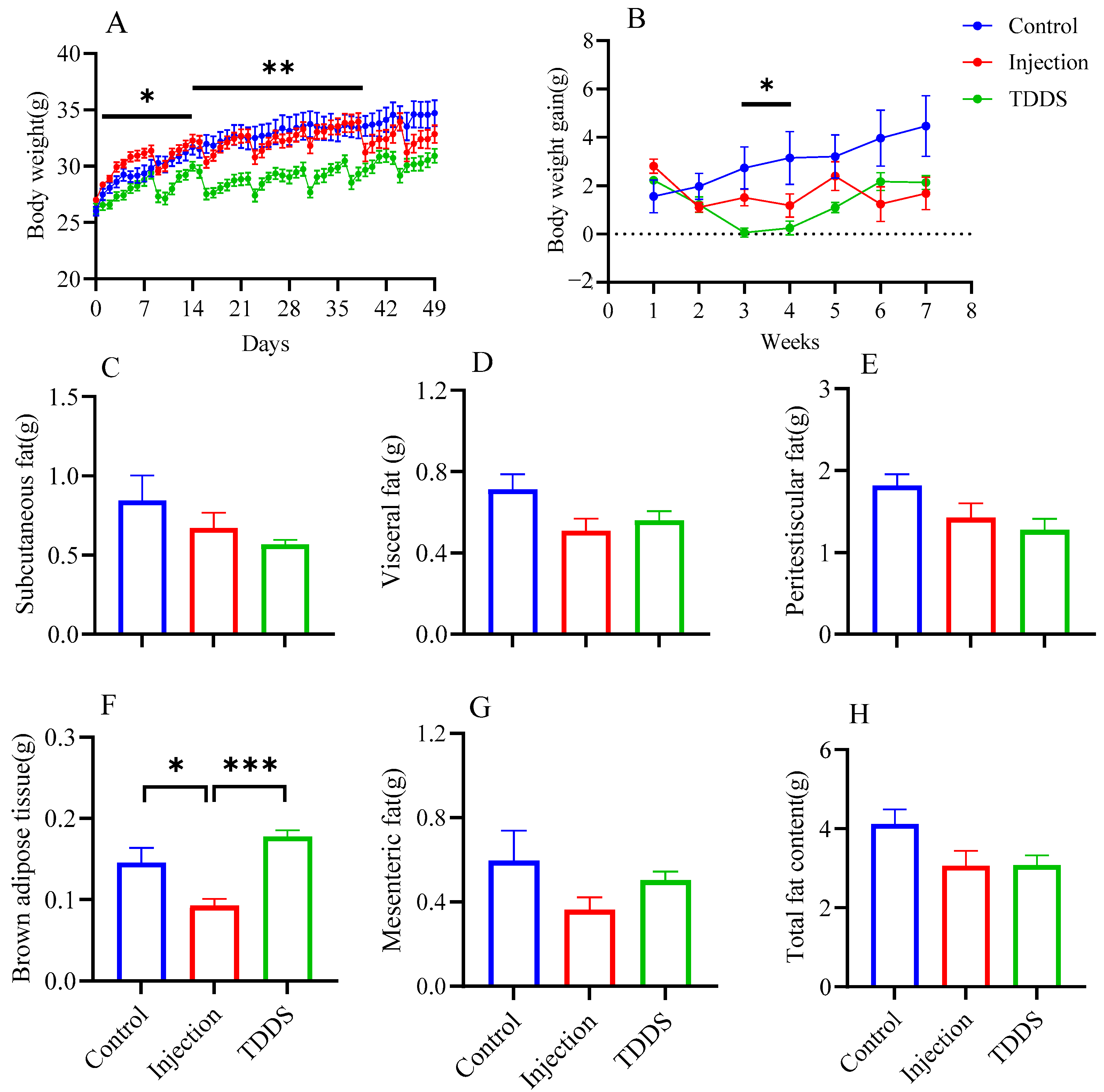
4.1. Body Weight and Fat Distribution
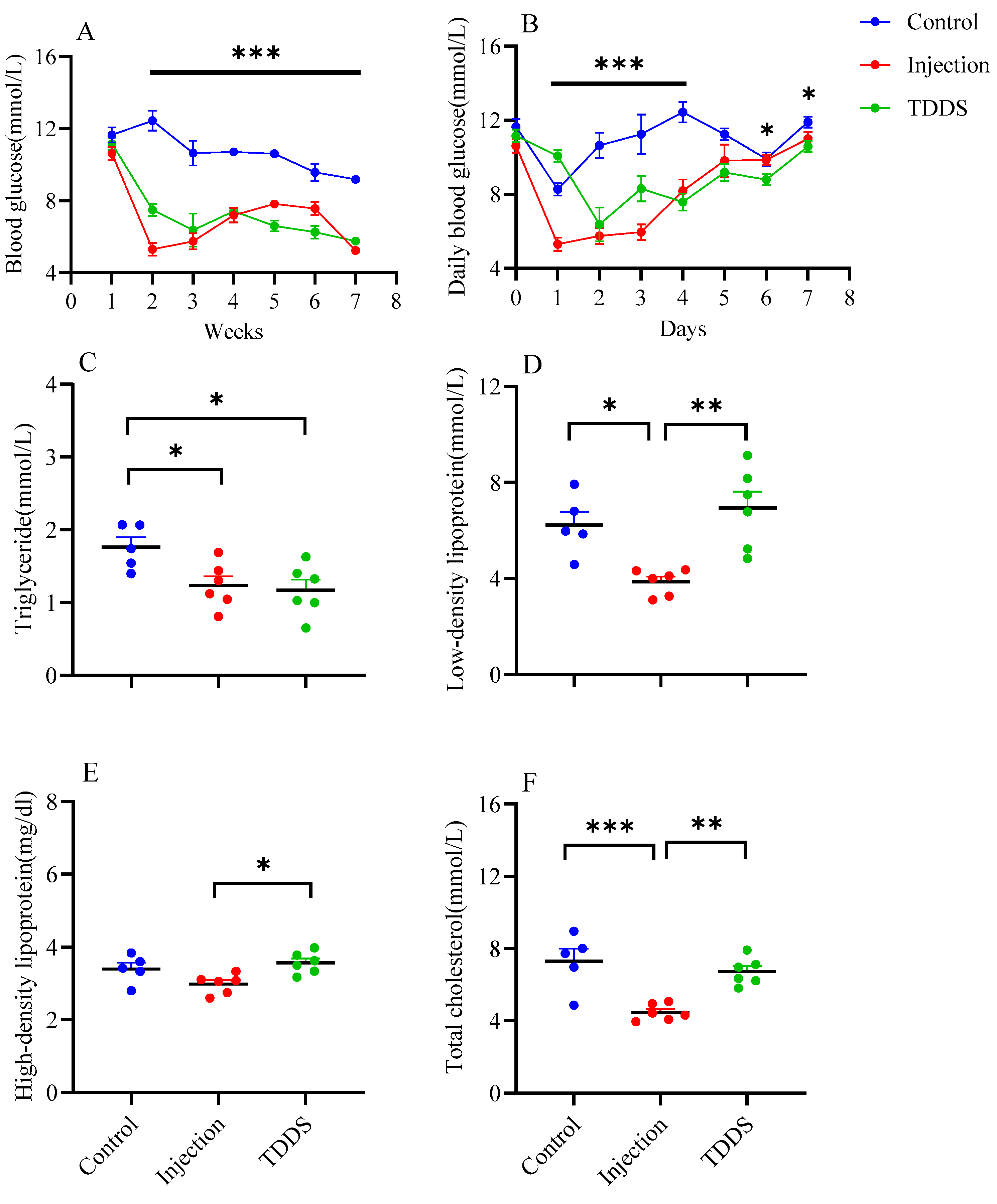
4.2. Blood Indexes
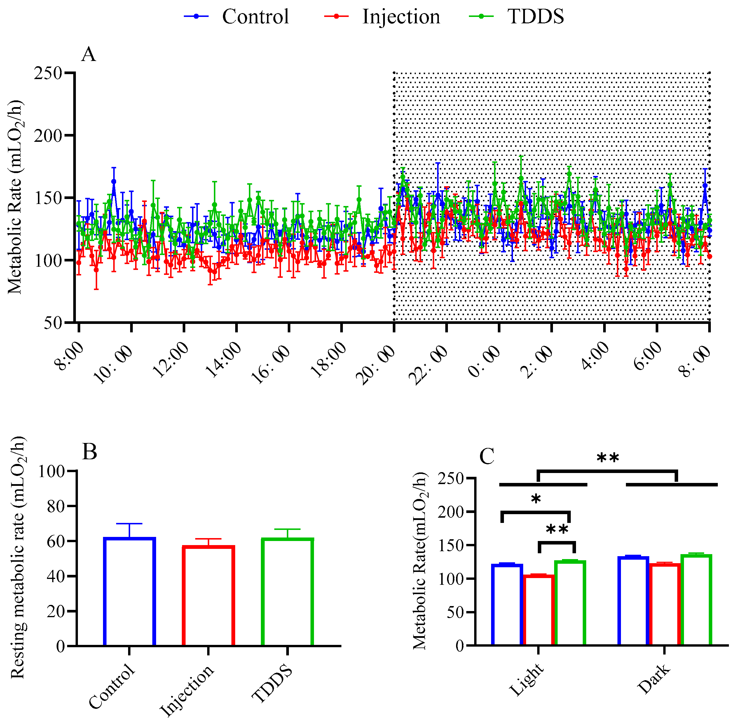
4.3. Metabolic Rate
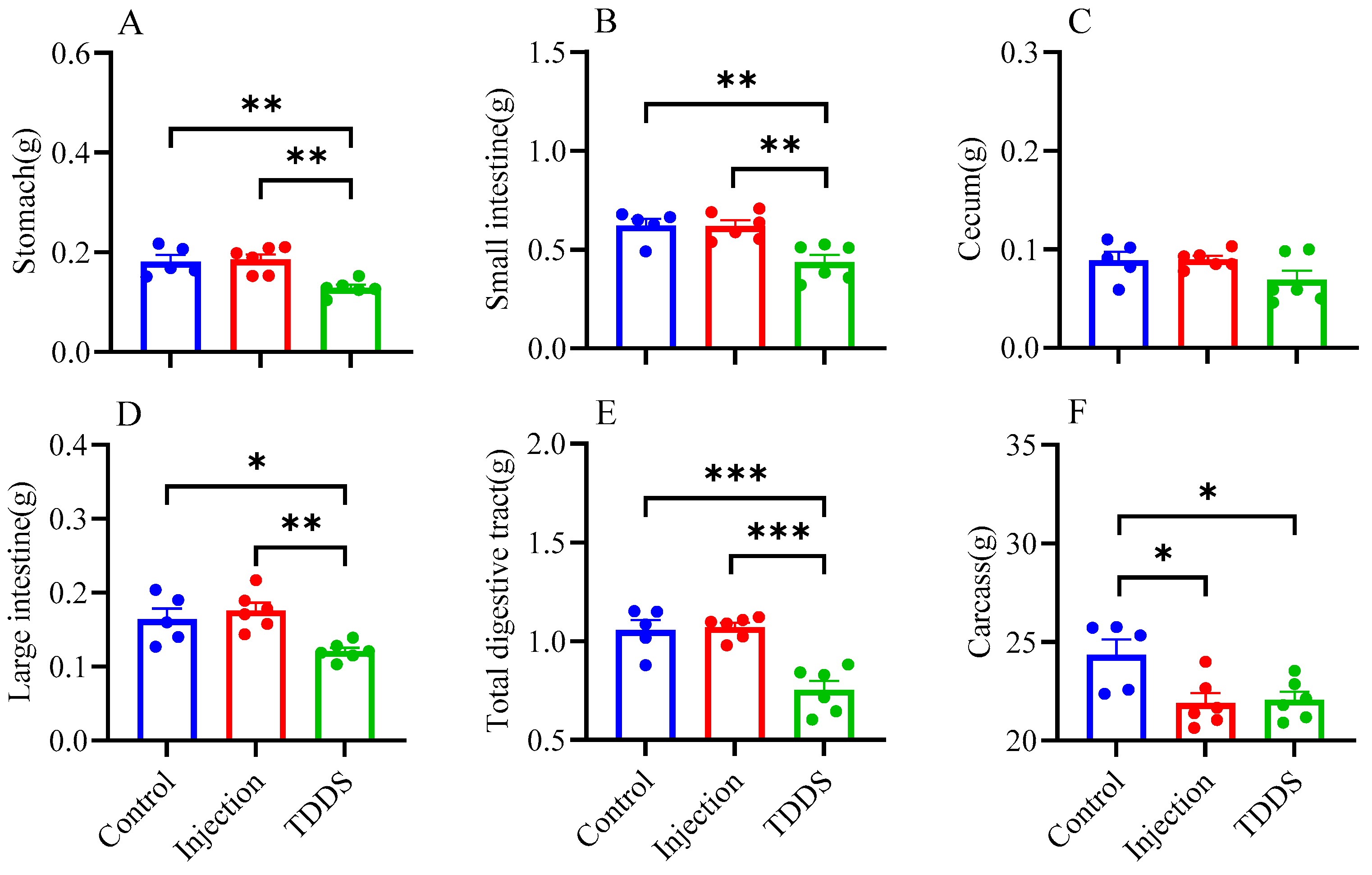
4.4. Organ Weight
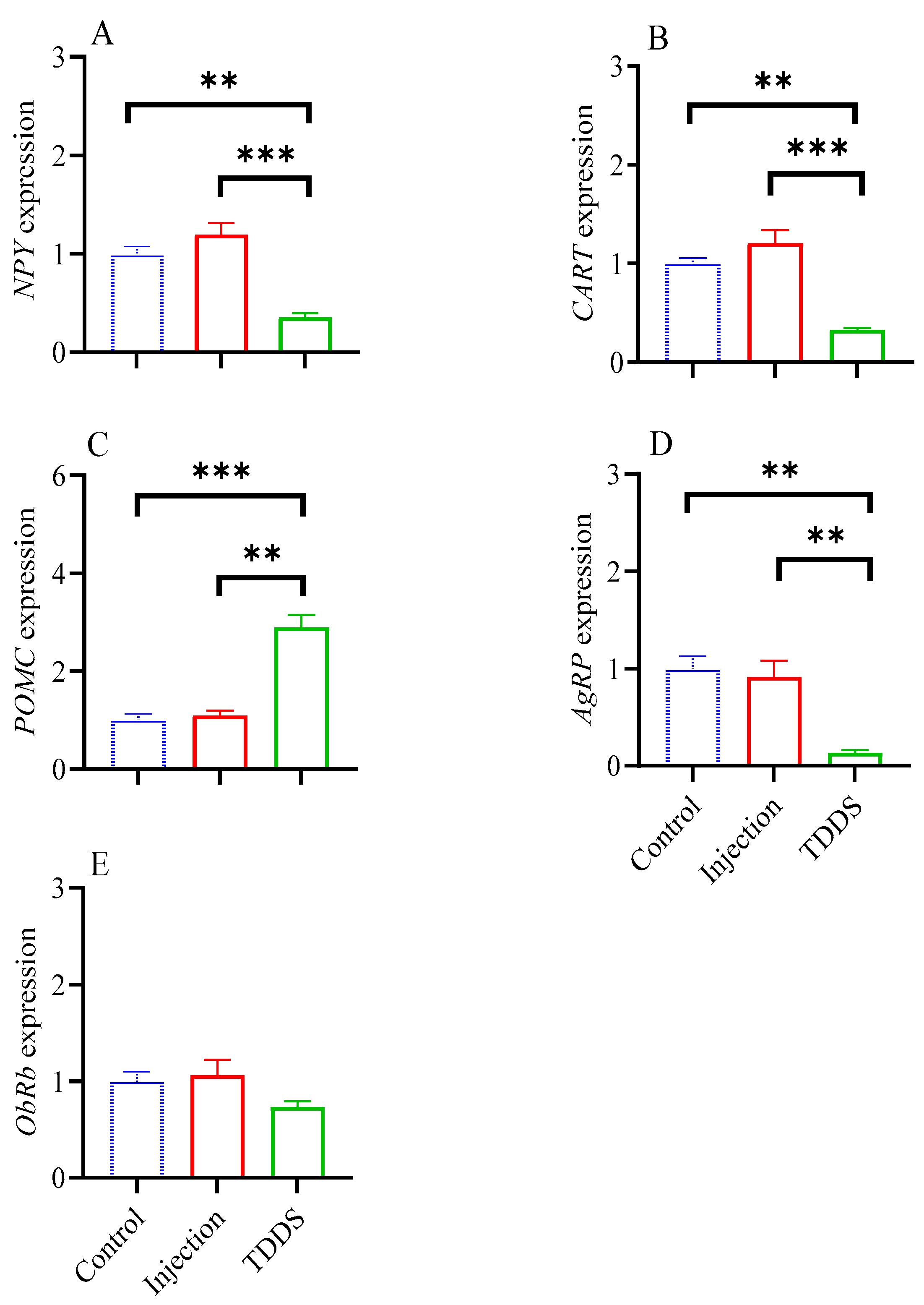
4.5. Expression of Feeding-Related Neuropeptides
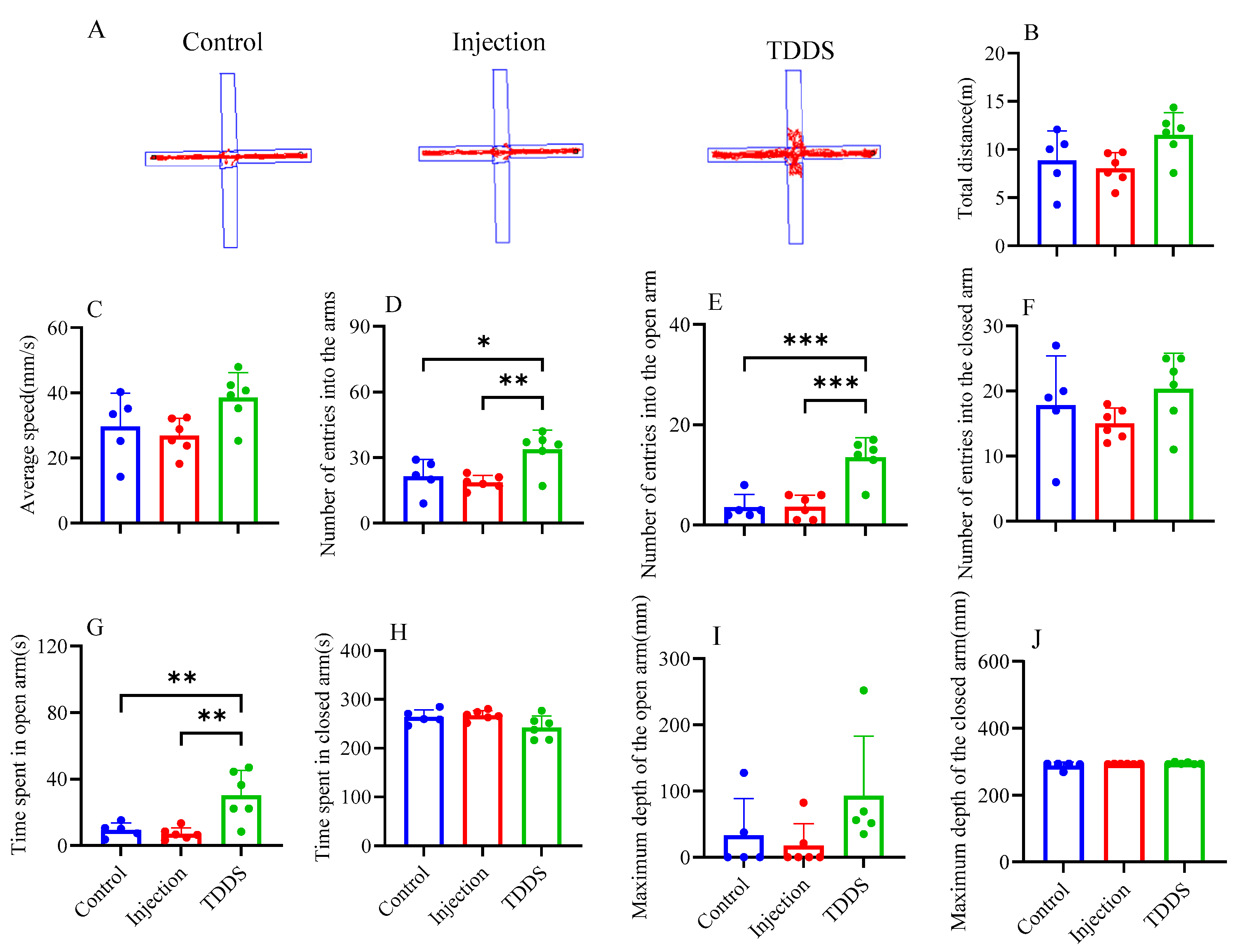
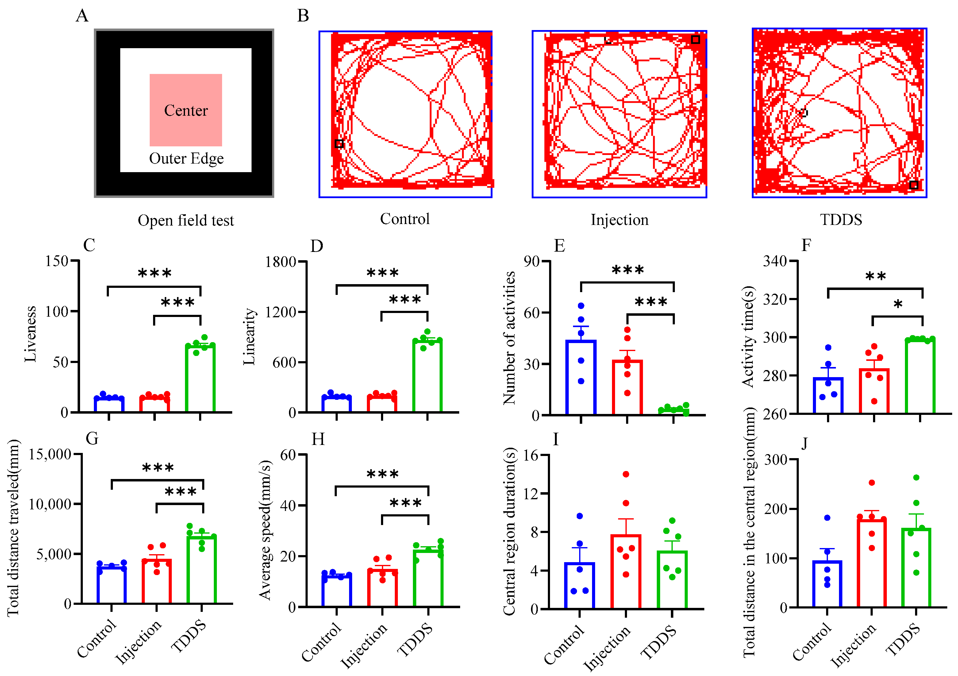
4.6. Anxiety-like and Exploratory Behaviors
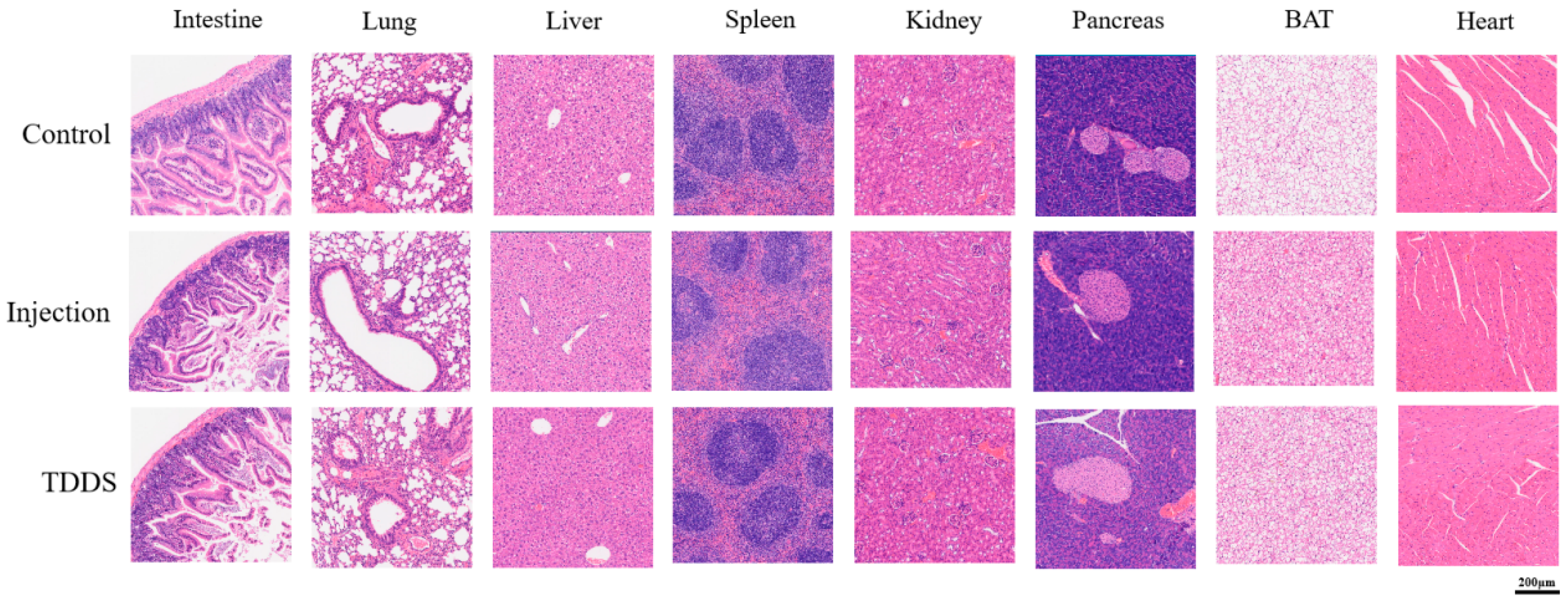
4.7. Hematoxylin-Eosin Staining Pathological Section
5. Discussion
5.1. Transdermal Semaglutide Delivery Is as Effective as Injection Treatment for Reducing Body Weights
5.2. Transdermal Semaglutide Delivery Is More Potent than the Injection Treatment
5.3. Transdermal Drug Delivery Has Good Security in Obesity Treatment
6. Conclusions
Author Contributions
Funding
Institutional Review Board Statement
Informed Consent Statement
Data Availability Statement
Acknowledgments
Conflicts of Interest
References
- Caruso, A.; Gelsomino, L.; Panza, S.; Accattatis, F.M.; Naimo, G.D.; Barone, I.; Giordano, C.; Catalano, S.; Andò, S. Leptin: A heavyweight player in obesity-related cancers. Biomolecules 2023, 13, 1084. [Google Scholar] [CrossRef] [PubMed]
- Caballero, B. Humans against obesity: Who will win? Adv. Nutr. 2019, 10, 4–9. [Google Scholar] [CrossRef]
- Broughton, D.E.; Moley, K.H. Obesity and female infertility: Potential mediators of obesity’s impact. Fertil. Steril. 2017, 107, 840–847. [Google Scholar] [CrossRef] [PubMed]
- Purdy, J.C.; Shatzel, J.J. The hematologic consequences of obesity. Eur. J. Haematol. 2021, 106, 306–319. [Google Scholar] [CrossRef] [PubMed]
- He, R.; Zheng, R.; Zheng, J.; Li, M.; Wang, T.; Zhao, Z.; Wang, S.; Lin, H.; Lu, J.; Chen, Y.; et al. Causal association between obesity, circulating glutamine levels, and depression: A mendelian randomization study. J. Clin. Endocrinol. Metab. 2023, 108, 1432–1441. [Google Scholar] [CrossRef]
- Biener, A.; Cawley, J.; Meyerhoefer, C. The impact of obesity on medical care costs and labor market outcomes in the US. Clin. Chem. 2018, 64, 108–117. [Google Scholar] [CrossRef]
- Ladenheim, E.E. Liraglutide and obesity: A review of the data so far. Drug Des. Dev. Ther. 2015, 9, 1867–1875. [Google Scholar] [CrossRef]
- Smits, M.M.; Van Raalte, D.H. Safety of semaglutide. Front. Endocrinol. 2021, 12, 645563. [Google Scholar] [CrossRef]
- Bunck, M.C.; Diamant, M.; Eliasson, B.; Cornér, A.; Shaginian, R.M.; Heine, R.J.; Taskinen, M.R.; Yki-Järvinen, H.; Smith, U. Exenatide affects circulating cardiovascular risk biomarkers independently of changes in body composition. Diabetes Care 2010, 33, 1734–1737. [Google Scholar] [CrossRef]
- Popoviciu, M.S.; Păduraru, L.; Yahya, G.; Metwally, K.; Cavalu, S. Emerging role of GLP-1 agonists in obesity: A comprehensive review of randomised controlled trials. Int. J. Mol. Sci. 2023, 24, 10449. [Google Scholar] [CrossRef]
- Berger, M.; Gray, J.A.; Roth, B.L. The expanded biology of serotonin. Annu. Rev. Med. 2009, 60, 355–366. [Google Scholar] [CrossRef] [PubMed]
- Zan, P.; Than, A.; Zhang, W.; Cai, H.X.; Zhao, W.; Chen, P. Transdermal photothermal-pharmacotherapy to remodel adipose tissue for obesity and metabolic disorders. ACS Nano 2022, 16, 1813–1825. [Google Scholar] [CrossRef] [PubMed]
- Wong, W.F.; Ang, K.P.; Sethi, G.; Looi, C.Y. Recent advancement of medical patch for transdermal drug delivery. Medicina 2023, 59, 778. [Google Scholar] [CrossRef]
- Morales, J.; Shubrook, J.H.; Skolnik, N. Practical guidance for use of oral semaglutide in primary care: A narrative review. Postgrad. Med. 2020, 132, 687–696. [Google Scholar] [CrossRef] [PubMed]
- Yao, H.Q.; Zhang, A.Q.; Li, D.; Wu, Y.; Wang, C.-Z.; Wan, J.-Y.; Yuan, C.-S. Comparative effectiveness of GLP-1 receptor agonists on glycaemic control, body weight, and lipid profile for type 2 diabetes: Systematic review and network meta-analysis. BMJ 2024, 384, e076410. [Google Scholar] [CrossRef]
- Wang, J.Y.; Wang, Q.W.; Yang, X.-Y.; Yang, W.; Li, D.-R.; Jin, J.-Y.; Zhang, H.-C.; Zhang, X.-F. GLP-1 receptor agonists for the treatment of obesity: Role as a promising approach. Front. Endocrinol. 2023, 14, 1085799. [Google Scholar] [CrossRef]
- Meng, F.; Qiao, X.; Xin, C.; Ju, X.; He, M. Recent progress of polymeric microneedle-assisted long-acting transdermal drug delivery. J. Pharm. Pharm. Sci. 2024, 27, 12434. [Google Scholar] [CrossRef]
- Lippi, G.; Cadamuro, J.; Danese, E.; Gelati, M.; Montagnana, M.; von Meyer, A.; Salvagno, G.L.; Simundic, A.M. Internal quality assurance of HIL indices on Roche Cobas c702. PLoS ONE 2018, 13, e0200088. [Google Scholar] [CrossRef]
- Chan, Y.K.; Davis, P.F.; Poppitt, S.D.; Sun, X.; Greenhill, N.S.; Krishnamurthi, R.; Przepiorski, A.; McGill, A.-T.; Krissansen, G.W. Influence of tail versus cardiac sampling on blood glucose and lipid profiles in mice. Lab. Anim. 2012, 46, 142–147. [Google Scholar] [CrossRef]
- Weir, J.B. New methods for calculating metabolic rate with special reference to protein metabolism. J. Physiol. 1949, 109, 1–9. [Google Scholar] [CrossRef]
- Lister, R.G. The use of a plus-maze to measure anxiety in the mouse. Psychopharmacology 1987, 92, 180–185. [Google Scholar] [CrossRef] [PubMed]
- Livak, K.J.; Schmittgen, T.D. Analysis of relative gene expression data using real-time quantitative PCR and the 2−ΔΔCT method. Methods 2001, 25, 402–408. [Google Scholar] [CrossRef] [PubMed]
- Meier, J.J. GLP-1 receptor agonists for individualized treatment of type 2 diabetes mellitus. Nat. Rev. Endocrinol. 2012, 8, 728–742. [Google Scholar] [CrossRef] [PubMed]
- Qin, W.H.; Yang, J.; Ni, Y.; Deng, C.; Ruan, Q.; Ruan, J.; Zhou, P.; Duan, K. Efficacy and safety of once-weekly tirzepatide for weight management compared to placebo: An updated systematic review and meta-analysis including the latest SURMOUNT-2 trial. Endocrine 2024, 86, 70–84. [Google Scholar] [CrossRef]
- Ren, Q.; Chen, S.; Chen, X.; Niu, S.; Yue, L.; Pan, X.; Li, Z.; Chen, X. An effective glucagon-like peptide-1 receptor agonists, semaglutide, improves sarcopenic obesity in obese mice by modulating skeletal muscle metabolism. Drug Des. Dev. Ther. 2022, 16, 3723–3735. [Google Scholar] [CrossRef]
- Choi, J.W.; Choe, H.W.; Pai, S.H. Serum lipid concentrations correlate more strongly with total body fat than with body mass index in obese humans. Clin. Chim. Acta 2003, 329, 83–87. [Google Scholar] [CrossRef]
- Wen, J.; Tan, S.; Qiao, Q.; Shi, L.; Huang, Y.; Zhao, Z. Strategies of behavior, energetic and thermogenesis of striped hamsters in response to food deprivation. Integr. Zool. 2018, 13, 70–83. [Google Scholar] [CrossRef]
- Garvey, W.T.; Batterham, R.L.; Bhatta, M.; Buscemi, S.; Christensen, L.N.; Frias, J.P.; Jódar, E.; Kandler, K.; Rigas, G.; Wadden, T.A.; et al. Two-year effects of semaglutide in adults with overweight or obesity: The STEP 5 trial. Nat. Med. 2022, 28, 2083–2091. [Google Scholar] [CrossRef]
- Liu, Q.S.; Wang, D.H. Effects of diet quality on phenotypic flexibility of organ size and digestive function in Mongolian gerbils (Meriones unguiculatus). J. Comp. Physiol. B 2007, 177, 509–518. [Google Scholar] [CrossRef]
- Zhang, J.Y.; Zhao, X.Y.; Wen, J.; Tan, S.; Zhao, Z.J. Plasticity in gastrointestinal morphology and enzyme activity in lactating striped hamsters (Cricetulus barabensis). J. Exp. Biol. 2016, 219, 1327–1336. [Google Scholar] [CrossRef]
- Piersma, T.; Lindström, A. Rapid reversible changes in organ size as a component of adaptive behaviour. Trends Ecol. Evol. 1997, 12, 134–138. [Google Scholar] [CrossRef] [PubMed]
- Kim, K.S.; Park, J.S.; Hwang, E.; Park, M.J.; Shin, H.Y.; Lee, Y.H.; Kim, K.M.; Gautron, L.; Godschall, E.; Portillo, B.; et al. GLP-1 increases preingestive satiation via hypothalamic circuits in mice and humans. Science 2024, 385, 438–446. [Google Scholar] [CrossRef]
- Cannon, B.; Nedergaard, J. Brown adipose tissue: Function and physiological significance. Physiol. Rev. 2004, 84, 277–359. [Google Scholar] [CrossRef] [PubMed]
- Corrales, P.; Vivas, Y.; Izquierdo-Lahuerta, A.; Horrillo, D.; Seoane-Collazo, P.; Velasco, I.; Torres, L.; Lopez, Y.; Martínez, C.; López, M.; et al. Long-term caloric restriction ameliorates deleterious effects of aging on white and brown adipose tissue plasticity. Aging Cell 2019, 18, e12948. [Google Scholar] [CrossRef]
- Wen, J.; Chi, Q.S.; Wang, D.H.; Zhao, Z.J. The responses of metabolic rate and neuropeptides to food deprivation in striped hamsters (Cricetulus barabensis) with different basal metabolic rate. J. Exp. Zool A Ecol. Integr. Physiol. 2020, 333, 483–492. [Google Scholar] [CrossRef] [PubMed]
- Ghadir, M.R.; Riahin, A.A.; Havaspour, A.; Nooranipour, M.; Habibinejad, A.A. The relationship between lipid profile and severity of liver damage in cirrhotic patients. Hepat. Mon. 2010, 10, 285–288. [Google Scholar]
- Prausnitz, M.R.; Langer, R. Transdermal drug delivery. Nat. Biotechnol. 2008, 26, 1261–1268. [Google Scholar] [CrossRef]
- Arillotta, D.; Floresta, G.; Guirguis, A.; Corkery, J.M.; Catalani, V.; Martinotti, G.; Sensi, S.L.; Schifano, F. GLP-1 receptor agonists and related mental health issues; insights from a eange of social media platforms ssing a mixed-methods approach. Brain Sci. 2023, 13, 1503. [Google Scholar] [CrossRef]
- Splinter, M.Y. Rotigotine: Transdermal dopamine agonist treatment of parkinson’s disease and restless legs syndrome. Ann. Pharmacother. 2007, 41, 285–295. [Google Scholar] [CrossRef]
- Segal, D.S.; Kuczenski, R. An escalating dose “binge” model of amphetamine psychosis: Behavioral and neurochemical characteristics. J. Neurosci. 1997, 17, 2551–2566. [Google Scholar] [CrossRef]
- Kornelius, E.; Huang, J.Y.; Lo, S.C.; Huang, C.N.; Yang, Y.S. The risk of depression, anxiety, and suicidal behavior in patients with obesity on glucagon like peptide-1 receptor agonist therapy. Sci. Rep. 2024, 14, 24433. [Google Scholar] [CrossRef] [PubMed]

| Gene | Sequence 5′-3′ | Size (bp) |
|---|---|---|
| NPY-f | TCGTGTGTTTGGGCATTCTG | 128 |
| NPY-r | TCTGGTGATGAGATTGATGTAGTG | |
| CART-f | ACGAGAAGGAGCTGCCAAG | 153 |
| CART-r | GCTCTCCAGCGTCACACAT | |
| POMC-f | GAAGATGCCGAGATTCTGCT | 175 |
| POMC-r | CTCCAGCGAGAGGTCGAGTT | |
| AgRP-f | ACCTTAGGGAGGCACCTCAT | 151 |
| AgRP-r | AGCAACATTGCAGTCAGCAT | |
| ObRb-f | TAAAGCTCTCGTGGCGCTCT | 195 |
| ObRb-r | TCCACACGAGCAAGAACAAC | |
| β-Actin-f | CGTAAAGACCTCTATGCCAA | 317 |
| β-Actin-r | GCGCAAGTTAGGTTTTGTC |
| Organs | Control | Injection | TDDS | p |
|---|---|---|---|---|
| Heart | 0.169 ± 0.014 | 0.148 ± 0.006 | 0.1712 ± 0.008 | ns |
| Liver | 1.236 ± 0.036 b | 1.0942 ± 0.018 a | 1.201 ± 0.042 b | * |
| Spleen | 0.072 ± 0.006 | 0.062 ± 0.003 | 0.062 ± 0.003 | ns |
| Lung | 0.149 ± 0.009 a | 0.161 ± 0.007 b | 0.182 ± 0.007 c | * |
| Kidney | 0.343 ± 0.012 | 0.329 ± 0.011 | 0.312 ± 0.006 | ns |
Disclaimer/Publisher’s Note: The statements, opinions and data contained in all publications are solely those of the individual author(s) and contributor(s) and not of MDPI and/or the editor(s). MDPI and/or the editor(s) disclaim responsibility for any injury to people or property resulting from any ideas, methods, instructions or products referred to in the content. |
© 2025 by the authors. Licensee MDPI, Basel, Switzerland. This article is an open access article distributed under the terms and conditions of the Creative Commons Attribution (CC BY) license (https://creativecommons.org/licenses/by/4.0/).
Share and Cite
Li, W.; Cai, R.; Yin, B.; Zhou, Y.; Dong, X.; Li, W.; Wen, J. Transdermal Semaglutide Administration in Mice: Reduces Body Weight by Suppressing Appetite and Enhancing Metabolic Rate. Biology 2025, 14, 575. https://doi.org/10.3390/biology14050575
Li W, Cai R, Yin B, Zhou Y, Dong X, Li W, Wen J. Transdermal Semaglutide Administration in Mice: Reduces Body Weight by Suppressing Appetite and Enhancing Metabolic Rate. Biology. 2025; 14(5):575. https://doi.org/10.3390/biology14050575
Chicago/Turabian StyleLi, Wenjing, Ruilin Cai, Binxin Yin, Yingying Zhou, Xinyuan Dong, Wenting Li, and Jing Wen. 2025. "Transdermal Semaglutide Administration in Mice: Reduces Body Weight by Suppressing Appetite and Enhancing Metabolic Rate" Biology 14, no. 5: 575. https://doi.org/10.3390/biology14050575
APA StyleLi, W., Cai, R., Yin, B., Zhou, Y., Dong, X., Li, W., & Wen, J. (2025). Transdermal Semaglutide Administration in Mice: Reduces Body Weight by Suppressing Appetite and Enhancing Metabolic Rate. Biology, 14(5), 575. https://doi.org/10.3390/biology14050575