Article

Identification of SNP Markers in the Glutamate Dehydrogenase (GDH) and Aspartate Aminotransferase 2 (AST2) Associated with Ammonia Nitrogen Tolerance in Penaeus monodon

1
Key Laboratory of South China Sea Fishery Resources Exploitation and Utilization, Ministry of Agriculture, South China Sea Fisheries Research Institute, Chinese Academy of Fishery Sciences, Guangzhou 510300, China
2
State Key Laboratory of Biocontrol, Southern Marine Science and Engineering Guangdong Laboratory (Zhuhai), School of Life Sciences, Sun Yat-sen University, Guangzhou 510275, China
3
Yancheng Institute of Technology, College of Marine and Bioengineering, Yancheng 224051, China
*
Author to whom correspondence should be addressed.
Biology 2025, 14(11), 1532; https://doi.org/10.3390/biology14111532
Submission received: 2 September 2025 / Revised: 26 October 2025 / Accepted: 30 October 2025 / Published: 31 October 2025

Simple Summary

Black tiger shrimp is a major farmed shrimp species in the world, which is negatively impacted by high ammonia nitrogen levels in high-density aquaculture. This study investigated the genetic basis of ammonia tolerance, focusing on two conserved genes (glutamate dehydrogenase and aspartate aminotransferase 2) involved in ammonia metabolism. Single nucleotide polymorphisms (SNPs) in these two genes were identified, genotyped and analyzed in different shrimp populations. Two specific SNPs (PmGDH-1227 and PmAST2-132) showed a significant distribution difference between ammonia-susceptible and ammonia-resistant shrimp. This study intuitively represented the interaction of PmGDH and PmAST2 to ammonia nitrogen and developed SNP markers linked to ammonia tolerance in these two genes from black tiger shrimp, providing valuable tools for molecular marker-assisted breeding programs to develop new strains with ammonia tolerance, thereby improving the survival rate and yield of black tiger shrimp under high-density farming conditions.

Abstract

Black tiger shrimp (Penaeus monodon) is the largest species of penaeid, being commercially cultured globally, ranking as the second most farmed species in the shrimp industry. However, with the transformation of shrimp aquaculture from semi-intensive to high-density farming, the concentration of ammonia nitrogen in the aquatic environment has increased, severely affecting the growth and survival of shrimps. To increase production efficiency, breeding new strains of shrimp with the trait of tolerance to high ammonia nitrogen is desired in the black tiger shrimp aquaculture. Previous studies have shown that glutamate dehydrogenase (GDH) and aspartate aminotransferase 2 (AST2) play important roles in the metabolism of ammonia nitrogen in crustaceans. In the present study, we conducted synteny analysis of PmGDH and PmAST2 in different species. The interactions of PmGDH with ammonium (NH4+) and PmAST2 with aspartate were analyzed by docking. To develop molecule markers associated with ammonia nitrogen tolerance, SNPs of PmGDH and PmAST2 were identified by direct sequencing, genotyped by the SNaPshot technique, and characterized through genotype-phenotype association analysis by PLINK software version 1.9 in the three geographical populations, two families with different ammonia tolerance, and 120 susceptible and resistant individuals of black tiger shrimp. The results indicate that the GDH and AST2 genes are evolutionarily conserved in vertebrates, except for the black tiger shrimp, which suggests divergence in selective pressure between invertebrates and vertebrates. Notably, PmGDH may interact with NH4+ via the residue Asp178 within loop 1 in the GdhA domain through a hydrogen bonding interaction, and PmAST2 may interact with aspartate via helix 1, sheet 1, loop 1, and loop 2, through both hydrogen bonding interactions and a salt bridge interaction. A total of 12 SNPs were detected in the exons of PmGDH and PmAST2. Among these candidate SNPs, the distributions of PmGDH-1227 and PmAST2-132 showed both significant differences in the genotype and allele association analysis between susceptible and resistant groups. Haplotype association analysis showed that three haplotypes exhibited significantly different distributions between susceptible and resistant groups. In conclusion, PmGDH-1227 and PmAST2-132 were associated with ammonia nitrogen tolerance, and these SNP markers are expected to contribute to marker-assisted selection (MAS) breeding programs to obtain new strains of Penaeus monodon.

1. Introduction

Shrimp farming is an important part of China’s aquaculture industry [1]. However, due to the development of intensive high-density aquaculture and the influence of climate change, the aquatic environment of aquaculture has deteriorated seriously in recent years, causing frequent diseases in aquaculture species. Ammonia nitrogen is the major pollutant in the aquatic environment and is one of the most important water quality parameters to monitor [2]. In an isolated aquaculture system, ammonia is one of the main concerns as it may accumulate over time, primarily from the decomposition of organics and from the excretion of the rearing animals [3]. High concentrations of ammonia may result in the retardation of shrimp growth and can cause tissue damage in gills, skin, and blood circulation; in extreme cases, it can cause shrimp mortality [3,4,5,6]. To increase production efficiency, shrimp with the trait of tolerance to high ammonia nitrogen are desired in shrimp farming. Thus, breeding new strains with this desirable trait has become the key to the sustainable development of the shrimp breeding industry.
With the development of molecular biology, marker-assisted selection (MAS) has breathed new life into the aquaculture breeding industry, for it enables selection to depend on genotypes and improves breeding efficiency [7,8]. Molecular markers can be used to detect quantitative trait loci (QTL) that control complex traits of interest and to select individuals for MAS programs [9]. As the third generation of genetic markers, single-nucleotide polymorphism (SNP) has the advantages of high genetic stability, high abundance and widespread distribution, representativeness, co-dominant, and easy to detect [10]. In recent years, SNPs have been widely used in the MAS breeding programs of aquatic animals. Many SNP markers were found to be associated with a variety of economic traits, such as growth, disease resistance, and ammonia tolerance traits [11,12,13,14,15,16,17]. In Epinephelus coioides, 25 SNPs were identified to be correlated with the ammonia tolerance, in which more than half of the SNPs were clustered on chromosomes 9 and 16, and the candidate gene sgk3 may be critical for its function to activate many ion channels and transporters [18]. In Litopenaeus vannamei, a SNP marker C/T545 in the hemocyanin gene was identified to be associated with both high growth and ammonia tolerance traits [19]. In Sinonovacula constricta, 56 associated SNPs embedded in or adjacent to 236 genes were identified to be associated with ammonia tolerance, in which Na+/K+-ATPase (NKA) was demonstrated to be involved in NH4+ excretion to reduce ammonia accumulation [20]. However, given that the ammonia tolerance is a quantitative trait with low heritability, currently developed SNP markers are insufficient for MAS breeding programs, especially in shrimps.
In black tiger shrimp (Penaeus monodon), ammonia stress is known to induce severe oxidative damage and trigger apoptosis, as revealed by transcriptome analyses [21]. The underlying mechanisms, further elucidated by single-cell sequencing, involve complex responses in oxidative stress, detoxification metabolism, and immune defense [22]. Specific genes, such as chitinase 4 and 5, have also been implicated in the response to ammonia stress [23,24]. Importantly, ammonia is commonly considered a toxic by-product of amino acid metabolism [25]. Our previous study has demonstrated that glutamate dehydrogenase (GDH) and aspartate aminotransferase 2 (AST2) in P. monodon contribute to ammonia detoxification [26,27,28]. Recent studies have proved that ammonia can be recycled through reductive amination catalyzed by GDH to central ammonia acid metabolism [25,29]. GDH is an NAD(P)H-dependent enzyme that can catalyze reductive amination of α-ketoglutarate to glutamate. In this process, excessive ammonia was assimilated, and the efficiency of nitrogen utilization was maximized [25]. Likewise, as a mitochondrial transaminase, AST2, also well-known as glutamate oxaloacetate transaminase 2 (GOT2), can catalyze the reversible reaction between glutamate and oxaloacetate, generating α-ketoglutarate and aspartate (Asp) [30,31,32]. Asp is further utilized to synthesize the amino acid asparagine (Asn) and is incorporated into the urea cycle [33]. This cycle will detoxify cellular NH4+ through urea excretion. In addition, two SNPs in the coding domain region of the GDH gene were significantly associated with ammonia tolerance in Sinonovacula constricta [34]. However, the SNP markers in the GDH and AST2 from shrimps have not yet been identified.
Black tiger shrimp is the largest species of penaeid being commercially cultured in many countries, especially in Southeast Asia. Up to now, several SNPs associated with growth, low salt tolerance, and diversity traits have been identified in black tiger shrimp [35,36,37]. However, little is known about the SNP markers associated with ammonia tolerance in the black tiger shrimp. In the present study, an acute ammonia stress test was conducted to test the tolerance capacity of the 28 families of black tiger shrimp to ammonia. The synteny of the PmGDH and PmAST2 in different species and the interactions of PmGDH with ammonium and PmAST2 with Asp were analyzed. The SNPs in the PmGDH and PmAST2 were screened by direct sequencing. SNaPshot was applied to process SNP genotyping in the three geographical populations, the 120 susceptible and resistant individuals, and two families with different ammonia tolerance of the black tiger shrimp. The Hardy–Weinberg equilibrium, the Mendelian genetic ratio, and the genotype-phenotype association analysis of the identified SNPs in the PmGDH and PmAST2 were analyzed. This study focused on the genetic basis of the ammonia tolerance trait and may contribute to MAS breeding programs for black tiger shrimp, aiming to reduce mortality and increase production.

2. Materials and Methods

2.1. Experimental Animals

The wild black tiger shrimp from different geographic populations were collected from four regions of the world (Figure S1). The Sanya population was collected in Sanya, Hainan province, China; the African population was collected in southern Africa near Mozambique; the Indonesian population was collected in Indonesia; and the Thai population was collected in Phuket, Thailand.
A total of 28 different families of healthy black tiger shrimp were selected and bred by our research group in the experimental base of the South China Sea Fisheries Research Institute (Shenzhen, China). According to our previous study, the ammonia-nitrogen tolerance of the No. 9 family was significantly stronger than that of the No. 3 family; thus, they were selected for SNP genotyping. The ID of the female parent of No. 3 family is ♀61109, which comes from the Thai population, and its male parent, which is numbered ♂61537, also belongs to the Thai population. The ID of the female parent of No. 9 family is ♀61116, which comes from the Thai population, and its male parent, which is numbered ♂61625, belongs to the African population.

2.2. Ammonia Nitrogen Stress Test

An ammonia nitrogen stress test was conducted at the Shenzhen base of the South China Sea Fisheries Research Institute. Shrimp (body weight: 8.50 ± 3.01 g, body length: 8.16 ± 1.67 cm) from 28 families of P. monodon marked with different fluorescent dyes were used (Figure S2). The shrimp were reared in a cement pond (1.5 m × 1.8 m × 1.5 m) for 14 days before the ammonia nitrogen stress test. Continuous aeration was provided in the rearing ponds, and shrimp were fed with a commercial shrimp diet at 1% body weight per day. Ammonium chloride (NH4Cl, analytically pure) was added to control the concentration of ammonia nitrogen, and the 96 h median lethal concentration (LC50) in the ammonia nitrogen stress test was 66 mg/L, which was calculated by preliminary experiments according to previous studies [3,38]. The experiments contained two groups (ammonia nitrogen test group and the control group), each group contained two replicates. In the test group, 2800 shrimp from 28 families were evenly assigned into two cement ponds containing 3 tons of seawater with NH4Cl added. In the control group, another 2800 shrimp from 28 families were evenly assigned into two cement ponds containing 3 tons of seawater without NH4Cl. During the experimental period, the dissolved oxygen was 6.5 ± 0.03 mg/L, the water temperature was 30 ± 0.5 °C, the pH was 7.0 ± 0.5, and the salinity was 30 ‰. Water in the experimental ponds was replaced every 24 h. The mortality of test shrimp in each pond was recorded, and the dead shrimp were collected every 2 h. The first 60 dead shrimp were defined as the susceptible group, and the last 60 surviving shrimp were defined as the resistant group. Shrimp were stored in 95 % ethanol. Shrimp were anesthetized on ice before sampling. All efforts were made to minimize the suffering of the shrimp.

2.3. Syntenic Analysis

JCVI was performed to explore gene synteny within 5 species at the whole genome level, and the results were visualized using TBtools 2.0. The genome annotation files of P. monodon (GCF_015228065.2), Homo sapiens (GCF_000001405.40), Mus musculus (GCA_000001635.9), Xenopus tropicalis (GCF_000004195.4) and Danio rerio (GCA_000002035.4) were downloaded from the NCBI database and inter-species protein sequence alignment was conducted using the same method to detect sequence homology, with the E-value < 1 × 10⁻⁵ and the minimum number of gene pairs set to 10. The gene synteny maps composed of GDH, AST2, and its neighbor genes of other species were learned by the browser of Genomicus v110.01 (https://www.genomicus.bio.ens.psl.eu/genomicus-110.01/cgi-bin/search.pl, accessed on 25 August 2025).

2.4. Protein-Ligand Docking

The PmGDH (XP_037799881.1) and PmAST2 (XP_037778756.1) protein sequence was obtained from the NCBI database, and their structures were modeled using AlphaFold3 software. The energy of the proteins was optimized using Rosetta Relax, and their structural preparations, such as dehydrogenation and hydrogenation, were performed on the acquired 3D structure of the proteins. The structures of NH4+ and aspartic acid (Asp) were obtained from the PubChem database (https://pubchem.ncbi.nlm.nih.gov/). The PmGDH-NH4+ complex and the PmAST2-Asp complex were modeled using blind docking on the CB-DOCK2 online server (https://cadd.labshare.cn/cb-dock2/php/blinddock.php, accessed on 4 July 2025), and docking was made using Autodock Vina 1.2.3. The protein-ligand force analysis was conducted by relying on the PLIP online server (https://plip-tool.biotec.tu-dresden.de/plip-web, accessed on 4 July 2025) analysis, and the protein-ligand 3D model was displayed using Pymol software version 2.5.4.

2.5. Sampling and DNA Extraction

Muscular tissues were dissected separately from fresh shrimp and stored immediately in 95% ethanol until the DNA samples were isolated. The total DNA was extracted from all collected tissues using the HiPure Tissue DNA Mini Kit (Magen, D3121, Guangzhou, China) according to the manuals. The concentration of DNA was measured by a NanoDrop 2000 spectrophotometer (Thermo Fisher Scientific, Shanghai, China). The integrity of DNA was assessed by electrophoresis on 1% agarose gel. The DNA samples were diluted to 100 ng/μL and stored at −20 °C.

2.6. Screen of SNPs in the PmGDH and PmAST2

The genome sequences of PmGDH (LOC119594874) and PmAST2 (LOC119575290) were obtained in the NCBI database, and the predicted genome structures were illustrated by IBS 2.0 software. According to their genome sequences, the potential domain region of PmGDH and PmAST2 was selected for further polymorphism analysis. Then Sanger sequencing was used to screen SNPs on exon 5 and exon 6 of PmGDH, and exon 1 and exon 2 of PmAST2. A total of 32 DNA samples were randomly selected from four geographic populations (Sanya, Indonesian, African, and Thai populations) of P. monodon as templates, and specific primers (Table S1) were designed to amplify specific sequence fragments mentioned above. PCR products were purified and sent to Beijing Genomics Institute (BGI, Shenzhen, China) for sequencing. Vector NTI Advance® 11.5 software was used to align the sequencing results to obtain SNPs.

2.7. Genotyping of SNPs in the PmGDH and PmAST2

After screening and evaluation, all the available SNPs obtained via the methods above were genotyped in 376 shrimp, which contained 50 individuals from African population, 56 individuals from Indonesian population, 50 individuals from Thai population, 50 individuals from the No. 3 family, 50 individuals from the No. 9 family, 60 individuals from the susceptible group and 60 individuals from the resistant group, using Multiplex SNaPshot system (Ruibiotech, Beijing, China) based on ABI PRISM 3730 XI Genetic Analyzer platform (Applied Biosystems, Woburn, MA, USA). The primers for SNPs amplification and extension primers for Multiplex SNaPshot genotyping were listed in Table S2. The SPSS 23.0 software (IBM, Armonk, NY, USA) was used to analyze the Mendelian genetic ratio in No. 3 and No. 9 families. Popgene 32 Version 1.32 software was used to verify the Hardy–Weinberg equilibrium of the three geographic groups.

2.8. Association Analysis Between Candidate SNPs and Ammonia Nitrogen Tolerance

To search for the SNP markers associated with ammonia nitrogen tolerance, the candidate SNPs were genotyped in the susceptible group and the resistant group by SNaPshot genotyping mentioned above. Only the SNP loci with the minor allele frequency (MAF) greater than 1% would be applied to the association analysis [39]. Differences in the distributions of genotype frequency of each SNP locus in susceptible and resistant groups were assessed using Pearson’s chi-square test, which was performed with statistical software SPSS 23.0. Distributions of allele frequency of each SNP locus in susceptible and resistant groups were analyzed by PLINK software version 1.9 with Fisher models. The odds ratios (OR) of alleles were also calculated. Linkage disequilibrium and haplotype-trait association analysis were analyzed using the Haploview software package v4.2 [40]. The test for normality and homogeneity of variances was performed before analysis.

2.9. Statistical Analysis

The survival rate in the ammonia nitrogen stress test was analyzed using the one-way ANOVA method followed by Tukey’s multiple comparison test in SPSS 23.0 and presented as mean ± the standard error of the mean (mean ± SEM). Figures were made using GraphPad Prism 8 software. It was considered statistically significant when p < 0.05 and highly statistically significant when p < 0.01.

3. Results

3.1. Susceptible and Resistant Families in the Ammonia-Nitrogen Stress Test

The ammonia nitrogen stress test lasted for about 128 h until all the shrimp from 28 families were dead. The first dead shrimp was observed approximately 2 h after ammonia nitrogen stress, and the mortality was increasing gradually until peaking at 24 h. The survival rate of 28 families at 48 h was calculated and presented in Figure 1. The most resistant family is the ♀61696 × ♂61629 group, whose female parent and male parent both belong to the African strain. The most susceptible family is the ♀61689 × ♂61504 group, whose female parent is from the African strain and male parent is from the Thailand strain, and the ♀61650 × ♂61673 group, whose female parent and male parent both belong to the African strain. The 48 h survival rate of the ♀61696 × ♂61629 group, the ♀61689 × ♂61504 group, and the ♀61650 × ♂61673 group is 50%, 8% and 8%, respectively. Accordingly, 60 shrimp that died within the first 8 h were classified into the susceptible group for their high sensitivity to ammonia nitrogen, and the last 60 shrimp that survived over 104 h post challenge were considered as resistant individuals. All the shrimp were still alive during the whole period in the control group.

3.2. Synteny Analysis of PmGDH and PmAST2

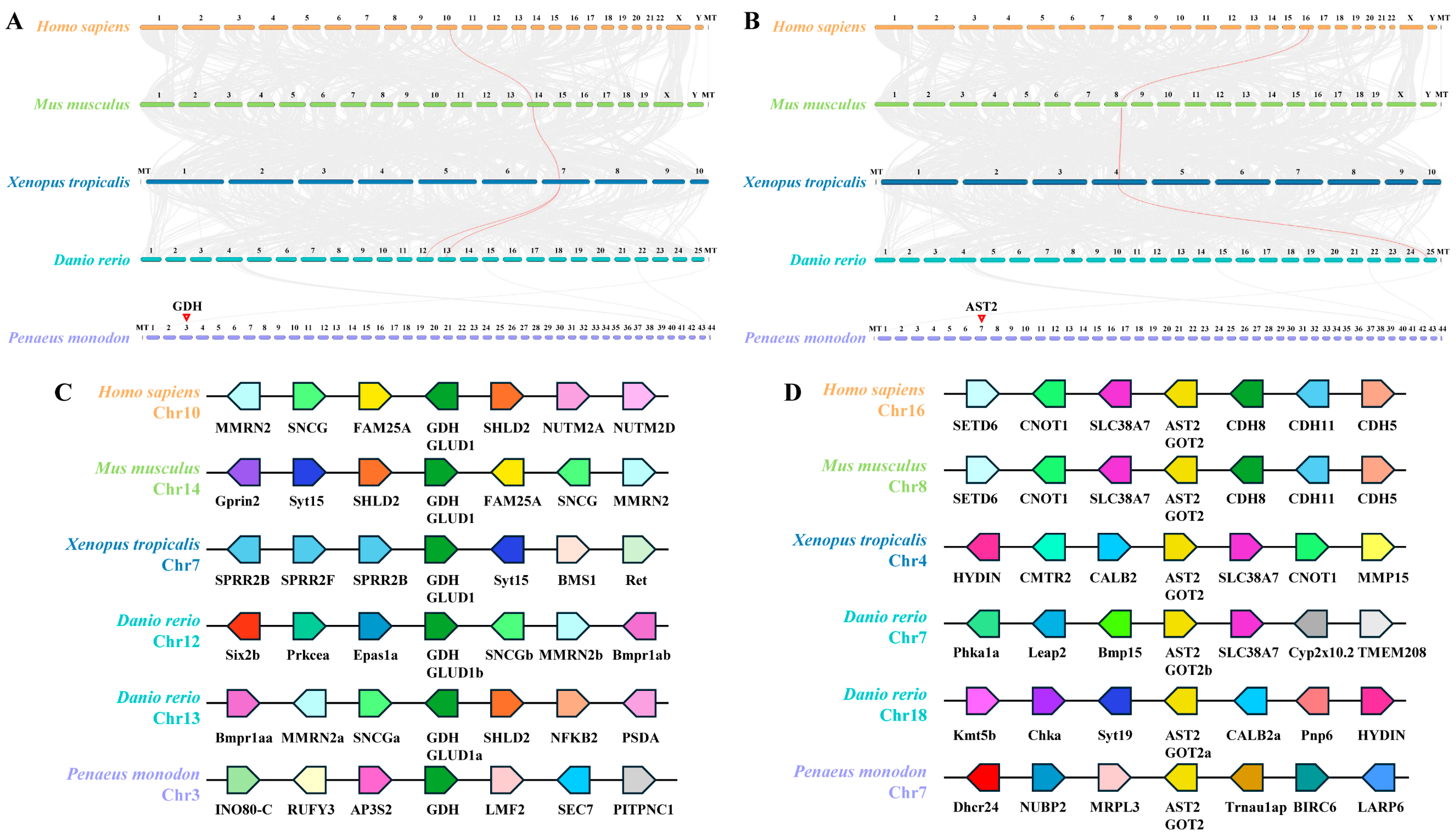
In P. monodon, the GDH gene is located on chromosome 3, and the AST2 gene is located on chromosome 7. Synteny analysis between P. monodon and other species (Homo sapiens, Mus musculus, Xenopus tropicalis, and Danio rerio) revealed 1, 1, 1, and 2 GDH genes with synteny. However, the GDH synteny relationship between Danio rerio and P. monodon did not exist (Figure 2A). Similarly, the synteny analysis between P. monodon and other species revealed 1 AST2 gene. But the synteny relationship of AST2 between Danio rerio and P. monodon did not exist (Figure 2B). The lack of synteny of GDH and AST2 in P. monodon and other species may be due to the divergence of selective pressure in invertebrates and vertebrates. In addition, gene synteny analysis exhibited that genes neighboring the GDH gene and AST2 are at variance. Mostly, adjoining genes of the GDH gene in Homo sapiens, Mus musculus, and Danio rerio are SNCG and MMRN2, respectively (Figure 2C). In addition, adjoining genes of the AST2 gene in Homo sapiens, Mus musculus, and Xenopus tropicalis are SLC38A7 and CNOT1, respectively, and SLC38A7 is also conserved in Danio rerio (Figure 2D). Notably, there are 2 GDH genes (GLUD1a on Chr13 and GLUD1b on Chr12) and 2 AST2 genes (GOT2a on Chr18 and GOT2b on Chr7) in the Danio rerio (Figure 2C,D).

3.3. The PmGDH-NH4+ Complex and the PmAST2-Asp Complex

To intuitively demonstrate the interaction of PmGDH and PmAST2 with ammonia nitrogen, molecular docking was performed. To identify the key amino acids involved in the binding of PmGDH to ammonium ion and PmAST2 to aspartic acid, the interactions in the PmGDH-NH4+ complex and the PmAST2-Asp complex were analyzed. Upon protein-ligand docking, a hydrogen bonding interaction was formed between the PmGDH protein and NH4+ (Figure 3A,C). Within loop 1 of the PmGDH protein, amino acid residue Asp178 forms a hydrogen bonding interaction with NH4+ at a distance of 3.3 Å (Figure 3C). The binding energy between the PmGDH protein and NH4+ was predicted to be −1.4 kcal/mol according to Vina’s binding energy prediction algorithm based on the AMBER force field. Thus, we hypothesized that loop 1 in the GdhA domain of PmGDH might be involved in its binding to NH4+. Meanwhile, the binding interface between the PmAST2 protein and aspartic acid encompasses predominantly hydrogen bonding interactions, with the addition of a salt bridge interaction (Figure 3B,D). Aspartic acid can interact with helix 1, sheet 1, loop 1, and loop 2 of the PmAST2 protein. Aspartic acid can form hydrogen bonding interactions with amino acid residues Thr132 and Gly131 of helix 1, Ser273 of sheet 1, Ser130 of loop 1, Ser275 and Lys276 of loop 2 in the PmAST2 protein at 2.9/3.0 Å, 3.2 Å, 3.1 Å, 3.0 Å, 3.2 Å, and 3.7 Å, respectively (Figure 3D). Aspartic acid can also form a salt-bridge interaction with amino acid residue Arg284 of loop 2 in the PmAST2 protein at 3.8 Å (Figure 3D). The binding energy between the PmAST2 protein and aspartic acid was predicted to be −5.6 kcal/mol according to Vina’s binding energy prediction algorithm based on the AMBER force field. Thus, we hypothesized that helix 1, sheet 1, loop 1, and loop 2 in the AAT_I (Aspartate aminotransferase superfamily fold type I) domain of PmAST2 might be involved in its binding to aspartic acid.

3.4. Identification of SNPs in Exons of PmGDH and PmAST2

The complete genome sequence of the PmGDH gene was approximately 9546 bp, including 9 exons and 8 introns (Figure 4A). The results of direct sequencing showed that a total of seven SNPs were detected in PmGDH, including four SNPs in exon 5 and three SNPs in exon 6 (Figure 4B). The base count was noted as 1 at the beginning of the initiation codon. Among them, PmGDH-1086, PmGDH-1101, and PmGDH-1227 loci were transitions, PmGDH-1212 and PmGDH-1338 were transversions, and PmGDH-1083 and PmGDH-1091 loci were deletion loci (Table 1). Alignment of the nucleotide from exon 5 of PmGDH, including PmGDH-1083, PmGDH-1086, PmGDH-1091, and PmGDH-1101, in the different individuals belonging to the African group, Thai group, Sanya group, and Indonesian group was shown in Figure 4C. The complete genome sequence of PmAST2 is about 14,436 bp, including 7 exons and 6 introns (Figure 5A). There are two SNPs in exon 1 and three SNPs in exon 2 of PmAST2, respectively (Figure 5B), including deletion loci PmAST2-44, transversion loci PmAST2-78, and transition loci PmAST2-132, PmAST2-180, and PmAST2-225 (Table 1). Alignment of the nucleotide from exon 2 of PmAST2, including PmAST2-132, PmAST2-180, and PmAST2-225, in the different individuals belonging to the African group, Thai group, Sanya group, and Indonesian group was shown in Figure 5C. The minor allele frequency (MAF) of all SNP loci is greater than 1% in this study, and most of them are synonymous mutations.

3.5. Genotyping of Potential SNPs

According to the standard of longer flanking sequence, better quality, and suitable for designing SNaPshot primer, PmGDH-1101, PmGDH-1212, PmGDH-1227, PmAST2-132, and PmAST2-225 loci were chosen for SNaPshot genotyping. The No. 3 and No. 9 families were applied to evaluate the Mendelian inheritance of five polymorphic SNPs. The results of SNP genotyping in families are shown in Table 2. In the No. 9 family, parental genotypes of all the loci were homozygous except PmAST2-132 with parental genotype CC × CT, among which PmGDH-1101 and PmAST2-132 were not in line with Mendel’s laws of inheritance (p < 0.05). In No. 3 family, the parental genotypes of PmGDH-1101, PmGDH-1212, and PmAST2-132 were heterozygous, and the parental genotypes of PmGDH-1227 and PmAST2-225 were homozygous. In the No. 3 family, all SNP loci were segregated and assorted independently, fitting well with Mendel’s inheritance law.
The African, Indonesian, and Thai geographic populations were used to verify the Hardy–Weinberg equilibrium of each SNP locus (Table 3). In the African population, almost all SNP loci were deviated significantly from the Hardy–Weinberg equilibrium at p < 0.01 except PmGDH-1227. Alleles of all the SNP loci are spread relatively evenly in the Indonesian population, and all of them are in accordance with the Hardy–Weinberg equilibrium. In the Thai population, genotyping results of all SNP loci showed significant fitness to the Hardy–Weinberg equilibrium except PmAST2-132 (p < 0.05).

3.6. SNPs Are Associated with Ammonia Nitrogen Tolerance in P. monodon

The results of the association analysis with the chi-square test and Fisher model are shown in Table 4 and Table 5. Two SNPs were identified to be associated with ammonia nitrogen tolerance. In detail, the genotype frequency distributions of PmGDH-1101, PmGDH-1227, PmAST2-132, and PmAST2-225 were significantly different between the susceptible group and the resistant group (p < 0.01). And the allele frequency distributions of PmGDH-1227 and PmAST2-132 were also significantly different between the susceptible group and the resistant group (p < 0.05, FDR < 0.05, OR < 1). Shrimp containing these alleles would be more resistant to ammonia nitrogen. These two SNPs in PmGDH and PmAST2, associated with ammonia nitrogen tolerance, were further analyzed to investigate the Linkage disequilibrium of the SNPs. The results showed that the SNPs located on the same gene fragment were in Linkage disequilibrium.
Haplotype association analysis showed that there were three haplotypes whose frequency distributions were significantly different between resistant and susceptible groups (p < 0.01) (Table 6). The haplotype frequencies of PmGDH-1227-G/PmAST2-132-C in the resistant group were significantly higher than those in the susceptible group (p < 0.01), while the frequencies of PmGDH-1227-A/PmAST2-132-T and PmGDH-1227-A/PmAST2-132-C in the resistant group were significantly lower than those in the susceptible group (p < 0.05).

4. Discussion

High concentrations of ammonia nitrogen in water can inhibit the excretion of ammonia nitrogen in shrimps, leading to an increased concentration of ammonia nitrogen in their hemolymph. This condition results in a decrease in hemocyanin content and an increase in free amino acid levels, ultimately causing hypoxia, metabolic disorders, and reduced immunity in shrimps [41,42]. Given that different individuals and families of shrimp may exhibit varying tolerances to ammonia, family selection is a practical traditional breeding method in which the entire family is considered as a selection unit. This approach is particularly effective for phenotypic traits with low heritability, such as fecundity, survival rates, and resistance [3]. In this study, we compared the survival rate of 28 black tiger shrimp families to evaluate their tolerance capacity to ammonia nitrogen stress. The most resistant family, whose female parent and male parent are numbered ♀61,696 × ♂61,629, was obtained. This family can be utilized in future family selection for new strains of black tiger shrimp with ammonia nitrogen resistance, and the shrimp aquaculture industry to maximize commercial productivity. The susceptible and resistant individuals were also obtained, which is useful for the genotyping and genotype-phenotype association analysis of SNP markers associated with ammonia tolerance.
Glutamate dehydrogenase (GDH) can catalyze the oxidative deamination of glutamate, and aspartate aminotransferase 2 (AST2) is an efficient catalyst for ammonia transfer reaction; they all play important roles in the metabolism of ammonia nitrogen in crustaceans [43,44]. The results of synteny analysis revealed that the GDH and AST2 genes are evolutionarily conserved in vertebrates, suggesting the same function of GDH and AST2 in the detoxification of ammonia in vertebrates. However, synteny between black tiger shrimp and vertebrates was not observed, including GDH and AST2, suggesting that the invertebrates and vertebrates might cope with different selective pressures. The ligand-protein docking revealed the interaction models between PmGDH and ammonium and between PmAST2 and amino acids, elucidating the mechanisms by which GDH and AST2 exert their ammonia detoxification functions. In mammals, as a ubiquitous by-product of cellular metabolism, excess ammonia can be recycled directly by GDH to build new amino acids, such as glutamate, which also plays an important role in ammonia detoxification by forming glutamine [25]. In addition, the deficiency of GOT2 increased ammonia levels in mice [45].
In the present study, a total of 12 SNPs in exons of PmGDH and PmAST2 were identified by direct sequencing; most of them were synonymous mutations. Previous studies generally agree that in the genome, especially in the coding region of genes, synonymous mutations are far more common than non-synonymous mutations. This is due to the biological evolution mechanism that tends to have the self-elimination of harmful mutations [46]. The PmGDH and PmAST2 genes are relatively conserved and strongly influenced by negative selection, so the frequency of synonymous mutations in the encoding region is higher than that of non-synonymous mutations [26,27,47]. Then, five eligible SNPs were chosen to be genotyped in 376 shrimp by the SNaPshot genotyping technique, and the success rate of genotyping is 94.9%. SNaPshot is a highly accurate genotyping method that allows for the simultaneous detection of multiple sites and can identify contaminated samples, making it suitable for projects with medium throughput and large sample sizes [48]. According to the result of genotyping in families and geographical populations, several SNPs in the No. 9 family and the African population were deviated significantly from Mendel’s inheritance law and Hardy–Weinberg equilibrium. In this study, the No. 9 family is stronger in ammonia nitrogen tolerance than the No. 3 family. In addition, the African population is genetically distant from the Southeast Asian populations [37,49]. In this situation, SNPs in these groups are influenced more frequently by environmental selection, and the allele frequencies of SNPs associated with ammonia nitrogen tolerance are higher than normal.
In the previous studies of other species, the loci associated with QTLs always showed allelic heterogeneity between the analyzed groups with significantly different phenotypes [50,51,52]. Similar results were shown for some ammonia nitrogen tolerance-associated makers in this research. After marker-trait association analysis using Pearson’s chi-square test, Fisher model, and Logistic model, three SNP markers were identified to be associated with ammonia nitrogen tolerance. The genotype frequency distributions of four SNPs were significantly different in the resistant group compared to the susceptible group (p < 0.05). The allele frequency distributions of three SNP loci showed a significant difference between susceptible and resistant groups (p < 0.05). Among these candidate SNPs, the distributions of PmGDH-1227 and PmAST2-132 showed both significant differences with the genotype and allele analysis between the susceptible and resistant groups (p < 0.05). In Litopenaeus vannamei, 12 SNP markers from the 1826 ammonia-responsive genes were identified to be associated with ammonia tolerance, containing 10 loci with significantly different allele frequencies and 10 loci with significantly different genotyping frequencies [53]. Another study in Litopenaeus vannamei has identified six SNPs to be significantly associated with ammonia tolerance, and 7 candidate genes (pdi, ozf, upf2, vps16, tmem19, mycbp2, and hox7) were retrieved around these SNPs [54]. Haplotype association analysis showed that three haplotypes exhibited significantly different distributions between susceptible and resistant groups (p < 0.05). It is of note that alleles of different SNPs associated with ammonia nitrogen tolerance tend to appear in one individual simultaneously. Shrimp with these SNPs may be stronger in tolerance of ammonia nitrogen, and these two SNPs may be appropriate breeding markers for new strains of black tiger shrimp.
This study has many limitations, such as the sample sizes for SNP screens and association analysis. In future studies, we will employ larger numbers of extreme individuals for genotype-phenotype association analyses, such as 200–300 individuals per group. We will also adopt more advanced methods like genome-wide association study (GWAS) to analyze ammonia nitrogen tolerance loci at the genome-wide level, alongside more rigorous quality control and data analysis methodologies. The loci PmGDH-1227 and PmAST2-132 found in this study will also be validated further for applying them to MAS programs.

5. Conclusions

In conclusion, 12 SNP markers were identified in the exons of PmGDH and PmAST2 in this study, in which PmGDH-1227 and PmAST2-132 proved to be associated with ammonia nitrogen tolerance of black tiger shrimp. To improve the accuracy of results, SNP genotyping was performed using the SNaPshot method in two families, three different geographic populations, and the susceptible and resistant individuals. The genotype-phenotype association analysis was processed by PLINK with Pearson’s chi-square test, logistic regression, and Fisher models. This is the first report for the SNP markers associated with ammonia nitrogen tolerance in P. monodon. These SNP markers provided important potential for MAS and will be applied to construct genetic linkage maps and breed new strains of black tiger shrimp with ammonia nitrogen tolerance.

Supplementary Materials

The following supporting information can be downloaded at: https://www.mdpi.com/article/10.3390/biology14111532/s1, Figure S1: Map of sampling sites of geographical groups of P. monodon; Figure S2: Shrimp from different families of P. monodon marked with fluorescent dyes; Table S1: Primers used in detecting SNPs on PmGDH and PmAST2; Table S2: Primers used for Multiplex SNaPshot genotyping.

Author Contributions

Conceptualization, Y.D. and S.J. (Shigui Jiang); methodology, J.C.; software, S.J. (Song Jiang); validation, Q.Y., Y.L., and Y.Y.; formal analysis, Y.D.; investigation, J.C.; resources, J.H.; data curation, J.S.; writing—original draft preparation, Y.D.; writing—review and editing, F.Z.; visualization, L.Y.; supervision, F.Z.; project administration, J.C.; funding acquisition, F.Z. All authors have read and agreed to the published version of the manuscript.

Funding

This work was supported by National Key Research and Development Program of China (2022YFD2400104), the China Agriculture Research System of MOF and MARA (CARS-48), 2024 Provincial Rural Revitalization Strategy Special Fund Seed Industry Revitalization Project (2024-SPY-00-005), the earmarked fund for HNARS (HNARS-10-ZJ01), the Central Public-interest Scientific Institution Basal Research Fund, CAFS (NO. 2025XT0703), the Central Public-interest Scientific Institution Basal Research Fund, CAFS (NO. 2023TD34), and the Central Public-interest Scientific Institution Basal Research Fund, South China Sea Fisheries Research Institute, CAFS (NO. 2024TS02).

Institutional Review Board Statement

All the experiments in this study were approved by the Animal Care and Use Committee of the South China Sea Fisheries Research Institute, Chinese Academy of Fishery Sciences (nhdf2023-20), and the collection and handling of the experimental animals were performed according to the regulations and guidelines established by this committee.

Informed Consent Statement

Not applicable.

Data Availability Statement

The original data can be downloaded at Zenodo, https://doi.org/10.5281/zenodo.17454008.

Conflicts of Interest

The authors declare no conflicts of interest.

References

  1. Zhao, W.; Shen, H. A statistical analysis of China’s fisheries in the 12th five-year period. Aquac. Fish. 2016, 1, 41–49. [Google Scholar] [CrossRef]
  2. Yue, F.; Pan, L.; Xie, P.; Zheng, D.; Li, J. Immune responses and expression of immune-related genes in swimming crab Portunus trituberculatus exposed to elevated ambient ammonia-N stress. Comp. Biochem. Physiol. Part A Mol. Integr. Physiol. 2010, 157, 246–251. [Google Scholar] [CrossRef]
  3. Chen, J.; Zhou, F.; Huang, J.; Ma, Z.; Jiang, S.; Qiu, L.; Qin, J.G. Ammonia and salinity tolerance of Penaeus monodon across eight breeding families. Springerplus 2016, 5, 171. [Google Scholar] [CrossRef]
  4. Alcaraz, G.; Espinoza, V.; Vanegas, C.; Carrara, X.C. Acute Effect of Ammonia and Nitrite on Respiration of Penaeus setiferus Postlarvae under Different Oxygen Levels. J. World Aquac. Soc. 2010, 30, 98–106. [Google Scholar] [CrossRef]
  5. Ostrensky, A.; Wasielesky, W. Acute toxicity of ammonia to various life stages of the So Paulo shrimp, Penaeus paulensis Pérez-Farfante, 1967. Aquaculture 1995, 132, 339–347. [Google Scholar] [CrossRef]
  6. Wang, W.N.; Wang, Y.; Wang, A.L. Effect of supplemental l-ascorbyl-2-polyphosphate (APP) in enriched live food on the immune response of Penaeus vannamei exposed to ammonia-N. Aquaculture 2006, 256, 552–557. [Google Scholar] [CrossRef]
  7. Dekkers, J.C.M.; Hospital, F. The use of molecular genetics in the improvement of agricultural populations. Nat. Rev. Genet. 2002, 3, 22. [Google Scholar] [CrossRef]
  8. Tang, L.; Xu, Z.; Ge, J. Application of DNA molecular markers in genetics and breeding of aquatic animals. J. Aquac. 2013, 34, 44–48. [Google Scholar]
  9. Fuji, K.; Hasegawa, O.; Honda, K.; Kumasaka, K.; Sakamoto, T.; Okamoto, N. Marker-assisted breeding of a lymphocystis disease-resistant Japanese flounder (Paralichthys olivaceus). Aquaculture 2007, 272, 291–295. [Google Scholar] [CrossRef]
  10. Bradley, K.M.; Elmore, J.B.; Breyer, J.P.; Yaspan, B.L. A major zebrafish polymorphism resource for genetic mapping. Genome Biol. 2007, 8, R55. [Google Scholar] [CrossRef] [PubMed]
  11. Li, H.; Li, J.; Tang, Y.; Ren, H.; Xia, Z.; Yu, J. Genetic diversity and association analysis of two duplicated ODC genes polymorphisms with weight gain in Cyprinus carpio L. Aquaculture 2016, 459, 14–18. Aquaculture 2016, 459, 14–18. [Google Scholar] [CrossRef]
  12. Liu, J.; Zhou, N.; Fu, R.; Cao, D.; Si, Y. The polymorphism of chicken-type lysozyme gene in Japanese flounder (Paralichthys olivaceus) and its association with resistance/susceptibility to Listonella anguillarum. Fish Shellfish. Immunol. 2017, 66, 43–49. [Google Scholar] [CrossRef] [PubMed]
  13. Yu, Y.; Liu, J.; Li, F.; Zhang, X.; Zhang, C.; Xiang, J. Gene set based association analyses for the WSSV resistance of Pacific white shrimp Litopenaeus vannamei. Sci. Rep. 2017, 7, 40549. [Google Scholar] [CrossRef]
  14. Yao, D.; Su, H.; Zhu, J.; Zhao, X.; Aweya, J.J.; Wang, F.; Zhong, M.; Zhang, Y. SNPs in the Toll1 receptor of Litopenaeus vannamei are associated with immune response. Fish Shellfish Immunol. 2018, 72, 410–417. [Google Scholar] [CrossRef]
  15. Zhang, S.; Yue, X.; Jiang, F.; Wang, H.; Liu, B. Identification of an MITF gene and its polymorphisms associated with the Vibrio resistance trait in the clam Meretrix petechialis. Fish Shellfish Immunol. 2017, 68, 466–473. [Google Scholar] [CrossRef]
  16. Zhu, Z.X.; Lin, Y.L.; Qin, H.; Xiong, Y.Y.; Jiang, D.L.; Lin, H.R.; Yu, Z.L.; Xia, J.H. Identifying a genome-wide QTL interval controlling for ammonia-nitrogen tolerance on chrLG1 of Nile tilapia. Aquaculture 2021, 543, 736946. [Google Scholar] [CrossRef]
  17. Chi, Y.; Fraslin, C.; Mukiibi, R.; Hu, Y.; Li, W.; Robledo, D.; Chen, S.; Li, Y. First genome-wide association and genomic prediction of ammonia-nitrogen tolerance in tiger pufferfish (Takifugu rubripes). Aquaculture 2025, 600, 742260. [Google Scholar] [CrossRef]
  18. Xu, T.; Zhang, X.; Ruan, Z.; Yu, H.; Chen, J.; Jiang, S.; Bian, C.; Wu, B.; Shi, Q.; You, X. Genome resequencing of the orange-spotted grouper (Epinephelus coioides) for a genome-wide association study on ammonia tolerance. Aquaculture 2019, 512, 734332. [Google Scholar] [CrossRef]
  19. Janpoom, S.; Kaewduang, M.; Prasertlux, S.; Rongmung, P.; Ratdee, O.; Lirdwitayaprasit, T.; Klinbunga, S.; Khamnamtong, B. A SNP of the hemocyanin gene (LvHc) is a marker for high growth and ammonia-tolerance in Pacific white shrimp Litopenaeus vannamei. Fish Shellfish Immunol. 2020, 106, 491–501. [Google Scholar] [CrossRef] [PubMed]
  20. Lv, L.; Hu, C.; Xu, H.; Ren, J.; Wu, B.; Dong, Y.; Lin, Z. Insight into the genetic basis of ammonia tolerance in razor clam Sinonovacula constricta by genome-wide association study. Aquaculture 2023, 569, 739351. [Google Scholar] [CrossRef]
  21. Li, Y.; Zhou, F.; Huang, J.; Yang, L.; Jiang, S.; Yang, Q.; He, J.; Jiang, S. Transcriptome reveals involvement of immune defense, oxidative imbalance, and apoptosis in ammonia-stress response of the black tiger shrimp (Penaeus monodon). Fish Shellfish Immunol. 2018, 83, 162–170. [Google Scholar] [CrossRef] [PubMed]
  22. Li, Y.; Zhou, F.; Yang, Q.; Jiang, S.; Huang, J.; Yang, L.; Ma, Z.; Jiang, S. Single-Cell Sequencing Reveals Types of Hepatopancreatic Cells and Haemocytes in Black Tiger Shrimp (Penaeus monodon) and Their Molecular Responses to Ammonia Stress. Front. Immunol. 2022, 13, 883043. [Google Scholar] [CrossRef] [PubMed]
  23. Zhou, K.; Zhou, F.; Huang, J.; Yang, Q.; Jiang, S.; Qiu, L.; Yang, L.; Zhu, C.; Jiang, S. Characterization and expression analysis of a chitinase gene (PmChi-4) from black tiger shrimp (Penaeus monodon) under pathogen infection and ambient ammonia nitrogen stress. Fish Shellfish Immunol. 2017, 62, 31–40. [Google Scholar] [CrossRef]
  24. Zhou, F.; Zhou, K.; Huang, J.; Yang, Q.; Jiang, S.; Qiu, L.; Yang, L.; Jiang, S. Characterization and expression analysis of a chitinase gene (PmChi-5) from black tiger shrimp (Penaeus monodon) under pathogens infection and ambient ammonia-N stress. Fish Shellfish Immunol. 2018, 72, 117–123. [Google Scholar] [CrossRef] [PubMed]
  25. Spinelli, J.B.; Yoon, H.; Ringel, A.E.; Jeanfavre, S.; Clish, C.B.; Haigis, M.C. Metabolic recycling of ammonia via glutamate dehydrogenase supports breast cancer biomass. Science 2017, 358, 941–946. [Google Scholar] [CrossRef]
  26. Chen, J.; Jian, S.; Huang, J.; Yang, Q.; Ma, X.; Zhou, F. Molecular cloning and expression analysis of aspartate aminotransferase(AST) in Penaeus monodon under ambient ammonia stress. South China Fish. Sci. 2017, 13, 73–82. [Google Scholar]
  27. Falin, Z.; Jinsong, C.; Jianhua, H.; Qibin, Y.; Lihua, Q.; Zhenhua, M.A.; Shigui, J. Molecular cloning and expression analysis of glutamate dehydrogenase (GDH) in Penaeus monodon under ammonia nitrogen stress. J. Fish. Sci. China 2016, 23, 1236–1246. [Google Scholar]
  28. Ding, Y.; Jiang, S.; Jiang, S.; Li, Y.; Yang, Q.; Yang, L.; Huang, J.; Shi, J.; Li, P.; Diao, H.; et al. Metabolic Response of Black Tiger Shrimp (Penaeus monodon) to Acute Ammonia Nitrogen Stress. Biology 2025, 14, 501. [Google Scholar] [CrossRef]
  29. Jin, L.; Li, D.; Alesi, G.N.; Fan, J.; Kang, H.B.; Lu, Z.; Boggon, T.J.; Jin, P.; Yi, H.; Wright, E.R.; et al. Glutamate Dehydrogenase 1 Signals through Antioxidant Glutathione Peroxidase 1 to Regulate Redox Homeostasis and Tumor Growth. Cancer Cell 2015, 27, 257–270. [Google Scholar] [CrossRef]
  30. Abrego, J.; Sanford-Crane, H.; Oon, C.; Xiao, X.; Betts, C.B.; Sun, D.; Nagarajan, S.; Diaz, L.; Sandborg, H.; Bhattacharyya, S.; et al. A Cancer Cell-Intrinsic GOT2-PPARδ Axis Suppresses Antitumor Immunity. Cancer Discov. 2022, 12, 2414–2433. [Google Scholar] [CrossRef]
  31. Fink, B.D.; Som, R.; Rauckhorst, A.J.; Taylor, E.B.; Yu, L.; Sivitz, W.I. Hepatic glutamic-oxaloacetic transaminase promotes mitochondrial respiration energized at complex II and alters whole body metabolism. J. Biol. Chem. 2025, 301, 110261. [Google Scholar] [CrossRef]
  32. Cooper, A.J. The role of glutamine synthetase and glutamate dehydrogenase in cerebral ammonia homeostasis. Neurochem. Res. 2012, 37, 2439–2455. [Google Scholar] [CrossRef] [PubMed]
  33. Kerk, S.A.; Garcia-Bermudez, J.; Birsoy, K.; Sherman, M.H.; Shah, Y.M.; Lyssiotis, C.A. Spotlight on GOT2 in Cancer Metabolism. Onco Targets Ther. 2023, 16, 695–702. [Google Scholar] [CrossRef]
  34. Sun, G.; Dong, Y.; Sun, C.; Yao, H.; Lin, Z. Vital Role of Glutamate Dehydrogenase Gene in Ammonia Detoxification and the Association Between its SNPs and Ammonia Tolerance in Sinonovacula constricta. Front. Physiol. 2021, 12, 664804. [Google Scholar] [CrossRef]
  35. Prasertlux, S.; Yocawibun, P.; Janpoom, S.; Klinbunga, S.; Menasveta, P.; Khamnamtong, B. Differential expression of X-box binding protein 1 during ovarian development and association between its SNP and growth-related parameters of the giant tiger shrimp Penaeus monodon. Aquaculture 2015, 448, 531–538. [Google Scholar] [CrossRef]
  36. Si, M.R.; Li, Y.D.; Jiang, S.G.; Yang, Q.B.; Jiang, S.; Yang, L.S.; Huang, J.H.; Chen, X.; Zhou, F.L. A CSDE1/Unr gene from Penaeus monodon: Molecular characterization, expression and association with tolerance to low salt stress. Aquaculture 2022, 561, 738660. [Google Scholar] [CrossRef]
  37. Vu, N.T.T.; Jerry, D.R.; Edmunds, R.C.; Jones, D.B.; Zenger, K.R. Development of a global SNP resource for diversity, provenance, and parentage analyses on the Indo-Pacific giant black tiger shrimp (Penaeus monodon). Aquaculture 2023, 563, 738890. [Google Scholar] [CrossRef]
  38. Li, Y.; Yang, Q.B.; Su, T.F. The toxicity of ammonia-N on Penaeus monodon and immune parameters. J. Shanghai Ocean. Univ. 2012, 21, 358–362. [Google Scholar]
  39. Vignal, A.; Milan, D.; SanCristobal, M.; Eggen, A. A review on SNP and other types of molecular markers and their use in animal genetics. Genet. Sel. Evol. 2002, 34, 275–305. [Google Scholar] [CrossRef]
  40. Barrett, J.C.; Fry, B.; Maller, J.; Daly, M.J. Haploview: Analysis and visualization of LD and haplotype maps. Bioinformatics 2005, 21, 263–265. [Google Scholar] [CrossRef]
  41. Nan, C.F.H. Oxygen Consumption and Ammonia-N Excretion of Penaeus chinensis (Osbeck, 1765) Juveniles at Different Salinity Levels (Decapoda, Penaeidae). Crustaceana 1995, 68, 712–719. [Google Scholar]
  42. Cheng, S.Y.; Chen, J.C. Hemocyanin oxygen affinity, and the fractionation of oxyhemocyanin and deoxyhemocyanin for Penaeus monodon exposed to elevated nitrite. Aquat. Toxicol. 1999, 45, 35–46. [Google Scholar] [CrossRef]
  43. Li, E.; Arena, L.; Chen, L.; Wormhoudt, Q.A.V. Characterization and Tissue-Specific Expression of the Two Glutamate Dehydrogenase cDNAs in Pacific White Shrimp, Litopenaeus vannamei. J. Crustac. Biol. 2009, 29, 379–386. [Google Scholar] [CrossRef]
  44. Shaofei, L.I.; Yuying, H.E.; Jitao, L.I.; Jian, L.I.; Ping, L.; Qianqian, G.E. Cloning and expression analysis of aspartate aminotransferase c DNA in Fenneropenaeus chinensis following ambient ammonia stresses. J. Fish. Sci. China 2014, 21, 1125–1133. [Google Scholar]
  45. Broeks, M.H.; van Karnebeek, C.D.M.; Wanders, R.J.A.; Jans, J.J.M.; Verhoeven-Duif, N.M. Inborn disorders of the malate aspartate shuttle. J. Inherit. Metab. Dis. 2021, 44, 792–808. [Google Scholar] [CrossRef] [PubMed]
  46. Cargill, M.; Altshuler, D.; Ireland, J.; Sklar, P.; Lander, E.S. Characterization of single-nucleotide polymorphisms in coding regions of human genes. Nat. Genet. 1999, 22, 231. [Google Scholar] [CrossRef] [PubMed]
  47. Nielsen, R. Molecular Signatures of Natural Selection. Annu. Rev. Genet. 2005, 39, 197–218. [Google Scholar] [CrossRef]
  48. Filippini, S.; Blanco, A.; Fernández-Marmiesse, A.; Alvarez-Iglesias, V.; Ruíz-Ponte, C.; Carracedo, A.; Vega, A. Multiplex SNaPshot for detection of BRCA1/2 common mutations in Spanish and Spanish related breast/ovarian cancer families. BMC Med. Genet. 2007, 8, 40. [Google Scholar] [CrossRef] [PubMed]
  49. Aguirre-Pabón, J.; Chasqui, L.; Muñoz, E.; Narváez-Barandica, J. Multiple origins define the genetic structure of tiger shrimp Penaeus monodon in the colombian Caribbean Sea. Heliyon 2023, 9, e17727. [Google Scholar] [CrossRef]
  50. Araneda, C.; Lam, N.; Díaz, N.F.; Cortez, S.; Pérez, C.; Neira, R.; Iturra, P. Identification, development, and characterization of three molecular markers associated to spawning date in Coho salmon (Oncorhynchus kisutch). Aquaculture 2009, 296, 21–26. [Google Scholar] [CrossRef]
  51. Correa, K.; Lhorente, J.P.; Bassini, L.; López, M.E.; Di Genova, A.; Maass, A.; Davidson, W.S.; Yánez, J.M. Genome wide association study for resistance to Caligus rogercresseyi in Atlantic salmon (Salmo salar L.) using a 50K SNP genotyping array. Aquaculture 2016, 472 (Suppl. 1), 61–65. [Google Scholar] [CrossRef]
  52. Wang, G.; Li, X.; Li, J. Association between SNPs in interferon regulatory factor 2 (IRF-2) gene and resistance to Aeromonas hydrophila in freshwater mussel Hyriopsis cumingii. Fish Shellfish Immunol. 2013, 34, 1366–1371. [Google Scholar] [CrossRef] [PubMed]
  53. Lu, X.; Kong, J.; Meng, X.; Cao, B.; Luo, K.; Dai, P.; Luan, S. Identification of SNP markers associated with tolerance to ammonia toxicity by selective genotyping from de novo assembled transcriptome in Litopenaeus vannamei. Fish Shellfish Immunol. 2018, 73, 158–166. [Google Scholar] [CrossRef]
  54. Fu, S.; Liu, J. Genome-wide association study identified genes associated with ammonia nitrogen tolerance in Litopenaeus vannamei. Front. Genet. 2022, 13, 961009. [Google Scholar] [CrossRef] [PubMed]
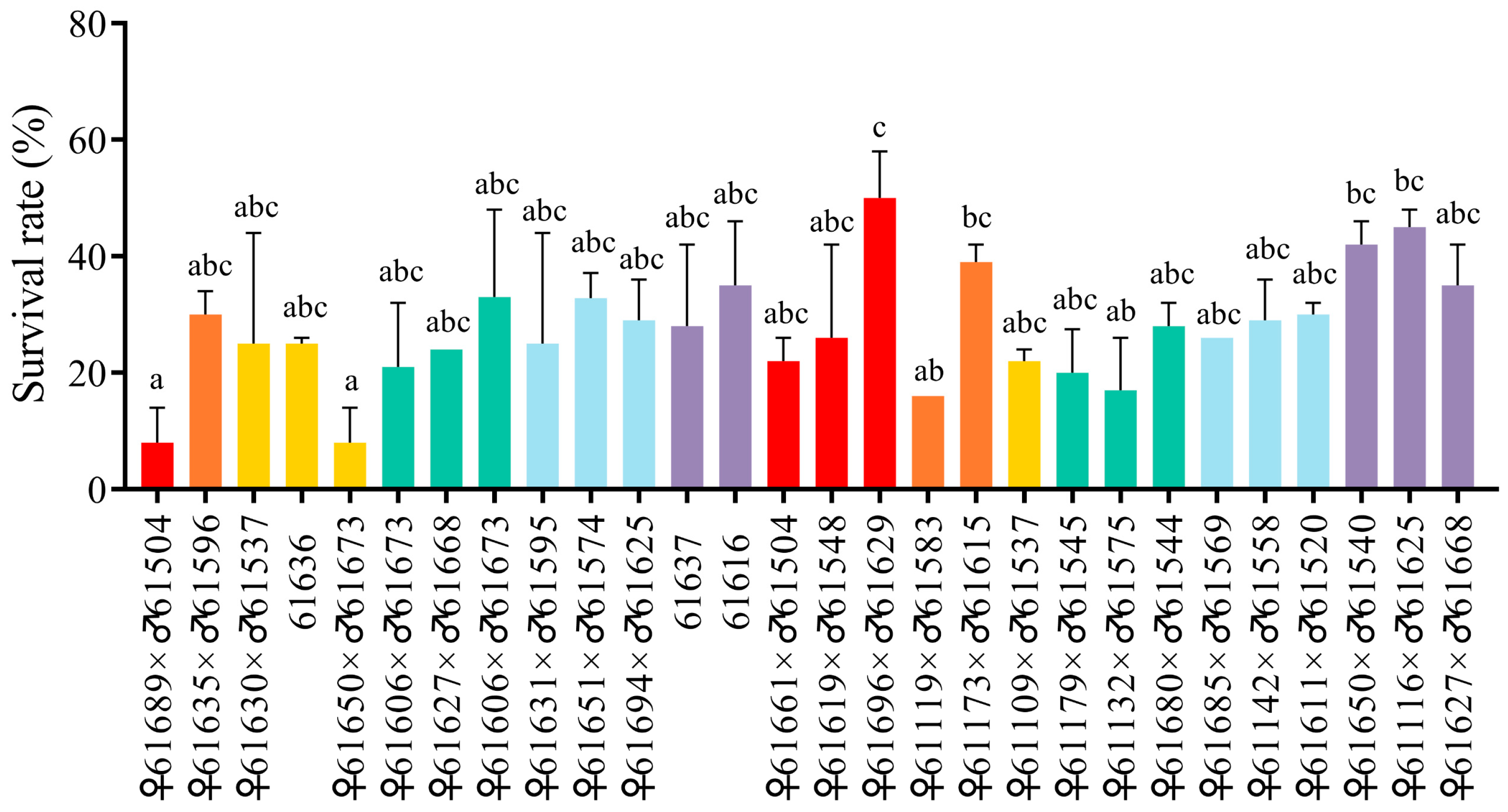
Figure 1. The 48 h survival rate of 28 families in P. monodon in the ammonia nitrogen test. The histogram represents the 48 h survival rate (%) of 28 families in P. monodon after ammonia nitrogen stress. The horizontal axis represents the number of parents in the families. Values (expressed as mean ± SEM, n = 100) with different letters are significantly different from each other (p < 0.05).
Biology 14 01532 g001
Figure 2. Syteney analysis of PmGDH and PmAST2. (A) Synteny analysis across the genomes of P. monodon, Homo sapiens, Mus musculus, Xenopus tropicalis, and Danio rerio. Syntenic blocks between P. monodon and other species’ genomes were represented by gray lines, while red lines highlight the syntenic relationship of the GDH gene. (B) Synteny analysis across the genomes of P. monodon, Homo sapiens, Mus musculus, Xenopus tropicalis, and Danio rerio. Syntenic blocks between P. monodon and other species’ genomes were represented by gray lines, while red lines highlight the syntenic relationship of the AST2 gene. (C) Analysis of the GDH gene collinearity in representative species. The same genes were represented with the same color, and the direction of transcription was indicated by the acute angle of the pentagon. (D) Analysis of AST2 gene collinearity in representative species. The same genes were represented with the same color, and the direction of transcription was indicated by the acute angle of the pentagon.
Biology 14 01532 g002
Figure 3. The predicted structural model of PmGDH-NH4+ complex and PmAST2-Asp complex. (A) Predicted structural model of the PmGDH-NH4+ complex. The secondary structures of the PmGDH protein are shown in different colors. Helix, blue. Sheet, red. Loop, orange. (B) Predicted structural model of the PmAST2-Asp complex. The secondary structures of the PmGDH protein are shown in different colors. Helix, blue. Sheet, red. Loop, orange. (C) A detailed view of the binding mode of the PmGDH protein to the ammonium ion (NH4+). The interacting residue Asp178 on loop 1 of PmGDH and NH4+ are colored, respectively. The pink atoms are O atoms of Asp178 of PmGDH, and the dark blue atoms are N atoms of NH4+. The hydrogen bonding interaction is indicated in a red dotted line. The number next to the dotted line indicates the force bond length, measured in angstroms (Å). (D) A detailed view of the binding mode of the PmAST2 protein to the aspartic acid (Asp) molecule. The interacting residues of PmAST2 and Asp are colored, respectively. The red atoms are O atoms, the dark blue atoms are N atoms, and the light blue atoms are C atoms of protein residues. The purple atoms represent the C atoms of the Asp molecules. The hydrogen bonding interactions are indicated in red dotted lines, and the salt bridge interaction is indicated in a yellow dotted line. The numbers next to the dotted lines indicate the force bond length, measured in angstroms (Å). Three replicates were performed to predict the complex structure models.
Biology 14 01532 g003
Figure 4. SNPs in exon 5 and exon 6 of PmGDH. (A) Genome structure of PmGDH, including 9 exons (blue) and 8 introns (red). (B) Nucleotide and amino acid sequences of exon 5 and exon 6 of PmGDH. Exon 5 is marked in blue. Exon 6 is marked in red. SNPs are marked in green. The serial number on both sides of each row refers to the location of the nucleotides and amino acids. (C) Alignment of the nucleotide from exon 5 of PmGDH in the different individuals belonging to the African group, Thai group, Sanya group, and Indonesian group, respectively. SNPs in exon 5 are marked in blue.
Biology 14 01532 g004
Figure 5. SNPs in exon 1 and exon 2 of PmAST2. (A) Genome structure of PmAST2, including 7 exons (blue) and 6 introns (red). (B) Nucleotide and amino acid sequences of exon 1 and exon 2 of PmAST2. Exon 1 is marked in blue. Exon 2 is marked in red. SNPs are marked in green. The serial number on both sides of each row refers to the location of the nucleotides and amino acids. (C) Alignment of the nucleotide from exon 2 of PmAST2 in the different individuals belonging to the African group, Thai group, Sanya group, and Indonesian group, respectively. SNPs in exon 2 are marked in blue.
Biology 14 01532 g005
Table 1. Characteristics of the 12 polymorphic SNP loci enrolled in this study.
SNP IDLocationPosition from Start Codon (bp)Mutation TypeVariationAmino Acid
PmGDH-1083Exon 5 of PmGDH1083AbsenceGGAGlycine
PmGDH-1086Exon 5 of PmGDH1086Transition, synonymousGGA/GGGGlycine
PmGDH-1091Exon 5 of PmGDH1091AbsenceAACAsparagine
PmGDH-1101Exon 5 of PmGDH1101Transition, synonymousTAC/TATTyrosine
PmGDH-1212Exon 6 of PmGDH1212Transversion, synonymousACC/ACAThreonine
PmGDH-1227Exon 6 of PmGDH1227Transition, synonymousCAA/CAGGlutamine
PmGDH-1338Exon 6 of PmGDH1338Transversion, synonymousGGT/GGAGlycine
PmAST2-44Exon 1 of PmAST244AbsenceAACAsparagine
PmAST2-78Exon 1 of PmAST278Transversion, synonymousGCA/GCTAlanine
PmAST2-132Exon 2 of PmAST2132Transition, synonymousGTT/GTCValine
PmAST2-180Exon 2 of PmAST2180Transition, synonymousGGC/GGTGlycine
PmAST2-225Exon 2 of PmAST2225Transition, synonymousCCG/CCAProline
Table 2. SNP genotyping of PmGDH and PmAST2 in No. 3 and No. 9 families.
Family IDSample SizeSNP IDParents (♀ × ♂)Genotype Frequency (%)p-Value for
Mendelian Ratio
Chi-Square Value
No. 950PmGDH-1101TT × CCCT 96.0CC 4.00.043 *4.082
50PmGDH-1212CC × CCCC 100\\\
50PmGDH-1227GG × GGGG 100\\\
50PmAST2-132CC × TCCT 96.0TT 4.00.000 **64.438
50PmAST2-225GG × GGGG 100\\\
No. 349PmGDH-1101CT × CCCC 44.9CT 55.10.4790.501
49PmGDH-1212CA × CCCA 49.0CC 51.00.8880.020
49PmGDH-1227GG × GGGG 98.0GA 2.00.1552.020
49PmAST2-132TT × CTTT 40.8CT 59.20.2011.633
49PmAST2-225GG × GGGG 98.0GA 2.00.1552.020
Significant differences are indicated with an asterisk: * p < 0.05, ** p < 0.01.
Table 3. SNP genotyping of PmGDH and PmAST2 in three geographic groups.
Geographic GroupsSample SizeSNP IDGenotype Frequency (%)p-Value for
Hardy–Weinberg Equilibrium
Chi-Square Value
African group50PmGDH-1101TT 10.0CT 6.0CC 84.0<0.01 **29.20
50PmGDH-1212AA 2.0AC 2.0CC 96.0<0.01 **32.33
50PmGDH-1227GA 8.0GG 92.0 0.790.06
50PmAST2-132TT 6.0CT76.0CC 18.0<0.01 **22.17
50PmAST2-225AA 4.0GA 8.0GG 88.0<0.01 **14.95
Indonesian group56PmGDH-1101TT 33.9CT 41.1CC 25.00.181.83
56PmGDH-1212AC 10.7CC 89.3 0.700.15
56PmGDH-1227AA 1.8GA 35.7GG 62.50.350.86
56PmAST2-132TT 44.6CT 51.8CC 3.60.083.17
56PmAST2-225AA 1.8GA 28.6GG 69.60.700.15
Thai group50PmGDH-1101TT 38.0CT 42.0CC 20.00.311.01
50PmGDH-1212AA 2.0AC 20.0CC 78.00.650.21
50PmGDH-1227GA 0.32GG 0.68 0.191.69
50PmAST2-132TT 42.0CT 56.0CC 2.00.02 *5.27
50PmAST2-225GA 28.0GG 72.0 0.271.22
Significant differences are indicated with an asterisk: * p < 0.05, ** p < 0.01.
Table 4. Genotype frequency of SNPs in PmGDH and PmAST2 in the association analysis with ammonia nitrogen tolerance.
SNP IDGenotypeGenotype Frequency (%)Chi-Square Valuep-Value
RGSG
PmGDH1101CT12 (20.3)6 (8.6)4.880.027 *
CC47 (79.7)52 (91.4)
PmGDH1212AC2 (3.4)5 (8.6)3.1910.074
CC57 (96.6)53 (91.4)
PmGDH1227GA8 (13.8)20 (34.5)28.1510.000 **
GG48 (82.8)28 (48.3)
AA2 (3.4)10 (17.2)
PmAST132TC11 (18.3)16 (28.1)12.9080.002 **
CC40 (66.7)24 (42.1)
TT9 (15.0)17 (29.8)
PmAST225GA2 (3.3)8 (14.0)9.5170.009 **
GG57 (95.0)46 (80.7)
AA1 (1.7)3 (5.3)
RG: resistant group; SG: susceptible group; Significant differences are indicated with an asterisk: * p < 0.05, ** p < 0.01.
Table 5. Allele frequency of SNPs in PmGDH and PmAST2 in the association analysis with ammonia nitrogen tolerance.
SNP IDA1F_AF_UA2MAFNCHROBSFrequency p-Value for FisherORFDR
PmGDH-1101T0.1017 0.0431 C0.0727 234 0.9274 0.1290 2.5130 0.3606
PmGDH-1212A0.0170 0.0431 C0.0299 234 0.9701 0.2785 0.3828 0.3606
PmGDH-1227A0.1034 0.3448 G0.2241 232 0.7759 0.0000 ** 0.2192 0.0001 **
PmAST2-132T0.2417 0.4386 C0.3376 234 0.6624 0.0015 ** 0.4079 0.0077 **
PmAST2-225A0.0333 0.1228 G0.0769 234 0.9231 0.0131 * 0.2463 0.0522
A1: Minor allele name; F_A: Frequency of A1 in resistant group; F_U: Frequency of A1 in susceptible group; A2: Major allele name; MAF: Minor allele frequency; NCHROBS: Number of alleles at the observed SNP; OR: Odds ratio; Significant differences are indicated with an asterisk: * p < 0.05, ** p < 0.01.
Table 6. Haplotype analysis of SNPs involved in ammonia nitrogen tolerance in resistant and susceptible groups.
LocusHaplotypeSusceptible
Frequency (%)
Resistant
Frequency (%)
Chi-Square Valuep-Value
PmGDH-1227/
PmAST2-132
GC99 (45.0)163 (70.3)29.5670.000 **
GT43 (19.5)45 (19.4)0.0020.968
AT49 (22.3)11 (4.7)26.5110.000 **
AC29 (13.2)13 (5.6)7.6940.006 **
** p < 0.01, significantly different distributions of the haplotype frequency between these two groups.
Disclaimer/Publisher’s Note: The statements, opinions and data contained in all publications are solely those of the individual author(s) and contributor(s) and not of MDPI and/or the editor(s). MDPI and/or the editor(s) disclaim responsibility for any injury to people or property resulting from any ideas, methods, instructions or products referred to in the content.