Article

Isolation and Characterization of Chlorpyrifos-Degrading Gut Bacteria from Field-Collected Larvae of Spodoptera frugiperda (J.E. Smith) (Lepidoptera: Noctuidae)

1
Department of Microbiology, Faculty of Agriculture, Cairo University, Giza 12613, Egypt
2
School of Biotechnology, Nile University, Giza 12677, Egypt
3
Department of Economic Entomology and Pesticides, Faculty of Agriculture, Cairo University, Giza 12613, Egypt
4
Pesticide Residues and Environmental Pollution Department, Central Agricultural Pesticide Laboratory, Agricultural Research Center, Dokki, Giza 12618, Egypt
5
Department of Biology, College of Science and Humanities, Prince Sattam Bin Abdulziz University, Al-Kharj 11942, Saudi Arabia
*
Author to whom correspondence should be addressed.
Biology 2025, 14(11), 1468; https://doi.org/10.3390/biology14111468
Submission received: 13 September 2025 / Revised: 16 October 2025 / Accepted: 19 October 2025 / Published: 22 October 2025

Simple Summary

The fall armyworm, Spodoptera frugiperda (J.E. Smith) (Lepidoptera: Noctuidae), is a highly invasive and economically important pest that inflicts significant damage on a wide range of host crops, particularly maize. One of the most challenging aspects of managing S. frugiperda populations is their capacity to develop resistance rapidly to various classes of chemical insecticides. Symbiotic microbes play vital roles in insect physiology and behavior, contributing to fundamental processes such as food digestion, nutrient acquisition, immune defense against pathogens, and even mating preferences, as well as insecticide resistance. In this study, four bacterial isolates capable of degrading chlorpyrifos were isolated from the gut of a field-collected population of S. frugiperda. These findings substantiate the concept of “detoxifying symbiosis,” wherein insect gut microbiota strengthen host survival in toxic environments. Harnessing such pesticide-degrading symbionts could pioneer innovative, microbe-based pest-management strategies that minimize reliance on synthetic chemicals.

Abstract

Exploration of new niches for microorganisms capable of degrading recalcitrant molecules is still required. We hypothesized that the gut microbiota associated with the field S. frugiperda population carries pesticide-degrading bacteria that would enhance the host’s ability to metabolize pesticides. Three strategies were implemented to address this principle: (i) isolation and identification of chlorpyrifos-degrading gut bacteria from field-collected S. frugiperda larvae; (ii) evaluation of chlorpyrifos biodegradation capacity through in vitro assays; and (iii) assessment of the impact of specific bacterial taxa capable of degrading chlorpyrifos directly within the gut. In this study, we successfully isolated four chlorpyrifos-degrading gut bacterial isolates from a field-collected population of S. frugiperda. These isolates were identified using 16S rDNA sequencing as Klebsiella quasipneumoniae strain 60D (PP504878), Klebsiella pneumoniae strain 64D (PP504879), Klebsiella pneumoniae strain 66D (PP504880), and Klebsiella pneumoniae strain 71D (PP504881). In vitro chlorpyrifos degradation assays revealed that all isolates exhibited strong degradative capacities, with Klebsiella pneumoniae strain 64D achieving the highest degradation rate, 80.38%, after one day of inoculation. In contrast, in vivo chlorpyrifos biodegradation assessment demonstrated a clear protective effect of gut bacteria on host survival. Among the mono-associated groups, larvae colonized with Klebsiella pneumoniae strain 66D exhibited the most pronounced reduction in mortality by 19.16-fold compared to antibiotic-treated larvae following exposure to chlorpyrifos suspension.

1. Introduction

The fall armyworm (FAW), Spodoptera frugiperda (J.E. Smith) (Lepidoptera: Noctuidae), is a polyphagous pest native to tropical and subtropical Americas. The Food and Agriculture Organization (FAO) classified FAW as a global quarantine pest in 2016. Maize is the preferred host for FAW among infested countries, with yield losses of maize crops reaching up to 70% [1]. For a long time, chemical pesticides have been the main tactic for the management of FAWs [2]. Among them, chlorpyrifos is a broad-spectrum organophosphate insecticide commonly used against lepidopteran pests, which induces neurotoxicity and ultimately neuronal death [3]. The FAW is a pest with a high capacity to develop resistance to pesticides, as evidenced by the wide range of cases worldwide [2]. When exposed to pesticides, resistant insect pests enhance their tolerance through multiple mechanisms, including overexpression of pesticide detoxification and degradation enzymes [4,5] mutation of pesticide receptors [4,6], and altered cuticle thickness and composition [7]. Apart from these host-driven mechanisms, the gut microbiota of insects may also play an important role in alleviating the adverse effects of different insecticidal compounds [8].
More broadly, the diversity, abundance, and evolutionary success of insects are to some extent related to their relationships with symbiotic bacteria, which directly affect the performance of insects in nature [9]. Recent studies have revealed that the insect microbiome shapes their response to pesticides, either through direct degradation of insecticides by microbes that influence the host immune system [10] or through microbe-mediated activation or enhancement of insecticide toxicity [11,12]. Accompanying roles in nutrient provisioning and immune system stimulation, some microbial symbionts can break down complex molecules such as insecticides and promote insecticide resistance [10,13,14,15]. It is also notable that pathogenic bacteria can reside in host guts, only initiating or facilitating pathogenesis under certain conditions [16,17].
Insecticide-degrading bacteria are ubiquitous in nature and have been found in several orders of insects [18], such as Coleoptera [19,20], Diptera [21], Hemiptera [15], and Lepidoptera [22,23]. In Lepidoptera, in vitro assays demonstrated the potential of bacteria isolated from the gut of Plutella xylostella (Linnaeus) (Lepidoptera: Plutellidae) [23] and from resistant strains of S. frugiperda [22] to degrade several insecticides, including lambda-cyhalothrin, deltamethrin, chlorpyrifos ethyl, spinosad, and lufenuron. Moreover, insecticide-guided selection of strains of S. frugiperda led to the selection of insecticide-degrading bacteria, which were not present in the microbiota of susceptible, unselected larvae [22]. Similarly, Siddiqui et al. (2025) reported the isolation of several culturable bacterial species, including Klebsiella variecola, from S. frugiperda larvae treated with profluanilide, spinosad, and indoxacarb, further supporting the adaptive role of gut microbiota in pesticide detoxification [24].
To better understand this symbiotic interaction, we used the destructive invasive pest S. frugiperda to evaluate the effects of pesticide exposure on the diversity and ability of the gut bacterial community to metabolize pesticides. This study aimed to identify bacterial strains associated with pesticide resistance and uncover potential mechanisms. We hypothesized that gut bacteria would enhance the host’s ability to metabolize pesticides. To address this principle, the following methods were hypothesized: (i) isolate and identify the chlorpyrifos-degrading gut bacteria from the field collection of the invasive pest S. frugiperda larvae, (ii) evaluate chlorpyrifos biodegradation ability through an in vitro assay, and (iii) assess the impact of particular bacterial taxa that have the ability to degrade chlorpyrifos directly in the gut.

2. Materials and Methods

2.1. Sampling

A total of 50 S. frugiperda larvae were obtained from a maize field in Giza Governorate located at the following coordinates: “29°42′49.4″ N 31°17′12.4″ E” during summer 2022. In this field, insecticides are used periodically for the management of S. frugiperda. The collected larvae were transported directly to the laboratory in a cold container. The larvae were starved for 24 h before dissection.

2.2. Pesticides

Commercial-grade insecticide chlorpyrifos (chlorpyrifos® 50% EC, Dow Agro Sciences, Manchester, UK), pyrethroid insecticide lambda-cyhalothrin (Axone® 5% EC, 5 ppm; Jiangsu Changqing Agrochemical, Yangzhou, China), the diamide insecticide chlorantraniliprole (Coragen 20% SC, 0.6 ppm; Coragen® 20% SC, DuPont, Nemours, France), and the carbamate insecticide Lannate (Lannit® 90% WP, 0.5 ppm; Rotam Agrochemical, Zone Kunshan, Jiangsu, China) were used in the current study. All the tested insecticides are diluted in distilled water.

2.3. Isolation of Chlorpyrifos-Degrading Gut Bacteria

All larvae were surface-disinfected by immersion in 70% ethanol for 2 min, followed by two rinses with sterile distilled water. Under aseptic conditions and using a stereomicroscope (Bergamo, Italy), intact guts of ten S. frugiperda larvae were dissected and pooled into a sterile 15 mL Falcon tube containing 9 mL of sterile M9 salt solution [25]. To isolate chlorpyrifos-degrading gut bacteria, an enrichment culture technique was employed. One milliliter of the pooled gut homogenate was inoculated into M9 mineral medium (MM9) at a final concentration of 20% (v/v). The MM9 medium was prepared using the following components: 100 mL of 10× M9 salts (Na2HPO4·2H2O, 75.2 g/L; KH2PO4, 30 g/L; NaCl, 5 g/L; NH4Cl, 5 g/L), 1 mL of 1 M MgSO4·7H2O, and 1 mL of 1 M CaCl2, supplemented with chlorpyrifos (20% EC) at a concentration of 240 ppm as the sole carbon source.
The pH of the medium was adjusted to 7.0 ± 0.2. Cultures were incubated at 30 °C on a rotary shaker at 160 rpm for 14 days. Enrichment samples were collected on days 5, 7, 10, and 14. Aliquots (1 mL) were serially diluted and plated onto an MM9 agar medium containing chlorpyrifos (240 ppm) using the pour plate technique. Plates were incubated at 30 °C for 7 days. Emerging bacterial colonies were purified by repeated subculturing and preserved in 20% glycerol stocks at −80 °C for further identification and biodegradation analysis.

2.4. Molecular Identification of Bacterial Isolates

The purified isolates were grown on nutrient broth at 30 °C for 48 h. Total genomic DNA from the selected strains was extracted and purified using the Thermo Scientific GeneJET Kit, Waltham, MA, USA (K0721, K0722) according to the manufacturer’s protocol. 16S rRNA gene fragments were amplified using the Thermo Scientific Dream Taq Green PCR MasterMix (2×) kit according to the supplier’s protocol. PCR was carried out using forward (5′ CCAGCAGCCGCGGTAATACG 3′) and reverse (5′ATCGG(C/T)TACCTTGTTACGACTTC 3′) 16S rRNA primers [26,27]. 16S rRNA PCR amplicons were purified using a QIAquick PCR Purification Kit according to the supplier’s instructions (QIAGEN, Hilden, Germany). DNA sequencing was performed by the Sanger method [28] and performed by Macrogen (Seoul, Korea). The BLASTN program was used to submit the nucleotide sequences at the NCBI BLAST server (https://blast.ncbi.nlm.nih.gov/Blast.cgi, accessed on 28 March 2024). The partial 16S rDNA sequences (ranging from 1096 to 1328 bp and the average read quality (Q ≥ 30)) were deposited in NCBI GenBank under accession numbers PP504878-PP504881. The phylogenetic tree (neighbor-joining with bootstrap method) was constructed using MEGA11 [29]. The analysis featured 28 sequences, which correspond to the nearest matches found in the GenBank database.

2.5. Factors Affecting the Growth of Chlorpyrifos-Degrading Isolates

To optimize the growth of chlorpyrifos-degrading bacterial isolates, we assessed the effect of incubation temperature, pH, and incubation time. The flasks were loaded in triplicate with 20% of sterile MM9 broth media supplemented with 250 ppm chlorpyrifos. Each set of triplicate flasks was inoculated with 2% of fresh bacterial cultures (approx. 106 CFU/mL). Each optimized parameter will be passed to the following one [25].
The effect of temperature was investigated by incubation at different temperatures (20, 25, 30, 35, and 40 °C) at pH 7 and 160 rpm for 5 days on a rotary shaker (VISION SCIENTIFIC CO., LTD., Model: VS-8480, Daejeon-Si, Korea). While studying the effect of pH, it was carried out with different pH ranges (4–9) and then incubated at the optimum temperature for each isolate and 160 rpm for 5 days on a rotary shaker. The optimum incubation time, which is required for maximum chlorpyrifos-degrading isolate growth, was determined at 0, 1, 2, 3, 4, 5, 6, 7, and 8 days. Followed by incubation at optimum temperature and pH for each isolate and 160 rpm on a rotary shaker. For growth study analysis of all factors, an aliquot of 1 mL of culture was withdrawn daily, and growth was evaluated as viable cell counts on nutrient agar media.

2.6. In Vitro Chlorpyrifos Biodegradation Assay

Four bacterial isolates—Klebsiella quasipneumoniae strain 60D, Klebsiella pneumoniae strain 64D, Klebsiella pneumoniae strain 66D, and Klebsiella pneumoniae strain 71D (approximately 106 CFU/mL)—were used to inoculate MM9 broth medium supplemented with chlorpyrifos (250 ppm). Cultures were incubated at the optimal temperature for each isolate on a rotary shaker at 160 rpm for 5 days. Samples were collected at 1-day intervals. The colony-forming units (CFU/mL) of the incubated cultures were determined using the dilution plate counting method.
For pesticide biodegradation analysis, a modified QuEChERS extraction protocol was applied [30]. For sample preparation, 10 mL from each culture (in triplicate) was transferred into a 50 mL centrifuge tube, followed by the addition of 10 mL acetonitrile. Tubes were sealed and placed in a freezer at −18 °C for 15 min. Subsequently, 4 g of MgSO4 and 1 g of NaCl were added. Tubes were hand-shaken vigorously for 1 min, then centrifuged at 4000 rpm for 5 min at 5 °C. One milliliter of the supernatant was transferred to a sealed GC vial for analysis. Residual chlorpyrifos in the media was quantified by GC–MS analysis using an HP 6890 gas chromatograph equipped HPLC system (Agilent HPLC 1260 infinite series; Agilent Technologies, Santa Clara, CA, USA) with an HP 7673 autosampler. The external standard method was employed to determine the insecticide content in each sample, and the degradation rate was calculated using the following formula:
ρ = (1 − A1/A0) × 100%
where A1 represents the insecticide content in the MM9 medium after bacterial inoculation, and A0 represents the insecticide content in the uninoculated control [31]. Each sample was analyzed in three technical replicates, and its mean result was considered one biological replicate.

2.7. Screening of Biodegradation Capabilities by Chlorpyrifos-Degrading Isolates to Other Pesticides

To evaluate the broader biodegradation potential of the chlorpyrifos-degrading isolates, each isolate was streaked in triplicate onto MM9 agar plates supplemented with one of three different pesticide groups, each serving as the sole carbon source. The tested pesticides included the pyrethroid lambda-cyhalothrin (Axone 5% EC, 5 ppm), the diamide chlorantraniliprole (Coragen 20% SC, 0.6 ppm), and the carbamate Lannate (Lannate 90% WP, 0.5 ppm), each applied at its recommended field concentration. A negative control (uninoculated plates) was also included to ensure that no growth occurred in the absence of bacterial inoculation. The inoculated plates were incubated at 30 °C for 7 days. Bacterial growth on these selective media was considered indicative of the isolate’s potential biodegradation capability for the respective pesticide. This screening served as a preliminary assessment of the metabolic versatility of the isolates in degrading structurally and functionally distinct classes of insecticides.

2.8. In Vivo Chlorpyrifos Biodegradation Assay

2.8.1. Experimental Design

A laboratory strain of S. frugiperda was used to determine the biodegradability of chlorpyrifos-degrading isolates in vivo. Egg clusters of S. frugiperda were used in two main groups, as demonstrated in Figure 1. First group (mono-associated): The egg clusters were immersed in 70% ethanol for 45 s and rinsed again in autoclaved distilled water three times. Disinfected eggs were maintained in a 90 mm plastic Petri dish and allowed to dry on a clean bench at room temperature. After hatching, larvae were reared on castor leaves (Ricinus communis L.) dipped in an antibiotic mixture containing Penicillin G, Polymyxin B, and erythromycin dissolved in sterile water at a concentration of 150, 150, and 600 μg/mL, respectively, to disrupt the S. frugiperda gut microbiome following protocols in [12,14]; leaves were replaced every 24 h, and antibiotic treatment was applied until the larvae molted to the third instar. The third instar larvae were reared individually in a clean, sterile plastic cup. The larvae were orally inoculated individually by daily feeding with castor leaf disks (5 cm) that had been dipped for 45 s in suspensions (approx. 106 CFU/mL) of the chlorpyrifos-degrading isolates Klebsiella quasipneumoniae strain 60D, Klebsiella pneumoniae strain 64D, Klebsiella pneumoniae strain 66D, and Klebsiella pneumoniae strain 71D and, subsequently, air-dried under aseptic conditions at room temperature. The second group (control groups): Egg clusters were divided into two subgroups: (i) egg clusters that were disinfected as mentioned above and treated with an antibiotic mixture until they reached the 3rd larval instar, and (ii) egg clusters that were not treated with antibiotics, and the larvae were reared on castor leaves without any additional treatment. The third larval instar of the control groups was maintained daily with castor leaf disks (5 cm) dipped in distilled water until it reached the 4th larval instar. To validate the effect of antibiotics on the gut bacteria, larval weight and gut bacterial count were estimated. The thirty-third larval instar of S. frugiperda in all the above-mentioned treatments was weighed, and then the thirty larvae were freshly dissected in 10 mL of sterilized M9 salts, and bacterial counting was applied on both nutrient agar and MM9 agar media containing chlorpyrifos (250 ppm).

2.8.2. Bioassay of S. frugiperda 4th Instar Larvae

Chlorpyrifos toxicity bioassays on newly ecdysed 4th instar larvae of S. frugiperda were performed using 24 ppm. The leaf-dipping technique was used according to [32] as follows: For experiments, castor leaves were dipped in chlorpyrifos suspension (24 ppm) for 20 s and then allowed to dry in the air. Each treatment had five replicates; each replicate consisted of 50 larvae (n = 250). Leaves dipped in water were used in the control group. The larvae were allowed to feed on treated leaves for 24 h, and the surviving larvae were then transferred to a clean, dry container containing fresh, untreated leaves [33]. Larval mortality was recorded daily for five consecutive days after post-treatment was calculated. In addition, detoxification enzyme activity, pesticide residue in larval feces, and bacterial count in larval feces were investigated.

2.8.3. Biochemical Assays

Insect Treatment and Sample Preparation
Fourth instar larvae of Spodoptera frugiperda were exposed to a chlorpyrifos suspension (24 ppm) as described above. At 24, 72, and 120 h post-treatment, 50 mg of fresh body weight from surviving larvae were collected for enzymatic activity analysis. Each time point included five biological replicates. Larvae were homogenized in 0.1 M phosphate buffer, pH 7.8 for acetylcholinesterase (AChE) activity, and pH 6.5 for glutathione S-transferase (GST) activity. The homogenates were centrifuged at 7000 rpm for 15 min at 4 °C, and the resulting supernatants were used for enzyme activity and total protein measurements. Crude enzyme extracts were prepared separately for each enzyme assay. Total protein content in each replicate was determined using Coomassie Brilliant Blue dye, with bovine serum albumin as the standard, following Bradford’s method [34].
Acetylcholine Esterase (AChE) Assay
AChE activity was determined following the method of Ellman et al. 1961 [35]. The reaction mixture consisted of 100 µL of enzyme extract and 50 µL of 0.075 M acetylthiocholine iodide (ATChI) as the substrate. The reaction was initiated by adding 50 µL of 0.01 M dithiobisnitrobenzoic acid (DTNB). The absorbance was recorded at 412 nm at one-minute intervals over a 5-min period. Enzyme activity was expressed in units per mg of protein [36].
Glutathione S-Transferase (GST) Assay
GST activity was measured according to the method described by [37]. The reaction mixture contained 10 µL of enzyme extract, 25 µL of 30 mM 1-chloro-2,4-dinitrobenzene (CDNB) as the substrate, 25 µL of 50 mM reduced glutathione (GSH), and 940 µL of 50 mM potassium phosphate buffer (pH 6.5), bringing the total volume to 1 mL. The optical density was measured at 340 nm at one-minute intervals over 5 min. GST activity was calculated using the extinction coefficient (ε = 9.6 mM−1 cm−1) [38].

2.8.4. Quantification of Chlorpyrifos Residues in Larval Feces

The extraction and analysis of chlorpyrifos residues from S. frugiperda feces were performed with slight modifications to the method described by [39]. Approximately 50 mg of feces were collected and transferred to a PTFE tube, to which 3 mL of acetonitrile was added. The samples were vortexed for 1 min, left to stand for 15 min, and then centrifuged at 3500 rpm for 15 min. The supernatants were frozen at −20 °C for 1 h to precipitate fats, then filtered through 0.22 μm PTFE syringe filters. For solid-phase extraction (SPE) cleanup, each sample was mixed with 500 mg of MgSO4 and 75 mg of PSA (primary secondary amine). The mixture was vortexed for 60 s and centrifuged again at 3500 rpm for 5 min. Approximately 1 mL of the supernatant was filtered through a 0.22 μm PTFE filter in preparation for GC-ECD analysis.
In Schwantes et al., 2020, gas chromatography was used for chlorpyrifos residue quantification with some modifications [40], a gas chromatograph (HP 6890) equipped with an autosampler (HP 7673) and a dual micro-electron capture detector (μECD), using a capillary column (30 m × 0.32 mm × 0.25 μm, HP-5, 5% phenylmethyl polysiloxane). The injector and detector temperatures were set at 250 °C and 300 °C, respectively, with nitrogen as the carrier gas at a flow rate of 1.5 mL/min. The GC oven temperature program was set as follows: initial temperature at 160 °C held for 2 min, then increased at 10 °C/min to 200 °C and held for 5 min. An amount of 1 µL aliquot of the prepared sample was injected, and the retention time for chlorpyrifos was 6.5 min.

2.8.5. Bacterial Count in Larval Feces

To ensure the definite isolates’ presence, 50 mg of feces were collected from each plastic cup. These feces samples were homogenized with 450 μL of sterilized PBS solution, and 100 μL of the homogenized feces were plated on MM9 media supplemented with chlorpyrifos and incubated for 5 days at 30 °C.

2.9. Data Analysis

Data was coded and entered using the statistical package SPSS V.22 and R (version 3.2.5). Data were tested for satisfying assumptions of parametric tests, and continuous variables were subjected to the Shapiro–Wilk and Kolmogorov–Smirnov tests for normality. Probability and percentile data were standardized for normality using Arcsine Square Root. Data were presented as mean and standard deviation. ANOVA followed by Tukey’s post hoc pairwise comparison was performed for all experimental groups to evaluate the recorded growth factors, degradation rate, enzymatic activities (AChE and GST), bacterial counting, and in vitro assays. Each group was analyzed using at least three replicates (five replicates for the in vivo assay). p-values < 0.05 were considered statistically significant. Data were visualized, when applicable, using R software (version 3.2.5).

3. Results

3.1. Isolation and Molecular Identification of Chlorpyrifos-Degrading Gut Bacteria

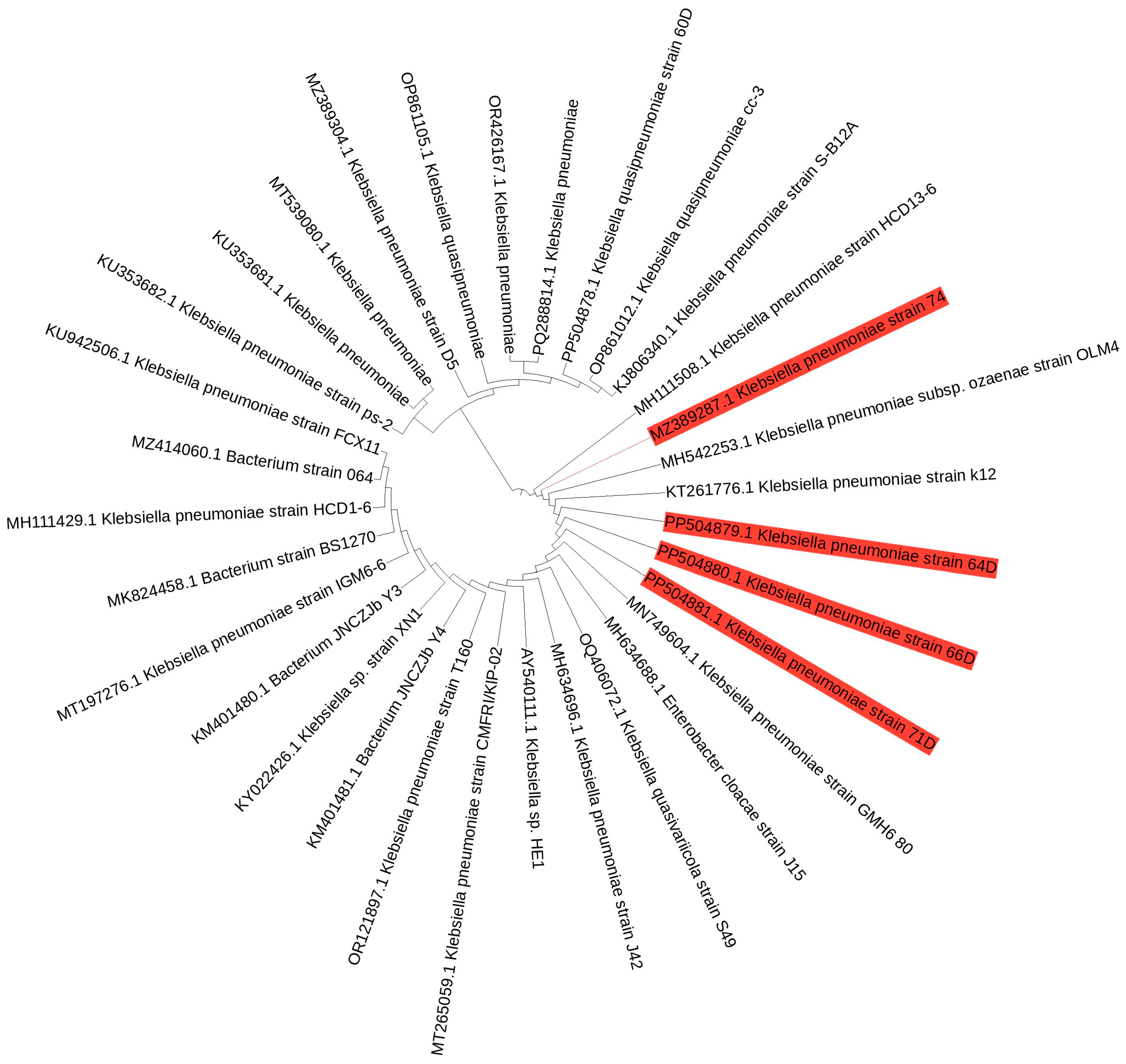
Four bacterial isolates were obtained and purified. Then, the NCBI BLAST software 2.17.0 was employed to sequence and identify the four strains, facilitating a comparison with analogous sequences found in the GenBank database. The BLAST analysis of the 16S rDNA sequences from strains 60D, 64D, 66D, and 71D exposed a 100% similarity to Klebsiella quasipneumoniae strain 60D, Klebsiella pneumoniae strain 64D, Klebsiella pneumoniae strain 66D, and Klebsiella pneumoniae strain 71D, respectively, as shown in Figure 2. The 16S rRNA sequences were deposited and assigned to GenBank, with accession numbers that span from PP504878 to PP504881. The phylogenetic tree, constructed through the UPGMA for the bacterial isolates, illustrated substantial phylogenetic relatedness (Figure 2).

3.2. Factors Affecting the Growth of Chlorpyrifos-Degrading Isolates

A series of experiments was carried out in batch cultures to determine the ideal culture conditions for obtaining the highest growth of four chlorpyrifos-degrading strains: Klebsiella quasipneumoniae 60D, Klebsiella pneumoniae 64D, Klebsiella pneumoniae 66D, and Klebsiella pneumoniae 71D.

3.2.1. Effect of Incubation Temperature

All Klebsiella strains demonstrated temperature-dependent growth, with optimal ranges typically from 25 °C to 35 °C. The highest growth was significantly (p < 0.05) observed at 30 °C for strains K. quasipneumoniae 60D and K. pneumoniae 71D, while for strains K. pneumoniae 64D and K. pneumoniae 66D, the optimum temperature was 35 °C (Figure 3). Bacterial counts are expressed as CFU/mL on a logarithmic scale. The obtained data showed consistently high counts in K. pneumoniae 64D and K. pneumoniae 66D (approx. 9.1 Log10), followed by K. quasipneumoniae 60D (approx. 8.8 Log10), then K. pneumoniae 71D (approx. 8.3 Log10). All strains exhibited a significant decline in viability at 20 °C and 40 °C.

3.2.2. Effect of pH

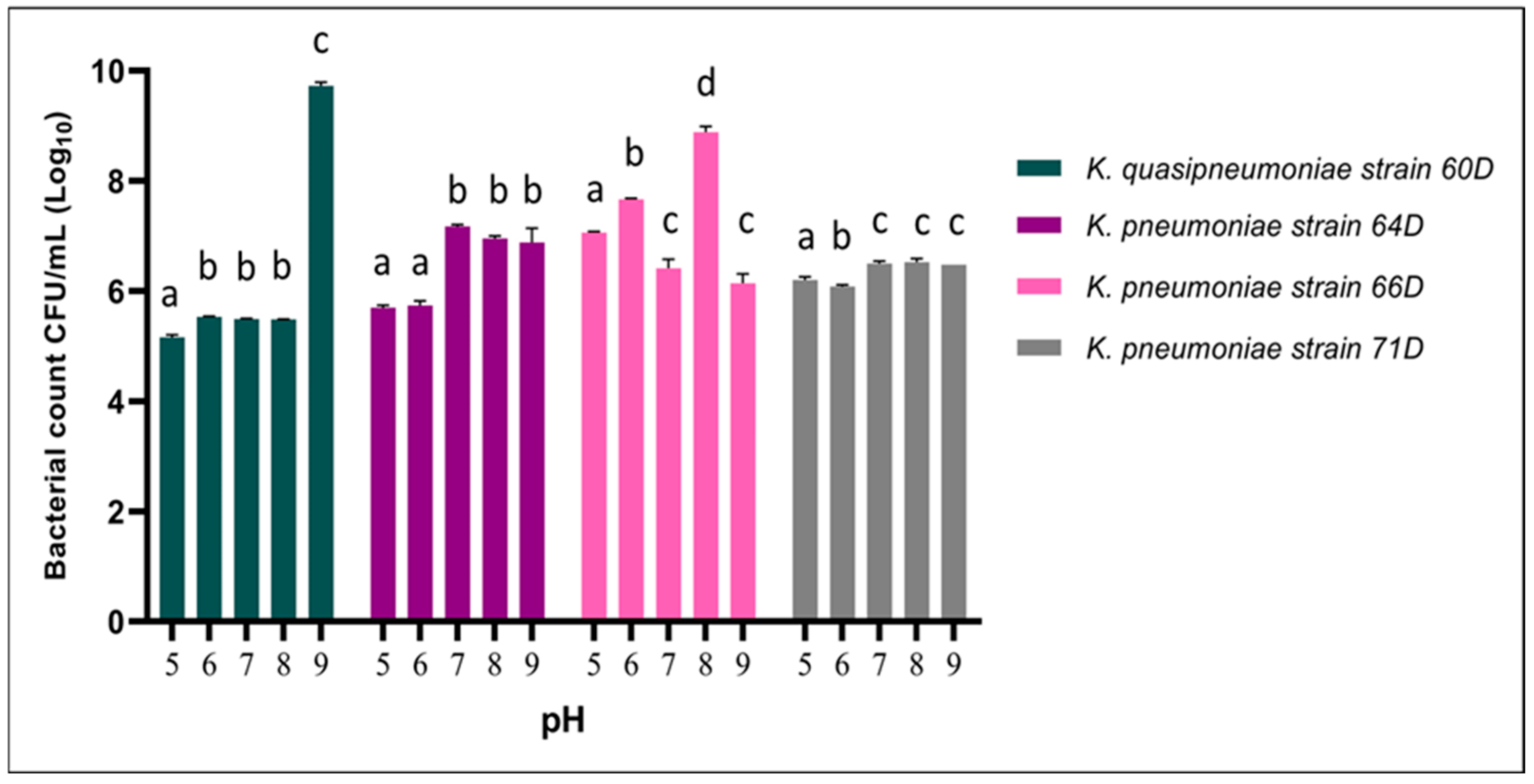
The influence of pH (ranging from 5 to 9) on the growth of K. quasipneumoniae 60D and K. pneumoniae strains 64D, 66D, and 71D was evaluated by measuring bacterial counts (CFU/mL, Log10). K. quasipneumoniae 60D exhibited a markedly enhanced growth at pH 9 (p < 0.05), reaching approximately 9 Log10, while maintaining significantly lower but relatively stable growth (approx. 5.4–5.6 Log10) across pH 5 to 8, suggesting a narrower pH optimum. K. pneumoniae 64D displayed a higher growth at a pH range from 7 to 9 with no significant difference between these pH ranges (approx. 6.9–7.1 Log10). In contrast, K. pneumoniae 66D demonstrated optimal growth at pH 8 (approx. 8.9 Log10), with reduced viability at both acidic and high alkaline conditions. K. pneumoniae 71D showed a uniform growth profile over the tested pH ranges (approx. 6.1–6.5 Log10), indicating a broad pH tolerance but without a distinct optimum. These findings highlight pH-dependent variability in growth responses among Klebsiella strains, with optimum growth at neutral to alkaline pH, and all strains, specifically K. quasipneumoniae 60D, were stimulated at a pH of 9, as shown in Figure 4.

3.2.3. Effect of Incubation Time

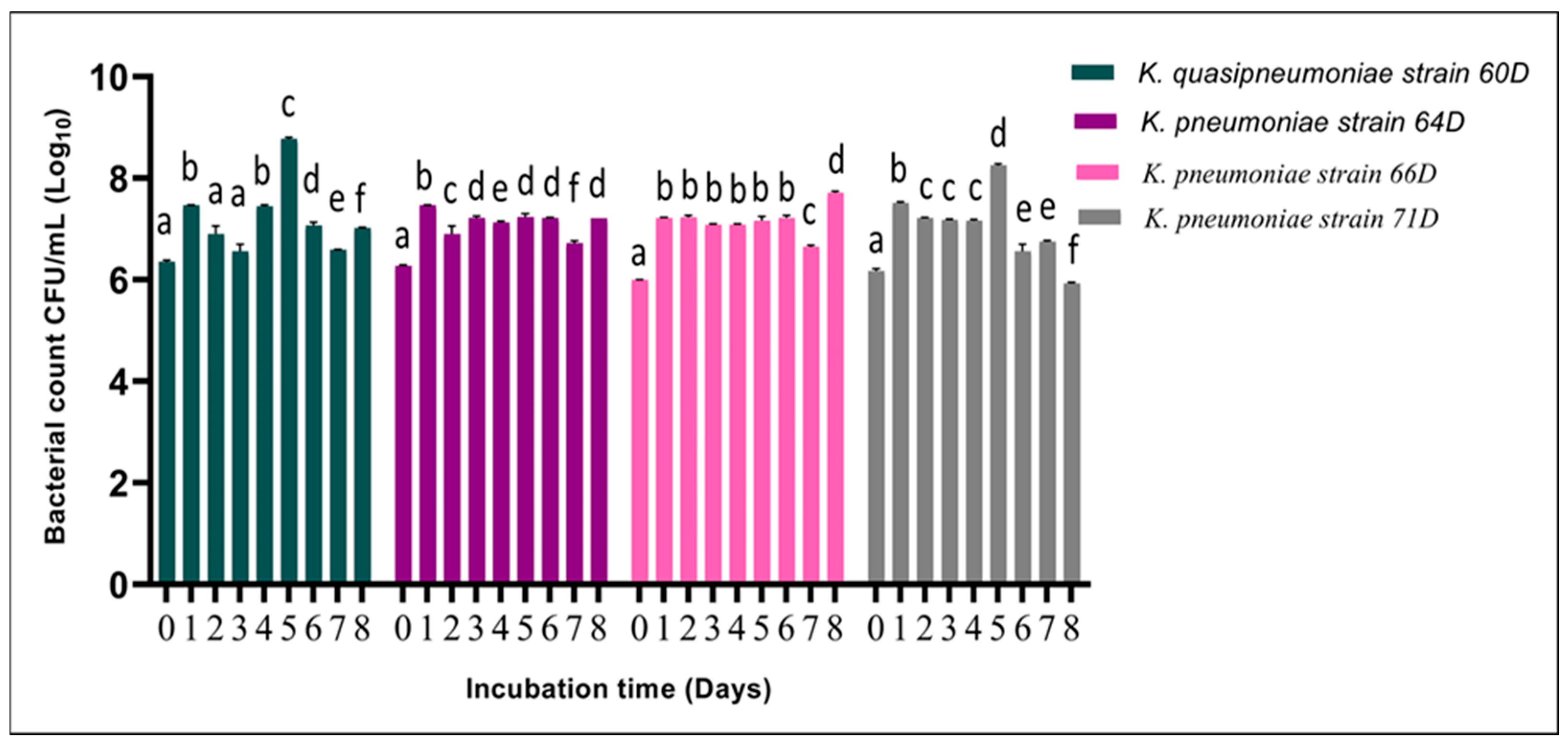
The growth kinetics of four Klebsiella strains—K. quasipneumoniae 60D and K. pneumoniae strains 64D, 66D, and 71D—were monitored over an 8-day incubation period, with bacterial counts expressed as CFU/mL (Log10), Figure 5. All strains exhibited distinct growth patterns over time, with significant inter-strain differences. K. quasipneumoniae 60D exhibited a marked increase in growth, reaching a peak at day 5 (approx. 8.8 Log10), followed by a significant moderate decline through day 8 (p < 0.05). K. pneumoniae 64D demonstrated early and stable growth, with a high growth peak starting at day 1 (approx. 7.4 Log10), while cell counts plateaued with nominal oscillation from day 2 through day 8 (approx. 6.8–7.2 Log10). K. pneumoniae 66D showed sustained high-level growth from day 1 through day 6 (approx. 7–7.2 Log10), with minimal fluctuation, before peaking slightly at day 8 (approx. 7.7 Log10). Conversely, K. pneumoniae 71D exhibited a more gradual growth trajectory, achieving its highest count (approx. 8.2 Log10) on day 5, followed by a significant reduction in viability by day 8 (approx. 5.9 Log10), p < 0.05.

3.3. In Vitro Insecticide Biodegradability of Isolated Gut Bacteria

All four isolated bacterial strains exhibited strong chlorpyrifos-degrading capabilities. The highest degradation rate was observed after one day of inoculation, with Klebsiella pneumoniae strain 64D achieving 80.38%, followed by K. quasipneumoniae strain 60D (77.9%), K. pneumoniae strain 71D (65.52%), and K. pneumoniae strain 66D (51.3%) (Table 1). However, after five days of incubation, K. pneumoniae strain 66D showed the highest degradation efficiency at 61.14%. Nevertheless, it is not always possible to extrapolate biodegradation rates calculated for higher pesticide doses to lower concentrations [41], as strain 64D exhibited the lowest degradation rate at 25%.

3.4. Screening of Biodegradation Capabilities by Chlorpyrifos-Degrading Isolates to Other Pesticides

All four strains demonstrated the ability to degrade chlorantraniliprole (Coragen) and the carbamate insecticide (Lannate). Notably, only K. quasipneumoniae strain 60D exhibited the capacity to biodegrade the pyrethroid lambda-cyhalothrin (Table 2).

3.5. In Vivo Chlorpyrifos Biodegradation Assay

3.5.1. Bacterial Counting in Larval Gut

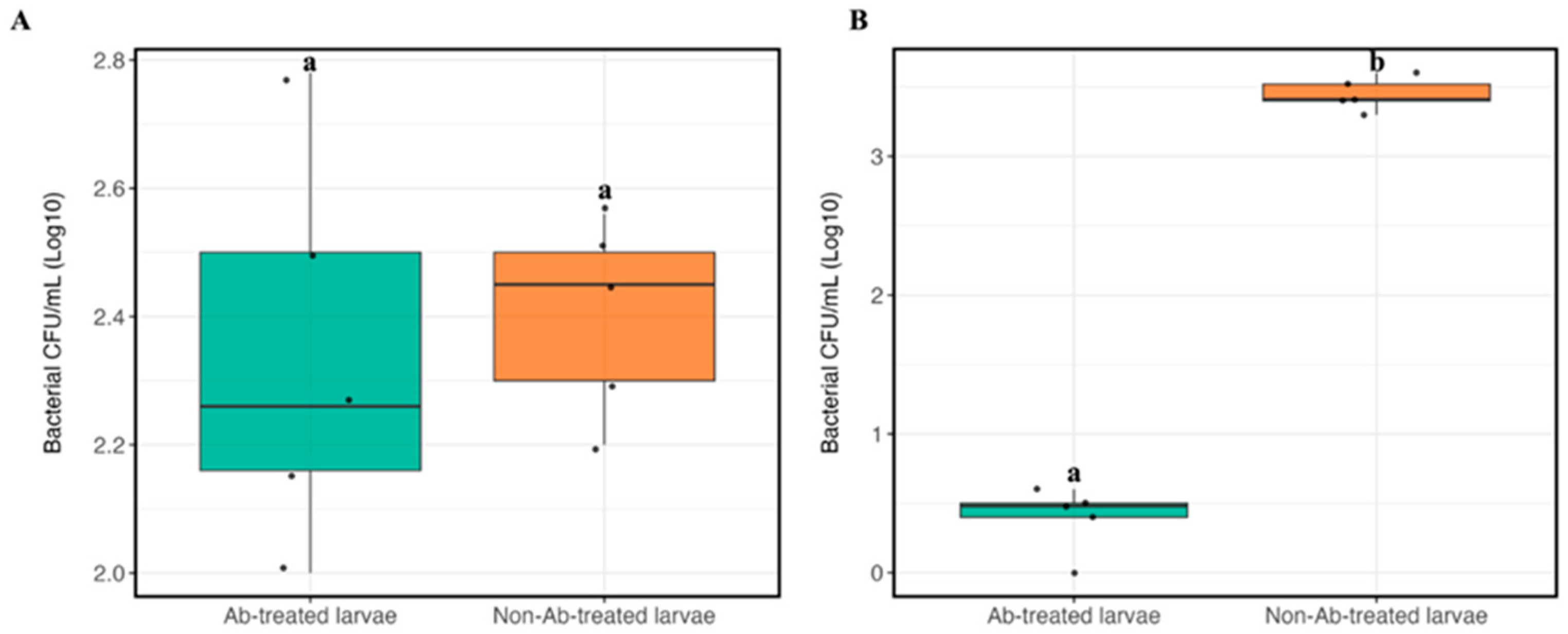
The bacterial CFU/mL (Log10) of chlorpyrifos-degrading gut bacteria was determined under different conditions using NA (Figure 6A) and MM9 mineral media supplemented with chlorpyrifos (Figure 6B). A comparison of antibiotic-treated and non-antibiotic-treated larval groups revealed significant variations in bacterial counts (p < 0.05) grown on MM9 media supplemented with chlorpyrifos (approx. 2.2 Log10). However, the bacterial counts in antibiotic-treated and non-antibiotic-treated larvae groups on NA media did not differ significantly. These results suggest that MM9 mineral media, supplemented with chlorpyrifos as the sole carbon source, selectively enriched chlorpyrifos-degrading bacteria, particularly in non-antibiotic-treated larvae. Furthermore, while bacterial survival is similar across the groups on rich media, antibiotic treatment decreases bacterial colonization with chlorpyrifos under nutrient-limited settings.

3.5.2. Larval Weight

The data presented in Table 3 indicate that S. frugiperda larvae treated with an antibiotic mixture for six days post-hatching exhibited a significant reduction in weight (8.57 mg) compared to the non-antibiotic-treated group (12.73 mg). This suggests that smaller body mass in antibiotic-treated larvae may result from the disruption of the gut microbiota, which adversely affects larval development and growth [10,22,42], or reduction in nutrient uptake [43].

3.5.3. Mortality

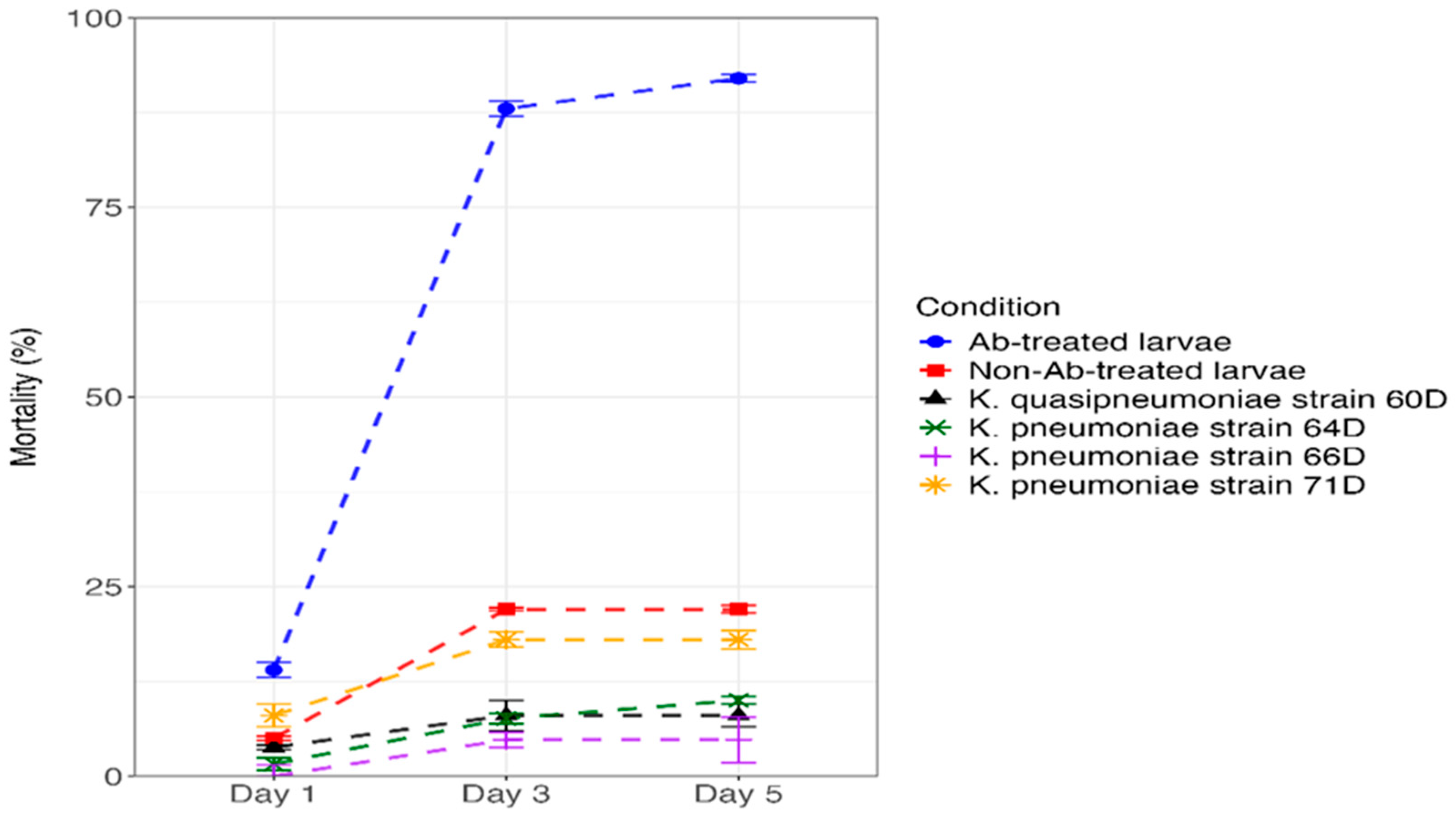
The mortality rate of 4th instar S. frugiperda larvae after being exposed to chlorpyrifos suspension at 24 ppm exhibited varying levels of toxicity. As shown in Figure 7 and TS1, the Ab-treated larvae caused the maximum mortality rate (14%) after 24 h of treatment. The second-highest mortality rate was recorded with K. pneumoniae strain 71D (8%), followed by non-Ab-treated larva (5%), K. quasipneumoniae strain 60D (3.8%), K. pneumoniae strain 64D (1.6%), and K. pneumoniae strain 66D (0%) after 24 h of treatment. After 5 days of treatment, Ab-treated larvae exhibited higher larval mortality of 92%, followed by non-Ab-treated larvae (22%), K. pneumoniae strain 71D (18%), K. pneumoniae strain 64D (10%), K. quasipneumoniae strain 60D (8%), and K. pneumoniae strain 66D (4.8%).

3.5.4. Detoxification Enzymes Activity

After being exposed to chlorpyrifos suspension (24 ppm), the AchE enzyme activity of S. frugiperda larvae was significantly reduced after 24 h compared with antibiotic-treated larvae. The mono-associated K. quasipneumoniae strain 60D shows the highest inhibition in AchE enzyme activity after being exposed to chlorpyrifos suspension by 3.5-fold compared to antibiotic-treated larvae. Followed by K. pneumoniae strain 64D, K. pneumoniae strain 71D, and K. pneumoniae strain 66D by about 2.96, 1.8, and 1.2-fold, respectively (Table 4). After three and five days from exposure to chlorpyrifos suspension, there is no significant difference in AchE enzyme activity of S. frugiperda larvae. The GST activity exhibited a different pattern; there was no significant difference in GST enzyme activity in S. frugiperda larvae after 1 and 3 days of exposure to chlorpyrifos suspension. A significant difference has been observed in the mono-associated K. pneumoniae strain 66D, K. pneumoniae strain 71D, K. quasipneumoniae strain 60D, and K. pneumoniae strain 64D by about 3.0-, 2.9-, 2.2-, and 1.8-fold inhibition, respectively, compared with antibiotic-treated larvae.

3.5.5. Pesticide Residues in Feces (Larval Frass)

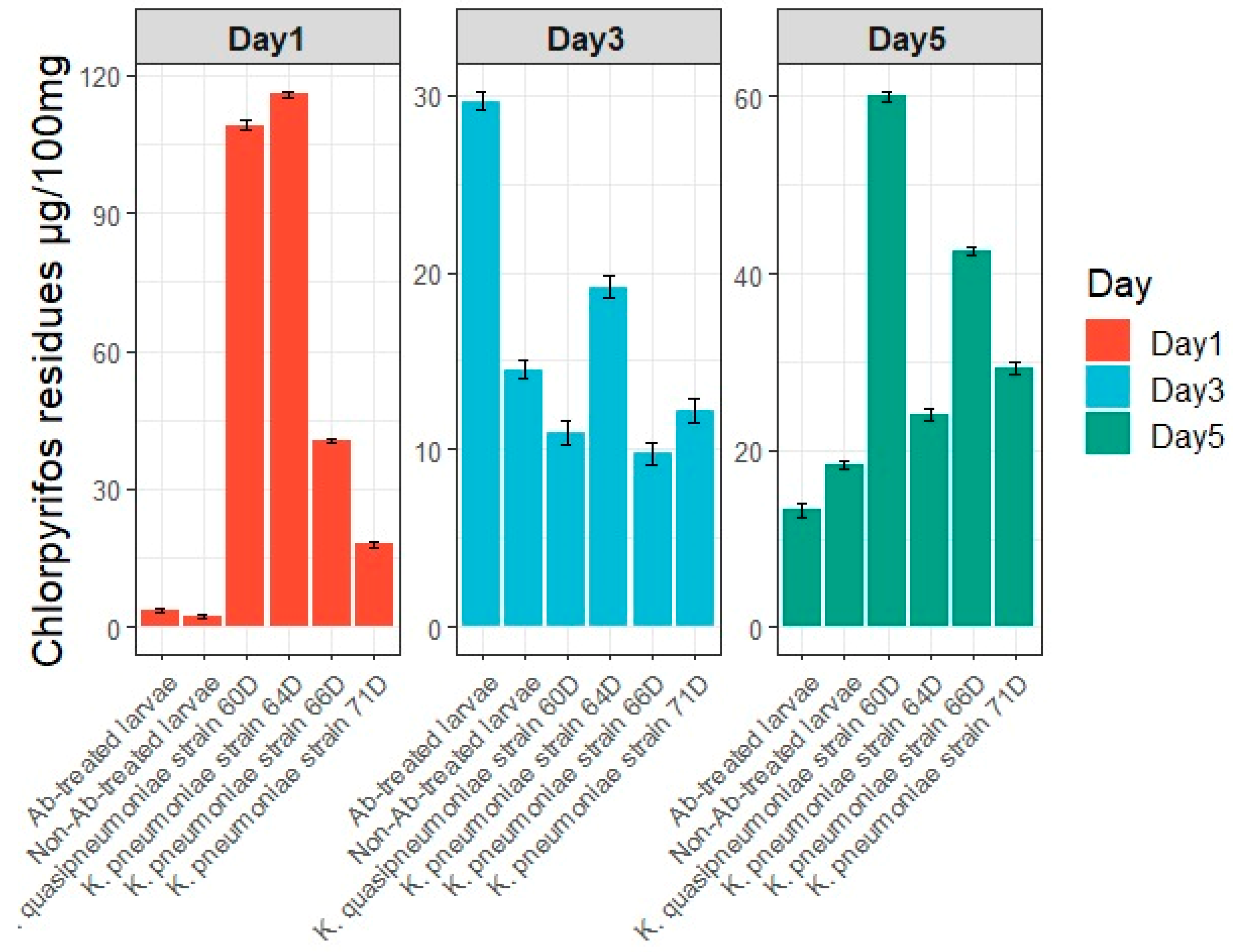
Residues of chlorpyrifos were detected in the feces of S. frugiperda larvae after 1, 3, and 5 days of exposure to chlorpyrifos suspension (Figure 8). On day 1, mono-associated larvae with K. quasipneumoniae strain 60D (approximately 119 μg/100 mg feces) and K. pneumoniae strain 64D (approximately 115 μg/100 μg feces) (p < 0.0001) showed the highest chlorpyrifos residues. By day 5, K. quasipneumoniae strain 60D still exhibited the highest fecal chlorpyrifos residues (approximately 61 μg/100 μg feces), followed by K. pneumoniae strain 66D (approximately 42 μg/100 μg feces) (p < 0.0001).

3.5.6. Bacteria Counting in Larval Feces

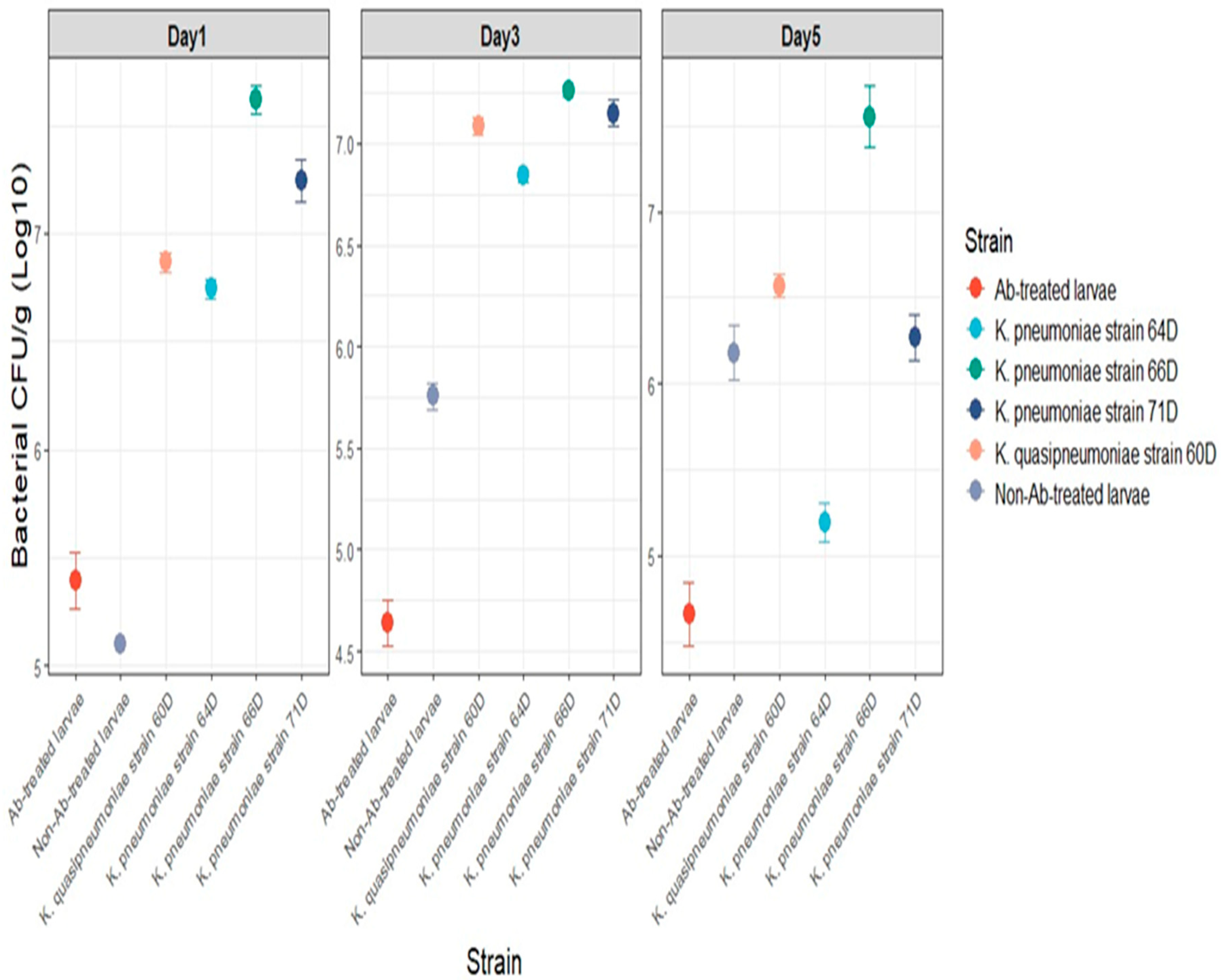
The bacterial CFU counts (Log10) of chlorpyrifos-degrading gut bacteria in larval feces were assessed over time (Figure 9). These findings show that bacterial growth differed between antibiotic-treated larvae, non-antibiotic-treated larvae, and several isolated strains. Bacterial CFU counts in antibiotic-treated larvae were consistently low at all time points, with no development by day 5. Bacterial counts grew gradually in non-antibiotic-treated larvae, reaching around 6 log10 CFU/g by day 5. Individual bacterial isolates, including Klebsiella strains 60D, 66D, and 71D, followed similar growth trends, with CFU counts increasing from day 1 to day 5. Interestingly, strain 66 had the highest CFU counts, reaching over 7 log10 CFU/g by day 5, demonstrating its strong ability to degrade chlorpyrifos. Strain 64D showed a lower count by day 5 (5.2 log10 CFU/g). The results indicate that non-antibiotic-treated larvae include a resilient microbial population capable of efficiently degrading chlorpyrifos.

4. Discussion

Spodoptera frugiperda, as a key invasive pest, has the capacity to rapidly develop resistance to insecticides [44]. At the field level, the insect, alongside its microbial community, remains subject to constant selection pressures. An increase in the resistance of S. frugiperda to different insecticides has various underlying mechanisms, including elevated activity of the detoxifying enzymes, as well as insensitivity of the target [45]. In addition, as observed in other insects, resistance may also be associated with detoxification processes mediated by symbiotic microorganisms [15,19,21]. We analyzed the symbiotic gut bacteria in S. frugiperda, revealing their pivotal role in the biodegradation of pesticides. In this study, we successfully isolated four isolates of chlorpyrifos-degrading bacteria from a field-collected population of S. frugiperda. Our isolations identified isolates belonging to the phylum Pseudomonadota (formerly Proteobacteria), an abundant bacterial group in the insect gut that plays a significant role in insecticide degradation [13,24]. These isolates were identified using 16S rDNA sequencing as Klebsiella quasipneumoniae strain 60D (PP504878), Klebsiella pneumoniae strain 64D (PP504879), Klebsiella pneumoniae strain 66D (PP504880), and Klebsiella pneumoniae strain 71D (PP504881), as shown in (Figure 2). Notably, previous studies have demonstrated the chlorpyrifos-degrading potential of several bacterial species, including the genus Klebsiella [46,47,48].
When these gut bacteria were cultured with insecticides as the sole carbon source, we anticipated they would metabolize the insecticides ingested by their hosts, thereby contributing to insecticide tolerance of the host system [49]. To evaluate the degradation ability of the four chlorpyrifos-degrading gut bacterial isolates, the degradation rate was assessed by measuring the residual chlorpyrifos in MM9 medium using GC–MS analysis. Most of the isolates obtained from the midgut of S. frugiperda exhibited strong chlorpyrifos-degrading capabilities, as shown in Table 2. Our results indicated that Klebsiella pneumoniae strain 64D showed the highest degradation rate after 1 day (80.38%), but its efficiency declined to 25% after 5 days, whereas K. pneumoniae strain 66D maintained the highest activity at this stage (61.14%). The decline in strain 64D may reflect enzyme kinetic limitations or substrate/product inhibition, where reduced chlorpyrifos availability or accumulation of metabolites suppresses further enzymatic activity and bacterial growth, reflecting the complex interplay between enzyme induction, substrate concentration, and metabolic feedback regulation [50].
The importance of gut microbiota in insecticide degradation and resistance is becoming increasingly recognized. Bacillus cereus isolated from Plutella xylostella can degrade up to 20% of indoxacarb, highlighting its role in host growth and metabolism [23]. Moreover, Enterococcus sp. has been shown to enhance resistance to chlorpyrifos in P. xylostella, whereas proteobacteria, including Enterobacter sp. and Serratia sp., reduce it, suggesting complex and species-specific interactions between gut microbiota and insecticide resistance mechanisms [10]. With growing evidence demonstrating that gut microbiota confers insecticide tolerance and resistance across different arthropods [49,51]. Presumably, bacteria can influence this resistance through three non-exclusive mechanisms: (1) mitigating the fitness costs associated with insecticide resistance; (2) directly degrading insecticides; and (3) regulating the host’s detoxification metabolism [49,51,52]. To gain deeper insight into the interaction between gut bacteria and insecticide resistance, we examined the effect of gut bacterial communities in S. frugiperda. The results of the in vivo chlorpyrifos biodegradation assessment demonstrated that symbiotic gut bacteria play a critical role in enhancing host tolerance to chlorpyrifos. The mortality rate of 4th instar S. frugiperda larvae exposed to a 24 ppm chlorpyrifos suspension varied significantly, indicating differential toxicity levels. Notably, larvae mono-associated with K. pneumoniae strain 66D exhibited the greatest reduction in mortality by 19.16-fold compared to antibiotic-treated larvae. This was followed by larvae colonized with K. quasipneumoniae strain 60D (11-fold mortality reduction), K. pneumoniae strain 64D (9.2-fold mortality reduction), and K. pneumoniae strain 71D (5.11-fold mortality reduction) after five days of exposure. These findings indicate that specific isolates contribute to chlorpyrifos detoxification and enhance host survival [15,48]. Residues of chlorpyrifos were detected in the feces of S. frugiperda larvae after 1, 3, and 5 days of exposure to the chlorpyrifos suspension. The mono-associated larvae with K. quasipneumoniae strain 60D and K. pneumoniae strain 64D exhibited the highest chlorpyrifos residues in their feces after one day of exposure (Figure 6). This pattern is consistent with the observed mortality results, in which larvae colonized with K. quasipneumoniae strain 60D and K. pneumoniae strain 64D showed 11-fold and 9.2-fold reductions in mortality, respectively.
Further supporting a role for gut symbionts in pesticide detoxification, acetylcholinesterase (AChE) activity was significantly inhibited in larvae exposed to chlorpyrifos compared to antibiotic-treated controls. Mono-association with K. quasipneumoniae strain 60D resulted in the strongest AChE inhibition (3.5-fold), followed by strains 64D (2.96-fold), 71D (1.8-fold), and 66D (1.2-fold), as shown in Table 4. This inhibition is likely related to the chlorpyrifos mode of action, an organophosphate insecticide that irreversibly inhibits acetylcholinesterase (AChE), leading to the accumulation of acetylcholine at synaptic junctions [53]. These observations support those of Xiao et al., 2022, who reported that the gut symbiont Stenotrophomonas maltophilia can modulate host detoxification enzyme responses under chlorpyrifos exposure [54]. In contrast, GST activity exhibited a different pattern. No significant changes were observed in GST activity during the three days of chlorpyrifos exposure. However, by the fifth day, significant inhibition of GST activity was observed in larvae mono-associated with K. pneumoniae strain 66D, K. pneumoniae strain 71D, K. quasipneumoniae strain 60D, and K. pneumoniae strain 64D, with reductions of approximately 3.0-, 2.9-, 2.2-, and 1.8-fold, respectively, compared to antibiotic-treated larvae. Chlorpyrifos detoxification primarily occurs through conjugation with glutathione (GSH), a process catalyzed by glutathione S-transferase (GST) [55,56]. Moreover, chlorpyrifos can induce the generation of reactive oxygen species (ROS) that disrupt normal cellular development and differentiation [57,58]. These findings further support the role of gut microbiota in modulating host detoxification pathways, specifically through the regulation of AChE and GST activity [54]. Overall, this study extends current knowledge on pesticide–microbe interactions by demonstrating chlorpyrifos biodegradation in a different Spodoptera species and by identifying Klebsiella spp. among the key bacterial taxa involved, thereby providing novel perspectives on the microbial basis of pest resistance.

5. Conclusions

In summary, the isolation of Klebsiella strains from the gut of Spodoptera frugiperda and their remarkable capability to degrade chlorpyrifos both in vitro (up to 80.4% by strain 64D) and in vivo (notably, strain 66D afforded a 19.16-fold reduction in larval mortality relative to antibiotic-treated controls) underscores the pivotal role of gut-associated microbes in insecticide detoxification. These findings substantiate the concept of “detoxifying symbiosis,” wherein insect gut microbiota strengthen host survival in toxic environments. Supporting this, acetylcholinesterase (AChE) activity was significantly inhibited in larvae exposed to chlorpyrifos compared to antibiotic-treated control groups. Overall, harnessing such pesticide-degrading symbionts could pioneer innovative, microbe-based pest-management strategies that minimize reliance on synthetic chemicals.

Author Contributions

Conceptualization, A.S.E.-Z. and M.A.; methodology, A.S.E.-Z., M.S., and M.A.; validation, A.S.E.-Z., D.E.E.-H., N.A.A., and M.A.; formal analysis, A.S.E.-Z., M.N.A., D.E.E.-H., D.E.-S., H.M.F., and M.A.; investigation, A.S.E.-Z., M.N.A., D.E.-S., and M.A.; data curation, A.S.E.-Z., M.S., D.E.E.-H., N.A.A., H.M.F., and M.A.; writing—original draft preparation, A.S.E.-Z., M.N.A., D.E.E.-H., and M.A.; writing—review and editing, A.S.E.-Z., M.N.A., D.E.-S., and M.A.; supervision, A.S.E.-Z. and M.A. All authors have read and agreed to the published version of the manuscript.

Funding

This research received no external funding.

Institutional Review Board Statement

Not applicable.

Informed Consent Statement

Not applicable.

Data Availability Statement

All data from the study have been presented in the manuscript.

Acknowledgments

Special thanks to Charles J. Mason for providing us with the chlorpyrifos-degrading gut bacteria isolation protocol, editing the English version, reviewing the manuscript, and providing valuable comments for its improvement. Charles is a research biologist at the Tropical Pest Genetics and Molecular Biology Research Unit, Daniel K. Inouye U.S. Pacific Basin Agricultural Research Center, Agricultural Research Service, USDA, Hilo, HI, USA; charles.mason@usda.gov.

Conflicts of Interest

The authors declare no competing interests.

References

  1. Briefing Note on FAO Actions on Fall Armyworm. Available online: https://openknowledge.fao.org/items/a33e9a05-d913-4701-bf99-1158839be003 (accessed on 5 March 2019).
  2. Garlet, C.G.; Gubiani, P.d.S.; Palharini, R.B.; Moreira, R.P.; Godoy, D.N.; Farias, J.R.; Bernardi, O. Field-evolved resistance to chlorpyrifos by Spodoptera frugiperda (Lepidoptera: Noctuidae): Inheritance mode, cross-resistance patterns, and synergism. Pest Manag. Sci. 2021, 77, 5367–5374. [Google Scholar] [CrossRef]
  3. Smegal, D.C. Human Health Risk Assessment Chlorpyrifos; US Environmental Protection Agency, Office of Prevention, Pesticides and Toxic Substances, Office of Pesticide Programs, Health Effects Division, US Government Printing Office: Washington, DC, USA, 2000; pp. 1–131. [Google Scholar]
  4. Simma, E.A.; Dermauw, W.; Balabanidou, V.; Snoeck, S.; Bryon, A.; Clark, R.M.; Yewhalaw, D.; Vontas, J.; Duchateau, L.; Van Leeuwen, T. Genome-wide gene expression profiling reveals that cuticle alterations and P450 detoxification are associated with deltamethrin and DDT resistance in Anopheles arabiensis populations from Ethiopia. Pest Manag. Sci. 2019, 75, 1808–1818. [Google Scholar] [CrossRef]
  5. Khan, H.A.A. Characterization of permethrin resistance in a Musca domestica strain: Resistance development, cross-resistance potential and realized heritability. Pest Manag. Sci. 2019, 75, 2969–2974. [Google Scholar] [CrossRef] [PubMed]
  6. Sindhu, T.; Venkatesan, T.; Prabhu, D.; Jeyakanthan, J.; Gracy, G.R.; Jalali, S.K.; Rai, A. Insecticide-resistance mechanism of Plutella xylostella (L.) associated with amino acid substitutions in acetylcholinesterase-1: A molecular docking and molecular dynamics investigation. Comput. Biol. Chem. 2018, 77, 240–250. [Google Scholar] [CrossRef] [PubMed]
  7. Balabanidou, V.; Grigoraki, L.; Vontas, J. Insect cuticle: A critical determinant of insecticide resistance. Curr. Opin. Insect Sci. 2018, 27, 68–74. [Google Scholar] [CrossRef]
  8. Peterson, B.F. Microbiome toxicology—Bacterial activation and detoxification of insecticidal compounds. Curr. Opin. Insect Sci. 2024, 63, 101192. [Google Scholar] [CrossRef] [PubMed]
  9. Engel, P.; Moran, N.A. The gut microbiota of insects–diversity in structure and function. FEMS Microbiol. Rev. 2013, 37, 699–735. [Google Scholar] [CrossRef]
  10. Xia, X.; Sun, B.; Gurr, G.M.; Vasseur, L.; Xue, M.; You, M. Gut microbiota mediate insecticide resistance in the diamondback moth, Plutella xylostella (L.). Front. Microbiol. 2018, 9, 25. [Google Scholar] [CrossRef]
  11. Malook Su Arora, A.K.; Wong, A.C.N. The role of microbiomes in shaping insecticide resistance: Current insights and emerging paradigms. Curr. Opin. Insect Sci. 2025, 69, 101346. [Google Scholar] [CrossRef] [PubMed]
  12. ElKraly, O.A.; Awad, M.; El-Saadany, H.M.; Hassanein, S.E.; Elrahman, T.A.; Elnagdy, S.M. Impact of gut microbiota composition on black cutworm, Agrotis ipsilon (hufnagel) metabolic indices and pesticide degradation. Anim. Microbiome 2023, 5, 44. [Google Scholar] [CrossRef]
  13. Gomes, A.F.F.; Omoto, C.; Cônsoli, F.L. Gut bacteria of field-collected larvae of Spodoptera frugiperda undergo selection and are more diverse and active in metabolizing multiple insecticides than laboratory-selected resistant strains. J. Pest Sci. 2020, 93, 833–851. [Google Scholar] [CrossRef]
  14. Chen, B.; Zhang, N.; Xie, S.; Zhang, X.; He, J.; Muhammad, A.; Sun, C.; Lu, X.; Shao, Y. Gut bacteria of the silkworm Bombyx mori facilitate host resistance against the toxic effects of organophosphate insecticides. Environ. Int. 2020, 143, 105886. [Google Scholar] [CrossRef]
  15. Kikuchi, Y.; Hayatsu, M.; Hosokawa, T.; Nagayama, A.; Tago, K.; Fukatsu, T. Symbiont-mediated insecticide resistance. Proc. Natl. Acad. Sci. USA 2012, 109, 8618–8622. [Google Scholar] [CrossRef]
  16. Mason, C.J.; Peiffer, M.; Hoover, K.; Felton, G. Tomato chemical defenses intensify corn earworm (Helicoverpa zea) mortality from opportunistic bacterial pathogens. J. Chem. Ecol. 2023, 49, 313–324. [Google Scholar] [CrossRef]
  17. Mason, C.J.; Peiffer, M.; Felton, G.W.; Hoover, K. Host-Specific larval lepidopteran mortality to pathogenic Serratia mediated by poor diet. J. Invertebr. Pathol. 2022, 194, 107818. [Google Scholar] [CrossRef]
  18. Itoh, H.; Tago, K.; Hayatsu, M.; Kikuchi, Y. Detoxifying symbiosis: Microbe-mediated detoxification of phytotoxins and pesticides in insects. Nat. Prod. Rep. 2018, 35, 434–454. [Google Scholar] [CrossRef]
  19. Akami, M.; Njintang, N.Y.; Gbaye, O.A.; Andongma, A.A.; Rashid, M.A.; Niu, C.-Y.; Nukenine, E.N. Gut bacteria of the cowpea beetle mediate its resistance to dichlorvos and susceptibility to Lippia adoensis essential oil. Sci. Rep. 2019, 9, 6435. [Google Scholar] [CrossRef]
  20. Shen, S.; Dowd, P. Detoxification spectrum of the cigarette beetle symbiont Symbiotaphrina kochii in culture. Entomol. Exp. Appl. 1991, 60, 51–59. [Google Scholar] [CrossRef]
  21. Cheng, T.; Wu, J.; Wu, Y.; Chilukuri, R.V.; Huang, L.; Yamamoto, K.; Feng, L.; Li, W.; Chen, Z.; Guo, H. Genomic adaptation to polyphagy and insecticides in a major East Asian noctuid pest. Nat. Ecol. Evol. 2017, 1, 1747–1756. [Google Scholar] [CrossRef] [PubMed]
  22. Almeida, L.G.D.; Moraes, L.A.B.D.; Trigo, J.R.; Omoto, C.; Consoli, F.L. The gut microbiota of insecticide-resistant insects houses insecticide-degrading bacteria: A potential source for biotechnological exploitation. PLoS ONE 2017, 12, e0174754. [Google Scholar] [CrossRef]
  23. Ramya, S.L.; Venkatesan, T.; Srinivasa Murthy, K.; Jalali, S.K.; Verghese, A. Detection of carboxylesterase and esterase activity in culturable gut bacterial flora isolated from diamondback moth, Plutella xylostella (Linnaeus), from India and its possible role in indoxacarb degradation. Braz. J. Microbiol. 2016, 47, 327–336. [Google Scholar] [CrossRef]
  24. Siddiqui, J.A.; Fan, R.; Liu, Y.; Syed, A.H.; Benlin, Y.; Chu, Q.; Ding, Z.; Ghani, M.I.; Liu, X.; Wakil, W.; et al. The larval gut of Spodoptera frugiperda harbours culturable bacteria with metabolic versatility after insecticide exposure. Insect Mol. Biol. 2025, 34, 452–469. [Google Scholar] [CrossRef]
  25. Adams, N.H.; Umar, R.; Ishaya, S.; Nweke, O.D.; Ilyasu, N.S.; Jagaba, A.H.; Usman, S.; Yakasai, H.M. Chlorpyrifos degradation by Bacillus sp. strain UPMB10 isolated from polluted environment: Analysis and characterization of the metabolite by GC-MS. Case Stud. Chem. Environ. Eng. 2024, 9, 100608. [Google Scholar] [CrossRef]
  26. Turner, S.; Pryer, K.M.; Miao, V.P.; Palmer, J.D. Investigating deep phylogenetic relationships among cyanobacteria and plastids by small subunit rRNA sequence analysis 1. J. Eukaryot. Microbiol. 1999, 46, 327–338. [Google Scholar] [CrossRef] [PubMed]
  27. Lane, D. 16S/23S rRNA sequencing. In Nucleic acid Techniques in Bacterial Systematics; Wiley: Chichester, UK, 1991; pp. 125–175. [Google Scholar]
  28. Sanger, F.; Nicklen, S.; Coulson, A.R. DNA sequencing with chain-terminating inhibitors. Proc. Natl. Acad. Sci. USA 1977, 74, 5463–5467. [Google Scholar] [CrossRef] [PubMed]
  29. Tamura, K.; Stecher, G.; Kumar, S. MEGA11: Molecular evolutionary genetics analysis version 11. Mol. Biol. Evol. 2021, 38, 3022–3027. [Google Scholar] [CrossRef] [PubMed]
  30. Abdel Ghani, S.B.; Hanafi, A.H. QuEChERS method combined with GC-MS for pesticide residues determination in water. J. Anal. Chem. 2016, 71, 508–512. [Google Scholar] [CrossRef]
  31. Li, H.; Qiu, Y.; Yao, T.; Ma, Y.; Zhang, H.; Yang, X.; Li, C. Evaluation of seven chemical pesticides by mixed microbial culture (PCS-1): Degradation ability, microbial community, and Medicago sativa phytotoxicity. J. Hazard. Mater. 2020, 389, 121834. [Google Scholar] [CrossRef]
  32. Moustafa, M.A.; Fouad, E.A.; Ibrahim, E.; Erdei, A.L.; Kárpáti, Z.; Fónagy, A. The comparative toxicity, biochemical and physiological impacts of chlorantraniliprole and indoxacarb on Mamestra brassicae (Lepidoptera: Noctuidae). Toxics 2023, 11, 212. [Google Scholar] [CrossRef]
  33. Moustafa, M.A.; Fouad, E.A.; Abdel-Mobdy, Y.; Hamow, K.Á.; Mikó, Z.; Molnár, B.P.; Fónagy, A. Toxicity and sublethal effects of chlorantraniliprole and indoxacarb on Spodoptera littoralis (Lepidoptera: Noctuidae). Appl. Entomol. Zool. 2021, 56, 115–124. [Google Scholar] [CrossRef]
  34. Bradford, M.M. A rapid and sensitive method for the quantitation of microgram quantities of protein utilizing the principle of protein-dye binding. Anal. Biochem. 1976, 72, 248–254. [Google Scholar] [CrossRef]
  35. Ellman, G.L.; Courtney, K.D.; Andres, V., Jr.; Featherstone, R.M. A new and rapid colorimetric determination of acetylcholinesterase activity. Biochem. Pharmacol. 1961, 7, 88–95. [Google Scholar] [CrossRef]
  36. Moustafa, M.A.; El-Said, N.A.; Ahmed, F.S.; Amer, A.; Awad, M.; Alfuhaid, N.A. In vitro and silico exploration of the insecticidal properties of Lavandula multifida L. essential oil and its binary combinations with cyantraniliprole and emamectin benzoate on Spodoptera frugiperda (Lepidoptera: Noctuidae). Crop Prot. 2025, 187, 106969. [Google Scholar] [CrossRef]
  37. Awad, M.; El Kenawy, A.H.; Alfuhaid, N.A.; Ibrahim, E.-D.S.; Jósvai, J.K.; Fónagy, A.; Moustafa, M.A. Lethal and sublethal effects of cyantraniliprole on the Biology and Metabolic enzyme activities of two lepidopteran Pests, Spodoptera littoralis and Agrotis ipsilon, and A generalist predator, Chrysoperla carnea (Neuroptera: Chrysopidae). Insects 2024, 15, 450. [Google Scholar] [CrossRef] [PubMed]
  38. Habig, W.; Pabst, M.J.; Jakoby, W. The first enzymatic step in mercapturic acid formation. Glutathione-S-transferase. J. Biol. Chem. 1974, 249, 7130–7139. [Google Scholar] [CrossRef] [PubMed]
  39. Lalander, C.; Senecal, J.; Calvo, M.G.; Ahrens, L.; Josefsson, S.; Wiberg, K.; Vinnerås, B. Fate of pharmaceuticals and pesticides in fly larvae composting. Sci. Total Environ. 2016, 565, 279–286. [Google Scholar] [CrossRef] [PubMed]
  40. Schwantes, D.; Celso Gonçalves, A., Jr.; Conradi Junior, É.; Campagnolo, M.A.; Zimmermann, J. Determination of CHLORPYRIFOS by GC/ECD in water and its sorption mechanism study in a RHODIC FERRALSOL. J. Environ. Health Sci. Eng. 2020, 18, 149–162. [Google Scholar] [CrossRef]
  41. Wirsching, J.; Pagel, H.; Ditterich, F.; Uksa, M.; Werneburg, M.; Zwiener, C.; Berner, D.; Kandeler, E.; Poll, C. Biodegradation of pesticides at the limit: Kinetics and microbial substrate use at low concentrations. Front. Microbiol. 2020, 11, 2107. [Google Scholar] [CrossRef]
  42. Douglas, A.E. Multiorganismal insects: Diversity and function of resident microorganisms. Annu. Rev. Entomol. 2015, 60, 17–34. [Google Scholar] [CrossRef]
  43. Lü, D.; Dong, Y.; Yan, Z.; Liu, X.; Zhang, Y.; Yang, D.; He, K.; Wang, Z.; Wang, P.; Yuan, X. Dynamics of gut microflora across the life cycle of Spodoptera frugiperda and its effects on the feeding and growth of larvae. Pest Manag. Sci. 2023, 79, 173–182. [Google Scholar] [CrossRef]
  44. Diez-Rodríguez, G.I.; Omoto, C. Inheritance of lambda-cyhalothrin resistance in Spodoptera frugiperda (JE Smith) (Lepidoptera: Noctuidae). Neotrop. Entomol. 2001, 30, 311–316. [Google Scholar] [CrossRef]
  45. Yu, S.; Nguyen, S.; Abo-Elghar, G. Biochemical characteristics of insecticide resistance in the fall armyworm, Spodoptera frugiperda (JE Smith). Pestic. Biochem. Physiol. 2003, 77, 1–11. [Google Scholar] [CrossRef]
  46. Elshikh, M.S.; Alarjani, K.M.; Huessien, D.S.; Elnahas, H.A.; Esther, A.R. Enhanced Biodegradation of Chlorpyrifos by Bacillus cereus CP6 and Klebsiella pneumoniae CP19 from municipal waste water. Environ. Res. 2022, 205, 112438. [Google Scholar] [CrossRef]
  47. Maya, K.; Singh, R.; Upadhyay, S.; Dubey, S.K. Kinetic analysis reveals bacterial efficacy for biodegradation of chlorpyrifos and its hydrolyzing metabolite TCP. Process Biochem. 2011, 46, 2130–2136. [Google Scholar] [CrossRef]
  48. Vidya Lakshmi, C.; Kumar, M.; Khanna, S. Biodegradation of chlorpyrifos in soil by enriched cultures. Curr. Microbiol. 2009, 58, 35–38. [Google Scholar] [CrossRef]
  49. Wang, Z.; Wang, W.; Lu, Y. Biodegradation of insecticides by gut bacteria isolated from stored grain beetles and its implication in host insecticide resistance. J. Stored Prod. Res. 2022, 96, 101943. [Google Scholar] [CrossRef]
  50. Akbar, S.; Sultan, S. Soil bacteria showing a potential of chlorpyrifos degradation and plant growth enhancement. Braz. J. Microbiol. 2016, 47, 563–570. [Google Scholar] [CrossRef] [PubMed]
  51. Pietri, J.E.; Liang, D. The links between insect symbionts and insecticide resistance: Causal relationships and physiological tradeoffs. Ann. Entomol. Soc. Am. 2018, 111, 92–97. [Google Scholar] [CrossRef]
  52. Liu, X.-D.; Guo, H.-F. Importance of endosymbionts Wolbachia and Rickettsia in insect resistance development. Curr. Opin. Insect Sci. 2019, 33, 84–90. [Google Scholar] [CrossRef]
  53. Greer, J.B.; Magnuson, J.T.; Hester, K.; Giroux, M.; Pope, C.; Anderson, T.; Liu, J.; Dang, V.; Denslow, N.D.; Schlenk, D. Effects of chlorpyrifos on cholinesterase and serine lipase activities and lipid metabolism in brains of rainbow trout (Oncorhynchus mykiss). Toxicol. Sci. 2019, 172, 146–154. [Google Scholar] [CrossRef]
  54. Xiao, T.; Lu, K. Functional characterization of CYP6AE subfamily P450s associated with pyrethroid detoxification in Spodoptera litura. Int. J. Biol. Macromol. 2022, 219, 452–462. [Google Scholar] [CrossRef]
  55. Singh, B.K.; Walker, A. Microbial degradation of organophosphorus compounds. FEMS Microbiol. Rev. 2006, 30, 428–471. [Google Scholar] [CrossRef] [PubMed]
  56. Tang, J.; Cao, Y.; Rose, R.L.; Brimfield, A.A.; Dai, D.; Goldstein, J.A.; Hodgson, E. Metabolism of chlorpyrifos by human cytochrome P450 isoforms and human, mouse, and rat liver microsomes. Drug Metab. Dispos. 2001, 29, 1201–1204. [Google Scholar] [PubMed]
  57. Slotkin, T.A. Guidelines for developmental neurotoxicity and their impact on organophosphate pesticides: A personal view from an academic perspective. Neurotoxicology 2004, 25, 631–640. [Google Scholar] [CrossRef] [PubMed]
  58. Abdollahi, M.; Ranjbar, A.; Shadnia, S.; Nikfar, S.; Rezaie, A. Pesticides and oxidative stress: A review. Med. Sci. Monit. 2004, 10, 141–147. [Google Scholar]
Figure 1. Experimental design of the in vivo chlorpyrifos biodegradation assay in Spodoptera frugiperda larvae.
Biology 14 01468 g001
Figure 2. A neighbor-joining phylogenetic tree based on 16S rRNA gene sequences of four bacterial isolates (highlighted in red color) and their closest matches from the NCBI database.
Biology 14 01468 g002
Figure 3. Effect of incubation temperature on the growth of Klebsiella strains. Bacterial counts (expressed as Log10 CFU/mL) of K. quasipneumoniae strain 60D, K. pneumoniae strain 64D, K. pneumoniae strain 66D, and K. pneumoniae strain 71D. Different letters denote statistically significant differences (p < 0.05).
Biology 14 01468 g003
Figure 4. Effect of pH range on the growth of Klebsiella strains. Bacterial counts (expressed as Log10 CFU/mL) of K. quasipneumoniae strain 60D, K. pneumoniae strain 64D, K. pneumoniae strain 66D, and K. pneumoniae strain 71D. Different letters denote statistically significant differences (p < 0.05).
Biology 14 01468 g004
Figure 5. Effect of incubation time on the growth of Klebsiella strains. Bacterial counts (expressed as Log10 CFU/mL) of K. quasipneumoniae strain 60D, K. pneumoniae strain 64D, K. pneumoniae strain 66D, and K. pneumoniae strain 71D. Different letters denote statistically significant differences (p < 0.05).
Biology 14 01468 g005
Figure 6. Bacterial load in Ab-treated and non-Ab-treated larvae grown on different media. (A) Bacterial load (log10 CFU/mL) on nutrient agar (NA), (B) bacterial load on minimal medium (MM9). Different letters denote statistically significant differences (p < 0.05).
Biology 14 01468 g006
Figure 7. Mortality rates (%) of 4th S. frugiperda larvae after being exposed to chlorpyrifos.
Biology 14 01468 g007
Figure 8. In vivo analysis of chlorpyrifos biodegradation by gut bacteria, as detected in the feces of Spodoptera frugiperda larvae after 1, 3, and 5 days of exposure to a chlorpyrifos suspension.
Biology 14 01468 g008
Figure 9. Bacterial CFU counts (Log10 CFU/g) of chlorpyrifos-degrading gut bacteria recovered from larval feces across three time points (day 1, day 3, and day 5).
Biology 14 01468 g009
Table 1. Degradation rate % (±SD) of chlorpyrifos in MM9 media after 1, 3, and 5 days of bacteria inoculation (n = 3).
Groups1 Day3 Days5 Days
Klebsiella quasipneumoniae strain 60D77.9 ± 0.91 b45.13 ± 1.94 b57.94 ± 1.81 ab
Klebsiella pneumoniae strain 64D80.38 ± 0.68 a69.8 ± 0.32 a25 ± 1.82 c
Klebsiella pneumoniae strain 66D51.3 ± 0.15 d46.93 ± 1.64 b61.14 ± 2.12 a
Klebsiella pneumoniae strain 71D65.52 ± 0.74 c50.15 ± 2.06 b53.08 ± 1.82 b
Means that those that do not share a letter are significantly different.
Table 2. The biodegradation capabilities of chlorpyrifos-degrading isolates to other pesticides.
Strain/PesticideChlorantraniliprole
(Coragen)
0.6 ppm
Carbamate
(Lannate)
0.5 ppm
Pyrethroid
(Lambda-Cyhalothrin)
5 ppm
K. quasipneumoniae 60DPositivePositivePositive
K. pneumoniae 64DPositivePositiveNegative
K. pneumoniae 66DPositivePositiveNegative
K. pneumoniae 71DPositivePositiveNegative
Table 3. Larval weight (mg) of antibiotic-treated and non-antibiotic-treated application.
Antibiotic-Treated (mg)Non-Antibiotic-Treated (mg)t-Testp-Value
Larval weight (mg)8.57 ± 1.8912.73 ± 0.47−3.020.039
Table 4. Mean (±SD) of enzymatic activity of S. frugiperda after 1, 3, and 5 days of being exposed to chlorpyrifos.
EnzymeGroups1 Day3 Days5 Days
AchE(Mmole/mg of protein)Ab-treated larva0.74 ± 0.12 a0.61 ± 0.02 a0.43 ± 0.05 a
Non-Ab-treated larva0.32 ± 0.05 cd0.43 ± 0.07 a0.36 ± 0.03 a
Klebsiella quasipneumoniae strain 60D0.21 ± 0.06 d0.48 ± 0.06 a0.42 ± 0.01 a
Klebsiella pneumoniae strain 64D0.25 ± 0.05 cd0.58 ± 0.08 a0.28 ± 0.08 a
Klebsiella pneumoniae strain 66D0.58 ± 0.06 ab0.61 ± 0.03 a0.46 ± 0.09 a
Klebsiella pneumoniae strain 71D0.41 ± 0.03 bcd0.66 ± 0.03 a0.26 ± 0.02 a
P-Value0.0010.1710.020
F-Value(df)14.54(6)1.80(6)3.71(6)
GST (Mmole/mg of protein)Ab-treated larva0.44 ± 0.25 a0.38 ± 0.07 b0.87 ± 0.07 a
Non-Ab-treated larva0.3 ± 0.05 a0.79 ± 0.04 a0.46 ± 0.03 b
Klebsiella quasipneumoniae strain 60D0.2 ± 0.03 a0.24 ± 0.08 b0.39 ± 0.03 b
Klebsiella pneumoniae strain 64D0.28 ± 0.01 a0.45 ± 0.09 b0.47 ± 0.16 b
Klebsiella pneumoniae strain 66D0.41 ± 0.14 a0.35 ± 0.05 b0.29 ± 0.13 b
Klebsiella pneumoniae strain 71D0.35 ± 0.07 a0.8 ± 0.12 a0.3 ± 0.11 b
P-Value0.4790.0010.001
F-Value(df)0.97(6)10.96(6)8.94(6)
Means that do not share a letter are significantly different.
Disclaimer/Publisher’s Note: The statements, opinions and data contained in all publications are solely those of the individual author(s) and contributor(s) and not of MDPI and/or the editor(s). MDPI and/or the editor(s) disclaim responsibility for any injury to people or property resulting from any ideas, methods, instructions or products referred to in the content.