Article

Neuroprotective Effect of Marrubium vulgare Extract in Scopolamine-Induced Cognitive Impairment in Rats: Behavioral and Biochemical Approaches

1
Institute of Neurobiology, Bulgarian Academy of Science, 1113 Sofia, Bulgaria
2
Laboratory of Biologically Active Substances, Institute of Organic Chemistry with Centre of Phytochemistry, Bulgarian Academy of Sciences, 4000 Plovdiv, Bulgaria
3
Institute of Plant Physiology and Genetics, Bulgarian Academy of Sciences, Acad. Georgi Bonchev Str., Block 21, 1113 Sofia, Bulgaria
*
Authors to whom correspondence should be addressed.
Biology 2024, 13(6), 426; https://doi.org/10.3390/biology13060426
Submission received: 20 May 2024 / Revised: 5 June 2024 / Accepted: 7 June 2024 / Published: 9 June 2024
(This article belongs to the Special Issue Animal Models of Neurodegenerative Diseases)

Abstract

:

Simple Summary

Cognitive deficits, including spatial working and recognition memory impairment, are a common feature of Alzheimer’s disease with current therapies offering limited efficacy. Marrubium vulgare, a member of the Lamiaceae family, has shown potential to alleviate spatial memory impairment in a model of experimental dementia in rats through its antioxidant and acetylcholinesterase inhibitory activities. The aim of this study was to examine the effect of M. vulgare on recognition memory in healthy and dementia-affected rats after 21 days of oral administration. Memory performance was evaluated by the novel object recognition test. Levels of neurotransmitters acetylcholine, noradrenaline (NA), and serotonin, as well as the protein expression of the brain-derived neurotrophic factor (BDNF) and the phosphorylation of the cAMP response element-binding protein (p-CREB), were measured. The expression levels of BDNF and CREB were evaluated via RT-PCR in the cortex and hippocampus. Our result revealed that M. vulgare ameliorated recognition memory impairment in dementia rats by preserving cholinergic function in the hippocampus, increasing NA levels in the brain, and restoring pCREB expression in the cortex following their reduction in the experimental model used. In healthy rats, the extract upregulated the expression of BDNF and pCREB in the cortex. These findings suggest that M. vulgare has potential as a therapeutic agent for cognitive impairments in various neurodegenerative diseases.

Abstract

The potential of Marrubium vulgare to alleviate scopolamine (Sco)-induced deficits in spatial working memory has drawn considerable scientific interest. This effect is partly attributed to its potent antioxidant and acetylcholinesterase inhibitory (AChEI) activities. This study examined the effects of M. vulgare extract, standardized to marrubiin content, on recognition memory in healthy and Sco-treated rats. Male Wistar rats (200–250 g) were divided into four groups. The extract was orally administered for 21 days and Sco (2 mg/kg) was intraperitoneally injected for 11 consecutive days. Memory performance was assessed using the novel object recognition test. Levels of acetylcholine (ACh), noradrenaline (NA), serotonin (Sero), and brain-derived neurotrophic factor (BDNF) and the phosphorylation of cAMP response element-binding protein (p-CREB) were evaluated in the cortex and hippocampus via ELISA. BDNF and CREB expression levels were assessed using RT-PCR. The results showed that M. vulgare significantly alleviated Sco-induced memory impairment, preserved cholinergic function in the hippocampus, increased NA levels in the brain, and restored pCREB expression in the cortex following Sco-induced reduction. In healthy rats, the extract upregulated BDNF, pCREB, and Bcl2 expression. Our findings indicate that the neuroprotective effects of M. vulgare may be linked to the modulation of cholinergic function, regulation of NA neurotransmission, and influence on key memory-related molecules.

1. Introduction

Alzheimer’s disease (AD) stands out as one of the most common forms of dementia in the elderly population [1,2]. Dementia involves a decline in cognitive abilities due to the death of brain cells [3], which consequently impairs cognitive functions such as memory, reasoning, and learning capacity [4].
The cognitive impairment in AD is predominantly linked to reductions in cholinergic neurotransmission systems and neuronal dysfunction [5,6]. Decreased levels of brain acetylcholine (ACh), the overexpression of acetylcholinesterase (AChE) and glutamate excitotoxicity [7], decreased levels of the ACh synthesizing enzyme choline acetyltransferase (ChAT), the altered function of both muscarinic and nicotinic ACh receptors [5,8,9,10,11,12,13,14,15,16], and increased oxidative stress [17] are some of the main features of the disease.
Correspondingly, the use of cholinergic receptor antagonists such as scopolamine (Sco) is a widely used method for inducing cognitive impairment in rodents by blocking the cholinergic system [18]. This approach represents the most commonly used model of pharmacologically induced dementia [19,20]. Sco administration leads to changes in exploratory behavior, as well as spatial, associative, and recognition memory in experimental animals [20,21,22,23,24,25]. In addition, impairment in antioxidant defense systems, increased oxidative stress, mitochondrial dysfunction, apoptosis, and neuroinflammation have also been reported [26]. These neurochemical disturbances mainly affect the hippocampus and prefrontal cortex of experimental animals.
Currently, medications for Alzheimer’s disease primarily focus on symptom management, with AChE inhibitors being the most effective treatment. Four AChE inhibitors are approved and in use for AD treatment: donepezil, rivastigmine, tacrine, and galantamine [27,28,29,30,31]. These drugs alleviate cognitive deficits by increasing the bioavailability of acetylcholine in cholinergic synapses [32,33]. Despite their therapeutic benefits [34], they neither delay nor stop the progression of the disease. Their prolonged use is associated with severe adverse effects, including nausea, diarrhea, vomiting, dizziness, bradycardia, headache, insomnia, and liver toxicity [35,36].
A new hope for AD treatment lies in the emergence of disease-modifying agents capable of altering disease progression. In 2021, the U.S. Food and Drug Administration (FDA) approved aducanumab, and in 2023, lecanemab, which are passive immunotherapy antibodies capable of reducing amyloid β (Aβ) deposition [37,38]. Clinical research has shown that over a period of 18 months, lecanemab reduced brain amyloid burden and cognitive decline by 27% in patients compared to a placebo [38]. These new drugs represent a fundamentally new approach to disease treatment. However, there is no definitive evidence yet that removing amyloid deposits will be therapeutically beneficial for all individuals diagnosed with AD, especially for patients at more advanced stages of the disease.
Given the limitations of current medications for memory impairment therapy, including their relatively low efficacy and severe adverse effects with prolonged use, there is a critical need to develop novel agents with improved therapeutic outcomes.
For centuries, the plant kingdom has been a vital source of medicinal compounds [39,40,41,42,43,44,45]. Moreover, the use of medicinal plants appears to result in fewer side effects and interactions with drugs [46]. Due to their rich diversity of secondary phytochemical metabolites, plants have shown potential in treating various neurodegenerative diseases, including AD.
Lamiaceae is one of the largest families of flowering plants, with high expectations for its potential benefits in the field of neurodegenerative diseases. Numerous articles have discussed the neurobiological effects of various Lamiaceae species. S. officinalis, M. officinalis, and R. officinalis are Lamiaceae plants known in different cultures as potent memory-enhancing agents [47,48,49].
Potential in vitro anti-Alzheimer effects have been reported for S. officinalis, L. an-gustifolia, M. piperita, M. officinalis, O. onites, T. vulgaris L., O. majorana, O. vulgare L., S. congesta, and D. hastata, which are also all Lamiacea species [50]. Potential anticholinesterase activity has been reported from Nepeta, Origanum, Salvia, and Mentha—species from the genus Lavandula [51].
Our previous research with Sideritis scardica and Clinopodium vulgare, also members of the Lamiaceae family, showed an ameliorative memory effect in experimental models of dementia in rodents. They positively impact spatial working and recognition memory, affected by scopolamine [22]. Notably, the combination of S. scardica and C. vulgare demonstrated the best recognition memory-preserving effect, strong antioxidant activity, and a notable monoaminergic preservation effect in the cortex. The strongest antioxidant potential and AChE inhibitory activity of C. vulgare have also been demonstrated. In addition, our findings indicate that S. scardica and C. vulgare mitigate the Sco-induced downregulation of the brain-derived neurotrophic factor (BDNF) and the phosphorylation of the cAMP response element-binding protein (p-CREB) signaling pathway suggests a neuroprotective effect. The preservation and enhancement of neurotrophic factors like BDNF, which supports neuronal survival and synaptic plasticity, constitute a key focus as a new strategy for AD treatment.
Perry et al. [52] reported that a mixture of sage, rosemary, and melissa supported verbal episodic memory in healthy subjects under 63 years old. M. officinalis significantly improved the memory and cognitive performance of young people and AD patients between 65 and 80 years of age [48,53]. The effectiveness of S. officinalis in the treatment of AD and memory deficits in clinical trials has been also reported [54].
Marrubium vulgare, also a member of the Lamiaceae family, is commonly known as white horehound. It is widely used globally as an expectorant for cough, as well as to alleviate bloating, flatulence, and temporary loss of appetite [55]. This plant is abundant in polyphenolic compounds, such as caffeic acid, p-coumaric acid, ferulic acid, rosmarinic acid, quercetin, and apigenin [23], which are recognized for their antioxidant properties and can affect the cardiovascular, gastrointestinal, immune, and endocrine systems [56,57,58,59,60]. Additionally, recent studies have shown various pharmacological effects of M. vulgare on the central nervous system, including the neuroprotection of short-term memory in mice using an in vivo model of traumatic brain injury, as well as antioxidant and AChEI activities [61].
Our preceding research has shown that a water extract of M. vulgare effectively ameliorates spatial memory impairment in a Sco-induced experimental model of AD in rats [23]. The treatment not only decreased the AChE activity in the hippocampus by 20% but also reduced oxidative stress induced by Sco in the brain. A decrease in thiobarbituric acid-reactive substances and the restoration of antioxidant enzyme activities, such as superoxide dismutase (SOD), catalase (CAT), and glutathione peroxidase (GPx), are particularly evident in the cortex of the Sco-treated rats [23].
Building on these findings, we have embarked on a further investigation of the neuroprotective potential of M. vulgare against memory deficits associated with neurodegeneration. We conducted an additional in vivo study assessing its effects in a Sco-induced rat model of dementia. Our study examined the impact of the plant extract on recognition memory using the Novel Object Recognition test and monitored changes in the levels of ACh and biogenic amines, along with the expression of BDNF and pCREB in the brain cortex and hippocampus. Furthermore, we assessed the relative expression levels of BDNF, CREB, and Bcl2 in the cortex of healthy rats using quantitative real-time PCR (qRT-PCR). This comprehensive approach aims to elucidate the therapeutic potential of M. vulgare, paving the way for future treatments that harness the neuroprotective properties of natural compounds.

2. Materials and Methods

2.1. Plant Material and Extract Preparation

Marrubium vulgare plants were obtained using micropropagation techniques and cultivated in the experimental field located in Elin Pelin (near Sofia city, at an altitude of 560 m). The aerial parts of the plants were collected in July and subsequently dried at room temperature (19–21 °C).
Dried aerial parts were ground into a fine powder using a laboratory mill. Five grams of the powder were added to 200 mL water (90 °C) and incubated for 15 min. The mixture was centrifuged at 6000× g and the supernatant was lyophilized for 96 h using an Alpha 1–4 LD plus laboratory freeze drier (Martin Christ Gefriertrocknungsanlagen GmbH, Osterode am Harz, Germany).

2.2. Determination of Marrubiin Content

Marrubiin content in the extract was determined using a slightly modified method based on the European Pharmacopoeia (Ph Eur) monograph 1835 [62]. Chromatographic analyses were conducted using a Nexera-i LC2040C Plus UHPLC system (Shimadzu Corporation, Kyoto, Japan), equipped with a UV-VIS detector and a binary pump. The analytical column employed was a Poroshell 120 EC-C18 (3 mm × 100 mm, 2.7 μm), thermostated at a temperature of 26 °C. The flow rate was set at 1.5 mL/min, with an injection volume of 20 μL. Derivatives were detected at a wavelength of 330 nm. The mobile phase consisted of component A: acetonitrile, and component B: 0.5 mL of phosphoric acid R diluted to 1000 mL with water. The gradient condition was applied as specified.
Time, minA (%)B (%)
0–1540–9060–10
15–2090–4010–60
20–254060
The content of marrubiin in the extract, expressed in grams per 100 grams (g/100 g), was calculated using the following formula:
A 1 × M 2 × P × 2.5 A 2 × M 1
where:
  • A1 = the area of the marrubiin in the chromatogram of the test solution.
  • A2 = the area of the marrubiin peak in the chromatogram of the reference solution.
  • M1 = the mass of the extract being examined, in milligrams.
  • M2 = the mass of marrubiin R, in milligrams.
  • P = the percentage content of marrubiin in the marrubiin standard

2.3. Animals

Male Wistar rats weighing 200–250 g were obtained from the animal house Erboj, Slivniza, Bulgaria. The rats were housed under standard laboratory conditions with temperatures maintained at 25 ± 3 °C and a consistent 12/12 h light/dark cycle. They had ad libitum access to a pellet diet and water. To acclimate the rats to the environment, they underwent a five-day habituation period prior to the experiment. The experimental protocol followed the guidelines of the European Communities Council Directive (86/609/EEC) and received approval from the Bulgarian Food Safety Agency (Approval No. 340/13.12.22 for working with laboratory animals).

2.4. Experimental Design

The animals were randomly assigned into four groups, each consisting of 12 rats: 1. Control; 2. Sco; 3. Sco + M. vulgare; 4. M. vulgare. For 21 days, the Control and Sco groups received distilled water orally (0.5 mL/100 g body weight). The M. vulgare and Sco + M. vulgare groups received an oral dosage of the plant extract at 200 mg/kg body weight for the same duration [23,56]. The Control and M. vulgare groups received intraperitoneal (i.p.) injections of 0.9% NaCl solution (0.5 mL/100 g body weight) for 11 consecutive days. The Sco and Sco + M. vulgare groups received i.p. injections of scopolamine hydrobromide (2 mg/kg) [25,63,64,65,66,67] also for 11 consecutive days to develop the experimental model of dementia. The intraperitoneal injections with Sco or 0.9% NaCl solution began 10 days after the first oral administration of the plant extract or distiller water and continued for 11 days simultaneously with the oral treatments (Figure 1).
Scopolamine was freshly dissolved in distilled water ex tempore before each administration.

2.5. Assessment of Cognitive Function: Novel Object Recognition Test

After completing the 21-day treatment regimen, rats from all groups underwent the Novel Object Recognition (NOR) behavioral test to evaluate their recognition memory performance (Figure 1). This behavioral assessment was conducted in a dimly lit room between 9 a.m. and 12 p.m., providing consistent environmental conditions for accurate observation of cognitive responses.
The NOR test utilizes the natural propensity of rodents to explore new objects more than familiar ones [68]. This test was conducted over two days. On the first day, a habituation session was conducted in which each rat was individually placed in an enclosed area (box measuring 60 × 60 × 60 cm) containing two identical objects, where they were allowed to explore for 3 min. The test session began 24 h after habituation and was divided into two phases, separated by a one-hour interval. In the first phase, each rat explored two identical objects in the recognition box for 4 min. An hour later, one of the original objects was replaced with a new one, and the rat was given 3 min to explore both objects. Recognition memory was assessed using the discrimination index (DI), calculated as follows: DI = (time spent on the new object/total time spent exploring both objects) × 100%. An object was considered to be explored when the animal placed its nose within 2 cm of the zone where the object was located.

2.6. Brain Dissection Technique

One hour after completing the test, the animals were humanely euthanized using mild CO2 inhalation followed by decapitation. The skin covering the skull was incised along the midline and removed to expose the dorsal skull plates. Using scissors, the plates were carefully split along the midline and then twisted outward along the lateral border to reveal the brain. The membrane enveloping the brain was delicately removed with fine forceps. The brain was then carefully removed from the skull and subjected to microdissection to isolate memory-specific regions, such as the frontal cortex and hippocampus. For hippocampus isolation, two short microspatulas were used. One spatula was positioned just above the cerebellum near its junction with the cortex, while the other gently peeled the cortical hemisphere laterally to expose the hippocampus, which was meticulously scooped out. The frontal cortex was then dissected. Throughout the procedure, the tissue was periodically rinsed with fresh ice-cold saline (0.9% NaCl) using a pipette.

2.7. Determination of Acetylcholine and Monoamine Content

The ACh content in tissue lysate was determined using an Enzyme-Linked Immunosorbent Assay (ELISA) kit (cat. No. E-EL-0081, Elabscience, Wuhan, China) following homogenization. The absorbance was measured using a microplate reader set to 450 nm. The results were calculated and expressed in picograms per milliliter (pg/mL).
For the determination of biogenic amines, specifically noradrenalin (NA), dopamine (DA), and serotonin (Sero), in the cortex and hippocampus of the experimental animals, the method established by Jacobowitz and Richardson [69] was employed. In brief, the extraction of NA and DA was carried out into a phosphate buffer; Sero extraction was performed using 0.1N HCl. The subsequent fluorescence reaction included the use of ethylenediaminetetraacetic acid (EDTA), an iodide solution, alkaline sulfite, and 5N CH3COOH for NA and DA. For Sero, o-phthaldehyde was used. Fluorescence intensity was then measured at specific wavelengths optimized for each monoamine: λ = 385/485 nm for NA, λ = 320/385 nm for DA, and λ = 360/470 nm for Sero. The fluorescence levels of the monoamines were calculated based on the fluorescence of the standard solution and the results are expressed in ng/g of fresh tissue.

2.8. Determination of BDNF and pCREB Concentrations

The concentrations of Brain-Derived Neurotrophic Factor (BDNF) and phosphorylated cAMP response element-binding (pCREB) in the frontal cortex and hippocampus of rats were quantified using ELISA. According to the manufacturer instructions, the following kits were utilized: the Rat BDNF ELISA Kit (cat. no. E-EL-R1235) from Elabscience and the Rat Phospho cAMP response element binding protein (pCREB) ELISA Kit (cat. no. SL1344Ra) from Sunlong Biotech Co., Ltd. (Hangzhou, China) Measurements were performed with a microplate reader set to 450 nm. The results were quantified and reported in picograms per milliliter (pg/mL).

2.9. RNA Extraction and Reverse Transcription

RNA was isolated from the cortex using the PRImeZOL reagent (Canvax), following the manufacturer’s protocol. The purity and concentration of the extracted RNA were assessed using a NanoDrop 2000 spectrophotometer (Thermo Scientific, Waltham, MA, USA). To eliminate DNA contamination, the RNA samples were treated with DNase I (Thermo Scientific). Reverse transcription was carried out using the FIREScript RT cDNA Synthesis Mix, which included oligo (dT) and random primers (Solis BioDyne, Tartu, Estonia), according to the instructions provided by the manufacturer.

2.10. Quantitative Real-Time PCR

The genes BDNF, CREB, and Bcl2 were analyzed using HOT FIREPol EvaGreen qPCR Mix Plus (Solis BioDyne). The RT-PCR reactions were performed in duplicate on a 96-well plate using a PikoReal 96-RT PCR system (Thermo Scientific). The thermal cycling conditions consisted of initial denaturation at 95 °C for 12 min, followed by 35 cycles consisting of 15 s at 95 °C, 30 s at annealing temperatures of 59 °C or 58 °C, and 30 s at 72 °C. The primer sequences and their corresponding product sizes are detailed in Table 1. GAPDH was used as the reference gene for the normalization of the qRT-PCR data. The primer sequences for BDNF and CREB were based on Tancheva et al. [25], while those for Bcl2 and GAPDH were sourced from Ramezani et al. [70] and Kharroubi et al. [71], respectively. The Relative Expression Software Tool V2.0.13 (REST) [72] facilitated the group comparison and statistical analysis of relative expression results obtained from RT-PCR experiments.

2.11. Statistical Analysis

Data are presented as the mean ± standard error of the mean (SEM). Analyses were performed with Prism 8.0 software (GraphPad Software, Inc., San Diego, CA, USA). Statistical testing involved one-way analysis of variance (ANOVA) followed by Tukey’s post hoc comparison test to determine significant differences between groups. A p-value of less than 0.05 (p < 0.05) was considered statistically significant.

3. Results

3.1. Effects of M. vulgare Treatment on Recognition Memory Performance in Healthy and Scopolamine-Treated Rats

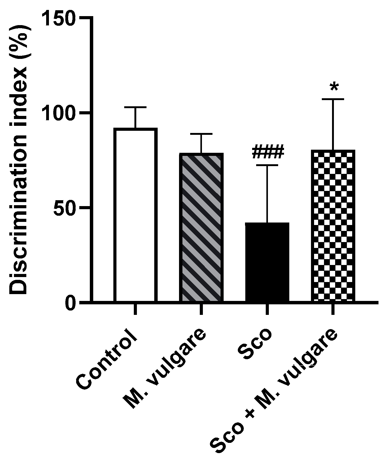
In this study, we evaluated recognition memory performance using the Discrimination Index (DI) as illustrated in Figure 2. Analysis of the results showed that 11 days of Sco treatment significantly impaired recognition memory with a 54.29% decrease in DI (p < 0.001, n = 6). This indicated that the Sco-treated rats spent significantly less time interacting with the new object compared to the control group. After 21 days, treatment with M. vulgare effectively reversed the memory deficits induced by Sco. The value of DI in the Sco + M. vulgare group increased by 91.10% compared to the untreated Sco group. In healthy rats treated with M. vulgare, there was no significant change in DI compared to the saline-treated group.

3.2. Neuromodulatory Activity of M. vulgare in the Frontal Cortex and Hippocampus of Healthy and Scopolamine-Treated Rats

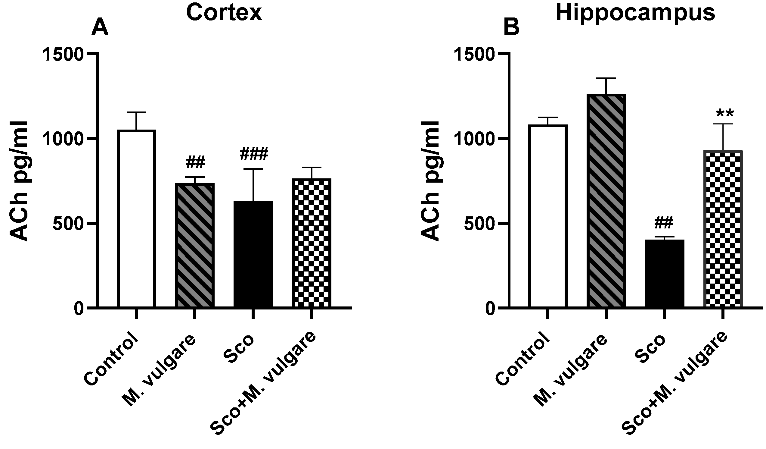
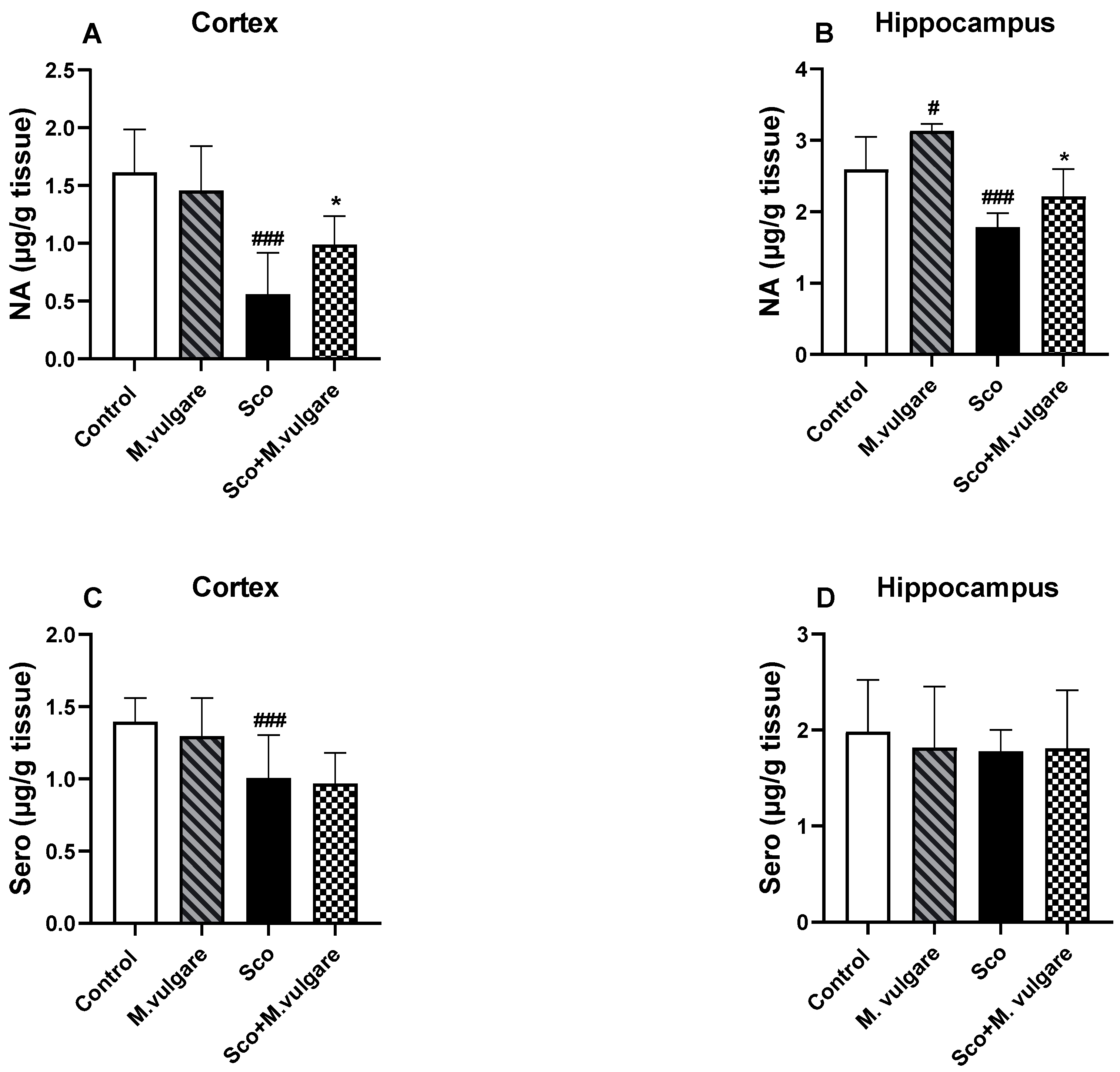
The neuromodulatory effects of M. vulgare extract were evaluated by measuring the levels of ACh, noradrenaline (NA) and Sero in the cortex and hippocampus of rats from all experimental groups (Figure 3A,B and Figure 4A–D). Our findings revealed that Sco treatment significantly decreased the levels of ACh and monoamines in rats with dementia-like symptoms. In the cortex of these animals, ACh levels decreased by 39.96% (p < 0.001, n = 6), NA by 65.26% (p < 0.001, n = 6), and Sero by 28.08% (p < 0.001, n = 6), compared to the control group. In the hippocampus, ACh and NA levels decreased by 62.73% (p < 0.01, n = 6) and 31.12% (p < 0.001, n = 6), respectively, compared to the saline-treated rats. However, Sero levels in the hippocampus of Sco-treated animals did not show significant changes following 21 days of M. vulgare treatment (Figure 4D).
Treatment with M. vulgare for 21 days demonstrated a protective effect on ACh and NA levels and did not significantly alter Sero levels in the brains of Sco-treated rats. In the cortex, the plant extract increased NA levels by 76.33% (p < 0.05, n = 6) compared to Sco-treated rats. In the hippocampus, ACh content was increased by 130.89% (p < 0.01, n = 6) and NA levels were enhanced by 23.91% (p < 0.05, n = 6) compared to the dementia-like animals.
In healthy rats, M. vulgare treatment had a significant impact only on ACh and NA levels in the brain. In the cortex, ACh levels were decreased by 30.11% (p < 0.01, n = 6), while NA and Sero levels remained largely unaffected compared to the saline-treated animals. In the hippocampus, the plant extract showed a trend toward increasing ACh content (although not significantly) and raised NA levels by 20.57% (p < 0.05, n = 6), without significant changes in Sero levels compared to controls.

3.3. Effect of M. vulgare on the Expression Levels of BDNF and pCREB in the Frontal Cortex and Hippocampus of Healthy and Scopolamine-Treated Rats

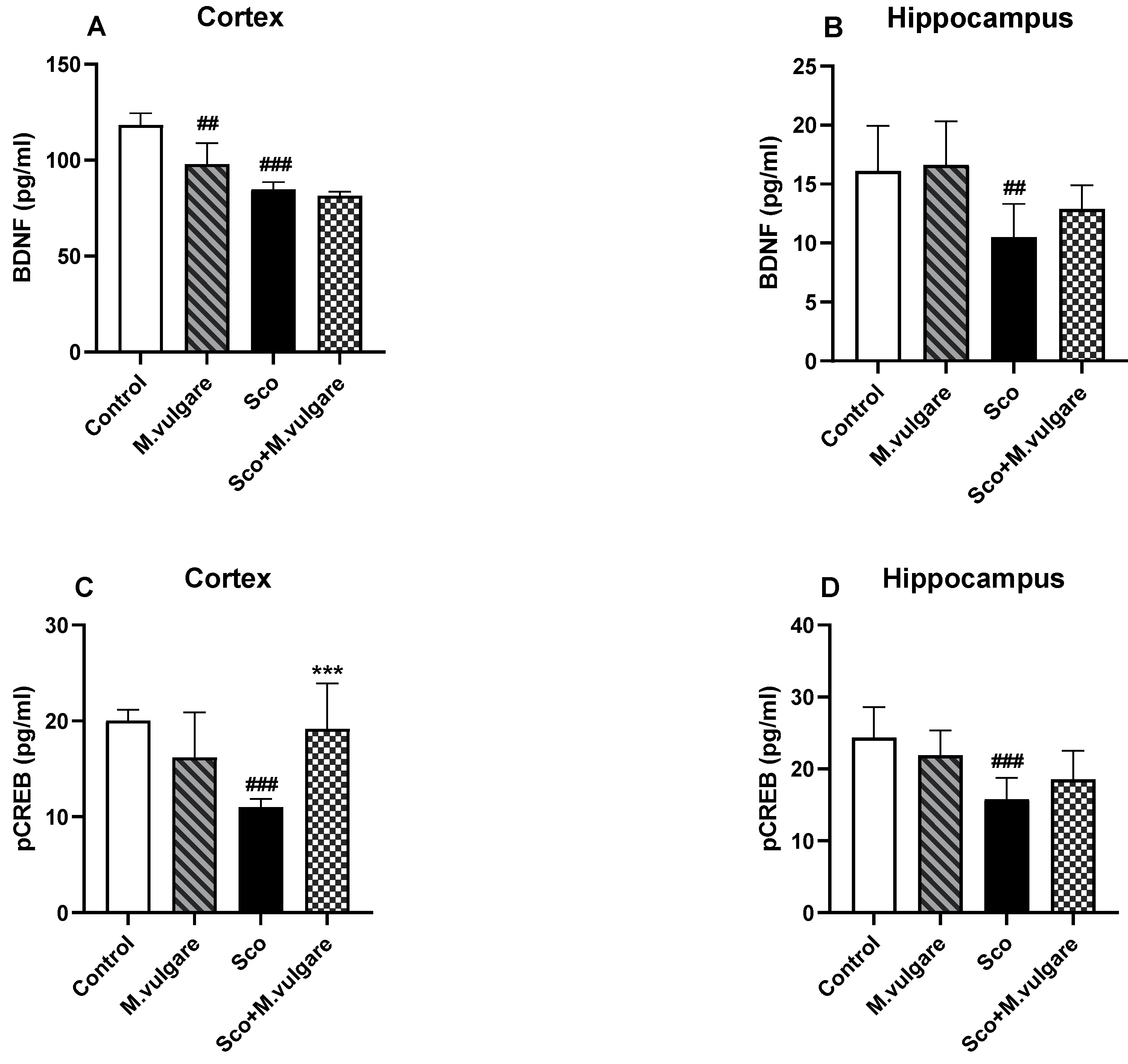
The expression levels of BDNF and pCREB in the cerebral cortex and hippocampus were evaluated by the ELISA method. According to our results, 11 days of Sco administration significantly decreased the expression of BDNF and pCREB in these brain regions (Figure 5A–D). Specifically, in the cortex, BDNF expression was reduced by 28.44% (p < 0.001, n = 6) and pCREB by 45.24% (p < 0.001, n = 6) (Figure 5A,C). In the hippocampus, BDNF expression decreased by 34.98% (p < 0.01, n = 6) and pCREB by 35.15% (p < 0.001, n = 6) (Figure 5B,D). These comparisons were made against saline-treated rats.
Treatment with M. vulgare for 21 days significantly counteracted the Sco-induced changes in pCREB expression in the cortex. Specifically, pCREB levels increased by 74.77% (p < 0.001, n = 6) compared to the untreated Sco group.
In healthy animals, ELISA analysis indicated that the plant extract treatment generally did not result in significant changes in pCREB/BDNF signaling in the frontal cortex and hippocampus. The sole exception was observed in the cortex, where BDNF expression decreased by 17.26% (p < 0.01, n = 6) compared to the control group.

3.4. Effect of M. vulgare Treatment on Relative Expression Levels of BDNF, CREB, and Bcl2 in the Cortex of Healthy Rats

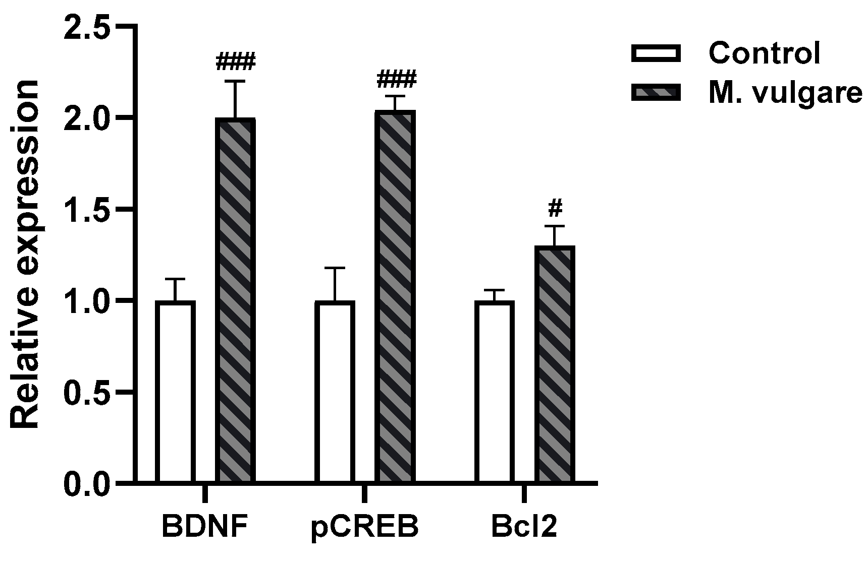
We employed an RT-PCR assay to investigate potential differences in the expression of the BDNF, CREB, and Bcl2 genes between M. vulgare-treated and control rats (Figure 6). Our results revealed significant upregulation in the expression of all three genes. Specifically, BDNF expression increased to 2.0±0.20 (p < 0.001, n = 10), while CREB and Bcl2 expressions rose to 2.04 ± 0.08 (p < 0.001, n = 10) and 1.3 ± 0.11 (p < 0.05, n = 10), respectively, compared to the control group.

4. Discussion

In this research, we demonstrated that treatment with M. vulgare extracts substantially improved recognition memory in rats with Sco-induced cognitive deficits. A 21-day oral administration of the extract appeared to stabilize the cholinergic system, as evidenced by elevated levels of ACh in the hippocampus. Moreover, our findings suggest that the plant extract also impacted the NA system and key signaling molecules involved in memory formation and neural plasticity, namely BDNF and CREB. These observations support our previous research, which demonstrated that a water extract of M. vulgare improved spatial memory in a dementia rat model through mechanisms involving antioxidant and AChEI activities [23]. These results not only affirm the neuroprotective properties of M. vulgare but also highlight its potential to counteract the biochemical pathways of memory decline typically observed in Sco-induced models of dementia.
Sco is a muscarinic antagonist known to induce cognitive deficits in healthy humans and rodents by blocking cholinergic signaling [18]. Its intraperitoneal administration is widely accepted as a pharmacological model of AD in rodents, aligning with the cholinergic hypothesis. This hypothesis suggests that defects in the central cholinergic system or alterations in hippocampal function in the brain are implicated in human memory disorders [73]. The administration of Sco altered the behavior and biochemical profiles of the experimental animals. Deficiency in spatial memory, associative memory, and recognition memory performance are reported in our studies and by other researchers [20,21,22,23,24,25,64]. In addition to disrupting the brain cholinergic system (evidenced by increased AChE activity and reduced ACh levels), Sco treatment also escalated oxidative stress, diminished brain monoamine levels (DA, NA, and Sero), and attenuated BDNF/CREB signaling in the brain [22,23,24,25,63,74,75,76].
In this study, we employed the NOR test to evaluate the effect of the M. vulgare treatment on Sco-induced memory impairment in rats. The NOR test is a widely used method for evaluating cognitive deficits in experimental animals, capitalizing on the innate inclination of rodents to explore new objects more than familiar ones. This task incorporates elements of exploratory behavior and memory retention, requiring that animals adequately explore the familiar object during the pretest phase to enable them to differentiate it from a novel object later in the test phase [77]. Animals with preserved memory formation tend to spend more time with the new object during the actual test. Our findings show that rats treated with Sco displayed reduced exploration of novel objects compared to controls. This aligns with our previous observations, where Sco treatment significantly lowers the discrimination index in the NOR task and decreases the exploratory behavior of animals in other behavioral tests like the Hole board test [22,25,64].
Our analyses showed that 21 days of M. vulgare treatment did not significantly change recognition memory in healthy rats versus controls; however, it effectively mitigated memory impairment in rats with Sco-induced cognitive decline. These results indicate that the extract sustained memory formation under conditions of cognitive stress. This is consistent with our previous research, which demonstrated that M. vulgare preserved spatial memory against Sco-induced damage, as evidenced in the T-maze test. Moreover, we have verified that the memory-related effects of Sco and M. vulgare are not due to changes in motor function or sedation of the experimental animals, underscoring the specific cognitive benefits of the plant extract treatment [23].
The ameliorated memory effects of the extract may be attributed to the diversity of its secondary phytochemical metabolites. Our analysis revealed that M. vulgare extract is rich in polyphenolic compounds, such as caffeic acid, p-coumaric acid, ferulic acid, rosmarinic acid, quercetin, and apigenin [23]. Marrubiin, a well-known diterpenoid lactone prevalent in horehound and other medicinal plants of the Lamiaceae family, was present in the extract standardized to 2.5% marrubiin content. Marrubiin is recognized for its broad pharmacological properties, including high safety, low turnover, high stability, and minimal catabolism. Additionally, it has revealed a range of important biological activities including antinociceptive, antigenotoxic, cardioprotective, vasorelaxant, gastroprotective, antispasmodic, immunomodulating, antioedematogenic, analgesic, and antidiabetic properties [78]. The neuroprotective potential of marrubiin following traumatic brain injury in experimental mice [61] and potent AChEI activity [79] have also been reported and could explain the results of our study. Furthermore, literature data indicate that rosmarinic acid, another phenolic constituent found in horehound, has a beneficial effect on Sco-induced learning and memory impairment in rat models [80]. In addition, apigenin has been reported to improve cognitive dysfunction and neuroinflammation by upregulating the ERK/CREB pathway in APP/PS1 transgenic AD mice [81]. These findings collectively provide a comprehensive explanation for the cognitive benefits observed in our study with M. vulgare extract.
To identify the specific components of cholinergic neurotransmission affected by M. vulgare treatment, we measured ACh levels in the cortex and hippocampus of healthy and dementia rats. This analysis complements our prior findings, which highlighted the AChEI activity of M. vulgare, particularly noting its pronounced effects in the hippocampus [23]. ACh, a key component of the cholinergic system, has been a focal point of memory research over the last decade. It is well documented that fluctuations in ACh levels or AChE activity can disrupt cholinergic transmission, resulting in learning and memory deficits similar to those seen in AD [82]. Therefore, assessing acetylcholinesterase activity and ACh levels in the brain can provide valuable information on cholinergic function, which is closely connected to cognitive performance.
Our results clearly indicate that Sco administration to experimental animals led to a significant reduction in brain ACh levels compared to the control group. The subsequent administration of the plant extract for three weeks elevated ACh levels in the hippocampus of rats suffering from Sco-induced memory impairment. Consequently, we could suggest that the positive effects of M. vulgare extract and its major diterpene component, marrubiin, in mitigating Sco-induced memory deficits may be linked to increased central cholinergic functioning.
The brain monoaminergic system is another key factor in memory-related processes. NA, a component of this system, is essential for the formation of hippocampus-based declarative memory [83].
The locus coeruleus (LC) is the primary source of NA in the central nervous system and plays a crucial role in memory formation by projecting to key brain areas, such as the hippocampus [84], amygdala [85], and prefrontal cortex [86]. It is noteworthy that the LC is activated in response to novelty [87] and arousal [88], and its degeneration is a hallmark of AD [89]. Sco treatment has been shown to depress the monoaminergic systems in the cortex and hippocampus, a finding corroborated by our previous research [22,25,64]. Treatment with M. vulgare has been observed to positively affect the NA system but did not significantly impact the Sero system.
There are data indicating a close correlation between reduced expression of BDNF and CREB in the hippocampus and cognitive impairments. This suggests that the transcription of BDNF, which is regulated by CREB, plays a critical role in the adaptive neuronal responses that underpin learning and memory functions [90,91].
Traditionally recognized as a neurotrophic factor essential for neuronal cell survival and the prevention of neurodegeneration, BDNF also plays a pivotal role in modulating synaptic function and plasticity in the central nervous system, particularly in learning and memory processes [92]. CREB, a transcription factor, is instrumental in regulating the expression of genes involved in neuroplasticity, cell survival, and cognition [93]. Phosphorylated CREBs can bind to the cAMP response elements of target genes associated with long-term memory formation [94]. BDNF is a prominent target of CREB signaling [95,96], indicating that BDNF transcription, regulated by CREB, is vital in the adaptive neuronal responses that support learning and memory function [90].
In the current study, the expression levels of BDNF and pCREB in the hippocampus and frontal cortex of the animals were evaluated by the ELISA method. Analysis revealed that, as expected, Sco suppressed hippocampal and cortical pCREB/BDNF protein expression, leading to memory impairment in rats. This aligns with literature data and findings from our previous research [22,25,97]. Treatment with M. vulgare significantly increased pCREB expression in the cortex of the animals with dementia. In the hippocampus, there was only a tendency for an ameliorative effect of the extract on suppressed BDND/pCREB signaling by Sco.
In the healthy animals, M. vulgare treatment led to complex neurochemical alterations: after 21 days of administration, there was a decrease in ACh levels and BDNF expression in the cortex, while NA levels in the hippocampus increased. Interestingly, the gene expression of the neurotrophic factor BDNF in the cortex of healthy rats was elevated post-treatment. In addition, in our genetic studies, we also observed significant changes in the expression of the cellular transcription factor CREB and the antiapoptotic factor Bcl2.
Despite the promising results on the neuroprotective activity of M. vulgare extract, this study has limitations, including the absence of histopathological evaluations of the frontal cortex and hippocampus. Future research will focus on addressing these gaps. Considering the safety profile and demonstrated benefits of M. vulgare, its integration into existing treatment paradigms for cognitive impairment could improve therapeutic outcomes and enhance the quality of life for patients with neurodegenerative conditions.

5. Conclusions

Our research demonstrated the memory-protective effects of M. vulgare in a Sco-induced model of dementia, evaluated using the NOR test. The observed benefits on memory appear to be linked to several neural mechanisms: the modulation of cholinergic function in the hippocampus, the regulation of NA neurotransmission in the brain, and alterations in the gene and peptide expression of key memory-related molecules such as BDNF and CREB. These findings suggest that M. vulgare could influence critical pathways involved in memory formation and retention. Given these results, there is a compelling need for more in-depth studies to explore the mechanisms of action of M. vulgare extract. Expanding this line of inquiry could further delineate its mechanisms of action and reinforce its potential as a therapeutic agent for cognitive impairments in various neurodegenerative diseases.

Author Contributions

Conceptualization, M.L. and K.T.; methodology, M.L.; software, M.L. and P.D.; validation, M.L. and M.S.; formal analysis, M.L., K.T., T.T., and M.S.; data curation, M.L. and V.V.; writing—original draft preparation, M.L., V.V., T.T., and P.D.; writing—review and editing, M.L., K.T., V.V., and P.D.; supervision, M.L. and K.T.; project administration, K.T.; funding acquisition, K.T. All authors have read and agreed to the published version of the manuscript.

Funding

This research was funded by the National Science Fund—Bulgaria, grant number KP-06-N56/16.

Institutional Review Board Statement

The animal study protocol was in accordance with the requirements of the European Communities Council Directive (86/609/EEC) and the Bulgarian Food Safety Agency (Approval for working with laboratory animals no. 340/13 December 2022).

Informed Consent Statement

Not applicable.

Data Availability Statement

All data are contained in the manuscript.

Conflicts of Interest

The authors declare no conflicts of interest.

References

  1. Anand, A.; Khurana, P.; Chawla, J.; Sharma, N.; Khurana, N. Emerging treatments for the behavioral and psychological symptoms of dementia. CNS Spectr. 2018, 23, 361–369. [Google Scholar] [CrossRef] [PubMed]
  2. Sharma, N.; Khurana, N.; Muthuraman, A. Lower vertebrate and invertebrate models of Alzheimer’s disease—A review. Eur. J. Pharmacol. 2017, 815, 312–323. [Google Scholar] [CrossRef]
  3. Goo, M.J.; Choi, S.M.; Kim, S.H.; Ahn, B.O. Protective effects of acetyl-L-carnitine on neurodegenerative changes in chronic cerebral ischemia models and learning-memory impairment in aged rats. Arch. Pharm. Res. 2012, 35, 145–154. [Google Scholar] [CrossRef] [PubMed]
  4. Puri, A.; Srivastava, P.; Pandey, P.; Yadav, R.S.; Bhatt, P.C. Scopolamine induced behavioral and biochemical modifications and protective effect of Celastrus paniculatous and Angelica glauca in rats. Int. J. Nutr. Pharmacol. Neurol. Dis. 2014, 4, 158–169. [Google Scholar]
  5. Francis, P.T.; Palmer, A.M.; Snape, M.; Wilcock, G.K. The cholinergic hypothesis of Alzheimer’s disease: A review of progress. J. Neurol. Neurosurg. Psychiatry 1999, 66, 137–147. [Google Scholar]
  6. Blennow, K.; de Leon, M.J.; Zetterberg, H. Alzheimer’s disease. Lancet 2006, 368, 387–403. [Google Scholar] [CrossRef] [PubMed]
  7. Francis, P.T. The interplay of neurotransmitters in Alzheimer’s disease. CNS Spectr. 2005, 10, 6–9. [Google Scholar] [CrossRef] [PubMed]
  8. Coyle, J.T.; Price, D.L.; DeLong, M.R. Alzheimer’s disease: A disorder of cortical cholinergic innervation. Science 1983, 219, 1184–1190. [Google Scholar] [CrossRef] [PubMed]
  9. Sims, N.R.; Bowen, D.M.; Allen, S.J.; Smith, C.C.T.; Neary, D.; Thomas, D.J.; Davison, A.N. Presynaptic cholinergic dysfunction in patients with dementia. J. Neurochem. 1983, 40, 503–509. [Google Scholar] [CrossRef]
  10. Davies, P.; Maloney, A.J. Selective loss of central cholinergic neurons in Alzheimer’s disease. Lancet 1976, 2, 1403. [Google Scholar] [CrossRef]
  11. Perry, E.K.; Gibson, P.H.; Blessed, G.; Perry, R.H.; Tomlinson, B.E. Neurotransmitter enzyme abnormalities in senile dementia. Choline acetyltransferase and glutamic acid decarboxylase activities in necropsy brain tissue. J. Neurol. Sci. 1977, 34, 247–265. [Google Scholar] [CrossRef] [PubMed]
  12. Whitehouse, P.J.; Price, D.L.; Struble, R.G.; Clark, A.W.; Coyle, J.T.; Delon, M.R. Alzheimer’s disease and senile dementia: Loss of neurons in the basal forebrain. Science 1982, 215, 1237–1239. [Google Scholar] [CrossRef] [PubMed]
  13. Ikonomovic, M.D.; Wecker, L.; Abrahamson, E.E.; Wuu, J.; Counts, S.E.; Ginsberg, S.D.; Mufson, E.J.; Dekosky, S.T. Cortical alpha7 nicotinic acetylcholine receptor and beta-amyloid levels in early Alzheimer disease. Arch. Neurol. 2009, 6, 646–651. [Google Scholar]
  14. Jiang, S.; Li, Y.; Zhang, C.; Zhao, Y.; Bu, G.; Xu, H.; Zhang, Y.W. M1 muscarinic acetylcholine receptor in Alzheimer’s disease. Neurosci. Bull. 2014, 30, 295–307. [Google Scholar] [CrossRef] [PubMed]
  15. Wang, H.Y.; Stucky, A.; Liu, J.; Shen, C.; Trocme-Thibierge, C.; Morain, P. Dissociating beta-amyloid from alpha 7 nicotinic acetylcholine receptor by a novel therapeutic agent, S 24795, normalizes alpha 7 nicotinic acetylcholine and NMDA receptor function in Alzheimer’s disease brain. J. Neurosci. 2009, 29, 10961–10973. [Google Scholar] [CrossRef] [PubMed]
  16. Zuchner, T.; Schliebs, R.; Perez-Polo, J.R. Down-regulation of muscarinic acetylcholine receptor M2 adversely affects the expression of Alzheimer’s disease-relevant genes and proteins. J. Neurochem. 2005, 95, 20–32. [Google Scholar] [CrossRef] [PubMed]
  17. Huang, W.-J.; Zhang, X.; Chen, W.-W. Role of oxidative stress in Alzheimer’s disease. Biomed. Rep. 2016, 4, 519–522. [Google Scholar] [CrossRef] [PubMed]
  18. Klinkenberg, I.; Blokland, A. The validity of scopolamine as a pharmacological model for cognitive impairment: A review of animal behavioral studies. Neurosci. Biobehav. Rev. 2010, 34, 1307–1350. [Google Scholar] [CrossRef] [PubMed]
  19. Lee, B.; Sur, B.; Shim, J.; Hahm, D.H.; Lee, H. Acupuncture stimulation improves scopolamine-induced cognitive impairment via activation of cholinergic system and regulation of BDNF and CREB expressions in rats. BMC Complement. Altern. Med. 2014, 14, 338. [Google Scholar] [CrossRef]
  20. Goverdhan, P.; Sravanthi, A.; Mamatha, T. Neuroprotective effects of meloxicam and selegiline in scopolamine-induced cognitive impairment and oxidative stress. Int. J. Alzheimer’s Dis. 2012, 2012, 974013. [Google Scholar] [CrossRef]
  21. Abd-El-Fattah, M.A.; Abdelakader, N.F.; Zaki, H.F. Pyrrolidine dithiocarbamate protects against scopolamine-induced cognitive impairment in rats. Eur. J. Pharmacol. 2014, 723, 330–338. [Google Scholar] [CrossRef] [PubMed]
  22. Lazarova, M.; Tsvetanova, E.; Georgieva, A.; Stefanova, M.; Uzunova, D.; Denev, P.; Vassileva, V.; Tasheva, K. Extracts of Sideritis scardica and Clinopodium vulgare alleviate cognitive impairments in scopolamine-induced rat dementia. Int. J. Mol. Sci. 2024, 25, 1840. [Google Scholar] [CrossRef] [PubMed]
  23. Lazarova, M.I.; Tsvetanova, E.R.; Georgieva, A.P.; Stefanova, M.O.; Uzunova, D.N.; Denev, P.N.; Tasheva, K.N. Marrubium vulgare extract improves spatial working memory and oxidative stress damage in scopolamine-treated rats. J. Alzheimers Dis. 2024, 99, S157–S169. [Google Scholar] [CrossRef] [PubMed]
  24. Lazarova, M.; Tancheva, L.; Alexandrova, A.; Tsvetanova, E.; Georgieva, A.; Stefanova, M.; Tsekova, D.; Vezenkov, L.; Kalfin, R.; Uzunova, D.; et al. Effects of new galantamine derivatives in a scopolamine model of dementia in mice. J. Alzheimers Dis. 2021, 84, 671–690. [Google Scholar] [CrossRef] [PubMed]
  25. Tancheva, L.; Lazarova, M.; Velkova, L.; Dolashki, A.; Uzunova, D.; Minchev, B.; Petkova-Kirova, P.; Hassanova, Y.; Gavrilova, P.; Tasheva, K.; et al. Beneficial effects of snail Helix aspersa extract in an experimental model of Alzheimer’s type dementia. J. Alzheimers Dis. 2022, 88, 155–175. [Google Scholar] [CrossRef] [PubMed]
  26. Tang, K.S. The cellular and molecular processes associated with scopolamine-induced memory deficit: A model of Alzheimer’s biomarkers. Life Sci. 2019, 233, 116695. [Google Scholar] [CrossRef] [PubMed]
  27. Campos, C.; Rocha, N.B.; Vieira, R.T.; Rocha, S.A.; Telles Correia, D.; Paes, F.; Yuan, T.; Nardi, A.E.; Arias-Carrion, O.; Machado, S.; et al. Treatment of cognitive deficits in Alzheimer’s disease: A psychopharmacological review. Psychiatr. Danub. 2016, 28, 2–12. [Google Scholar] [PubMed]
  28. McGleenon, B.M.; Dynan, K.B.; Passmore, A.P. Acetylcholinesterase inhibitors in Alzheimer’s disease. Br. J. Clin. Pharmacol. 1999, 8, 471–480. [Google Scholar] [CrossRef] [PubMed]
  29. Craig, L.A.; Hong, N.S.; McDonald, R.J. Revisiting the cholinergic hypothesis in the development of Alzheimer’s disease. Neurosci. Biobehav. Rev. 2011, 35, 1397–1409. [Google Scholar] [CrossRef]
  30. Mehta, M.; Adem, A.; Sabbagh, M. New acetylcholinesterase inhibitors for Alzheimer’s disease. Int. J. Alzheimer’s Dis. 2012, 2012, 728983. [Google Scholar] [CrossRef]
  31. Colović, M.B.; Krstić, D.Z.; Lazarević-Pašti, T.D.; Bondžić, A.M.; Vasić, V.M. Acetylcholinesterase inhibitors: Pharmacology and toxicology. Curr. Neuropharmacol. 2013, 11, 315–335. [Google Scholar] [CrossRef] [PubMed]
  32. Corbett, A.; Smith, J.; Creese, B.; Ballard, C. Treatment of behavioral and psychological symptoms of Alzheimer’s disease. Curr. Treat. Options Neurol. 2012, 14, 113–125. [Google Scholar] [CrossRef] [PubMed]
  33. Bogdan, A.; Manera, V.; Koenig, A.; David, R. Pharmacologic approaches for the management of apathy in neurodegenerative disorders. Front. Pharmacol. 2020, 10, 1581. [Google Scholar] [CrossRef] [PubMed]
  34. Fink, H.A.; Linskens, E.J.; MacDonald, R.; Silverman, P.C.; McCarten, J.R.; Talley, K.M.C.; Forte, M.L.; Desai, P.J.; Nelson, V.A.; Miller, M.A.; et al. Benefits and harms of prescription drugs and supplements for treatment of clinical Alzheimer-type dementia. Ann. Intern. Med. 2020, 172, 656–668. [Google Scholar] [CrossRef] [PubMed]
  35. Gauthier, S. Cholinergic adverse effects of cholinesterase inhibitors in Alzheimer’s disease: Epidemiology and management. Drugs Aging 2001, 18, 853–862. [Google Scholar] [CrossRef] [PubMed]
  36. Watkins, P.B.; Zimmerman, H.J.; Knapp, M.J.; Gracon, S.I.; Lewis, K.W. Hepatotoxic effects of tacrine administration in patients with Alzheimer’s disease. JAMA 1994, 271, 992–998. [Google Scholar] [CrossRef] [PubMed]
  37. Cummings, J.; Lee, G.; Ritter, A.; Sabbagh, M.; Zhong, K. Alzheimer’s disease drug development pipeline: 2020. Alzheimers Dement. 2020, 6, e12050. [Google Scholar] [CrossRef] [PubMed]
  38. van Dyck, C.; Swanson, C.; Aisen, P.; Bateman, R.; Chen, C.; Gee, M.; Kanekiyo, M.; Li, D.; Reyderman, L.; Cohen, S.; et al. Lecanemab in early Alzheimer’s disease. N. Engl. J. Med. 2023, 388, 9–21. [Google Scholar] [CrossRef] [PubMed]
  39. Dias, D.A.; Urban, S.; Roessner, U. A historical overview of natural products in drug discovery. Metabolites 2012, 2, 303–336. [Google Scholar] [CrossRef]
  40. Newman, D.J.; Cragg, G.M. Natural products as sources of new drugs from 1981 to 2014. J. Nat. Prod. 2016, 79, 629–661. [Google Scholar] [CrossRef]
  41. Sharifi-Rad, J.; Ayatollahi, S.A.; Varoni, E.M.; Salehi, B.; Kobarfard, F.; Sharifi-Rad, M.; Iriti, M.; Sharifi-Rad, M. Chemical composition and functional properties of essential oils from Nepeta schiraziana Boiss. Farmacia 2017, 65, 802–812. [Google Scholar]
  42. Sharifi-Rad, J.; Sharifi-Rad, M.; Salehi, B.; Iriti, M.; Roointan, A.; Mnayer, D.; Soltani-Nejad, A.; Afshari, A. In vitro and in vivo assessment of free radical scavenging and antioxidant activities of Veronica persica Poir. Cell. Mol. Biol. 2018, 64, 57–64. [Google Scholar] [CrossRef] [PubMed]
  43. Sharifi-Rad, J.; Melgar-Lalanne, G.; Hernández-Álvarez, A.J.; Taheri, Y.; Shaheen, S.; Kregiel, D.; Antolak, H.; Pawlikowska, E.; Brdar-Jokanovi’c, M.; Rajkovic, J.; et al. Malva species: Insights on its chemical composition towards pharmacological applications. Phytother. Res. 2020, 34, 546–567. [Google Scholar] [CrossRef]
  44. Patti, F.; Taheri, Y.; Sharifi-Rad, J.; Martorell, M.; C Cho, W.; Pezzani, R. Erythrina suberosa: Ethnopharmacology, phytochemistry and biological activities. Medicines 2019, 6, 105. [Google Scholar] [CrossRef] [PubMed]
  45. Salehi, B.; Stojanović-Radić, Z.; Matejić, J.; Sharopov, F.; Antolak, H.; Kręgiel, D.; Sen, S.; Sharifi-Rad, M.; Acharya, K.; Sharifi-Rad, R.; et al. Plants of genus Mentha: From farm to food factory. Plants 2018, 7, 70. [Google Scholar] [CrossRef] [PubMed]
  46. Al-Megrin, W.A.; Alkhuriji, A.F.; Yousef, A.O.S.; Metwally, D.M.; Habotta, O.A.; Kassab, R.B.; Abdel Moneim, A.E.; El-Khadragy, M.F. Antagonistic efficacy of luteolin against lead acetate exposureassociated with hepatotoxicity is mediated via antioxidant, anti-inflammatory, and anti-apoptotic activities. Antioxidants 2019, 9, 10. [Google Scholar] [CrossRef]
  47. Kennedy, D.O.; Pace, S.; Haskell, C.; Okello, E.J.; Milne, A.; Scholey, A.B. Effects of cholinesterase inhibiting sage (Salvia officinalis) on mood, anxiety and performance on a psychological stressor battery. Neuropsychopharmacology 2006, 31, 845–852. [Google Scholar] [CrossRef]
  48. Kennedy, D.O.; Scholey, A.B.; Tildesley, N.T.J.; Perry, E.K.; Wesnes, K.A. Modulation of mood and cognitive performance following acute administration of Melissa officinalis (lemon balm). Pharmacol. Biochem. Behav. 2002, 72, 953–964. [Google Scholar] [CrossRef]
  49. Ozarowski, M.; Mikolajczak, P.L.; Bogacz, A.; Gryszczynska, A.; Kujawska, M.; Jodynis-Liebert, J.; Piasecka, A.; Napieczynska, H.; Szulc, M.; Kujawski, R.; et al. Rosmarinus officinalis L. leaf extract improves memory impairment and affects acetylcholinesterase and butyrylcholinesterase activities in rat brain. Fitoterapia 2013, 91, 261–271. [Google Scholar] [CrossRef]
  50. Gürbüz, P.; Martinez, A.; Pérez, C.; Martínez-González, L.; Göger, F.; Ayran, İ. Potential anti-Alzheimer effects of selected Lamiaceae plants through polypharmacology on glycogen synthase kinase-3β, β-secretase, and casein kinase 1δ. Ind. Crops Prod. 2019, 138, 111431. [Google Scholar] [CrossRef]
  51. Topcu, G.; Kusman, T. Lamiaceae Family Plants as a Potential Anticholinesterase Source in the Treatment of Alzheimer’s Disease. Bezmialem Sci. 2014, 1, 1–25. [Google Scholar] [CrossRef]
  52. Perry, N.; Menzies, R.; Hodgson, F.; Wedgewood, P.; Howes, M.J.; Brooker, H.; Wesnes, K.; Perry, E. A randomised double-blind placebo-controlled pilot trial of a combined extract of sage, rosemary and melissa, traditional herbal medicines, on the enhancement of memory in normal healthy subjects, including influence of age. Phytomedicine 2018, 39, 42–48. [Google Scholar] [CrossRef] [PubMed]
  53. Akhondzadeh, S.; Noroozian, M.; Mohammadi, M.; Ohadinia, S.; Jamshidi, A.H.; Khani, M. Melissa officinalis extract in the treatment of patients with mild to moderate Alzheimer’s disease: A double blind, randomised, placebo controlled trial. J. Neurol. Neurosurg. Psychiatry 2003, 74, 863–866. [Google Scholar] [CrossRef] [PubMed]
  54. Akhondzadeh, S.; Noroozian, M.; Mohammadi, M.; Ohadinia, S.; Jamshidi, A.H.; Khani, M. Salvia officinalis extract in the treatment of patients with mild to moderate Alzheimer’s disease: A double blind, randomized and placebo-controlled trial. J. Clin. Pharm. Ther. 2003, 28, 53–59. [Google Scholar] [CrossRef] [PubMed]
  55. European Medicine Agency. Committee on Herbal Medicinal Products (HMPC) Community Herbal Monograph on Marrubium vulgare L., Herba, 604271/2012. 2013. Available online: https://www.ema.europa.eu/en/medicines/herbal/marrubii-herba (accessed on 20 May 2024).
  56. Boudjelal, A.; Henchiri, C.; Siracusa, L.; Sari, M.; Ruberto, G. Compositional analysis and in vivo anti-diabetic activity of wild Algerian Marrubium vulgare L. infusion. Fitoterapia 2012, 83, 286–292. [Google Scholar] [CrossRef] [PubMed]
  57. Ghedadba, N.; Bousselsela, H.; Hambaba, L.; Benbia, S.; Mouloud, Y. Évaluation de l’activité antioxydante et antimicrobienne des feuilles et des sommités fleuries de Marrubium vulgare L. Phytothérapie 2014, 12, 15–24. [Google Scholar] [CrossRef]
  58. Masoodi, M.; Ahmed, B.; Zargar, I.; Khan, S.; Khan, S.; Singh, P. Antibacterial activity of whole plant extract of Marrubium vulgare. AJB 2008, 7, 086–087. [Google Scholar]
  59. Paula de Oliveira, A.; Santin, J.R.; Lemos, M.; Klein Junior, L.C.; Couto, A.G.; Meyre da Silva Bittencourt, C.; Filho, V.C.; Faloni de Andrade, S. Gastroprotective activity of methanol extract and marrubiin obtained from leaves of Marrubium vulgare L. (Lamiaceae). J. Pharm. Pharmacol. 2011, 63, 1230–1237. [Google Scholar] [CrossRef]
  60. Kanyonga, P.; Faouzi, M.; Meddah, B.; Mpona, M.; Essassi, E.; Cherrah, Y. Assessment of methanolic extract of Marrubium vulgare for anti-inflammatory, analgesic and anti-microbiologic activities. J. Chem. Pharm. Res. 2011, 3, 199–204. [Google Scholar]
  61. Nidhi; Singh, G.; Valecha, R.; Shukla, G.; Kaushik, D.; Rahman, M.A.; Gautam, R.K.; Madan, K.; Mittal, V.; Singla, R.K. Neurobehavioral and biochemical evidences in support of protective effect of marrubiin (furan labdane diterpene) from Marrubium vulgare Linn. and its extracts after traumatic brain injury in experimental mice. Evid. Based Complement. Alternat Med. 2022, 2022, 4457973. [Google Scholar]
  62. White Horehound. Ph Eur Monograph 1835, European Pharmacopoeia, 7th ed.; Council Of Europe, European Directorate for the Quality of Medicines and Healthcare: Strasbourg, France, 2013. [Google Scholar]
  63. Tzvetanova, E.; Georgieva, A.; Alexandrova, A.; Tancheva, L.; Lazarova, M.; Dragomanova, S.; Alova, L.; Stefanova, M.; Kalfin, R. Antioxidant mechanisms in neuroprotective action of lipoic acid on learning and memory of rats with experimental dementia. Bul. Chem. Commun. 2018, 50, 52–57. [Google Scholar]
  64. Staykov, H.; Lazarova, M.; Hassanova, Y.; Stefanova, M.; Tancheva, L.; Nikolov, R. Neuromodulatory mechanisms of a memory loss-preventive effect of alpha-lipoic acid in an experimental rat model of dementia. J. Mol. Neurosci. 2022, 72, 1018–1025. [Google Scholar] [CrossRef] [PubMed]
  65. Lee, B.; Sur, B.; Shim, I.; Lee, H.; Hahm, D. Phellodendron amurense and its major alkaloid compound, berberine ameliorates scopolamine-induced neuronal impairment and memory dysfunction in rats. Korean J. Physiol. Pharmacol. 2012, 16, 79–89. [Google Scholar] [CrossRef] [PubMed]
  66. Upadhyay, P.; Shukla, R.; Kavindra Tiwari, K.N.; Dubey, G.P.; Mishra, S.K. Neuroprotective effect of Reinwardtia indica against scopolamine induced memory-impairment in rat by attenuating oxidative stress. Metab. Brain Dis. 2020, 35, 709–725. [Google Scholar] [CrossRef] [PubMed]
  67. Shivakumar, S.; Ilango, K.; Agrawal, A.; Dubey, G.P. Efect of hippophae rhamnoides on cognitive enhancement via neurochemical modulation in scopolamine induced Sprague Dawely rats. Int. J. Pharm. Sci. Res. 2014, 6, 4153–4158. [Google Scholar]
  68. Ennaceur, A.; Delacour, J. A new one-trial test for neurobiological studies of memory in rats. 1: Behavioral data. Behav. Brain Res. 1988, 31, 47–59. [Google Scholar] [CrossRef] [PubMed]
  69. Jacobowitz, D.M.; Richardson, J.S. Method for the rapid determination of norepinephrine, dopamine and serotonin in the same brain region. Pharmacol. Biochem. Behav. 1978, 8, 515–519. [Google Scholar] [CrossRef] [PubMed]
  70. Ramezani, N.; Vanaky, B.; Shakeri, N.; Soltanian, Z.; Fakhari Rad, F.; Shams, Z. Evaluation of Bcl-2 and Bax expression in the heart of diabetic rats after four weeks of high intensity interval training. MLJ 2019, 13, 15–20. [Google Scholar] [CrossRef]
  71. El Kharroubi, A.; Piras, G.; Stewart, C.L. DNA demethylation reactivates a subset of imprinted genes in uniparental mouse embryonic fibroblasts. J. Biol. Chem. 2001, 276, 8674–8680. [Google Scholar] [CrossRef]
  72. Pfaffl, M.W.; Horgan, G.W.; Dempfle, L. Relative expression software tool (REST) for group-wise comparison and statistical analysis of relative expression results in Real-time PCR. Nucleic Acids Res. 2002, 30, e36. [Google Scholar] [CrossRef]
  73. Giacobini, E. Long-term stabilizing effect of cholinesterase inhibitors in the therapy of Alzheimer’ disease. J. Neural Transm. Suppl. 2002, 62, 181–187. [Google Scholar]
  74. Yamada, K.; Nabeshima, T. Brain-derived neurotrophic factor/TrkB signaling in memory processes. J. Pharmacol. Sci. 2003, 91, 267–270. [Google Scholar] [CrossRef] [PubMed]
  75. Bekinschtein, P.; Cammarota, M.; Izquierdo, I.; Medina, J.H. Reviews: BDNF and memory formation and storage. Neuroscientist 2008, 14, 147–156. [Google Scholar] [CrossRef] [PubMed]
  76. El-Sherbiny, D.A.; Khalifa, A.E.; Attia, A.S.; Eldenshary, D. Hypericum perforatum extract demonstrates antioxidant properties against elevated rat brain oxidative status induced by amnestic dose of scopolamine. Pharmacol. Biochem. Behav. 2003, 76, 525–533. [Google Scholar] [CrossRef] [PubMed]
  77. Soellner, D.E.; Grandys, T.; Joseph, L.; Nunez, J.L. Chronic prenatal caffeine exposure impairs novel object recognition and radial arm maze behaviors in adult rats. Behav. Brain Res. 2009, 205, 191–199. [Google Scholar] [CrossRef] [PubMed]
  78. Popoola, O.K.; Elbagory, A.M.; Ameer, F.; Hussein, A.A. Marrubiin. Molecules 2013, 18, 9049–9060. [Google Scholar] [CrossRef] [PubMed]
  79. Eltahawy, N.; Ali, A.; Ibrahim, S.; Nafie, M.; Sindi, A.; Alkharobi, H.; Almalki, A.; Badr, J.; Elhady, S.; Abdelhameed, R. Analysis of marrubiin in Marrubium alysson L. extract using advanced HPTLC: Chemical profiling, acetylcholinesterase inhibitory activity, and molecular docking. Metabolites 2024, 14, 27. [Google Scholar] [CrossRef] [PubMed]
  80. Hasanein, P.; Mahtaj, A.K. Ameliorative effect of rosmarinic acid on scopolamineinduced memory impairment in rats. Neurosci. Lett. 2015, 585, 23–27. [Google Scholar] [CrossRef] [PubMed]
  81. Zhao, L.; Wang, J.L.; Liu, R.; Li, X.X.; Li, J.F.; Zhang, L. Neuroprotective, antiamyloidogenic and neurotrophic effects of apigenin in an Alzheimer’s disease mouse model. Molecules 2013, 18, 9949–9965. [Google Scholar] [CrossRef]
  82. Micheau, J.; Marighetto, A. Acetylcholine and memory: A long, complex and chaotic but still living relationship. Behav. Brain Res. 2011, 221, 424–429. [Google Scholar] [CrossRef]
  83. Eichenbaum, H. Memory: Organization and control. Annu. Rev. Psychol. 2016, 68, 19–45. [Google Scholar] [CrossRef]
  84. Loughlin, S.E.; Foote, S.L.; Grzanna, R. Efferent projections of nucleus locus coeruleus: Morphologic subpopulations have different efferent targets. Neuroscience 1986, 18, 307–319. [Google Scholar] [CrossRef] [PubMed]
  85. Fallon, J.H.; Koziell, D.A.; Moore, R.Y. Catecholamine innervations of the basal forebrain. II. Amygdala, suprarhinal cortex and entorhinal cortex. J. Com. Neurol. 1978, 180, 509–532. [Google Scholar] [CrossRef]
  86. Loughlin, S.E.; Foote, S.L.; Fallon, J.H. Locus coeruleus projections to cortex: Topography, morphology and collateralization. Brain Res. Bull. 1982, 9, 287–294. [Google Scholar] [CrossRef]
  87. Vankov, A.; Hervé-Minvielle, A.; Sara, S.J. Response to novelty and its rapid habituation in locus coeruleus neurons of the freely exploring rat. Eur. J. Neurosci. 1995, 7, 1180–1187. [Google Scholar] [CrossRef] [PubMed]
  88. Rajkowski, J.; Kubiak, P.; Aston-Jones, G. Locus coeruleus activity in monkey: Phasic and tonic changes are associated with altered vigilance. Brain Res. Bull. 1994, 35, 607–616. [Google Scholar] [CrossRef] [PubMed]
  89. Theofilas, P.; Ehrenberg, A.J.; Dunlop, S.; Di Lorenzo Alho, A.T.; Nguy, A.; Leite, R.E.P.; Rodriguez, R.D.; Mejia, M.B.; Suemoto, C.K.; Ferretti-Rebustini, R.E.; et al. Locus coeruleus volume and cell population changes during Alzheimer’s disease progression: A stereological study in human postmortem brains with potential implication for early-stage biomarker discovery. Alzheimers Dement. 2017, 13, 236–246. [Google Scholar] [CrossRef]
  90. Tyler, W.J.; Alonso, M.; Bramham, C.R.; Pozzo-Miller, L.D. From acquisition to consolidation: On the role of brain-derived neurotrophic factor signaling in hippocampal-dependent learning. Learn. Mem. 2002, 9, 224–237. [Google Scholar] [CrossRef]
  91. Xu, J.; Rong, S.; Xie, B.; Sun, Z.; Deng, Q.; Wu, H.; Bao, W.; Wang, D.; Yao, P.; Huang, F.; et al. Memory impairment in cognitively impaired aged rats associated with decreased hippocampal CREB phosphorylation: Reversal by procyanidins extracted from the lotus seedpod. J. Gerontol. A Biol. Sci. Med. Sci. 2010, 65, 933–940. [Google Scholar] [CrossRef]
  92. Vaynman, S.; Ying, Z.; Gomez-Pinilla, F. Interplay between brain-derived neurotrophic factor and signal transduction modulators in the regulation of the effects of exercise on synaptic-plasticity. Neuroscience 2003, 122, 647–657. [Google Scholar] [CrossRef]
  93. Kida, S.; Josselyn, S.A.; Peña de Ortiz, S.; Kogan, J.H.; Chevere, I.; Masushige, S.; Silva, A.J. CREB required for the stability of new and reactivated fear memories. Nat. Neurosci. 2002, 5, 348–355. [Google Scholar] [CrossRef] [PubMed]
  94. Guzowski, J.F.; McGaugh, J.L. Antisense oligodeoxynucleotidemediated disruption of hippocampal cAMP response element binding protein levels impairs consolidation of memory for water maze training. Proc. Natl. Acad. Sci. USA 1997, 94, 2693–2698. [Google Scholar] [CrossRef] [PubMed]
  95. Sheng, M.; Thompson, M.A.; Greenberg, M.E. CREB: A Ca2+-regulated transcription factor phosphorylated by calmodulin-dependent kinases. Science 1991, 252, 1427–1430. [Google Scholar] [CrossRef] [PubMed]
  96. Finkbeiner, S.; Tavazoie, S.F.; Maloratsky, A.; Jacobs, K.M.; Harris, K.M.; Greenberg, M.E. CREB: A major mediator of neuronal neurotrophin responses. Neuron 1997, 19, 1031–1047. [Google Scholar] [CrossRef]
  97. Chen, Z.; Huang, C.; Ding, W. Z-guggulsterone improves the scopolamine-induced memory impairments through enhancement of the BDNF signal in C57BL/6 J mice. Neurochem. Res. 2016, 41, 3322–3332. [Google Scholar] [CrossRef]
Figure 1. Schematic timeline of the experimental paradigm.
Biology 13 00426 g001
Figure 2. Effect of M. vulgare extract on cognitive performance of healthy rats and those with scopolamine-induced memory impairment. Recognition memory was evaluated through the Novel Object Recognition test after a 21-day administration of M. vulgare extract. Data are presented as mean values ± standard error of the mean (SEM) for groups of 12 animals each. Statistical significance is indicated as follows: ### p < 0.001 compared to the saline-treated group; * p < 0.05 compared to the scopolamine-treated group.
Biology 13 00426 g002
Figure 3. Effect of M. vulgare extract on acetylcholine levels in the cortex (A) and hippocampus (B) of healthy rats and those with scopolamine-induced memory impairment. Acetylcholine levels were evaluated after a 21-day administration of M. vulgare extract. Data are presented as mean values ± SEM (n = 6 animals per group). Statistical significance is indicated as follows: ### p < 0.001, ## p < 0.01 compared to the saline-treated group; ** p < 0.01 compared to the scopolamine-treated group.
Biology 13 00426 g003
Figure 4. Effect of M. vulgare extract on the content of brain biogenic amines in healthy rats and those with scopolamine-induced memory impairment. The content of noradrenaline (A,B) and serotonin (C,D) was assessed in the cortex and hippocampus of rats after a 21-day administration of M. vulgare extract. Data are presented as mean values ± SEM (n = 6 animals per group). Statistical significance is indicated as follows: # p < 0.05, ### p < 0.001 compared to the saline-treated group; * p < 0.05 compared to the scopolamine-treated group.
Biology 13 00426 g004
Figure 5. Effect of M. vulgare extract on the expression of BDNF and pCREB in the brains of healthy rats and those with scopolamine-induced memory impairment. The concentrations of BDNF and pCREB were assessed using the Enzyme-Linked Immunosorbent Assay (ELISA) in the rat cortex and hippocampus after a 21-day administration of M. vulgare extract. (A) BDNF levels in the cortex. (B) BDNF levels in the hippocampus. (C) pCREB levels in the cortex. (D) pCREB levels in the hippocampus. Data are presented as mean values ± SEM (n = 6 animals per group). Statistical significance is indicated as follows: ## p < 0.01; ### p < 0.001 compared to the saline-treated group; *** p < 0.001 compared to the scopolamine-treated group.
Biology 13 00426 g005
Figure 6. Relative expression levels of BDNF, CREB, and Bcl2 in the cortex of healthy rats. Gene expression was analyzed using qRT-PCR after a 21-day administration of M. vulgare extract. Data represented as mean values ± SEM (n = 10 animals per group). Statistical significance is indicated as follows: # p < 0.05, ### p < 0.001 compared to the saline-treated group.
Biology 13 00426 g006
Table 1. Primer sequences, annealing temperatures, and product sizes for genes analyzed in Real-Time PCR.
Gene.Primer sequences (5′-3′)Annealing Temperature (°C)Product Size (bp)
BDNFTGGCTGACACTTTTGAGCAC58188
GTTTGCGGCATCCAGGTAAT
CREBTGCACAGACCACTGATGGAC59286
TTCAAGCACTGCCACTCTGT
Bcl2GGATTGTGGCCTTCTTTGAGTTC5988
AGAGCGATGTTGTCCACCAG
GAPDHACCACAGTCCATGCCATCACTGCCAC58447
TCCACCACCCTGTTGCTGTA
Disclaimer/Publisher’s Note: The statements, opinions and data contained in all publications are solely those of the individual author(s) and contributor(s) and not of MDPI and/or the editor(s). MDPI and/or the editor(s) disclaim responsibility for any injury to people or property resulting from any ideas, methods, instructions or products referred to in the content.