Article

Trunk Injection Delivery of Biocontrol Strains of Trichoderma spp. Effectively Suppresses Nut Rot by Gnomoniopsis castaneae in Chestnut (Castanea sativa Mill.)

by
Alessandra Benigno
1,*,
Chiara Aglietti
1,
Santa Olga Cacciola
2 and
Salvatore Moricca
1
1
Department of Agricultural, Food, Environmental and Forestry Science and Technology (DAGRI), Plant Pathology and Entomology Section, University of Florence, Piazzale delle Cascine 28, 50144 Florence, Italy
2
Department of Agriculture, Food and Environment, University of Catania, 95123 Catania, Italy
*
Author to whom correspondence should be addressed.
Biology 2024, 13(3), 143; https://doi.org/10.3390/biology13030143
Submission received: 30 January 2024 / Revised: 15 February 2024 / Accepted: 22 February 2024 / Published: 23 February 2024

Abstract

:

Simple Summary

The cultivation of chestnut trees for fruit production has historically played a fundamental role in the survival of the populations of the poorest and most disadvantaged mountain areas of Southern Europe. Starting from the 2000s, a new fruit parasite, the fungus Gnomoniopsis castaneae, agent of the brown or chalky nut rot, has put chestnut cultivation in crisis. The control of this pathogen in the forest is difficult due to its endophytic lifestyle, wide distribution and the inability to resort to chemical control, given the need for environmental protection. In this study, strains of three Trichoderma species, T. viride, T. harzianum and T. atroviride, were tested for their ability to inhibit G. castaneae, both in the forest and in vitro. The inoculation of the antagonists was in the stem of adult chestnut trees using endotherapy, in two consecutive years. Statistically significant results demonstrated that the three biocontrol agents effectively suppressed nut rot in both chestnut stands and in vitro tests. Endotherapic treatments have proven to be an innovative and effective solution for the biological control of this emerging disease.

Abstract

Gnomoniopsis castaneae is responsible for brown or chalky nut rot in sweet chestnut (Castanea sativa), causing heavy reductions in nut production. Controlling it is challenging, due to its inconspicuous infections, erratic colonization of host tissues and endophytic lifestyle. Fungicides are not applicable because they are prohibited in chestnut forests and strongly discouraged in fruit chestnut groves. Trichoderma species are safe and wide-spectrum biocontrol agents (BCAs), with a variety of beneficial effects in plant protection. This study tested selected strains of T. viride, T. harzianum and T. atroviride for their ability to suppress G. castaneae. Field experiments were conducted in four chestnut groves (two test plots plus two controls) at two sites with a different microclimate. As the size of the trees were a major drawback for uniform and effective treatments, the Trichoderma strains were delivered directly by trunk injection, using the BITE® (Blade for Infusion in TrEes) endotherapic tool. The BCA application, repeated twice in two subsequent years, significantly reduced nut rot incidence, with a more marked, presumably cumulative, effect in the second year. Our data showed the tested Trichoderma strains retain great potential for the biological control of G. castaneae in chestnut groves. The exploitation of Trichoderma spp. as biopesticides is a novelty in the forestry sector and proves the benefits of these microbes in plant disease protection.

1. Introduction

Chestnuts are multi-purpose tree species, domesticated as crops since ancient times in various parts of the world, supplying nuts, timber, tannins and, indirectly, flour and honey [1]. Among these species, the sweet chestnut (Castanea sativa Mill.) has, for centuries, played a key role in many European countries as a primary source of livelihood for people living in mountainous areas [2].
In Italy, the second biggest producer of chestnut fruits in Europe (approximately 300,000 t in 2022 according to an FAO report) [3], the cultivation of sweet chestnut began to decline in coincidence with rural depopulation, an effect of the process of industrialization, which came with an unmanageable trend towards urbanization [4]. This abandonment of chestnut cultivation, essentially due to socio-economic development, was further accentuated starting from the period around World War II, this time due to biological factors, i.e., the advent of new diseases and pests. Among these are the introduction of the harmful chestnut blight pathogen Cryphonectria parasitica (Murr.) Barr.; the recrudescence of old infections by Phytophthora cambivora [(Petri) Buisman], the oomycete agent of “ink disease”; the more recent arrival of the cosmopolitan, plurivorous root rot pathogen Phytophthora cinnamomi Rands; as well as the introduction, in the last 20 years, of the Asian chestnut gall wasp Dryocosmus kuriphilus Yasumatsu. To make matters worse, biotic stresses were compounded by abiotic disorders, the major driver of which is climate change; climate warming, with high temperatures, low humidity and unusual weather events (e.g., increased recurrence of heat waves and drought during the growing seasons) cause severe defoliation and fruits abortion or non-ripening on chestnuts, with harmful consequences to chestnut cultivation and local economies [5].
The long-term crisis of chestnut cultivation, due to the depopulation of mountainous territories, the succession of parasitic attacks and climate anomalies [6] has shown, however, a marked reversal of trend in the last few years [7]. In recent years, in fact, chestnut cultivation has acquired a new and significant role, brought about by a new appreciation of the health and nutritional qualities of the fruit, as well as of the nutraceutical values of chestnut flours [8]. Furthermore, chestnut formations are being revalued enormously, due to a rediscovery of the range of ecosystem services these stands are able to offer, including their high landscape and cultural value, with an awakening of the demand from citizens for mountain traditions and environmental awareness [9].
Fruit spoilage by diseases, pests and abiotic factors was always one of the main causes of product loss in the chestnut production chain, but normally a low, tolerable percentage of fruits was affected in storage [10]. This situation changed drastically with the advent of a new fungal disease of the fruit. The disease was reported for the first time almost contemporarily in Australia and Northern Italy around 2012, with two different fungal species, now considered synonyms, which were recognized at that time as the causative agents: Gnomoniopsis castaneae Tamietti and Gnomoniopsis smithogilvyi L. A. Shuttleworth, E. C. Y. Liew, and D. I. Guest [11]. The fungus, belonging to the Gnomoniaceae, induces nut rot, with alterations in the internal tissue and organoleptic features of the fruit, which become white-brown in color, chalky and sponge-like in texture and takes on an unpleasant taste, becoming inedible. The heavy product losses caused by this pathogen today represent the most serious threat to chestnuts production [12]. Reports of the presence of this fungus are referred mainly to post-harvest investigations, with disease incidences that can vary depending on climate trend and environmental conditions of the growing areas but do not rarely reach 90% [13]. Besides causing nut rot in Castanea spp., this ascomycete was also reported to cause cankers on seedlings and young chestnut trees. It is also found as an endophyte in asymptomatic tissues of various tree and shrub species, including Fraxinus ornus, Quercus ilex, Quercus cerris, Corylus avellana, Pinus pinaster and Buxus sempervirens [14].
Due to the inconspicuous infections and the endophytic colonization of internal tree and nut tissue by G. castaneae, control measures against this harmful pathogen are, to date, limited and often ineffective [15]. Currently, most of the control methods rely on water curing, sterilization and the application of chemicals post-harvest [10,16]. However, the efficacy of these methods is strictly linked to disease severity, often being ineffective with high infection levels. The application of fungicides in chestnut stands, on the other hand, raises concerns for environmental problems, linked to their deleterious impact on non-target organisms and the emergence of pathogens’ resistant strains [6]. These effects, coupled with the risk chemical applications pose to human health, have led authorities to ban the use of pesticides in forest environments. Hence, alternative solutions that are effective against G. castaneae are highly demanded.
The exploitation of the natural microbial enemies of plant pathogens as biocontrol agents (BCAs) is a new, promising strategy for sustainable crop protection, strongly promoted in Europe by national and EU agricultural and environmental policies [17]. Indeed, biocontrol enables the effective and environmentally friendly control of plant diseases by harnessing the natural ability of certain microorganisms to antagonize phytopathogens through various mechanisms. These mechanisms include competition for space and nutrients, direct parasitism, production of antimicrobial compounds and interaction with the plant system. This interaction enhances host growth and resistance by activating both ISR (induced systemic resistance) and SAR (systemic acquired resistance) pathways [18]. BCAs can be selected based on the targeted host–pathogen system among different organisms including fungi, bacteria and viruses that can be found in the ecosystem but among these, some genera (e.g., Agrobacterium, Ampelomyces candida, Bacillus, Coniothyrium, Pseudomonas, Streptomyces and Trichoderma) have acquired great importance, being marketed and promoted worldwide due to their BCA effectiveness [19]. Fungi of the genus Trichoderma have received much interest from both science and the commercial market, having been recognized as potential BCAs since the 1930s [20]. Trichoderma species are soilborne, free-living fungi that, depending on the strain and species, can effectively compete with naturally occurring microorganisms, restricting their growth for weeks or months. In many plant/pathogen/microbial antagonist interactions, these beneficial fungi revealed the ability to suppress pathogen growth in the rootsphere, endosphere and phyllosphere, either by direct mycoparasitism or indirectly, by releasing antifungal compounds (e.g., antibiotics) [21]. The use of Trichoderma species as BCAs, biostimulants and biofertilizers has become widespread in agricultural and horticultural systems, relying on different methods of application that range from seed coating to post-harvest and from soil to foliar treatments [22].
Contrary to a now widespread application in agriculture, biocontrol is still rarely applied in forestry, with the few attempts to apply BCA-based control referring mostly to lab, glasshouse or nursery investigations [23,24]. Schubert et al. [25] tested the use of Trichoderma spp. for controlling wood decay fungi in adult trees, obtaining effective results. However, the application of biocontrol in forests is particularly challenging due to the following several drawbacks: the steepness of the territory (for the effective execution of treatments with machinery), the absence of roads, and the large size of trees. All these difficulties can make a biopesticide treatment inconspicuous, erratic or incomplete, not to mention that some treatment types (e.g., foliar applications) would cause a large waste of the BCA due to drift and its dispersal into the environment, and this could negatively affect the ecological resilience of forest ecosystems, creating further damage [26]. The application of biocontrol agents by endotherapy could overcome some of these issues, by not provoking dispersal of the injected BCA into the environment and strongly reducing (practically zeroing out) the doses needed for treatment. The efficacy of endotherapic treatments coupled with the use of Trichoderma spp. has already been demonstrated by Berger et al. [27], who tested the method against Phytophthora species on Quercus robur and Fagus sylvatica. The aim of this study was to assess the effectiveness of some Trichoderma species for controlling G. castaneae rot in chestnut fruits, by delivering selected strains of these BCAs in cultivated C. sativa trees by trunk injections, using a minimally invasive technique. The test was repeated twice, in two consecutive growing seasons (2021 and 2022).

2. Materials and Methods
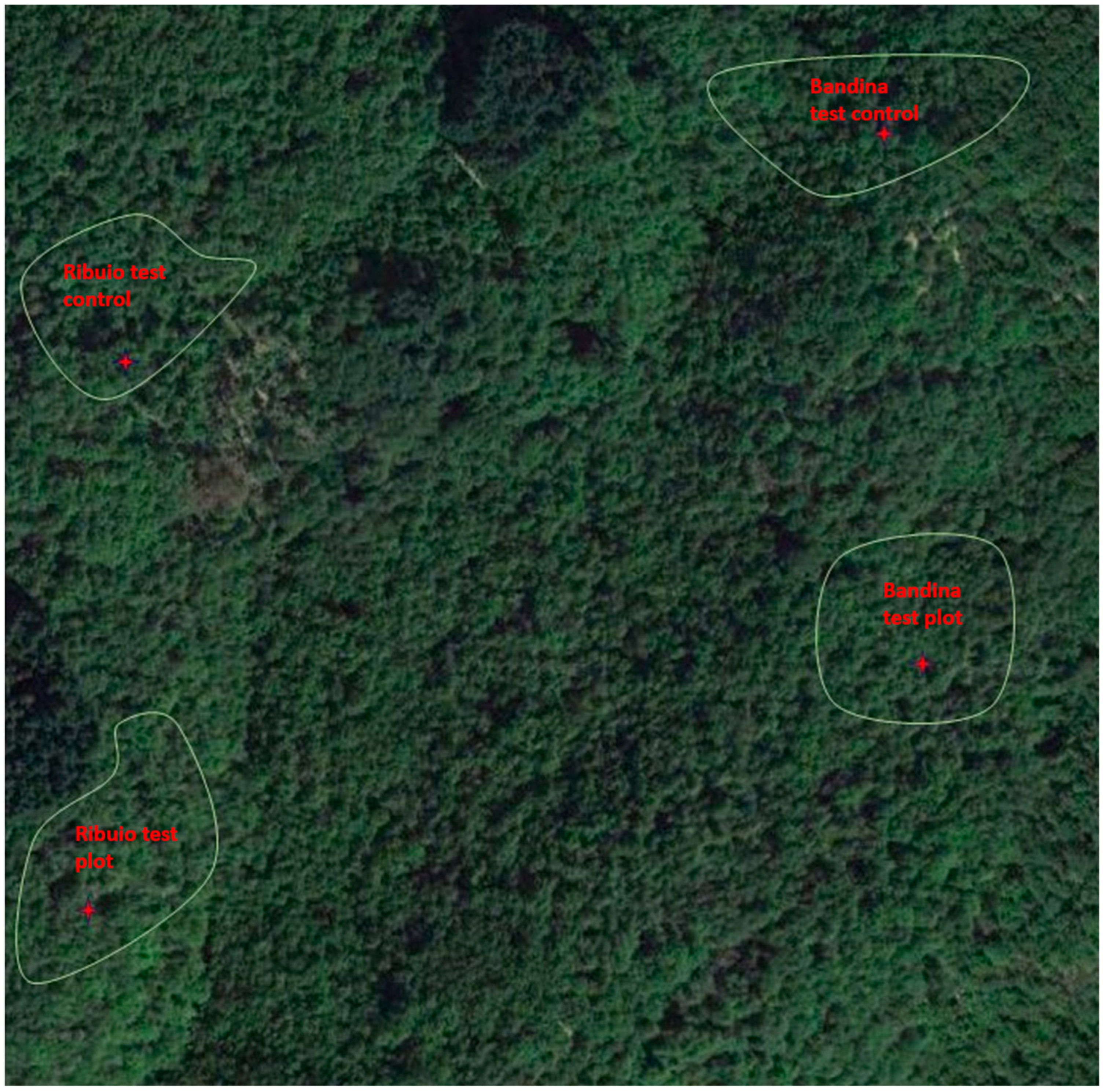
2.1. Study Sites

The study was conducted in chestnut groves at localities “Ribuio” and “Bandina”, in the Municipality of Ortignano-Raggiolo (AR), in the Casentino Valley (Arezzo, Tuscany), in central Italy. These localities were about 1300 m apart from each other, in two separate valleys with different microclimatic conditions (Ribuio test plot 43.683688 11.704197 690 S; Ribuio control plot 43.682456 11.705163 620 S; Bandina test plot 43.671514 11.700513 820 E; Bandina control plot 43.675462 11.705967 700 E (Figure 1). Two plots (ca. 0.4 Ha each), one per each locality, were established for the experimental trials. Two additional same-surface plots, homogeneous for tree cultivar and size, soil type and microclimate, were set up as a control in the immediate vicinity of each of the plots selected for treatments.
The “Ribuio” plot was on a skeleton in the form of outcropping rocks on a steep slope, in a small gorge, near a stand of Pinus sp. and was characterized by high humidity, due to the proximity of a river. It had a south-east exposure and an average altitude of 650 m a.s.l. (above sea level). The “Bandina” plot had no water course nearby, and the land was slightly sloping and was exposed to the east, with less outcropping rocks and fairly deep soil. The thermo-hygrometric conditions were therefore markedly different between the two plots (and in the adjacent control plots): “Ribuio” was more humid and colder; “Bandina” was hotter (because it was sunnier) and drier. The following parameters were recorded at each plot selected for treatments: number of trees, DBH (diameter at breast height: 1.30 m), height of each tree and age class (young < 30 years; adult 30–80 years; mature 80–150 years; overripe > 150 years height) (Table 1).

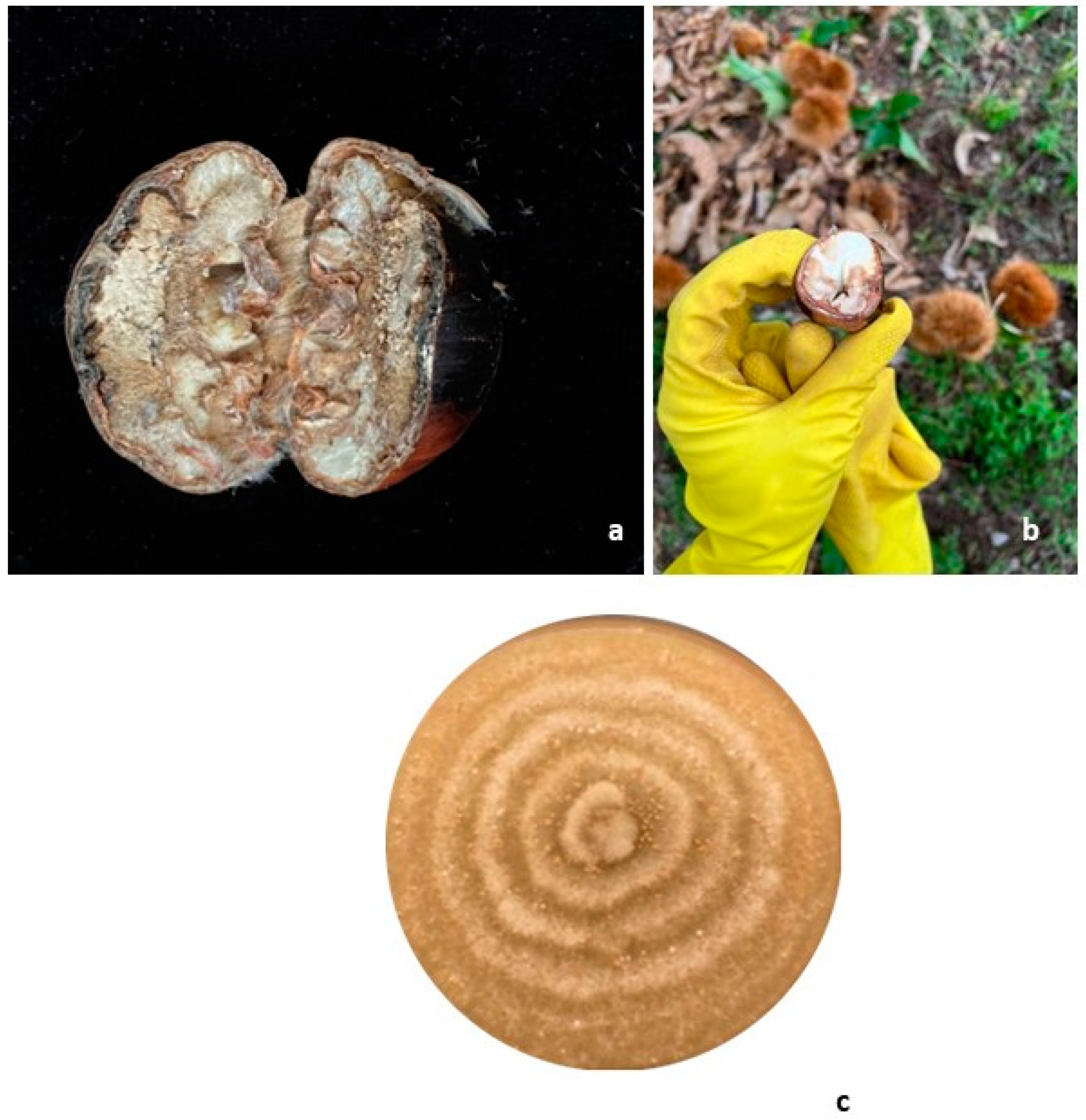
2.2. Soil Sampling and Trichoderma Isolation

A total of 20 soil samples were collected from the four study plots. Each area was divided into five homogeneous subplots, with one soil sample being collected from each subplot. Each sample was placed in a nylon bag, labeled and stored at 4 °C until use. For the isolation of Trichoderma from soil, modified malt extract agar (MEA, DIFCO, Detroit, MI, USA) was used. Six-fold serial dilutions of each soil sample were then prepared in sterilized, distilled water and 1 mL of diluted sample was poured onto the surface of the MEA, amended with 25 mg/L of streptomycin sulfate (Sigma Aldrich, Steinheim, Germany). The plates were incubated in the dark at 24 ± 1 °C for 7 days, according to the species requirements. Morphologically different colonies appearing on the plates were sub-cultured onto MEA and stored at 4 °C.

2.3. Identification of G. castaneae and Trichoderma spp. Strains

G. castaneae strains were isolated from infected nuts, whereas T. viride and T. harzianum strains were retrieved from soil samples. The appearance and morphology of the colonies (surface topography, texture, compactness, mycelium pigmentation and margin type) were determined on malt extract agar (MEA). For each morphotype, tufts of mycelium were picked off by scraping colony surfaces with a sterile dissecting needle, then mounted on glass slides in a drop of 0.5% KOH or Lactophenol for direct examination under the light microscope. Conidial micromorphology was determined under a Zeiss light microscope (ZEISS, Jena, Germany) at ×40 magnifications, by averaging 200 measurements per fungal taxon. Images were captured with an Optikam 4083.B5 microscopic Digital USBCamera operated with OptikaView version 7 acquisition software (OptikaSrl, Ponteranica, Italy). The identity of representative strains of each species was confirmed through sequence analysis of the region spanning the 5.8S rRNA gene and flanking Internal Transcribed Spacers 1 and 2 (ITS1-5.8 S-ITS2). For DNA extraction, fungal strains were grown on sterile cellophane in 90 mm Petri dishes containing 1% potato dextrose agar (PDA, DIFCO, Detroit, MI, USA) and maintained in the dark at 20 °C. After 7 days of incubation, ca. 70 mg (fresh weight) of mycelium was scraped off the cellophane surface and stored at −20 °C in a 2 mL Eppendorf tube until use. Genomic DNA was extracted using the GenElute plant Genomic DNA Miniprep extraction kit (Sigma Aldrich, St. Louis, MO, USA), following the manufacturer’s instructions, and stored at −20 °C. Internal transcribed spacer (ITS) region PCR amplification was performed on extracted samples by using ITS1 (5′-TCC GTA GGT GAA CCT GCG G-3′) and ITS4 (5′-TCC TCC GCT TAT TGA TAT GC-3′) universal primers [28]. Cycling and sequencing conditions were as described in Moricca et al. [29]. The identification of retrieved fungal taxa was undertaken by processing the relative sequences with the nucleotide–nucleotide BLAST® search tool (Basic Local Alignment Search Tool; http://www.ncbi.nlm.nih.gov/BLAST accessed on 11 October 2023). Generated sequences were submitted and deposited in NCBI GenBank database.

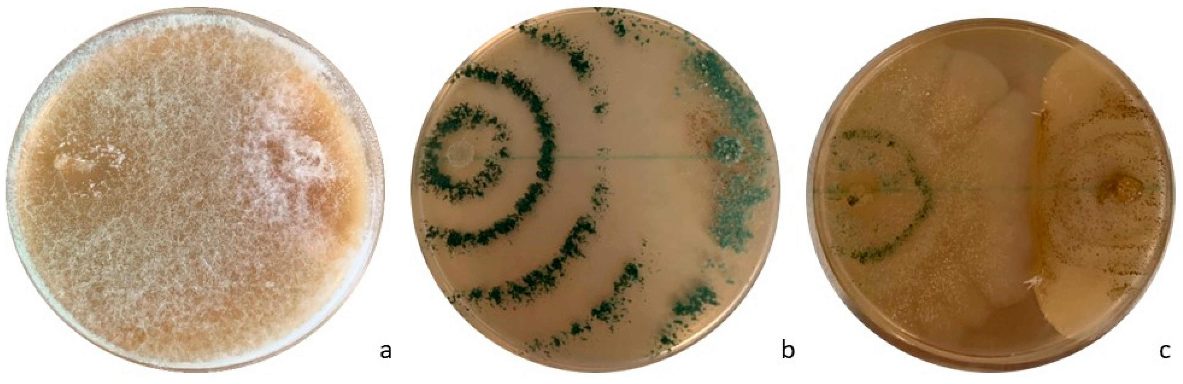
2.4. In Vitro Antagonism Tests

The T. harzianum and T. viride isolates retrieved from the soil were confronted with G. castaneae for their ability to inhibit the pathogen’s mycelial growth (%) in in vitro dual culture assays. For comparison, the test also included a commercial strain (SC1) of T. atroviride obtained from a Vintec® biological commercial formula (Certis Belchim B.V., Utrecht, The Netherlands). Each test was performed in a 9 cm diameter Petri dish containing 12 mL of MEA. Two plugs (diameter, 4 mm) retrieved from 7-day-old colonies of G. castaneae and of each one of the selected antagonists, were inserted in each dish, 6 cm apart from each other. Mycelial interactions were determined according to Badalayan et al. [30]. The experiment was conducted with 10 replicates for each antagonist and for the control, represented by two isolates of G. castaneae. Dishes were incubated at 25 °C in the dark for 8 days. Mycelial radial growth was measured after 8 days in each dish, calculating the growth area of each fungal species using the ImageJ software (version 1.8.0). Results were then compared to those obtained for the controls. The inhibition index percentage (I%) was calculated as follows [31]:
I % = R M r m R M × 100
where RM is the radius of the G. castaneae colony in the control plate, and rm is the radius of the colonies in the direction of the antagonist.
Hyphal interactions between colonies were assessed at 7 and 14 days under an optical microscope (ZEISS, Jena, Germany), with the antagonistic ability of T. viride, T. harzianum and T. atroviride specifically determined following Badalayan et al. [30], using the rating scale described in Table 2.

2.5. Biocontrol Agent Mixture Preparation
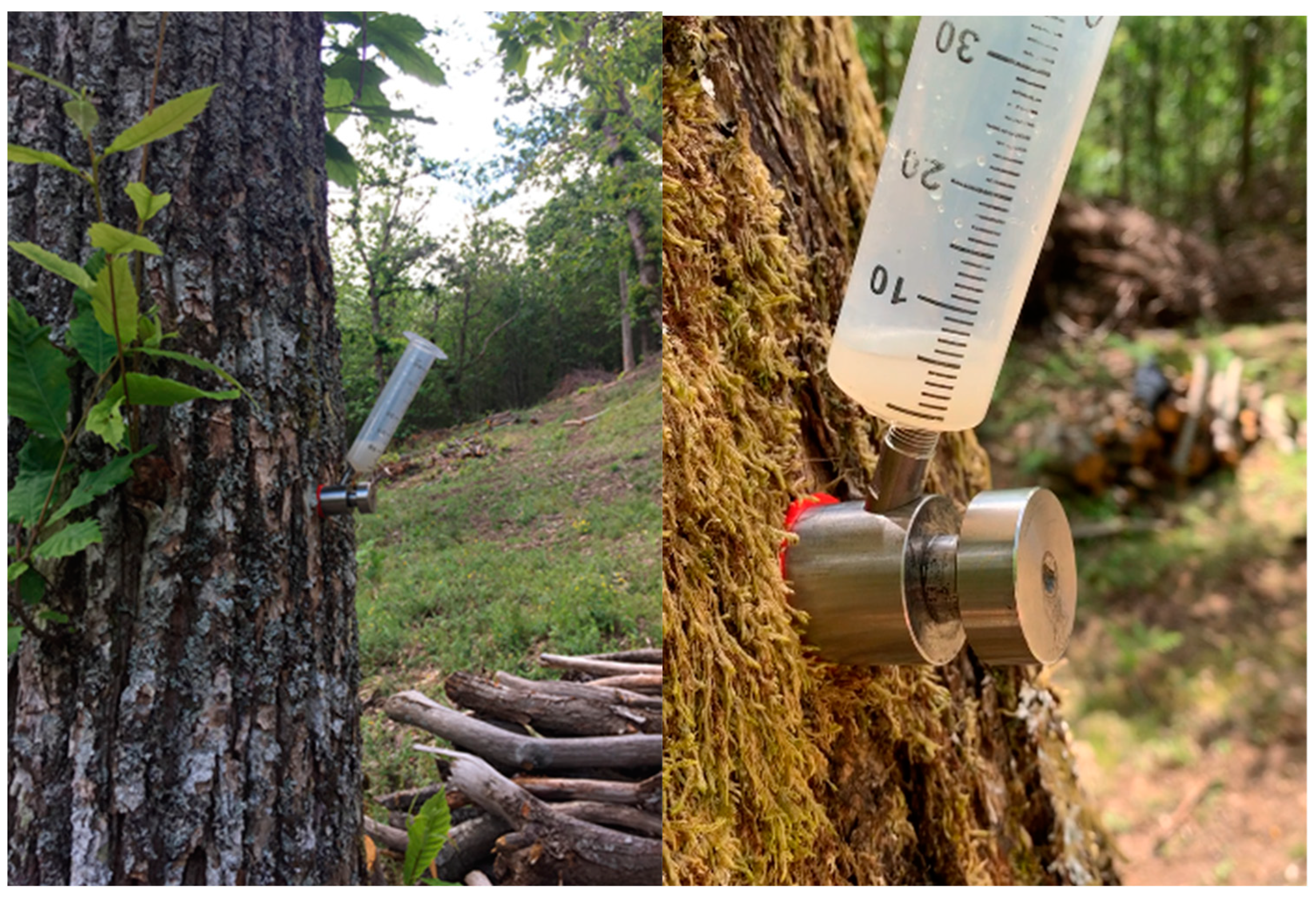
The solution used for endotherapic treatments was prepared by collecting T. harzianum and T. viride conidia from pure 7–10-day-old MEA cultures. For T. atroviride, the preparation was made according to the manufacturer’s instructions for the Vintec® (Belchim Crop Protection, Londerzeel, Belgium) biological formulation. The collected fungal mixture was filtered through filter paper (Whatman® Grade 1, Buckinghamshire, UK, diameter: 9 cm, pore size: 11 µm) to remove mycelium fragments. The concentration of conidial suspensions was then adjusted to 108 conidia/mL of water by using a counting chamber. The final dose of the fungal mixture utilized in trunk injections was 0.8 mL/10 cm of trunk circumference, following Berger et al. [27].

2.6. In Vivo Tests

In order to investigate the ability of Trichoderma species to antagonize the nut rot agent G. castaneae in planta, selected strains of the BCAs were delivered by means of trunk injections into the 66 adult chestnut trees growing in the two chestnut groves Bandina (34 trees) and Ribuio (32 trees) (see Table 1 above). Endotherapic treatments were performed at breast height (about 1.5/1.7 m from the ground) by using the BITE® (Blade for Infusion in TrEes) injection tool from De Rebus Plantarum (Vicenza, Italy). This instrument enables a targeted delivery of the biopesticide, without producing holes in the tree, but rather by causing a minimal (a few mm) vertical lesion, which the plant normally heals in a couple of weeks [32]. The Trichoderma solution was applied in June, when the trees were actively growing, with the crown fully expanded, and the full hydraulic tension of the transpiration flow guaranteed the maximum absorption of the solution (Figure 2). Treatments were performed during two subsequent growing seasons, 2020 and 2021.

2.7. Assessments of G. castaneae Incidence

Plants were monitored for their health conditions throughout the duration of the field experiment, with plant material (foliage, shoots, green curls with unripe nuts) being collected and analyzed periodically. Up to 400 fully ripened nuts were finally randomly collected from chestnut trees in each treated and untreated stand in October (Figure 3a,b). Fresh chestnuts were transported to the laboratory and stored at 4 °C before isolations. Each chestnut was surface washed with 75% ethanol (1 min) and 3% sodium hypochlorite (NaOCl) (3 min), then rinsed three times in sterile water. A sterile scalpel was used to remove the outer lignified shell and open the nut by cutting it in half. Five fragments (approximately 1 × 1 × 2 mm in size) were randomly excised from the tissues of each fruit and plated in 90 mm diameter Petri dishes filled with 2% MEA. All dishes were incubated in the dark at 25 °C for 3 days, according to species requirements. The presence of G. castaneae was analyzed for each dish by comparing macro- and micro-morphological features of each obtained mycelium with that of a G. castaneae isolate, whose identity was previously assessed by ITS sequencing (Figure 3c).

2.8. Statistical Analyses

For all the traits considered, the means and standard deviations were calculated for all the combinations considered. A one-way ANOVA was performed to test the significance of the in vitro antagonistic activity of three Trichoderma spp. versus G. castaneae. All data refer to the four chestnut stands (two treated and two untreated). Homogeneity of variance and normality tests were performed using the Levene and Shapiro–Wilk tests. SPSS V.28 (IBM Corporate, Endicott, NY, USA) was used for the statistical analysis, with χ2 square test, to identify significant differences among treatments. The square test was applied to analyze the significance of the differences between trees treated with Trichoderma spp. and the controls (with no BCA application). The incidence of G. castaneae was calculated for each treated and untreated stand as the ratio (%) between the number of affected nuts and the total number of nuts analyzed.

3. Results

3.1. Gnomoniopsis castaneae and Trichoderma spp. Identification

The colonies of G. castaneae were initially white, turning to light gray after 7 days of incubation. The mycelium appeared either flat or thick and densely woolly. Margins were diffuse and developed in concentric circles. In the innermost portion of the colony, acervuli, ranging in color from orange to red, developed after 10 days. They were either superficial or erupting on the upper surface, circular, solitary or gregarious. Conidia were hyaline, one-celled, ovoid–oblong, straight or curved. Colony phenotypes, as well the size and shape of conidia, matched exactly the original descriptions of Visentin et al. [33] and Shuttleworth et al. [34]. DNA sequencing confirmed the representative morphotype as belonging to G. castaneae (PP326312), with BLAST searches that revealed complete (100%) sequence homology with those of the pathogen already deposited in the GenBank database. Trichoderma harzianum exhibited a cottony white, slightly yellowish mycelium, with a flat growth profile. Colonies developed at 8 days showed several concentric rings with dark green conidial production. Trichoderma viride colonies showed a uniform appearance with light green-yellowish conidia evenly distributed across their surfaces (without differentiating concentric rings). Growth rates and cultural characteristics of the two Trichoderma species differed on the same medium at a constant temperature of 24 °C. T. viride, from all soil types, demonstrated the highest growth rate compared to T. harzianum. Trichoderma harzianum conidia were from globose to sub-globose, with a dark green color. T. viride presented globose conidia, with a color ranging from light yellow to green.
Sequence analysis confirmed the identities of T. harzianum and T. viride (GenBank acc. nos. PP326311 and PP326313, respectively), with BLAST searches that revealed complete (100%) homology with sequences of the two species retrieved from the GenBank database.

3.2. In Vitro Tests

A priori in vitro assays were useful for assessing Trichoderma species for their ability to inhibit G. castaneae. T. viride and T. harzianum strains were highly effective against G. castaneae. The parasitization of the pathogen was already visible at 96 h. After 8 days, G. castaneae mycelium turned out completely overgrown and replaced by strains of these two Trichoderma species (Figure 4a,b). The growth of T. atroviride at 25 °C was lower than that of T. viride and T. harzianum, with only a partial interaction with G. castaneae that was observed at the margin of the two colonies (Figure 4c). Hyphal interactions for T. viride and T. harzianum resulted in the CA2 subtype of Badalyan’s scale; T. atroviride ranked as CA1 of the same scale. Statistical analyses confirmed the different behaviors/inhibitory effects of the three Trichoderma species. The one-way ANOVA statistical criterion showed that T. atroviride’s area value differed significantly after 6 days from that of the other two antagonistic isolates (p > 0.05). The highest inhibition capability was found for T. harzianum (99.14%), followed by T. viride (78.12%) and T. atroviride (51.82%) (Table 3).

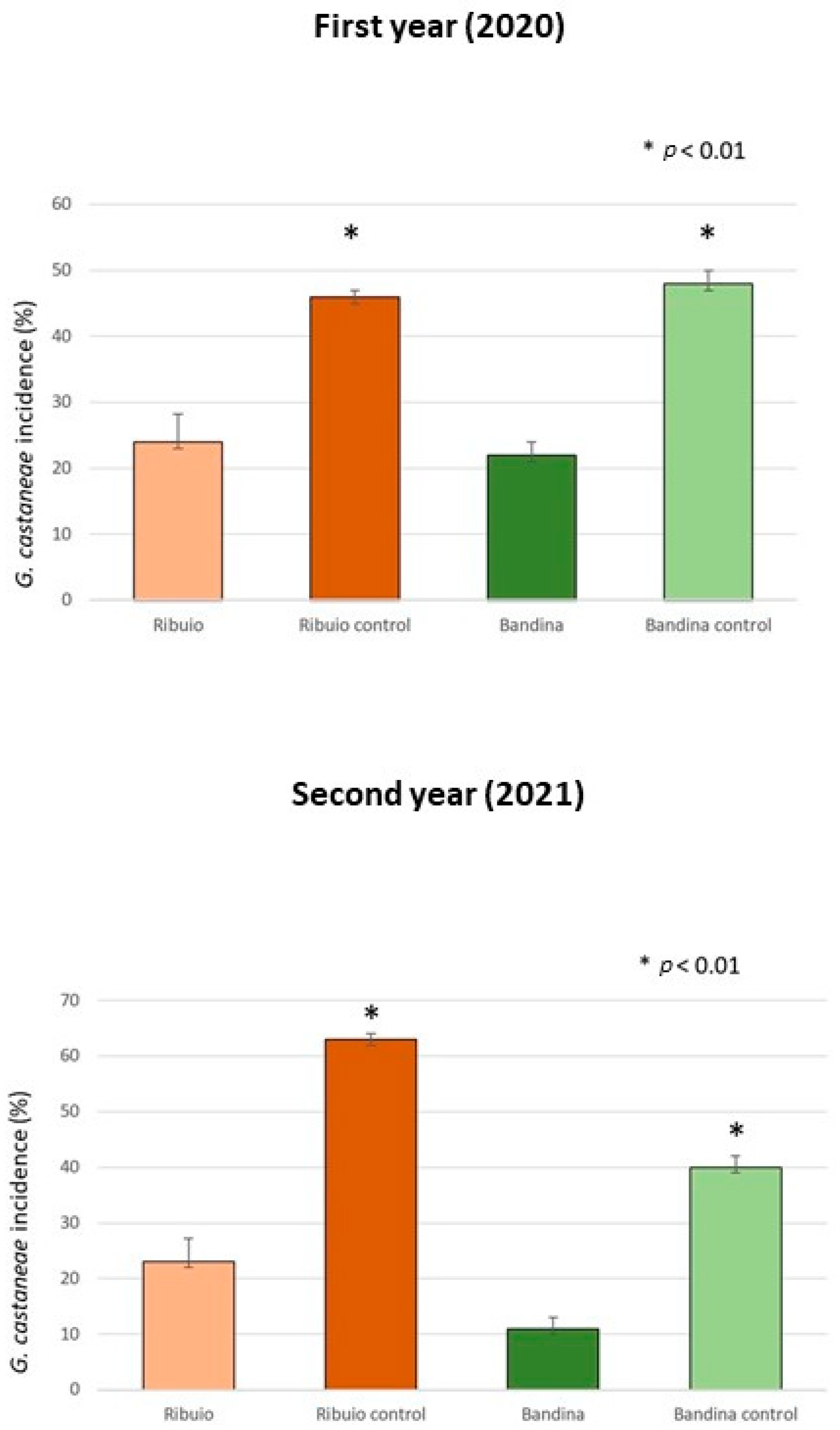
3.3. Endotherapic Treatments in the Field

The field trials proved the effectiveness of treatments with T. viride, T. harzianum and T. atroviride in curtailing G. castaneae, with the pathogen’s incidence being significantly reduced in the investigated chestnut groves. Data from the first and second year of treatments showed a reduction in G. castaneae incidence, with this trend increasing in the second year. In fact, in 2020, different percentages of G. castaneae incidence were obtained in the two treated chestnut groves, which were 24% and 22%, respectively, compared to the two control areas (46% and 48%). In the second year of treatment (2021), the incidence of the target pathogen was 23% and 11% in the treated plots and 63% and 40% in the in the control plots. The decrease in G. castaneae incidence between treated and untreated plots resulted in percentages of 26% and 22% in 2020 and of 40% and 29% in 2021 (Figure 5). The chi-squared statistical analysis confirmed the significant differences (p ≤ 0.01) between treated and untreated chestnut groves during the two years of analysis. No other nut rot pathogens were isolated from the analyzed nut samples.

4. Discussion

Nut rot by G. castaneae today represents the main cause of chestnut fruit deterioration. The disease strongly curtails nut production in the areas where it is present, the pathogen irremediably compromising nut quality and organoleptic features, making the fruit tissue chalky and inedible [11,13,35,36,37,38,39]. As the fungus can live as an endophyte in latency in chestnut organs and tissues, the application of effective control measures is troublesome and relies only on post-harvest treatments: water curing, sterilization and chemicals application [10,16]. Since a chestnut grove is considered in all respects a forest, chemical treatments are prohibited for environmental reasons. But even if they were not prohibited, chemical treatments would still be difficult to implement due to the morphology and slope of the terrain on which chestnut groves are often located, the environmental and spatial heterogeneity within the plantation and the large size of the trees. For all these reasons, in this work, biological control based on the application of Trichoderma species was evaluated as an alternative strategy for the control of G. castaneae in chestnut groves. The implementation of biological control against plant pathogens in agro-ecosystems by releasing competent Trichoderma strains is a safe technology that could solve issues related to environmental and health risks inherent with conventional control strategies [40,41]. Fungi of the genus Trichoderma are innate with the environment, being common inhabitants of soils, both agricultural and forest, and are often also found as endophytes in many plant species [42]. The effectiveness of Trichoderma species as BCAs against plant pathogens has been known for a long time and has been successfully applied in a variety of agricultural crops, especially in horticulture [43,44,45,46,47,48,49].
The application of biological control methods against forest pathogens, on the contrary, has been scarcely investigated and few attempts were made on adult trees [25,26]. In this work, endotherapy was exploited to deliver Trichoderma BCAs against G. castaneae directly in the stem of cultivated chestnut trees. Results were promising and consistent with those obtained by Berger et al. [27], who injected T. atroviride in Quercus robur and Fagus sylvatica using endotherapy as a preventive strategy against Phytophthora spp. infections. Indeed, our results showed that, in 2020, the incidences of G. castaneae were 24% and 22% in the treated groves (Ribuio and Bandina), in comparison to their respective adjacent control plots (46% and 48%). A similar behavior was observed in the second year of treatment (2021), registering pathogen incidences of 23% and 11% in the treated plots (Ribuio and Bandina), and 63% and 40% in the control plots (Ribuio and Bandina). The decrease in G. castaneae incidence between treated and untreated stands resulted in percentages of 22% and 26% for 2020, and of 29% and 40% for 2021. The difference between the two locations, in the incidence of G. castaneae both in treated and in control plots, is ascribed to the different thermo-hygrometric conditions of the two sites. In fact, Ribuio is characterized by a particular microclimate: it is in a gorge, at the base of which a stream flows, which generates constant humidity and poor sunlight on the ground. Bandina, on the other hand, has a different exposure, is not located near streams and experiences high levels of sunshine. The particular temperature and humidity conditions of Ribuio may have favored G. castaneae infections.
The results of in vitro tests performed in this study have further confirmed the effectiveness of Trichoderma application as a biocontrol agent, as it inhibited G. castaneae in culture. Indeed, the inhibition of G. castaneae was observed by each tested Trichoderma species, with inhibition index percentages of 99.14% for T. harzianum, 78.12% for T. viride and 51.82% for T. atroviride. To the best of our knowledge, no other authors have reported a detailed assessment of the effects and interactions among T. viride, T. harzianum and T. atroviride on G. castaneae using dual culturing. Our in vitro results are in line with those of other authors who tested these Trichoderma species against different plant pathogens [24,50,51].
The treatment with three Trichoderma spp. was able to significantly reduce the necrotic surfaces of chestnuts caused by G. castaneae, and no systematic studies of resistance have been undertaken to date [11]. The results obtained in this study can also be compared with those reported by Pasche et al. [23], who applied T. atroviride against C. parasitica and G. castaneae by soaking chestnut scions in fungal propagule suspensions. These authors observed that the endophytic behavior of Trichoderma in chestnut tissues and the presence of T. atroviride were able to influence the infection of G. castaneae. Indeed, in agreement with our study, only 15% of treated scions analyzed by Pasche et al. [23] showed G. castaneae symptoms, while the percentage of G. castaneae symptoms in control scions was 75%. These authors ascribed the effectiveness of their biological control treatment to the possibility that inoculated Trichoderma had spread and colonized the totality of woody tissues.
The endotherapic method applied in this work in chestnut trees could improve the efficacy of biological applications, facilitating the entrance of Trichoderma in the plant vascular system by trunk injection. Endotherapy offers important advantages over other treatment methods: it lowers (reducing practically to zero) the dispersion of the BCA into the environment, and the doses needed for treatments are enormously reduced, making them more sustainable than traditional methods of application (e.g., spraying). The results obtained in this study suggest that the biological control method employed could represent a key advantage for chestnut growers, since it reduces the decay of fresh and processed nuts and the resulting economic losses.
Little is known about the duration of protection conferred by treatment with Trichoderma BCAs in forestry. However, the fact that in the second year a greater reduction in the incidence of rot was achieved means that there was an additive effect between the first and second year treatments. The mechanisms of action of Trichoderma strains (e.g., direct parasitism, lytic enzyme production, antibiosis, competition for nutrients and space) have been widely studied [18,19,21,22,50,52,53,54]. It is possible that the injected Trichoderma species utilized one or more of the above mechanisms, as well as also synthesizing bioactive compounds that elicited plant defense molecular responses, through the activation of either the ISR (induced systemic resistance) or the SAR (systemic acquired resistance) pathways. All these protection mechanisms may have restricted G. castaneae, conferring long-lasting defense to chestnut trees [18,41].

5. Conclusions

This is the first work in which BCAs of the genus Trichoderma have been administered on chestnut trees by endotherapy. The choice of endotherapic applications, dictated by technical–operational needs (we needed to administer the biopesticide in uncomfortable conditions, on large-sized plants and, last but not least, to reach the target pathogen in the most distal portions of the foliage), turned out to be a valid option. Further research is required to confirm the promising results obtained here and to better elucidate unknown aspects of Trichoderma mycoparasitism when conferring these BCAs to restrict G. castaneae in chestnut groves.

Author Contributions

A.B. and S.M.: conceptualization; A.B., S.M. and C.A.: field survey, sample collection and assay; A.B. and S.O.C.: data analysis; S.M.: funding acquisition; A.B.: writing—draft preparation; A.B., C.A., S.O.C. and S.M.: review and manuscript editing. All authors have read and agreed to the published version of the manuscript.

Funding

This research was funded in part by Regione Toscana, PS-GO 2017, PSR—FEASR 2014–2020, Project INGECA—“Strategie INnovative a basso impatto per la GEstione delle avversità dei CAstagneti da frutto”, and in part by Fondazione CRF (Cassa di Risparmio di Firenze), Project “Innovazione e Recupero Sostenibile in alcune Filiere Agroalimentari Pedemontane nell’Appennino Toscano”.

Institutional Review Board Statement

Not applicable.

Informed Consent Statement

Not applicable.

Data Availability Statement

Data are available by e-mail on reasonable request.

Acknowledgments

The authors thank the chestnut growers Giorgini Riccardo and Giovannuzzi Andrea for making their plots available for the experimental trials.

Conflicts of Interest

The authors declare no conflict of interest.

References

  1. Aglietti, C.; Cappelli, A.; Andreani, A. From Chestnut Tree (Castanea sativa) to Flour and Foods: A Systematic Review of the Main Criticalities and Control Strategies towards the Relaunch of Chestnut Production Chain. Sustainability 2022, 14, 12181. [Google Scholar] [CrossRef]
  2. Mattioni, C.; Martin, M.A.; Chiocchini, F.; Cherubini, M.; Gaudet, M.; Pollegioni, P.; Velichkov, I.; Jarman, R.; Chambers, F.M.; Damian, V.L.; et al. Landscape genetics structure of European sweet chestnut (Castanea sativa Mill): Indications for conservation priorities. Tree Genet. Genomes. 2017, 13, 39. [Google Scholar] [CrossRef]
  3. FAO. World Food and Agriculture Statistical Yearbook 2022; FAO: Rome, Italy, 2022. [Google Scholar]
  4. López-Sáez, J.A.; Glais, A.; Robles-López, S.; Alba-Sánchez, F.; Pérez-Díaz, S.; Abel-Schaad, D.; Luelmo-Lautenschlaeger, R. Unraveling the naturalness of sweet chestnut forests (Castanea sativa Mill.) in central Spain. Veg. Hist. Archaeobot. 2017, 26, 167–182. [Google Scholar] [CrossRef]
  5. Bussotti, F.; Papitto, G.; Di Martino, D.; Cocciufa, C.; Cindolo, C.; Cenni, E.; Bettini, D.; Iacopetti, G.; Pollastrini, M. Defoliation, recovery and increasing mortality in Italian forests: Levels, patterns and possible consequences for forest multifunctionality. Forests 2021, 12, 1476. [Google Scholar] [CrossRef]
  6. Fernandes, P.; Colavolpe, M.B.; Serrazina, S.; Costa, R.L. European and American Chestnuts: An Overview of the Main Threats and Control Efforts. Front. Plant Sci. 2022, 13, 951844. [Google Scholar] [CrossRef] [PubMed]
  7. Agnoletti, M. The degradation of traditional landscape in a mountain area of Tuscany during the 19th and 20th centuries: Implications for biodiversity and sustainable management. For. Ecol. Manag. 2007, 249, 5–17. [Google Scholar] [CrossRef]
  8. Frati, A.; Landi, D.; Marinelli, C.; Gianni, G.; Fontana, L.; Migliorini, M.; Pierucci, F.; Garcia-Gil, M.; Meacci, E. Nutraceutical Properties of Chestnut Flours: Beneficial Effects on Skeletal Muscle Atrophy. Food Funct. 2014, 5, 2870–2882. [Google Scholar] [CrossRef]
  9. Cevasco, R.; Moreno, D.; Balzaretti, R.; Watkins, C. Historical chestnut cultures, climate and rural landscapes in the Apennines. In The Future of Heritage as Climates Change: Loss, Adaptation and Creativity; Harvey, D., Perry, J., Eds.; Routledge: London, UK, 2015; pp. 130–147. [Google Scholar]
  10. Morales-Rodríguez, C.; Bastianelli, G.; Caccia, R.; Bedini, G.; Massantini, R.; Moscetti, R.; Thomidis, T.; Vannini, A. Impact of ‘brown rot’caused by Gnomoniopsis castanea on chestnut fruits during the post-harvest process: Critical phases and proposed solutions. J. Sci. Food Agric. 2022, 102, 680–687. [Google Scholar] [CrossRef]
  11. Shuttleworth, L.A.; Liew, E.C.Y.; Guest, D.I. Survey of the incidence of chestnut rot in south-eastern Australia. Australas. Plant Pathol. 2013, 42, 63–72. [Google Scholar] [CrossRef]
  12. Sieber, T.N.; Jermini, M.; Conedera, M. Effects of the harvest method on the infestation of chestnuts (Castanea sativa) by insects and moulds. J. Phytopathol. 2007, 155, 497–504. [Google Scholar] [CrossRef]
  13. Lione, G.; Danti, R.; Fernandez-Conradi, P.; Ferreira-Cardoso, J.V.; Lefort, F.; Marques, G.; Meyer, J.B.; Prospero, S.; Radócz, L.; Robin, C.; et al. The emerging pathogen of chestnut Gnomoniopsis castaneae: The challenge posed by a versatile fungus. Eur. J. Plant Pathol. 2019, 153, 671–685. [Google Scholar] [CrossRef]
  14. Vettraino, A.M.; Luchi, N.; Rizzo, D.; Pepori, A.L.; Pecori, F.; Santini, A. Rapid diagnostics for Gnomoniopsis smithogilvyi (syn. Gnomoniopsis castaneae) in chestnut nuts: New challenges by using LAMP and real-time PCR methods. AMB Express 2021, 11, 105. [Google Scholar] [CrossRef] [PubMed]
  15. Lema, F.; Baptista, P.; Oliveira, C.; Ramalhosa, E. Brown Rot Caused by Gnomoniopsis smithogilvyi (syn. Gnomoniopsis castaneae) at the Level of the Chestnut Tree (Castanea sativa Mill.). Appl. Sci. 2023, 13, 3969. [Google Scholar] [CrossRef]
  16. Silva-Campos, M.; Nadiminti, P.; Cahill, D. Rapid and Accurate Detection of Gnomoniopsis smithogilvyi the Causal Agent of Chestnut Rot, through an Internally Controlled Multiplex PCR Assay. Pathogens 2022, 11, 907. [Google Scholar] [CrossRef]
  17. Robin, D.C.; Marchand, P.A. Evolution of the biocontrol active substances in the framework of the European Pesticide Regulation (EC) No. 1107/2009. Pest. Manag. Sci. 2019, 75, 950–958. [Google Scholar] [CrossRef]
  18. Harman, G.E.; Howell, C.R.; Viterbo, A.; Chet, I.; Lorito, M. Trichoderma species—Opportunistic, avirulent plant symbionts. Nat. Rev. Microbiol. 2004, 2, 43–56. [Google Scholar] [CrossRef] [PubMed]
  19. Ghazanfar, M.U.; Raza, M.; Raza, W.; Qamar, M.I. Trichoderma as Potential Biocontrol Agent, Its Exploitation in Agriculture: A Review. Plant Prot. 2018, 2, 109–135. [Google Scholar]
  20. Weindling, R. Trichoderma lignorum as a parasite of other soil fungi. Phytopathology 1932, 22, 837–845. [Google Scholar]
  21. Lorito, S.; Selva, J.; Basili, R.; Romano, F.; Tiberti, M.M.; Piatanesi, A. Probabilistic hazard for seismically induced tsunamis: Accuracy and feasibility of inundation maps. Geophys. J. Int. 2015, 200, 574–588. [Google Scholar] [CrossRef]
  22. Alfiky, A.; Weisskopf, L. Deciphering Trichoderma–Plant–Pathogen Interactions for Better Development of Biocontrol Applications. J. Fungi 2021, 7, 61. [Google Scholar] [CrossRef]
  23. Pasche, S.; Crovadore, J.; Pelleteret, P.; Jermini, M.; Mauch-Mani, B.; Oszako, T.; Lefort, F. Biological control of the latent pathogen Gnomoniopsis smithogylvyi in European chestnut grafting scions using Bacillus amyloliquefaciens and Trichoderma atroviride. Dendrobiology 2016, 75, 113–122. [Google Scholar] [CrossRef]
  24. Murolo, S.; Concas, J.; Romanazzi, G. Use of biocontrol agents as potential tools in the management of chestnut blight. Biol. Control 2019, 132, 102–109. [Google Scholar] [CrossRef]
  25. Schubert, M.; Fink, S.; Schwarze, F.W.M.R. Evaluation of Trichoderma spp. as a biocontrol agent against wood decay fungi in urban trees. Biol. Control 2008, 45, 111–123. [Google Scholar] [CrossRef]
  26. Prospero, S.; Botella, L.; Santini, A.; Robin, C. Biological control of emerging forest diseases: How can we move from dreams to reality? For. Ecol. Manag. 2021, 496, 119377. [Google Scholar] [CrossRef]
  27. Berger, G.; Czarnocka, K.; Cochard, B.; Oszako, T.; Lefort, F. Biocontrol Endotherapy with Trichoderma spp. and Bacillus amyloliquefaciens against Phytophthora spp.: A Comparative Study with Phosphite Treatment on Quercus robur and Fagus sylvatica. J. Agric. Sci. Technol. A 2015, 5, 428–439. [Google Scholar]
  28. White, T.J.; Bruns, T.; Lee, S.; Taylor, J. Amplification and direct sequencing of fungal ribosomal RNA genes for phylogenetics. PCR Protoc. Guide Methods Appl. 1990, 18, 315–322. [Google Scholar]
  29. Moricca, S.; Ginetti, B.; Ragazzi, B. Species and organ-specificity in endophytes colonizing healthy and declining Mediterranean oaks. Phytopathol. Mediterr. 2012, 51, 587–598. [Google Scholar]
  30. Badalyan, S.M.; Garibyan, N.G.; Innocenti, G. Antagonistic activity of xylotrophic mushrooms against pathogenic fungi of cereals in dual culture. Antagonistic activity of xylotrophic mushrooms against pathogenic fungi of cereals in dual culture. Phytopathol. Med. 2002, 41, 220–225. [Google Scholar]
  31. Idris, H.A.; Labuschagne, N.; Korsten, L. Screening rhizobacteria for biological control of Fusarium root and crown rot of sorghum in Ethiopia. Biol. Control 2007, 40, 97–106. [Google Scholar] [CrossRef]
  32. Montecchio, L. A Venturi Effect Can Help Cure Our Trees. J. Vis. Exp. 2013, 80, e51199. [Google Scholar]
  33. Visentin, I.; Gentile, S.; Valentino, D.; Gonthier, P.; Tamietti, G.; Cardinale, F. Gnomoniopsis castanea sp. nov. (Gnomoniaceae, Diaporthales) as the causal agent of nut rot in sweet chestnut. J. Plant. Pathol. 2012, 94, 411–419. [Google Scholar]
  34. Shuttleworth, L.A.; Guest, D.I.; Liew, E.C.Y. Fungal planet description sheet 108–Gnomoniopsis smithogilvyi L.A. Shuttleworth, E.C.Y. Liew & D.I. Guest, sp. nov. Persoonia 2012, 28, 142–143. [Google Scholar]
  35. Beccaro, G.L.; Alma, A.; Gonthier, P.; Mellano, M.G.; Ferracini, C.; Giordano, L.; Lione, G.; Donno, D.; Boni, I.; Ebone, A.; et al. Chestnut R&D Centre, Piemonte (Italy): 10 years of activity. Acta Hortic. 2018, 1220, 133–140. [Google Scholar]
  36. Beccaro, G.; Alma, A.; Bounous, G.; Gomes-Laranjo, J. The Chestnut Handbook: Crop & Forest Management; CRC Press: Boca Raton, FL, USA, 2019; 378p. [Google Scholar]
  37. Conti, V.; Salusti, P.; Romi, M.; Cantini, C. Effects of drying methods and temperatures on the quality of chestnut flours. Foods 2022, 11, 1364. [Google Scholar] [CrossRef] [PubMed]
  38. Nerva, L.; Costa, L.D.; Ciacciulli, A.; Sabbadini, S.; Pavese, V.; Dondini, L.; Vendramin, E.; Caboni, E.; Perrone, I.; Moglia, A.; et al. The role of Italy in the use of advanced plant genomic techniques on fruit trees: State of the art and future perspectives. Int. J. Mol. 2023, 24, 977. [Google Scholar] [CrossRef] [PubMed]
  39. Drais, M.I.; Gusella, G.; Mazzaglia, A.; Polizzi, G. A quantitative PCR assay for the detection and quantification of Septoria pistaciarum, the causal agent of pistachio leaf spot in Italy. PLoS ONE 2023, 18, e0286130. [Google Scholar] [CrossRef]
  40. Rodrigues, A.O.; De Mio, L.L.M.; Soccol, C.R. Trichoderma as a powerful fungal disease control agent for a more sustainable and healthy agriculture: Recent studies and molecular insights. Planta 2023, 257, 31. [Google Scholar] [CrossRef] [PubMed]
  41. Woo, S.L.; Hermosa, R.; Lorito, M.; Monte, E. Trichoderma: A multipurpose, plant-beneficial microorganism for eco-sustainable agriculture. Nat. Rev. Microbiol. 2023, 21, 312–326. [Google Scholar] [CrossRef]
  42. Klein, D.; Eveleigh, D.E. Ecology of Trichoderma. In Trichoderma & Gliocladium, Basic Biology, Taxonomy and Genetics; Kubicek, C.P., Harmna, G.E., Eds.; Taylor and Francis: London, UK, 1998; Volume 1, pp. 57–69. [Google Scholar]
  43. Chandra, S.; Singh, B.K. Trichoderma spp.: As potential bio-control agents (BCAs) against fungal plant pathogens. Indian J. Life Sci. 2016, 5, 105. [Google Scholar]
  44. Hidayah, B.N. Biological Control Potential of Trichoderma Species and Bacterial Antagonists against Sclerotinia sclerotiorum on Canola in Western Australia. Int. J. Agric. Biol. 2022, 27, 215–227. [Google Scholar] [CrossRef]
  45. Asad, S.A. Mechanisms of Action and Biocontrol Potential of Trichoderma against Fungal Plant Diseases—A Review. Ecol. Complex. 2022, 49, 100978. [Google Scholar] [CrossRef]
  46. Shalaby, T.A.; Taha, N.; El-Beltagi, H.S.; El-Ramady, H. Combined Application of Trichoderma harzianum and Paclobutrazol to Control Root Rot Disease Caused by Rhizoctonia solani of Tomato Seedlings. Agronomy 2022, 12, 3186. [Google Scholar] [CrossRef]
  47. Natsiopoulos, D.; Tziolias, A.; Lagogiannis, I.; Mantzoukas, S.; Eliopoulos, P.A. Growth-Promoting and Protective Effect of Trichoderma atrobrunneum and T. simmonsii on Tomato against Soil-Borne. Fungal Pathogens. Crops 2022, 2, 202–217. [Google Scholar] [CrossRef]
  48. Sehim, A.E.; Hewedy, O.A.; Altammar, K.A.; Alhumaidi, M.S.; Abd Elghaffar, R.Y. Trichoderma asperellum empowers tomato plants and suppresses Fusarium oxysporum through priming responses. Front. Microbiol. 2023, 14, 1140378. [Google Scholar] [CrossRef] [PubMed]
  49. Poveda, J.; Millen, M.R.; Bailey, A.M. Analysis of Trichoderma as an effective biological control agent against the honey fungus (Armillaria spp.). Biol. Control 2023, 188, 105424. [Google Scholar] [CrossRef]
  50. Benítez, T.; Rincón, A.M.; Limón, M.C.; Codón, A.C. Biocontrol mechanisms of Trichoderma strains. Int. Microbiol. 2004, 7, 249–260. [Google Scholar]
  51. Bunbury-Blanchette, A.; Walker, A. Trichoderma species show biocontrol potential in dual culture and greenhouse bioassays against Fusarium basal rot of onion. Biol. Control 2018, 130, 127–135. [Google Scholar] [CrossRef]
  52. Gajera, H.; Domadiya, R.; Patel, S.; Kapopara, M.; Golakiya, B. Molecular mechanism of Trichoderma as bio-control agents against phytopathogen system—A review. Curr. Res. Microbiol. Biotechnol. 2013, 1, 133–142. [Google Scholar]
  53. Nawrocka, J.; Małolepsza, U. Diversity in Plant Systemic Resistance Induced by Trichoderma. Biol. Control 2013, 67, 149–156. [Google Scholar] [CrossRef]
  54. Mukherjee, P.K.; Mendoza-Mendoza, A.; Zeilinger, S.; Horwitz, B.A. Mycoparasitism as a Mechanism of Trichoderma-Mediated Suppression of Plant Diseases. Fungal Biol. Rev. 2022, 39, 15–33. [Google Scholar] [CrossRef]
Figure 1. Aerial view of the four plots (Ribuio test, Ribuio control, Bandina test and Bandina control); the plots under investigation are delineated in green.
Biology 13 00143 g001
Figure 2. Application of conidial suspensions of the BCAs on C. sativa stems with the BITE® injection tool.
Biology 13 00143 g002
Figure 3. Symptoms caused by G. castaneae in nuts: (a) a fruit with a completely dehydrated endosperm; (b) a freshly collected nut and cut in the field displaying a spongy or chalky appearance with distinctive dark-brown lesions. This discoloration further underscores the altered composition, indicating potential physiological changes within the fruit; (c) a G. castaneae culture grown on malt extract agar (MEA) after two weeks.
Biology 13 00143 g003
Figure 4. In vitro dual cultures of Trichoderma species against G. castaneae after two weeks of incubation at 25 °C: (a) T. viride/G. castaneae interaction; (b) T. harzianum/G. castaneae interaction; and (c) T. atroviride/G. castaneae interaction.
Biology 13 00143 g004
Figure 5. Average percentage of nut fruits found infected by G. castaneae in test plots at the “Ribuio” and “Bandina” chestnut groves and in their respective control plots. Black bars represent standard errors. * Significant differences between numbers of infected nuts found between localities (χ2, p < 0.01).
Biology 13 00143 g005
Table 1. Age class, DBH (cm) and height (m) of treated trees in each chestnut stand.
Ribuio Chestnut Stand
NumberAge (Y = Young, <30 years; M = Mature, 80–150 years; O = Overripe, >150 years)Diameter at Breast Height DBH (cm)Height (m)
1O79.5811.8
2O42.9715.9
3O86.2614
4O95.4912.3
5O44.5610
6M38.214.7
7M34.0613.8
8O113.3212.8
9M53.1616.3
10M33.4214.1
11O65.5710.9
12O108.2310.9
13O11311.1
14O66.859.4
15O82.767.6
16O64.37.8
17O43.9312.8
18O69.397.7
19M36.616.1
20NA28.87NA
21O62.716.9
22O70.666.8
23O60.48NA
24NA60.48NA
25O63.66NA
26NA49.34NA
27O92.31NA
28O98.04NA
29O127.32NA
30M49.34NA
31O119.37NA
32O93.9NA
Bandina chestnut stand
1M70.0310
2M38.8311.9
3M55.710.02
4M53.1611.06
5M59.2113.6
6M41.3813.4
7M61.7515.1
8M51.5714.1
9M45.214
10M48.3816.1
11M74.89.5
12M44.5612.9
13M67.1610.5
14M73.2111.1
15M49.3416.8
16M53.4816
17M46.4711.4
18M48.3812.2
19M59.8414
20M60.811.9
21M50.9311.5
22M66.8511.5
23M58.8910.3
24O194.1710.9
25Y30.2413.6
26Y35.0113.4
27M48.713.5
28M49.0212.4
29Y29.9216.3
30M59.2114.2
31M37.2414.5
32M42.3414.7
33M40.1114.3
34M61.7515.6
NA = Not Available.
Table 2. Rating scale of the interaction types among the antagonistic fungus and investigated pathogen, following Badalayan et al. [30].
Type of InteractionInteractionValue
AStop of colony growth by contact with mutual inhibition1
BRemote stop without mycelial contact2
CGrowth of one colony over another without initial stop3
CA1Partial growth of one colony over another after contact arrest3.5
CA2Complete growth of one colony upon another after contact arrest4.5
CB1Partial growth of one colony upon another after remote arrest4
CB2Complete growth of one colony upon another after remote arrest5
Table 3. Antagonism of T. viride, T. harzianum and T. atroviride against G. castaneae assessed after 6 days by (A) average Trichoderma spp. area; (I) inhibition index percentage of G. castaneae mycelial growth; and (CI) competitive interactions.
IsolatesA (cm2) *I (%) *CI **
Trichoderma viride63.5878.12 bCA2
Trichoderma harzianum63.5899.14 cCA2
Trichoderma atroviride41.0351.82 aCA1
* Mean values (10 repetitions) followed by different letters indicate significant differences at p < 0.05. ** Badalyan rating scale [30]: CA2 = complete growth of one colony upon another after remote arrest; CA1 = partial growth of one colony over another after contact arrest.
Disclaimer/Publisher’s Note: The statements, opinions and data contained in all publications are solely those of the individual author(s) and contributor(s) and not of MDPI and/or the editor(s). MDPI and/or the editor(s) disclaim responsibility for any injury to people or property resulting from any ideas, methods, instructions or products referred to in the content.

