Article

A Bacillus subtilis Strain ZJ20 with AFB1 Detoxification Ability: A Comprehensive Analysis

1
Luoyang Key Laboratory of Live Carrier Biomaterial and Animal Disease Prevention and Control, College of Animal Science and Technology, Henan University of Science and Technology, Luoyang 471023, China
2
Laboratory of Functional Microbiology and Animal Health, College of Animal Science and Technology, Henan University of Science and Technology, Luoyang 471003, China
*
Authors to whom correspondence should be addressed.
Biology 2023, 12(9), 1195; https://doi.org/10.3390/biology12091195
Submission received: 29 July 2023 / Revised: 28 August 2023 / Accepted: 29 August 2023 / Published: 31 August 2023
(This article belongs to the Section Microbiology)

Abstract

:

Simple Summary

AFB1 is the most toxic mycotoxin known and is considered a class of carcinogens with serious damaging effects on various tissues and organs, especially the liver. In this study, we screened a Bacillus subtilis ZJ20 that effectively degraded AFB1 (84.23% degradation rate) and sequenced and analyzed its complete genome. The general traits of Bacillus subtilis ZJ20 and its genome were outlined, and further genetic analyses showed that the strain had strong enzyme content, metabolic activity, and antioxidant capacity. Potential antibacterial, antifungal, and antiviral capacities of ZJ20 were hypothesized by metabolic prediction. In addition, genes encoding AFB1-degrading enzymes, including chitinase, laccase, lactonase, and manganese peroxidase, were identified in the whole genome of ZJ20, demonstrating that ZJ20 degrades AFB1 through multiple enzymes.

Abstract

As a class I carcinogen, aflatoxin can cause serious damage to various tissues and organs through oxidative stress injuries. The liver, as the target organ of AFB1, is the most seriously damaged. Biological methods are commonly used to degrade AFB1. In our study, the aflatoxin B1-degrading strain ZJ20 was screened from AFB1-contaminated feed and soil, and the degradation of AFB1 by ZJ20 was investigated. The whole genome of strain ZJ20 was analyzed, revealing the genomic complexity of strain ZJ20. The 16S rRNA analysis of strain ZJ20 showed 100% identity to Bacillus subtilis IAM 12118. Through whole gene functional annotation, it was determined that ZJ20 has high antioxidant activity and enzymatic activity; more than 100 CAZymes and 11 gene clusters are involved in the production of secondary metabolites with antimicrobial properties. In addition, B. subtilis ZJ20 was predicted to contain a cluster of genes encoding AFB1-degrading enzymes, including chitinase, laccase, lactonase, and manganese oxidase. The comprehensive analysis of B. subtilis provides a theoretical basis for the subsequent development of the biological functions of ZJ20 and the combinatorial enzyme degradation of AFB1.

1. Introduction

Aflatoxin (AFT) is the most stable and toxic of the known mycotoxins, and it is a bifuranocyclic toxin produced by certain strains of Aspergillus flavus and Aspergillus parasiticus [1,2]. There are more than 20 aflatoxin derivatives, of which aflatoxinB1 (AFB1) is the most common and most toxic and is classified as a group I carcinogen due to its strongest carcinogenic and mutagenic properties [3]. Aflatoxins are widely present in agricultural products such as legumes and grains, and they reach animal tissues, milk, and eggs through a “carry-over” mechanism after feeding animals with contaminated feedstuffs [4]. If humans eat food containing AFB1, it can cause damage to various organs such as the gastrointestinal tract [5], nervous system [6], lungs [7], and liver [8]. The liver is the most affected by aflatoxins and can be damaged by oxidative stress, leading to impaired liver cell function or apoptosis [9].
Genomics, proteomics, transcriptomics, metabolomics, and other omics techniques are widely used in the study of bacteria, toxins, and viruses. Genomics is the study of all the genes of an organism. Genomics allows the identification of relevant genes that play a role in the degradation of mycotoxins by bacteria and the analysis of the structure, function, evolution, localization, editing, and role of the relevant genomes [10]. Genomics includes functional genomics, structural genomics, epigenomics, and macro-genomics. Different genomic analyses allow for describing the functions and interactions of genes and proteins, the three-dimensional structure of proteins, and epigenetically modified genetic material [11]. Genome-wide analysis plays an important role in discovering gene functions and better understanding the biological functions, pathogenic mechanisms, toxin degradation mechanisms, and beneficial mechanisms of bacteria. For example, genome-wide analysis can reveal the active enzyme of Bacillus licheniformis BL-0101 that acts on AFB1 [12]. In addition, the mechanisms by which food-grade bacteria function in the gut were revealed by postgenomics, comparative genomics, and other analyses [13].
Microbial degradation of AFB1 is mostly through the destruction of toxic sites or biotransformation, and enzymes, as the substances that play a major catalytic role in the study of AFB1 degradation mechanisms, occupy an important position in the biodegradation of AFB1. Enzymes with the ability to degrade AFB1 toxin have been reported to include laccases [14], manganese peroxidase [15], dye-decolorizing peroxidase [16], and lactonase [17], as well as chitinase, which is antagonistic to Aspergillus flavus strains. These degradation enzymes act on Aspergillus flavus strains and aflatoxins through different degradation mechanisms. Chitinases degrade the chitin that makes up the fungal cell wall and inhibit the growth of fungal strains [18], oxidases and peroxidases oxidize AFB1 to non-toxic or less toxic products through oxidation reactions, and lactases open the inner lipid ring structure to degrade AFB1 [19].

2. Materials and Methods

2.1. Screening of AFB1-Degrading Strains

Five grams of moldy feed was mixed with 100 mL of sterile water; 100 μL of the upper suspension was taken after 20 min of resting, diluted to 10−1, 10−2, 10−3, 10−4, and 10−5 with a gradient of sterile water, and inoculated with 2% in liquid medium containing coumarin.
After incubation in a shaker at 37 °C for 24 h, the bacterial solution was taken and coated in solid medium containing coumarin, and single colonies were selected for purification and preserved for primary screening of bacteria.
The bacteria from the initial screening were inoculated in Lysogeny broth (LB) liquid medium containing 0.5 μg/mL AFB1, incubated at 37 °C for 48 h, and then sampled to determine the content of AFB1 for bacterial re-screening.

2.2. Detection of AFB1 Degradation Ability

The content of AFB1 was determined by high-performance liquid chromatography (HPLC). The supernatant was mixed with acetonitrile and water (acetonitrile/water = 84:16) and 1 mL of n-hexane, shaken, and left for 1 h. Then, 200 μL of AFB1 degradation solution was taken through a 0.22 filter membrane and added to the injection bottle for determination.
Chromatographic conditions: Waters XBridge C18 (5 μm 4.6 × 250 mm) column. Mobile phase: phase A: water; phase B: acetonitrile–methanol solution (50 + 50). Gradient elution: 24%B (0–6 min), 35%B (8.0–10.0 min), 100%B (10.2–11.2 min), 24%B (11.5–13.0 min). Flow rate: 1.0 mL/min; column temperature: 40 °C; injection volume: 50 μL. Detection wavelength: excitation wavelength 360 nm; emission wavelength 440 nm.

2.3. General Characteristics of ZJ20

The ZJ20 strain was inoculated in LB solid medium, and the morphological characteristics of single colonies were observed. Single colonies were selected and inoculated in the liquid medium, and the morphological characteristics of the strain were observed under the microscope by taking the Gram stain of the bacterial solution.

2.4. Genome Sequence Determination and Assembly of Strain ZJ20

Individual colonies of strain ZJ20 were inoculated in an LB solid medium and incubated at 37 °C in an incubator shaker for 12 h. The whole genome of ZJ20 was extracted using the Bacterial Genomic DNA Extraction Kit DP302 (TIANGEN Biochemical Technology, Beijing, China) according to the manufacturer’s instructions. The DNA was stored at −20 °C for further use.
Whole genome sequencing of ZJ20 was performed by Sangon Biotech Co., Ltd. (Shanghai, China). Sequencing was performed using the Illumina platform [20], and after accounting for information such as raw data quality, the sequencing data quality of the samples was assessed visually using FastQC 0.11.2 [21].
The sequencing data source was spliced using SPAdes 3.5.0 [22] after filtering for fragment reads (less than 35 nt) that contained spliced, low-quality, and segmented fragments. After splicing, GapFiller 1.11 [23] was used to complement the GAP of the contig obtained by splicing, and finally, sequence correction was performed with PrlnSeS-G (primer-initiated sequence synthesis for genomes) 1.0.0 [24].
Gene, tRNA, rRNA, and other gene elements were predicted with Prokka1.10 [25].

2.5. Molecular Confirmation of B. subtilis ZJ20

The macro-genome of ZJ20 was compared with the Non-Redundant (NR) database in the National Center for Biotechnology Information (NCBI) to check the proximity of the species transcript sequences to similar species and the functional information of homologous sequences. The spliced sequence files were compared with the data in the NCBI 16S database for Blast comparison analysis. Twelve bacterial strains with high homology were selected, and a phylogenetic tree was constructed using MEGA 11.0.13 [26] software, and the Maximum Likelihood method was used for phylogenetic analysis.

2.6. Functional Annotation of the ZJ20 Genome

The whole genome of ZJ20 was annotated using the Prokka online site (https://proksee.ca/projects/new accessed on 1 March 2023) and a circular map of the strain ZJ20 genome was constructed using CGviewer (https://paulstothard.github.io/cgview/ accessed on 1 March 2023) [27].
The protein sequence of the ZJ20 gene was compared with Clusters of Orthologous Genes (COG) [28], Swissprot, and TrEMBL databases using NCBI Blast+ [29] to obtain the corresponding functional annotation information. The Gene Ontology (GO) functional annotation information was obtained based on the results of gene annotation with Swissprot and TrEMBL.
Finally, the annotation information for the gene Kyoto Encyclopedia of Genes and Genomes (KEGG) [30] was obtained using KAAS (KEGG Automatic Annotation Server) [31].

2.7. Prediction of Genes Encoding for CAZymes and Secondary Metabolites in B. subtilis ZJ20

The predicted B. subtilis ZJ20 protein sequence was aligned with the carbohydrate-active enzymes (CAZymes) [32] database using the dbCAN [33] online annotation platform (https://bcb.unl.edu/dbCAN2/index.php accessed on 14 February 2023) to predict the gene encoding the carbohydrate-active enzyme in ZJ20.
Identification of secondary metabolite gene clusters using the antiSMASH (antibiotics and Secondary Metabolite Analysis Shell) [34] online annotation site (http://antismash.secondarymetabolites.org/ accessed on 6 March 2023).

2.8. Mining of Potential Aflatoxin AFB1-Degrading Enzymes of B. subtilis ZJ20

The coding sequences of reported AFB1-degrading enzymes in bacteria were downloaded from NCBI, and similar sequences of the downloaded sequences were screened in the whole genome of ZJ20 using TBtools 1.115 [35] software. Then, multiple sequence comparisons were performed using the ClustalW tool of MEGA 11.0.13 software.
The aligned sequences were used for protein secondary structure prediction and the phylogenetic tree of the enzyme using Jalview 2.11.2.6 [36] software. Protein tertiary structure prediction is performed through the online site SWISS-MODEL [37] (https://swissmodel.expasy.org accessed on 20 March 2023).
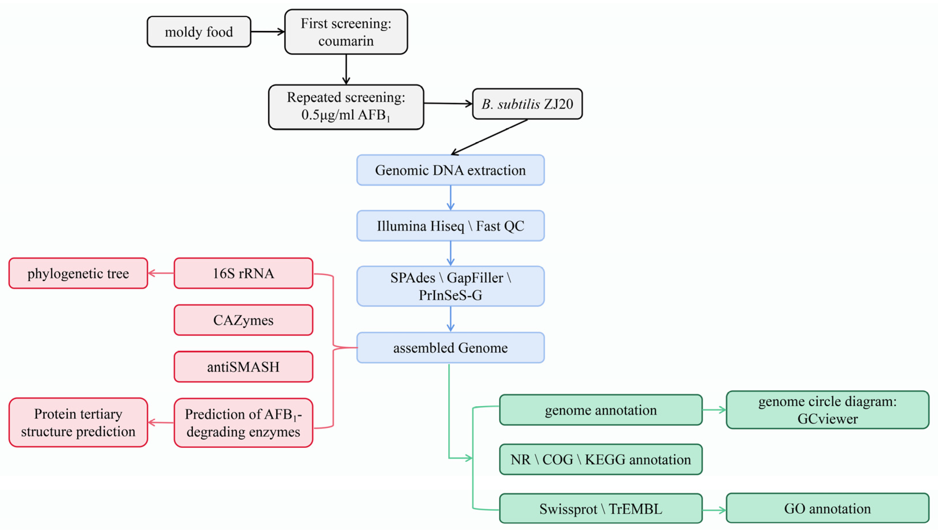
The experimental flow is shown in Figure 1.

3. Results

3.1. Screening of AFB1-Degrading Strains

The bacteria in the moldy feed were enriched and cultured, and 79 strains of bacteria were isolated. These 79 strains were subjected to primary screening in a primary screening medium containing coumarin as the only carbon source, and 12 strains with stable growth and good condition were obtained. The 12 strains were numbered as ZJ3, ZJ10, ZJ11, ZJ16, ZJ6, ZJ13, ZJ20, ZJ27, ZJ31, ZJ32, ZJ8, ZJ22.
The 12 strains obtained from the initial screening were co-cultured with LB medium containing 0.5 μg/mL for 48 h. The residual amount of AFB1 in the medium was detected by HPLC and the degradation efficiency of the strains was calculated, and the results are shown in Table 1. The results showed that the degradation efficiency of strain ZJ20 on AFB1 was the highest, which could reach 84.23%.

3.2. Bacillus Identification

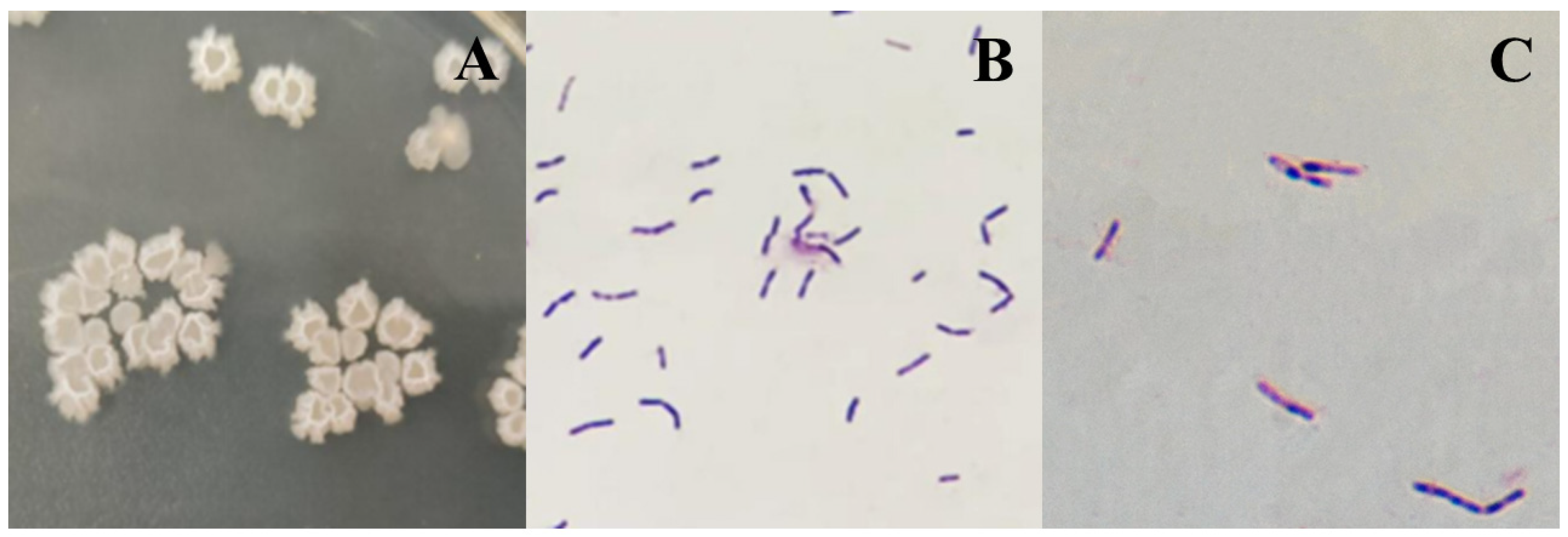
Strain ZJ20 is a Gram-positive bacillus that grows fast and often forms a white film on the liquid surface in an LB liquid medium. The colonies in the solid medium are larger, white, and irregular in shape, with smooth depressions in the middle and ruffled elevations all around.
Gram staining microscopy for blue-purple rods, uniform coloring, and no pods can form endophytic resistant bacilli, located in the center of the body or slightly off (Figure 2).
The genome of the B. subtilis ZJ20 strain was compared with the NR database to make a pie chart of the homology distribution of ZJ20. Each sector in the chart represents one species, and the larger the area of the sector, the greater the number of sequences compared to that species. As shown in Figure 3, strain ZJ20 has high homology with Bacillus spp. and Bacillus substilis species.
The 16sRNA sequence of strain ZJ20 was 1454 bp in length, and the sequence was submitted to GenBank with the accession number OR453222. The complete 16S rRNA of ZJ20 was compared to Blast, and the comparison showed that B. subtilis ZJ20 was the closest to the Bacillus subtilis strain (more than 99% identity), which was the same as the result of the NR database comparison.
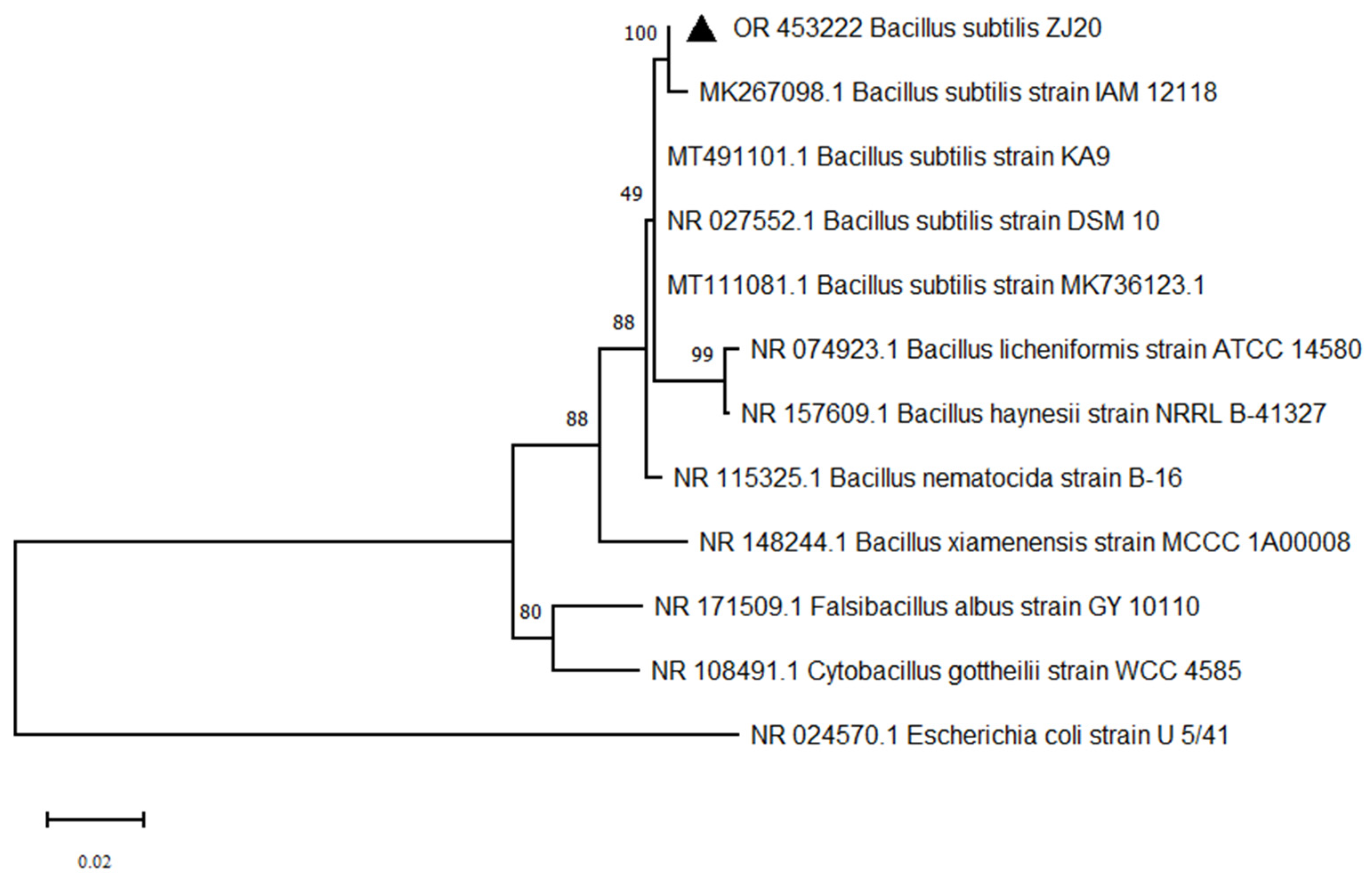
Four Bacillus subtilis strains and four other Bacillus spp. homologous ZJ20 strains were selected to construct a bacterial phylogenetic tree. As shown in Figure 4, strain ZJ20 was in the same branch as Bacillus subtilis strain IAM12118 and had the highest homology. The ZJ20 strain can be identified in Bacillus subtilis species (Bacillus subtilis), named Bacillus subtilis ZJ20, abbreviated as B. subtilis ZJ20.

3.3. Basic Characteristics of the Genome

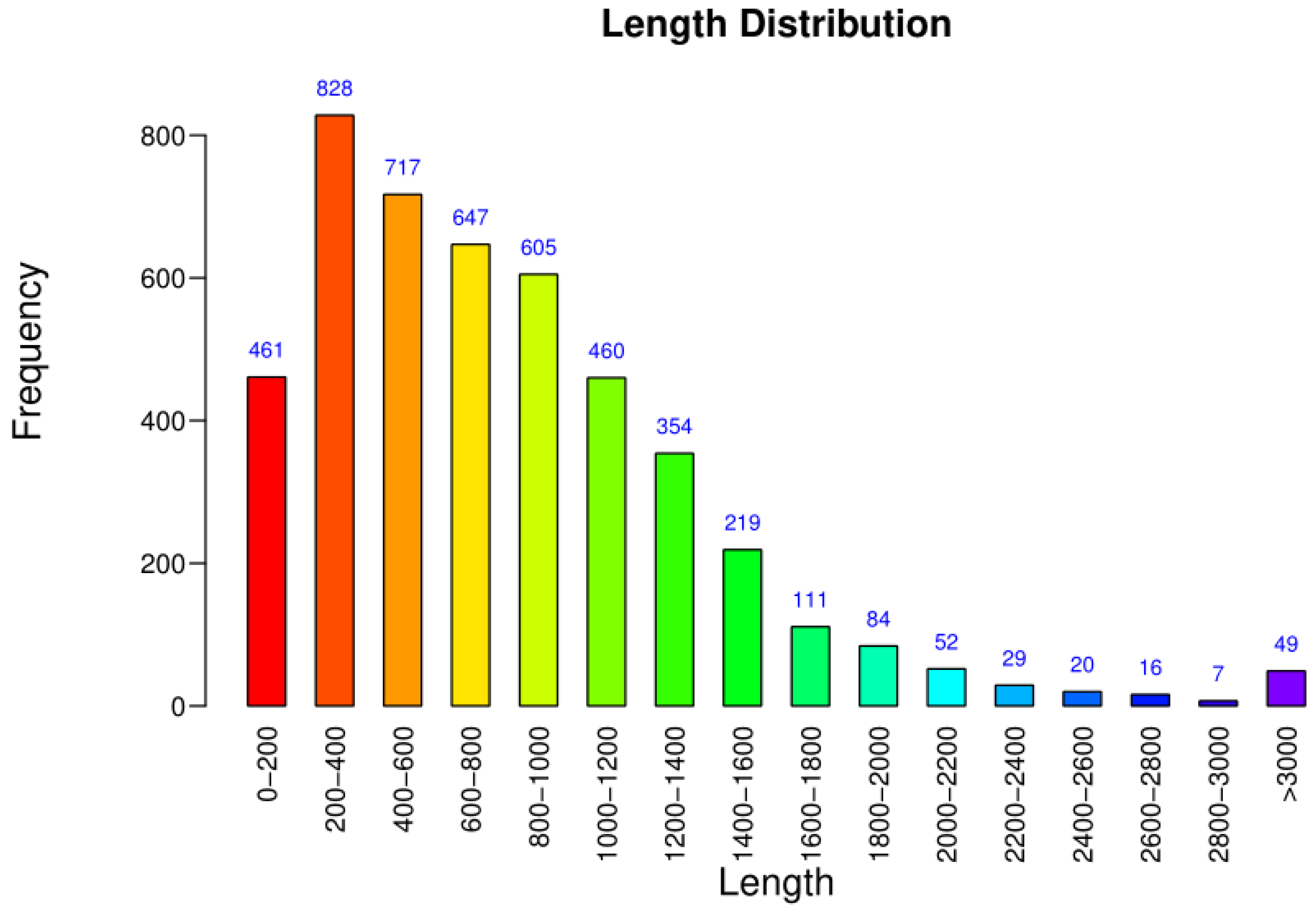
The genomic statistics of B. subtilis ZJ20 are shown in Table 2. The total length of all coding genes was 4,326,340 bp, and the length of coding genes ranged from 45 to 10,806 bp, with an average length of 817.69 bp. Most of the genes were between 200 and 1000 bp (Figure 5), with an average G + C content of 42.9%.
The B. subtilis ZJ20 genome contains 11 copies of rRNA manipulators (16S, 23S, and 5S RNAs), 85 tRNA genes, and 4659 coding sequences (CDS), of which 4432 are functionally annotated. This Whole Genome Shotgun project has been deposited at GenBank under the accession JAVFVM000000000. The version described in this paper is version JAVFVM010000000.

3.4. COG Annotation Results

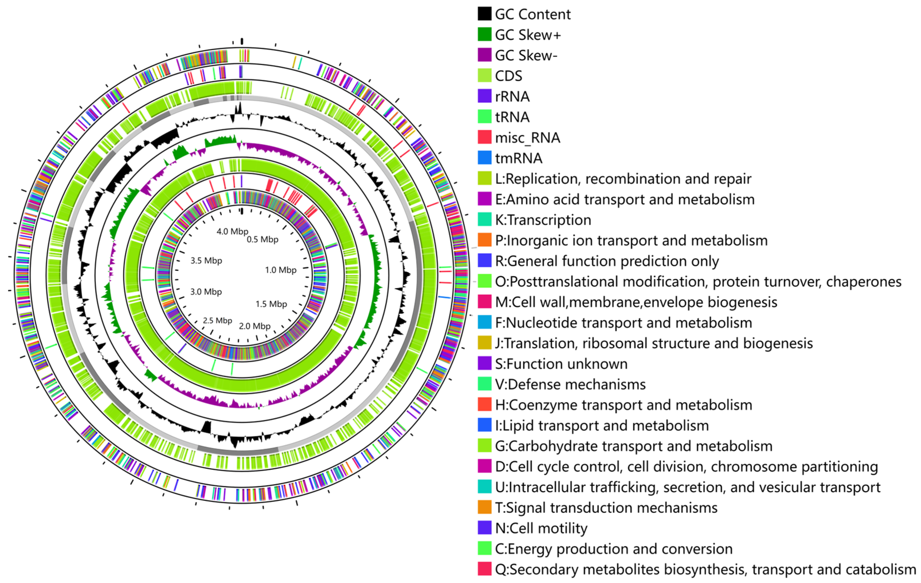
All protein-coding genes were annotated with the protein direct homology cluster database (COG). The functional categories defined according to the COGs showed that B. subtilis ZJ20 contains a large number of proteins involved in amino acid transport and metabolism (COG E) and transcription (COG K), followed by carbohydrate transport and metabolism (COG G) (Table 3). The annotated gene circle map is shown in Figure 6.

3.5. GO Annotation Results

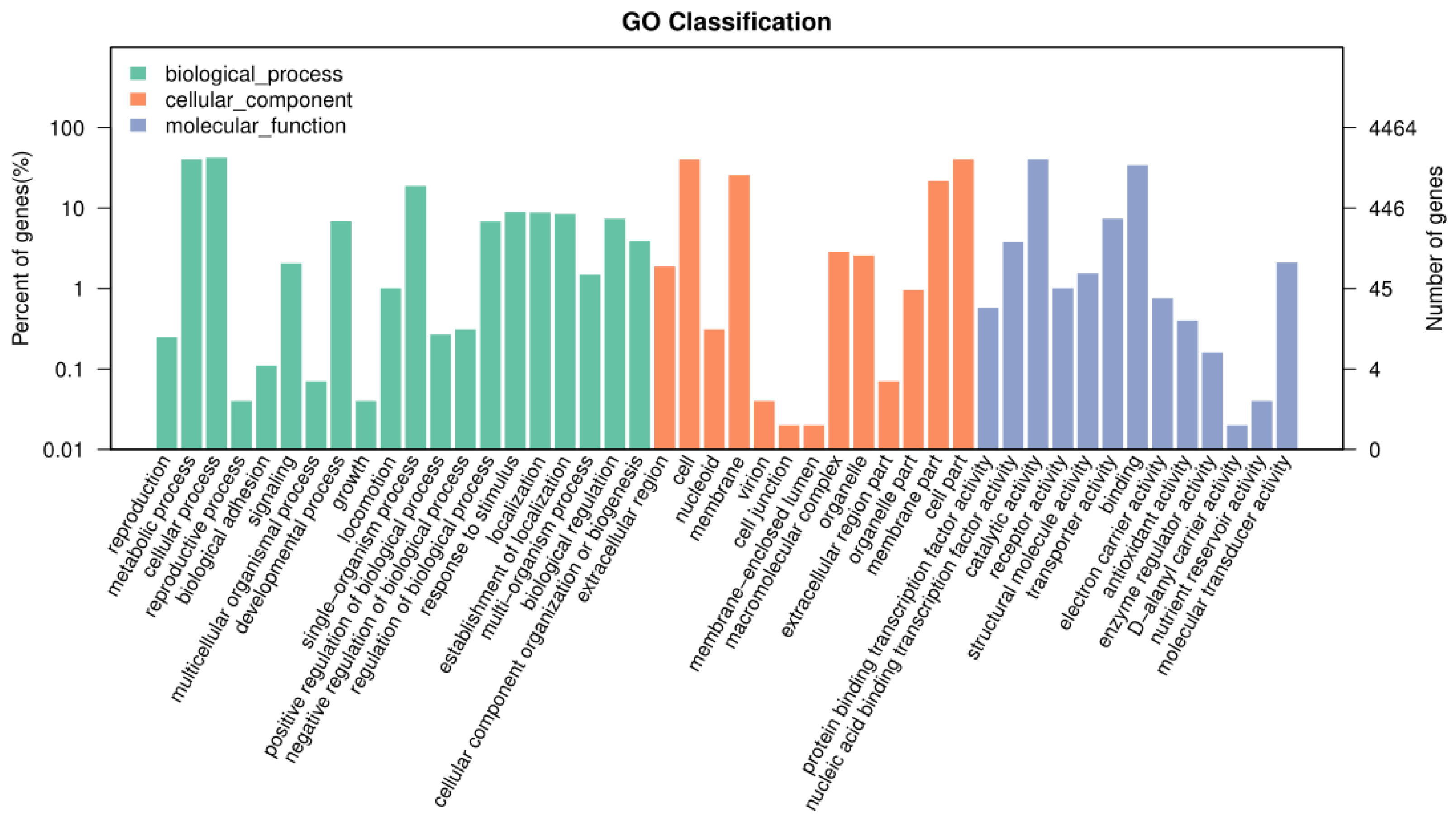
The gene sequences encoding the protein encoded by B. subtilis ZJ20 were aligned with the Gene Ontology (GO) database to obtain functional annotation information for the protein encoded by B. subtilis ZJ20. The proteins of B. subtilis ZJ20 are divided into three main categories: Cellular Component (CC); Biological Process (BP); and Molecular Function (MF).
ZJ20 has a large number of genes responsible for metabolic processes, cellular processes, cells, binding, and catalytic activity, followed by more genes coding for antioxidant activity and enzyme regulator activity (Figure 7). This suggests that B. subtilis ZJ20 may have high enzyme content, metabolic activity, and antioxidant capacity.

3.6. KEGG Annotation Results

The whole genome of B. subtilis ZJ20 was compared with the KEGG database, and most of the genes in all KEGG pathways were involved in carbohydrate metabolism, membrane transport, and amino acid metabolism (Figure 8). Most of the genes in amino acid metabolism are related to cysteine, methionine, phenylalanine, D-alanine, histidine, lysine, complexion, arginine, proline, tryptophan metabolism, and the synthesis of glycine, valine, and lysine.
In carbohydrate metabolism, most genes are involved in the synthesis of pantothenic acid, CoA, fatty acids, unsaturated fatty acids, folic acid, and peptidoglycans; and fatty acid, biotin, propionic acid, starch, sucrose, glutathione, fructose, galactose, mannose, purine, glyoxylate, and dicarboxylic acid metabolism. There are also genes involved in biological pathways such as the citric acid cycle (TCA cycle), ABC transporter, tetracycline biosynthesis, and glycolysis (Supplementary Table S1).
It shows that B. subtilis ZJ20 has strong biochemical metabolism, membrane permeability, and reproduction ability, which provide the basis for the production of a large number of functional enzymes.

3.7. CAZy Database Annotation Result

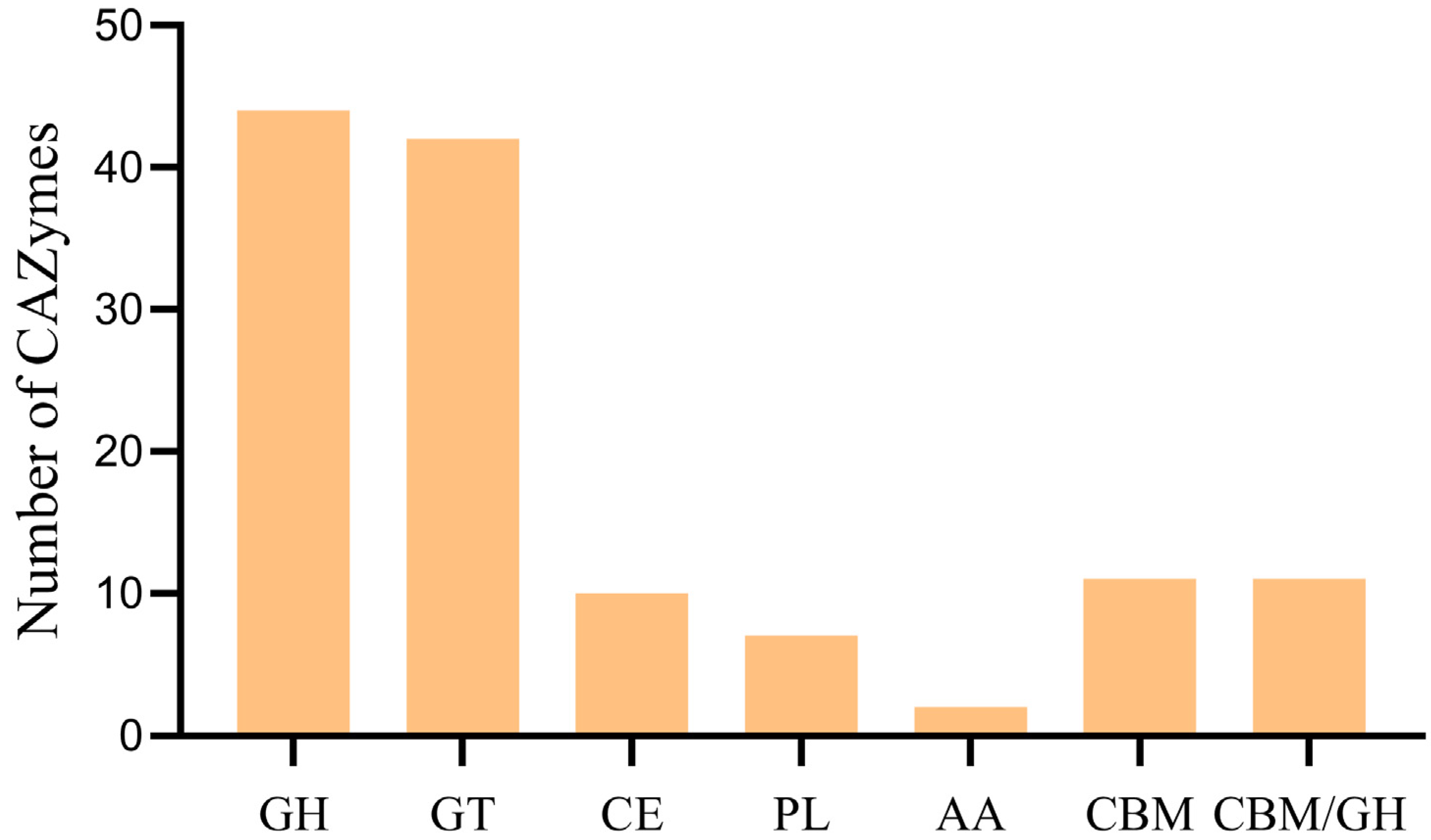
CAZymes are carbohydrate-active enzymes that play a key role in carbohydrates. In the CAZy database, CAZymes are classified into five categories, namely GH (glycoside hydrolase), GT (glycosyltransferase), CE (carbohydrate esterase), PL (polysaccharide lyase), and AA (auxiliary active).
Sequence analysis of the ZJ20 genome identified 127 CAZymes family enzymes, including 43 GH, 42 GT, 11 CBM, 11 CE, 7 PL, and 2 AA. In addition, 11 enzymes of the CBM/GH complex type were identified (Figure 9). Glycoside hydrolases and glycosyltransferases had the highest number of genes, accounting for 33.9% and 33.1% of the total number of CAZymes genes, respectively.

3.8. Prediction of Secondary Metabolites of B. subtilis ZJ20

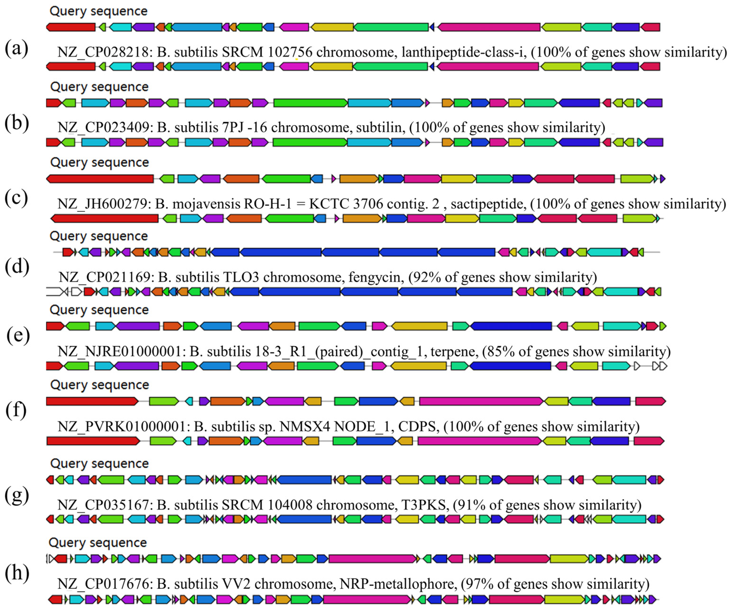
The obtained whole genome sequence of ZJ20 was compared to the secondary metabolite database, and a variety of secondary metabolites encoding antibacterial properties were identified in B. subtilis ZJ20.
It contains five gene clusters encoding NRPS (non-ribosomal peptide synthase); two gene clusters encoding terpenoids; and two gene clusters encoding class I lactose peptides, one of which shows 100% similarity to genes involved in the synthesis of chymotrypsin. There was also one gene cluster each encoding T3PKS (class III polyketide synthase), CDPS (cyclic dipeptide synthase), and bacillus lysozyme; one gene cluster encoding a cecropeptide class showed 100% similarity to Subtilosin A. Among the five gene clusters encoding NRPS, two gene clusters showed 100% similarity to fengycins and bacillibactin, respectively (Figure 10).

3.9. Mining Potential AFT-Degrading Enzymes of Strain B. subtilis ZJ20

In addition to the chitinase with possible AFB1 degradation potential identified in the above analysis of carbohydrase, a laccase, two lactonases, and four protein fragments of manganese peroxidase were identified in ZJ20.
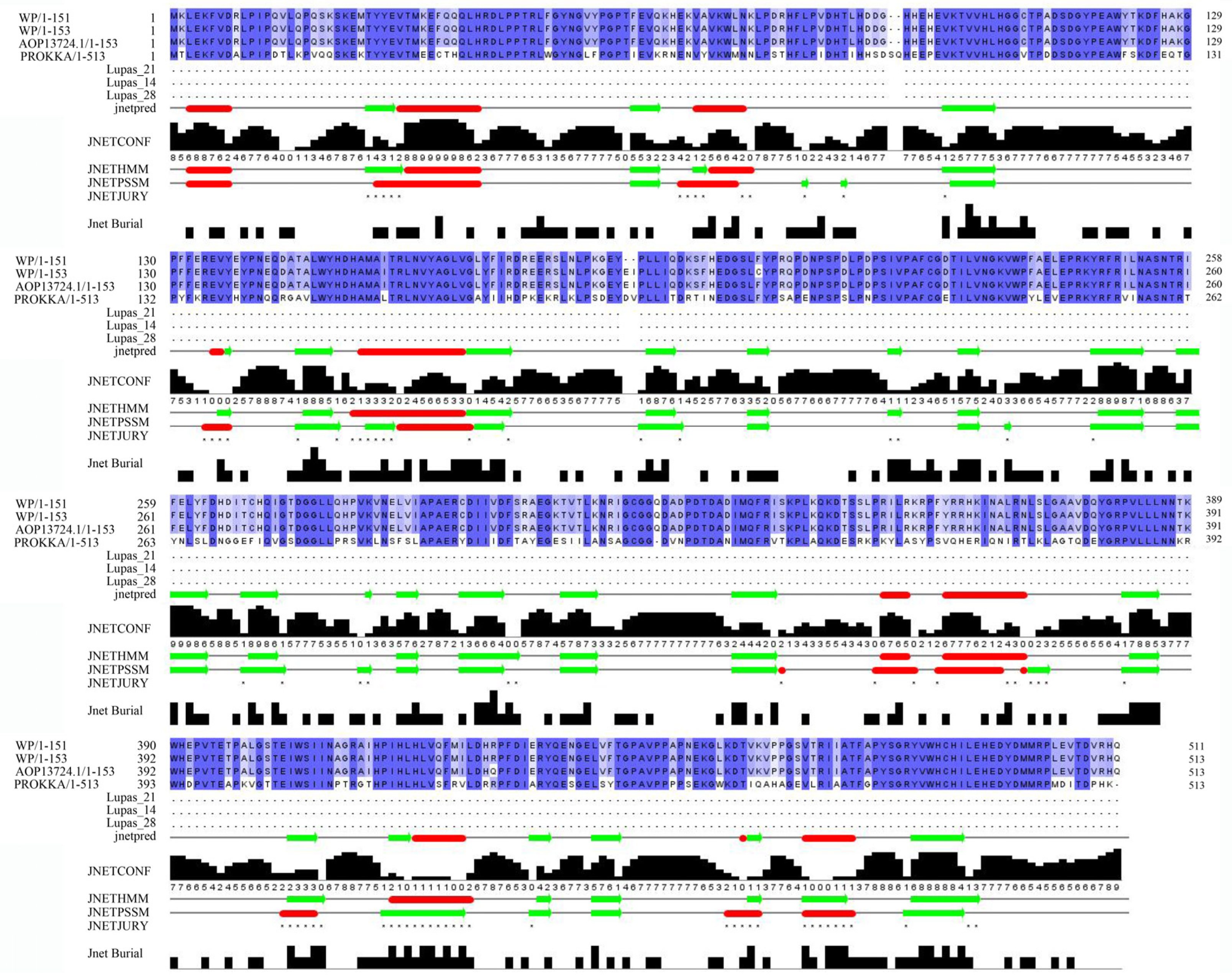
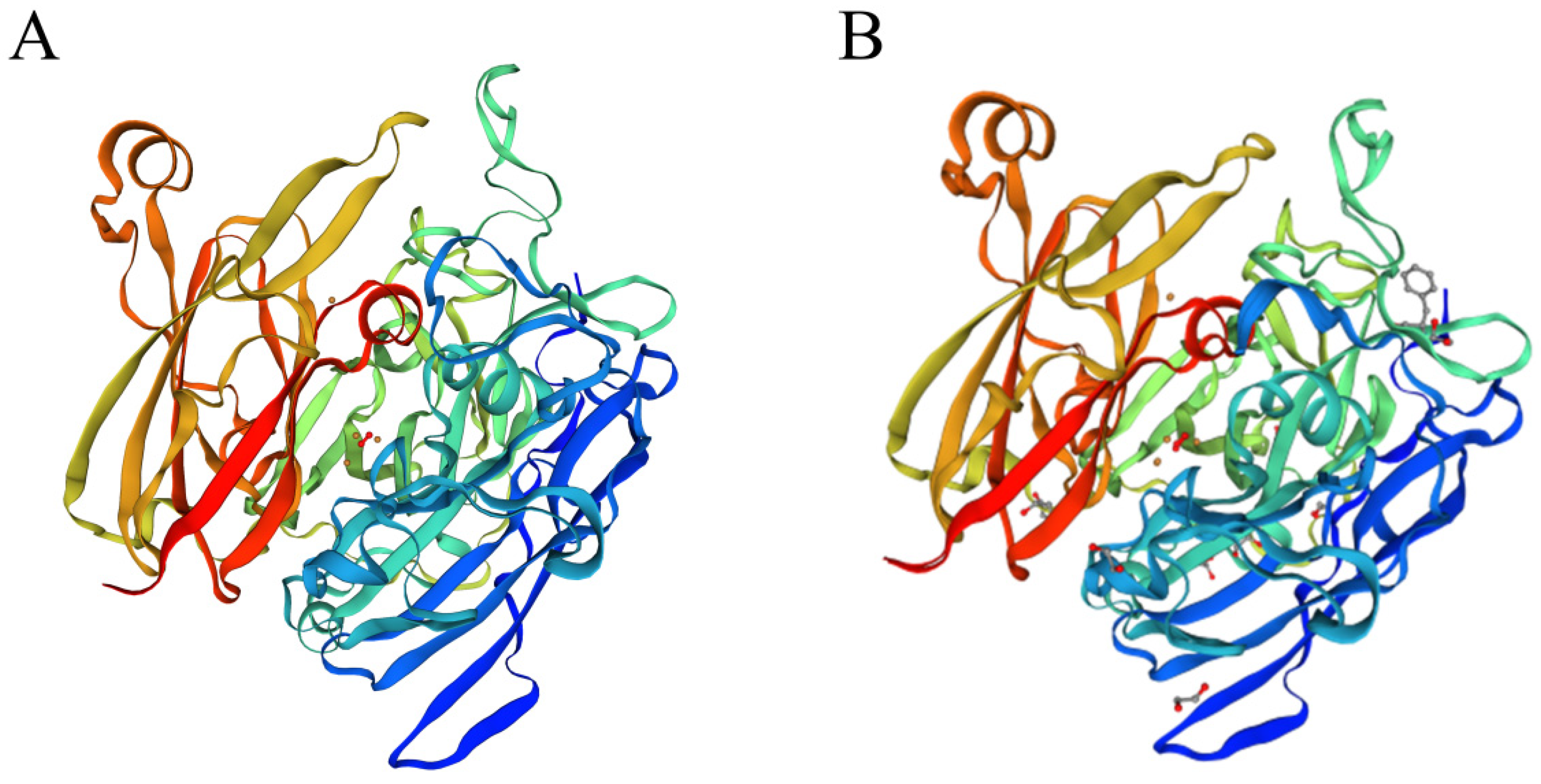
The laccase sequence in ZJ20 was compared with three segments of laccase protein sequences for multiple sequence comparison as well as secondary structure prediction (Figure 11). In addition, the tertiary structure of the protein was also very similar to the known tertiary structure of laccase (Figure 12), indicating that the laccase produced by ZJ20 has the potential to degrade AFB1.
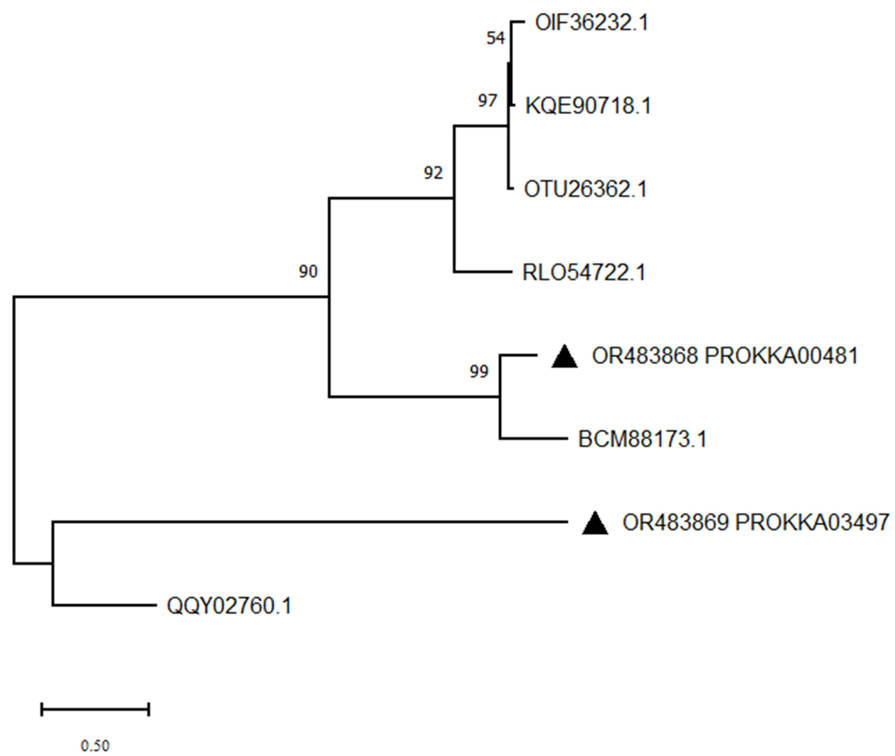
ZJ20 contains the protein sequences of two lactonases, and these two protein sequences were used to construct a phylogenetic tree together with six lactonases of different origins (Figure 13). The results indicated that PROKKA03497 was distantly related to other lactonases and that it was a new lactonase to be investigated.
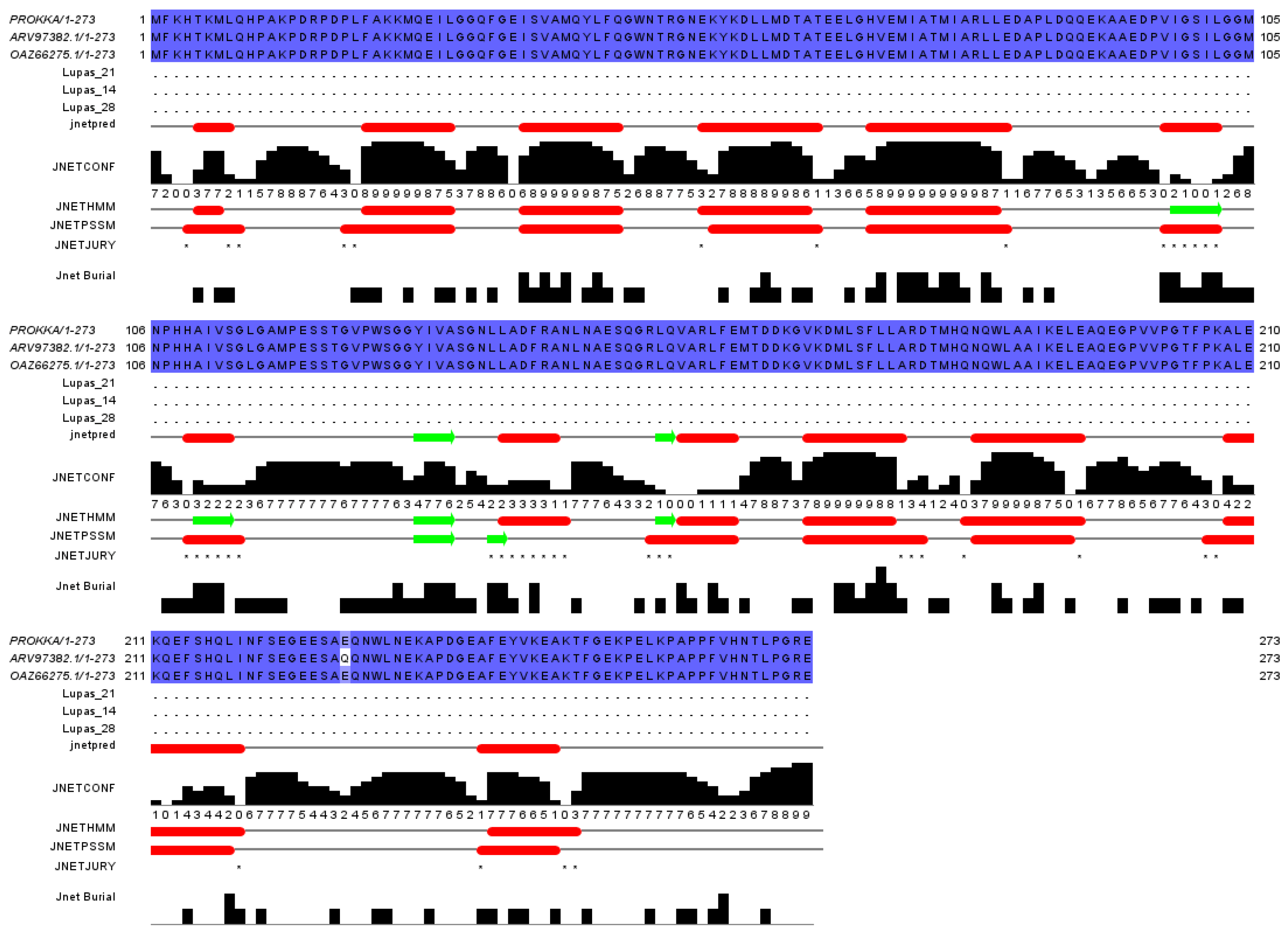
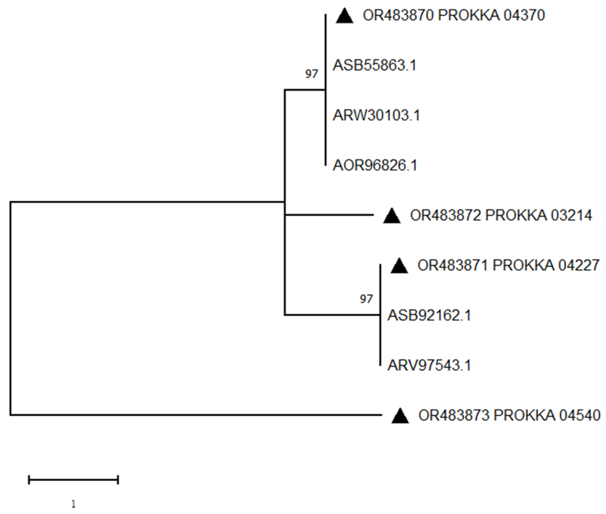
Of the four manganese peroxidase protein fragments, one had 100% sequence identity with the identified manganese peroxidase coding sequence. As shown in Figure 14, five different manganese peroxidase protein sequences were selected to construct a phylogenetic tree with the four sequences in ZJ20 (Figure 15). The results showed that PROKKA04540 was distantly related to the other proteins, and the protein might belong to a different family from other proteins, which has a greater potential for exploitation.

4. Discussion

Aflatoxins are natural carcinogens with multiple toxicities and are present in moldy feed and cereals, with AFB1 being a class I carcinogen that can damage a variety of tissues and organs of the body. A variety of enzymes have been reported to degrade aflatoxin, and the focus of current research is to develop an efficient degrading enzyme for aflatoxin to reduce the risk of aflatoxin to humans and animals.
In our study, B. subtilis ZJ20 showed good degradation of AFB1 and produced effects through extracellular enzymes. B. subtilis ZJ20 encoded a total of four AFB1-degrading enzymes, including chitinase, laccase, lactonase, and manganese peroxidase. AFB1 was degraded by a combination of multiple enzymes to achieve higher degradation efficiency.
Thereafter, whole genome sequencing was combined with bioinformatics analysis to provide more information to explore more biological functions of strain ZJ20.
Amino acids are small molecules that constitute proteins and play an important role in protein synthesis, maintaining normal body metabolism, improving body immunity, and increasing energy [38]. ABC transporter proteins are a large and functionally diverse family of transmembrane transport proteins responsible for the transmembrane transport of a variety of substances that are essential for cellular detoxification, membrane homeostasis, and lipid transport [39]. Fatty acids maintain normal cellular physiological functions, maintain the relative fluidity of cell membranes, and provide energy to cells. And some fatty acids, short-chain fatty acids (SCFAs), are involved in the immune system, regulating intestinal pH, intestinal permeability, intestinal microbiota, and reducing intestinal inflammation [40]. Biotin, a member of the B vitamin family, enhances the host’s immune response, resists infection, and maintains normal growth and development [41]. Tetracycline, as a broad-spectrum antibacterial agent, has a good inhibitory effect on both Gram-positive and Gram-negative bacteria [42]. In our study, we found that B. subtilis ZJ20 contains genes related to amino acids, fatty acid metabolism and synthesis, biotin synthesis, and tetracycline biosynthesis. This suggests that B. subtilis ZJ20 can exert beneficial effects on animals as a probiotic by controlling the metabolism of amino acids and fatty acids, enhancing the digestion and absorption of nutrients in animals, and inhibiting the growth of intestinal pathogenic bacteria.
Carbohydrates are essential for the survival of microorganisms, acting not only as a carbon source for bacteria but also as a means of attachment to the host and as a barrier to host infection [43]. Glycoside hydrolases play an important role in the synthesis of oligosaccharides, the synthesis of alkyl and aromatic glycosides, the glycosylation of amino acids and peptides, and the glycosylation of antibiotics. Glycosyltransferases use activated sugar donors to drive glycosyl transfer to appropriate acceptors, and the formation of glycosidic bonds affects the metabolic choreography of small molecules and the formation of numerous glycolipids, glycopeptides, and lipopolysaccharides [44].
The more numerous CAZymes gene families in B. subtilis ZJ20 were 5 GH23, 4 GH32, 17 GT2, and 10 GT4 family genes, respectively (Supplementary Table S2). The GH23 family of glycoside hydrolases mainly includes lysozyme type G, chitinase, and peptidoglycan lyase, which can degrade fungal and pathogenic cell walls and thus inhibit pathogenic growth. Chitinase also has the potential to degrade fungal toxins such as AFB1. In addition, the β-glucosidases GH1 and GH4, xyloglucanase GH16, and endoglucanase GH51 gene families also have potential antifungal activity, and fructosyltransferase GH32 can also stimulate the growth of intestinal probiotics [45].
GT4, as the largest class of the GT family, contains some enzymes that have potential therapeutic significance. GT4, along with GT2, as the ancestral reversal and retention family of the GT family, can modulate antibiotic activity by glycosylating antibiotics and changing the location, type, and amount of sugar [44]. The binding properties of CBM improve the catalytic function of CAZymes by targeting the enzyme to the substrate, increasing substrate–enzyme proximity, and disrupting the crystallinity of the insoluble substrate fraction [46]. Carbohydrate-active enzymes can break down complex carbohydrates such as cellulose, starch, glycogen, and polysaccharides into small-molecule compounds, which facilitate nutrient absorption from the intestinal epithelium. These genes encoding CAZymes allow B. subtilis ZJ20 to produce carbohydrate-active enzymes that synthesize carbohydrates and metabolize sugars and have antifungal, pathogen-inhibiting, antibiotic-activating, and nutrient-absorbing effects.
In addition, genes encoding NRPS, terpenes, class I lactose peptides, T3PKS, CDPS, bacillus lysozyme, and sequestered peptides were identified in the analysis of secondary metabolites. Various secondary metabolites encoded by ZJ20 have been reported to have antifungal [47,48], broad-spectrum antibacterial [49,50], antiviral [51], and antioxidant [52] effects, and some metabolites have also been predicted to have immunomodulatory, antitumor, and antiparasitic effects [53,54], laying the groundwork for the excavation of other biological effects of ZJ20. Among the secondary metabolites encoded by ZJ20, there is also a metal carrier that exhibits 100% similarity to the iron carrier and can chelate Fe3+, Al3+, Pb2+, Cu2+, and other metal ions bound [55] to bioremediate metal-contaminated soil. B. subtilis ZJ20 has seen large developments not only in the farming and pharmaceutical industries, but also in environmental control and plant biocontrol. The potential of B. subtilis ZJ20 is not only in the farming and pharmaceutical industries but also in environmental control and plant biocontrol.
Overall, B. subtilis ZJ20 can function as a feed additive, plant and environmental biocontrol bacterium, and bioengineering bacterium due to its ability to produce a variety of enzymes, antibiotics, and other biologically active substances. ZJ20 can effectively degrade AFB1, so when it is added as a probiotic to the feed, it can not only alleviate the effect of AFB1 in moldy feed on the animal’s body, but also improve the animal’s intestinal flora balance.

5. Conclusions

In summary, our screened strain, ZJ20, could effectively degrade aflatoxin B1 with a degradation rate of 84.23%. ZJ20 was identified as Bacillus subtilis according to the general morphological characteristics of the strain and 16Sr RNA comparison. After sequencing and assembling the whole bacterial genome, we understood the basic genomic features of ZJ20 and functionally annotated its protein-coding genes.
We identified genes involved in amino acid metabolism, lipid metabolism, and vitamin metabolism and synthesis in a genome-wide KEGG analysis of B. subtilis ZJ20, confirming the probiotic properties of ZJ20. Active enzymes with inhibitory effects on fungi and bacteria were identified in CAZymes and secondary metabolite analyses, and four potential AFB1-degrading enzymes were found in B. subtilis ZJ20 by multiple sequence comparison. The comprehensive analysis of ZJ20 in this paper lays a theoretical foundation for the subsequent development of more biological functions of ZJ20 and the combined degradation of AFB1 by multiple biological enzymes.
For future research, on the one hand, we can continue to explore the biological functions of B. subtilis ZJ20 as a probiotic so that it can perform the corresponding probiotic functions. On the other hand, the cloning of AFB1-degrading enzymes to degrade AFB1 more effectively is a very worthwhile research direction.

Supplementary Materials

The following supporting information can be downloaded at: https://www.mdpi.com/article/10.3390/biology12091195/s1, Table S1: KEGG annotation results. Table S2: CAZy database annotation results. Table S3: Prediction of secondary metabolites of B. subtilis ZJ20.

Author Contributions

M.H. and Z.Y. conceived and designed the study; methodology, M.H. and J.G.; validation, J.G., Z.Y. and K.D.; data curation, Z.Y.; writing—original draft preparation, M.H.; writing—review and editing, Z.Y., Y.J., C.L., L.H., J.L., Y.W., S.C., J.C., K.S. and R.G.; visualization, M.H. and J.G.; supervision, Z.Y. All authors have read and agreed to the published version of the manuscript.

Funding

This research is sponsored by the National Natural Science Foundation of China (grant nos. 32072771) and the Natural Science Foundation of Henan Province (grant no. 182300410052).

Institutional Review Board Statement

Not applicable.

Informed Consent Statement

Not applicable.

Data Availability Statement

The whole-genome sequence generated in this study was submitted to GenBank with the accession number JAVFVM000000000.

Conflicts of Interest

The authors declare no conflict of interest.

References

  1. Gizachew, D.; Chang, C.H.; Szonyi, B.; De La Torre, S.; Ting, W.E. Aflatoxin B1 (AFB1) production by Aspergillus flavus and Aspergillus parasiticus on ground Nyjer seeds: The effect of water activity and temperature. Int. J. Food Microbiol. 2019, 296, 8–13. [Google Scholar] [CrossRef]
  2. Wang, P.; Ma, L.; Jin, J.; Zheng, M.; Pan, L.; Zhao, Y.; Sun, X.; Liu, Y.; Xing, F. The anti-aflatoxigenic mechanism of cinnamaldehyde in Aspergillus flavus. Sci. Rep. 2019, 9, 10499. [Google Scholar] [CrossRef] [PubMed]
  3. Kensler, T.W.; Roebuck, B.D.; Wogan, G.N.; Groopman, J.D. Aflatoxin: A 50-year odyssey of mechanistic and translational toxicology. Toxicol. Sci. Off. J. Soc. Toxicol. 2011, 120 (Suppl. 1), S28–S48. [Google Scholar] [CrossRef]
  4. Emmanuel, K.T.; Els, V.P.; Bart, H.; Evelyne, D.; Els, V.H.; Els, D. Carry-over of some Fusarium mycotoxins in tissues and eggs of chickens fed experimentally mycotoxin-contaminated diets. Food Chem. Toxicol. 2020, 145, 111715. [Google Scholar] [CrossRef] [PubMed]
  5. Wang, X.; Yang, F.; Na, L.; Jia, M.; Ishfaq, M.; Zhang, Y.; Liu, M.; Wu, C. Ferulic acid alleviates AFB1-induced duodenal barrier damage in rats via up-regulating tight junction proteins, down-regulating ROCK, competing CYP450 enzyme and activating GST. Ecotoxicol. Environ. Saf. 2022, 241, 113805. [Google Scholar] [CrossRef] [PubMed]
  6. Huang, B.; Chen, Q.; Wang, L.; Gao, X.; Zhu, W.; Mu, P.; Deng, Y. Aflatoxin B1 Induces Neurotoxicity through Reactive Oxygen Species Generation, DNA Damage, Apoptosis, and S-Phase Cell Cycle Arrest. Int. J. Mol. Sci. 2020, 21, 6517. [Google Scholar] [CrossRef]
  7. Guindon-Kezis, K.A.; Mulder, J.E.; Massey, T.E. In vivo treatment with aflatoxin B1 increases DNA oxidation, base excision repair activity and 8-oxoguanine DNA glycosylase 1 levels in mouse lung. Toxicology 2014, 321, 21–26. [Google Scholar] [CrossRef]
  8. Zhu, Q.; Ma, Y.; Liang, J.; Wei, Z.; Li, M.; Zhang, Y.; Liu, M.; He, H.; Qu, C.; Cai, J.; et al. AHR mediates the aflatoxin B1 toxicity associated with hepatocellular carcinoma. Signal Transduct. Target. Ther. 2021, 6, 299. [Google Scholar] [CrossRef]
  9. Abdel-Daim, M.M.; Abdeen, A.; Jalouli, M.; Abdelkader, A.; Megahed, A.; Alkahtane, A.; Almeer, R.; Alhoshani, N.M.; Al-Johani, N.S.; Alkahtani, S.; et al. Fucoidan supplementation modulates hepato-renal oxidative stress and DNA damage induced by aflatoxin B1 intoxication in rats. Sci. Total Environ. 2021, 768, 144781. [Google Scholar] [CrossRef]
  10. Eshelli, M.; Qader, M.M.; Jambi, E.J.; Hursthouse, A.S.; Rateb, M.E. Current Status and Future Opportunities of Omics Tools in Mycotoxin Research. Toxins 2018, 10, 433. [Google Scholar] [CrossRef] [PubMed]
  11. Deng, Y.; Wang, H.; Hamamoto, R.; Schaffer, D.; Duan, S. Functional genomics, genetics, and bioinformatics. BioMed Res. Int. 2015, 2015, 184824. [Google Scholar] [CrossRef] [PubMed]
  12. He, S.; Feng, K.; Ding, T.; Huang, K.; Yan, H.; Liu, X.; Zhang, Z. Complete genome sequence of Bacillus licheniformis BL-010. Microb. Pathog. 2018, 118, 199–201. [Google Scholar] [CrossRef]
  13. De Vos, W.M.; Bron, P.A.; Kleerebezem, M. Post-genomics of lactic acid bacteria and other food-grade bacteria to discover gut functionality. Curr. Opin. Biotechnol. 2004, 15, 86–93. [Google Scholar] [CrossRef]
  14. Bian, L.; Zheng, M.; Chang, T.; Zhou, J.; Zhang, C. Degradation of Aflatoxin B1 by recombinant laccase extracellular produced from Escherichia coli. Ecotoxicol. Environ. Saf. 2022, 244, 114062. [Google Scholar] [CrossRef] [PubMed]
  15. Wang, X.; Qin, X.; Hao, Z.; Luo, H.; Yao, B.; Su, X. Degradation of Four Major Mycotoxins by Eight Manganese Peroxidases in Presence of a Dicarboxylic Acid. Toxins 2019, 11, 566. [Google Scholar] [CrossRef]
  16. Qin, X.; Su, X.; Tu, T.; Zhang, J.; Wang, X.; Wang, Y.; Wang, Y.; Bai, Y.; Yao, B.; Luo, H.; et al. Enzymatic Degradation of Multiple Major Mycotoxins by Dye-Decolorizing Peroxidase from Bacillus subtilis. Toxins 2021, 13, 429. [Google Scholar] [CrossRef]
  17. Xing, F.; Wang, L.; Liu, X.; Selvaraj, J.N.; Wang, Y.; Zhao, Y.; Liu, Y. Aflatoxin B1 inhibition in Aspergillus flavus by Aspergillus niger through down-regulating expression of major biosynthetic genes and AFB1 degradation by atoxigenic A. flavus. Int. J. Food Microbiol. 2017, 256, 1–10. [Google Scholar] [CrossRef]
  18. Xie, X.H.; Fu, X.; Yan, X.Y.; Peng, W.F.; Kang, L.X. A Broad-Specificity Chitinase from Penicillium oxalicum k10 Exhibits Antifungal Activity and Biodegradation Properties of Chitin. Mar. Drugs 2021, 19, 356. [Google Scholar] [CrossRef]
  19. González Pereyra, M.L.; Martínez, M.P.; Cavaglieri, L.R. Presence of aiiA homologue genes encoding for N-Acyl homoserine lactone-degrading enzyme in aflatoxin B1-decontaminating Bacillus strains with potential use as feed additives. Food Chem. Toxicol. 2019, 124, 316–323. [Google Scholar] [CrossRef]
  20. Bolger, A.M.; Lohse, M.; Usadel, B. Trimmomatic: A flexible trimmer for Illumina sequence data. Bioinformatics 2014, 30, 2114–2120. [Google Scholar] [CrossRef] [PubMed]
  21. Brown, J.; Pirrung, M.; McCue, L.A. FQC Dashboard: Integrates FastQC results into a web-based, interactive, and extensible FASTQ quality control tool. Bioinformatics 2017, 33, 3137–3139. [Google Scholar] [CrossRef]
  22. Bankevich, A.; Nurk, S.; Antipov, D.; Gurevich, A.A.; Dvorkin, M.; Kulikov, A.S.; Lesin, V.M.; Nikolenko, S.I.; Pham, S.; Prjibelski, A.D.; et al. SPAdes: A new genome assembly algorithm and its applications to single-cell sequencing. J. Comput. Biol. J. Comput. Mol. Cell Biol. 2012, 19, 455–477. [Google Scholar] [CrossRef]
  23. Boetzer, M.; Pirovano, W. Toward almost closed genomes with GapFiller. Genome Biol. 2012, 13, R56. [Google Scholar] [CrossRef] [PubMed]
  24. Massouras, A.; Hens, K.; Gubelmann, C.; Uplekar, S.; Decouttere, F.; Rougemont, J.; Cole, S.T.; Deplancke, B. Primer-initiated sequence synthesis to detect and assemble structural variants. Nat. Methods 2010, 7, 485–486. [Google Scholar] [CrossRef]
  25. Seemann, T. Prokka: Rapid prokaryotic genome annotation. Bioinformatics 2014, 30, 2068–2069. [Google Scholar] [CrossRef] [PubMed]
  26. Kumar, S.; Stecher, G.; Li, M.; Knyaz, C.; Tamura, K. MEGA X: Molecular Evolutionary Genetics Analysis across Computing Platforms. Mol. Biol. Evol. 2018, 35, 1547–1549. [Google Scholar] [CrossRef]
  27. Grant, J.R.; Stothard, P. The CGView Server: A comparative genomics tool for circular genomes. Nucleic Acids Res. 2008, 36, W181–W184. [Google Scholar] [CrossRef]
  28. Tatusov, R.L.; Galperin, M.Y.; Natale, D.A.; Koonin, E.V. The COG database: A tool for genome-scale analysis of protein functions and evolution. Nucleic Acids Res. 2000, 28, 33–36. [Google Scholar] [CrossRef]
  29. Altschul, S.F.; Madden, T.L.; Schäffer, A.A.; Zhang, J.; Zhang, Z.; Miller, W.; Lipman, D.J. Gapped BLAST and PSI-BLAST: A new generation of protein database search programs. Nucleic Acids Res. 1997, 25, 3389–3402. [Google Scholar] [CrossRef]
  30. Kanehisa, M.; Goto, S. KEGG: Kyoto encyclopedia of genes and genomes. Nucleic Acids Res. 2000, 28, 27–30. [Google Scholar] [CrossRef]
  31. Moriya, Y.; Itoh, M.; Okuda, S.; Yoshizawa, A.C.; Kanehisa, M. KAAS: An automatic genome annotation and pathway reconstruction server. Nucleic Acids Res. 2007, 35, W182–W185. [Google Scholar] [CrossRef]
  32. Lombard, V.; Golaconda Ramulu, H.; Drula, E.; Coutinho, P.M.; Henrissat, B. The carbohydrate-active enzymes database (CAZy) in 2013. Nucleic Acids Res. 2014, 42, D490–D495. [Google Scholar] [CrossRef]
  33. Zheng, J.; Hu, B.; Zhang, X.; Ge, Q.; Yan, Y.; Akresi, J.; Piyush, V.; Huang, L.; Yin, Y. dbCAN-seq update: CAZyme gene clusters and substrates in microbiomes. Nucleic Acids Res. 2023, 51, D557–D563. [Google Scholar] [CrossRef]
  34. Medema, M.H.; Blin, K.; Cimermancic, P.; de Jager, V.; Zakrzewski, P.; Fischbach, M.A.; Weber, T.; Takano, E.; Breitling, R. antiSMASH: Rapid identification, annotation and analysis of secondary metabolite biosynthesis gene clusters in bacterial and fungal genome sequences. Nucleic Acids Res. 2011, 39, W339–W346. [Google Scholar] [CrossRef]
  35. Chen, C.; Chen, H.; Zhang, Y.; Thomas, H.R.; Frank, M.H.; He, Y.; Xia, R. TBtools: An Integrative Toolkit Developed for Interactive Analyses of Big Biological Data. Mol. Plant 2020, 13, 1194–1202. [Google Scholar] [CrossRef]
  36. Waterhouse, A.M.; Procter, J.B.; Martin, D.M.; Clamp, M.; Barton, G.J. Jalview Version 2--a multiple sequence alignment editor and analysis workbench. Bioinformatics 2009, 25, 1189–1191. [Google Scholar] [CrossRef]
  37. Waterhouse, A.; Bertoni, M.; Bienert, S.; Studer, G.; Tauriello, G.; Gumienny, R.; Heer, F.T.; de Beer, T.A.P.; Rempfer, C.; Bordoli, L.; et al. SWISS-MODEL: Homology modelling of protein structures and complexes. Nucleic Acids Res. 2018, 46, W296–W303. [Google Scholar] [CrossRef]
  38. Swain, B.K.; Johri, T.S. Effect of supplemental methionine, choline and their combinations on the performance and immune response of broilers. Br. Poult. Sci. 2000, 41, 83–88. [Google Scholar] [CrossRef]
  39. Domenichini, A.; Adamska, A.; Falasca, M. ABC transporters as cancer drivers: Potential functions in cancer development. Biochim. Biophys. Acta. Gen. Subj. 2019, 1863, 52–60. [Google Scholar] [CrossRef]
  40. Metzler-Zebeli, B.U.; Koger, S.; Sharma, S.; Sener-Aydemir, A.; Ruczizka, U.; Kreutzmann, H.; Ladinig, A. Short-Chain Fatty Acids Modulate Permeability, Motility and Gene Expression in the Porcine Fetal Jejunum Ex Vivo. Nutrients 2022, 14, 2524. [Google Scholar] [CrossRef]
  41. Kuroishi, T. Regulation of immunological and inflammatory functions by biotin. Can. J. Physiol. Pharmacol. 2015, 93, 1091–1096. [Google Scholar] [CrossRef]
  42. Nelson, M.L.; Levy, S.B. The history of the tetracyclines. Ann. N. Y. Acad. Sci. 2011, 1241, 17–32. [Google Scholar] [CrossRef] [PubMed]
  43. Wardman, J.F.; Bains, R.K.; Rahfeld, P.; Withers, S.G. Carbohydrate-active enzymes (CAZymes) in the gut microbiome. Nat. Rev. Microbiol. 2022, 20, 542–556. [Google Scholar] [CrossRef] [PubMed]
  44. Martinez-Fleites, C.; Proctor, M.; Roberts, S.; Bolam, D.N.; Gilbert, H.J.; Davies, G.J. Insights into the synthesis of lipopolysaccharide and antibiotics through the structures of two retaining glycosyltransferases from family GT4. Chem. Biol. 2006, 13, 1143–1152. [Google Scholar] [CrossRef]
  45. Kleessen, B.; Hartmann, L.; Blaut, M. Oligofructose and long-chain inulin: Influence on the gut microbial ecology of rats associated with a human faecal flora. Br. J. Nutr. 2001, 86, 291–300. [Google Scholar] [CrossRef]
  46. Sidar, A.; Albuquerque, E.D.; Voshol, G.P.; Ram, A.F.J.; Vijgenboom, E.; Punt, P.J. Carbohydrate Binding Modules: Diversity of Domain Architecture in Amylases and Cellulases from Filamentous Microorganisms. Front. Bioeng. Biotechnol. 2020, 8, 871. [Google Scholar] [CrossRef]
  47. Hanif, A.; Zhang, F.; Li, P.; Li, C.; Xu, Y.; Zubair, M.; Zhang, M.; Jia, D.; Zhao, X.; Liang, J.; et al. Fengycin Produced by Bacillus amyloliquefaciens FZB42 Inhibits Fusarium graminearum Growth and Mycotoxins Biosynthesis. Toxins 2019, 11, 295. [Google Scholar] [CrossRef]
  48. Krishnan, N.; Velramar, B.; Velu, R.K. Investigation of antifungal activity of surfactin against mycotoxigenic phytopathogenic fungus Fusarium moniliforme and its impact in seed germination and mycotoxicosis. Pestic. Biochem. Physiol. 2019, 155, 101–107. [Google Scholar] [CrossRef]
  49. Delves-Broughton, J.; Blackburn, P.; Evans, R.J.; Hugenholtz, J. Applications of the bacteriocin, nisin. Antonie Van Leeuwenhoek 1996, 69, 193–202. [Google Scholar] [CrossRef] [PubMed]
  50. Shelburne, C.E.; An, F.Y.; Dholpe, V.; Ramamoorthy, A.; Lopatin, D.E.; Lantz, M.S. The spectrum of antimicrobial activity of the bacteriocin subtilosin A. J. Antimicrob. Chemother. 2007, 59, 297–300. [Google Scholar] [CrossRef] [PubMed]
  51. De Carvalho, M.P.; Abraham, W.R. Antimicrobial and biofilm inhibiting diketopiperazines. Curr. Med. Chem. 2012, 19, 3564–3577. [Google Scholar] [CrossRef] [PubMed]
  52. González-Burgos, E.; Gómez-Serranillos, M.P. Terpene compounds in nature: A review of their potential antioxidant activity. Curr. Med. Chem. 2012, 19, 5319–5341. [Google Scholar] [CrossRef] [PubMed]
  53. Kuttan, G.; Pratheeshkumar, P.; Manu, K.A.; Kuttan, R. Inhibition of tumor progression by naturally occurring terpenoids. Pharm. Biol. 2011, 49, 995–1007. [Google Scholar] [CrossRef] [PubMed]
  54. Sweeney-Jones, A.M.; Gagaring, K.; Antonova-Koch, J.; Zhou, H.; Mojib, N.; Soapi, K.; Skolnick, J.; McNamara, C.W.; Kubanek, J. Antimalarial Peptide and Polyketide Natural Products from the Fijian Marine Cyanobacterium Moorea producens. Mar. Drugs 2020, 18, 167. [Google Scholar] [CrossRef]
  55. Von Wirén, N.; Khodr, H.; Hider, R.C. Hydroxylated phytosiderophore species possess an enhanced chelate stability and affinity for iron (III). Plant Physiol. 2000, 124, 1149–1158. [Google Scholar] [CrossRef] [PubMed]
Figure 1. Experimental technology roadmap. (Black: strain screening; blue: genome sequencing and assembly; red: bacterial product prediction; green: genome annotation.)
Biology 12 01195 g001
Figure 2. General characteristics of strain ZJ20. (A) Colony morphology. (B) Gram stain microscopy (1000×). (C) Microscopic examination of the Gram stain of budding spores (1000×).
Biology 12 01195 g002
Figure 3. Pie chart of the homology distribution of B. subtilis ZJ20.
Biology 12 01195 g003
Figure 4. Phylogenetic tree construction based on the full 16S rRNA of Bacillus subtilis ZJ20 by the maximum likelihood method.
Biology 12 01195 g004
Figure 5. Distribution of coding gene lengths. The horizontal axis represents the length of the protein sequence, and the vertical axis represents the number of genes within the interval.
Biology 12 01195 g005
Figure 6. Genome circle map of B. subtilis ZJ20. From edge to center: forward strand of protein−coding genes with COG data annotation information; forward strand colored by RNA category; forward strand of protein−coding genes; forward strand colored according to GC content and GC skewness; reverse strand of protein−coding genes; reverse strand colored by RNA category; reverse strand of protein-coding genes with COG data annotation information.
Biology 12 01195 g006
Figure 7. GO annotation results of B. subtilis ZJ20.
Biology 12 01195 g007
Figure 8. Taxonomic map annotation of the genomic KEGG metabolic pathway of B. subtilis ZJ20.
Biology 12 01195 g008
Figure 9. Distribution of the carbohydrate active enzyme (CAZy) family protein identified in the genome of B. subtilis ZJ20.
Biology 12 01195 g009
Figure 10. Analysis of secondary metabolites of B. subtilis ZJ20: (a) lanthipeptide-class-i; (b) subtilin; (c) sactipeptide; (d) fengycin; (e) terpene; (f) CDPS; (g) T3PKS; (h) NRP-metallophore.
Biology 12 01195 g010
Figure 11. Laccase comparison and secondary structure prediction in the ZJ20 genome: the red bar is α-helix and the green arrow is β-fold. The black color indicates the number of identical amino acids and the blue color indicates the sequence similarity, the darker the color, the higher the sequence similarity.
Biology 12 01195 g011
Figure 12. (A) Prediction of the laccase tertiary structure in the ZJ20 genome. (B) The predicted tertiary structure of laccase has been reported.
Biology 12 01195 g012
Figure 13. Phylogenetic tree of lactonase protein sequences in ZJ20 constructed by the maximum likelihood method: triangles indicate endostatin protein sequences in ZJ20.
Biology 12 01195 g013
Figure 14. Comparison and secondary structure prediction of the protein sequence PROKKA04370 encoding manganese peroxidase in ZJ20: the red bar is α-helix and the green arrow is β-fold. The black color indicates the number of identical amino acids and the blue color indicates the sequence similarity, the darker the color, the higher the sequence similarity.
Biology 12 01195 g014
Figure 15. Phylogenetic tree of manganese peroxidase protein sequences in ZJ20 constructed by the maximum likelihood method: triangles indicate the manganese peroxidase protein sequences in ZJ20.
Biology 12 01195 g015
Table 1. Degradation rate of AFB1-degrading strains.
Strain NumberAFB1 Degradation Rate (%)Strain NumberAFB1 Degradation Rate (%)
ZJ340.26 ± 0.22ZJ1645.86 ± 1.36
ZJ652.09 ± 1.23ZJ2084.23 ± 0.13
ZJ842.86 ± 2.16ZJ2238.73 ± 0.32
ZJ1052.06 ± 1.16ZJ2754.28 ± 2.03
ZJ1135.86 ± 1.14ZJ3141.49 ± 0.12
ZJ1368.47 ± 0.18ZJ3253.29 ± 1.24
Table 2. General genome features of B. subtilis ZJ20.
ClassNumber
Size (base)4,326,240
G + C content (%)42.9
Protein Coding Genes4659
Min length (base)45
Max length (base)10,806
Average length (base)817.69
Total coding gene (base)3,809,638
Coding ratio (%)88.06
tRNA85
rRNA11
Repeat Region
Repeat Region Count0
Total Repeat Region (base)0
Repeat Ratio (%)0
Table 3. COG categories of B. subtilis ZJ20.
COG CodeNumberProportion (%)Description
C1645.35Energy production and conversion
D371.21Cell cycle control, cell division, and chromosome partitioning
E2488.09Amino acid transport and metabolism
F822.68Nucleotide transport and metabolism
G2608.48Carbohydrate transport and metabolism
H1254.08Coenzyme transport and metabolism
I722.35Lipid transport and metabolism
J1685.48Translation, ribosomal structure, and biogenesis
K2578.38Transcription
L1334.34Replication, recombination, and repair
M1856.04Cell wall/membrane/envelope biogenesis
N240.78Cell motility
O912.97Posttranslational modification, protein turnover, chaperones
P1715.58Inorganic ion transport and metabolism
Q601.96Secondary metabolite biosynthesis, transport, and catabolism
R40013.05General function prediction only
S35611.62Function unknown
T1294.21Signal transduction mechanisms
U451.47Intracellular trafficking, secretion, and vesicular transport
V581.89Defense mechanisms
Disclaimer/Publisher’s Note: The statements, opinions and data contained in all publications are solely those of the individual author(s) and contributor(s) and not of MDPI and/or the editor(s). MDPI and/or the editor(s) disclaim responsibility for any injury to people or property resulting from any ideas, methods, instructions or products referred to in the content.