Review

Glucocorticoids and Their Receptor Isoforms: Roles in Female Reproduction, Pregnancy, and Foetal Development

1
Mater Research Institute, Faculty of Medicine, The University of Queensland, Brisbane 4067, Australia
2
Department of Diabetes and Endocrinology, Princess Alexandra Hospital, Metro South Health, Brisbane 4102, Australia
3
School of Biomedical Sciences, The University of Queensland, Brisbane 4067, Australia
*
Author to whom correspondence should be addressed.
Biology 2023, 12(8), 1104; https://doi.org/10.3390/biology12081104
Submission received: 29 June 2023 / Revised: 4 August 2023 / Accepted: 7 August 2023 / Published: 9 August 2023
(This article belongs to the Special Issue Maternal Adaptation in Physiological and Pathological Pregnancy)

Abstract

:

Simple Summary

This review explores how glucocorticoids influence female fertility, reproduction, and foetal development. Glucocorticoids interact with specific receptors located in various parts of the reproductive system, potentially altering their function and impacting fertility. When a pregnant woman is exposed to stress or excess glucocorticoids, these effects can be long-lasting and alter the growth and development of the baby. Interestingly, there are different versions of the glucocorticoid receptor found in different parts of the reproductive system and the placenta. These various receptor types could explain why there is a range of responses to stress, different pregnancy outcomes, and even why males and females respond differently to changes in glucocorticoid concentrations. This review sums up our current knowledge about these different receptor types and their roles in hormone signalling, reproduction, and foetal growth.

Abstract

Alterations in the hypothalamic–pituitary–adrenal (HPA) axis and associated changes in circulating levels of glucocorticoids are integral to an organism’s response to stressful stimuli. Glucocorticoids acting via glucocorticoid receptors (GRs) play a role in fertility, reproduction, placental function, and foetal development. GRs are ubiquitously expressed throughout the female reproductive system and regulate normal reproductive function. Stress-induced glucocorticoids have been shown to inhibit reproduction and affect female gonadal function by suppressing the hypothalamic–pituitary–gonadal (HPG) axis at each level. Furthermore, during pregnancy, a mother’s exposure to prenatal stress or external glucocorticoids can result in long-lasting alterations to the foetal HPA and neuroendocrine function. Several GR isoforms generated via alternative splicing or translation initiation from the GR gene have been identified in the mammalian ovary and uterus. The GR isoforms identified include the splice variants, GRα and GRβ, and GRγ and GR-P. Glucocorticoids can exert both stimulatory and inhibitory effects and both pro- and anti-inflammatory functions in the ovary, in vitro. In the placenta, thirteen GR isoforms have been identified in humans, guinea pigs, sheep, rats, and mice, indicating they are conserved across species and may be important in mediating a differential response to stress. Distinctive responses to glucocorticoids, differential birth outcomes in pregnancy complications, and sex-based variations in the response to stress could all potentially be dependent on a particular GR expression pattern. This comprehensive review provides an overview of the structure and function of the GR in relation to female fertility and reproduction and discusses the changes in the GR and glucocorticoid signalling during pregnancy. To generate this overview, an extensive non-systematic literature search was conducted across multiple databases, including PubMed, Web of Science, and Google Scholar, with a focus on original research articles, meta-analyses, and previous review papers addressing the subject. This review integrates the current understanding of GR variants and their roles in glucocorticoid signalling, reproduction, placental function, and foetal growth.

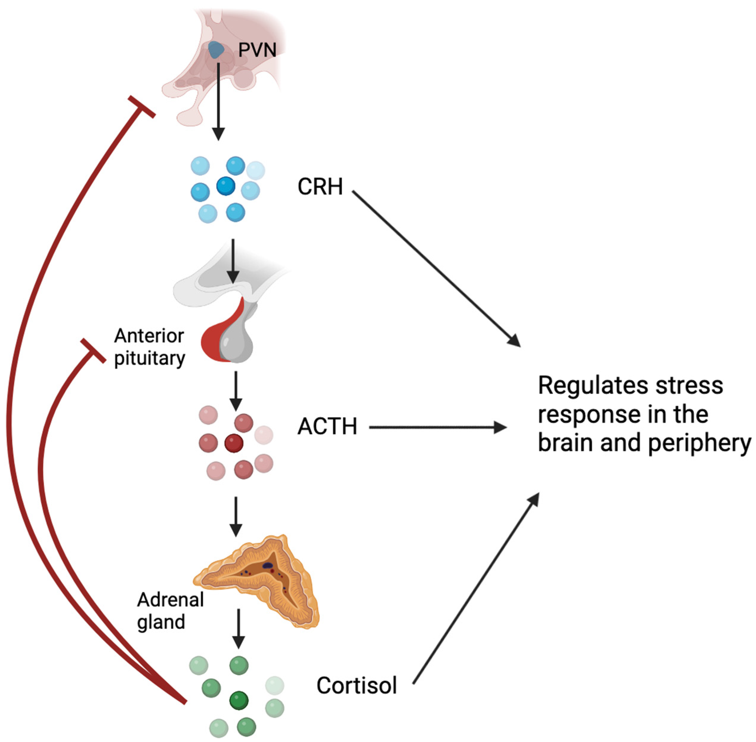
1. Glucocorticoids and the Hypothalamic–Pituitary–Adrenal Axis

Glucocorticoids, named for their effects on glucose metabolism, play a crucial role in regulating metabolic homeostasis, the cardiovascular system, cell proliferation and survival, growth, cognition and behaviour, immune function, and reproduction [1]. Glucocorticoids are steroid hormones that are synthesised and released by the adrenal cortex under the regulation of the hypothalamic–pituitary–adrenal (HPA) axis (Figure 1) [2]. In the presence of psychological or physiological stress, hypothalamic neurons in the paraventricular nucleus (PVN) synthesise and release corticotropin-releasing hormone (CRH) into the hypophysial portal vessels, which stimulates the release of adrenocorticotropic hormone (ACTH) from the anterior pituitary into the bloodstream [3]. ACTH primarily acts on the adrenal cortex, stimulating the synthesis and secretion of glucocorticoids from the zona fasciculata [2]. Glucocorticoids can inhibit CRH expression and secretion and ACTH output via negative feedback mechanisms [4,5]. Under normal, unstressed conditions, glucocorticoids are released in a circadian and ultradian rhythm. Exposure to acute stressors activates the HPA axis and leads to a temporary increase in glucocorticoids, which returns to baseline following resolution of the stressor; conversely, chronic stress can lead to disruption of the circadian rhythm and derangement of the ultradian rhythm of glucocorticoid secretion [2,6]. While glucocorticoids are typically adaptive to stress, excessive or inadequate activation of the HPA axis may contribute to the development of pathological conditions [6].

2. Functions of Glucocorticoids

Glucocorticoids play vital roles in a wide range of physiological processes crucial for maintaining homeostasis in the body. They upregulate enzymes involved in gluconeogenesis and glycogen synthesis in the liver and inhibit glucose uptake and utilisation in the muscle and adipose tissues [7,8,9]. This not only helps maintain glucose homeostasis during normal times but also, during periods of stress such as starvation or physical exertion, ensures that glucose remains the primary source of energy for vital organs such as the brain and skeletal muscles. Cortisol is also involved in sodium and water homeostasis to ensure sufficient blood volume and the maintenance of appropriate blood pressure [10,11]. It also plays an important role in the nervous system, with increased levels of glucocorticoids associated with various mental health disorders, including schizophrenia, post-traumatic stress disorder (PTSD), anxiety, depression, and addiction to drugs [12,13,14]. Prior to birth, glucocorticoids are recognised for their role in promoting the maturation of foetal lungs [15,16], but they can also lead to the development and differentiation of other essential organs [17].
The immunomodulatory actions of glucocorticoids are highly complex and include both immune stimulatory and immunosuppressive actions. Glucocorticoids are widely prescribed for their anti-inflammatory and immunosuppressive properties to treat inflammatory conditions, autoimmune disorders, haematological malignancies, and many other diverse uses [18]. Physiologically, glucocorticoids enhance innate immunity while limiting inflammation, supporting pathogen clearance, and preventing excessive tissue damage, to restore homeostasis following an inflammatory response [19]. These dichotomous impacts on inflammation are dictated by the cellular lineage and activation state, the stage of the inflammatory response, and glucocorticoid concentration [20]. In the innate immune response, glucocorticoids upregulate pattern-recognition receptors such as Toll-like receptor 2 (TLR2) and NOD-like receptors (NLRP1, NLRP3, and NLRC4) [21] to improve detection of pathogen-associated or damage-associated molecular patterns (PAMP/DAMPs). Downstream of these receptors, pro-inflammatory cytokines (such as IL-6, IL-1β, and TNF-α) are produced under the control of transcription factors nuclear factor kappa-light-chain-enhancer of activated B cells (NF-κB) and activator protein 1 (AP-1) [19,22]. These pro-inflammatory cytokines initiate an immune and acute-phase response that promotes glucocorticoid secretion, creating a feedforward mechanism that limits inflammation [22]. Glucocorticoids directly inhibit AP-1 and NF-κB, and upregulate their inhibitors, effectively dampening the inflammatory response and contributing to the resolution of inflammation [19]. Glucocorticoids further influence the resolution phase by inhibiting neutrophil recruitment, promoting apoptosis in neutrophils and lymphocytes, and activating macrophages at the injury site for tissue repair [19]. However, when administered in therapeutic doses, glucocorticoids primarily exhibit immunosuppressive effects. These effects include inhibiting leukocyte migration into tissues, inducing eosinophil apoptosis and preventing neutrophil emergence from the marrow, reducing circulating lymphocyte counts (more so T cells than B cells) through apoptosis and sequestration, and subsequently reducing immunoglobulin levels [23,24,25,26].

3. Glucocorticoid Bioavailability

A number of factors can impact glucocorticoid bioavailability. Roughly 95% of circulating glucocorticoids are bound to cortisol-binding globulin (CBG) or albumin following secretion, and the expression of these binding proteins impacts the level of freely available, biologically active glucocorticoids [27]. Endogenous glucocorticoids in circulation have an inconsistent half-life (66–120 min); however, those bound to CBG tend to have a longer half-life compared to the unbound hormone [27].
Given the lipophilic nature of glucocorticoids, free hormones can traverse the plasma membrane through simple diffusion. In cells that express multi-drug resistance p-glycoprotein, intracellular glucocorticoid concentration can be limited through their active expulsion [2]. Once inside the cell, glucocorticoids’ conversion to active forms (in humans, cortisone to cortisol) by the enzyme 11β-HSD 1 or to inactive forms (cortisol to cortisone) by 11β-HSD 2 regulates the availability of ligands to the glucocorticoid receptor (GR) [2]. Through these mechanisms, glucocorticoid availability to activate GR and hence downstream signalling can be finely tuned [2].

4. Glucocorticoid Receptors

The cellular action of glucocorticoids is mediated by an intracellular protein, the glucocorticoid receptor (GR) [28]. A number of GR isoforms, originating from different splice variants and transcription initiation sites, have been discovered. These isoforms diversify the range of glucocorticoid actions and play a role in determining cellular responsiveness to glucocorticoids [29]. While not the only determinant of glucocorticoid action, the composition of different glucocorticoid receptor isoforms differs between cell types and likely mediates differences in cellular activation state. Furthermore, differences in cellular responses to high or low glucocorticoid doses, natural or synthetic glucocorticoids, as well as changes that occur over time, are likely mediated by glucocorticoid receptor subtypes.
The GR is a member of the highly conserved nuclear receptor subfamily 3 [30]. All members of this superfamily share a similar domain structure, consisting of an N-terminal ligand-independent transactivation (AF1) domain, a central DNA-binding domain (DBD), and a C-terminal ligand-binding domain (LBD) [31]. Apart from containing the ligand-binding pocket, the LBD also contains crucial sequences for receptor dimerisation and nuclear localisation, as well as a second transactivation domain (AF2) that facilitates interactions with coregulators in a ligand-dependent manner [31]. A small flexible hinge region separates the DBD and LBD and is involved in conformational changes induced after ligand-binding [31].
The human GR, encoded by the NR3C1 gene, is composed of nine exons [31]. Exon 1 forms the 5′-untranslated region, while exon 2 encodes the N-terminal domain (NTD), which is poorly conserved across nuclear receptors [31]. Exons 3 and 4 constitute the DBD, the most conserved domain in the nuclear receptor family, while exons 5–9 encode the hinge region and ligand-binding domain (LBD) [31]. The NR3C1 gene undergoes alternative splicing, leading to the production of human GRα, GRβ, GRγ, GR-A, and GR-P transcriptional isoforms (Figure 2) [31,32,33]. Most research has focused on glucocorticoid signalling through GRα and GRβ, proteins identical up to amino acid 727 but differing in their C-terminal exon 9 [34]. The GRβ variant lacks sequences encoding the 11th and 12th helices of the LBD, making it incapable of binding glucocorticoids [34]. Though initially thought to serve primarily as a dominant negative regulator of GRα, transcriptional analysis shows that GRβ has inherent transcriptional activity when overexpressed [35,36,37].
The sequence of GR-γ differs due to an arginine residue included between exons 3 and 4 of the DBD, resulting from the use of an alternative intronic splice donor site [38]. The insertion of a single amino acid significantly reduces the transcriptional activity of GR-γ, demonstrating the functional significance of this region [39,40]. Moreover, changes in the DBD of GR-γ alter its DNA binding sequence specificity, leading to a unique transcriptome [41,42]. The in vivo functions of the GR-A and GR-P isoforms, which contain incomplete LBDs, are not well understood, but both have been demonstrated in glucocorticoid-resistant malignant cell lines [33,43].
All GR splice variants could possibly generate additional isoforms through alternative translation initiation mechanisms. However, these translational isoforms have only been described for GRα to date. In exon 2, eight highly conserved AUG start codons generate various GR isoforms through ribosomal leaky scanning and shunting, including GRα-A, -B, -C1, -C2, -C3, -D1, -D2, and -D3 (Figure 2). The relative expression of both splice and translation GR isoforms can vary among tissues and within individual cells, influencing their transcriptional potential [44]. Each GR isoform is also subject to several post-translational modifications, such as phosphorylation, acetylation, ubiquitination, and sumoylation, further influencing receptor activity [1].
Figure 2. Splice and translation initiation isoforms of the glucocorticoid receptor. Exons of the glucocorticoid receptor gene with the subsequent splice (left) and translation initiation (right) isoforms. AF-1—transcriptional activation function 1; DBD—DNA binding domain; GR—glucocorticoid receptor; LBD—ligand-binding domain; NTD—N-terminal domain. Created on bio render. Adapted from Ramamoorthy and Cidlowski [45].
Biology 12 01104 g002
The traditional explanation for GR signalling focused on glucocorticoids passing through the plasma membrane and binding to GR anchored within the cytoplasm, which induced translocation to the nucleus to regulate gene transcription. More recent discoveries have expanded our understanding of GR function to include a multitude of mechanisms by which GRs impact gene regulation, the varying impact of GR isoforms on regulating cellular response, and non-genomic effects of GR. There are methodological limitations in identifying and understanding the effects of individual GR isoforms. These limitations include a lack of specific antibodies for each GR isoform, leading to the use of the semi-quantitative technique of Western blot for the characterisation of the GR isoform profile in different cell types and tissues, distinguishing different isoforms by molecular weight. Furthermore, identifying the role of each specific GR isoform has been limited to overexpression experiments in GR null cell lines [44], which does not account for the interaction of multiple GR isoforms that are expressed endogenously.

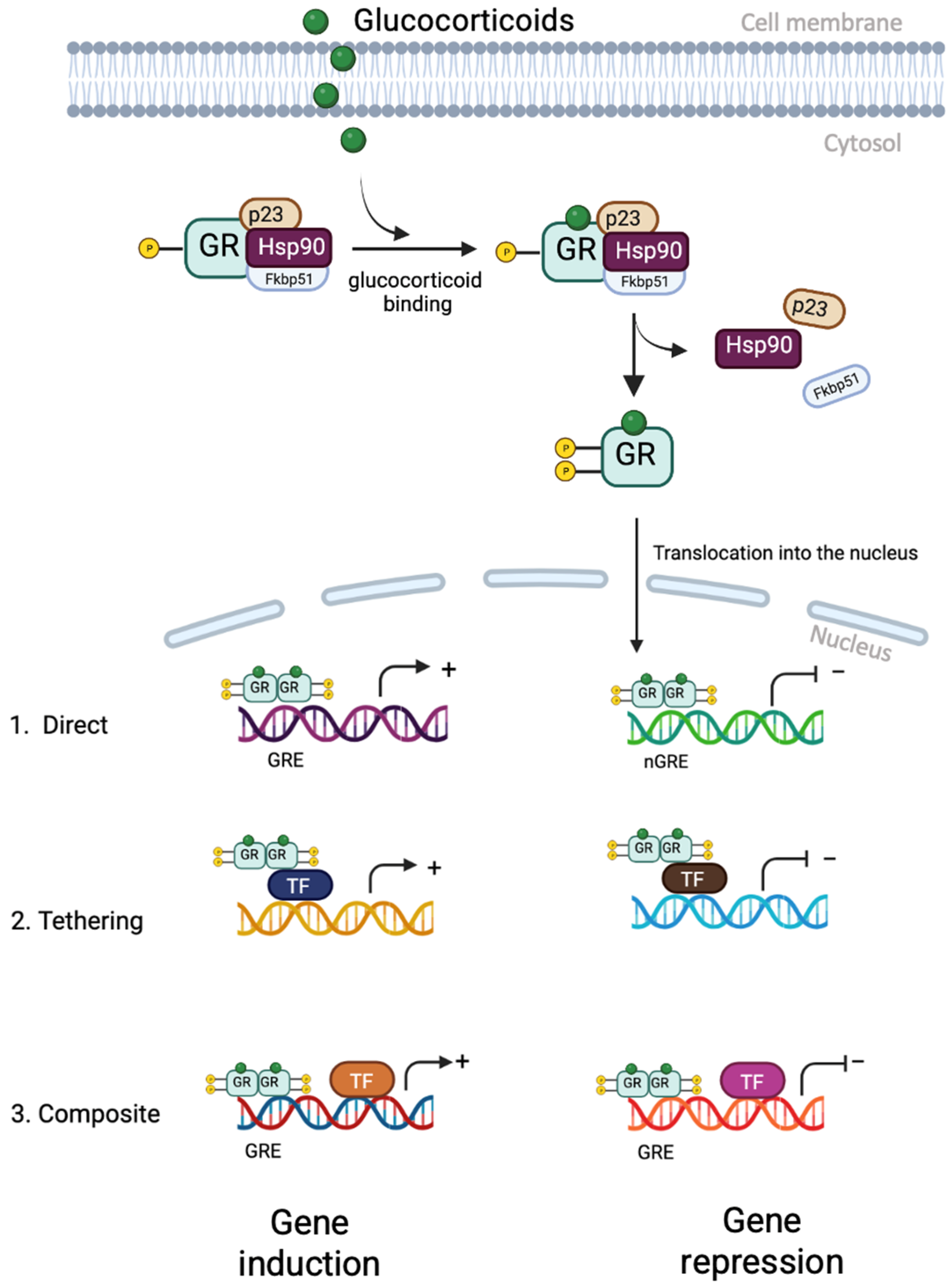
5. Glucocorticoid Receptor Signalling

More recent understanding of glucocorticoid function has demonstrated that they act through both genomic (predominant) and non-genomic pathways. The genomic pathway is mediated by GRs (Figure 3). In the absence of glucocorticoids, GRs exist principally anchored in the cytoplasm as part of a large complex of proteins, including chaperones (hsp90, hsp70, and p23) and immunophilins (FKBP51 and FKBP52), which help maintain GRs in a conformation that allows them to bind to ligands with high affinity [46]. Once glucocorticoids bind to GRs, receptor conformational changes lead to the dissociation of the protein complex and the exposure of 2 nuclear localisation signals [45]. The ligand-bound GR is then quickly transported into the nucleus through nuclear pores. Inside the nucleus, the GR binds directly to glucocorticoid response elements (GREs) as a dimer and activates the expression of target genes [45]. This binding leads to further conformational changes in the GR, allowing it to recruit coregulators and chromatin-remodelling complexes that influence the activity of RNA polymerase II and activate gene transcription and repression [45]. Binding to negative glucocorticoid-responsive elements (nGREs) can recruit corepressors (NCoR1 and SMRT) and histone deacetylases (HDACs) to repress the activity of target genes [47]. In addition to its direct interaction with GREs, the GR can also interact with members of the signal transducer and activator of transcription (STAT) family to enhance transcription of specific target genes [47]. The anti-inflammatory effects of glucocorticoids are mainly mediated by a negative regulatory mechanism called trans-repression. In this, the ligand-bound GR is recruited to genes by directly interacting with DNA-bound transcription factors, particularly NF-κB and activator protein-1 (AP-1) [48]. There are a number of proteins within the cell that can interact with the GR and potentiate trans-repression. For instance, O-linked-N-acetylglucosamine transferase (OGT), a placental stress biomarker, can interact with GRs and impact NF-kB-mediated transcription [49]. The GR directly binds to the p65 subunit of NF-kB and the Jun subunit of AP-1, inhibiting their transcriptional activation [48]. For some genes, GRs function in a composite manner, binding directly to a GRE and physically associating with AP1 or NF-kB bound to a neighbouring site on the DNA, while for others, GRs bind to transcription factors without interacting directly with DNA [45].
The non-genomic effects of glucocorticoids, which occur without requiring gene transcription, are rapid and mediated through physiochemical interactions of cytosolic or membrane-bound GRs [50]. These effects happen within seconds to minutes of activation and involve the activity of kinases such as phosphoinositide 3-kinase, AKT, Src kinase, mitogen-activated protein kinases (MAPKs), or intracellular Ca2+ signalling [51,52,53,54]. Glucocorticoids can non-genomically interact with membrane lipids and proteins, cytoplasmic proteins, including various kinases, transcription factors, and glucocorticoid transporters, to influence cell function [53]. High doses of glucocorticoids can directly interact with membrane lipids to alter membrane fluidity and affect ATP use and ion cyclicity [55]. Glucocorticoids can also enhance Ca2+ signalling by binding to voltage-dependent calcium channels in brain synapses. For example, in cultured rat hippocampal cells, stress-induced elevated levels of corticosterone rapidly prolonged NMDA receptor-mediated Ca2+ signalling via the activation of a protein kinase C-dependent process by membrane GRs [56]. Similarly, glucocorticoids have been found to non-genomically impact neurotransmission by affecting GABA, glutamate, serotonin, acetylcholine, and vasopressin receptors [57,58,59,60]. Furthermore, glucocorticoids can engage with a variety of cytoplasmic proteins, including members of the MAPK family (extracellular signal-regulated protein kinases (ERK), c-Jun NH2-terminal protein kinase (JNK), and MAPK p38), phospholipases, and protein kinases, both genomically and non-genomically. MAPKs can regulate processes such as cell proliferation, development, differentiation, and apoptosis [61,62]. Some examples of these interactions in relation to reproductive organs will be discussed in the forthcoming sections. Glucocorticoid administration to mouse thymocytes induced apoptosis via activation of phospholipase C, non-genomically, in vitro [63]. Its administration to A549 cells in vitro prevented cell growth due to inhibition of prostaglandin E2 via a reduction in the activity of phospholipase A [64]. Glucocorticoids can also secondarily activate MAPKs [65], Ca2+ [66], and nitric oxide (NO) [67] signalling and impact ion transport [68] by activating protein kinases A, B, and C. Glucocorticoids can bind the extracellular site of membrane-bound GRs, which can then interact with and activate the stimulatory G protein, Gαs, to rapidly increase the levels of 3′,5′-cyclic adenosine monophosphate (cAMP), a common secondary messenger involved in the regulation of various biological processes and the actions of non-steroidal hormones [69,70]. Lastly, when glucocorticoids bind to GRs, components of the multi-protein anchoring complex are released, which participate in secondary signalling pathways. The accessory proteins that have been reported to mediate these non-genomic effects of glucocorticoids include HSP-70, HSP-90, and MAPKs such as Src [71,72]. Moreover, the ligand-bound GR can also interact with other proteins, including ZAP-90, leading to further downstream signalling [73].
This non-genomic signalling of the glucocorticoid receptor increases the complexity and diversity of biological actions that are dependent on glucocorticoids [45].

6. Glucocorticoids on Hypothalamic–Pituitary–Gonadal Axis

The hypothalamus and pituitary are also central to reproductive physiology, serving as hubs for both receiving and delivering endocrine signals [74]. The hypothalamus generates and releases gonadotropin-releasing hormone (GnRH), which travels through the hypophyseal portal system to reach the anterior pituitary [75]. There, GnRH stimulates the synthesis and release of follicle-stimulating hormone (FSH) and luteinising hormone (LH) from the gonadotrophs, leading to the secretion of estradiol and progesterone from the ovaries [76]. A regulatory negative feedback mechanism exists in which estradiol from the ovaries inhibits the secretion of GnRH and thus the production of FSH and LH [76]. Increased glucocorticoid exposure, whether resulting from stress or exogenous administration, can lead to significant reproductive disturbances through their notable impacts on the hypothalamus and pituitary [77,78,79]. Glucocorticoids influence the HPG axis by inhibiting the release of GnRH from the hypothalamus and the synthesis and release of gonadotropins from the pituitary [77,79].
The presence of GR in the hypothalamus implies that glucocorticoids could have direct effects [80]. Indeed, dexamethasone-induced GR signalling in hypothalamic cell lines that secrete GnRH suppresses GnRH promoter activity and subsequent GnRH mRNA levels in vitro by interacting with a multiprotein complex at negative GRE sites [81]. A study analysing the reproductive function in adult male rats treated with corticosterone over an extended period reported decreased hypothalamic GnRH mRNA and decreased serum LH but not FSH secretion [82]. The same study examining the acute effects of dexamethasone on reproductive function in adult female rats reported a significant decrease in pituitary FSHβ but not LHβ mRNA levels [81]. The findings from this study suggest that chronic increases in levels of glucocorticoid/stress suppress gonadotropin levels by reducing the expression of GnRH levels, while acute glucocorticoid administration prevents gonadotropin secretion via other processes, including the reduction of FSHβ mRNA levels. However, this effect of acute stress and short-term increases in glucocorticoid levels on gonadotrophin secretion can vary, either inhibiting or stimulating it. GnRH stimulates the production and release of LH and FSH by binding to the GnRHR in pituitary gonadotropes [83,84]. Evidence from the mouse pituitary gonadotrope cell line LβT2 treated with dexamethasone suggests that glucocorticoids could directly induce the expression of the GnRHR gene, leading to increases in LH and FSH secretion [85]. In addition, in primary cultured cells from the anterior pituitary, glucocorticoids have been found to increase FSHβ mRNA levels [82,86]. These are inconsistent with the findings from Gore et al. [82], which may suggest that the way glucocorticoids control the expression of gonadotropin synthesis is dependent on the context. Our understanding of the regulation of gonadotrophins by glucocorticoids is presently based on findings from cell line studies, which may not accurately depict the regulation by endogenous glucocorticoids in vivo [79].
Besides directly controlling GnRH, GnRHR, and pituitary gonadotropes, glucocorticoids can also regulate their inhibitory effects on HPG function via novel intermediaries. Glucocorticoids can inhibit GnRH expression by regulating the expression of two neuropeptides, kisspeptin (KISS1) and gonadotropin-inhibitory hormone (GnIH) [79]. These neuropeptides exert contrasting impacts on GnRH secretion from the hypothalamus and react to elevated glucocorticoid levels. KISS1 promotes GnRH secretion via its receptor, KISS-1R, which is co-expressed in GnRH neurons. KISS1 neurons located in the anteroventral periventricular nucleus and the periventricular nucleus continuum of the preoptic area in the hypothalamus express GRs, indicating that glucocorticoids can directly act on these neurons [87]. Corticosterone administration in mice inhibited ovarian cyclicity by disrupting the preovulatory hormonal cascade required for the LH surge [88]. The study also reported a significant decrease in the activation and percentage of KISS1 neurons as well as the expression of KISS1 mRNA in the hypothalamus during an estradiol-induced LH surge [88]. This suggests that glucocorticoids can suppress the HPG axis by inhibiting the expression of KISS1 neurons. GnIH neurons suppress the functions of GnRH and KISS1 neurons and gonadotrophs [89]. Both acute and chronic stress in sheep led to an increase in the expression and activity of GnIH neurons and increased the interaction between GnIH fibres and GnRH cells [90]. However, stress had no effect on GnIH levels in the hypophysial portal blood [90]. Moreover, this effect on GnIH gene expression disappeared after adrenalectomy, implying that adrenal-derived factors, most likely corticosterone, mediated the stress-induced inhibition of reproductive functions [90]. GnIH neurons co-express GR, and the exposure of quail and rat hypothalamic neuronal cells to corticosterone resulted in increased GnIH expression [90]. This increase was facilitated through glucocorticoid response elements (GREs) in the promoter region of GnIH [90]. In short, glucocorticoids can indirectly influence the reproductive functions of the HPG axis by regulating GnRH expression and secretion by KISS1 and GnIH.

7. Effect of Glucocorticoids on Reproductive Organs

As glucocorticoids are known to be a key regulator of basal and stress-related homeostasis, it is highly likely that stress-related increases in glucocorticoid levels could contribute to decreased fertility [77]. Stress-related compromises of reproductive function can be reported across various species. Mammals such as rats [91,92,93], along with birds and reptiles [94,95,96,97], all exhibit a reduction in both male and female reproductive abilities in response to physical stress [3]. The response to stress in humans is also detrimental to reproductive function throughout the life cycle. For example, high perceived stress during pregnancy is a risk factor for preterm labour and poor outcomes in the offspring [98,99]. For female athletes, there is a high prevalence of delayed puberty, menstrual disorders, and long-term secondary amenorrhoea, especially among those participating in aesthetic sports and endurance sports [100,101]. This reproductive dysfunction could be caused by the alteration of HPG axis activity due to excess cortisol associated with physiological and psychological stress on the body. Glucocorticoids are also known to interfere with sexual maturation, as patients with hypocortisolism (Addison’s disease) and hypercortisolism (Cushing’s syndrome) have altered onsets of puberty [102,103]. A study revealed a positive correlation between circulating glucocorticoid levels and the age of puberty onset in prepubertal girls [103]. This is consistent with findings from animal studies [104,105,106]. Studies in mice and rats report delayed onset of puberty in offspring of mothers subjected to stress or with synthetic glucocorticoid administration [106,107,108]. Prepubertal exposure to synthetic glucocorticoids delayed the onset of vaginal opening, an indicator of puberty in female rats [109].

7.1. Ovary

Apart from influencing the ovarian cycle via the hypothalamus and pituitary, glucocorticoids can also directly affect ovarian functions by controlling the activities of granulosa cells, oocytes, cumulus cells, and luteal cells through GRs [88]. It was previously thought that the ovary did not produce glucocorticoids locally. Hence, it was presumed that glucocorticoids in the ovary were transported from the adrenal glands via the bloodstream [110]. These glucocorticoids, probably inactive (cortisone), were then proposed to diffuse into the granulosa cells of the periovulatory follicle, where 11β-HSD 1 converted them to cortisol, which could then exert its effects via co-localised GRs [110]. However, a recent study by Jeon et al. [111] demonstrated that human primary luteinising granulosa cells produce cortisol in vitro. The mRNA levels of CYP11B1 and CYP21A2, two enzymes required for the biosynthesis of cortisol, were detected in primary cultured human granulosa/lutein cells (hGLC), and their levels were found to increase following ovulatory administration of human chorionic gonadotropin (hCG) as a substitute for LH [111]. CYP21A2, an enzyme necessary for the metabolism of 17α-hydroxyprogesterone into 11-deoxycortisol, and CYP11B1, an enzyme responsible for the conversion of 11-deoxycortisol into cortisol, are both required for the synthesis of cortisol from progesterone (Figure 4) [111]. This is consistent with findings from Amin et al. [112], who reported the levels of two cortisol precursors, 11-deoxycorticosterone and 11-deoxycortisol, in the follicular fluid to be positively correlated with the lipid content in human luteinised granulosa cells (LGCs) and demonstrated the expression of CYP21A2 mRNA in LGCs in vitro. Given that during ovulation, human granulosa cells generate a substantial amount of progesterone, the presence of enzymes that convert progesterone to cortisol suggests that these cells could produce cortisol [111]. However, it is still likely that the majority of the cortisol acting on the ovaries is derived from the adrenal glands instead of being locally produced, as this evidence is based on in vitro studies and might not accurately represent in vivo physiology.
Glucocorticoid concentrations are intracellularly regulated in the ovary by 11β-HSD 1 and 2. 11β-HSD has been shown to be present in various ovarian cell types such as oocytes, cumulus cells, granulosa cells, theca cells, granulosa-lutein cells, corpus luteum, and ovarian surface epithelium [113,114,115]. During the LH surge, there is a rapid increase in the production of progesterone and cortisol. Before the LH surge, follicular levels of inactive glucocorticoids (cortisone) were higher than those of cortisol [116]. However, post-surge, this reverses, with the concentration of cortisol being 4.5-fold higher than that of cortisone [116]. This switch is proposed to be caused by a change in the predominance of the 11β-HSD isotype from 2 to 1 [116,117]. In non-luteinised granulosa cells from IVF patients, 11β-HSD 2 was readily detected, but it was not detectable in the luteinising granulosa cells [118]. More recent studies have also demonstrated a swift decrease in 11β-HSD 2 (mRNA) and increased 11β-HSD 1 (mRNA and protein) levels in human granulosa cells collected after hCG stimulation in comparison to cells obtained prior to the in vivo LH surge [119], as well as in vitro [111]. The increased expression of 11β-HSD 1 persisted in post-ovulatory follicles, suggesting it has a role during ovulation and the initial phase of luteal formation by modulating intracellular glucocorticoid concentrations. Furthermore, 11β-HSD 1 expression was mainly observed in granulosa cells from late ovulatory follicles and granulosa-lutein cells of post-ovulatory follicles, indicating the location of cortisol synthesis [111]. These findings suggest that active glucocorticoid concentrations are reduced during follicular growth and maturation, but they rise during the ovulation process initiated by the LH surge.
Preovulatory follicles undergo an acute inflammatory event during ovulation that is associated with rupture, the subsequent healing process, and metabolic alterations linked with luteinisation. Thus, the coinciding increase in expression of anti-inflammatory glucocorticoids [120] by 11β-HSD 1 activation of cortisone to cortisol may be a physiological compensatory mechanism to reduce the ovarian inflammatory process [111]. To determine the effect of cortisol on periovulatory follicles, Jeon et al. [111] looked at the effect of ovulatory hCG stimulation on the expression of genes involved in cortisol synthesis. The expression of 11β-HSD 1, NR3C1, and FKBP5 was increased after hCG administration in periovulatory follicles, both in vitro and in vivo [111]. Moreover, it was reported through immunohistochemistry that GR was localised in endothelial cells and leukocytes within and near the late and post-ovulatory follicles [111]. These findings suggest that glucocorticoids probably operate during ovulation and the initial stages of corpus luteum formation by exerting their effect directly on follicular cells, endothelial cells, and leukocytes, all of which express GR. Since cortisol plays a role in ovulation and corpus luteum (CL) formation, this could contribute to the fact that women with dysregulated levels of cortisol frequently experience menstrual irregularities and anovulatory infertility.
Furthermore, stress-induced glucocorticoid levels have been linked to a decrease in oocyte competence. Chronic heat stress in pigs has been shown to modify ovarian expression of genes involved in steroidogenesis and increase signalling via the insulin-mediated phosphoinositide 3-Kinase (PI3K) pathway [121]. Heat stress can also alter the composition of follicular fluid, thereby changing the oocyte’s microenvironment [122,123]. In mice, the damage to oocyte competence was found to be dependent on the severity of the restraint stress, with more significant effects observed after prolonged stress [124]. Administration of exogenous glucocorticoids in female rats led to a decline in growth factor levels and alterations in the estrogen-to-progesterone ratio [125].

7.1.1. Dual Role of Glucocorticoids in the Ovary

Despite the studies aimed at determining the role of glucocorticoids in the ovary, their function is not completely understood, with contradictory findings from multiple studies. Several studies have looked at the effect of glucocorticoid administration on mammalian oocytes and its influence on oocyte maturation, with inconsistent results. In some in vitro mouse studies, no effect was found on oocyte maturation when they were subjected to cortisol (up to 28 μM) and dexamethasone (up to 100 μM) [126,127,128,129]. However, in other mouse studies, slightly higher concentrations of cortisol (138 μM) and dexamethasone (204 μM) resulted in inhibition of oocyte maturation in vitro [126,128,129]. Similar to studies in mice, cortisol and dexamethasone administration (between 0.26 and 138 μM) inhibited oocyte maturation in porcine oocytes in vitro [130]. Conversely, a study on horses reported similar doses of cortisol (0.28–2.8 μM) to have no effect on oocyte maturation in vitro [131]. Glucocorticoids can either selectively induce or inhibit ovarian steroidogenesis. Dexamethasone administration in rats is associated with altered production of progesterone from granulosa cells by either increasing or decreasing the expression of steroidogenic proteins like steroidogenic acute regulatory protein (StAR) [132,133,134]. This indicates that in vitro glucocorticoids exhibit both stimulatory and inhibitory effects in the ovary. Such discrepancies could be attributed to variations in the administered dosage, the phase of follicular development, or the differential expression of GR isoforms.
Similarly, glucocorticoids can have both pro- and anti-inflammatory roles in the ovary [135]. Cortisol treatment has been shown to decrease germ cell density by increasing the apoptosis of oogonia in the developing human ovary, impair oocyte development potential, and increase the apoptosis of granulosa cells in animal models [136]. However, glucocorticoids have also been reported to inhibit apoptosis and promote survival in ovarian granulosa cells by non-genomically phosphorylating ERK 1 and 2, and Akt/protein kinase B [137,138]. In primary bovine luteal cells, cortisol has been reported to prevent apoptosis induced by TNF and interferon γ (IFN-γ) by inhibiting the expression of caspase 8 and 3, thus preserving the function of the corpus luteum [139]. The anti-apoptotic effects of glucocorticoids could be a mechanism through which glucocorticoids exert their anti-inflammatory effects in the ovary, as they induce the death of pro-inflammatory cells and promote the survival of cells undergoing the inflammatory response. These anti-apoptotic mechanisms could also contribute to reducing the damage associated with follicle rupture. Glucocorticoids, via GR signalling, reduce the expression and function of extracellular matrix metalloproteinase 9 in human ovarian surface epithelial cells, which helps inhibit proteolytic injury to the ovarian surface during ovulation with the associated inflammation [140]. These opposing effects of glucocorticoids on the ovary cannot be explained by the expression of a single isoform of GR and could potentially be a result of differential expression of the GR isoforms.

7.1.2. Glucocorticoid Receptor Isoforms in the Ovary

GRs are found in the follicles, corpus luteum, and cells of the ovarian surface epithelium in both rats and humans [141]. In human studies, GR protein has been identified in ovarian surface epithelium cells, in vitro [142]. As mentioned above, in vivo studies have also found GR protein to be localised in granulosa cells, theca cells, endothelial cells, and leukocytes in blood vessels in the vicinity of late ovulatory and post-ovulatory follicles [111]. The expression levels of GR in the follicle remain unchanged during the stages of follicular maturation and ovulation in rats [143], implying that the main regulatory processes in these cell types could be determined by the concentration of active glucocorticoids. Current evidence proposes that glucocorticoids have a direct influence on the preliminary phases of reproduction, such as the maturation of oocytes, fertilisation, and preimplantation embryo growth [144,145]. However, the observed effects can vary based on factors like the type of glucocorticoid molecule, dosage, stage of development, and species under study [145]. As variation in the expression of GR isoforms may lead to distinct cellular responses to glucocorticoids, investigating the expression of various splice and translational isoforms of the GR in oocytes carries significant importance.
Very few studies have looked at the expression of the different isoforms of the GR in the mammalian ovary. The ovary is a complex organ, with its function highly linked to the active follicles that undergo constant change across the ovarian cycle. This could possibly lead to a constant shift in the expression profiles of the different GR isoforms. A recent study examining mouse oocytes found the two splice variants GRα and GRγ (mRNA) to be highly and consistently expressed, while GRβ and GR-P (mRNA) were undetectable [145]. This study also reported the presence of a doublet band around 94 and 91 kDa in oocytes, and this doublet likely represents mouse isoforms GRα-A and B (GR protein) [145]. Another study [146] examining the presence of GRβ in various tissues reported the absence of GRβ in SK-OV-3 (ovarian adenocarcinoma) cells in vitro, consistent with the results from the study by Cikos et al. [145]. Since GRβ is known to antagonise the effects of GRα, the pro-inflammatory or contradictory effects of cortisol have often been attributed to an increased GRβ:GRα ratio. However, since GRβ is undetectable in the ovary, the varying responses of ovarian cells to high concentrations of glucocorticoids could be attributed to the presence of the other splice or translational isoforms present in the ovary. The detection of various GR isoforms in ovarian cells needs to be further examined to better elucidate their contribution to ovarian glucocorticoid function.

7.2. Uterus

The influence of glucocorticoids on uterine biology has been typically depicted as an antagonist of estrogen activity, but it has been recently studied as an independent moderator of uterine functionality. Dexamethasone impedes estrogen-induced uterine growth and proliferation in mice and, when administered before estradiol-facilitated implantation, can lead to a decrease in implantation sites [147,148]. Gene identification and ontology studies looking at the expression of genes regulated by dexamethasone and estradiol in a human uterine epithelial cell line point to glucocorticoids regulating important biological processes, including immune function, cell cycle, and embryonic development [149]. For instance, dexamethasone (but not estradiol) upregulated the expression of nuclear factor of kappa light chain inhibitor alpha (NFκB1A), an inhibitor of the NFκB complex. NFκB signalling in the uterus controls the apoptotic function of caspase 3 during pregnancy [149]. The expression of NFκB1A escalates during the late secretory phase, coinciding with an increased incidence of apoptosis in the uterus [150,151]. Thus, the regulatory role of glucocorticoids on NFκB1A could contribute to the balance between cell death and survival in the uterus [149]. Furthermore, expression of the gene left-right determination factor 1 (LEFTY1) was upregulated with dexamethasone treatment (restored with estradiol administration) [149]. LEFTY1 is a gene that encodes a soluble cytokine of the TGF-β superfamily [152]. When LEFTY1 is introduced into the uterine horn of pregnant mice, it significantly reduces uterine decidualisation and results in a decreased litter size [153]. Thus, glucocorticoid regulation of key fertility genes, like LEFTY1, offers additional evidence that glucocorticoids may function as a bridge between the immune and reproductive systems in the uterus [149].
Dexamethasone administration in mice also singularly regulates the expression of genes associated with cellular development, proliferation, and signalling. In the uterus of neonatal mice, dexamethasone reduces epithelial cell proliferation through mechanisms that are both dependent on and independent of GR [154]. Human endometrial cells that have undergone decidualisation also exhibit a distinct transcriptome dependent on the GR [155]. This GR-dependent transcriptome is enriched with Krüppel-associated box domain containing zinc-finger proteins, which are a family of transcriptional repressors involved in heterochromatin formation [155]. In fact, decidualisation of human endometrial stromal cells was associated with an increase in GR expression [154]. Gene expression analysis of GR knockout HESCs that were undergoing differentiation reported an increase in trimethylated H3K9 levels in decidualising cells [154]. This suggests that GR can directly manage gene expression and globally modify transcription through the regulation of histone alterations in human uterine cells [154].
The semi-allogenic foetus’s immunological tolerance and a successful pregnancy require pre-emptive immune system adjustments that support endometrial receptivity [79]. These changes begin before the implantation of the embryo and escalate throughout the early stages of pregnancy [156,157,158]. During this time, immune cells are mobilised and activated in the uterus, establishing a dynamic immune response that is crucial for implantation and tolerance [79]. When this immune balance is disrupted, it can contribute to complications such as miscarriage, preeclampsia, or premature labour. Glucocorticoids can regulate the uterine immune system by directly or indirectly acting on these immune cells. Studies involving conditional ablation of GRs in mouse uteri have suggested the essential role of endometrial glucocorticoid signalling in recruiting appropriate immune cells [159]. In addition, in these GR knockouts, the expression of genes required for implantation was altered. This implies that the glucocorticoid signalling in the uterus might play a critical role in establishing the molecular groundwork that determines uterine receptivity [159].
Administration of exogenous glucocorticoids affects the number and function of uterine immune cells [160]. In the initial stages of pregnancy, uterine natural killer (uNK) cells control trophoblast invasion and changes in the vascular functions that help placental growth [161,162]. GRs are expressed in uNK cells, and their cytotoxic activity is responsive to cortisol treatment [163]. The administration of prednisolone in women experiencing recurrent miscarriage correlates with decreased uNK cell counts; however, there is yet to be a demonstrated connection between this glucocorticoid-induced reduction in uNK cells and the success of the pregnancy [164]. Macrophages exist in two distinct forms: an M1, or pro-inflammatory phenotype, and an M2 phenotype associated with tissue remodelling, immunosuppression, and angiogenesis [165]. During the peri-implantation period, endometrial macrophages lean towards the M1 phenotype and shift towards the M2 phenotype post-implantation [166,167]. GRs are expressed in human endometrial macrophages during the secretory phase, which coincides with the uterine receptivity period, implantation, and the menstrual phase, as well as at full term, but the function of glucocorticoid signalling in these macrophages remains largely unexplored [168,169]. Dendritic cells and regulatory T cells play a vital role in determining whether the blastocyst is accepted or rejected. However, the physiological impacts of glucocorticoids on these cell types within the endometrium remain under-researched [170].
In the peripheral immune system, glucocorticoids exhibit a broad range of direct influences on the development and function of immune cells, which can potentially affect the composition of immune cells available for recruitment to the reproductive tract [19]. They restrict the formation of immature dendritic cells from monocytes and inhibit the maturation of immature dendritic cells in vitro [171]. Elevations in serum glucocorticoid levels have been linked to the rapid increase of mature NK cells in peripheral circulation in vivo [172]. Furthermore, glucocorticoids stimulate substantial apoptosis in T and B cells, mature dendritic cells, basophils, and eosinophils [20]. Glucocorticoids suppress the transcription of pro-inflammatory cytokines and chemokines in macrophages and guide macrophages towards the M2 phenotype [160]. Dexamethasone can also trigger components of the inflammasome, thereby enhancing the innate immune response in macrophages [173]. The response of the immune system to glucocorticoids relies on both the physiological or stress-induced hormone levels and the duration of the stimulus. This suggests that the immune response that oversees reproduction is partly managed through physiological glucocorticoid signalling by the GR and is susceptible to hormone fluctuations.

GR Isoforms in the Uterus

GR is found in the uterus, predominantly within the stromal fibroblasts, lymphocytes, and endothelial cells, with only minimal presence in the glandular epithelium. Contrary to the cyclical changes reported with the estrogen receptor (ER) and progesterone receptor (PR), GR expression remains steady throughout the menstrual cycle [174]. During labour, there is a significant increase in total GR and GRα protein levels, while the levels of GRβ do not undergo any change [175]. Given the limited research on the expression of GR isoforms in the uterus, further studies are necessary to investigate if differential expression of these isoforms is behind the contradictory effects of glucocorticoids reported in the uterus.

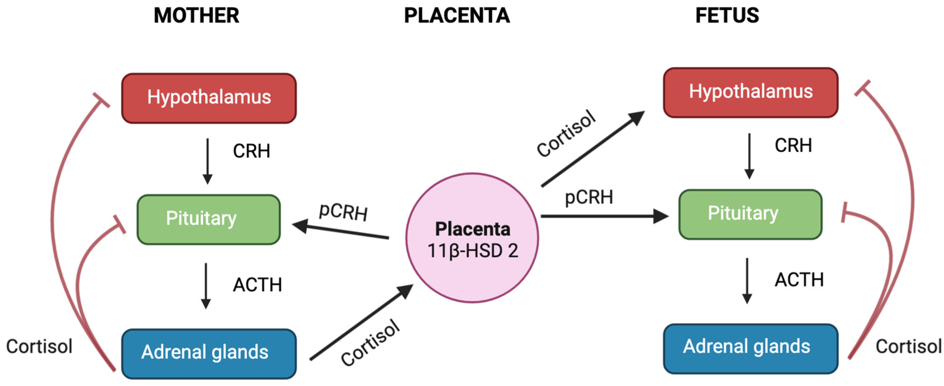
8. Pregnancy and Parturition

During the second and third trimesters of pregnancy, the placenta begins to produce CRH, which has the same biological activity as the CRH produced by the hypothalamus (Figure 5) [99]. The placental CRH (pCRH) is secreted into both the maternal and foetal compartments. Unlike its inhibitory effect on the CRH-producing cells of the hypothalamus, cortisol stimulates the synthesis and release of pCRH [99], leading to a shift from negative feed-back regulation of the maternal HPA axis to a positive feed-forward cycle. Consequently, the circulating CRH levels in pregnant women are 1000 to 10,000 times higher than those of non-pregnant individuals [176]. Throughout gestation, most of the CRH is inactively bound to the CRH-binding protein (CRH-BP); however, during the third trimester, a spike in CRH occurs without a corresponding increase in CRH-BP levels in the maternal circulation, leading to an increase in the levels of active CRH [177]. It has been proposed that this rise in CRH levels may determine the length of gestation and initiate uterine contractility at the start of labour, acting as a “placental clock” [176]. However, if certain abnormalities occur during pregnancy, such as preeclampsia or maternal stress, these alterations may occur prematurely resulting from excess cortisol, which may lead to adverse maternal and/or foetal outcomes, including preterm birth and foetal growth restriction [99].
During pregnancy and parturition, uterine contractility can be separated into at least four stages. The first stage, known as phase 0, is the pregnancy period, where the uterus stays relatively quiescent through the action of inhibitors of uterine contractility such as progesterone [179]. In the first phase of parturition (phase 1), the uterus responds to mechanical stretch or uterotrophic priming by activating a set of genes essential for myometrial contraction [179]. As parturition enters phase 2, the uterus is stimulated by various uterotonins such as prostaglandins (PGs), oxytocin, and CRH [179]. The third and final phase involves uterine involution following the delivery of the foetus and placenta, a process primarily influenced by oxytocin [179]. Glucocorticoids help with two major events that lead to parturition [180]. Firstly, glucocorticoids stimulate the production of the placental enzyme cytochrome P450 17α hydroxylase (P450c17), which converts C21 steroids (pregnenolone and progesterone) into C19 steroid aromatase precursors [181]. This triggers a decrease in progesterone along with an increase in estrogen.
The rising estrogen then primes the myometrium from a state of quiescence to a contractile state by upregulating the expression of contraction-associated proteins (CAPs) such as connexin 43 [Cx43], oxytocin receptor (OTR), and prostaglandin (PG) receptor. After myometrium priming, sufficient uterotonins, such as prostaglandins, are synthesised to complete the feed-forward process of parturition [181]. Apart from initiating myometrial contractions, prostaglandins E2 and F2α (PGE2 and PGF2α) also facilitate cervical ripening, membrane rupture prior to parturition, and promote the expression of placental P450c17 [181]. In humans, prostaglandin production is compartmentalised within the tissues of the pregnant uterus [182]. While chorion and decidua also contribute, amnion is predominantly responsible for the formation of PGE2, and its output surges at labour onset [182]. Glucocorticoids have been found to elevate PGE2 and PGF2α synthesis in the amnion and chorion by inducing the expression of prostaglandin H synthase (PGHS-2), an enzyme involved in the rate-limiting step in prostaglandin synthesis [183,184]. Furthermore, PGE2 and PGF2α enhance 11β-HSD-1 and decrease 11β-HSD-2 activity in chorion, leading to increased cortisol production [185]. These feedback loops serve to increase both local cortisol and local PG concentrations [185]. 15-Hydroxyprostaglandin dehydrogenase (PGDH), an enzyme present in the chorion, metabolises PGs generated from the amnion and the chorion and prevents their passage to underlying tissues [182]. Its activity is usually decreased in preterm births, and as a result of the reduced metabolic barrier, the PGs are thought to prematurely stimulate myometrial contractility. Glucocorticoids have been shown to decrease the expression and activity of human chorionic PGDH [182].
Apart from stimulating the synthesis and inhibiting the metabolism of PG, glucocorticoids can also promote the rupture of the amnion. Apoptosis of the amnion epithelial layer occurs before the rupture of the foetal membrane, a key factor for both term and preterm delivery. Studies on human amnion epithelial cells in ex vivo cultures have demonstrated that cortisol triggers this apoptosis through the extrinsic apoptotic pathway [186]. Cortisol also reduces the quantity of collagen proteins and lysyl oxidase, an enzyme responsible for collagen cross-linking, in amnion cells through a process called lysosome-mediated autophagy [187]. The tensile strength of the amnion is determined by collagen content, and a reduction in collagen content has been observed in women experiencing preterm membrane rupture [188]. Therefore, glucocorticoids’ degenerative impact on amnion cells could be a contributing factor to the premature rupture of foetal membranes, leading to preterm labour.
Placental CRH also directly influences the uterus and cervix by increasing the changes instigated by estrogen within these structures. It stimulates dehydroepiandrosterone (DHEA) production from the foetal adrenal gland, which is required for the rise in placental estrogen needed to allow for the increase in uterine oxytocin and prostaglandin receptors, which allow the onset of parturition [189,190]. At the same time, cortisol upregulates prostaglandin production from the placenta and foetal membranes to induce membrane rupture and enhance contraction [190].
Numerous studies have been conducted looking at the impact of externally administered glucocorticoids on inducing parturition. In humans, intra-amniotic glucocorticoids can induce labour, whether at >41 weeks gestation with 20 mg of betamethasone [191] or in patients who are >12 days past their expected date of delivery and were given 500 mg of hydrocortisone [192]. In pregnancies involving triplets or quadruplets, maternal administration of betamethasone led to an increase in uterine contractions, premature labour, and alterations in the cervix [193]. Furthermore, the average gestation period in a cohort of term-born infants was reduced by an average of 4 days in pregnant women who received glucocorticoid treatment (a single dose of 12 mg of dexamethasone or betamethasone given intramuscularly twice over a 24 h period) during their 30th week of pregnancy as opposed to those who did not undergo glucocorticoid treatment [194]. Another concern relates to the excessive administration of synthetic glucocorticoids to stimulate foetal lung maturation in women at risk of preterm labour [195]. Despite numerous beneficial effects of endogenous glucocorticoids, such as foetal organ maturation [195], exogenously administered corticosteroids given to pregnant women at risk of preterm labour [193] and to animals [196] have been demonstrated to stimulate uterine activity. Thus, it is clear that both term and preterm births are a result of processes leading to an increase in PG production, with glucocorticoids playing an important part.

Effect of Excess Cortisol on Myometrial Contractility during Parturition

Increased levels of cortisol due to maternal stress or an overactivated foetal HPA axis could lead to increased myometrial contractility and an increased chance of preterm birth.
Contrary to expectations, stress endured either prior to pregnancy or during gestation does not predict a preterm birth outcome, despite numerous studies suggesting an association. Women experiencing preterm labour often show significantly elevated CRH levels in comparison to controls at the same gestational stage [189,197]. Interestingly, these heightened CRH levels can emerge weeks ahead of preterm labour onset [179]. Under normal circumstances, cortisol levels increase towards the final stages of gestation and during the initiation of labour [135,189,197]. However, maternal stress and high cortisol levels can lead to the activation of labour-related mechanisms in the placental and foetal membranes, potentially triggering labour prematurely [189,197]. Understanding how stress can induce preterm labour in some women but not others may be related to the expression of different GR isoforms. The differential expression of GR isoforms between individuals may contribute to the risk of preterm delivery under stressful conditions, but this needs to be investigated further.

9. Foetus

During pregnancy, the foetus relies heavily on glucocorticoids for appropriate development. The concentrations of cortisol required by the foetus change as pregnancy advances, with the placenta allowing increasing levels of glucocorticoid transfer as the foetus approaches term. Indeed, maternal glucocorticoids contribute to the growth, development, and survival of the foetus in all mammals [198]. Of particular clinical importance is the role of glucocorticoids in ensuring appropriate lung maturation, but they are also vitally important for the development of the kidney, heart, adrenal, genital, and almost all other foetal tissues [199]. However, exposure to high levels of cortisol due to maternal stress can have significant consequences for foetal development [198]. While some of the negative effects of elevated maternal glucocorticoids are caused by disruption of placental formation [200,201,202], they can also directly impact the formation of key systems, including the cardiovascular system, kidneys, brain, and immune system [203,204,205]. Consequently, it can elevate the risk of future health issues such as cardiovascular, kidney, neurological, respiratory, and metabolic disorders, along with the likelihood of allergies, in the child’s later life [206]. Synthetic glucocorticoids are also commonly administered to women who are at risk of giving birth prematurely, as well as those with certain medical conditions like asthma, systemic lupus erythematosus, and hyperemesis gravidarum [207]. Excessive exposure to synthetic glucocorticoids during gestation can have far-reaching impacts on an individual’s health throughout their lifespan. Previous reviews have focused on describing the wide-ranging, programmed effects of glucocorticoids [207]. Below are a few examples. Elevations in maternal glucocorticoids have been shown to program an altered stress response [208], motor developmental delays, and higher blood pressure [209].
Studies on rodents have also shown that administering synthetic glucocorticoids during pregnancy can lead to sex-specific changes in placental development [201,210] and result in negative health outcomes for offspring [211,212]. For example, dexamethasone administration in mice led to reduced placental size and reduced foetal growth in female placentas [211]. Similarly, in sheep studies, exogenous administration of betamethasone led to reduced foetal growth [211], increased placental apoptosis [213], and decreased levels of insulin-like growth factors [213], suggesting that excess maternal glucocorticoids can impact foetal growth and nutrient transport through changes in the placenta.

9.1. Sex Differences

There is a large body of evidence indicating that the effects of prenatal stress on offspring differ between males and females [214]. For example, in asthmatic mothers, increased cortisol levels and decreased cortisol metabolism by 11β-HSD2 in the placenta were associated with reduced birth weights and suppressed foetal adrenal function only in female foetuses [215]. Animal studies have also shown that maternal stress or exposure to cortisol can cause sex-specific changes in placental dysfunction [200,216] and negative health outcomes for offspring [216,217]. The female placenta appears to adapt to fluctuations in glucocorticoid levels, as indicated by alterations in cortisol metabolism, placental cytokine expression, IGF axis signalling, adrenal functionality, and growth [218,219]. In contrast, the male placenta seems less responsive to changes in glucocorticoid levels, with no changes in cortisol-regulated pathways [217]. This lack of adaptation in the male placenta is associated with a higher likelihood of intrauterine growth restriction, preterm birth, or stillbirth [219]. On the other hand, female placentas adapt in a way that may lead to reduced growth but enhanced survival [217].
The female placenta has been observed to potentially allow greater glucocorticoid exposure due to the altered activity of enzymes responsible for cortisol metabolism, including 11β-HSD1 2 [218,220]. Intriguingly, unique transcriptional profiles emerge from the differential expression of GR isoforms in response to maternal dexamethasone exposure, and this expression varies between male and female mouse placentas [44,221].
Research also suggests that the impacts of prenatal maternal stress are not only sex-specific but can also extend beyond the neonatal period [222]. For instance, exposure to stress during foetal life in females has lingering effects into early childhood and adolescence, as evidenced by increased anxiety levels, compromised executive function, and neurologic markers linked to these behaviours [223]. Early exposure to high maternal cortisol levels in pregnancy led to a significantly larger amygdala and increased anxiety levels in female offspring, but not in males [223]. Additionally, maternal stress correlated with negative emotionality in female offspring, but this association was not found in males [224]. This evidence suggests that exposure to stress in early life can lead to sex-specific outcomes in foetal programming. Such programmed disease outcomes have even been shown to have impacts that persist in the next generation [225,226]. Thus, to understand how excess cortisol or synthetic glucocorticoids affect foetal development and mediate sex-specific changes in foetal outcome and programming, it is important to study the glucocorticoid receptor and its downstream signalling pathways [214,227].

9.2. Glucocorticoid Receptor Isoform in the Placenta

There are thirteen glucocorticoid receptor (GR) protein isoforms in the placenta of humans [222,223], guinea pigs [224], sheep [225], mice [141], and rats [226] that vary in relation to gestational age at delivery, foetal sex, circulating glucocorticoid concentration, foetal growth, and maternal stress. Changes in GR isoforms in the placenta may be a novel mechanism to explain how different placentae have varied responses to pregnancy complications, including pregnancies complicated by asthma. For instance, an increase in cortisol levels is associated with a rise in the expression of GRβ protein in the cytoplasm of male placentae in pregnancies complicated by asthma and in both the cytoplasm and nucleus of placentae in small for gestational age (SGA) pregnancies [222]. Thus, in scenarios of increased cortisol exposure, males may establish a state of glucocorticoid resistance through the increased expression and nuclear localisation of GRβ, which then suppresses the activity of GRα in a dominant-negative manner [222]. This resistance state might also be influenced by the nuclear co-localisation of GR-A and GR-P in the male placental trophoblast [222].
The interaction between different GR isoforms could also lead to the activation of different downstream pathways and target genes. For example, under basal conditions, GRα and GRβ are co-expressed in neutrophils [227]. However, when these cells are exposed to IL-8, there is an increase in GRβ, which subsequently leads to a decrease in corticosteroid-induced apoptosis [227]. Similarly, an increase in the GRβ: GRα ratio in male foetuses from pregnancies complicated by asthma could potentially allow these male foetuses to continue growing despite a high glucocorticoid environment, by upregulating genes related to growth and preventing apoptosis [222]. Unlike male foetuses, female foetuses have reduced growth in high-glucocorticoid environments [209]. In SGA female placentae, there is a decrease in the nuclear expression of GRβ [222]. This change is coupled with an increase in the nuclear expression of GRα-D3 and an increase in the cytoplasmic expression of GRα-C [222]. The interaction of GRα-A with GRα-C or GRα-D3 could contribute to maintaining glucocorticoid sensitivity in female placentae [222], potentially inducing higher rates of apoptosis, thereby contributing to reduced foetal growth. In preterm placentae, males displayed elevated cytoplasmic levels of GRα-C and GR-A, while female placentae showed increased nuclear levels of GRα-C compared to term placentae [223]. GRα-C is recognised as a powerful activator of glucocorticoid-triggered apoptosis, a process that includes the stimulation of the mitochondrial BIM pathway, consequently leading to the activation of caspases 9 and 3, resulting in cell death [228]. The placenta of preterm pregnancies has been shown to exhibit increased expression of BIM and BAD [229]. This upregulation of GRα-C might be crucial in understanding the pathophysiology of preterm birth. It is possible that an escalation in glucocorticoid-induced cell death, mediated by GRα-C, could be a precursor to preterm labour [209]. These findings suggest that in a high glucocorticoid environment, responsiveness to glucocorticoids hinges on the presence or absence of different GR variants, as well as the concurrent expression and interaction between different GR isoforms within placental tissue.

10. Conclusions

Glucocorticoids play a central role in regulating female reproduction and can act as a mediator of immune suppression or an activator of inflammation, depending on the GR isoform profile. GRs have been identified in many tissues and cell types of the reproductive tract and have a central role in pregnancy. However, the lack of data identifying the expression of different GR isoforms and their role in regulating the reproductive cycle is an area that requires further investigation.

Author Contributions

Conceptualisation, V.L.C.; writing—original draft preparation, S.B.; writing—review and editing, S.B., V.L.C., J.L. and J.C.; All authors have read and agreed to the published version of the manuscript.

Funding

This research received no external funding.

Conflicts of Interest

The authors declare no conflict of interest.

References

  1. Oakley, R.H.; Cidlowski, J.A. Cellular Processing of the Glucocorticoid Receptor Gene and Protein: New Mechanisms for Generating Tissue-specific Actions of Glucocorticoids. J. Biol. Chem. 2011, 286, 3177–3184. [Google Scholar] [CrossRef] [Green Version]
  2. Timmermans, S.; Souffriau, J.; Libert, C. A General Introduction to Glucocorticoid Biology. Front. Immunol. 2019, 10, 1545. [Google Scholar] [CrossRef] [Green Version]
  3. Smith, S.M.; Vale, W.W. The role of the hypothalamic-pituitary-adrenal axis in neuroendocrine responses to stress. Dialogues Clin. Neurosci. 2006, 8, 383–395. [Google Scholar] [CrossRef]
  4. Aguilera, G. Regulation of Pituitary ACTH Secretion during Chronic Stress. Front. Neuroendocr. 1994, 15, 321–350. [Google Scholar] [CrossRef] [PubMed]
  5. Sawchenko, P. Evidence for a local site of action for glucocorticoids in inhibiting CRF and vasopressin expression in the paraventricular nucleus. Brain Res. 1987, 403, 213–224. [Google Scholar] [CrossRef]
  6. Munck, A.; Guyre, P.M.; Holbrook, N.J. Physiological Functions of Glucocorticoids in Stress and Their Relation to Pharmacological Actions. Endocr. Rev. 1984, 5, 25–44. [Google Scholar] [CrossRef]
  7. Exton, J.H. Regulation of Gluconeogenesis by Glucocorticoids. Monogr. Endocrinol. 1979, 12, 535–546. [Google Scholar] [CrossRef]
  8. Olefsky, J.M. Effect of dexamethasone on insulin binding, glucose transport, and glucose oxidation of isolated rat adipocytes. J. Clin. Investig. 1975, 56, 1499–1508. [Google Scholar] [CrossRef] [Green Version]
  9. Watts, L.M.; Manchem, V.P.; Leedom, T.A.; Rivard, A.L.; McKay, R.A.; Bao, D.; Neroladakis, T.; Monia, B.P.; Bodenmiller, D.M.; Cao, J.X.-C.; et al. Reduction of Hepatic and Adipose Tissue Glucocorticoid Receptor Expression With Antisense Oligonucleotides Improves Hyperglycemia and Hyperlipidemia in Diabetic Rodents Without Causing Systemic Glucocorticoid Antagonism. Diabetes 2005, 54, 1846–1853. [Google Scholar] [CrossRef] [PubMed] [Green Version]
  10. Marver, D.; Ackermann, D.; Gresko, N.; Carrel, M.; Loffing-Cueni, D.; Habermehl, D.; Gomez-Sanchez, C.; Rossier, B.C.; Loffing, J.; De Matteo, R.; et al. Evidence of corticosteroid action along the nephron. Am. J. Physiol. Physiol. 1984, 246, F111–F123. [Google Scholar] [CrossRef] [PubMed]
  11. Hammer, F.; Stewart, P.M. Cortisol metabolism in hypertension. Best Pract. Res. Clin. Endocrinol. Metab. 2006, 20, 337–353. [Google Scholar] [CrossRef] [PubMed]
  12. Silverman, M.N.; Sternberg, E.M. Glucocorticoid regulation of inflammation and its functional correlates: From HPA axis to glucocorticoid receptor dysfunction. Ann. N. Y. Acad. Sci. 2012, 1261, 55–63. [Google Scholar] [CrossRef] [PubMed]
  13. Desrivières, S.; Lourdusamy, A.; Müller, C.; Ducci, F.; Wong, C.P.; Kaakinen, M.; Pouta, A.; Hartikainen, A.-L.; Isohanni, M.; Charoen, P.; et al. Glucocorticoid receptor (NR3C1) gene polymorphisms and onset of alcohol abuse in adolescents. Addict. Biol. 2010, 16, 510–513. [Google Scholar] [CrossRef] [PubMed] [Green Version]
  14. Wei, Q.; Fentress, H.M.; Hoversten, M.T.; Zhang, L.; Hebda-Bauer, E.K.; Watson, S.J.; Seasholtz, A.F.; Akil, H. Early-Life Forebrain Glucocorticoid Receptor Overexpression Increases Anxiety Behavior and Cocaine Sensitization. Biol. Psychiatry 2012, 71, 224–231. [Google Scholar] [CrossRef] [Green Version]
  15. Ballard, P.L.; Ertsey, R.; Gonzales, L.W.; Gonzales, J. Transcriptional regulation of human pulmonary surfactant proteins SP-B and SP-C by glucocorticoids. Am. J. Respir. Cell Mol. Biol. 1996, 14, 599–607. [Google Scholar] [CrossRef]
  16. Grier, D.G.; Halliday, H.L. Effects of Glucocorticoids on Fetal and Neonatal Lung Development. Treat. Respir. Med. 2004, 3, 295–306. [Google Scholar] [CrossRef]
  17. Busada, J.T.; Cidlowski, J.A. Mechanisms of Glucocorticoid Action During Development. Curr. Top. Dev. Biol. 2017, 125, 147–170. [Google Scholar] [CrossRef]
  18. Liu, D.; Ahmet, A.; Ward, L.; Krishnamoorthy, P.; Mandelcorn, E.D.; Leigh, R.; Brown, J.P.; Cohen, A.; Kim, H. A practical guide to the monitoring and management of the complications of systemic corticosteroid therapy. Allergy Asthma Clin. Immunol. 2013, 9, 30. [Google Scholar] [CrossRef] [Green Version]
  19. Busillo, J.M.; Cidlowski, J.A. The five Rs of glucocorticoid action during inflammation: Ready, reinforce, repress, resolve, and restore. Trends Endocrinol. Metab. 2013, 24, 109–119. [Google Scholar] [CrossRef] [Green Version]
  20. Cruz-Topete, D.; Cidlowski, J.A. One Hormone, Two Actions: Anti- and Pro-Inflammatory Effects of Glucocorticoids. Neuroimmunomodulation 2015, 22, 20–32. [Google Scholar] [CrossRef] [Green Version]
  21. Chinenov, Y.; Rogatsky, I. Glucocorticoids and the innate immune system: Crosstalk with the Toll-like receptor signaling network. Mol. Cell. Endocrinol. 2007, 275, 30–42. [Google Scholar] [CrossRef] [PubMed]
  22. Besedovsky, H.; Del Rey, A.; Klusman, I.; Furukawa, H.; Arditi, G.M.; Kabiersch, A. Cytokines as modulators of the hypothalamus-pituitary-adrenal axis. J. Steroid Biochem. Mol. Biol. 1991, 40, 613–618. [Google Scholar] [CrossRef] [PubMed]
  23. Coutinho, A.E.; Chapman, K.E. The anti-inflammatory and immunosuppressive effects of glucocorticoids, recent developments and mechanistic insights. Mol. Cell. Endocrinol. 2011, 335, 2–13. [Google Scholar] [CrossRef]
  24. Galati, A.; Brown, E.S.; Bove, R.; Vaidya, A.; Gelfand, J. Glucocorticoids for therapeutic immunosuppression: Clinical pearls for the practicing neurologist. J. Neurol. Sci. 2021, 430, 120004. [Google Scholar] [CrossRef]
  25. Fauci, A.S.; Dale, D.C.; Balow, J.E. Glucocorticosteroid Therapy: Mechanisms of Action and Clinical Considerations. Ann. Intern. Med. 1976, 84, 304–315. [Google Scholar] [CrossRef] [PubMed]
  26. Settipane, G.A.; Pudupakkam, R.; McGowan, J.H. Corticosteroid effect on immunoglobulins. J. Allergy Clin. Immunol. 1978, 62, 162–166. [Google Scholar] [CrossRef] [PubMed]
  27. Hammond, G.L. Plasma steroid-binding proteins: Primary gatekeepers of steroid hormone action. J. Endocrinol. 2016, 230, R13–R25. [Google Scholar] [CrossRef] [Green Version]
  28. Nicolaides, N.C.; Galata, Z.; Kino, T.; Chrousos, G.P.; Charmandari, E. The human glucocorticoid receptor: Molecular basis of biologic function. Steroids 2010, 75, 1–12. [Google Scholar] [CrossRef] [Green Version]
  29. Cain, D.W.; Cidlowski, J.A. Specificity and sensitivity of glucocorticoid signaling in health and disease. Best Pr. Res. Clin. Endocrinol. Metab. 2015, 29, 545–556. [Google Scholar] [CrossRef]
  30. Lu, N.Z.; Wardell, S.E.; Burnstein, K.L.; Defranco, D.; Fuller, P.J.; Giguere, V.; Hochberg, R.B.; McKay, L.; Renoir, J.-M.; Weigel, N.L.; et al. International Union of Pharmacology. LXV. The Pharmacology and Classification of the Nuclear Receptor Superfamily: Glucocorticoid, Mineralocorticoid, Progesterone, and Androgen Receptors. Pharmacol. Rev. 2006, 58, 782–797. [Google Scholar] [CrossRef] [Green Version]
  31. Vandevyver, S.; Dejager, L.; Libert, C. Comprehensive Overview of the Structure and Regulation of the Glucocorticoid Receptor. Endocr. Rev. 2014, 35, 671–693. [Google Scholar] [CrossRef] [PubMed] [Green Version]
  32. Bamberger, C.M.; Bamberger, A.M.; de Castro, M.; Chrousos, G.P. Glucocorticoid receptor beta, a potential endogenous inhibitor of glucocorticoid action in humans. J. Clin. Investig. 1995, 95, 2435–2441. [Google Scholar] [CrossRef] [PubMed] [Green Version]
  33. Moalli, P.A.; Pillay, S.; Krett, N.L.; Rosen, S.T. Alternatively spliced glucocorticoid receptor messenger RNAs in glucocorticoid-resistant human multiple myeloma cells. Cancer Res. 1993, 53, 3877–3879. [Google Scholar]
  34. Hollenberg, S.M.; Weinberger, C.; Ong, E.S.; Cerelli, G.; Oro, A.; Lebo, R.; Thompson, E.B.; Rosenfeld, M.G.; Evans, R.M. Primary structure and expression of a functional human glucocorticoid receptor cDNA. Nature 1985, 318, 635–641. [Google Scholar] [CrossRef] [PubMed]
  35. Oakley, R.H.; Sar, M.; Cidlowski, J.A. The Human Glucocorticoid Receptor β Isoform: Expression, biochemical properties, and putative function. J. Biol. Chem. 1996, 271, 9550–9559. [Google Scholar] [CrossRef] [Green Version]
  36. Kino, T.; Manoli, I.; Kelkar, S.; Wang, Y.; Su, Y.A.; Chrousos, G.P. Glucocorticoid receptor (GR) β has intrinsic, GRα-independent transcriptional activity. Biochem. Biophys. Res. Commun. 2009, 381, 671–675. [Google Scholar] [CrossRef] [Green Version]
  37. He, B.; Cruz-Topete, D.; Oakley, R.H.; Xiao, X.; Cidlowski, J.A. Human Glucocorticoid Receptor β Regulates Gluconeogenesis and Inflammation in Mouse Liver. Mol. Cell. Biol. 2016, 36, 714–730. [Google Scholar] [CrossRef] [Green Version]
  38. Rivers, C.; Levy, A.; Hancock, J.; Lightman, S.; Norman, M. Insertion of an Amino Acid in the DNA-Binding Domain of the Glucocorticoid Receptor as a Result of Alternative Splicing. J. Clin. Endocrinol. Metab. 1999, 84, 4283–4286. [Google Scholar] [CrossRef]
  39. Ray, D.W.; Davis, J.; White, A.; Clark, A.J. Glucocorticoid receptor structure and function in glucocorticoid-resistant small cell lung carcinoma cells. Cancer Res. 1996, 56, 3276–3280. [Google Scholar]
  40. Meijsing, S.H.; Pufall, M.A.; So, A.Y.; Bates, D.L.; Chen, L.; Yamamoto, K.R. DNA binding site sequence directs glucocorticoid receptor structure and activity. Science 2009, 324, 407–410. [Google Scholar] [CrossRef] [Green Version]
  41. Thomas-Chollier, M.; Watson, L.C.; Cooper, S.B.; Pufall, M.A.; Liu, J.S.; Borzym, K.; Vingron, M.; Yamamoto, K.R.; Meijsing, S.H. A naturally occuring insertion of a single amino acid rewires transcriptional regulation by glucocorticoid receptor isoforms. Proc. Natl. Acad. Sci. USA 2013, 110, 17826–17831. [Google Scholar] [CrossRef]
  42. Morgan, D.J.; Poolman, T.M.; Williamson, A.J.K.; Wang, Z.; Clark, N.R.; Ma’ayan, A.; Whetton, A.D.; Brass, A.; Matthews, L.C.; Ray, D.W. Glucocorticoid receptor isoforms direct distinct mitochondrial programs to regulate ATP production. Sci. Rep. 2016, 6, 26419. [Google Scholar] [CrossRef] [PubMed] [Green Version]
  43. Gaitan, D.; DeBold, C.R.; Turney, M.K.; Zhou, P.; Orth, D.N.; Kovacs, W.J. Glucocorticoid receptor structure and function in an adrenocorticotropin-secreting small cell lung cancer. Mol. Endocrinol. 1995, 9, 1193–1201. [Google Scholar] [CrossRef]
  44. Lu, N.Z.; Cidlowski, J.A. Translational Regulatory Mechanisms Generate N-Terminal Glucocorticoid Receptor Isoforms with Unique Transcriptional Target Genes. Mol. Cell 2005, 18, 331–342. [Google Scholar] [CrossRef] [PubMed]
  45. Ramamoorthy, S.; Cidlowski, J.A. Corticosteroids: Mechanisms of action in health and disease. Rheum. Dis. Clin. 2016, 42, 15–31. [Google Scholar] [CrossRef] [PubMed]
  46. Pratt, W.B.; Toft, D.O. Steroid Receptor Interactions with Heat Shock Protein and Immunophilin Chaperones. Endocr. Rev. 1997, 18, 306–360. [Google Scholar] [CrossRef] [PubMed] [Green Version]
  47. Surjit, M.; Ganti, K.P.; Mukherji, A.; Ye, T.; Hua, G.; Metzger, D.; Chambon, P. Faculty Opinions recommendation of Widespread negative response elements mediate direct repression by agonist-liganded glucocorticoid receptor. Cell 2011, 145, 224–241. [Google Scholar] [CrossRef] [Green Version]
  48. Rogatsky, I.; Ivashkiv, L.B. Glucocorticoid modulation of cytokine signaling. Tissue Antigens 2006, 68, 1–12. [Google Scholar] [CrossRef]
  49. Pantaleon, M.; Steane, S.E.; McMahon, K.; Cuffe, J.S.; Moritz, K.M. Placental O-GlcNAc-transferase expression and interactions with the glucocorticoid receptor are sex specific and regulated by maternal corticosterone exposure in mice. Sci. Rep. 2017, 7, 2017. [Google Scholar] [CrossRef] [Green Version]
  50. Groeneweg, F.L.; Karst, H.; de Kloet, E.R.; Joëls, M. Mineralocorticoid and glucocorticoid receptors at the neuronal membrane, regulators of nongenomic corticosteroid signalling. Mol. Cell. Endocrinol. 2012, 350, 299–309. [Google Scholar] [CrossRef]
  51. Samarasinghe, R.A.; Witchell, S.F.; DeFranco, D.B. Cooperativity and complementarity: Synergies in non-classical and classical glucocorticoid signaling. Cell Cycle 2012, 11, 2819–2827. [Google Scholar] [CrossRef] [Green Version]
  52. Ayroldi, E.; Cannarile, L.; Migliorati, G.; Nocentini, G.; Delfino, D.V.; Riccardi, C.; Lee, J.; Machin, M.; Russell, K.E.; Pavlidis, S.; et al. Mechanisms of the anti-inflammatory effects of glucocorticoids: Genomic and nongenomic interference with MAPK signaling pathways. FASEB J. 2012, 26, 4805–4820. [Google Scholar] [CrossRef]
  53. Haller, J.; Mikics, É.; Makara, G.B. The effects of non-genomic glucocorticoid mechanisms on bodily functions and the central neural system. A critical evaluation of findings. Front. Neuroendocr. 2008, 29, 273–291. [Google Scholar] [CrossRef]
  54. Urbach, V.; Walsh, D.; Mainprice, B.; Bousquet, J.; Harvey, B. Rapid non-genomic inhibition of ATP-induced Cl secretion by dexamethasone in human bronchial epithelium. J. Physiol. 2002, 545, 869–878. [Google Scholar] [CrossRef]
  55. Buttgereit, F.; Scheffold, A. Rapid glucocorticoid effects on immune cells. Steroids 2002, 67, 529–534. [Google Scholar] [CrossRef]
  56. Takahashi, T.; Kimoto, T.; Tanabe, N.; Hattori, T.-A.; Yasumatsu, N.; Kawato, S. Corticosterone acutely prolonged N-methyl-d-aspartate receptor-mediated Ca2+ elevation in cultured rat hippocampal neurons. J. Neurochem. 2002, 83, 1441–1451. [Google Scholar] [CrossRef] [Green Version]
  57. Di, S.; Malcher-Lopes, R.; Halmos, K.C.; Tasker, J.G. Nongenomic Glucocorticoid Inhibition via Endocannabinoid Release in the Hypothalamus: A Fast Feedback Mechanism. J. Neurosci. 2003, 23, 4850–4857. [Google Scholar] [CrossRef] [Green Version]
  58. Inoue, M.; Kuriyama, H. Glucocorticoids inhibit acetylcholine-induced current in chromaffin cells. Am. J. Physiol. Physiol. 1989, 257, C906–C912. [Google Scholar] [CrossRef]
  59. Sze, P.Y. Glucocorticoid regulation of the serotonergic system of the brain. Adv. Biochem. Psychopharmacol. 1976, 15, 251–265. [Google Scholar]
  60. Liu, X.; Wang, C.-A.; Chen, Y.-Z. Nongenomic Effect of Glucocorticoid on the Release of Arginine Vasopressin from Hypothalamic Slices in Rats. Neuroendocrinology 1995, 62, 628–633. [Google Scholar] [CrossRef]
  61. Cargnello, M.; Roux, P.P. Activation and Function of the MAPKs and Their Substrates, the MAPK-Activated Protein Kinases. Microbiol. Mol. Biol. Rev. 2012, 76, 496. [Google Scholar] [CrossRef] [Green Version]
  62. Roux, P.P.; Blenis, J. ERK and p38 MAPK-Activated Protein Kinases: A Family of Protein Kinases with Diverse Biological Functions. Microbiol. Mol. Biol. Rev. 2004, 68, 320–344. [Google Scholar] [CrossRef] [PubMed] [Green Version]
  63. Bruscoli, S.; Di Virgilio, R.; Donato, V.; Velardi, E.; Baldoni, M.; Marchetti, C.; Migliorati, G.; Riccardi, C. Genomic and non-genomic effects of different glucocorticoids on mouse thymocyte apoptosis. Eur. J. Pharmacol. 2006, 529, 63–70. [Google Scholar] [CrossRef]
  64. Croxtall, J.D.; Van Hal, P.T.W.; Choudhury, Q.; Gilroy, D.; Flower, R.J. Different glucocorticoids vary in their genomic and non-genomic mechanism of action in A549 cells. Br. J. Pharmacol. 2002, 135, 511–519. [Google Scholar] [CrossRef] [PubMed] [Green Version]
  65. Qi, A.-Q.; Qiu, J.; Xiao, L.; Chen, Y.-Z. Rapid activation of JNK and p38 by glucocorticoids in primary cultured hippocampal cells. J. Neurosci. Res. 2005, 80, 510–517. [Google Scholar] [CrossRef]
  66. Han, J.Z.; Lin, W.; Chen, Y.Z. Inhibition of ATP-induced calcium influx in HT4 cells by glucocorticoids: Involvement of protein kinase A. Acta Pharmacol. Sin. 2005, 26, 199–204. [Google Scholar] [CrossRef]
  67. Hafezi-Moghadam, A.; Simoncini, T.; Yang, Z.; Limbourg, F.P.; Plumier, J.C.; Rebsamen, M.C.; Liao, J.K. Faculty Opinions recommendation of Acute cardiovascular protective effects of corticosteroids are mediated by non-transcriptional activation of endothelial nitric oxide synthase. Nat. Med. 2007, 8, 473–479. [Google Scholar] [CrossRef] [PubMed]
  68. Urbach, V.; Verriere, V.; Grumbach, Y.; Bousquet, J.; Harvey, B. Rapid anti-secretory effects of glucocorticoids in human airway epithelium. Steroids 2006, 71, 323–328. [Google Scholar] [CrossRef]
  69. Nuñez, F.J.; Johnstone, T.B.; Corpuz, M.L.; Kazarian, A.G.; Mohajer, N.N.; Tliba, O.; Panettieri RAJr Koziol-White, C.; Roosan, M.R.; Ostrom, R.S. Glucocorticoids rapidly activate cAMP production via Gαs to initiate non-genomic signaling that contributes to one-third of their canonical genomic effects. FASEB J. 2020, 34, 2882. [Google Scholar] [CrossRef] [Green Version]
  70. Ostrom, K.F.; LaVigne, J.E.; Brust, T.F.; Seifert, R.; Dessauer, C.W.; Watts, V.J.; Ostrom, R.S. Physiological roles of mammalian transmembrane adenylyl cyclase isoforms. Physiol. Rev. 2022, 102, 815–857. [Google Scholar] [CrossRef]
  71. Tumlin, J.A.; Lea, J.P.; Swanson, C.E.; Smith, C.L.; Edge, S.S.; Someren, J.S. Aldosterone and dexamethasone stimulate calcineurin activity through a transcription-independent mechanism involving steroid receptor-associated heat shock proteins. J. Clin. Investig. 1997, 99, 1217–1223. [Google Scholar] [CrossRef] [PubMed] [Green Version]
  72. Marchetti, M.C.; Di Marco, B.; Cifone, G.; Migliorati, G.; Riccardi, C. Dexamethasone-induced apoptosis of thymocytes: Role of glucocorticoid receptor–associated Src kinase and caspase-8 activation. Blood 2003, 101, 585–593. [Google Scholar] [CrossRef] [PubMed] [Green Version]
  73. Bartis, D.; Boldizsár, F.; Kvell, K.; Szabó, M.; Pálinkás, L.; Németh, P.; Monostori, É.; Berki, T. Intermolecular relations between the glucocorticoid receptor, ZAP-70 kinase, and Hsp-90. Biochem. Biophys. Res. Commun. 2007, 354, 253–258. [Google Scholar] [CrossRef]
  74. Dagklis, T.; Ravanos, K.; Makedou, K.; Kourtis, A.; Rousso, D. Common features and differences of the hypothalamic–pituitary–gonadal axis in male and female. Gynecol. Endocrinol. 2014, 31, 14–17. [Google Scholar] [CrossRef]
  75. Gharib, S.D.; Wierman, M.E.; Shupnik, M.A.; Chin, W.W. Molecular Biology of the Pituitary Gonadotropins. Endocr. Rev. 1990, 11, 177–199. [Google Scholar] [CrossRef]
  76. Richards, J.S.; Pangas, S.A. The ovary: Basic biology and clinical implications. J. Clin. Investig. 2010, 120, 963–972. [Google Scholar] [CrossRef] [Green Version]
  77. Whirledge, S.; Cidlowski, J.A. A Role for Glucocorticoids in Stress-Impaired Reproduction: Beyond the Hypothalamus and Pituitary. Endocrinology 2013, 154, 4450–4468. [Google Scholar] [CrossRef] [PubMed] [Green Version]
  78. Geraghty, A.C.; Kaufer, D. Glucocorticoid regulation of reproduction. In Glucocorticoid Signaling: From Molecules to Mice to Man; Springer: Berlin/Heidelberg, Germany, 2015; pp. 253–278. [Google Scholar]
  79. Whirledge, S.; Cidlowski, J.A. Glucocorticoids and Reproduction: Traffic Control on the Road to Reproduction. Trends Endocrinol. Metab. 2017, 28, 399–415. [Google Scholar] [CrossRef]
  80. Poisson, M.; Pertuiset, B.F.; Moguilewsky, M.; Magdelenat, H.; Martin, P.M. Steroid receptors in the central nervous system. Implications in neurology. Rev. Neurol. 1984, 140, 233–248. [Google Scholar]
  81. Chandran, U.R.; Attardi, B.; Friedman, R.; Dong, K.W.; Roberts, J.L.; DeFranco, D.B. Glucocorticoid receptor-mediated repression of gonadotropin-releasing hormone promoter activity in GT1 hypothalamic cell lines. Endocrinology 1994, 134, 1467–1474. [Google Scholar] [CrossRef]
  82. Gore, A.C.; Attardi, B.; DeFranco, D.B. Glucocorticoid repression of the reproductive axis: Effects on GnRH and gonadotropin subunit mRNA levels. Mol. Cell. Endocrinol. 2006, 256, 40–48. [Google Scholar] [CrossRef]
  83. Hyde, C.L.; Moriarty, G.C.; Wahl, L.M.; Naor, Z.; Catt, K.J. Preparation of Gonadotroph-Enriched Cell Populations From Adult Rat Anterior Pituitary Cells By Centrifugal Elutriation. Endocrinology 1982, 111, 1421–1423. [Google Scholar] [CrossRef]
  84. Cheng, K.W.; Leung, P.C. The expression, regulation and signal transduction pathways of the mammalian gonadotropin-releasing hormone receptor. Can. J. Physiol. Pharmacol. 2000, 78, 1029–1052. [Google Scholar] [CrossRef] [PubMed]
  85. Maya-Núñez, G.; Conn, P. Transcriptional regulation of the GnRH receptor gene by glucocorticoids. Mol. Cell. Endocrinol. 2003, 200, 89–98. [Google Scholar] [CrossRef] [PubMed]
  86. Kilen, S.M.; Szabo, M.; Strasser, G.A.; McAndrews, J.M.; Ringstrom, S.J.; Schwartz, N.B. Corticosterone selectively increases follicle-stimulating hormone beta-subunit messenger ribonucleic acid in primary anterior pituitary cell culture without affecting its half-life. Endocrinology 1996, 137, 3802–3807. [Google Scholar] [CrossRef] [Green Version]
  87. Takumi, K.; Iijima, N.; Higo, S.; Ozawa, H. Immunohistochemical analysis of the colocalization of corticotropin-releasing hormone receptor and glucocorticoid receptor in kisspeptin neurons in the hypothalamus of female rats. Neurosci. Lett. 2012, 531, 40–45. [Google Scholar] [CrossRef] [PubMed]
  88. Luo, E.; Stephens, S.B.Z.; Chaing, S.; Munaganuru, N.; Kauffman, A.S.; Breen, K.M. Corticosterone Blocks Ovarian Cyclicity and the LH Surge via Decreased Kisspeptin Neuron Activation in Female Mice. Endocrinology 2015, 157, 1187–1199. [Google Scholar] [CrossRef] [PubMed]
  89. Ubuka, T.; Morgan, K.; Pawson, A.J.; Osugi, T.; Chowdhury, V.S.; Minakata, H.; Tsutsui, K.; Millar, R.P.; Bentley, G.E. Identification of Human GnIH Homologs, RFRP-1 and RFRP-3, and the Cognate Receptor, GPR147 in the Human Hypothalamic Pituitary Axis. PLoS ONE 2009, 4, e8400. [Google Scholar] [CrossRef] [Green Version]
  90. Clarke, I.J.; Bartolini, D.; Conductier, G.; Henry, B.A. Stress Increases Gonadotropin Inhibitory Hormone Cell Activity and Input to GnRH Cells in Ewes. Endocrinology 2016, 157, 4339–4350. [Google Scholar] [CrossRef]
  91. Herrenkohl, L.R. Prenatal Stress Reduces Fertility and Fecundity in Female Offspring. Science 1979, 206, 1097–1099. [Google Scholar] [CrossRef]
  92. Carney, E.W.; Crissman, J.W.; Liberacki, A.B.; Clements, C.M.; Breslin, W.J. Assessment of adult and neonatal reproductive parameters in Sprague-Dawley rats exposed to propylene glycol monomethyl ether vapors for two generations. Toxicol. Sci. 1999, 50, 249–258. [Google Scholar] [CrossRef] [Green Version]
  93. Crump, C.; Chevins, P. Prenatal stress reduces fertility of male offspring in mice, without affecting their adult testosterone levels. Horm. Behav. 1989, 23, 333–343. [Google Scholar] [CrossRef] [PubMed]
  94. De Rensis, F.; Scaramuzzi, R.J. Heat stress and seasonal effects on reproduction in the dairy cow—A review. Theriogenology 2003, 60, 1139–1151. [Google Scholar] [CrossRef] [PubMed]
  95. Schmid, B.; Tam-Dafond, L.; Jenni-Eiermann, S.; Arlettaz, R.; Schaub, M.; Jenni, L. Modulation of the adrenocortical response to acute stress with respect to brood value, reproductive success and survival in the Eurasian hoopoe. Oecologia 2013, 173, 33–44. [Google Scholar] [CrossRef]
  96. Lucas, L.D.; French, S.S. Stress-Induced Tradeoffs in a Free-Living Lizard across a Variable Landscape: Consequences for Individuals and Populations. PLoS ONE 2012, 7, e49895. [Google Scholar] [CrossRef]
  97. Akers, S.W.; Mitchell, C.A. Seismic stress effects on reproductive structures of tomato, potato, and marigold. HortScience 1985, 20, 684–686. [Google Scholar] [CrossRef]
  98. Hobel, C.J.; Goldstein, A.; Barrett, E.S. Psychosocial Stress and Pregnancy Outcome. Clin. Obstet. Gynecol. 2008, 51, 333–348. [Google Scholar] [CrossRef] [PubMed]
  99. Mulder, E.J.H.; de Medina, P.G.R.; Huizink, A.C.; Bergh, B.R.H.V.D.; Buitelaar, J.K.; Visser, G.H.A. Prenatal maternal stress: Effects on pregnancy and the (unborn) child. Early Hum. Dev. 2002, 70, 3–14. [Google Scholar] [CrossRef]
  100. Beals, K.A.; Meyer, N.L. Female Athlete Triad Update. Clin. Sports Med. 2007, 26, 69–89. [Google Scholar] [CrossRef]
  101. Stafford, D.E. Altered hypothalamic-pituitary-ovarian axis function in young female athletes: Implications and recommendations for management. Treat. Endocrinol. 2005, 4, 147–154. [Google Scholar] [CrossRef]
  102. Marilus, R.; Dickerman, Z.; Kaufman, H.; Varsano, I.; Laron, Z. Addison’s disease associated with precocious sexual development in a boy. Acta Pædiatrica 1981, 70, 587–589. [Google Scholar] [CrossRef] [PubMed]
  103. Zadik, Z.; Cooper, M.; Chen, M.; Stern, N. Cushing’s disease presenting as pubertal arrest. J. Pediatr. Endocrinol. 1993, 6, 201–204. [Google Scholar] [PubMed]
  104. Kinouchi, R.; Matsuzaki, T.; Iwasa, T.; Gereltsetseg, G.; Nakazawa, H.; Kunimi, K.; Kuwahara, A.; Yasui, T.; Irahara, M. Prepubertal exposure to glucocorticoid delays puberty independent of the hypothalamic Kiss1-GnRH system in female rats. Int. J. Dev. Neurosci. 2012, 30, 596–601. [Google Scholar] [CrossRef] [PubMed]
  105. Killian, D.B.; Kiesling, D.O.; Wulff, F.P.; Stewart, A. Effects of Adrenalectomy and Glucocorticoids on Puberty in Gilts Reared in Confinement. J. Anim. Sci. 1987, 64, 231–236. [Google Scholar] [CrossRef]
  106. Smith, J.T.; Waddell, B.J. Increased Fetal Glucocorticoid Exposure Delays Puberty Onset in Postnatal Life. Endocrinology 2000, 141, 2422–2428. [Google Scholar] [CrossRef]
  107. Politch, J.A.; Herrenkohl, L.R. Prenatal ACTH and corticosterone: Effects on reproduction in male mice. Physiol. Behav. 1984, 32, 135–137. [Google Scholar] [CrossRef]
  108. Politch, J.A.; Herrenkohl, L.R. Effects of prenatal stress on reproduction in male and female mice. Physiol. Behav. 1984, 32, 95–99. [Google Scholar] [CrossRef]
  109. Benešová, O.; Pavlík, A. Perinatal treatment with glucocorticoids and the risk of maldevelopment of the brain. Neuropharmacology 1989, 28, 89–97. [Google Scholar] [CrossRef]
  110. Omura, T.; Morohashi, K.-I. Gene regulation of steroidogenesis. J. Steroid Biochem. Mol. Biol. 1995, 53, 19–25. [Google Scholar] [CrossRef]
  111. Jeon, H.; Choi, Y.; Brännström, M.; Akin, J.W.; Curry, T.E.; Jo, M. Cortisol/glucocorticoid receptor: A critical mediator of the ovulatory process and luteinization in human periovulatory follicles. Hum. Reprod. 2023, 38, 671–685. [Google Scholar] [CrossRef]
  112. Amin, M.; Simerman, A.; Cho, M.; Singh, P.; Briton-Jones, C.; Hill, D.; Grogan, T.; Elashoff, D.; Clarke, N.J.; Chazenbalk, G.D.; et al. 21-Hydroxylase-Derived Steroids in Follicles of Nonobese Women Undergoing Ovarian Stimulation for In Vitro Fertilization (IVF) Positively Correlate With Lipid Content of Luteinized Granulosa Cells (LGCs) as a Source of Cholesterol for Steroid Synthesis. J. Clin. Endocrinol. Metab. 2014, 99, 1299–1306. [Google Scholar] [CrossRef] [PubMed]
  113. Albiston, A.L.; Obeyesekere, V.R.; Smith, R.E.; Krozowski, Z.S. Cloning and tissue distribution of the human 1 lβ-hydroxysteroid dehydrogenase type 2 enzyme. Mol. Cell. Endocrinol. 1994, 105, R11–R17. [Google Scholar] [CrossRef] [PubMed]
  114. Benediktsson, R.; Yau, J.L.W.; Low, S.; Brett, L.P.; Cooke, B.E.; Edwards, C.R.W.; Seckl, J.R. 11β-Hydroxysteroid dehydrogenase in the rat ovary: High expression in the oocyte. J. Endocrinol. 1992, 135, 53–58. [Google Scholar] [CrossRef]
  115. Condon, J.; Ricketts, M.; Whorwood, C.; Stewart, P. Ontogeny and sexual dimorphic expression of mouse type 2 11β-hydroxysteroid dehydrogenase. Mol. Cell. Endocrinol. 1997, 127, 121–128. [Google Scholar] [CrossRef]
  116. Kushnir, M.M.; Naessen, T.; Kirilovas, D.; Chaika, A.; Nosenko, J.; Mogilevkina, I.; Rockwood, A.L.; Carlström, K.; Bergquist, J. Steroid Profiles in Ovarian Follicular Fluid from Regularly Menstruating Women and Women after Ovarian Stimulation. Clin. Chem. 2009, 55, 519–526. [Google Scholar] [CrossRef] [Green Version]
  117. Chapman, K.; Holmes, M.; Seckl, J.; Clemmer, J.S.; Pruett, W.A.; Coleman, T.G.; Hall, J.E.; Hester, R.L.; Rossier, B.C.; Baker, M.E.; et al. 11β-Hydroxysteroid Dehydrogenases: Intracellular Gate-Keepers of Tissue Glucocorticoid Action. Physiol. Rev. 2013, 93, 1139–1206. [Google Scholar] [CrossRef] [PubMed] [Green Version]
  118. Tetsuka, M.; Thomas, F.J.; Thomas, M.J.; Anderson, R.A.; Mason, J.I.; Hillier, S.G. Differential expression of messenger ribonucleic acids encoding 11beta-hydroxysteroid dehydrogenase types 1 and 2 in human granulosa cells. J. Clin. Endocrinol. Metab. 1997, 82, 2006–2009. [Google Scholar]
  119. Poulsen, L.C.; Bøtkjær, J.A.; Østrup, O.; Petersen, K.B.; Andersen, C.Y.; Grøndahl, M.L.; Englund, A.L.M. Two waves of transcriptomic changes in periovulatory human granulosa cells. Hum. Reprod. 2020, 35, 1230–1245. [Google Scholar] [CrossRef] [PubMed]
  120. Fateh, M.; Ben-Rafael, Z.; Benadiva, C.A.; Mastroianni, L., Jr.; Flickinger, G.L. Cortisol levels in human follicular fluid. Fertil. Steril. 1989, 51, 538–541. [Google Scholar] [CrossRef] [PubMed]
  121. Nteeba, J.; Sanz-Fernandez, M.V.; Rhoads, R.P.; Baumgard, L.H.; Ross, J.W.; Keating, A.F. Heat Stress Alters Ovarian Insulin-Mediated Phosphatidylinositol-3 Kinase and Steroidogenic Signaling in Gilt Ovaries. Biol. Reprod. 2015, 92, 148. [Google Scholar] [CrossRef]
  122. Gosden, R.G.; Hunter, R.H.F.; Telfer, E.; Torrance, C.; Brown, N. Physiological factors underlying the formation of ovarian follicular fluid. Reproduction 1988, 82, 813–825. [Google Scholar] [CrossRef] [Green Version]
  123. Fortune, J.E. Ovarian Follicular Growth and Development in Mammals. Biol. Reprod. 1994, 50, 225–232. [Google Scholar] [CrossRef] [Green Version]
  124. Gao, Y.; Chen, F.; Kong, Q.-Q.; Ning, S.-F.; Yuan, H.-J.; Lian, H.-Y.; Luo, M.-J.; Tan, J.-H. Stresses on Female Mice Impair Oocyte Developmental Potential:Effects of Stress Severity and Duration on Oocytes at the Growing Follicle Stage. Reprod. Sci. 2016, 23, 1148–1157. [Google Scholar] [CrossRef] [PubMed]
  125. Yuan, H.-J.; Han, X.; He, N.; Wang, G.-L.; Gong, S.; Lin, J.; Gao, M.; Tan, J.-H. Glucocorticoids impair oocyte developmental potential by triggering apoptosis of ovarian cells via activating the Fas system. Sci. Rep. 2016, 6, 24036. [Google Scholar] [CrossRef] [PubMed] [Green Version]
  126. Zhang, S.-Y.; Wang, J.-Z.; Li, J.-J.; Wei, D.-L.; Sui, H.-S.; Zhang, Z.-H.; Zhou, P.; Tan, J.-H. Maternal Restraint Stress Diminishes the Developmental Potential of Oocytes. Biol. Reprod. 2010, 84, 672–681. [Google Scholar] [CrossRef] [PubMed] [Green Version]
  127. Andersen, C.Y. Effect of glucocorticoids on spontaneous and follicle-stimulating hormone induced oocyte maturation in mouse oocytes during culture. J. Steroid Biochem. Mol. Biol. 2003, 85, 423–427. [Google Scholar] [CrossRef]
  128. Van Merris, V.; Van Wemmel, K.; Cortvrindt, R. In vitro effects of dexamethasone on mouse ovarian function and pre-implantation embryo development. Reprod. Toxicol. 2007, 23, 32–41. [Google Scholar] [CrossRef]
  129. Gong, S.; Sun, G.-Y.; Zhang, M.; Yuan, H.-J.; Zhu, S.; Jiao, G.-Z.; Luo, M.-J.; Tan, J.-H. Mechanisms for the species difference between mouse and pig oocytes in their sensitivity to glucorticoids. Biol. Reprod. 2017, 96, 1019–1030. [Google Scholar] [CrossRef]
  130. Yang, J.-G.; Chen, W.-Y.; Li, P.S. Effects of glucocorticoids on maturation of pig oocytes and their subsequent fertilizing capacity in vitro. Biol. Reprod. 1999, 60, 929–936. [Google Scholar] [CrossRef] [Green Version]
  131. Scarlet, D.; Ille, N.; Ertl, R.; Alves, B.; Gastal, G.; Paiva, S.; Gastal, M.; Gastal, E.; Aurich, C. Glucocorticoid metabolism in equine follicles and oocytes. Domest. Anim. Endocrinol. 2016, 59, 11–22. [Google Scholar] [CrossRef]
  132. Yuan, X.-H.; Yang, B.-Q.; Hu, Y.; Fan, Y.-Y.; Zhang, L.-X.; Zhou, J.-C.; Wang, Y.-Q.; Lu, C.-L.; Ma, X. Dexamethasone altered steroidogenesis and changed redox status of granulosa cells. Endocrine 2014, 47, 639–647. [Google Scholar] [CrossRef]
  133. Huang, T.-J.; Li, P.S. Dexamethasone Inhibits Luteinizing Hormone-Induced Synthesis of Steroidogenic Acute Regulatory Protein in Cultured Rat Preovulatory Follicles. Biol. Reprod. 2001, 64, 163–170. [Google Scholar] [CrossRef] [Green Version]
  134. Michael, A.E.; Pester, L.A.; Curtis, P.; Shaw, R.W.; Edwards, C.R.W.; Cooke, B.A. Direct inhibition of ovarian steroidogenesis by Cortisol and the modulatory role of 11β-hydroxysteroid dehydrogenase. Clin. Endocrinol. 1993, 38, 641–644. [Google Scholar] [CrossRef]
  135. Joseph, D.N.; Whirledge, S. Stress and the HPA Axis: Balancing Homeostasis and Fertility. Int. J. Mol. Sci. 2017, 18, 2224. [Google Scholar] [CrossRef] [Green Version]
  136. Poulain, M.; Frydman, N.; Duquenne, C.; N′ Tumba-Byn, T.; Benachi, A.; Habert, R.; Rouiller-Fabre, V.; Livera, G. Dexamethasone induces germ cell apoptosis in the human fetal ovary. J. Clin. Endocrinol. Metab. 2012, 97, e1890–e1897. [Google Scholar] [CrossRef] [PubMed] [Green Version]
  137. Sasson, R.; Amsterdam, A. Pleiotropic anti-apoptotic activity of glucocorticoids in ovarian follicular cells. Biochem. Pharmacol. 2003, 66, 1393–1401. [Google Scholar] [CrossRef] [PubMed]
  138. Sasson, R.; Shinder, V.; Dantes, A.; Land, A.; Amsterdam, A. Activation of multiple signal transduction pathways by glucocorticoids: Protection of ovarian follicular cells against apoptosis. Biochem. Biophys. Res. Commun. 2003, 311, 1047–1056. [Google Scholar] [CrossRef]
  139. Komiyama, J.; Nishimura, R.; Lee, H.-Y.; Sakumoto, R.; Tetsuka, M.; Acosta, T.J.; Skarzynski, D.J.; Okuda, K. Cortisol Is a Suppressor of Apoptosis in Bovine Corpus Luteum. Biol. Reprod. 2008, 78, 888–895. [Google Scholar] [CrossRef] [PubMed] [Green Version]
  140. Rae, M.T.; Price, D.; Harlow, C.R.; Critchley, H.O.; Hillier, S.G. Glucocorticoid receptor-mediated regulation of MMP9 gene expression in human ovarian surface epithelial cells. Fertil. Steril. 2009, 92, 703–708. [Google Scholar] [CrossRef] [PubMed]
  141. Schreiber, J.R.; Nakamura, K.; Erickson, G.F. Rat ovary glucocorticoid receptor: Identification and characterization. Steroids 1982, 39, 569–584. [Google Scholar] [CrossRef] [PubMed]
  142. Rae, M.T.; Niven, D.; Critchley, H.O.D.; Harlow, C.R.; Hillier, S.G. Antiinflammatory Steroid Action in Human Ovarian Surface Epithelial Cells. J. Clin. Endocrinol. Metab. 2004, 89, 4538–4544. [Google Scholar] [CrossRef] [PubMed] [Green Version]
  143. Tetsuka, M.; Milne, M.; Simpson, G.; Hillier, S. Expression of 11β-Hydroxysteroid Dehydrogenase, Glucocorticoid Receptor, and Mineralocorticoid Receptor Genes in Rat Ovary. Biol. Reprod. 1999, 60, 330–335. [Google Scholar] [CrossRef] [PubMed] [Green Version]
  144. Michael, A.E.; Papageorghiou, A.T. Potential significance of physiological and pharmacological glucocorticoids in early pregnancy. Hum. Reprod. Updat. 2008, 14, 497–517. [Google Scholar] [CrossRef] [PubMed] [Green Version]
  145. Čikoš, Š.; Babeľová, J.; Špirková, A.; Burkuš, J.; Kovaříková, V.; Šefčíková, Z.; Fabian, D.; Koppel, J. Glucocorticoid receptor isoforms and effects of glucocorticoids in ovulated mouse oocytes and preimplantation embryos. Biol. Reprod. 2018, 100, 351–364. [Google Scholar] [CrossRef]
  146. Oakley, R.H.; Webster, J.C.; Sar, M.; Parker Jr, C.R.; Cidlowski, J.A. Expression and subcellular distribution of the β-isoform of the human glucocorticoid receptor. Endocrinology 1997, 138, 5028–5038. [Google Scholar] [CrossRef]
  147. Rhen, T.; Grissom, S.; Afshari, C.; Cidlowski, J.A. Dexamethasone blocks the rapid biological effects of 17β-estradiol in the rat uterus without antagonizing its global genomic actions. FASEB J. 2003, 17, 1849–1870. [Google Scholar] [CrossRef] [Green Version]
  148. Johnson, D.C.; Dey, S.K. Role of Histamine in Implantation: Dexamethasone Inhibits Estradiol-Induced Implantation in the Rat. Biol. Reprod. 1980, 22, 1136–1141. [Google Scholar] [CrossRef]
  149. Whirledge, S.; Xu, X.; Cidlowski, J.A. Global Gene Expression Analysis in Human Uterine Epithelial Cells Defines New Targets of Glucocorticoid and Estradiol Antagonism. Biol. Reprod. 2013, 89, 66. [Google Scholar] [CrossRef]
  150. Dmowski, W.P.; Ding, J.; Shen, J.; Rana, N.; Fernandez, B.; Braun, D. Apoptosis in endometrial glandular and stromal cells in women with and without endometriosis. Hum. Reprod. 2001, 16, 1802–1808. [Google Scholar] [CrossRef] [Green Version]
  151. Ponce, C.; Torres, M.; Galleguillos, C.; Sovino, H.; Boric, M.A.; Fuentes, A.; Johnson, M.C. Nuclear factor κB pathway and interleukin-6 are affected in eutopic endometrium of women with endometriosis. Reproduction 2009, 137, 727–737. [Google Scholar] [CrossRef] [Green Version]
  152. Meno, C.; Saijoh, Y.; Fujii, H.; Ikeda, M.; Yokoyama, T.; Yokoyama, M.; Toyoda, Y. Left–right asymmetric expression of the TGFβ-family member lefty in mouse embryos. Nature 1996, 381, 151–155. [Google Scholar] [CrossRef] [PubMed]
  153. Tang, M.; Naidu, D.; Hearing, P.; Handwerger, S.; Tabibzadeh, S. LEFTY, a Member of the Transforming Growth Factor-β Superfamily, Inhibits Uterine Stromal Cell Differentiation: A Novel Autocrine Role. Endocrinology 2010, 151, 1320–1330. [Google Scholar] [CrossRef] [Green Version]
  154. Nanjappa, M.K.; Medrano, T.I.; Lydon, J.P.; Bigsby, R.M.; Cooke, P.S. Maximal Dexamethasone Inhibition of Luminal Epithelial Proliferation Involves Progesterone Receptor (PR)- and Non-PR-Mediated Mechanisms in Neonatal Mouse Uterus. Biol. Reprod. 2015, 92, 122. [Google Scholar] [CrossRef] [PubMed]
  155. Kuroda, K.; Venkatakrishnan, R.; Salker, M.S.; Lucas, E.S.; Shaheen, F.; Kuroda, M.; Blanks, A.; Christian, M.; Quenby, S.; Brosens, J.J. Induction of 11β-HSD 1 and Activation of Distinct Mineralocorticoid Receptor- and Glucocorticoid Receptor-Dependent Gene Networks in Decidualizing Human Endometrial Stromal Cells. Mol. Endocrinol. 2013, 27, 192–202. [Google Scholar] [CrossRef] [PubMed] [Green Version]
  156. Zhang, J.; Dunk, C.; Croy, A.B.; Lye, S.J. To serve and to protect: The role of decidual innate immune cells on human pregnancy. Cell Tissue Res. 2015, 363, 249–265. [Google Scholar] [CrossRef]
  157. Figueiredo, A.S.; Schumacher, A. The T helper type 17/regulatory T cell paradigm in pregnancy. Immunology 2016, 148, 13–21. [Google Scholar] [CrossRef] [PubMed] [Green Version]
  158. Vinketova, K.; Mourdjeva, M.; Oreshkova, T. Human Decidual Stromal Cells as a Component of the Implantation Niche and a Modulator of Maternal Immunity. J. Pregnancy 2016, 2016, 8689436. [Google Scholar] [CrossRef] [Green Version]
  159. Whirledge, S.D.; Oakley, R.H.; Myers, P.H.; Lydon, J.P.; DeMayo, F.; Cidlowski, J.A. Uterine glucocorticoid receptors are critical for fertility in mice through control of embryo implantation and decidualization. Proc. Natl. Acad. Sci. USA 2015, 112, 15166–15171. [Google Scholar] [CrossRef]
  160. Robertson, S.A.; Jin, M.; Yu, D.; Moldenhauer, L.M.; Davies, M.J.; Hull, M.L.; Norman, R.J. Corticosteroid therapy in assisted reproduction—Immune suppression is a faulty premise. Hum. Reprod. 2016, 31, 2164–2173. [Google Scholar] [CrossRef] [Green Version]
  161. Mori, M.; Bogdan, A.; Balassa, T.; Csabai, T.; Szekeres-Bartho, J. (Eds.) The decidua—The maternal bed embracing the embryo—Maintains the pregnancy. In Seminars in Immunopathology; Springer: Berlin/Heidelberg, Germany, 2016. [Google Scholar]
  162. Bartmann, C.; Segerer, S.E.; Rieger, L.; Kapp, M.; Sütterlin, M.; Kämmerer, U. Quantification of the Predominant Immune Cell Populations in Decidua Throughout Human Pregnancy. Am. J. Reprod. Immunol. 2013, 71, 109–119. [Google Scholar] [CrossRef]
  163. Henderson, T.A.; Saunders, P.T.K.; Moffett-King, A.; Groome, N.P.; Critchley, H.O.D. Steroid Receptor Expression in Uterine Natural Killer Cells. J. Clin. Endocrinol. Metab. 2003, 88, 440–449. [Google Scholar] [CrossRef] [PubMed]
  164. Quenby, S.; Kalumbi, C.; Bates, M.; Farquharson, R.; Vince, G. Prednisolone reduces preconceptual endometrial natural killer cells in women with recurrent miscarriage. Fertil. Steril. 2005, 84, 980–984. [Google Scholar] [CrossRef]
  165. Porta, C.; Riboldi, E.; Ippolito, A.; Sica, A. Molecular and epigenetic basis of macrophage polarized activation. Semin. Immunol. 2015, 27, 237–248. [Google Scholar] [CrossRef] [PubMed]
  166. Gustafsson, C.; Mjösberg, J.; Matussek, A.; Geffers, R.; Matthiesen, L.; Berg, G.; Sharma, S.; Buer, J.; Ernerudh, J. Gene Expression Profiling of Human Decidual Macrophages: Evidence for Immunosuppressive Phenotype. PLoS ONE 2008, 3, e2078. [Google Scholar] [CrossRef] [Green Version]
  167. Heikkinen, J.; Möttönen, M.; Komi, J.; Alanen, A.; Lassila, O. Phenotypic characterization of human decidual macrophages. Clin. Exp. Immunol. 2003, 131, 498–505. [Google Scholar] [CrossRef] [PubMed]
  168. Thiruchelvam, U.; Maybin, J.A.; Armstrong, G.M.; Greaves, E.; Saunders, P.T.K.; Critchley, H.O.D. Cortisol regulates the paracrine action of macrophages by inducing vasoactive gene expression in endometrial cells. J. Leukoc. Biol. 2015, 99, 1165–1171. [Google Scholar] [CrossRef] [PubMed] [Green Version]
  169. Horton, J.; Yamamoto, S.; Bryant-Greenwood, G. Relaxin Modulates Proinflammatory Cytokine Secretion from Human Decidual Macrophages. Biol. Reprod. 2011, 85, 788–797. [Google Scholar] [CrossRef] [Green Version]
  170. Plaks, V.; Birnberg, T.; Berkutzki, T.; Sela, S.; BenYashar, A.; Kalchenko, V.; Mor, G.; Keshet, E.; Dekel, N.; Neeman, M.; et al. Uterine DCs are crucial for decidua formation during embryo implantation in mice. J. Clin. Investig. 2008, 118, 3954–3965. [Google Scholar] [CrossRef] [Green Version]
  171. Matasić, R.; Dietz, A.B.; Vuk-Pavlović, S. Dexamethasone inhibits dendritic cell maturation by redirecting differentiation of a subset of cells. J. Leukoc. Biol. 1999, 66, 909–914. [Google Scholar] [CrossRef]
  172. Bigler, M.B.; Egli, S.B.; Hysek, C.M.; Hoenger, G.; Schmied, L.; Baldin, F.S.; Marquardsen, F.A.; Recher, M.; Liechti, M.E.; Hess, C.; et al. Stress-Induced In Vivo Recruitment of Human Cytotoxic Natural Killer Cells Favors Subsets with Distinct Receptor Profiles and Associates with Increased Epinephrine Levels. PLoS ONE 2015, 10, e0145635. [Google Scholar] [CrossRef]
  173. Busillo, J.M.; Azzam, K.M.; Cidlowski, J.A. Glucocorticoids Sensitize the Innate Immune System through Regulation of the NLRP3 Inflammasome. J. Biol. Chem. 2011, 286, 38703–38713. [Google Scholar] [CrossRef] [Green Version]
  174. Moutsatsou, P.; Sekeris, C.E. Steroid Receptors in the Uterus: Implications in Endometriosis. Ann. N. Y. Acad. Sci. 2003, 997, 209–222. [Google Scholar] [CrossRef] [PubMed]
  175. Gupta, S.; Gyomorey, S.; Lye, S.J.; Gibb, W.; Challis, J.R. Effect of labor on glucocorticoid receptor (GRTotal, GRα, and GRβ) proteins in ovine intrauterine tissues. J. Soc. Gynecol. Investig. JSGI 2003, 10, 136–144. [Google Scholar] [CrossRef]
  176. Thomson, M. The physiological roles of placental corticotropin releasing hormone in pregnancy and childbirth. J. Physiol. Biochem. 2012, 69, 559–573. [Google Scholar] [CrossRef] [PubMed]
  177. Linton, E.A.; Perkins, A.V.; Woods, R.J.; Eben, F.; Wolfe, C.D.; Behan, D.P.; Potter, E.; Vale, W.W.; Lowry, P.J. Corticotropin releasing hormone-binding protein (CRH-BP): Plasma levels decrease during the third trimester of normal human pregnancy. J. Clin. Endocrinol. Metab. 1993, 76, 260–262. [Google Scholar] [CrossRef]
  178. St-Pierre, J.; Laurent, L.; King, S.; Vaillancourt, C. Effects of prenatal maternal stress on serotonin and fetal development. Placenta 2015, 48, S66–S71. [Google Scholar] [CrossRef]
  179. Challis, J.R.; Sloboda, D.M.; Alfaidy, N.; Lye, S.J.; Gibb, W.; Patel, F.A. Prostaglandins and mechanisms of preterm birth. Reproduction 2002, 124, 1–17. [Google Scholar] [CrossRef]
  180. Li, X.; Zhu, P.; Myatt, L.; Sun, K. Roles of glucocorticoids in human parturition: A controversial fact? Placenta 2014, 35, 291–296. [Google Scholar] [CrossRef]
  181. Anderson, A.B.M.; Flint, A.P.F.; Turnbull, A.C. Mechanism Of Action Of Glucocorticoids In Induction Of Ovine Parturition: Effect On Placental Steroid Metabolism. J. Endocrinol. 1975, 66, 61–70. [Google Scholar] [CrossRef]
  182. Challis, J.R.; Lye, S.J.; Gibb, W.; Whittle, W.; Patel, F.; Alfaidy, N. Understanding preterm labor. Ann. N. Y. Acad. Sci. 2001, 943, 225–234. [Google Scholar] [CrossRef]
  183. Liggins, G.C.; Grieves, S. Possible Role for Prostaglandin F2α in Parturition in Sheep. Nature 1971, 232, 629–631. [Google Scholar] [CrossRef] [PubMed]
  184. Gyomorey, S.; Lye, S.; Gibb, W.; Challis, J. Fetal-to-maternal progression of prostaglandin H(2) synthase-2 expression in ovine intrauterine tissues during the course of labor. Biol. Reprod. 2000, 62, 797–805. [Google Scholar] [CrossRef] [PubMed] [Green Version]
  185. Challis, J.R.; Matthews, S.G.; Gibb, W.; Lye, S.J. Endocrine and paracrine regulation of birth at term and preterm. Endocr. Rev. 2000, 21, 514–550. [Google Scholar]
  186. Wang, W.; Liu, C.; Sun, K. Induction of Amnion Epithelial Apoptosis by Cortisol via tPA/Plasmin System. Endocrinology 2016, 157, 4487–4498. [Google Scholar] [CrossRef] [Green Version]
  187. Mi, Y.; Wang, W.; Zhang, C.; Liu, C.; Lu, J.; Li, W.; Zuo, R.; Myatt, L.; Sun, K. Autophagic Degradation of Collagen 1A1 by Cortisol in Human Amnion Fibroblasts. Endocrinology 2017, 158, 1005–1014. [Google Scholar] [CrossRef]
  188. Hampson, V.; Liu, D.; Billett, E.; Kirk, S. Amniotic membrane collagen content and type distribution in women with preterm premature rupture of the membranes in pregnancy. BJOG Int. J. Obstet. Gynaecol. 1997, 104, 1087–1091. [Google Scholar] [CrossRef]
  189. Wadhwa, P.D.; Garite, T.J.; Porto, M.; Glynn, L.; Chicz-DeMet, A.; Dunkel-Schetter, C.; Sandman, C.A. Placental corticotropin-releasing hormone (CRH), spontaneous preterm birth, and fetal growth restriction: A prospective investigation. Am. J. Obstet. Gynecol. 2004, 191, 1063–1069. [Google Scholar] [CrossRef] [Green Version]
  190. Challis, J.; Matthews, S.; Van Meir, C.; Ramirez, M. Current topic: The placental corticotrophin-releasing hormone-adrenocorticotrophin axis. Placenta 1995, 16, 481–502. [Google Scholar] [CrossRef]
  191. Craft, I.; Day, J.; Brummer, V.; Horwell, D.; Morgan, H. Betamethazone Induction Of Labour. Obstet. Gynecol. Surv. 1977, 32, 580–828. [Google Scholar] [CrossRef]
  192. Nwosu, U.C.; Wallach, E.E.; Bolognese, R.J. Initiation of labor by intraamniotic cortisol instillation in prolonged human pregnancy. Obstet. Gynecol. 1976, 47, 137–142. [Google Scholar]
  193. Elliott, J.; Radin, T. The effect of corticosteroid administration on uterine activity and preterm labor in high-order multiple gestations. Obstet. Gynecol. 1995, 85, 250–254. [Google Scholar] [CrossRef] [PubMed]
  194. Alexander, N.; Rosenlöcher, F.; Stalder, T.; Linke, J.; Distler, W.; Morgner, J.; Kirschbaum, C. Impact of Antenatal Synthetic Glucocorticoid Exposure on Endocrine Stress Reactivity in Term-Born Children. J. Clin. Endocrinol. Metab. 2012, 97, 3538–3544. [Google Scholar] [CrossRef] [PubMed] [Green Version]
  195. Ballard, P.L.; Ballard, R.A. Scientific basis and therapeutic regimens for use of antenatal glucocorticoids. Am. J. Obstet. Gynecol. 1995, 173, 254–262. [Google Scholar] [CrossRef] [PubMed]
  196. Liggins, G.C. Premature parturition after infusion of corticotrophin or cortisol into foetal lambs. J. Endocrinol. 1968, 42, 323–329. [Google Scholar] [CrossRef]
  197. García-Blanco, A.; Diago, V.; De La Cruz, V.S.; Hervás, D.; Cháfer-Pericás, C.; Vento, M. Can stress biomarkers predict preterm birth in women with threatened preterm labor? Psychoneuroendocrinology 2017, 83, 19–24. [Google Scholar] [CrossRef] [PubMed]
  198. Murphy, V.E.; Smith, R.; Giles, W.B.; Clifton, V.L. Endocrine Regulation of Human Fetal Growth: The Role of the Mother, Placenta, and Fetus. Endocr. Rev. 2006, 27, 141–169. [Google Scholar] [CrossRef]
  199. Class, Q.A.B.; Lichtenstein, P.; Långström, N.; D’Onofrio, B.M. Timing of Prenatal Maternal Exposure to Severe Life Events and Adverse Pregnancy Outcomes: A Population Study of 2.6 Million Pregnancies. Psychosom. Med. 2011, 73, 234–241. [Google Scholar] [CrossRef] [Green Version]
  200. Gluckman, P.D.; Cutfield, W.; Hofman, P.; Hanson, M.A. The fetal, neonatal, and infant environments—The long-term consequences for disease risk. Early Hum. Dev. 2005, 81, 51–59. [Google Scholar] [CrossRef]
  201. Barker, D.J. The developmental origins of chronic adult disease. Acta Pædiatrica 2004, 93, 26–33. [Google Scholar] [CrossRef]
  202. Singh, R.R.; Cuffe, J.S.; Moritz, K.M. Short- and long-term effects of exposure to natural and synthetic glucocorticoids during development. Clin. Exp. Pharmacol. Physiol. 2012, 39, 979–989. [Google Scholar] [CrossRef]
  203. Erni, K.; Shaqiri-Emini, L.; La Marca, R.; Zimmermann, R.; Ehlert, U. Psychobiological Effects of Prenatal Glucocorticoid Exposure in 10-Year-Old-Children. Front. Psychiatry 2012, 3, 104. [Google Scholar] [CrossRef] [PubMed] [Green Version]
  204. Huh, S.Y.; Andrew, R.; Rich-Edwards, J.W.; Kleinman, K.P.; Seckl, J.R.; Gillman, M.W. Association between umbilical cord glucocorticoids and blood pressure at age 3 years. BMC Med. 2008, 6, 25–28. [Google Scholar] [CrossRef] [PubMed] [Green Version]
  205. Cuffe, J.; Dickinson, H.; Simmons, D.; Moritz, K. Sex specific changes in placental growth and MAPK following short term maternal dexamethasone exposure in the mouse. Placenta 2011, 32, 981–989. [Google Scholar] [CrossRef] [PubMed]
  206. O’Connell, B.A.; Moritz, K.M.; Roberts, C.T.; Walker, D.W.; Dickinson, H. The Placental Response to Excess Maternal Glucocorticoid Exposure Differs Between the Male and Female Conceptus in Spiny Mice. Biol. Reprod. 2011, 85, 1040–1047. [Google Scholar] [CrossRef] [PubMed]
  207. O’Sullivan, L.; Cuffe, J.S.M.; Paravicini, T.M.; Campbell, S.; Dickinson, H.; Singh, R.R.; Gezmish, O.; Black, M.J.; Moritz, K.M. Prenatal Exposure to Dexamethasone in the Mouse Alters Cardiac Growth Patterns and Increases Pulse Pressure in Aged Male Offspring. PLoS ONE 2013, 8, e69149. [Google Scholar] [CrossRef] [Green Version]
  208. Braun, T.; Meng, W.; Shang, H.; Li, S.; Sloboda, D.M.; Ehrlich, L.; Lange, K.; Xu, H.; Henrich, W.; Dudenhausen, J.W.; et al. Early Dexamethasone Treatment Induces Placental Apoptosis in Sheep. Reprod. Sci. 2014, 22, 47–59. [Google Scholar] [CrossRef] [PubMed] [Green Version]
  209. Clifton, V.; Cuffe, J.; Moritz, K.; Cole, T.; Fuller, P.; Lu, N.; Kumar, S.; Chong, S.; Saif, Z. Review: The role of multiple placental glucocorticoid receptor isoforms in adapting to the maternal environment and regulating fetal growth. Placenta 2016, 54, 24–29. [Google Scholar] [CrossRef] [Green Version]
  210. Murphy, V.E.; Gibson, P.G.; Giles, W.B.; Zakar, T.; Smith, R.; Bisits, A.M.; Kessell, C.G.; Clifton, V.L. Maternal Asthma Is Associated with Reduced Female Fetal Growth. Am. J. Respir. Crit. Care Med. 2003, 168, 1317–1323. [Google Scholar] [CrossRef]
  211. Cuffe, J.S.M.; Burgess, D.J.; O’Sullivan, L.; Singh, R.R.; Moritz, K.M. Maternal corticosterone exposure in the mouse programs sex-specific renal adaptations in the renin-angiotensin-aldosterone system in 6-month offspring. Physiol. Rep. 2016, 4, e12754. [Google Scholar] [CrossRef] [Green Version]
  212. Bronson, S.L.; Bale, T.L. Prenatal Stress-Induced Increases in Placental Inflammation and Offspring Hyperactivity Are Male-Specific and Ameliorated by Maternal Antiinflammatory Treatment. Endocrinology 2014, 155, 2635–2646. [Google Scholar] [CrossRef] [Green Version]
  213. Cheong, J.N.; Cuffe, J.S.M.; Jefferies, A.J.; Anevska, K.; Moritz, K.M.; Wlodek, M.E. Sex-Specific Metabolic Outcomes in Offspring of Female Rats Born Small or Exposed to Stress During Pregnancy. Endocrinology 2016, 157, 4104–4120. [Google Scholar] [CrossRef] [Green Version]
  214. Carpenter, T.; Grecian, S.M.; Reynolds, R.M. Sex differences in early-life programming of the hypothalamic–pituitary–adrenal axis in humans suggest increased vulnerability in females: A systematic review. J. Dev. Orig. Heal. Dis. 2017, 8, 244–255. [Google Scholar] [CrossRef] [Green Version]
  215. Clifton, V.L. Sex and the Human Placenta: Mediating Differential Strategies of Fetal Growth and Survival. Placenta 2010, 31, S33–S39. [Google Scholar] [CrossRef]
  216. Mericq, V.; Medina, P.; Kakarieka, E.; Márquez, L.; Johnson, M.C.; Iñiguez, G. Differences in expression and activity of 11β-hydroxysteroid dehydrogenase type 1 and 2 in human placentas of term pregnancies according to birth weight and gender. Eur. J. Endocrinol. 2009, 161, 419–425. [Google Scholar] [CrossRef] [PubMed] [Green Version]
  217. Cuffe, J.S.M.; Saif, Z.; Perkins, A.V.; Moritz, K.M.; Clifton, V.L. Dexamethasone and sex regulate placental glucocorticoid receptor isoforms in mice. J. Endocrinol. 2017, 234, 89–100. [Google Scholar] [CrossRef] [PubMed]
  218. Simcock, G.; Kildea, S.; Elgbeili, G.; Laplante, D.P.; Cobham, V.; King, S. Prenatal maternal stress shapes children’s theory of mind: The QF2011 Queensland Flood Study. J. Dev. Orig. Heal. Dis. 2017, 8, 483–492. [Google Scholar] [CrossRef]
  219. Sandman, C.A.; Glynn, L.M.; Davis, E.P. Is there a viability–vulnerability tradeoff? Sex differences in fetal programming. J. Psychosom. Res. 2013, 75, 327–335. [Google Scholar] [CrossRef] [Green Version]
  220. Braithwaite, E.C.; Murphy, S.E.; Ramchandani, P.G.; Hill, J. Associations between biological markers of prenatal stress and infant negative emotionality are specific to sex. Psychoneuroendocrinology 2017, 86, 1–7. [Google Scholar] [CrossRef] [PubMed]
  221. Scott, N.M.; Hodyl, N.A.; Murphy, V.E.; Osei-Kumah, A.; Wyper, H.; Hodgson, D.M.; Smith, R.; Clifton, V.L. Placental Cytokine Expression Covaries with Maternal Asthma Severity and Fetal Sex. J. Immunol. 2009, 182, 1411–1420. [Google Scholar] [CrossRef] [Green Version]
  222. Saif, Z.; Hodyl, N.; Hobbs, E.; Tuck, A.; Butler, M.; Osei-Kumah, A.; Clifton, V. The human placenta expresses multiple glucocorticoid receptor isoforms that are altered by fetal sex, growth restriction and maternal asthma. Placenta 2014, 35, 260–268. [Google Scholar] [CrossRef]
  223. Saif, Z.; Hodyl, N.; Stark, M.; Fuller, P.; Cole, T.; Lu, N.; Clifton, V. Expression of eight glucocorticoid receptor isoforms in the human preterm placenta vary with fetal sex and birthweight. Placenta 2015, 36, 723–730. [Google Scholar] [CrossRef] [Green Version]
  224. Saif, Z.; Dyson, R.M.; Palliser, H.K.; Wright, I.M.R.; Lu, N.; Clifton, V.L. Identification of Eight Different Isoforms of the Glucocorticoid Receptor in Guinea Pig Placenta: Relationship to Preterm Delivery, Sex and Betamethasone Exposure. PLoS ONE 2016, 11, e0148226. [Google Scholar] [CrossRef] [Green Version]
  225. Clifton, V.L.; McDonald, M.; Morrison, J.L.; Holman, S.L.; Lock, M.C.; Saif, Z.; Meakin, A.; Wooldridge, A.L.; Gatford, K.L.; Wallace, M.J.; et al. Placental glucocorticoid receptor isoforms in a sheep model of maternal allergic asthma. Placenta 2019, 83, 33–36. [Google Scholar] [CrossRef]
  226. Akison, L.K.; Andersen IS, G.; Kent, N.L.; Steane, S.E.; Saif, Z.; Clifton, V.L.; Moritz, K.M. Glucocorticoid receptor isoform expression profile in the rat placenta and fetal liver in pregnancies exposed to periconceptional alcohol. Alcohol. Clin. Exp. Res. 2018, 42, 228A. [Google Scholar]
  227. Strickland, I.; Kisich, K.; Hauk, P.J.; Vottero, A.; Chrousos, G.P.; Klemm, D.J.; Leung, D.Y. High Constitutive Glucocorticoid Receptor β in Human Neutrophils Enables Them to Reduce Their Spontaneous Rate of Cell Death in Response to Corticosteroids. J. Exp. Med. 2001, 193, 585–594. [Google Scholar] [CrossRef] [PubMed]
  228. Gruver-Yates, A.L.; Cidlowski, J.A. Tissue-Specific Actions of Glucocorticoids on Apoptosis: A Double-Edged Sword. Cells 2013, 2, 202–223. [Google Scholar] [CrossRef] [PubMed] [Green Version]
  229. Whitehead, C.L.; Walker, S.P.; Lappas, M.; Tong, S. Circulating RNA coding genes regulating apoptosis in maternal blood in severe early onset fetal growth restriction and pre-eclampsia. J. Perinatol. 2013, 33, 600–604. [Google Scholar] [CrossRef] [Green Version]
Figure 1. Hypothalamic–pituitary–adrenal axis. Cells in the paraventricular nucleus produce CRH in response to physical or psychological stress. CRH binds to cells in the anterior pituitary gland to stimulate the production of ACTH, which acts on the adrenal glands to increase the production of glucocorticoids, which, in turn, regulates the stress response. Glucocorticoids can also exert their negative effects on CRH and ACTH via a negative feedback mechanism. PVN—paraventricular nucleus; CRH—corticotrophin-releasing hormone; ACTH—adrenocorticotrophic hormone. Created on bio render. Adapted from [6].
Biology 12 01104 g001
Figure 3. Genomic action of a glucocorticoid receptor (GR). The GR binds to glucocorticoids in the cytoplasm, leading to the dislocation of its chaperone protein complex and translocation into the nucleus. In the nucleus, the GR can either directly induce or repress gene expression by binding to GREs or nGREs, respectively. It can also interact with other TFs by tethering or composite actions. GR—glucocorticoid receptor; Hsp90—heat shock protein 90; Fkbp51—FK506-binding protein 51; GRE—glucocorticoid response element; nGRE—negative GRE; TF—transcription factor. Created on bio render. Adapted from [45].
Biology 12 01104 g003
Figure 4. Steroidogenesis pathway, illustrating the key factors involved in the conversion of cholesterol into progesterone and cortisol. STAR, steroidogenic acute regulatory protein; CYP11A1, cytochrome P450 family 11 subfamily A member 1; CYP17A1, cytochrome P450 family 17 subfamily A member 1; HSD3B, hydroxy-delta-5-steroid dehydrogenase, 3 beta- and steroid delta-isomerase; CYP21A2, cytochrome P450 family 21 subfamily A member 2; CYP11B1, cytochrome P450 family 11 subfamily B member 1; HSD11B1, hydroxysteroid 11-beta dehydrogenase 1; HSD11B2, hydroxysteroid 11-beta dehydrogenase 2. Created on bio render. Adapted from [111].
Biology 12 01104 g004
Figure 5. Stress axis during pregnancy. Under stress, the maternal HPA axis produces cortisol, which can act on the placenta to stimulate the production of pCRH. Most of the cortisol is metabolised by placental 11β-HSD 2, with 20% crossing over to the foetus. Increased levels of cortisol crossing over to the foetal compartment can over-activate the foetal HPA, leading to long-lasting effects on its growth and development. CRH—corticotrophin releasing hormone; ACTH—adrenocorticotropic hormone; 11β-HSD 2—type 2 11-beta hydroxysteroid dehydrogenase; pCRH—placental CRH; HPA—hypothalamus—pituitary—adrenal. Created on bio render. Adapted from [178].
Biology 12 01104 g005
Disclaimer/Publisher’s Note: The statements, opinions and data contained in all publications are solely those of the individual author(s) and contributor(s) and not of MDPI and/or the editor(s). MDPI and/or the editor(s) disclaim responsibility for any injury to people or property resulting from any ideas, methods, instructions or products referred to in the content.

