Article

Sex-Linked Growth Disorder and Aberrant Pituitary Gene Expression in Nestin-Cre-Mediated Egr1 Conditional Knockout Mice

1
Epigenomics and Computational Biology Lab, Fralin Life Sciences Institute of Virginia Tech, Blacksburg, VA 24061, USA
2
Department of Biomedical Sciences and Pathobiology, Virginia-Maryland College of Veterinary Medicine, Virginia Tech, Blacksburg, VA 24061, USA
3
Genetics, Bioinformatics and Computational Biology Program, Virginia Tech, Blacksburg, VA 24061, USA
4
Translational Biology, Medicine and Health Program, Virginia Tech, Blacksburg, VA 24061, USA
5
School of Neuroscience, Virginia Tech, Blacksburg, VA 24061, USA
*
Author to whom correspondence should be addressed.
Biology 2023, 12(7), 966; https://doi.org/10.3390/biology12070966
Submission received: 26 May 2023 / Revised: 22 June 2023 / Accepted: 30 June 2023 / Published: 6 July 2023
(This article belongs to the Section Developmental Biology)

Abstract

:

Simple Summary

Reduced growth hormone levels were observed in mice without the Egr1 gene and mice carrying a Nestin-Cre driver. It remains unknown why these two strains of mice share a similar phenotype and whether the dysregulation of hormone production in the pituitary occurs via the same mechanism. Previous studies have shown conflicting results regarding the effectiveness of Nestin-Cre in driving gene knockout in the pituitary. In this study, we found that while the Nestin-Cre driver successfully removed Egr1 expression in neuronal lineage cells, it did not effectively remove it from all sections of the pituitary. Moreover, the Nestin-Cre driver alone caused growth abnormalities in the mice and influenced the expression of genes related to growth factors in the pituitary. Sex differences were also observed in the mice that exhibited these growth and gene expression abnormalities, with female mice being more sensitive to the presence of Nestin-Cre and the loss of Egr1. Overall, this study highlights the limitations of the Nestin-Cre driver in removing Egr1 from pituitary cells and provides insights into the impact of Nestin-Cre on growth and gene expression, particularly in relation to sex differences.

Abstract

Genes that regulate hormone release are essential for maintaining metabolism and energy balance. Egr1 encodes a transcription factor that regulates hormone production and release, and a decreased in growth hormones has been reported in Egr1 knockout mice. A reduction in growth hormones has also been observed in Nestin-Cre mice, a model frequently used to study the nervous system. Currently, it is unknown how Egr1 loss or the Nestin-Cre driver disrupt pituitary gene expression. Here, we compared the growth curves and pituitary gene expression profiles of Nestin-Cre-mediated Egr1 conditional knockout (Egr1cKO) mice with those of their controls. Reduced body weight was observed in both the Nestin-Cre and Egr1cKO mice, and the loss of Egr1 had a slightly more severe impact on female mice than on male mice. RNA-seq data analyses revealed that the sex-related differences were amplified in the Nestin-Cre-mediated Egr1 conditional knockout mice. Additionally, in the male mice, the influence of Egr1cKO on pituitary gene expression may be overridden by the Nestin-Cre driver. Differentially expressed genes associated with the Nestin-Cre driver were significantly enriched for genes related to growth factor activity and binding. Altogether, our results demonstrate that Nestin-Cre and the loss of Egr1 in the neuronal cell lineage have distinct impacts on pituitary gene expression in a sex-specific manner.

1. Introduction

The pituitary gland is an organ that controls endocrine homeostasis by releasing a number of hormones to regulate growth, metabolism, stress responses, and the reproductive system. It can be divided into three sections: the anterior, intermediate, and posterior lobes [1]. Each section comprises distinct types of pituitary-specific cells characterized classically by the hormones they synthesize. Recent single-cell transcriptome profiling of the rat anterior pituitary identified folliculostellate cells, five hormone-producing cells, endothelial cells, and blood cells [2]. In the mouse pituitary, a single-cell analysis led to the identification of a “multi-hormonal” cell cluster that expressed growth hormone (GH), prolactin (PRL), and thyroid-stimulating hormone (TSH) simultaneously [3]. The well-characterized gonadotropes in the pituitary gland produce gonadotropins, luteinizing hormone (LH), and follicle-stimulating hormone (FSH). These hormones control gonadal function and development in both sexes. LH and FSH are heterodimeric glycoproteins that share a common α-glycoprotein hormone subunit but have distinct β subunits. The expression of the LH-β subunit (LHβ) is regulated by three key transcription factors: steroidogenic factor 1 (sf1), pituitary homeobox factor 1 (Pitx1), and early growth response factor 1 (Egr1) [4,5,6,7,8].
The Egr1 gene encodes a transcription factor with three zinc finger binding domains that recognize the consensus GC-rich genomic sequence 5′-GCG(T/G)GGGCG-3′, regardless of the methylation status of the cytosine [9]. As an immediate early gene, Egr1 can be rapidly and transiently induced in response to various environmental stimuli [10,11,12]. Activation of the Egr1 gene does not require de novo protein synthesis. During cell specification, the EGR1 protein has been found to serve as a critical epigenetic regulator that recruits a DNA demethylation enzyme to remove cytosine methylation for downstream gene activation [13]. In the anterior pituitary, Egr1 induction via gonadotropin-releasing hormone (GnRH) plays a critical role in regulating the synthesis of LHβ in gonadotrope cells [4,14]. A recent single-cell analysis demonstrated that gonadotrope cells exposed to GnRH exhibit the concentration-dependent bimodal response pattern of Egr1 expression [15]. Furthermore, as GnRH concentration increases, the proportion of Egr1-expressing cells increases accordingly. The functional role of Egr1 in the pituitary was demonstrated in transgenic mice with complete knockout (Egr1KO) of the Egr1 gene [16]. The loss of the Egr1 gene led to a deficiency in LHβ production and female infertility. These Egr1KO mice were of a smaller stature than the control mice, and the size of the pituitary gland was reduced in both sexes. It was determined that the Egr1 gene affects the cell differentiation of the somatotrophs and consequently results in a lower level of GH [17].
Many pituitary hormones have sex-specific functions and are synthesized in response to physiologic stimuli related to sex differences. A recent transcriptomic study revealed that significant sex differences in pituitary gene expression can be detected before puberty in mice; they can be detected as early as postnatal day 12 and persist post-puberty at least until postnatal day 37 [18]. Pituitary cellular composition can differ according to sex as well [3]. The percentage of gonadotropes in the male pituitary is approximately twice that in the female pituitary, and this difference is accompanied by a relative predominance of somatotropes over lactotrophs. In the female rat pituitary, the Egr1 gene promoter and first intron provide cis-regulatory elements that drive the cell type-specific expression of Egr1 in LHβ expressing cells [19,20]. In the male rat, Egr1 is expressed in a subset of somatotrophs and lactotrophs, and it is co-expressed with LHβ in gonadotrophs. Still, its transcription was not observed in pituitary precursor cells expressing TSH, adrenocorticotropic hormone (ACTH), or Sox2 [21]. Despite the important role of Egr1 in the development of gonadotropes, the male Egr1KO mice did not show reproduction defects as striking as those observed in the female mice. This sex difference was speculated to be associated with the dynamic regulation of Egr1 expression in gonadotrophs during the estrous cycle [16,17]. The contribution of the Egr1 gene to pituitary sexual dimorphism remains poorly understood.
The Nestin-Cre transgenic mouse is one of the mouse models most frequently used to study gene functions in the central nervous system. The Nestin gene encodes a member of the intermediate filament proteins, which are primary components of the cytoskeleton. It was initially discovered in neural stem cells, but has also been located in several different tissues throughout the body, including the pancreas, small bowel, muscles, and pituitary [22]. One of the first studies using Nestin-Cre mice targeted the glucocorticoid receptor and found the Nestin-Cre mice to be smaller than the control mice, and to exhibit lower anxiety responses [23]. Higher anxiety thresholds and smaller body sizes are consistently reported in Nestin-Cre transgenic mice [24]. This was speculated to be the result of the hypothalamic expression of the human growth hormone gene inserted downstream of the Cre recombinase. An early study on Nestin-Cre transgene expression found that it was present in the embryonic pituitary as well as in the stem cells of the adult pituitary [25]. However, it is still unknown whether it can achieve high efficiency in Nestin-Cre-mediated recombination in pituitary cells.
In this study, we utilized the Nestin-Cre driver to generate Egr1 conditional knockout (Egr1cKO) mice without Egr1 gene expression in their neuronal lineage cells. We monitored the growth rates and examined the sex-related differential gene expressions in the pituitary glands of the Egr1cKO mice, comparing them with those of the Nestin-Cre controls. Our aim was to gain insights into the potential interplay between the Egr1 gene and the Nestin-Cre driver in the disruption of pituitary gene expression. Additionally, we sought to investigate the effectiveness of Nestin-Cre-mediated recombination in removing the Egr1 gene specifically from pituitary cells.

2. Materials and Methods

2.1. Animals

All animal experiments were performed in accordance with the Virginia Tech (Blacksburg, VA, USA) Institutional Animal Care and Use Committee guidelines. The Egr1 conditional knockout mouse strain (Egr1_tm1a_A04, C57BL/6N-Egr1<tm1a(NCOM)Mfgc>/Tcp; MGI:5766027) was purchased from the Centre for Phenogenomics, Canada. The Nestin-Cre (B6.Cg-Tg (Nes-cre)1Kln/J; Jackson Lab, #003771) was a kind gift from Dr. Michael Fox’s lab. To create the Egr1cKO mice, a breeding scheme was implemented to cross individuals with varying degrees of floxed Egr1 alleles with heterozygous Nestin-Cre mice until the desired Nestin-Cre-driven Egr1cKO was achieved. The mice were maintained and bred in a 12 h light/dark cycle under standard pathogen-free conditions. Free food (Teklad Global 18% Protein Rodent Diet) and water access was provided.

2.2. Measurement of Growth

The weights of the Egr1cKO mice and their corresponding wild-type littermates, as well as those of the heterozygous Nestin-Cre positive mice and the controls, were measured for 12 weeks. Each data point represents the average of at least ten mice, and standard derivations (SDs) are indicated.

2.3. Genotyping PCR

The mice were genotyped at three weeks of age, during the time of weaning. A small distal portion of the tail was removed and incubated at 55 °C overnight in DirectPCR lysis reagent (Viagen, Cedar Park, TX, USA, cat# 101T) solution along with Proteinase K (Invitrogen, Carlsbad, CA, USA, cat# AM 2546). The next day, the sample was incubated at 80 °C for one hour to deactivate the proteinase K before preparing for PCR sampling. Genotyping PCR reactions were performed according to the Jackson Laboratory’s protocol for Nestin-Cre and the Centre for Phenogenomics’ protocol for Egr1-loxp. A GoTaq G2 Green Master Kit (Promega, Madison, WI, USA, cat# M7822) and Egr1 primers (Egr1KO-GT-For:5′-GGG AGG GTT TGT TTT GAT GA-3′ and Egr1KO-GT-Rev: 5′-CCA GCA CCC TAG TGGCTA CA-3′) or Nestin-Cre primers (Nes-CRE SET R: 5′-TGC ATG ATC TCC GGT ATT GA-3′ and Nes-CRE SET- F 5′-CGT ACT GAC GGT GGG AGA AT-3′) were used to create an Egr1 or Nestin-Cre PCR mixture. Agarose gel (General Purpose LE, El Cajon, CA, USA, Cat# 20-101) electrophoresis was used to identify the PCR products. A GeneRuler 100 bp DNA ladder (ThermoFisher, Waltham, MA, USA, Cat# SM0241) was used to determine the PCR product sizes.

2.4. Immunohistochemistry (IHC)

The Egr1cKO mice and the controls were rapidly and deeply anesthetized with isoflurane (Vet One, Boise, ID, USA, cat# 502017) and perfused transcardially with 10% formalin into the left ventricle. The right ventricle was opened to allow for exsanguination. After cardiac arrest was confirmed, the mice went through cervical dislocation. The brain was exposed and the maximum amount of bone was removed to allow for further tissue fixation in 10% formalin overnight. The pituitary was left in the sella turcica and dissected away from the sphenoid bone. It was then fixed for 48 h in 10% formalin before being removed. Both tissues, once fixed, were sent to the pathology laboratory of the Virginia–Maryland College of Veterinary Medicine for embedment. Coronal slices of the tissues in the wax block were taken and sent back to the pathology laboratory. Both the whole brain and the pituitary gland went through an automotive system involving a Ventana Discovery Ultra machine (DAB Detection kit Cat#: 760-159) and secondary antibody OMap anti-Rb HRP Cat#: 760-4311, purchased from Roche Diagnostic, Indianapolis, USA. Both the whole brain and the pituitary gland were stained with anti-Egr1 rabbit antibody (41542, purchased from Cell Signaling Technology, Danvers, MA, USA). Images were acquired using a MoticEasy Scan Pro 6 slide scanner.

2.5. RNAscope

Additional tissue samples were taken from the slices of embedded tissue used for IHC. An RNAscope procedure was carried out in accordance with the Advanced Cell Diagnostics protocol for RNAscope® Multiplex Fluorescent Reagent Kit v2 Assay (Document Number 323100-USM). Briefly, the FFPE sections were baked at 60 °C for 1 h in a dry oven, deparaffinized, and incubated with hydrogen peroxide at RT for 10 min. After target retrieval, the sections were incubated with probes and put through three sequential amplifications followed by signal development. Probes (Mm-Egr1-C3 REF 423371-C3, Mm-Lhb-C2 REF 478401-C2, and Mm-Gh REF 445361) were used for RNAscope detection. The slides were counterstained with DAPI and mounted with Prolong Gold anti-fade mountant (Invitrogen, cat# P36930). Images were taken using a Zeiss LSM 880 confocal microscope from the Virginia Tech Fralin Imaging Center.

2.6. RNA Extraction, RNA-Seq Library Construction, and Data Analysis

RNA was extracted from the pituitary using an RNaeasy Mini Kit (Qiagen, Hilden, Germany, Cat# 74104). The homogenized samples were subjected to the protocol provided by the manufacturer. The RNA concentrated on the silica membrane was eluted with RNase-free molecular biology-grade water. A total of 150 ng of RNA was collected from each tissue sample and shipped to Novogene Corporation Inc., Sacramento, CA, USA. for RNA-seq library construction. The libraries were sequenced on the Hiseq 4000 platform using the 150 bp paired-end mode (Illumina, San Diego, CA, USA). Trim Galore (version 0.6.5) was used to filter short or low-quality reads and trim adapter sequences from raw reads. Clean reads were mapped to the mm10 genome, and expression was quantified using STAR (version 2.7.3a). The raw counts were employed to pairwisely identify differentially expressed genes using R package DESeq2. Genes with a greater than 1.5-fold change and adjusted p-value of less than 0.05 were considered significant. R package glmmSeq was used to perform a mixed-model analysis and assess significant Egr1KO-dependent genes (adjusted p-value < 0.05).

2.7. GO Analysis

A gene ontology (GO) analysis was performed and visualized using R package clusterProfiler (v4.4.4). Default parameters were used for the biological process (BP) enrichment analysis. The resulting GO terms and corresponding p-values were then processed using R package rrvgo to reduce GO term redundancy.

2.8. Statistical Analysis

The data in the growth curves are expressed as mean ± SD. A Student’s t-test was used to determine the significant differences between the two groups with the following critical values: * for p < 0.05 and ** for p < 0.01.

3. Results

3.1. Generation and Characterization of Egr1cKO Mice

Both Nestin-Cre and Egr1KO mice show impaired pituitary functions. To explore the commonalities and sex differences resulting from the Nestin-Cre driver and Egr1 gene, a total of eight groups of mice with four different genotypes were included in this study (Figure 1A). Their genotypes are denoted as Egr1fNescre+, Egr1fNescre− Egr1wtNescre+, and Egr1wtNescre− in the text following Figure 1. Only heterozygous Nestin-Cre mice were used in this breeding scheme to ensure that only one copy of the Nestin-Cre driver would be present in the genome of the Nescre+ mice, while the Egr1f mice had two copies of Egr1-loxp to ensure the removal of the Egr1 gene from the two paired chromosomes. We performed genotyping PCR reactions to determine the presence or absence of the Nestin-Cre driver and Egr1-loxp, respectively (Figure 1B). The Nescre+ mice yielded a PCR product at 350 bp, while no PCR product could be obtained from the wild-type controls. Using primers provided by the Centre for Phenogenomics in Canada, PCR products with 102 bp and 378 bp were obtained from the homozygous Egr1f mice and the wild-type controls, respectively.

3.2. Growth Reduction Observed in Both Nestin-Cre and Egr1cKO Mice

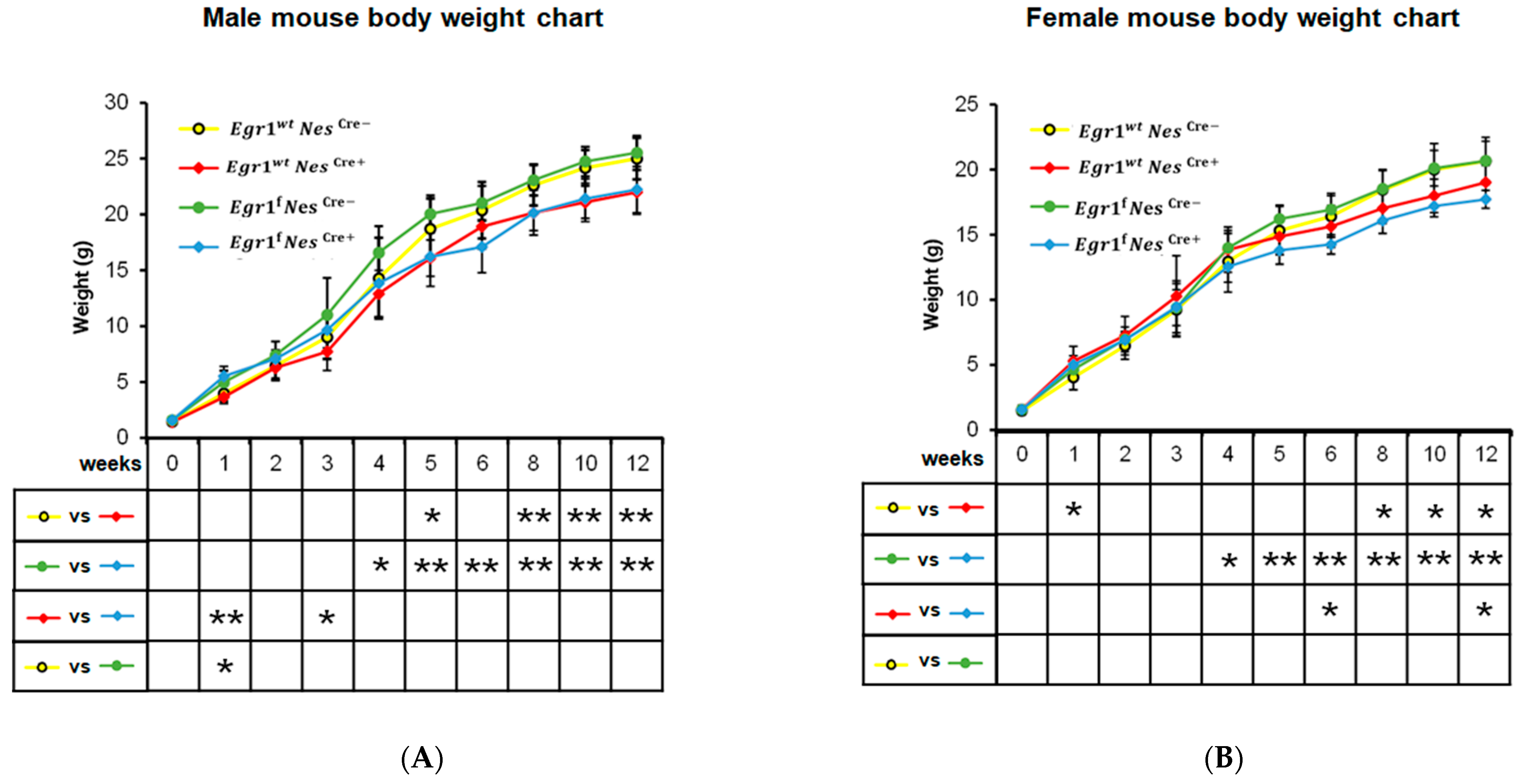
Despite the known impact of the Nestin-Cre driver and the loss of the Egr1 gene on body weight, no growth curve has been provided to demonstrate the progressive changes that occur in Egr1cKO and Nestin-Cre mice during postnatal development. Therefore, we monitored the eight groups of mice for twelve weeks after their birth (Figure 2). The difference the growth rate of the Egr1wtNescre+ and that of the Egr1wtNescre− mice became significant with the approach of young adulthood and was consistent after eight weeks. Compared with the female mice, the males showed more significant differences, with a p-value ≤ 0.01 from 8 to 12 weeks. Compared with the controls of the same sex, reductions in body weight of around 12% and 8% were observed in the male and female Egr1wtNescre+ mice, respectively. At the twelfth week, the average weight of a male Egr1wtNescre+ mouse was 22.0 ± 1.98 g, while the average weight of a male Egr1wtNescre− mouse was 25.0 ± 1.87 g. The average weight of a female Egr1wtNescre+ mouse was 19.0 ± 1.30 g, while the average weight of a female Egr1wtNescre− mouse was 20.7 ± 1.82 g.
A previous study indicated that the body weights of both male and female homozygous Egr1KO mice were significantly and consistently lower than those of normal controls of the same sex at ages older than two weeks [17]. In our study, the conditional Egr1 knockout delayed such differences between the sexes. Starting at four weeks of age, the Egr1fNescre+ and Egr1fNescre− mice exhibited reduced body weight steadily and with increasing significance. In the twelfth week, the average weight of a male Egr1fNescre+ mouse was 22.2 ± 2.07 g, and the average weight of a male Egr1fNescre− mouse was 25.5 ± 1.53 g, i.e., a greater than 12% body mass reduction in male Egr1cKO mice. Similarly, an approximately 14% reduction win body mass as observed for female Egr1cKO mice, which weighed on average 17.7 ± 0.68 g, while female Egr1fNescre− mice weighed 20.7 ± 1.50 g on average. In the early weeks, some significant differences in weight were sporadically observed. This is likely due to technical variations being enlarged by the lightweight litter. Interestingly, compared with female Egr1wtNescre+ mice, slightly decreased body weights were observed in the female Egr1fNescre+ mice, with p values ≤ 0.05 at six and twelve weeks. This result suggests that the loss of Egr1 might have a more noticeable impact on female mice than on male mice.

3.3. Sex Differences in Gene Expression Amplified in Nestin-Cre and Egr1cKO Mice

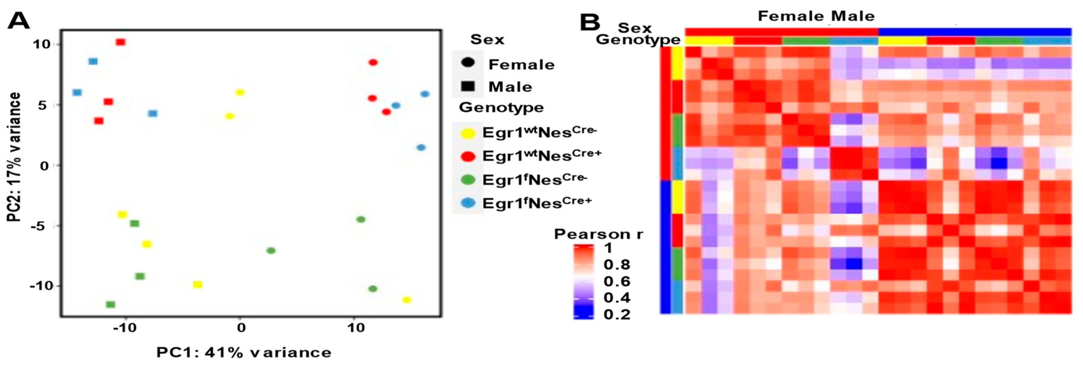
The growth differences observed in the Nestin-Cre and Egr1cKO mice inspired us to examine their pituitary gene expression profiles. RNA-seq analyses were performed in biological triplicates on adult pituitary tissues isolated from the eight groups of mice, with each sample taken at 12 weeks of age. A principal component analysis (PCA) indicated that, according to their gene expression patterns, the twenty-four samples could be segregated into two clusters, with females on the right side of the graph and males on the left (Figure 3A). Pearson correlation coefficients of the gene expressions across the samples further confirmed sex-specific clustering in general (Figure 3B). Intriguingly, strong correlations in the gene expressions in the pituitary tissues across all four genotypes were observed for males, but not for females. More specifically, despite a strong correlation among the other three biological replicates, the gene expression profiles from the female Egr1fNescre+ pituitary tissues showed a weak correlation (a Pearson’s r of around 0.2–0.4) with those of the Nescre− mice, but intermediate to strong correlations (Pearson’s rs of approximately 0.6–0.8) with those of both male and female Nescre+ mice. This result suggests that gene expression differences resulting from different genotypes (Nescre+ in particular) may prevail over sex differences.
We thus performed pairwise comparisons for the four genotypes separately to identify the genes associated with sex-specific pituitary expression. For the wild-type mice (Egr1wtNescre−), a total of 217 genes (99 downregulated and 118 upregulated) were observed in the females (Figure 4A, Supplementary Tables S1–S4). The number of differentially expressed genes (DEGs) involved in sex-related differences increased to 481 in the Egr1fNesCre− mice, 1925 in the Egr1wtNesCre+ mice, and 3822 in the Egr1fNesCre+ mice. A large number of the DEGs identified in the two sexes were genotype-specific (Figure 4B, Supplementary Tables S1–S4). Only 24 upregulated genes in females and 20 upregulated genes in males were found to be shared by all four genotypes. These shared genes are predominantly from the sex chromosomes and include Ddx3y and Kdm5d on the Y chromosome and Xist and Kdm6a on the X chromosome. For each pairwise comparison, we calculated the percentage of DEGs that were identified in autosomes and sex chromosomes (Figure 4C). Compared with the wild-type controls, the percentage of DEGs in autosomes increased by approximately 3.5 and 4.0 times for the female and male Egr1fNescre+ mice, respectively. For all four genotypes, the percentage of upregulated pituitary DEGs on the sex chromosomes in the males was approximately twice as high as that observed in the females. Altogether, the sex-related differences in pituitary gene expression were amplified in the Egr1f and Nescre+ mice and involved a thousand genes on autosomes.

3.4. Aberrant Gene Expression Associated with Nestin-Cre and Egr1cKO Mice

The substantial sex-related differences in pituitary gene expression observed in the mice of four different genotypes suggest that the Nestin-Cre driver may have a distinct influence on the development of male and female pituitary tissues. To better understand the effect of the Nestin-Cre driver, we focused on the Egr1wtNescre+ and Egr1wtNescre− mice. We still kept the male and female RNA-seq data separate to perform pairwise comparisons (Figure 5A, Supplementary Tables S5 and S6). In the females, 668 genes were downregulated, and the Nestin-Cre driver upregulated 259 genes. Fewer DEGs were found in the males (159 downregulated genes and 90 upregulated genes). Only 38 DEGs upregulated in Egr1wtNescre- and 4 DEGs upregulated in Egr1wtNescre+ pituitary glands were found to be shared by the male and female mice (Figure 5B). Significant differences in the enrichment of GO terms were observed between the female and male DEGs, and none of the GO terms were enriched for genes downregulated by the Nestin-Cre driver in the male pituitary, likely because only a small number of DEGs were detected (Figure 5C).
Five categories of GO terms in biological processes were found to be significantly enriched for DEGs associated with the Nestin-Cre driver. These GO terms are related to growth factors, ubiquitin protein ligase, protein kinase, cell adhesion molecules, and ion channel activity. Interestingly, the genes upregulated and downregulated by the Nestin-Cre driver in females were frequently found in similar GO categories. Although it remains unknown how these genes coordinate with each other in response to the Nestin-Cre driver, the identification of DEGs in the functional pathway related to growth factor may help explain the smaller stature of the Nestin-Cre mice. The expression patterns across 12 samples were examined for the 15 genes associated with the GO terms related to growth factors (Figure 5D). Except for interleukin 1 receptor type 1 (Il1r1), all of the genes were found to be Nestin-Cre-associated DEGs in females, but not in males. Compared with the wild-type controls, the expression of the Il1r1 gene decreased with a fold change of 0.22 in the males and 0.39 in the females. Interleukin 1 (IL-1) stimulates the expression of various kinds of growth factors, including fibroblast growth factor-2 [26] and nerve growth factor [27]. However, the mice without the Il1r1 gene exhibited no overt phenotype, but did exhibit altered innate immune and inflammatory responses [28].
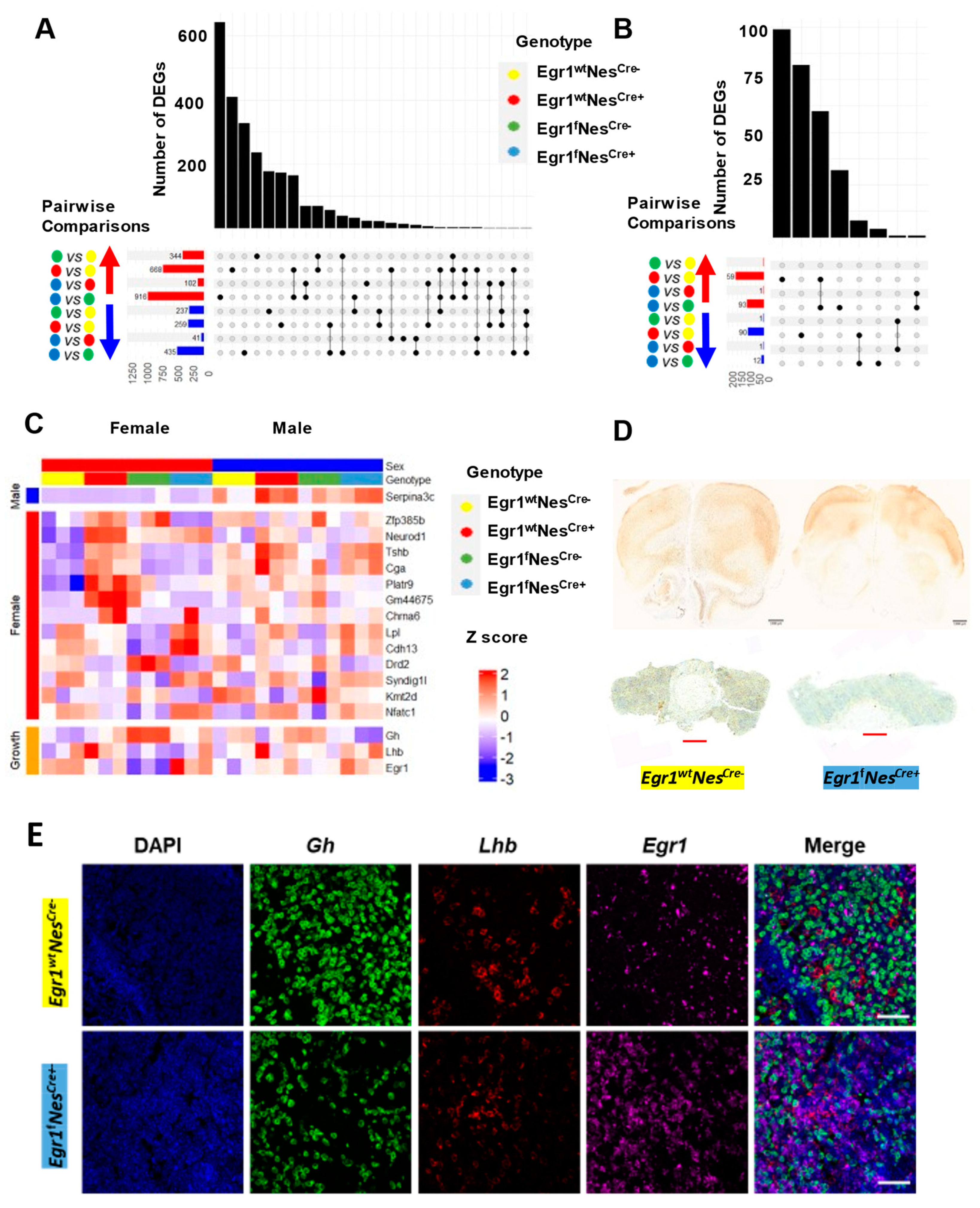
Since the Nestin-Cre driver led to a large number of genes being differentially expressed in the pituitaries of the two sexes, we performed four kinds of pairwise comparisons to examine the influence of Egr1cKO. In the females, more DEGs (916 upregulated and 435 downregulated) were identified between the Egr1fNescre+ and Egr1fNescre− mice than the number of DEGs (668 upregulated and 259 downregulated) identified between the Egr1wtNescre+ and Egr1wtNescre− mice (Figure 6A). However, in males, an opposite trend in the number of DEGs was observed; more DEGs were identified for the Nestin-Cre driver alone (Figure 6B). Since a pairwise comparison is not sufficient to scrutinize the influence of conditional knockout of the Egr1 gene, we adopted a mixed-gene model to analyze the pituitary gene expression profiles of the mice of all four genotypes (Supplementary Table S7). Only the Serpina3c gene was determined to be associated with Egr1 loss in the males, while eleven such genes were identified in the females (Figure 6C). We further examined the expression profiles of the Egr1, growth hormone, and LHβ genes, and they were not differentially expressed across genotypes. Altogether, our results indicate that the conditional knockout of the Egr1 gene has a more severe impact on pituitary gene expression in females than in males. In addition, in males, the influence of Egr1cKO on pituitary gene expression may be overridden by the Nestin-Cre driver.
To validate the presence of EGR1 proteins, immunohistochemistry using an anti-Egr1 antibody was performed on the brain sections and pituitary tissues obtained from the adult Egr1fNescre+ and Egr1wtNescre− mice (Figure 6D). EGR1 proteins were widely distributed across the cerebrum and highly expressed in the hippocampus in the Egr1wtNescre− mice, but they were substantially diminished in the brain sections derived from the Egr1fNescre+ mice. However, EGR1 proteins were detected in the pituitary tissues of both the Egr1wtNescre+ and Egr1fNescre− mice. This result indicates that the Nestin-Cre driver removes the expression of the Egr1 gene from neural lineage cells in the brain but fails to knock out the Egr1 gene in pituitary cells. Lastly, we further confirmed the expression of the Egr1, growth hormone, and LHβ genes in the pituitary tissues using RNAscope (Figure 6E). For each probe, individual images of the anterior pituitary were taken along with a DAPI control (Figure 6E). Consistent with IHC staining (Figure 6D), Egr1, LHβ, and GH mRNAs were detected in the pituitary glands of both the Egr1fNescre+ and Egr1wtNescre− mice. Such findings were observed consistently in both the IHC and RNAscope experiments using biological duplicates.

4. Discussion

Nestin-Cre transgenic mice have been widely used to study gene functions in the central nervous system. In this study, we found that the Nestin-Cre driver is able to successfully knock out Egr1 expression in a large number of brain cells, but that it fails to remove Egr1 from all three sections of the pituitary. Despite the high expression of Nestin previously found in the pituitary progenitor cells [25], a previous study did not find intensive expression of the Nestin-Cre transgene in the embryonic or adult pituitary [29]. Such inconsistency may be explained by the fact that different reporter mice were used in these studies, and by the unique characteristics of Nestin-Cre expression. During pituitary development, a high level of Nestin expression was observed in POU1F1 (POU domain class 1 transcription factor 1)-positive progenitor cells, but this was followed by a drastic reduction in the postnatal period [24]. The cells with Nestin expression in the postnatal period were terminally differentiated and located in the intermediate and posterior pituitary lobes. However, Nestin-Cre mediated recombination is extremely infrequent in early embryonic progenitor cells, but can reach nearly 100% in neural and glial progenitor cells during perinatal development [30]. In this study, our results concerning Egr1 mRNA and protein expression profiles have further confirmed the speculation that this specific strain of Nestin-Cre (B6.Cg-Tg (Nes-cre)1Kln/J; Jackson Lab, #003771) may not be suitable for pituitary gene knockout.
Despite this limitation, we made several interesting observations. Both Egr1 loss and the Nestin-Cre driver have been reported to be associated with growth abnormalities in transgenic mice [17,23]. In this study, for mice of four genotypes related to Egr1cKO driven by Nestin-Cre, we provided growth curves to monitor their postnatal changes in detail. The differences in growth rate resulting from the Nestin-Cre driver alone became significant as young adulthood approached and were consistent after eight weeks. Compared with the female groups, the Egr1wtNescre+ males with the Nestin-Cre driver showed more significant differences from the controls, but no additional weight loss was observed in the Egr1fNescre+ males with the combination of Nestin-Cre and Egr1 knockout in the neuronal lineage cells of the brain. However, Egr1 knockout led to slightly decreased body weight in the Egr1fNescre+ females compared with the Egr1wtNescre+ Females. This sex-related difference was further manifested in the gene expression profiles. Considering the significant role of the pituitary in the regulation of growth and development, we explored whether and how its expression profile may be altered in response to the loss of Egr1. To our surprise, the Egr1 gene was indeed removed from the neuronal lineage cells in the brain, but it was still expressed in the pituitary cells. This raises the possibility that the reductions in body size may have been due to the loss of Egr1 in the neuronal lineage cells. Regardless, we determined in this study that this loss of Egr1 expression resulted in substantial gene expression changes in the pituitary and affected the activities of growth factors.
In our study, a total of 217 genes were determined to be differentially expressed in the pituitary tissues of male and female wild-type controls. This difference increased approximately two-fold in the Egr1fNesCre− mice, five-fold in the Egr1wtNesCre+ mice, and nineteen-fold in the Egr1fNesCre+ mice. This result suggests that Egr1f and NesCre+ take on distinct functions, but that together they amplify their impact on the regulation of sex-related pituitary gene expression. The presence of the Nestin-Cre driver alone resulted in 927 differentially expressed genes in the female pituitary, but only 249 genes were affected in the male pituitary tissue. A GO term analysis showed that the pathway associated with growth factor was enriched in the list of differentially expressed genes. This may help explain the growth changes observed in the Nestin-Cre mice. Using a mixed-gene model, we identified a small set of differentially expressed genes associated with the loss of Egr1 in neuronal lineage cells, but the strong influence of the Nestin-Cre driver may undermine the impact of Egr1 on pituitary gene expression. Future studies using different Cre lines are highly desired to further understand the roles of Egr1 in pituitary functions.

5. Conclusions

Altogether, our results indicate that the Nestin-Cre driver was able to efficiently remove Egr1 genes from neuronal lineage cells in the brain, but not from pituitary cells. An RNA-seq analysis revealed that growth factor-related gene pathways were disrupted by the Nestin-Cre driver. In addition, sex differences in growth and gene expression were observed in the Egr1cKO and Nestin-Cre mice, and female mice were more sensitive to the presence of Nestin-Cre.

Supplementary Materials

The following supporting information can be downloaded at: https://www.mdpi.com/article/10.3390/biology12070966/s1, Supplementary Table S1. Full list of DEG sex differences, female vs. male (Egr1wtNescre−); Supplementary Table S2. Full list of DEG sex differences, female vs. male (Egr1fNescre−); Supplementary Table S3. Full list of DEG sex differences, female vs. male (Egr1wtNescre+); Supplementary Table S4. Full list of DEG sex differences, female vs. male (Egr1fNescre+); Supplementary Table S5. Full list of DEG differences between female Egr1fNescre+ and Egr1wtNescre+; Supplementary Table S6. Full list of DEG differences between male Egr1fNescre+ and Egr1wtNescre+; Supplementary Table S7. Full list of DEGs derived from mixed-model analysis.

Author Contributions

Conceptualization, C.S. and H.X.; resources, C.S., X.X., M.L., and K.Z.; data curation, Y.Z. and Y.L.; formal analysis, Y.Z., Y.L., C.S., and H.X; writing—original draft preparation, C.S., Y.L., X.X. and H.X.; writing—review and editing, C.S., Y.L., X.X. and H.X. All authors have read and agreed to the published version of the manuscript.

Funding

This work was supported by NIH grants T32OD028239, NS094574, MH120498, ES031521, and NSF1922428, the Center for One Health Research at the Virginia–Maryland College of Veterinary Medicine and the Edward Via College of Osteopathic Medicine, and the Fralin Life Sciences Institute faculty development fund for H X.

Institutional Review Board Statement

All procedures were performed in accordance with national and international guidelines and approved by the institutional board at Virginia Tech (Virginia Tech Institutional Animal Care and Use Committee 22-059 (BI)). This study was conducted in accordance with National Institute of Health guidelines.

Informed Consent Statement

Not applicable.

Data Availability Statement

The RNA-seq datasets generated in this study have been deposited in the NCBI Gene Expression Omnibus (GEO) under accession number GSE108768.

Conflicts of Interest

The authors declare no conflict of interest.

Abbreviations

GHGrowth hormone
LHLuteinizing hormone
PRLProlactin
TSHThyroid-stimulating hormone
FSHFollicle-stimulating hormone
Egr1Early growth response 1
sf1Steroidogenic factor 1
Pitx1Pituitary homeobox factor 1
EGR1Early growth response 1
GnRHGonadotropin-releasing hormone
PCAPrincipal component analysis
GOGene ontology

References

  1. Rizzoti, K. Genetic regulation of murine pituitary development. J. Mol. Endocrinol. 2015, 54, R55–R73. [Google Scholar] [CrossRef] [PubMed] [Green Version]
  2. Fletcher, P.A.; Smiljanic, K.; Prévide, R.M.; Iben, J.R.; Li, T.; Rokic, M.B.; Sherman, A.; Coon, S.L.; Stojilkovic, S.S. Cell Type- and Sex-Dependent Transcriptome Profiles of Rat Anterior Pituitary Cells. Front. Endocrinol. 2019, 10, 623. [Google Scholar] [CrossRef] [PubMed] [Green Version]
  3. Ho, Y.; Hu, P.; Peel, M.T.; Chen, S.; Camara, P.G.; Epstein, D.J.; Wu, H.; Liebhaber, S.A. Single-cell transcriptomic analysis of adult mouse pituitary reveals sexual dimorphism and physiologic demand-induced cellular plasticity. Protein Cell. 2020, 11, 565–583. [Google Scholar] [CrossRef] [PubMed] [Green Version]
  4. Wolfe, M.W.; Call, G.B. Early growth response protein 1 binds to the luteinizing hormone-beta promoter and mediates gonadotropin-releasing hormone-stimulated gene expression. Mol. Endocrinol. 1999, 13, 752–763. [Google Scholar] [PubMed] [Green Version]
  5. Quirk, C.C.; Lozada, K.L.; Keri, R.A.; Nilson, J.H. A single Pitx1 binding site is essential for activity of the LHbeta promoter in transgenic mice. Mol. Endocrinol. 2001, 15, 734–746. [Google Scholar]
  6. Jorgensen, J.S.; Quirk, C.C.; Nilson, J.H. Multiple and overlapping combinatorial codes orchestrate hormonal responsiveness and dictate cell-specific expression of the genes encoding luteinizing hormone. Endocr. Rev. 2004, 25, 521–542. [Google Scholar] [CrossRef] [PubMed] [Green Version]
  7. Kaiser, U.B.; Halvorson, L.M.; Chen, M.T. Sp1, steroidogenic factor 1 (SF-1), and early growth response protein 1 (egr-1) binding sites form a tripartite gonadotropin-releasing hormone response element in the rat luteinizing hormone-beta gene promoter: An integral role for SF-1. Mol. Endocrinol. 2000, 14, 1235–1245. [Google Scholar]
  8. Tremblay, J.J.; Drouin, J. Egr-1 is a downstream effector of GnRH and synergizes by direct interaction with Ptx1 and SF-1 to enhance luteinizing hormone beta gene transcription. Mol. Cell. Biol. 1999, 19, 2567–2576. [Google Scholar] [CrossRef] [Green Version]
  9. Hashimoto, H.; Olanrewaju, Y.O.; Zheng, Y.; Wilson, G.G.; Zhang, X.; Cheng, X. Wilms tumor protein recognizes 5-carboxylcytosine within a specific DNA sequence. Genes. Dev. 2014, 28, 2304–2313. [Google Scholar] [CrossRef] [Green Version]
  10. Veyrac, A.; Besnard, A.; Caboche, J.; Davis, S.; Laroche, S. The transcription factor Zif268/Egr1, brain plasticity, and memory. Progress. Mol. Biol. Transl. Sci. 2014, 122, 89–129. [Google Scholar]
  11. Li, L.; Carter, J.; Gao, X.; Whitehead, J.; Tourtellotte, W.G. The neuroplasticity-associated arc gene is a direct transcriptional target of early growth response (Egr) transcription factors. Mol. Cell. Biol. 2005, 25, 10286–10300. [Google Scholar] [CrossRef] [Green Version]
  12. Gashler, A.; Sukhatme, V.P. Early growth response protein 1 (Egr-1): Prototype of a zinc-finger family of transcription factors. Prog. Nucleic Acid. Res. Mol. Biol. 1995, 50, 191–224. [Google Scholar] [PubMed]
  13. Sun, Z.; Xu, X.; He, J.; Murray, A.; Sun, M.-A.; Wei, X.; Wang, X.; McCoig, E.; Xie, E.; Jiang, X.; et al. EGR1 recruits TET1 to shape the brain methylome during development and upon neuronal activity. Nat. Commun. 2019, 10, 3892. [Google Scholar] [CrossRef] [PubMed] [Green Version]
  14. Haisenleder, D.J.; Dalkin, A.C.; Ortolano, G.A.; Marshall, J.C.; Shupnik, M.A. A pulsatile gonadotropin-releasing hormone stimulus is required to increase transcription of the gonadotropin subunit genes: Evidence for differential regulation of transcription by pulse frequency in vivo. Endocrinology 1991, 128, 509–517. [Google Scholar] [CrossRef] [PubMed]
  15. Ruf-Zamojski, F.; Ge, Y.; Nair, V.; Zamojski, M.; Pincas, H.; Toufaily, C.; Tome-Garcia, J.; Stoeckius, M.; Stephenson, W.; Smith, G.R.; et al. Single-cell stabilization method identifies gonadotrope transcriptional dynamics and pituitary cell type heterogeneity. Nucleic Acids Res. 2018, 46, 11370–11380. [Google Scholar] [CrossRef]
  16. Lee, S.L.; Sadovsky, Y.; Swirnoff, A.H.; Polish, J.A.; Goda, P.; Gavrilina, G.; Milbrandt, J. Luteinizing hormone deficiency and female infertility in mice lacking the transcription factor NGFI-A (Egr-1). Science 1996, 273, 1219–1221. [Google Scholar] [CrossRef]
  17. Topilko, P.; Schneider-Maunoury, S.; Levi, G.; Trembleau, A.; Gourdji, D.; Driancourt, M.A.; Rao, C.V.; Charnay, P. Multiple pituitary and ovarian defects in Krox-24 (NGFI-A, Egr-1)-targeted mice. Mol. Endocrinol. 1998, 12, 107–122. [Google Scholar] [CrossRef]
  18. Hou, H.; Chan, C.; Yuki, K.E.; Sokolowski, D.; Roy, A.; Qu, R.; Uusküla-Reimand, L.; Faykoo-Martinez, M.; Hudson, M.; Corre, C.; et al. Postnatal developmental trajectory of sex-biased gene expression in the mouse pituitary gland. Biol. Sex. Differ. 2022, 13, 57. [Google Scholar] [CrossRef]
  19. Knight, C.; Slade, J.P.; Carter, D. The nuclear, 75 kDa form of early growth response protein-1/nerve growth factor-induced A protein is primarily restricted to LH beta-subunit-expressing cells in rat anterior pituitary. Eur. J. Endocrinol. 2000, 143, 817–821. [Google Scholar] [CrossRef] [Green Version]
  20. Man, P.S.; Wells, T.; Carter, D.A. Egr-1-d2EGFP transgenic rats identify transient populations of neurons and glial cells during postnatal brain development. Gene Expr. Patterns 2007, 7, 872–883. [Google Scholar] [CrossRef]
  21. Man, P.S.; Wells, T.; Carter, D.A. Cellular distribution of Egr1 transcription in the male rat pituitary gland. J. Mol. Endocrinol. 2014, 53, 271–280. [Google Scholar] [CrossRef] [PubMed] [Green Version]
  22. Bernal, A.; Arranz, L. Nestin-expressing progenitor cells: Function, identity and therapeutic implications. Cell. Mol. Life Sci. 2018, 75, 2177–2195. [Google Scholar] [CrossRef] [Green Version]
  23. Tronche, F.; Kellendonk, C.; Kretz, O.; Gass, P.; Anlag, K.; Orban, P.C.; Bock, R.; Klein, R.; Schütz, G. Disruption of the glucocorticoid receptor gene in the nervous system results in reduced anxiety. Nat. Genet. 1999, 23, 99–103. [Google Scholar] [CrossRef] [PubMed]
  24. Yoshida, S.; Kato, T.; Higuchi, M.; Yako, H.; Chen, M.; Kanno, N.; Ueharu, H.; Kato, Y. Rapid transition of NESTIN-expressing dividing cells from PROP1-positive to PIT1-positive advances prenatal pituitary development. J. Neuroendocrinol. 2013, 25, 779–791. [Google Scholar] [CrossRef] [PubMed]
  25. Gleiberman, A.S.; Michurina, T.; Encinas, J.M.; Roig, J.L.; Krasnov, P.; Balordi, F.; Fishell, G.; Rosenfeld, M.G.; Enikolopov, G. Genetic approaches identify adult pituitary stem cells. Proc. Natl. Acad. Sci. USA 2008, 105, 6332–6337. [Google Scholar] [CrossRef] [PubMed]
  26. Araujo, D.M.; Cotman, C.W. Basic FGF in astroglial, microglial, and neuronal cultures: Characterization of binding sites and modulation of release by lymphokines and trophic factors. J. Neurosci. 1992, 12, 1668–1678. [Google Scholar] [CrossRef] [Green Version]
  27. Friedman, W.J.; Thakur, S.; Seidman, L.; Rabson, A.B. Regulation of nerve growth factor mRNA by interleukin-1 in rat hippocampal astrocytes is mediated by NFkappaB. J. Biol. Chem. 1996, 271, 31115–31120. [Google Scholar] [CrossRef] [Green Version]
  28. Glaccum, M.B.; Stocking, K.L.; Charrier, K.; Smith, J.L.; Willis, C.R.; Maliszewski, C.; Livingston, D.J.; Peschon, J.J.; Morrissey, P.J. Phenotypic and functional characterization of mice that lack the type I receptor for IL-1. J. Immunol. 1997, 159, 3364–3371. [Google Scholar] [CrossRef]
  29. Galichet, C.; Lovell-Badge, R.; Rizzoti, K. Nestin-Cre mice are affected by hypopituitarism, which is not due to significant activity of the transgene in the pituitary gland. PLoS ONE 2010, 5, e11443. [Google Scholar] [CrossRef]
  30. Liang, H.; Hippenmeyer, S.; Ghashghaei, H.T. A Nestin-cre transgenic mouse is insufficient for recombination in early embryonic neural progenitors. Biol. Open 2012, 1, 1200–1203. [Google Scholar] [CrossRef] [Green Version]
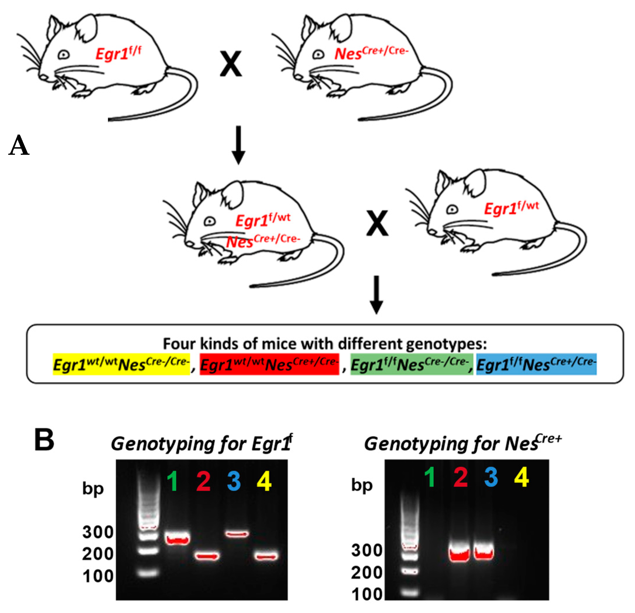
Figure 1. Generation of Egr1cKO mice. (A) Breeding scheme to obtain Egr1cKO mice. Four kinds of mice with different genotypes were included in this study, with Egr1f/f NesCre+ being the desired knockout genotype and the remaining genotypes being used as controls. The desired knockout genotype and the controls are all color coded throughout the paper. Egr1f represents a floxed Egr1 allele and NesCre+ represents the presence of Nestin-Cre in the loxp system, while NesCre− represents its absence. (B) Genotyping PCR to validate the presence of Nestin-Cre or Egr1 in the loxp systems of the mice. Egr1f is 291 bp while wild-type is 165 bp; NesCre+ is 350 bp while NesCre− has no band.
Biology 12 00966 g001
Figure 2. Growth curves for (A) male and (B) female mice of four genotypes from birth to 12 weeks. The data for each time point were derived from 10 individuals and are presented as means ± SEM. A group comparison was performed using a Student’s t-test. * denotes p-value < 0.05 and ** denotes p-value < 0.01.
Biology 12 00966 g002
Figure 3. RNA-seq data clustering. (A) Principal component analysis (PCA) and (B) Pearson correlations of pituitary gland transcriptome data. RNA-seq analyses were performed in triplicates for each group, and all samples were from mice 12 weeks of age.
Biology 12 00966 g003
Figure 4. Gene expression changes associated with sex difference. (A) Volcano plot for pairwise analysis between two sexes for four genotypes. DEGs were defined as genes with FCs ≥ 1.5 and adjusted p values ≤ 0.05. Blue dots indicate downregulated DEGs in males. Red dots indicate upregulated DEGs in males. Grey dots indicate no DEGs. (B) Venn diagram showing the overlapped DEGs identified in the pairwise comparisons. Corresponding genotypes are color-coded (blue for Egr1fNescre+, green for Egr1fNescre−, red for Egr1wtNescre+, and yellow for Egr1wtNescre−). (C) Chromosome distribution of DEGs.
Biology 12 00966 g004
Figure 5. Gene expression changes associated with the Nestin-Cre deriver. (A) Volcano plots for pairwise comparisons between female (left) and male (right) Egr1wtNescre+ and Egr1wtNescre− mice. (B) Venn diagram showing the overlapped DEGs identified in the pairwise comparisons for the two sexes. (C) GO analysis of identified DEGs. (D) Gene expression profiles of 15 DEGs were identified in the 3 GO categories associated with growth factors.
Biology 12 00966 g005
Figure 6. Gene expression changes associated with Egr1cKO. Barplots showing the number of intersecting DEGs identified in pairwise comparisons between (A) females and (B) males. Horizontal bars on the bottom left side of each plot show the numbers of DEGs determined to be upregulated (red arrow) or downregulated (blue arrow). Different intersection combinations of DEGs identified for each pairwise comparison are represented by the dotplot. The vertical barplots show the number of intersecting DEGs in the indicated combinations of pairwise comparisons. (C) Gene expression profiles of Egr1, growth hormone, and LHβ together with DEGs determined to be associated with Egr1cKO (1 DEG in males and 11 DEGs in females). (D) Immunohistochemistry staining of mouse brain in coronal view (scale bar = 2 mm) and pituitary tissues of Egr1cKO mouse and wild-type control. (scale bar = 400 μm). (E) RNAscope results for Egr1, growth hormone, and LHβ expression in the pituitary tissues of Egr1cKO and wild-type mice (scale bar = 50 μm). The experiments involving IHC and RNAscope were replicated using biological duplicates.
Biology 12 00966 g006
Disclaimer/Publisher’s Note: The statements, opinions and data contained in all publications are solely those of the individual author(s) and contributor(s) and not of MDPI and/or the editor(s). MDPI and/or the editor(s) disclaim responsibility for any injury to people or property resulting from any ideas, methods, instructions or products referred to in the content.