Article

Circular RNAs Could Encode Unique Proteins and Affect Cancer Pathways

1
Department of Translational Medicine, University of Ferrara, 44121 Ferrara, Italy
2
Genetics Unit, Institute for Maternal and Child Health, Scientific Institute for Research, Hospitalization and Healthcare (IRCCS) Burlo Garofolo, 34137 Trieste, Italy
3
Laboratory for Advanced Therapy Technologies (LTTA), University of Ferrara, 44121 Ferrara, Italy
4
Department of Oncology, Azienda Ospedaliero-Universitaria St. Anna di Ferrara, 44124 Ferrara, Italy
5
Laura and Isaac Perlmutter Cancer Center, New York University School of Medicine, NYU Langone Health, New York, NY 10016, USA
6
Division of Hematology and Hematological Malignancies, University of Utah, Salt Lake City, UT 84112, USA
7
Warsaw Genomics INC, 01-682 Warszawa, Poland
8
Human Cancer Genetics, Biological and Chemical Research Centre, University of Warsaw, 02-089 Warsaw, Poland
9
Genomics Core Facility, Centre of New Technologies, University of Warsaw, 02-097 Warsaw, Poland
10
CNBCh, Biological and Chemical Research Centre, University of Warsaw, 02-089 Warsaw, Poland
*
Author to whom correspondence should be addressed.
Biology 2023, 12(4), 493; https://doi.org/10.3390/biology12040493
Submission received: 7 February 2023 / Revised: 10 March 2023 / Accepted: 21 March 2023 / Published: 24 March 2023
(This article belongs to the Section Bioinformatics)

Abstract

:

Simple Summary

In this study, we investigated human genes encoding circular RNAs (circRNAs) in order to shed light on their functional role, which is still under debate. We identified 183 genes encoding circRNAs differentially expressed in cancer tissues with a novel coding potential. Our data suggest that circRNAs might directly affect cellular and systemic processes in cancer by generating novel members of the human proteome.

Abstract

circRNAs constitute a novel class of RNA, generally considered as non-coding RNAs; nonetheless, their coding potential has been under scrutiny. In this work, we systematically explored the predicted proteins of more than 160,000 circRNAs detected by exome capture RNA-sequencing and collected in the MiOncoCirc pan-cancer compendium, including normal and cancer samples from different types of tissues. For the functional evaluation, we compared their primary structure and domain composition with those derived from the same linear mRNAs. Among the 4362 circRNAs potentially encoding proteins with a unique primary structure and 1179 encoding proteins with a novel domain composition, 183 were differentially expressed in cancer. In particular, eight were associated with prognosis in acute myeloid leukemia. The functional classification of the dysregulated circRNA-encoded polypeptides showed an enrichment in the heme and cancer signaling, DNA-binding, and phosphorylation processes, and disclosed the roles of some circRNA-based effectors in cancer.

1. Introduction

CircRNAs are covalently closed molecules with a tissue- and cell-specific expression, whose biogenesis is specifically regulated [1]. They may exert biological functions by acting as microRNAs (“sponges”) and as protein inhibitors (“decoys”), but they could also be translated into proteins. Recent advances in RNA-sequencing (RNA-seq) and circRNA-specific decoding tools allowed for their quantification and characterization, leading to the identification of tens of thousands of potential circRNAs transcribed from the human genome [2,3] and modulated in breast cancer and other tumors, such as in leukemias [4,5]. In this context, we recently performed the first large-scale study of circRNAs considering over 400 total RNA-seq samples from patients with acute myeloid leukemia (AML) [6]. Nevertheless, the full functional role of circRNAs in cancer is still under debate [7], and several studies have asserted that circRNAs can act as templates for translation. Indeed, Abe et al. demonstrated that a pool of circRNAs comprised boundless Open Reading Frames (ORFs) that could be translated into a protein concatemer by a mechanism called “rolling circle amplification” [8]. Furthermore, Chen et al. confirmed the cytoplasmic localization of circRNAs in eukaryotic cells [9]. Different research groups have explained two cap-independent mechanisms of circRNAs translation: the internal ribosome entry sites (IRES) and the N6-methyladenosines (m6A)-mediated translation [10,11]. Currently, an increasing number of studies have taken into account the coding potential of circRNAs, as well as the role of the peptides encoded by both circRNAs and long non-coding RNAs, as it occurs in glioblastoma [12,13,14], liver cancer [15], and neurodegenerative diseases [16]. Here, we investigated the coding potential of circRNAs sourced by MiOncoCirc, a pan-cancer compendium of more than 160,000 cancerous circRNAs detected through a poly(A)-independent method and gene-body targeting, the exome capture RNA-seq [5,17], focusing on a restricted set of circRNAs with a high potential to generate novel isoforms.

2. Materials and Methods

2.1. Cancer circRNA Selection

The MiOncoCirc dataset (https://mioncocirc.github.io/download/, accessed on 15 June 2022) includes RNA-Seq data from a large number (n = 2036) of cancer samples, derived from several types of tissue (prostate, breast, lung, pancreatic, liver, etc.) [18,19,20] (Table S1). After excluding the read-through circRNAs located within intergenic sequences, we selected circRNAs expressed in at least 40 different samples. As alternative splicing events frequently occur within each circRNA, we considered only those at least 150 nucleotides long, characterized using their genomic coordinates in GENCODE (v.33) to determine all of the different spliced isoforms (n = 56,819). Using TransDecoder (v.5.5.0), we predicted the circular ORFs (circORFs) encoding for polypeptides at least 50 amino acids (aa) long, starting with a methionine and ending with a stop codon for each circRNA transcripts.

2.2. In Silico Characterization of Polypeptides Predicted from circRNAs

Using protein BLAST version 2.9.0 (https://rcc.uchicago.edu/docs/software/modules/blast/midway2/2.9.0.html, accessed on 15 June 2022) we determined the correspondence between the polypeptides encoded by the longest circORF and the cognate linear isoforms obtained from Ensembl 101 (https://www.ensembl.info/known-bugs/ensembl-101/, accessed on 15 June 2022), using a threshold E-value of 1.0 × 10⁻¹⁰. Therefore, we underline that no circRNAs had multiple putative ORFs. To restrict the cognate field, we aligned only proteins from the Consensus Coding Sequence (CCDS) database (https://www.ncbi.nlm.nih.gov/, accessed on 15 June 2022). We developed a Python script to isolate all circRNAs encoding proteins with a mismatch of at least one amino acid at the N- and/or C-terminus compared with their respective CCDS isoforms; these circORF encoded proteins were thus annotated accordingly: “canonical” or “internal” for the starting methionine, and/or with “premature” or “canonical” C-terminus. Lastly, the new peptides at the carboxy- and ammino-terminus were annotated as C-term and N-term, respectively.

2.3. The Domain Structure of circRNA Encoded Proteins

The domain composition of the circRNA proteins was globally investigated using HMMER HmmScan (https://www.ebi.ac.uk/Tools/pfa/hmmer_hmmscan/, accessed on 15 June 2022). We compared the domains of each circRNA protein with those of their cognate linear isoforms (GENCODE v. 33 (https://www.gencodegenes.org/human/release_33.html, accessed on 15 June 2022). We developed a Python script to identify the predicted circular proteins with domain mismatches in comparison with the linear isoforms, considering the following: (i) different order of the domains, (ii) partial overlap (missing one or more domains when compared to the parental isoforms), (iii) partial overlap with one or more additional domains, (iv) same parental domain structure with additional domains, or (v) no overlap with the domain structure of all cognate linear isoforms (Figure 1).

2.4. Expression Profile of circRNAs with Unique Protein-Coding Potential in Cancer

To investigate the cancer expression of circRNAs predicted with uniquely encoded polypeptides, we used the MiOncoCirc RNA-Seq data collection of clinical samples [5], cell lines, and normal tissues (n = 2036) [18,19,20], annotated in Table S2. Data were expressed as log2 reads per million (RPM) and normalized by quantile. A large number of samples showed overall very low levels of circRNAs in their transcriptome; we removed these samples from the following analyses, as they represented outliers, possibly related to intrinsic molecular characteristics or even to RNA processing. Thus, only samples with a high expression of circRNAs (n = 1018) were studied further, using the median of total log2 circRNA RPM counts as a threshold.

2.5. Functional Characterization of the circRNAs Encoding Proteins

The genes harboring circRNAs dysregulated in cancer and potentially translated were analyzed using PantherDB (http://pantherdb.org/, accessed on 15 June 2022). Correction for multiple testing was performed using the false discovery rate (FDR), with a threshold level of 0.05.

2.6. Mass Spectrometry Identification of Novel Peptides Derived from Coding circRNAs

To identify peptides corresponding to the novel N- or C-terminus predicted from circORFs, we used PeptideAtlas (http://www.peptideatlas.org/, build Human 2021-01, accessed on 1 August 2019), a compendium of peptides collected by tandem mass spectrometry experiments from humans and other organisms [21,22]. FASTA36 (version 36.3.8 h, accessed on 1 August 2019) was used for identifying circORF peptides in the PeptideAtlas database. Additionally, BLASTP 2.13.0+, and RefSeq were used to re-evaluate any peptides identified from Peptide Atlas.

2.7. Prediction of Internal Ribosome Entry Sites (IRES) in circRNAs with Coding Potential

IRESpy (https://irespy.shinyapps.io/IRESpy/, accessed on 20 October 2022), a high-throughput IRES tool based on the XGBoost model, was used to predict IRES in circRNAs with a novel primary structure and/or novel domain composition. Table S3 shows the circRNAs with a predicted probability (prob_IRES) higher than 0.3.

2.8. Identification of m6A Sites in Coding-circRNA Sequences

To investigate the overlapping regions, we intersected the chromosomal coordinates of the circRNAs differentially expressed in the Pan-cancer dataset (Table S12) with N6-adenosine methylation (m6A) peak information sourced by REPIC RNA Epitranscriptome Collection (https://repicmod.uchicago.edu/repic/download.php, dataset name: all human m6A peak information, Assembly:hg38, accessed on 27 February 2023).

3. Results

3.1. Cancer circRNAs Potentially Encode Novel Proteins

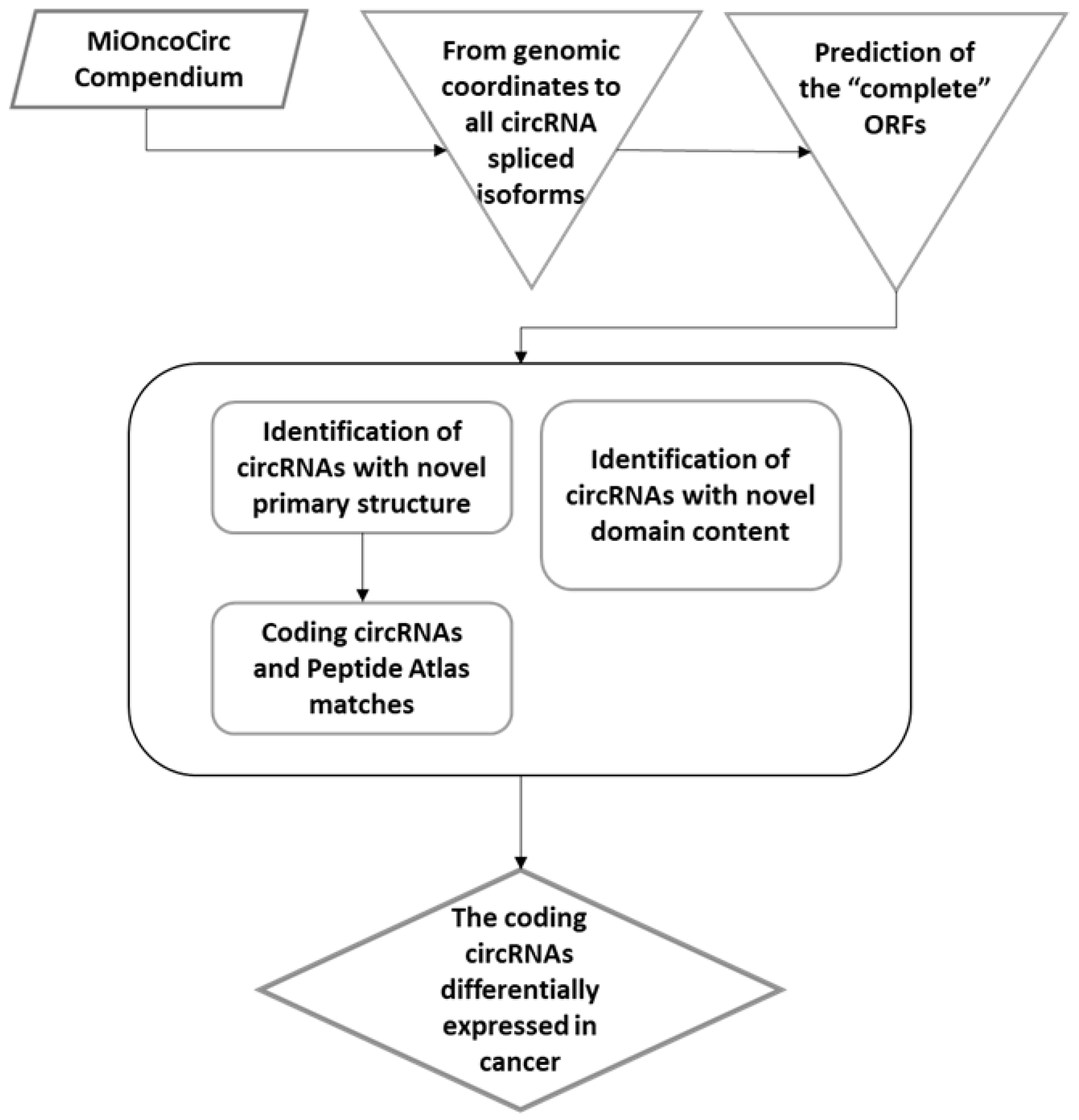
This study aimed to understand whether circRNAs expressed in cancer have a coding potential. We looked for the most relevant alterations compared with the canonical linear (cognate) isoforms. We hypothesized that circRNAs of interest in cancer should encode unique and novel peptide/protein, structurally and functionally different from the canonical proteins, encoded by the same loci. As function-related changes, we considered those including full or partially novel polypeptides (primary structure), as well as modifications of the canonical domain structure. The bioinformatics procedure, leading to the identification of such circRNAs, is illustrated in Figure 2.
We predicted the proteins encoded from circRNAs and considered those at least 50 aa residues long. Then, we looked for novel sequences among these circORFs-encoded proteins: for each circRNA, we focused on the longest circORF proteins (n = 4361) bearing a partial, but not complete, overlap (E-value lower than 1.0 × 10⁻¹⁰) with canonical cognate counterparts (from CCDS). Because of their conformation, the ORFs with partial overlap with the cognate CCDS sequences were bearing novel sequences either at the N-terminus, C-terminus, or at both. Most of these novel protein-coding circRNAs started at the canonical Methionine (canonicalMet), (n = 3363); a smaller number started at an internal Met (n = 824) (compared with the parental protein), while the remaining (n = 174) started at a novel 5′ Met, thus encoding for a novel N-terminus (N-term). As far as the C-terminus, most of the predicted circORFs encoded for novel sequences (n = 3865), while a few were terminated prematurely (prematureTerm) leading to a truncated protein (n = 223), or had the canonical stop codon (canonicalSTOP) (n = 273). Overall, the largest portion of circORFs had a canonical Met start and novel primary structure at the C-terminus (C-term). Only a small percentage possessed both novel N- and C-terminus (n = 115) (Figure S1).
The distribution of the length for the predicted novel peptide extensions, individually, at the N- and at C-terminus, alongside the descriptive statistics, are plotted in Figure S2. The average length of the novel peptides was slightly higher for N-terminus (mean = 24.1 aa) than the C-terminus (mean = 17 aa).
In addition, we looked for specific differences in the domain structure of circRNA-encodable proteins. Most of them shared the exact domain structure with their linear isoforms. The most frequent structural alteration was the loss of domains compared with the cognate linear mRNAs; indeed, 931 circORF proteins lacked one or more domains, whereas 120 showed a different domain order, 50 included an extra domain, 58 presented concurrent domain loss and the inclusion of extra domains, and 20 circORF proteins had a completely novel domain structure (Figure S3 and Tables S4–S9).
In a different approach, other investigators considered IRES in the circRNA sequence as necessary for efficient circRNA translation [23,24]. Accordingly, we used IRESPy [25] to predict IRES probability in the circRNAs with a coding potential and/or a novel domain composition. Among the 4664 circRNAs considered, 59 had an IRES probability higher than 0.3 (Table S3). The translation of circRNAs is a cap-independent event so it can rely on IRES or N6-methyladenosine (m6A) RNA modifications. Besides considering the IRES elements, we checked if any of the coding circRNAs overlapped with the m6A sites. Over the 183 total circRNAs differentially expressed, we identified 156 of them as overlapping with known m6A sites. As described by Wen S.Y. et al., m6A sites were enriched in circRNAs and functioned as IRES-like elements, so just a single m6A modification could drive the translation of these coding circRNAs [26].
To experimentally validate our results, we looked for the presence of the novel circRNA-specific N- and C-terminus, separately considered, among the peptides from a human organism sequenced by tandem mass spectrometry from the PeptideAtlas database. Among the total of 82 peptides, 70 were novel carboxy peptides and 12 were novel amino peptides, perfectly overlapped with sequences reported in the PeptideAtlas database (Table S10). Regarding the tissue distribution of the circRNA-peptide matching in PeptideAtlas, we also reported their localization and the number of observations in Table S10.
We highlighted five matched peptides also overlapping with the ORF of differentially expressed circRNAs encoded by SUCO, TBC1D31, PTBP3, GANAB, and RNF13 genes.

3.2. Expressed Coding circRNAs with Novel Coding Properties Are Involved in Cancer Pathways

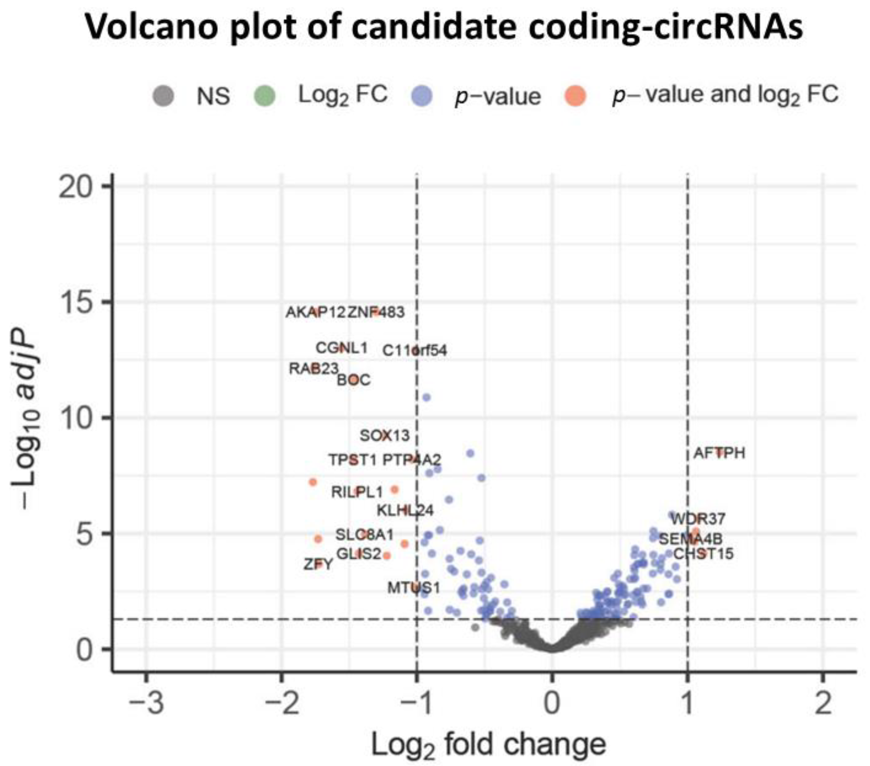
In this further step, we specifically investigated the expression profiles of coding circRNAs, with either novel amino-/carboxy-terminus or domain structures, in 1018 human samples from the MiOncoCirc compendium. Finally, 629 coding circRNAs had a highly variable expression across the remaining samples (IQR > 0.5) (Table S11). To investigate their possible cancer roles, we performed differential expression analysis identifying 183 coding circRNAs deregulated in cancer versus the control tissues. The Benjamini–Hochberg adjustment was applied for multiple testing (Figure S4 and Table S12) and the deregulated coding circRNAs were visualized using a volcano plot (Figure 3).
Among these, AKAP12 and ZNF483 emerged as the most differentially down-regulated coding circRNAs whereas AFTPH, CHST15 and WDR37 were differentially up-regulated in the pan-cancer RNA-Seq dataset.
Most of these coding circRNAs (n = 121) harbored N- and/or C-terminal novel sequence, 9 displayed only novel domain content, while 28 had both types of structural changes. Furthermore, 12 circRNAs started from an internal methionine (shorter N-terminal) and/or had premature termination, leading to truncated proteins without additional extra sequences. In Figure S5, we provide two alignment examples of the novel peptides at the C-term of FLI1 and HIPK3 coding circRNAs with the RefSeq protein database. Here, we highlight, in red, the novel sequence of circRNAs which overlap with the proteome of other organisms different from homo sapiens. We underline that the peptides overlapping with predicted new ORFs do not align anywhere else in the genome because they are specific. circRNAs potentially coding novel polypeptides could, in principle, bear novel functional roles. For example, a circRNA protein missing a domain could act as a dominant negative, or display altered cellular localization. Therefore, we studied the gene ontology (GO) and the molecular features of these 183 circORFs deregulated in cancer. We interrogated PantherDB [27] to perform an over-representation analysis of the circORF genes. The results showed a significant (BH corrected p value < 0.05) over-representation for biological processes, molecular functions, cellular components, and reactome pathways (Table 1 and Table S13).
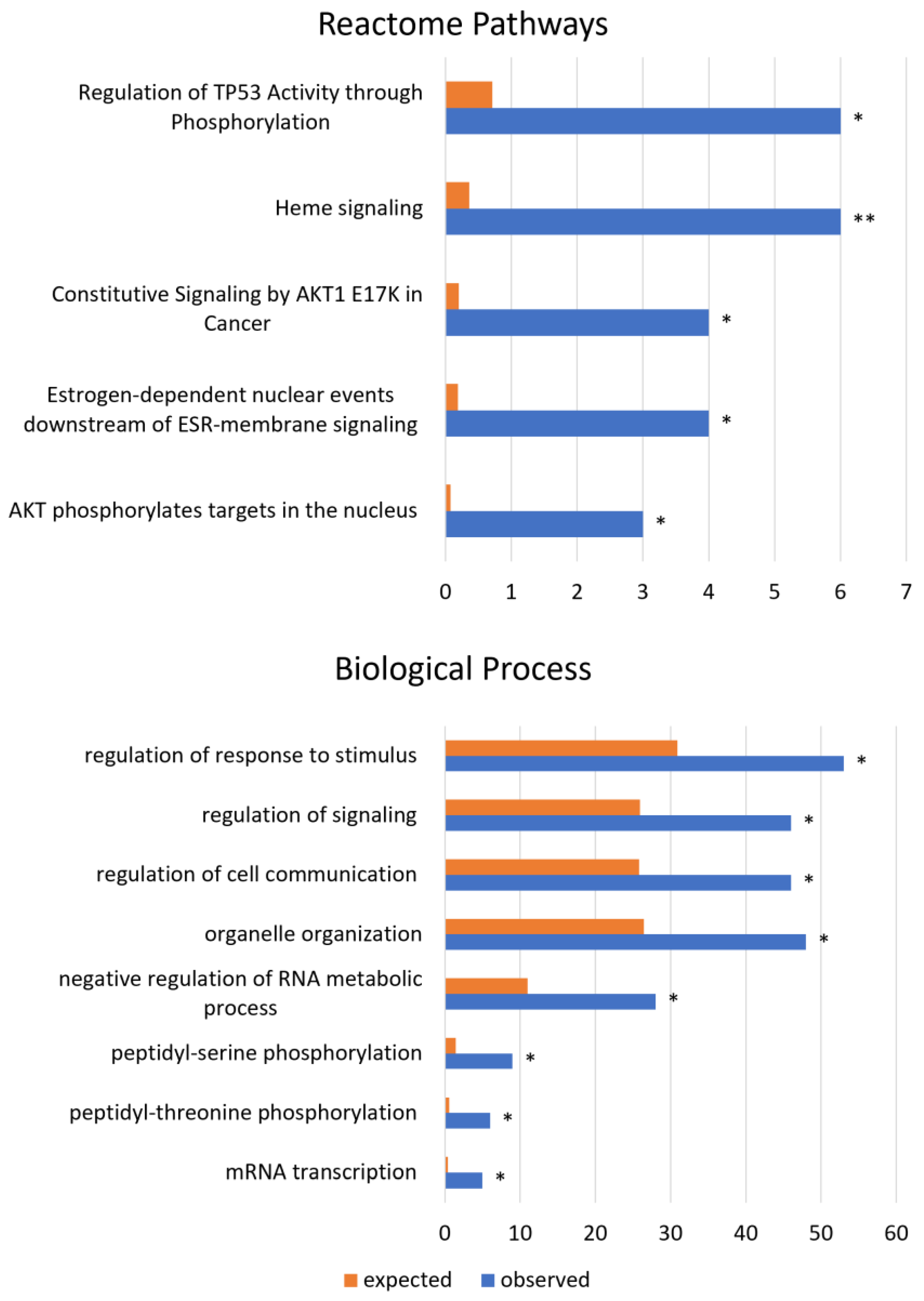
Among the reactome pathways associated with circORFs deregulated in cancer, we listed the regulation of TP53 activity through phosphorylation, heme signaling, and constitutive signaling by the AKT1 E17K mutation. The most significant biological processes overrepresented by coding circRNAs were the regulation of response to stimulus, signal transduction, cell communication, organelle organization, and peptidyl phosphorylation, which are illustrated in Figure 4.
Furthermore, we found molecular functions such as protein kinase activity; DNA binding; and cellular components, such as nuclear speck and rough endoplasmic reticulum, among those mapped by the circRNAs to have a coding potential and be differentially expressed in human cancer.

3.3. The Coding circRNAs Differentially Expressed in Cancer Are Also Involved in AML

In a previous study [6], we profiled the total RNA transcriptome of 345 patients affected by cytogenetically normal AML (CN-AML), identifying a set of circRNAs with prognostic value. We hypothesized that some AML prognostic circRNAs could also be among the cancer coding circRNAs we evidenced above. Interestingly, 24 circRNAs associated with prognosis in CN-AML (Table 2) were indeed present in the pan-cancer coding circRNAs profile, with 8 of them being differentially expressed in cancer (adjusted p value < 0.05). We performed a two tailed Fisher’s test to demonstrate that the presence of 8 coding circRNAs (indicated with an asterisk in Table 2) exceeded that expected by random association (p value < 0.001).

4. Discussion

Since their discovery, there has been much debate about the cellular roles of circRNAs. To date, an increasing number of reports have described the differential expression of circRNAs in normal and tumor samples [5], reporting their functions as “sponges” of microRNA [28,29] or decoys of protein [30,31]. A few studies have demonstrated that circRNAs act as messenger RNAs to be translated by ribosomes [32]. The aim of our study was to explore the latter possibility and to systematically investigate the coding potential of more than 160,000 species of circRNA expressed in cancer. For this purpose, we leveraged data obtained from the MiOncoCirc pan-cancer compendium, produced by total and exome capture RNA-seq. Using a bioinformatics approach, we predicted all of the polypeptides encodable from circORFs. Then, we focused on the predicted circORF proteins that, with respect to their same gene (or cognate) linear isoforms, had novelty in the (i) primary structure and/or (ii) domain structure. Critically, we highlighted those circRNAs with predicted polypeptides starting at the same Methionine as their cognate mRNAs (canonicalMet), and thus expected to be de facto translatable by the 5′-flanking site, where the protein translation machinery enables ribosomes binding. Overall, we identified 3723 circORFs potentially encoding for novel peptides at the C- or N-terminus in the absence of domain alterations, and 1179 such circRNAs encoding for proteins with a novel domain structure. Among the coding circRNAs considered, 59 of them also had an IRES probability higher than 0.3. To further pinpoint highly relevant coding circRNAs in cancer, we performed a differential expression analysis in 1018 human cancers, cell lines, and control samples and identified 183 genes that encode for circRNAs. In particular, ZNF483 and AKAP12 emerged as the strongest down-regulated circRNAs in cancer. Interestingly, the loss of AKAP12 and ZNF483 expression were described in prostate cancer [33] and acute lymphoblastic leukemia [34], respectively; on the other hand, AFTPH was the most up regulated circRNA in our analysis. This gene has been reported as a potential target and prognostic factor, because of its effects on proliferation, in different types of cancer such as breast cancer, diffuse large B-cell lymphoma, lung squamous cell carcinoma, and pancreatic adenocarcinoma, in which it is over-expressed [35]. Nevertheless, there are no studies to date about the role of these circRNAs.
Conversely, the circRNAs encoded by CHST15, WDR37, and SOX13, and their role in cancer have been previously delineated in the literature. CircCHST15 displayed a high expression in our study, according to the findings of Gui C.P. and Yang J., in clear cell renal cell carcinoma [36] and lung cancer [37], respectively. This circRNA is associated with proliferation, migration, invasion, and immune escape, through the activation of PD-L1. Thus, the over-expression of circCHST15 is an indication of a poor prognosis. CircWDR37, instead, exerts its role activating the PKR cascade, which results in the promotion of NF-κB activation, proliferation, and senescence-driven metastasis. In fact, experimentally lowered circWDR37 is correlated with chemotherapy response and favorable survival in nasopharyngeal carcinoma patients treated with gemcitabine or cisplatin [38]. Indeed, it could represent a possible therapeutic target. Finally, circSOX13 was recently found to be up-regulated in lung cancer [39] and its knockdown showed an inhibition of proliferation, invasion, and migration, also revealing that it might reduce cisplatin resistance. In contrast, in our analysis, circSOX13 exhibited a log Fold Change of −1.24 and an average expression of −4.75 as seen in Pedraz-Valdunciel’s experiments, the circRNA was downregulated even at the early stages of non-small cell lung cancer [40] and, as a result, it has the potential to be a cancer biomarker. We also found that 156 coding circRNAs differentially expressed in cancer overlapped m6A sites, a strong indicator of their potential translation. We highlight that the coding circRNAs with a differential expression in cancer were associated with (i) biological processes such as regulation of signaling and protein phosphorylation, (ii) molecular functions such as DNA binding and protein serine kinase activities, (iii) cellular components such as nuclear specks, and (iv) Reactome Pathways such as “constitutive signaling by AKT1 E17K in cancer” and “heme signaling”. Consistent with our working hypothesis, we found that some of the coding circRNAs in our list had already been reported in the literature. Strikingly, the β-catenin-370 aa isoform [15] was independently identified in our screen as a circORF with a novel C-terminus (with six extra residues). A second previously reported circRNA protein, FBXW7-185 [12,41], was also present among our circORFs, with a novel C-terminus (Table S11). Another 24 circRNAs were associated with prognosis in acute myeloid leukemia [6], and were also included in the pan-cancer coding circRNAs profile. These multiple findings converged to support a strong functional role for the coding circRNAs in cancer.
Finally, we aimed to look for experimental validation of our bioinformatics investigation. We underline that it is potentially challenging to identify the peptides derived from circRNA translation, because there are not many within the proteins and they might not be readily detectable with the usual analytical techniques, for example using liquid chromatography–tandem mass spectrometry [42]. We interrogated the mass spectrometry PeptideAtlas database to identify any match with circORFs’ peptides. More than 80 peptides generated from mass spectrometry (Table S10) were compatible with circORF specific amino acid sequences, corroborating our initial hypothesis and our circORF findings. We would also like to evidence that very few peptides encoded by circRNAs, currently about 13, have been validated in the literature [26], such as AKT3 [43], FBXW7-185aa [12], PINT-87aa [13], SHPRH-146aa [14], circPPP1R12A [44], and, more recently, CircSEMA4B [45].
As circRNA could display its function not only through its possible translated peptide, but also through its interaction with other molecules (ribosomes or miRNAs), and acting as a non-coding RNA molecule [46,47], in the future, both ways of action using antibodies especially designed and synthesized to be selective for the circular encoded peptide [12], and specific antisense oligonucleotide or knockout systems to suppress the transcribed circRNA form must be investigated [48].

5. Conclusions

Our study aims to contribute to unveiling the presence of a sizable group of circRNAs that have the potential to generate novel protein components in the cellular circuitry, specifically in cancer. Whether these coding circRNAs might constitute a core of more stable mRNA forms, be mass regulated by somatic mutations in the splicing machinery genes, and really impact key cancer pathways remains to be experimentally determined.

Supplementary Materials

The following supporting information can be downloaded at: https://www.mdpi.com/article/10.3390/biology12040493/s1. Figure S1: The percentage of circORFs characterized by novel or unexpected terminuses. The pie chart illustrates the number and the relative percentage of the circORFs with novel or unexpected combinations of terminuses compared to all linear counterparts. The circORFs annotated as “canonicalMet|canonicalSTOP” are not included in the pie chart. Figure S2: Density of the amino- and carboxy-terminus lengths from circRNAs with coding potential and the relative statistics. Figure S3: Domain structure of predicted circRNA proteins compared with the parental linear isoforms. The pie chart illustrates the number and the percentage of circRNAs with novel domain composition. The grey section represents the circRNAs with identical structure to the linear counterparts. Figure S4: CircRNAs, predicted to code for novel proteins, display differential expression in cancer and normal samples. The heatmap analysis shows the expression profiles of 183 circRNAs in 1018 cancer, cell lines and normal human samples from the MiOncoCirc compendium. Figure S5: Alignment examples of the novel peptides at C-term of the coding-circRNA FLI1 and HIPK3 with the RefSeq protein database by protein BLAST tool. Table S1: Types of cancer representation in pan-cancer dataset “MiOncoCirc”. Table S2: Sample annotations. Table S3: Prediction of internal ribosome entry site in circRNAs with coding potential. Table S4: CircRNAs with an identical domain structure than linear isoforms. Table S5: CircRNA with lacking domain than linear isoforms. Table S6: CircRNAs with a different order of domains than linear isoforms. Table S7: CircRNAs with lacking and extra domains than linear isoforms. Table S8: CircRNAs with extra domains than linear isoforms. Table S9: CircRNAs with a novel domain structure than linear isoforms. Table S10: CircRNAs with peptides (at least 10 aa long) matched in PeptideAtlas. Table S11: CircRNAs with highly variable expression across cancer and control human samples (IQR > 0.5). Table S12: CircRNAs differentially expressed in pan-cancer dataset “MiOncoCirc”. Benjamini Hochberg adjusted p value < 0.05. logFC, log fold change; AveExpr, average expression. Table S13: GO analysis of genes which encode for circRNAs with coding potential and differentially expressed in a pan-cancer dataset.

Author Contributions

Conceptualization, F.C. and S.V.; data curation, F.C., P.A. and N.B.; formal analysis, F.C.; funding acquisition, S.V.; investigation, F.C. and S.V.; methodology, F.C., D.P. and S.V.; project administration, S.V.; visualization, F.C. and A.T.; writing—original draft, F.C., S.V. and N.B.; writing—review and editing, D.P., A.T., P.G. (Paolo Gasparini), A.P.D., A.W., K.J., P.G. (Paweł Gaj), J.P., A.F., P.A. and R.G. All authors have read and agreed to the published version of the manuscript.

Funding

This research was funded by FIR2021: FIR2120538, FAR2021: FAR2120348 grants attributed to S. Volinia by University of Ferrara. Also, S. Volinia is recipient of a ULAM schoolarship by NAWA (Poland).

Institutional Review Board Statement

Not applicable.

Informed Consent Statement

Not applicable.

Data Availability Statement

Not applicable.

Conflicts of Interest

The authors declare no conflict of interest.

References

  1. Kristensen, L.S.; Andersen, M.S.; Stagsted, L.V.W.; Ebbesen, K.K.; Hansen, T.B.; Kjems, J. The biogenesis, biology and characterization of circular RNAs. Nat. Rev. Genet. 2019, 20, 675–691. [Google Scholar] [CrossRef] [PubMed]
  2. Memczak, S.; Jens, M.; Elefsinioti, A.; Torti, F.; Krueger, J.; Rybak, A.; Maier, L.; Mackowiak, S.D.D.; Gregersen, L.H.; Munschauer, M.; et al. Circular RNAs are a large class of animal RNAs with regulatory potency. Nature 2013, 495, 333–338. [Google Scholar] [CrossRef]
  3. Guo, J.U.; Agarwal, V.; Guo, H.; Bartel, D.P. Expanded identification and characterization of mammalian circular RNAs. Genome Biol. 2014, 15, 409–423. [Google Scholar] [CrossRef] [PubMed]
  4. Smid, M.; Wilting, S.M.; Uhr, K.; Rodríguez-González, F.G.; de Weerd, V.; Prager-Van der Smissen, W.J.C.; van der Vlugt-Daane, M.; van Galen, A.; Nik-Zainal, S.; Butler, A.; et al. The circular RNome of primary breast cancer. Genome Res. 2019, 29, 356–366. [Google Scholar] [CrossRef] [PubMed]
  5. Vo, J.N.; Cieslik, M.; Zhang, Y.; Shukla, S.; Xiao, L.; Zhang, Y.; Wu, Y.M.; Dhanasekaran, S.M.; Engelke, C.G.; Cao, X.; et al. The landscape of circular RNA in cancer. Cell 2019, 176, 869–881. [Google Scholar] [CrossRef] [PubMed] [Green Version]
  6. Papaioannou, D.; Volinia, S.; Nicolet, D.; Świerniak, M.; Petri, A.; Mrózek, K.; Bill, M.; Pepe, F.; Walker, C.J.; Walker, A.E.; et al. Clinical and functional significance of circular RNAs in cytogenetically normal AML. Blood Adv. 2020, 4, 239–251. [Google Scholar] [CrossRef] [Green Version]
  7. Qu, S.; Yang, X.; Li, X.; Wang, J.; Gao, Y.; Shang, R.; Sun, W.; Dou, K.; Li, H. Circular RNA: A new star of noncoding RNAs. Cancer Lett. 2015, 365, 141–148. [Google Scholar] [CrossRef]
  8. Abe, N.; Matsumoto, K.; Nishihara, M.; Nakano, Y.; Shibata, A.; Maruyama, H.; Shuto, S.; Matsuda, A.; Yoshida, M.; Ito, Y.; et al. Rolling Circle Translation of Circular RNA in Living Human Cells. Sci. Rep. 2015, 5, 16435. [Google Scholar] [CrossRef] [Green Version]
  9. Chen, L.; Huang, C.; Wang, X.; Shan, G. Circular RNAs in eukaryotic cells. Curr. Genom. 2015, 16, 312–318. [Google Scholar] [CrossRef] [Green Version]
  10. Chen, X.; Han, P.; Zhou, T.; Guo, X.; Song, X.; Li, Y. circRNADb: A comprehensive database for human circular RNAs with protein-coding annotations. Sci. Rep. 2016, 6, 34985–34991. [Google Scholar] [CrossRef]
  11. Wawrzyniak, O.; Zarębska, Ż.; Kuczyński, K.; Gotz-Więckowska, A.; Rolle, K. Protein-related circular RNAs in human pathologies. Cells 2020, 9, 1841. [Google Scholar] [CrossRef] [PubMed]
  12. Yang, Y.; Gao, X.; Zhang, M.; Yan, S.; Sun, C.; Xiao, F.; Huang, N.; Yang, X.; Zhao, K.; Zhou, H.; et al. Novel role of FBXW7 circular RNA in repressing glioma tumorigenesis. JNCI 2018, 110, 304–315. [Google Scholar] [CrossRef] [PubMed] [Green Version]
  13. Zhang, M.; Zhao, K.; Xu, X.; Yang, Y.; Yan, S.; Wei, P.; Liu, H.; Xu, J.; Xiao, F.; Zhou, H.; et al. A peptide encoded by circular form of LINC-PINT suppresses oncogenic transcriptional elongation in glioblastoma. Nat. Commun. 2018, 9, 4475–4492. [Google Scholar] [CrossRef] [PubMed] [Green Version]
  14. Zhang, M.; Huang, N.; Yang, X.; Luo, J.; Yan, S.; Xiao, F.; Chen, W.; Gao, X.; Zhao, K.; Zhou, H.; et al. A novel protein encoded by the circular form of the SHPRH gene suppresses glioma tumorigenesis. Oncogene 2018, 37, 1805–1814. [Google Scholar] [CrossRef] [PubMed]
  15. Liang, W.C.; Wong, C.W.; Liang, P.P.; Shi, M.; Cao, Y.; Rao, S.T.; Tsui, S.K.; Waye, M.M.; Zhang, Q.; Fu, W.M.; et al. Translation of the circular RNA circβ-catenin promotes liver cancer cell growth through activation of the Wnt pathway. Genome Biol. 2019, 20, 84–96. [Google Scholar] [CrossRef]
  16. Mo, D.; Li, X.; Raabe, C.A.; Rozhdestvensky, T.S.; Skryabin, B.V.; Brosius, J. Circular RNA encoded amyloid beta peptides-a novel putative player in Alzheimer’s Disease. Cells 2020, 9, 2196. [Google Scholar] [CrossRef]
  17. Cieslik, M.; Chugh, R.; Wu, Y.M.; Wu, M.; Brennan, C.; Lonigro, R.; Su, F.; Wang, R.; Siddiqui, J.; Mehra, R.; et al. The use of exome capture RNA-seq for highly degraded RNA with application to clinical cancer sequencing. Genome Res. 2015, 25, 1372–1381. [Google Scholar] [CrossRef] [Green Version]
  18. Robinson, D.; Van Allen, E.M.; Wu, Y.M.; Schultz, N.; Lonigro, R.J.; Mosquera, J.M.; Montgomery, B.; Taplin, M.E.; Pritchard, C.C.; Attard, G.; et al. Integrative clinical genomics of advanced prostate cancer. Cell 2015, 161, 1215–1228. [Google Scholar] [CrossRef] [Green Version]
  19. Robinson, D.R.; Wu, Y.M.; Lonigro, R.J.; Vats, P.; Cobain, E.; Everett, J.; Cao, X.; Rabban, E.; Kumar-Sinha, C.; Raymond, V.; et al. Integrative clinical genomics of metastatic cancer. Nature 2017, 548, 297–303. [Google Scholar] [CrossRef]
  20. Mody, R.J.; Wu, Y.M.; Lonigro, R.J.; Cao, X.; Roychowdhury, S.; Vats, P.; Frank, K.M.; Prensner, J.R.; Asangani, I.; Palanisamy, N.; et al. Integrative clinical sequencing in the management of refractory or relapsed cancer in youth. JAMA 2015, 314, 913–925. [Google Scholar] [CrossRef]
  21. Desiere, F.; Deutsch, E.W.; King, N.L.; Nesvizhskii, A.I.; Mallick, P.; Eng, J.; Chen, S.; Eddes, J.; Loevenich, S.N.; Aebersold, R. The PeptideAtlas project. Nucleic Acids Res. 2006, 34, D655–D658. [Google Scholar] [CrossRef] [PubMed] [Green Version]
  22. Schwenk, J.M.; Omenn, G.S.; Sun, Z.; Campbell, D.S.; Baker, M.S.; Overall, C.M.; Aebersold, R.; Moritz, R.L.; Deutsch, E.W. The human plasma proteome draft of 2017: Building on the human plasma PeptideAtlas from mass spectrometry and complementary assays. J. Proteome Res. 2017, 16, 4299–4310. [Google Scholar] [CrossRef] [PubMed] [Green Version]
  23. Wesselhoeft, R.A.; Kowalski, P.S.; Anderson, D.G. Engineering circular RNA for potent and stable translation in eukaryotic cells. Nat. Commun. 2018, 9, 2629–2639. [Google Scholar] [CrossRef] [Green Version]
  24. Legnini, I.; Di Timoteo, G.; Rossi, F.; Morlando, M.; Briganti, F.; Sthandier, O.; Fatica, A.; Santini, T.; Andronache, A.; Wade, M.; et al. Circ-ZNF609 is a circular RNA that can be translated and functions in myogenesis. Mol. Cell. 2017, 66, 22–37. [Google Scholar] [CrossRef] [PubMed] [Green Version]
  25. Wang, J.; Gribskov, M. IRESpy: An XGBoost model for prediction of internal ribosome entry sites. BMC Bioinform. 2019, 20, 409–424. [Google Scholar] [CrossRef]
  26. Wen, S.Y.; Qadir, J.; Yang, B.B. Circular RNA translation: Novel protein isoforms and clinical significance. Trends Mol. Med. 2022, 28, 405–420. [Google Scholar] [CrossRef]
  27. Mi, H.; Ebert, D.; Muruganujan, A.; Mills, C.; Albou, L.P.; Mushayamaha, T.; Thomas, P.D. PANTHER version 16: A revised family classification, tree-based classification tool, enhancer regions and extensive API. Nucleic Acids Res. 2021, 49, D394–D403. [Google Scholar] [CrossRef]
  28. Hansen, T.B.; Jensen, T.I.; Clausen, B.H.; Bramsen, J.B.; Finsen, B.; Damgaard, C.K.; Kjems, J. Natural RNA circles function as efficient microRNA sponges. Nature 2013, 495, 384–388. [Google Scholar] [CrossRef]
  29. Kulcheski, F.R.; Christoff, A.P.; Margis, R. Circular RNAs are miRNA sponges and can be used as a new class of biomarker. J. Biotechnol. 2016, 238, 42–51. [Google Scholar] [CrossRef]
  30. Li, J.; Sun, D.; Pu, W.; Wang, J.; Peng, Y. Circular RNAs in cancer: Biogenesis, function, and clinical significance. Trends Cancer 2020, 6, 319–336. [Google Scholar] [CrossRef]
  31. Fischer, J.W.; Leung, A.K. CircRNAs: A regulator of cellular stress. Crit. Rev. Biochem. Mol. 2017, 52, 220–233. [Google Scholar] [CrossRef] [PubMed] [Green Version]
  32. Pamudurti, N.R.; Bartok, O.; Jens, M.; Ashwal-Fluss, R.; Stottmeister, C.; Ruhe, L.; Hanan, M.; Wyler, E.; Perez-Hernandez, D.; Ramberger, E.; et al. Translation of CircRNAs. Mol. Cell. 2017, 66, 9–21. [Google Scholar] [CrossRef] [PubMed] [Green Version]
  33. Li, F.; Lu, T.; Liu, D.; Zhang, C.; Zhang, Y.; Dong, F. Upregulated PPARG2 facilitates interaction with demethylated AKAP12 gene promoter and suppresses proliferation in prostate cancer. Cell Death Dis. 2021, 12, 528–544. [Google Scholar] [CrossRef]
  34. Schroeder, M.P.; Bastian, L.; Eckert, C.; Gökbuget, N.; James, A.R.; Tanchez, J.O.; Schlee, C.; Isaakidis, K.; Häupl, B.; Baum, K.; et al. Integrated analysis of relapsed B-cell precursor Acute Lymphoblastic Leukemia identifies subtype-specific cytokine and metabolic signatures. Sci. Rep. 2019, 9, 4188–4199. [Google Scholar] [CrossRef] [PubMed] [Green Version]
  35. Zhu, T.; Chen, Y.; Liu, Z.; Leng, Y.; Tian, Y. Expression profiles and prognostic significance of AFTPH in different tumors. FEBS Open Bio 2020, 10, 2666–2677. [Google Scholar] [CrossRef] [PubMed]
  36. Gui, C.P.; Liao, B.; Luo, C.G.; Chen, Y.H.; Tan, L.; Tang, Y.M.; Li, J.Y.; Hou, Y.; Song, H.D.; Lin, H.S.; et al. CircCHST15 is a novel prognostic biomarker that promotes clear cell renal cell carcinoma cell proliferation and metastasis through the miR-125a-5p/EIF4EBP1 axis. Mol. Cancer 2021, 20, 169–190. [Google Scholar] [CrossRef] [PubMed]
  37. Yang, J.; Jia, Y.; Wang, B.; Yang, S.; Du, K.; Luo, Y.; Li, Y.; Zhu, B. Circular RNA CHST15 sponges miR-155-5p and miR-194-5p to promote the immune escape of lung cancer cells mediated by PD-L1. Front. Oncol. 2021, 11, 595609–595626. [Google Scholar] [CrossRef]
  38. Li, Q.; Zhao, Y.H.; Xu, C.; Liang, Y.L.; Zhao, Y.; He, Q.M.; Li, J.Y.; Chen, K.L.; Qiao, H.; Liu, N.; et al. Chemotherapy-induced senescence reprogramming promotes nasopharyngeal carcinoma metastasis by circRNA-mediated PKR activation. Adv. Sci. 2023, 10, e2205668. [Google Scholar] [CrossRef]
  39. Zhang, L.; Peng, H.; Xu, Z.; Yang, Q.; Wang, Y.; Wang, H.; Bu, L. Circular RNA SOX13 promotes malignant behavior and cisplatin resistance in non-small cell lung cancer through targeting microRNA-3194-3p/microtubule-associated protein RP/EB family member 1. Bioengineered 2022, 13, 1814–1827. [Google Scholar] [CrossRef]
  40. Pedraz-Valdunciel, C.; Giannoukakos, S.; Potie, N.; Giménez-Capitán, A.; Huang, C.Y.; Hackenberg, M.; Fernandez-Hilario, A.; Bracht, J.; Filipska, M.; Aldeguer, E.; et al. Digital multiplexed analysis of circular RNAs in FFPE and fresh non-small cell lung cancer specimens. Mol. Oncol. 2022, 16, 2367–2383. [Google Scholar] [CrossRef]
  41. Ye, F.; Gao, G.; Zou, Y.; Zheng, S.; Zhang, L.; Ou, X.; Xie, X.; Tang, H. CircFBXW7 inhibits malignant progression by sponging miR-197-3p and encoding a 185-aa protein in triple-negative breast cancer. Mol. Ther. Nucleic Acids 2019, 18, 88–98. [Google Scholar] [CrossRef] [PubMed] [Green Version]
  42. Chen, Z.; Qi, Z.; He, D.; Liu, J.; Xu, E.; Li, B.; Cai, S.; Sun, D.; Cheng, Y.; Shi, Q.; et al. Strategy for scanning peptide-coding circular RNAs in colorectal cancer based on bioinformatics analysis and experimental assays. Front. Cell Dev. Biol. 2022, 9, 815895–815906. [Google Scholar] [CrossRef] [PubMed]
  43. Xia, X.; Li, X.; Li, F.; Wu, X.; Zhang, M.; Zhou, H.; Huang, N.; Yang, X.; Xiao, F.; Liu, D.; et al. A novel tumor suppressor protein encoded by circular AKT3 RNA inhibits glioblastoma tumorigenicity by competing with active phosphoinositide-dependent Kinase-1. Mol. Cancer 2019, 18, 131–147. [Google Scholar] [CrossRef] [Green Version]
  44. Zheng, X.; Chen, L.; Zhou, Y.; Wang, Q.; Zheng, Z.; Xu, B.; Wu, C.; Zhou, Q.; Hu, W.; Wu, C.; et al. A novel protein encoded by a circular RNA circPPP1R12A promotes tumor pathogenesis and metastasis of colon cancer via Hippo-YAP signaling. Mol. Cancer 2019, 18, 47–60. [Google Scholar] [CrossRef] [PubMed] [Green Version]
  45. Wang, X.; Jian, W.; Luo, Q.; Fang, L. CircSEMA4B inhibits the progression of breast cancer by encoding a novel protein SEMA4B-211aa and regulating AKT phosphorylation. Cell Death Dis. 2022, 13, 794–810. [Google Scholar] [CrossRef] [PubMed]
  46. Wang, W.; Wang, L.; Yang, M.; Wu, C.; Lan, R.; Wang, W.; Li, Y. Circ-SIRT1 inhibits cardiac hypertrophy via activating SIRT1 to promote autophagy. Cell Death Dis. 2021, 12, 1069. [Google Scholar] [CrossRef] [PubMed]
  47. He, Y.; Hou, X.T.; Guan, Y.X.; Chen, Y. CircRNA SCMH1 regulates the miR-200a-3p/ZEB1 signaling axis to promote diabetes-induced retinal epithelial-mesenchymal transition. Exp. Eye Res. 2022, 224, 109264–109275. [Google Scholar] [CrossRef]
  48. He, Y.; Qiu, F.; Qiao, B.; Zhang, N.; Fang, Z.; Feng, L.; Zhang, S.; Qiu, W. Blocking circ-SCMH1 (hsa_circ_0011946) suppresses acquired DDP resistance of oral squamous cell carcinoma (OSCC) cells both in vitro and in vivo by sponging miR-338-3p and regulating LIN28B. Cancer Cell Int. 2021, 21, 412–429. [Google Scholar] [CrossRef]
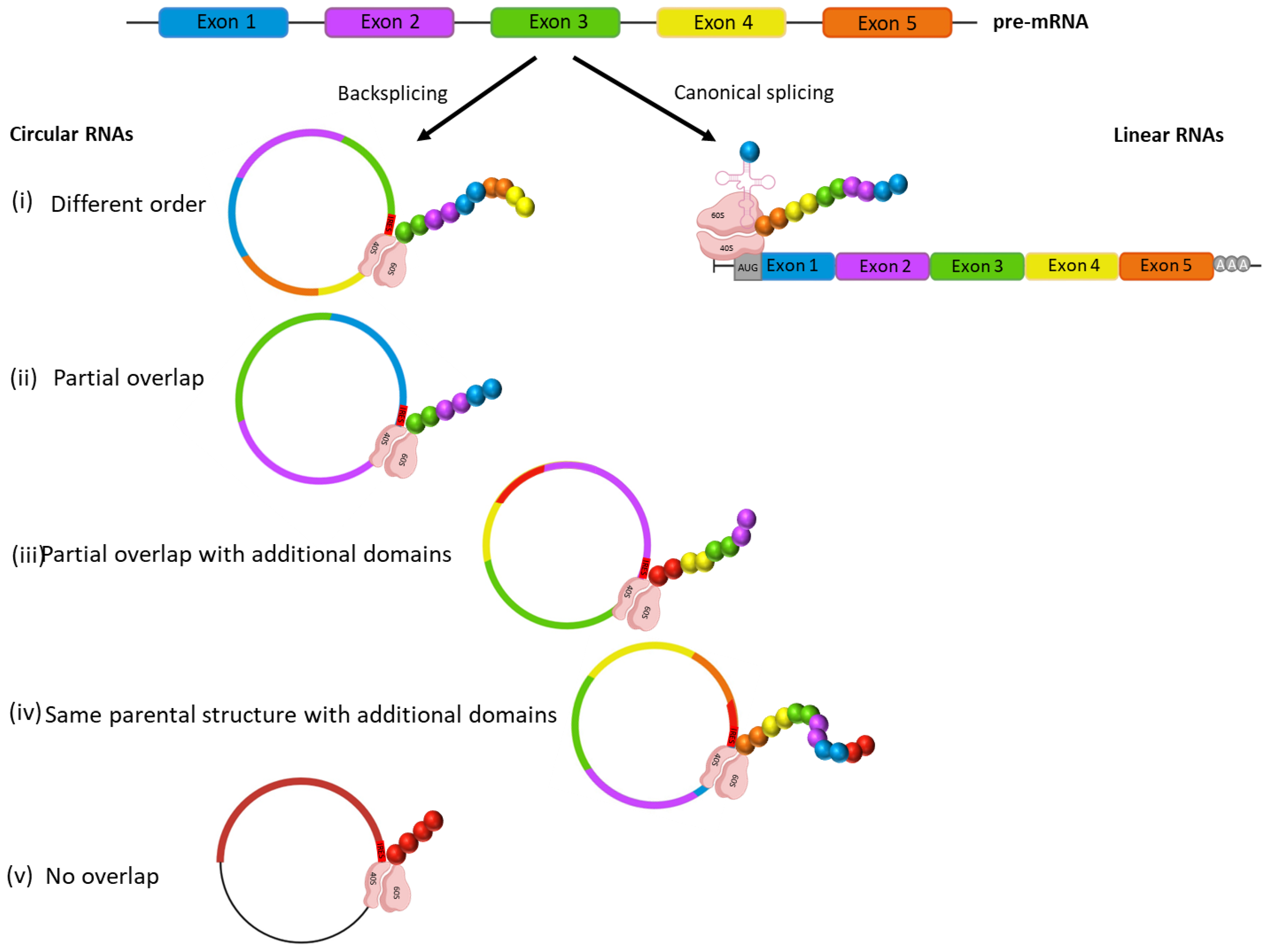
Figure 1. Schematic representation of the different circRNA proteins generated, focusing on their domain composition in comparison with the linear isoform.
Biology 12 00493 g001
Figure 2. The flow chart representation of the study. The diagram synthesizes the bioinformatics analysis flow, which led to the identification of the circRNAs with novel coding potential (structurally divergent proteins from those encoded by the linear mRNAs) and differentially expressed in a set of human cancer types.
Biology 12 00493 g002
Figure 3. Summary of the RNA-seq results. The volcano plot shows the differential expression of the candidate circRNA proteins in the cancer samples versus the control data sets. Orange points mark the genes with significantly increased or decreased expression (adjusted p value < 0.05). The x-axis shows log2 fold changes in expression and the y-axis the log10 adjusted p value of a gene being differentially expressed. A number of genes was filtered out the list of candidate coding circRNAs because of the log2 fold changes < 1 as absolute value (blue points) and adjusted p value > 0.05 (black points).
Biology 12 00493 g003
Figure 4. GO analysis: Reactome pathways and biological process for circRNAs novel peptides in cancer. The figure shows the statistical overrepresentation of the biological processes altered in cancer for the differentially expressed circRNAs with a novel coding potential (n = 183). The orange bars indicate the expected number of genes predicted for each category, while the blue bars show the observed number of genes in each of them. p values are adjusted using FDR correction: 0.05 < * < 0.01; 0.01 < ** < 0.001.
Biology 12 00493 g004
Table 1. Gene Ontology analysis for circRNAs novel peptides in cancer. The table illustrates the GO category significantly mapped and over-represented by the coding circRNAs differentially expressed in cancer. We used PantherDB to perform a statistical over-representation GO analysis. Fisher’s exact test was applied to evaluate the significance of the observed/expected gene ratio for each GO category and the BH method to adjust the p values (FDR < 0.05). MF, molecular function; BP, biological process; CC, cellular component; RP, reactome pathway; pol II, RNA polymerase II; obs, observed; exp, expected.
GO CategoriesGOObsExpFoldp ValueFDR
AKT phosphorylates targets in the nucleusRP30.0838.851.19 × 10−43.31 × 10⁻²
Estrogen-dependent nuclear events downstream of ESR-membrane signalingRP40.1921.585.89 × 10−52.45 × 10⁻²
Constitutive Signaling by AKT1 E17K in CancerRP40.219.927.79 × 10−52.77 × 10⁻²
Heme signalingRP60.3616.533.14 × 10−62.61 × 10⁻³
Regulation of TP53 Activity through PhosphorylationRP60.718.451.08 × 10−43.38 × 10⁻²
aryl hydrocarbon receptor bindingMF30.0743.169.23 × 10−53.78 × 10⁻²
protein serine kinase activityMF142.84.991.30 × 10−62.13 × 10⁻³
protein serine/threonine kinase activityMF143.344.29.06 × 10−66.35 × 10⁻³
protein serine/threonine/tyrosine kinase activityMF143.464.051.34 × 10−57.33 × 10⁻³
DNA bindingMF3919.32.021.57 × 10−57.68 × 10⁻³
rough endoplasmic reticulumCC50.637.95.53 × 10−44.70 × 10⁻²
transcription regulator complexCC153.913.841.20 × 10−51.63 × 10⁻³
nuclear speckCC113.193.454.41 × 10−44.09 × 10⁻²
centrosomeCC144.872.874.57 × 10−44.05 × 10⁻²
cytosolCC6542.041.559.16 × 10−51.04 × 10⁻²
mRNA transcriptionBP50.3813.215.80 × 10−53.49 × 10⁻²
peptidyl-threonine phosphorylationBP60.5810.363.72 × 10−53.43 × 10⁻²
peptidyl-serine phosphorylationBP91.426.331.85 × 10−53.23 × 10⁻²
negative regulation of RNA metabolic processBP2811.022.545.46 × 10−62.14 × 10⁻²
organelle organizationBP4826.441.822.44 × 10−53.19 × 10⁻²
regulation of cell communicationBP4625.811.785.71 × 10−53.58 × 10⁻²
regulation of signalingBP4625.911.788.60 × 10−54.65 × 10⁻²
regulation of response to stimulusBP5330.91.724.71 × 10−53.69 × 10⁻²
Table 2. Coding circRNAs associated with prognosis in CN-AML.
GenechrStartEndAnnotation
ABHD2chr158911372489116521C-term|canonicalMet
ANKRD12 *chr1891823819221999C-term|canonicalMet|conservedStructure
ARAP2 *chr43622858136229645C-term|canonicalMet|lackingDomain
CLNS1Achr117761960577625818canonicalSTOP|conservedStructure|internalMet
CPSF6chr126925112869262562canonicalSTOP|conservedStructure|internalMet
CSNK1G3chr5123545416123557564C-term|canonicalMet
FBXW7chr4152411302152412529C-term|canonicalMet
HIPK3 *chr113328641233287511C-term|canonicalMet|conservedStructure
KLHL8chr48719532387195690C-term|canonicalMet
MGAchr154166882741669958C-term|canonicalMet|lackingDomain
NCOA2 *chr87021390270216764C-term|canonicalMet|novelDomainStructure
OMA1chr15850605958539310C-term|canonicalMet|conservedStructure
PCMTD1chr85186084451861246C-term|canonicalMet|conservedStructure
PDE3Bchr111477193614789242C-term|internalMet
RELL1chr43763138437638504canonicalSTOP|internalMet
RNF13 *chr3149846010149921227C-term|canonicalMet|conservedStructure
RNF220chr14441198044412722C-term|canonicalMet
RSRC1chr3158122102158123991C-term|canonicalMet
SATB1 *chr31837816918420991C-term|canonicalMet|lackingDomain
SHOC2chr10110964124110985765lackingDomain
SLC38A1chr124622915246243314C-term|canonicalMet|conservedStructure
SLC8A1 *chr24042847240430304C-term|canonicalMet|conservedStructure
XPO1 *chr26152261061533903C-term|canonicalMet|conservedStructure
ZBTB44chr11130260855130261929C-term|canonicalMet|lackingDomain
* differentially and significantly expressed in pan-cancer dataset than normal samples; chr: chromosome (p value < 0.001).
Disclaimer/Publisher’s Note: The statements, opinions and data contained in all publications are solely those of the individual author(s) and contributor(s) and not of MDPI and/or the editor(s). MDPI and/or the editor(s) disclaim responsibility for any injury to people or property resulting from any ideas, methods, instructions or products referred to in the content.