An Enhanced 3D Model of Intravascular Lymphatic Valves to Assess Leaflet Apposition and Transvalvular Differences in Wall Distensibility

1
School of Mathematics & Statistics, University of Sydney, Sydney, NSW 2006, Australia
2
Department of Medical Pharmacology & Physiology, University of Missouri, Columbia, MO 65212, USA
*
Author to whom correspondence should be addressed.
Biology 2023, 12(3), 379; https://doi.org/10.3390/biology12030379
Submission received: 18 January 2023
/
Revised: 24 February 2023
/
Accepted: 24 February 2023
/
Published: 27 February 2023
(This article belongs to the Special Issue Physiology of Lymph Vessels: Structure, Function, Modelling, and Clinical Perspectives)
Simple Summary
The small size of lymphatic vessels, and thus of their one-way valves, brings problems that have resulted in a unique shape. This study extends a previous computer model of a lymphatic valve to deal with how the valve deforms at the end of closure, when the flexible opposite sides come together to prevent backward flow. The model is also extended to find out the effect of the wall of the vessel in which the valve exists having differing stiffness before and after the valve. In a small accompanying series of experiments, it was found in valves from two out of three locations that a measure of wall stiffness tended to be lower after the valve when the pressure inflating the vessel was low or moderate. The study adds to our knowledge of the behaviour of normal lymphatic valves, potentially aiding identification and management of abnormal behaviour in disease states.
Abstract
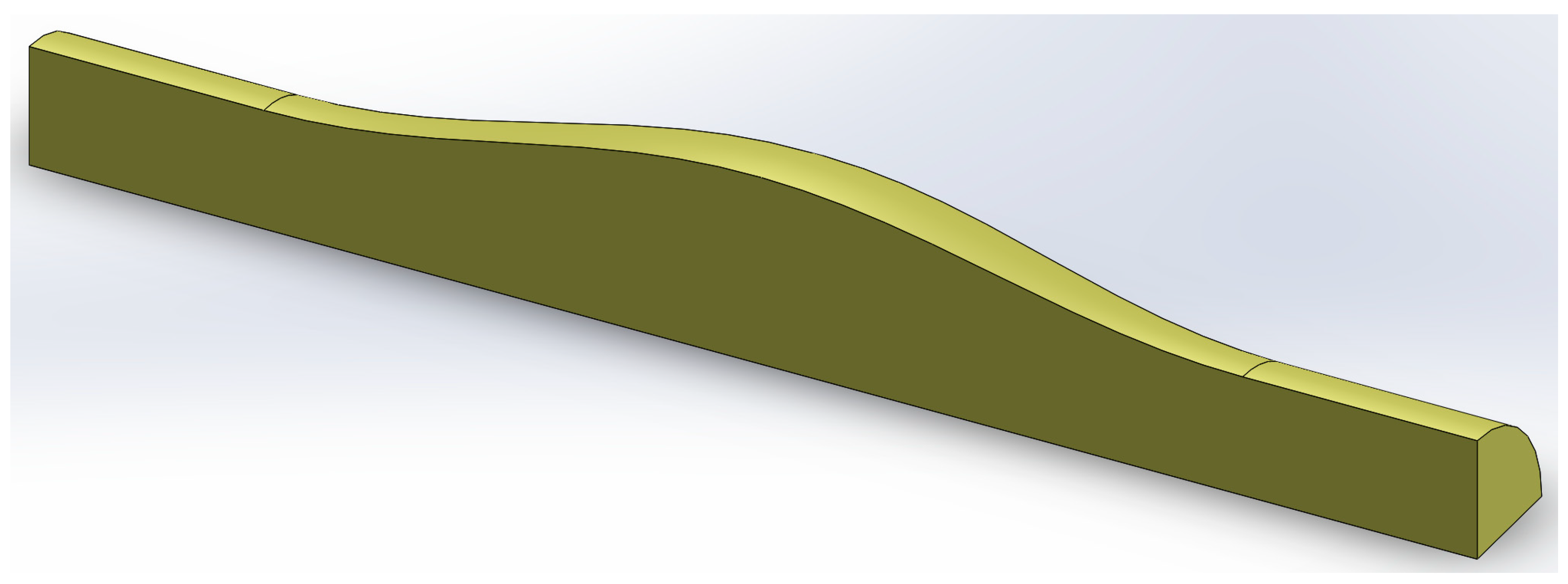
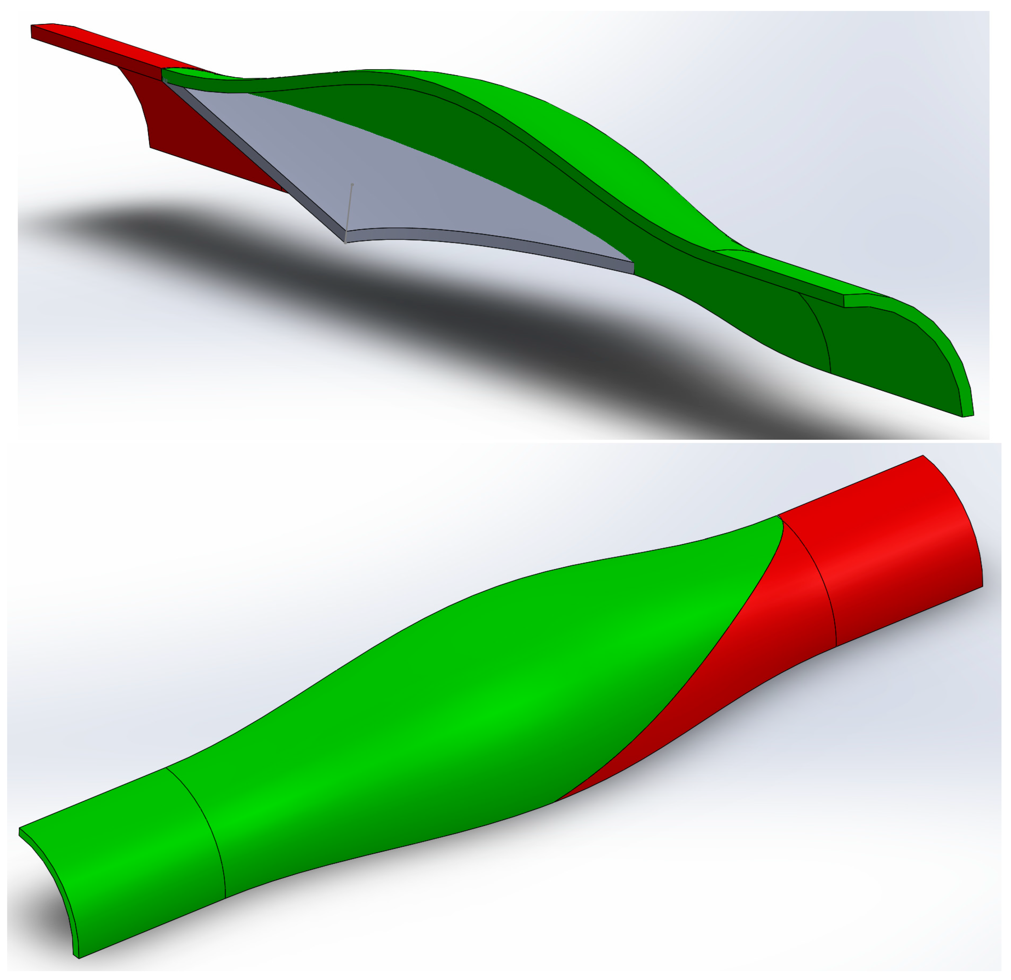
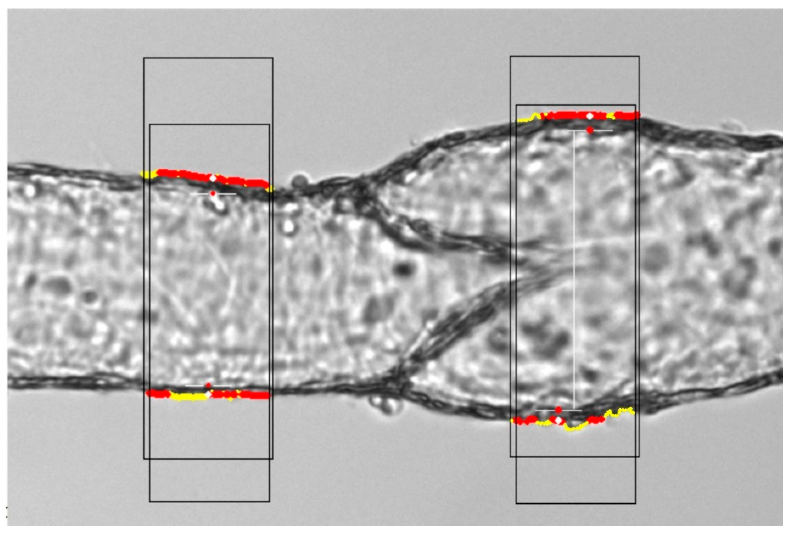
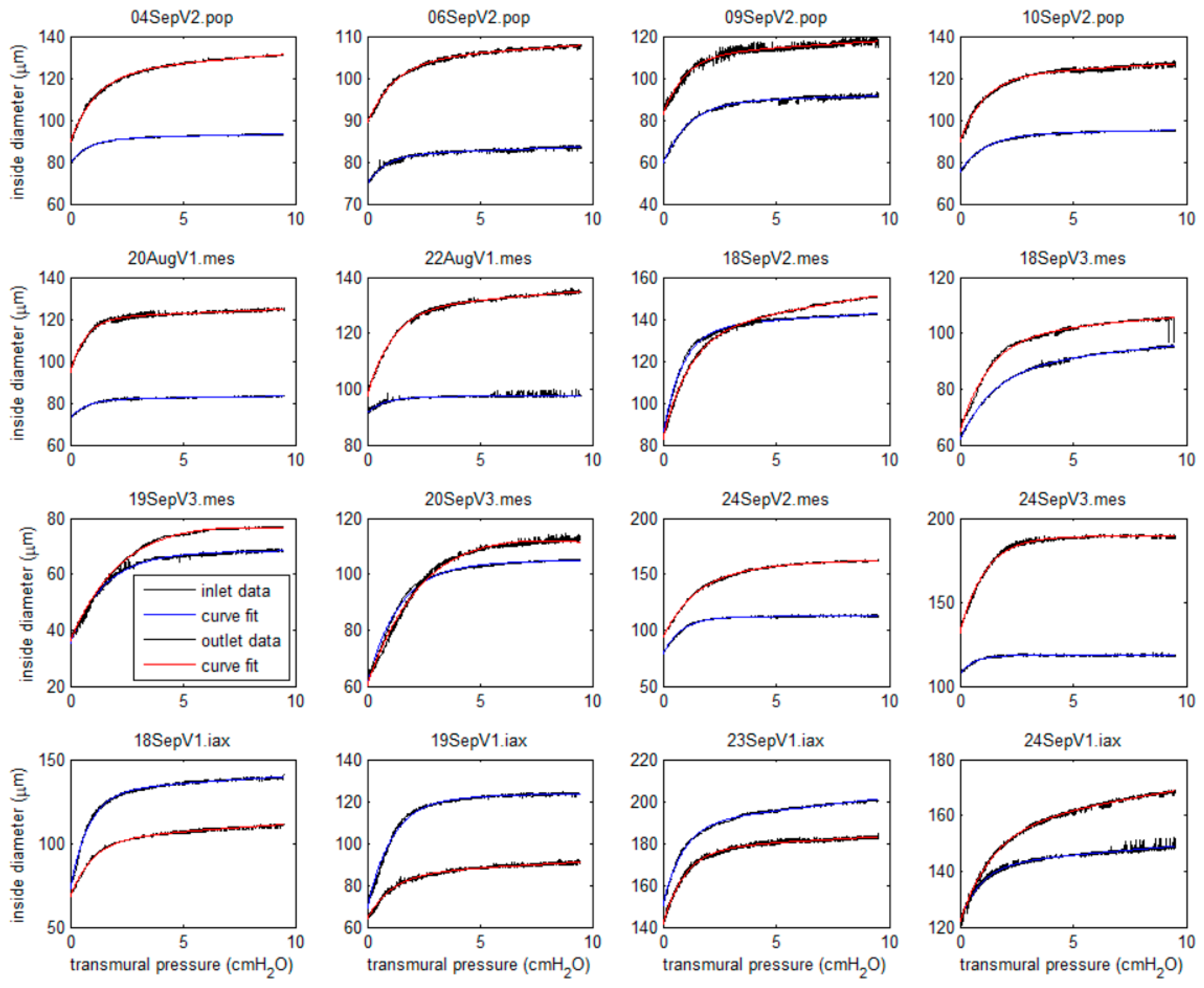
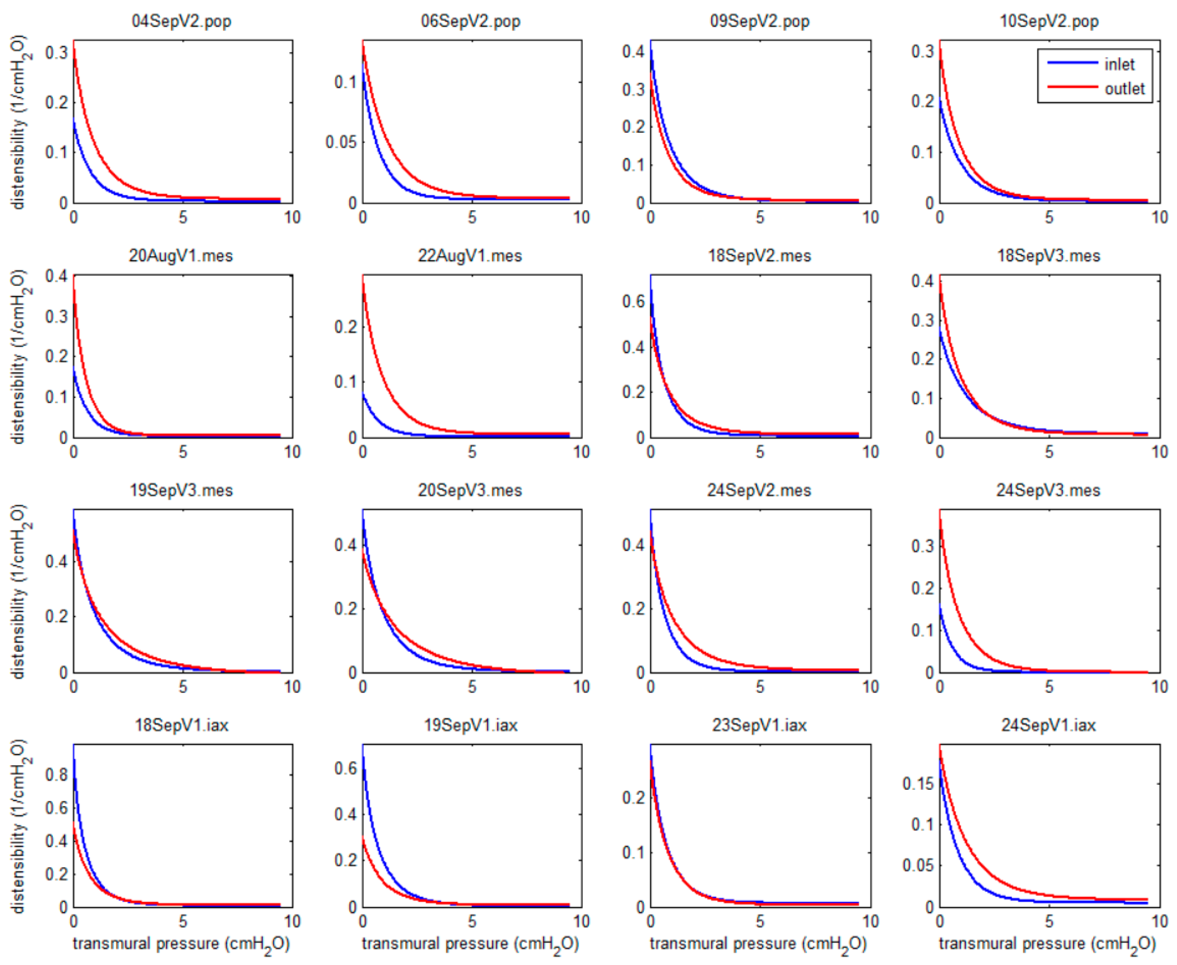
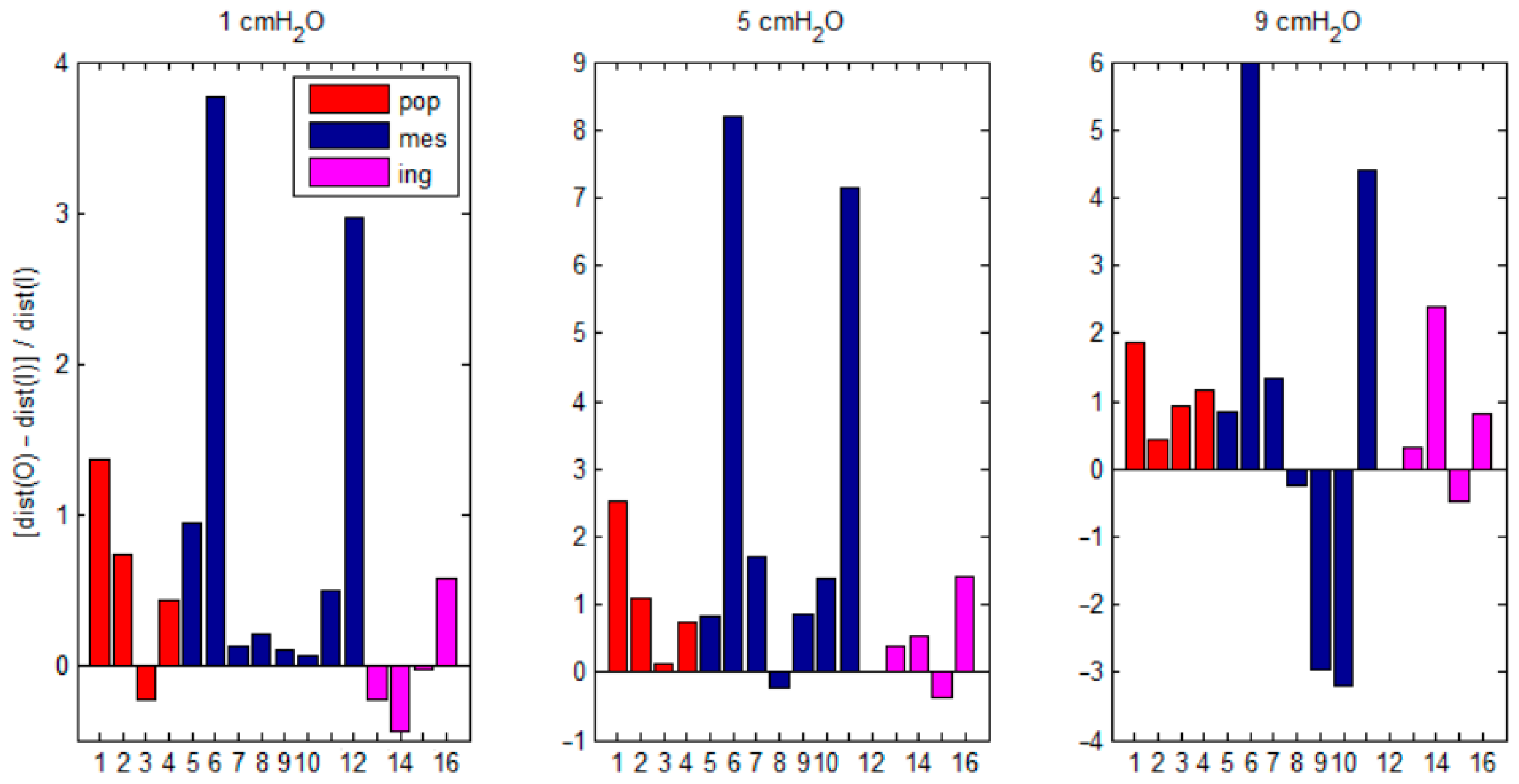
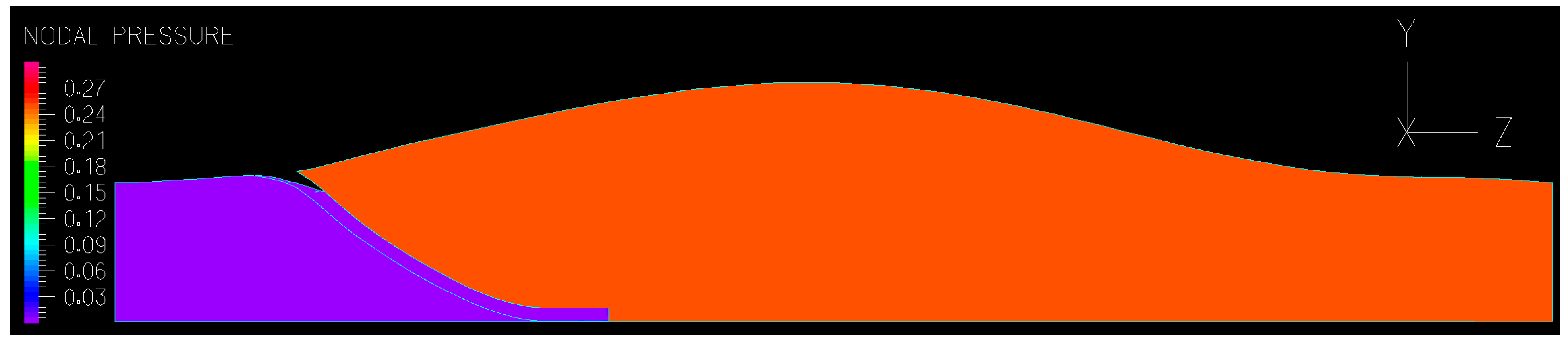
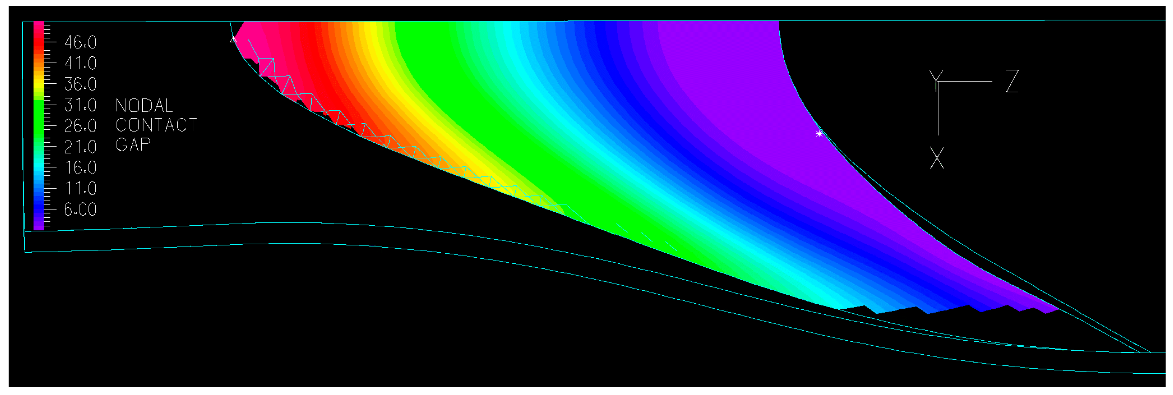
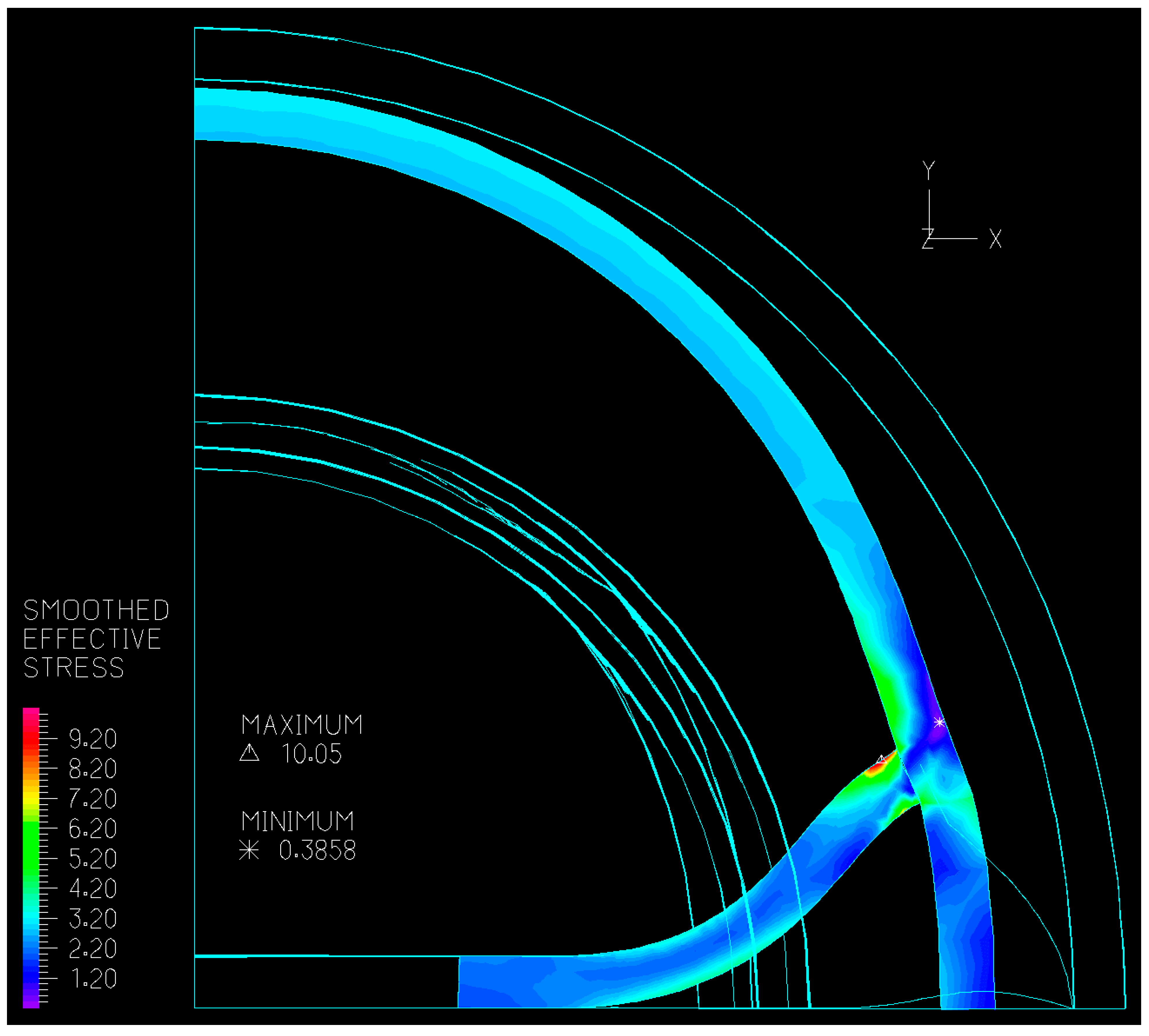
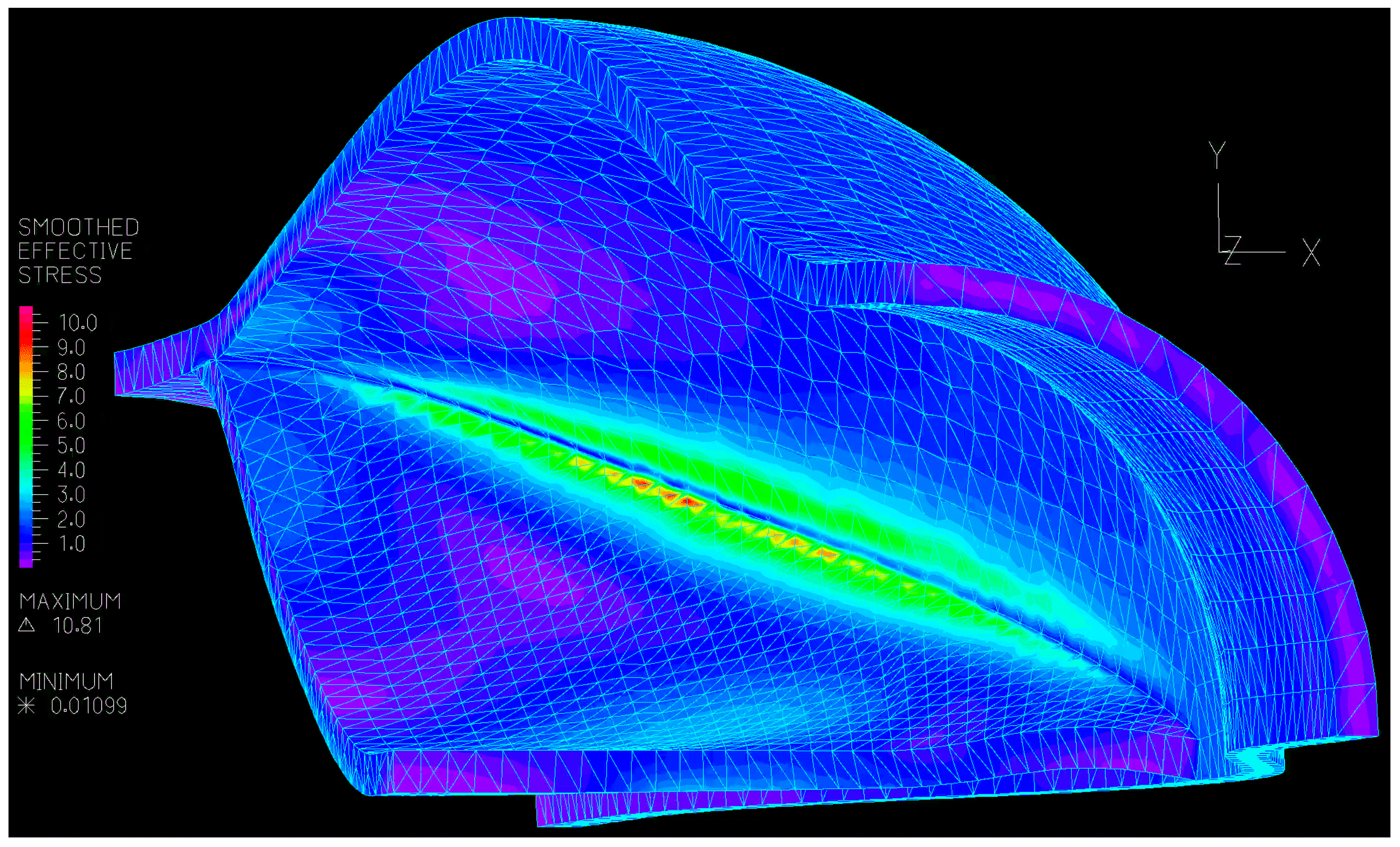
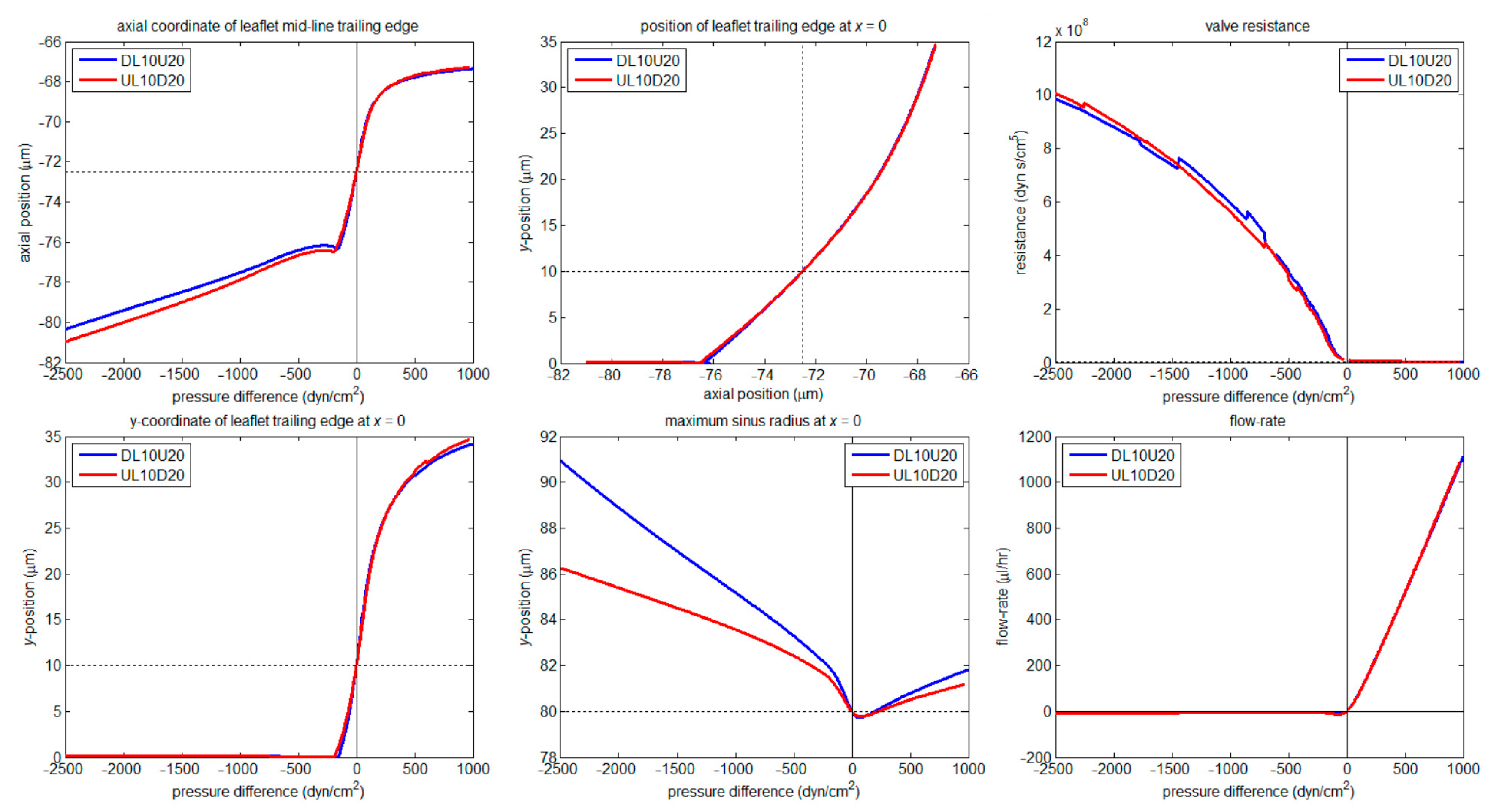
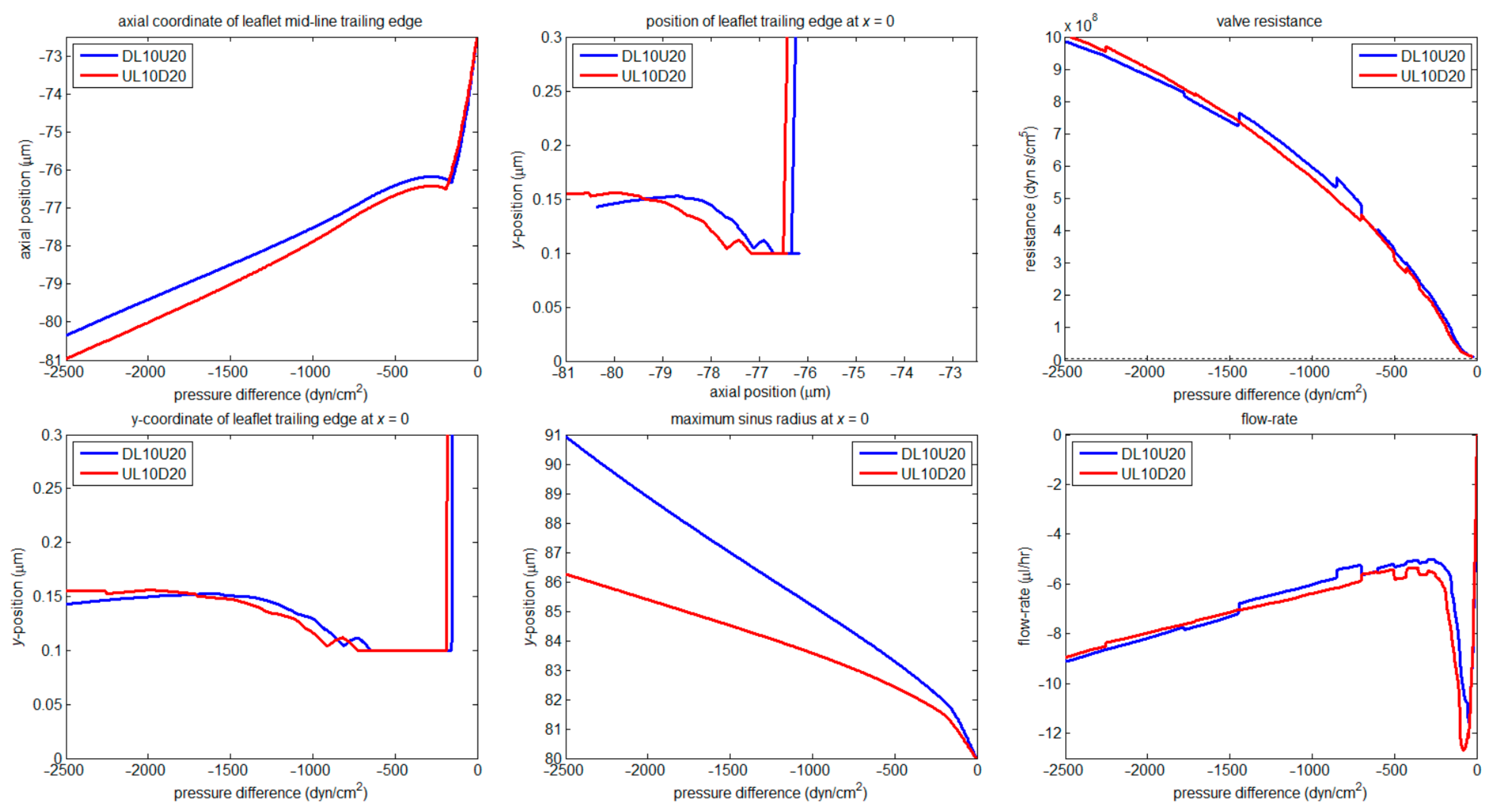
Lymphatic valves operate in a fluid-dynamically viscous environment that has little in common with that of cardiac valves, and accordingly have a different, axially lengthened, shape. A previously developed 3D fluid/structure interaction model of a lymphatic valve was extended to allow the simulation of stages of valve closure after the leaflets come together. This required that the numerical leaflet be prevented from passing into space occupied by the similar other leaflet. The resulting large deflections of the leaflet and lesser deflections of the rest of the valve were mapped as functions of the transvalvular pressure. In a second new development, the model was reconstructed to allow the vessel wall to have different material properties on either side of where the valve leaflet inserts into the wall. As part of this, a new pre-processing scheme was devised which allows easier construction of models with modified valve dimensions, and techniques for successfully interfacing the CAD software to the FE software are described. A two-fold change in wall properties either side of the leaflet made relatively little difference to valve operation apart from affecting the degree of sinus distension during valve closure. However, the numerically permitted strains were modest (<14%), and did not allow examination of the large-scale highly nonlinear elastic properties exhibited by real lymphatic vessels. A small series of murine popliteal, mesenteric, and inguinal-axillary lymphatic vessel segments containing a valve were experimentally investigated ex vivo. The pressure–diameter curves measured just upstream and just downstream of the valve were parameterised by computing the difference in tubular distensibility at three values of transmural pressure. In the popliteal and mesenteric segments, it was found that the distensibility was usually greater just downstream, i.e., in the sinus region, than upstream, at low and intermediate transmural pressure. However, there was wide variation in the extent of difference, and possible reasons for this are discussed.